EP1283989B1 - Diagnosis of liver fibrosis with serum marker algorithms - Google Patents
Diagnosis of liver fibrosis with serum marker algorithms Download PDFInfo
- Publication number
- EP1283989B1 EP1283989B1 EP01945050A EP01945050A EP1283989B1 EP 1283989 B1 EP1283989 B1 EP 1283989B1 EP 01945050 A EP01945050 A EP 01945050A EP 01945050 A EP01945050 A EP 01945050A EP 1283989 B1 EP1283989 B1 EP 1283989B1
- Authority
- EP
- European Patent Office
- Prior art keywords
- log
- score
- hyal
- col
- timp
- Prior art date
- Legal status (The legal status is an assumption and is not a legal conclusion. Google has not performed a legal analysis and makes no representation as to the accuracy of the status listed.)
- Expired - Lifetime
Links
Classifications
-
- G—PHYSICS
- G01—MEASURING; TESTING
- G01N—INVESTIGATING OR ANALYSING MATERIALS BY DETERMINING THEIR CHEMICAL OR PHYSICAL PROPERTIES
- G01N33/00—Investigating or analysing materials by specific methods not covered by groups G01N1/00 - G01N31/00
- G01N33/48—Biological material, e.g. blood, urine; Haemocytometers
- G01N33/50—Chemical analysis of biological material, e.g. blood, urine; Testing involving biospecific ligand binding methods; Immunological testing
- G01N33/68—Chemical analysis of biological material, e.g. blood, urine; Testing involving biospecific ligand binding methods; Immunological testing involving proteins, peptides or amino acids
- G01N33/6893—Chemical analysis of biological material, e.g. blood, urine; Testing involving biospecific ligand binding methods; Immunological testing involving proteins, peptides or amino acids related to diseases not provided for elsewhere
-
- G—PHYSICS
- G01—MEASURING; TESTING
- G01N—INVESTIGATING OR ANALYSING MATERIALS BY DETERMINING THEIR CHEMICAL OR PHYSICAL PROPERTIES
- G01N33/00—Investigating or analysing materials by specific methods not covered by groups G01N1/00 - G01N31/00
- G01N33/48—Biological material, e.g. blood, urine; Haemocytometers
- G01N33/50—Chemical analysis of biological material, e.g. blood, urine; Testing involving biospecific ligand binding methods; Immunological testing
- G01N33/68—Chemical analysis of biological material, e.g. blood, urine; Testing involving biospecific ligand binding methods; Immunological testing involving proteins, peptides or amino acids
- G01N33/6887—Chemical analysis of biological material, e.g. blood, urine; Testing involving biospecific ligand binding methods; Immunological testing involving proteins, peptides or amino acids from muscle, cartilage or connective tissue
-
- G—PHYSICS
- G01—MEASURING; TESTING
- G01N—INVESTIGATING OR ANALYSING MATERIALS BY DETERMINING THEIR CHEMICAL OR PHYSICAL PROPERTIES
- G01N2333/00—Assays involving biological materials from specific organisms or of a specific nature
- G01N2333/435—Assays involving biological materials from specific organisms or of a specific nature from animals; from humans
- G01N2333/46—Assays involving biological materials from specific organisms or of a specific nature from animals; from humans from vertebrates
- G01N2333/47—Assays involving proteins of known structure or function as defined in the subgroups
-
- G—PHYSICS
- G01—MEASURING; TESTING
- G01N—INVESTIGATING OR ANALYSING MATERIALS BY DETERMINING THEIR CHEMICAL OR PHYSICAL PROPERTIES
- G01N2333/00—Assays involving biological materials from specific organisms or of a specific nature
- G01N2333/435—Assays involving biological materials from specific organisms or of a specific nature from animals; from humans
- G01N2333/78—Connective tissue peptides, e.g. collagen, elastin, laminin, fibronectin, vitronectin, cold insoluble globulin [CIG]
-
- G—PHYSICS
- G01—MEASURING; TESTING
- G01N—INVESTIGATING OR ANALYSING MATERIALS BY DETERMINING THEIR CHEMICAL OR PHYSICAL PROPERTIES
- G01N2333/00—Assays involving biological materials from specific organisms or of a specific nature
- G01N2333/81—Protease inhibitors
- G01N2333/8107—Endopeptidase (E.C. 3.4.21-99) inhibitors
- G01N2333/8146—Metalloprotease (E.C. 3.4.24) inhibitors, e.g. tissue inhibitor of metallo proteinase, TIMP
-
- G—PHYSICS
- G01—MEASURING; TESTING
- G01N—INVESTIGATING OR ANALYSING MATERIALS BY DETERMINING THEIR CHEMICAL OR PHYSICAL PROPERTIES
- G01N2333/00—Assays involving biological materials from specific organisms or of a specific nature
- G01N2333/90—Enzymes; Proenzymes
- G01N2333/914—Hydrolases (3)
- G01N2333/948—Hydrolases (3) acting on peptide bonds (3.4)
- G01N2333/95—Proteinases, i.e. endopeptidases (3.4.21-3.4.99)
- G01N2333/964—Proteinases, i.e. endopeptidases (3.4.21-3.4.99) derived from animal tissue
- G01N2333/96425—Proteinases, i.e. endopeptidases (3.4.21-3.4.99) derived from animal tissue from mammals
- G01N2333/96427—Proteinases, i.e. endopeptidases (3.4.21-3.4.99) derived from animal tissue from mammals in general
- G01N2333/9643—Proteinases, i.e. endopeptidases (3.4.21-3.4.99) derived from animal tissue from mammals in general with EC number
- G01N2333/96486—Metalloendopeptidases (3.4.24)
-
- G—PHYSICS
- G01—MEASURING; TESTING
- G01N—INVESTIGATING OR ANALYSING MATERIALS BY DETERMINING THEIR CHEMICAL OR PHYSICAL PROPERTIES
- G01N2800/00—Detection or diagnosis of diseases
- G01N2800/08—Hepato-biliairy disorders other than hepatitis
- G01N2800/085—Liver diseases, e.g. portal hypertension, fibrosis, cirrhosis, bilirubin
Definitions
- hepatic stellate cells also called lipocytes, fat-storing or Ito cells
- This process is common to liver disease of all aetiologies.
- chronic viral hepatitis B and C and alcoholic liver disease as well as autoimmune and genetic liver diseases, all of which lead to clinical problems via the common final pathway of progressive liver fibrosis, with the eventual development of cirrhosis.
- Hepatic fibrosis is a reversible accumulation of extracellular matrix in response to chronic injury in which nodules have not yet developed
- cirrhosis implies an irreversible process, in which thick bands of matrix fully encircle the parenchyma, forming nodules. Consequently, any therapy must be directed towards patients with reversible disease (fibrosis), which will require early identification and monitoring of those at risk (2).
- liver fibrosis Severity and progression of liver fibrosis are difficult to assess, with liver biopsy currently remaining the most reliable clinical method.
- the qualitative evaluation of hepatic fibrosis by biopsies is limited by interobserver variability. Biopsies are clearly inadequate for the early clinical phase of drug development, where there is an imperative to employ less invasive methods that identify effective compounds within a commercially acceptable time frame, usually measured in weeks to a maximum of three months of experimental therapeutic exposure. Further disadvantages are the low diagnostic specificity and the risk of bleeding. Therefore there is a need for surrogate markers of liver fibrosis.
- Serum tests allow a non-invasive assessment of fibrogenesis and fibrolysis in the liver and can be done repeatedly and at short time intervals (3). Serum tests measuring the dynamic processes of extracellular matrix synthesis (fibrogenesis) and extracellular matrix degradation (fibrolysis) reflect the amount of extracellular matrix present, the degree of fibrosis or the ongoing process of architectural change of the liver (4).
- liver fibrosis The current state of the art in measuring surrogate markers of liver fibrosis is poorly developed. Previous studies have suggested that serum levels of extracellular matrix proteins (or their cleavage fragments) may be used to assess the severity and progression of liver fibrosis (4.5, US 5 316 914 , EP 0 283 779 ). Different serum markers have been investigated and correlations with liver biopsies and and severity of liver diseases have been found (6).
- the markers used for the assessment of liver fibrosis are PIIINP, Laminin, Hyaluronan, Collagen IV, TIMP-1, Tenascin, MMP-2 and Fibronectin. Markers are measured and their capability to assess liver fibrosis has been shown. Nevertheless, the diagnostic values of each single marker is not accurate and specific to assess fibrosis scores.
- serum markers of the extracellular matrix are assembled together in a panel leading to a set of markers whose measurement will enable the calculation of an algorithm and the use of such a derived algorithm for the prediction of liver fibrosis histological score.
- discriminant function analysis is used to determine which variables discriminate between the different fibrosis scores.
- the algorithms are derived from the set of markers N-terminal procollagen III propeptide (PIIINP), Collagen IV, Collagen VI, Tenascin, Laminin, Hyaluronan, MMP-2, TIMP-1 and MMP-9/TIMP-1 complex.
- the invention is directed to an in vitro method for diagnosis liver bibrosis wherein two or more diagnostic markers are measured in blood, serum or plasma and the measurements are combined by discriminant function analysis chareacterized in that the diagnostic markers are selected form the group of N-terminal procollagen III propeptide (PIIINP), Collagen IV, Collagen VI, Tenascin, Laminin, Hyaluronan, MMP-2- TIMP-1 and MMP-9/TIMP-1 complex wherein the algorithms is be used to support, predict or substitute or augment the histological score of a liver biopsy.
- PIIINP N-terminal procollagen III propeptide
- Collagen IV Collagen IV
- Collagen VI Tenascin
- Laminin Laminin
- Hyaluronan MMP-2- TIMP-1
- MMP-9/TIMP-1 MMP-9/TIMP-1
- liver biopsy The current "gold-standard" to assess fibrosis in the liver is the liver biopsy. With the biopsy, some randomly taken tissue out of the liver is cut into slices which become examined by an expert using a microscope. There are a lot of problems associated with liver biopsies inducing some uncertainty: distribution of fibrosis in the liver (clustered fibrosis and the needle might have hit regions of the liver not affected by fibrosis), failed sample preparation (e.g. not enough tissue material), and pathologist's individuality and preferences (individual assessments). Furthermore, the fibrotic state of the liver is usually described using scores and there are a lot of different, possibly incompatible scoring systems (e.g. Scheuer Score, Ishak Scores, etc.).
- the new technique is based on measuring serum parameters, which are directly associated with the fibrotic process, and combining them on a mathematical level yielding a fixed assessment procedure.
- the "gold-standard" is not the best but the only mean, since a priori both methods do not investigate comparable endpoints: whereas the serum parameters characterize dynamic processes, the biopsy characterizes the fibrotic manifestation at a fixed time-point. There may be a highly active fibrotic process in the liver although fibrotic tissue has not yet been developed. In contrast, there may be large clusters of fibrotic tissue in the liver but the fibrotic activity stopped or discontinued temporarily.
- Determination of concentrations of serum markers and subsequent calculation of an algorithm can also be used to make decisions whether or not a biopsy has to be taken and whether treatment should be started or continued or stopped. Therefore assignment of patients into a group of biopsy scores without taken a biopsy is advantageous. Categorization of patients into groups, e.g. mild versus serious fibrosis, by using algorithms is a benefit of the invention described.
- N-terminal procollagen III propeptide PIIINP
- Collagen IV Collagen IV
- Collagen VI Tenascin
- Laminin Hyaluronan
- MMP-2 TIMP-1 and MMP-9/TIMP-1 complex
- the markers are measured by making use of sandwich immunoassays.
- the immunoassays of the invention comprises reaction of two antibodies with human fluid samples, wherein the capture antibody specifically binds to one epitope of the marker.
- the second antibody of different epitope specificity is used to detect this complex.
- the antibodies are monoclonal antibodies and both antibodies of said two antibodies of the assay specifically bind to the protein.
- Antibody or other similar term used herein includes a whole immunoglobulin either monoclonal or polyclonal as well as antigenic fragments or immunoreactive fragments which specifically bind to the marker, including Fab, Fab', F(ab') 2 and F(v).
- Antibody includes also binding-proteins, especially Hyaluronic acid binding protein (HABP).
- HABP Hyaluronic acid binding protein
- the human fluid samples used in the assays of the invention can be any samples that contain the markers, e.g. blood, serum, plasma, urine, sputum or broncho alveolar lavage (BAL). Typically a serum or plasma sample is employed.
- markers e.g. blood, serum, plasma, urine, sputum or broncho alveolar lavage (BAL).
- BAL broncho alveolar lavage
- Antibodies of the invention can be prepared by techniques generally known in the art, and are typically generated to a sample of the markers.
- the second antibody is conjugated to a detector group, e.g. alkaline phosphatase, horseradish peroxidase, or a fluorescence dye.
- a detector group e.g. alkaline phosphatase, horseradish peroxidase, or a fluorescence dye.
- Conjugates are prepared by techniques generally known in the art.
- Concentration of the markers in human fluids are measured and algorithms calculated to assess the degree of fibrosis.
- Discriminant function analysis is used to determine which variables discriminate between two or more naturally occurring groups. Computationally, it is very similar to analysis of variance. The basic idea underlying discriminant function analysis is to determine whether groups differ with regard to the mean of a variable, and then to use that variable to predict group membership (e.g., of new cases). Stated in this manner, the discriminant function problem can be rephrased as a one-way analysis of variance (ANOVA) problem. Specifically, one can ask whether or not two or more groups are significantly different from each other with respect to the mean of a particular variable. If the means for a variable are significantly different in different groups, then we can say that this variable discriminates between the groups.
- ANOVA analysis of variance
- F is essentially computed as the ratio of the between-groups variance in the data over the pooled (average) within-group variance. If the between-group variance is significantly larger then there must be significant differences between means.
- the discrimination procedure develops a discriminant criterion to classify each observation into one of the groups.
- Post hoc predicting of what has happened in the past is not that difficult. It is not uncommon to obtain very good classification if one uses the same cases from which the discriminant criterion was computed.
- the discriminant criterion In order to get an idea of how well the current discriminant criterion "performs", one must classify (a priori) different cases, that is, cases that were not used to estimate the discriminant criterion. Only the classification of new cases allows us to assess the predictive validity of the discriminant criterion.
- the classification can be applied to other data sets.
- the data set used to derive the discriminant criterion is called the training or calibration data set.
- the discriminant criterion (function(s) or algorithm), is determined by a measure of generalized squared distance. It can be based on the pooled co-variance matrix yielding a linear function. Either Mahalanobis or Euclidean distance can be used to determine proximity.
- liver fibrosis scoring systems under view were
- Scoring Type Surrogate Parameters Scheurer Score: ln(TIMP-1) ln(Collagen VI / Hyaluronan) In(Hyaluronan / Laminin) Modified Ishak Score A - Interface Hepatitis: ln(TIMP-1) ln(Collagen VI / Hyaluronan) ln(Collagen VI / Tenascin) Modified Ishak Score B - Confluent Necrosis: ln(Hyaluronan) ln(Collagen VI / MMP-2) Modified Ishak Score C - Spotty Necrosis: In (Hyaluronan) In(MMP-9 / TIMP-1 / complex Tenascin) Modified Ishak Score D - Portal Inflammation: In(Laminin) In(Collagen VI / TIMP-1) Modified Ishak Score A - Interface Hepatitis: ln(TIMP-1) ln(Collagen VI /
- a corresponding discriminant analysis yielded the linear discriminating functions which can be used for calculation and prediction of biopsy score.
- the algorithms can be applied to every known scoring system (e.g. Scheuer Score, Ishak Score, Netavir Score, Ludwig Score, HAI Score).
- Algorithms can be used to predict the biopsy score of a patient (e.g. score 0, 1, 2, 3, ...) or to predict a group of scores (category) a patient belongs to (e.g. mild fibrosis; score 0 to 1).
- Discriminating functions used includes combinations of markers from the list of N-terminal procollagen III propeptide (PIIINP), Collagen IV, Collagen VI, Tenascin, Laminin, Hyaluronan, MMP-2, TRAP-1 and MMP-9/TIMP-1 complex and also factors between -1000 and +1000.
- PIIINP N-terminal procollagen III propeptide
- Collagen IV Collagen IV
- Collagen VI Tenascin
- Laminin Laminin
- Hyaluronan MMP-2
- TRAP-1 and MMP-9/TIMP-1 complex also factors between -1000 and +1000.
Landscapes
- Life Sciences & Earth Sciences (AREA)
- Health & Medical Sciences (AREA)
- Engineering & Computer Science (AREA)
- Biomedical Technology (AREA)
- Molecular Biology (AREA)
- Chemical & Material Sciences (AREA)
- Urology & Nephrology (AREA)
- Hematology (AREA)
- Immunology (AREA)
- Cell Biology (AREA)
- Analytical Chemistry (AREA)
- Biotechnology (AREA)
- Proteomics, Peptides & Aminoacids (AREA)
- Food Science & Technology (AREA)
- Medicinal Chemistry (AREA)
- Physics & Mathematics (AREA)
- Microbiology (AREA)
- Biochemistry (AREA)
- General Health & Medical Sciences (AREA)
- General Physics & Mathematics (AREA)
- Pathology (AREA)
- Investigating Or Analysing Biological Materials (AREA)
- Measuring And Recording Apparatus For Diagnosis (AREA)
- Other Investigation Or Analysis Of Materials By Electrical Means (AREA)
Abstract
Description
- Progressive fibrotic diseases of the liver are a major cause of morbidity and mortality throughout the world. Recent scientific advances demonstrate that the pathogenic process of fibrosis in liver is critically dependent on proliferation and activation of hepatic stellate cells (also called lipocytes, fat-storing or Ito cells) which synthesize and secrete excess extracellular matrix proteins (1). Moreover it is evident that this process is common to liver disease of all aetiologies. Of particular importance are chronic viral hepatitis B and C and alcoholic liver disease as well as autoimmune and genetic liver diseases, all of which lead to clinical problems via the common final pathway of progressive liver fibrosis, with the eventual development of cirrhosis.
- An important concept is the distinction between hepatic fibrosis and cirrhosis. Hepatic fibrosis is a reversible accumulation of extracellular matrix in response to chronic injury in which nodules have not yet developed, whereas cirrhosis implies an irreversible process, in which thick bands of matrix fully encircle the parenchyma, forming nodules. Consequently, any therapy must be directed towards patients with reversible disease (fibrosis), which will require early identification and monitoring of those at risk (2).
- Severity and progression of liver fibrosis are difficult to assess, with liver biopsy currently remaining the most reliable clinical method. The qualitative evaluation of hepatic fibrosis by biopsies is limited by interobserver variability. Biopsies are clearly inadequate for the early clinical phase of drug development, where there is an imperative to employ less invasive methods that identify effective compounds within a commercially acceptable time frame, usually measured in weeks to a maximum of three months of experimental therapeutic exposure. Further disadvantages are the low diagnostic specificity and the risk of bleeding. Therefore there is a need for surrogate markers of liver fibrosis. Serum tests allow a non-invasive assessment of fibrogenesis and fibrolysis in the liver and can be done repeatedly and at short time intervals (3). Serum tests measuring the dynamic processes of extracellular matrix synthesis (fibrogenesis) and extracellular matrix degradation (fibrolysis) reflect the amount of extracellular matrix present, the degree of fibrosis or the ongoing process of architectural change of the liver (4).
- The current state of the art in measuring surrogate markers of liver fibrosis is poorly developed. Previous studies have suggested that serum levels of extracellular matrix proteins (or their cleavage fragments) may be used to assess the severity and progression of liver fibrosis (4.5,
US 5 316 914 ,EP 0 283 779 ). Different serum markers have been investigated and correlations with liver biopsies and and severity of liver diseases have been found (6). - The J. P. TEARE ET AL. publication is related to "Comparison of serum procollagen III peptide concentrations and PGA index for assessment of hepatic fibrosis" (LANCET THE., vol. 342, no. 8876,9 October 1993 (1 993-1 0-09), pages 895-898),
- The X. JI ET AL. publication is related to "Clinical significance of serum 7s collagen and type VI collagen levels for the diagnosis of hepatic fibrosis." (CHINESE MEDICAL JOURNAL 1 ZHONGHUA Y1XUE ZAZHI YINGWEN BAN., vol. 1 10, no. 3,1997, pages 198- 201),
- The D. SCHUPPAN ET AL. publication is related to "Serummarker der Leberfibrose." (DEUTSCHE MEDIZINISCHE WOCHENSCHRIFT, vol. 124, no. 41,15 October 1999 (1999- 10-1 5), pages 1213-1218),
- G Castaldo, et.al. describes Differential diagnosis between hepatocellular carcinoma and cirrhosis through a discriminant function based on results for serum analytes (Clin. Chem., Aug 1996; 42: 1263 - 1269)
- A publication in DIGESTIVE DISEASES AND SCIENCES, vol. 39, no. 11, 1994, pages 2426-2432 has the title Alpha-2-macroglobulin and hepatic fibrosis.
- The markers used for the assessment of liver fibrosis are PIIINP, Laminin, Hyaluronan, Collagen IV, TIMP-1, Tenascin, MMP-2 and Fibronectin. Markers are measured and their capability to assess liver fibrosis has been shown. Nevertheless, the diagnostic values of each single marker is not accurate and specific to assess fibrosis scores.
- Therefore combinations of markers are discussed to increase the diagnostic value the simple biological index PGA combining prothrombin time (PT), serum gamma-glutamyl transpeptidase (GGT) and apolipoprotein A1 (Apo A1) and the index PGAA which includes alpha-2-macroglobulin (A2M) to the PGA index have been described for the diagnosis of alcoholic liver disease in drinkers (7,8). Although the PGA and PGAA index have been combined with single serum markers (9, 10) serum markers have not been used for establishing algorithms for the assessment of liver diseases.
- In this invention serum markers of the extracellular matrix are assembled together in a panel leading to a set of markers whose measurement will enable the calculation of an algorithm and the use of such a derived algorithm for the prediction of liver fibrosis histological score. For this purpose discriminant function analysis is used to determine which variables discriminate between the different fibrosis scores. The algorithms are derived from the set of markers N-terminal procollagen III propeptide (PIIINP), Collagen IV, Collagen VI, Tenascin, Laminin, Hyaluronan, MMP-2, TIMP-1 and MMP-9/TIMP-1 complex.
- Thus the invention is directed to an in vitro method for diagnosis liver bibrosis wherein two or more diagnostic markers are measured in blood, serum or plasma and the measurements are combined by discriminant function analysis chareacterized in that the diagnostic markers are selected form the group of N-terminal procollagen III propeptide (PIIINP), Collagen IV, Collagen VI, Tenascin, Laminin, Hyaluronan, MMP-2- TIMP-1 and MMP-9/TIMP-1 complex wherein the algorithms is be used to support, predict or substitute or augment the histological score of a liver biopsy.
- Generally, all new techniques have to be validated against existing standard techniques, provided there are any. The current "gold-standard" to assess fibrosis in the liver is the liver biopsy. With the biopsy, some randomly taken tissue out of the liver is cut into slices which become examined by an expert using a microscope. There are a lot of problems associated with liver biopsies inducing some uncertainty: distribution of fibrosis in the liver (clustered fibrosis and the needle might have hit regions of the liver not affected by fibrosis), failed sample preparation (e.g. not enough tissue material), and pathologist's individuality and preferences (individual assessments). Furthermore, the fibrotic state of the liver is usually described using scores and there are a lot of different, possibly incompatible scoring systems (e.g. Scheuer Score, Ishak Scores, etc.).
- For example, in a study with 24 patients, two independent pathologists had to score the same biopsy samples for each patient at two different time-points using two different scoring systems. The number of assessments where the two pathologists came to identical results ranged from only 36% to 46%.
- The new technique is based on measuring serum parameters, which are directly associated with the fibrotic process, and combining them on a mathematical level yielding a fixed assessment procedure.
- In order to validate the new technique the "gold-standard" is not the best but the only mean, since a priori both methods do not investigate comparable endpoints: whereas the serum parameters characterize dynamic processes, the biopsy characterizes the fibrotic manifestation at a fixed time-point. There may be a highly active fibrotic process in the liver although fibrotic tissue has not yet been developed. In contrast, there may be large clusters of fibrotic tissue in the liver but the fibrotic activity stopped or discontinued temporarily.
- Although, some mathematical functions of serum parameters yielded statistically significant different mean values in different biopsy score stages. Discriminant analyses using the "gold-standard" were chosen in order to investigate the diagnostic power of those mathematical functions of serum parameters.
- Determination of concentrations of serum markers and subsequent calculation of an algorithm can also be used to make decisions whether or not a biopsy has to be taken and whether treatment should be started or continued or stopped. Therefore assignment of patients into a group of biopsy scores without taken a biopsy is advantageous. Categorization of patients into groups, e.g. mild versus serious fibrosis, by using algorithms is a benefit of the invention described.
- The markers N-terminal procollagen III propeptide (PIIINP), Collagen IV, Collagen VI, Tenascin, Laminin, Hyaluronan, MMP-2, TIMP-1 and MMP-9/TIMP-1 complex are used for algorithms.
- The markers are measured by making use of sandwich immunoassays. The immunoassays of the invention comprises reaction of two antibodies with human fluid samples, wherein the capture antibody specifically binds to one epitope of the marker. The second antibody of different epitope specificity is used to detect this complex. Preferably the antibodies are monoclonal antibodies and both antibodies of said two antibodies of the assay specifically bind to the protein.
- Antibody or other similar term used herein includes a whole immunoglobulin either monoclonal or polyclonal as well as antigenic fragments or immunoreactive fragments which specifically bind to the marker, including Fab, Fab', F(ab')2 and F(v). Antibody includes also binding-proteins, especially Hyaluronic acid binding protein (HABP).
- The human fluid samples used in the assays of the invention can be any samples that contain the markers, e.g. blood, serum, plasma, urine, sputum or broncho alveolar lavage (BAL). Typically a serum or plasma sample is employed.
- Antibodies of the invention can be prepared by techniques generally known in the art, and are typically generated to a sample of the markers.
- The second antibody is conjugated to a detector group, e.g. alkaline phosphatase, horseradish peroxidase, or a fluorescence dye. Conjugates are prepared by techniques generally known in the art.
- Concentration of the markers in human fluids are measured and algorithms calculated to assess the degree of fibrosis.
- Discriminant function analysis is used to determine which variables discriminate between two or more naturally occurring groups. Computationally, it is very similar to analysis of variance. The basic idea underlying discriminant function analysis is to determine whether groups differ with regard to the mean of a variable, and then to use that variable to predict group membership (e.g., of new cases). Stated in this manner, the discriminant function problem can be rephrased as a one-way analysis of variance (ANOVA) problem. Specifically, one can ask whether or not two or more groups are significantly different from each other with respect to the mean of a particular variable. If the means for a variable are significantly different in different groups, then we can say that this variable discriminates between the groups. In the case of a single variable, the final significance test of whether or not a variable discriminates between groups is the F test. F is essentially computed as the ratio of the between-groups variance in the data over the pooled (average) within-group variance. If the between-group variance is significantly larger then there must be significant differences between means.
- Usually, one includes several variables in a study in order to see which one(s) contribute to the discrimination between groups. In that case, we have a matrix of total variances and co-variances; likewise, we have a matrix of pooled within-group variances and co-variances. We can compare those two matrices via multivariate F tests in order to determined whether or not there are any significant differences (with regard to all variables) between groups. This procedure is identical to multivariate analysis of variance or MANOVA. As in MANOVA, one could first perform the multivariate test, and, if statistically significant, proceed to see which of the variables have significantly different means across the groups. Thus, even though the computations with multiple variables are more complex, the principal reasoning still applies, namely, that we are looking for variables that discriminate between groups, as evident in observed mean differences.
- For a set of observations containing one or more quantitative variables and a classification variable defining groups of observations, the discrimination procedure develops a discriminant criterion to classify each observation into one of the groups. Post hoc predicting of what has happened in the past is not that difficult. It is not uncommon to obtain very good classification if one uses the same cases from which the discriminant criterion was computed. In order to get an idea of how well the current discriminant criterion "performs", one must classify (a priori) different cases, that is, cases that were not used to estimate the discriminant criterion. Only the classification of new cases allows us to assess the predictive validity of the discriminant criterion. In order to validate the derived criterion, the classification can be applied to other data sets. The data set used to derive the discriminant criterion is called the training or calibration data set.
- The discriminant criterion (function(s) or algorithm), is determined by a measure of generalized squared distance. It can be based on the pooled co-variance matrix yielding a linear function. Either Mahalanobis or Euclidean distance can be used to determine proximity.
- For the development of a discriminant algorithm, data of a group of subjects of an observational liver fibrosis of study were analyzed. Liver fibrosis scoring systems under view were
- the Scheuer Score (0-4),
- the Modified Ishak Score (HAI) A - Interface Hepatitis (0-4),
- the Modified Ishak Score (HAI) B - Confluent Necrosis (0-6),
- the Modified Ishak Score (HAI) C - Spotty Necrosis (0-4),
- the Modified Ishak Score (HAI) D - Portal Inflammation (0-4),
- the Modified HAI Score (Ishak Score) (0-6).
- Applying a stepwise discriminant analysis, for example the following functions of serum parameters showed to have major impact on the corresponding scoring type.
Scoring Type Surrogate Parameters Scheurer Score: ln(TIMP-1) ln(Collagen VI/ Hyaluronan) In(Hyaluronan / Laminin) Modified Ishak Score A - Interface Hepatitis: ln(TIMP-1) ln(Collagen VI/ Hyaluronan) ln(Collagen VI/ Tenascin) Modified Ishak Score B - Confluent Necrosis: ln(Hyaluronan) ln(Collagen VI/ MMP-2) Modified Ishak Score C - Spotty Necrosis: In (Hyaluronan) In(MMP-9/TIMP-1/ complex Tenascin) Modified Ishak Score D - Portal Inflammation: In(Laminin) In(Collagen VI/ TIMP-1) Modified Ishak Score - Stage: In(TIMP-1) In(Collagen VI/ Hyaluronan) In (Hyaluronan/ Laminin) - A corresponding discriminant analysis yielded the linear discriminating functions which can be used for calculation and prediction of biopsy score. The algorithms can be applied to every known scoring system (e.g. Scheuer Score, Ishak Score, Netavir Score, Ludwig Score, HAI Score).
- Algorithms can be used to predict the biopsy score of a patient (e.g. score 0, 1, 2, 3, ...) or to predict a group of scores (category) a patient belongs to (e.g. mild fibrosis; score 0 to 1).
- Discriminating functions used includes combinations of markers from the list of N-terminal procollagen III propeptide (PIIINP), Collagen IV, Collagen VI, Tenascin, Laminin, Hyaluronan, MMP-2, TRAP-1 and MMP-9/TIMP-1 complex and also factors between -1000 and +1000.
- Different scores need different algorithm form the list of markers and factors.
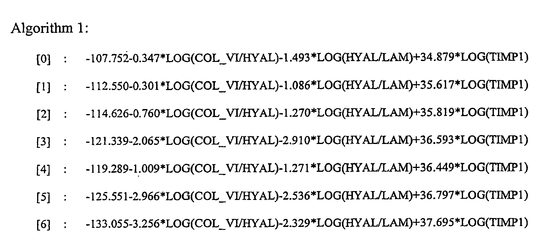
- The following algorithms 1, 2 and 3 were calculated by correlating biopsies assessed by the Scheuer scoring system and serum marker concentrations of a group of patients with liver diseases:


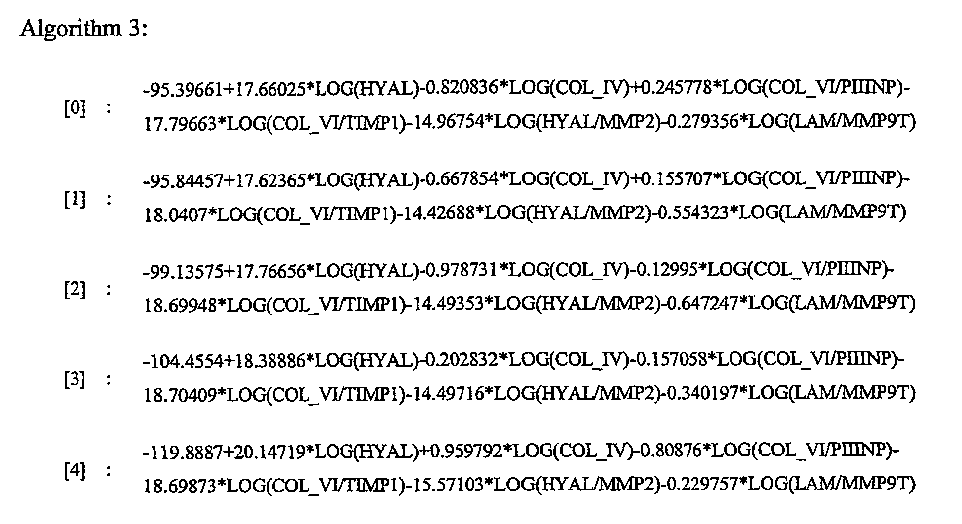
- The algorithms were used to predict biopsy scores of a separate group of patients. The calculated scores were compared with scores determined by a single pathologist (case B), with a consensus score of 3 pathologists (case C) and with the range covered by all pathologists (case A). Kappa values, negative predictive values (NPV) for score 0-1, positive predictive values (PPV) for score 2-4, sensitivities and specificities have also been calculated.
Algorithm 1 Algorithm 2 Algorithm 3 C A B C A B C A B Hit-Rate (%) [0] 33.3 38.9 35.0 17.1 40.0 13.7 20.0 41.4 16.8 Hit-Rate (%) [1] 36.8 42.7 36.0 80.7 81.6 75.8 74.6 77.2 71.7 Hit-Rate (%) [2] 25.8 42.4 19.0 0.0 36.8 0.0 0.0 34.2 5.1 Hit-Rate (%) [3] 26.1 34.8 22.2 6.4 17.0 5.2 12.8 21.3 9.3 Hit-Rate (%) [4] 63.0 63.0 55.9 62.5 62.5 52.9 43.8 43.8 47.1 Hit-Rate (%) All 35.9 42.9 33.8 42.2 54.2 36.7 39.5 51.2 36.7 N 468 468 793 301 301 626 301 301 626 Kappa 0.175 0.268 0.151 . 0.199 . 0.124 0.310 0.121 L_Kappa 0.119 0.211 0.109 . 0.134 . 0.056 0.235 0.077 U_Kappa 0.231 0.325 0.192 . 0.265 . 0.191 0.385 0.165 P(Kappa=0) <0.0001 <0.0001 <0.0001 . <0.0001 . <0.0001 <0.0001 <0.0001 NPV (%) [0-1] 61.6 63.8 62.8 91.8 92.4 85.5 88.0 89.1 81.6 PPV (%) [2-4] 66.1 75.1 66.3 31.6 46.2 35.4 35.0 48.7 39.0 Hit-Rate (%) All 63.5 68.4 64.2 68.4 74.4 65.8 67.4 73.4 64.9 Kappa 0.268 0.372 0.280 0.261 0.417 0.226 0.252 0.404 0.219 L_Kappa 0.182 0.291 0.214 0.160 0.315 0.152 0.146 0.299 0.142 U_Kappa 0.354 0.454 0.346 0.363 0.520 0.300 0.358 0.508 0.295 P(Kappa=0) <0.0001 <0.0001 <0.0001 < 0.0001 <0.0001 <0.0001 <0.0001 <0.0001 <0.0001 Sensitivity 0.539 0.584 0.540 0.712 0.794 0.613 0.651 0.740 0.578 Specificity 0.729 0.791 0.739 0.679 0.730 0.671 0.681 0.732 0.674 - The following algorithms 1, 2 and 3 were calculated by correlating biopsies assessed by the Ishak scoring system and serum marker concentrations of a group of patients with liver diseases:
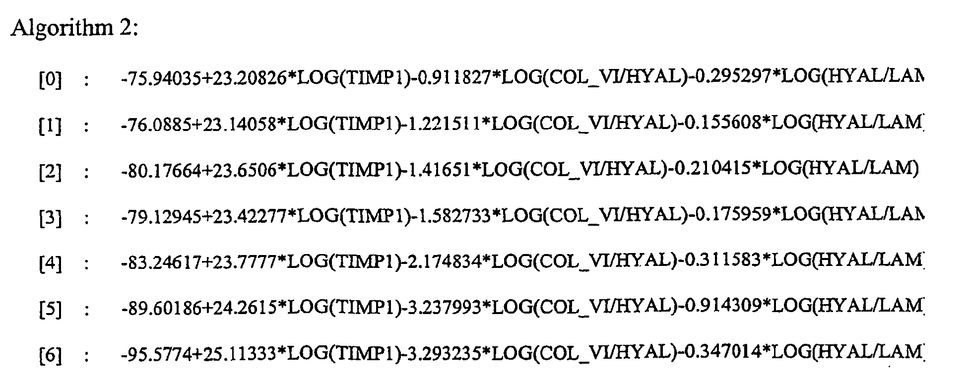

- The algorithms were used to predict biopsy scores of a separate group of patients. The calculated scores were compared with scores determined by a single pathologist (case B), with a consensus score of 3 pathologists (case C) and with the range covered by all pathologists (case A). Kappa values, negative predictive values (NPV) for score 0-2, positive predictive values (PPV) for score 3-6, sensitivities and specificities have also been calculated.
Algorithm 1 Algorithm 2 Algorithm 2 C A B C A B C A B Hit-Rate (%) [0] 28.7 31.5 29.5 45.7 58.6 41.0 45.7 57.1 39.8 Hit-Rate (%) [1] 25.0 34.0 29.2 50.8 60.7 50.8 27.9 41.0 37.7 Hit-Rate (%) [2] 10.7 24.0 9.7 0.0 22.4 1.1 1.7 15.5 1.1 Hit-Rate (%) [3] 23.0 27.9 20.2 0.0 3.1 0.0 9.4 12.5 6.9 Hit-Rate (%) [4] 22.2 37.8 25.6 0.0 0.0 0.0 0.0 7.7 3.3 Hit-Rate (%) [5] 32.0 44.0 24.4 4.5 18.2 2.6 0.0 18.2 2.6 Hit-Rate (%) [6] 57.4 57.4 51.1 71.9 71.9 60.9 43.8 43.8 52.2 Hit-Rate (%) All 27.1 34.6 27.3 28.9 39.5 28.1 22.3 32.6 25.2 N 468 468 794 301 301 627 301 301 627 Kappa 0.138 0.228 0.136 . . . 0.031 0.093 0.041 L_Kappa 0.090 0.177 0.100 . . . -0.02 0.039 0.006 U_Kappa 0.186 0.279 0.173 . . . 0.084 0.147 0.076 P(Kappa=0) < 0.0001 < 0.0001 < 0.0001 . . . 0.2293 0.0003 0.0152 NPV (%) [0-2] 57.2 59.4 59.1 89.9 91.0 83.9 77.8 80.4 75.1 PPV (%) [3-6] 71.9 79.5 74.2 40.2 50.9 41.5 47.3 55.4 48.1 Hit-Rate (%) All 63.0 67.3 65.0 71.4 76.1 67.6 66.4 71.1 64.8 Kappa 0.274 0.362 0.312 0.330 0.450 0.271 0.259 0.366 0.238 L_Kappa 0.191 0.283 0.249 0.223 0.346 0.195 0.147 0.257 0.160 U_Kappa 0.356 0.441 0.374 0.437 0.554 0.347 0.371 0.475 0.316 P(Kappa=0) <0.0001 <0.0001 <0.0001 <0.0001 < 0.0001 <0.0001 <0.0001 <0.0001 <0.0001 Sensitivity 0.524 0.561 0.537 0.703 0.770 0.617 0.558 0.626 0.547 Specificity 0.757 0.816 0.781 0.717 0.758 0.697 0.714 0.752 0.699 - Grouping the patients into categories no/mild fibrosis (score 0-1) and moderate/severe fibrosis (score 2-4) for the Scheuer score and calculating algorithms for the dichotomous outcome gave the following results:

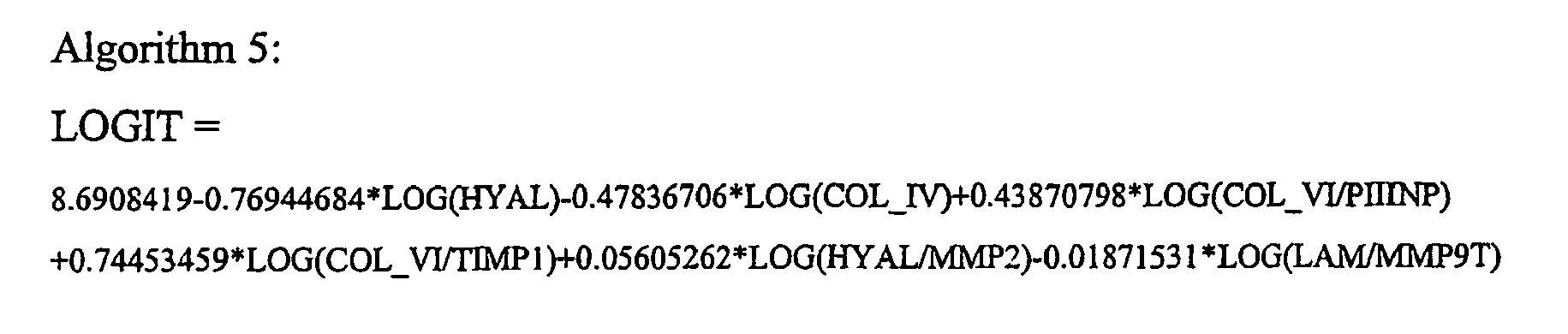
- The algorithms were used to calculate receiver operating characteristic curves for the categories no/mild fibrosis (score 0-1) and moderate/severe fibrosis (score 2-4) for the Scheuer score. The calculated scores were compared with scores determined by a single pathologist (case B), with a consensus score of 3 pathologists (case C) and with the range covered by all pathologists (case A). Area under curve (AUC) values have been calculated.
Algorithm 4 Algorithm 5 C A B C A B AUC 0.759 0.899 0.759 0.746 0.871 0.756 N 295 295 569 291 291 562 - Grouping the patients into categories no/mild fibrosis (score 0-2) and moderate/severe fibrosis (score 3-6) for the Ishak score and calculating algorithms for the dichotomous outcome gave the following results:


- The algorithms were used to calculate receiver operating characteristic curves for the categories no/mild fibrosis (score 0-2) and moderate/severe fibrosis (score 3-6) for the Ishak score. The calculated scores were compared with scores determined by a single pathologist (case B), with a consensus score of 3 pathologists (case C) and with the range covered by all pathologists (case A). Area under curve (AUC) values have been calculated.
Algorithm 4 Algorithm 5 C A B C A B AUC 0.763 0.887 0.763 0.751 0.861 0.757 N 295 295 570 292 292 564 -
- 1. Friedman SL The cellular basis of hepatic fibrosis:Mechanism and treatment strategies. N Engl J Med 1993; 328: 1828-1835
- 2. Friedman SL Molecular mechanism of hepatic fibrosis and principle of therapy J Gastroenterol 1997; 32: 424-430
- 3. Hayasaka A, Saisho H Scrum markers as tools to monitor liver fibrosis Digestion 1998; 59: 381-384
- 4. Schuppan D, Stolzel U, Oesterling C, Somasundaram R Serum assays for liver fibrosis. J Hepatol 1995; 22 (Suppl 2): 82-88
- 5. Murawaki Y, Ikuta Y, Nishimura Y, Koda M, Kawasaki H Serum markers for connective tissue turnover in patients with chronic hepatitis C: A comparative analysis. J Hepatol 1995; 23: 145-152
- 6. Wong VS, Hughes V, Trull A, Wight DGD, Petrik J, Alexander GJM Serum hyaluronic acid is a useful marker of liver fibrosis in chronic hepatitis C virus infection J Viral Hepatitis 1998; 5:187-192
- 7. Poynard T, Aubert A, Bedossa P, Abella A, Naveau S, Paraf F, Chapu JC A simple biological index for detection of alcoholic liver disease in drinkers Gastroenterology 1991; 100: 1397-1402
- 8. Naveau S, Poynard T, Benattat C, Bedossa P, Chaput JC Alpha-2 macroglobulin and hepatic fibrosis:diagnostic interest Dig Dis Sci 1994; 11: 2426-2432
- 9. Oberti F, Valsesia E, Pilette C, Rousselet MC, Bedossa P, Aube C, Gallois Y, Rifflet H, Maiga MY, Penneau-Fontbonne D, Cales P Noninvasive diagnosis of hepatic fibrosis and cirrhosis Gastroenterology 1997; 113: 1609-1616
- 10. Teare JP, Sherman D, Greenfield SM, Simpson J, Catterall AP, Murray-Lyon IM, Peters TJ, Williams R, Thompson RPH The Lancet 1993; 342: 895-898
Claims (5)
- An in vitro method for diagnosis liver fibrosis wherein two or more diagnostic markers are measured in blood, serum or plasma and the measurements are combined by discriminant function analysis characterized in that the diagnostic markers are selected form the group of N-terminal procollagen III propeptide (PIIINP), Collagen IV, Collagen VI, Tenascin, Laminin, Hyaluronan, MMP-2- TIMP-1 and MMP-9/TIMP-1 complex wherein the algorithms is be used to support, predict or substitute or augment the histological score of a liver biopsy.
- A method according claim 1 wherein the algorithms is adjusted to match known scoring system (e.g. Scheuer score, Ishak score, HAI score, Ludwig, score, Metavir score).
- A method according to claim 2 wherein treatment decision is supported by the calculated score or the calculated score group.
- A method according to claim 2 wherein decisions whether or not a biopsy should be taken is supported by the calculated score or the calculated score group.
- A method according claim 2 wherein patients are classified as suffering from no/mild or moderates/severe liver fibrosis.
Priority Applications (2)
| Application Number | Priority Date | Filing Date | Title |
|---|---|---|---|
| EP01945050A EP1283989B1 (en) | 2000-04-28 | 2001-04-26 | Diagnosis of liver fibrosis with serum marker algorithms |
| US10/868,437 US7668661B2 (en) | 2000-04-28 | 2004-06-15 | Liver disease-related methods and systems |
Applications Claiming Priority (6)
| Application Number | Priority Date | Filing Date | Title |
|---|---|---|---|
| EP00109063A EP1150123B1 (en) | 2000-04-28 | 2000-04-28 | Diagnosis of liver fibrosis with serum marker algorithms |
| EP00109063 | 2000-04-28 | ||
| IT2001MI000813A ITMI20010813A1 (en) | 2000-04-28 | 2001-04-13 | EVALUATION OF LIVER FIBROSIS WITH ALGORITHMS BASED ON SERUM MARKERS |
| ITMI000813 | 2001-04-13 | ||
| PCT/EP2001/004696 WO2001086304A2 (en) | 2000-04-28 | 2001-04-26 | Diagnostic of liver fibrosis with serum marker algorithms |
| EP01945050A EP1283989B1 (en) | 2000-04-28 | 2001-04-26 | Diagnosis of liver fibrosis with serum marker algorithms |
Publications (2)
| Publication Number | Publication Date |
|---|---|
| EP1283989A2 EP1283989A2 (en) | 2003-02-19 |
| EP1283989B1 true EP1283989B1 (en) | 2007-07-11 |
Family
ID=29713312
Family Applications (1)
| Application Number | Title | Priority Date | Filing Date |
|---|---|---|---|
| EP01945050A Expired - Lifetime EP1283989B1 (en) | 2000-04-28 | 2001-04-26 | Diagnosis of liver fibrosis with serum marker algorithms |
Country Status (7)
| Country | Link |
|---|---|
| US (1) | US7141380B2 (en) |
| EP (1) | EP1283989B1 (en) |
| AT (1) | ATE366939T1 (en) |
| AU (1) | AU6738001A (en) |
| DE (1) | DE60129314T2 (en) |
| ES (1) | ES2288962T3 (en) |
| WO (1) | WO2001086304A2 (en) |
Families Citing this family (23)
| Publication number | Priority date | Publication date | Assignee | Title |
|---|---|---|---|---|
| US7668661B2 (en) * | 2000-04-28 | 2010-02-23 | Siemens Healthcare Diagnostics Inc. | Liver disease-related methods and systems |
| US6631330B1 (en) | 2000-08-21 | 2003-10-07 | Assistance Publique-Hopitaux De Paris (Ap-Hp) | Diagnosis method of inflammatory, fibrotic or cancerous disease using biochemical markers |
| US6986995B2 (en) | 2002-02-28 | 2006-01-17 | Prometheus Laboratories, Inc. | Methods of diagnosing liver fibrosis |
| US7517951B2 (en) | 2002-12-24 | 2009-04-14 | Nitto Boseki Co., Ltd. | Marker proteins for diagnosing liver disease and method of diagnosing liver disease using the same |
| CA2511482A1 (en) * | 2002-12-24 | 2004-07-15 | Biosite Incorporated | Method and system for disease detection using marker combinations |
| US7670764B2 (en) * | 2003-10-24 | 2010-03-02 | Prometheus Laboratories Inc. | Methods of diagnosing tissue fibrosis |
| FR2870348B1 (en) * | 2004-05-14 | 2010-08-27 | Univ Angers | METHOD FOR DIAGNOSING THE PRESENCE AND / OR SEVERITY OF A HEPATHIC PATHOLOGY IN A SUBJECT |
| ES2282780T3 (en) * | 2004-08-12 | 2007-10-16 | F. Hoffmann-La Roche Ag | METHOD FOR THE DIAGNOSIS OF HEPATIC FIBROSIS. |
| US7809170B2 (en) * | 2006-08-10 | 2010-10-05 | Louisiana Tech University Foundation, Inc. | Method and apparatus for choosing and evaluating sample size for biometric training process |
| KR100999720B1 (en) | 2008-11-13 | 2010-12-08 | 아주대학교산학협력단 | Analytical Methods for Diagnosing Liver Cirrhosis |
| WO2010065541A2 (en) | 2008-12-02 | 2010-06-10 | Tzu-Ling Tseng | Biomarkers useful in liver fibrosis diagnosis |
| KR101018960B1 (en) | 2009-02-23 | 2011-03-02 | 아주대학교산학협력단 | Analytical Methods for Significant Liver Fibrosis Diagnosis |
| WO2010097472A1 (en) | 2009-02-26 | 2010-09-02 | Universite D'angers | Improved diagnosis of liver fibrosis or cirrhosis |
| EP2409154B1 (en) | 2009-03-19 | 2017-08-02 | Universite D'angers | Non-invasive method for assessing liver fibrosis progression |
| CN102020715B (en) * | 2010-10-22 | 2012-09-05 | 上海贝西生物科技有限公司 | Monoclonal antibody for resisting III-type procollagen amino terminal peptide and application thereof |
| EP2684513A1 (en) | 2012-07-13 | 2014-01-15 | Universite D'angers | Method for providing reliable non-invasive diagnostic tests |
| FR3023003B1 (en) | 2014-06-27 | 2016-07-15 | Bio-Rad Innovations | SYNERGISTIC COMBINATION OF BIOMARKERS FOR THE DETECTION AND EVALUATION OF A HEPATIC FIBROSIS |
| KR20180127416A (en) | 2016-03-17 | 2018-11-28 | 세다르스-신나이 메디칼 센터 | Diagnosis of Inflammatory Bowel Disease through RNASET2 |
| JP7007361B2 (en) | 2016-08-01 | 2022-01-24 | センター ホスピタリア ユニバーシタイア ダンガース | Multi-target fibrosis test |
| PL425251A1 (en) * | 2018-04-17 | 2019-10-21 | Uniwersytet Kazimierza Wielkiego | Analytical panel as one of the elements of the multi-aspect process designed for determination of health condition, liver dysfunction, intended for persons with the alcohol dependence syndrome |
| CA3121162A1 (en) | 2018-11-29 | 2020-06-04 | Cedars-Sinai Medical Center | Rnaset2 compositions and methods of treatment therewith |
| US20250253047A1 (en) * | 2022-04-20 | 2025-08-07 | Siemens Healthcare Diagnostics Inc. | Biomarkers for idiopathic pulmonary fibrosis and methods of producing and using same |
| ES2957479B2 (en) * | 2022-06-07 | 2025-07-28 | Baigene S L | METHOD FOR OBTAINING USEFUL DATA FOR PREDICTING A SUBJECT'S RISK OF SUFFERING FROM FIBROSIS |
Family Cites Families (1)
| Publication number | Priority date | Publication date | Assignee | Title |
|---|---|---|---|---|
| US6060255A (en) * | 1997-10-03 | 2000-05-09 | Tosoh Corporation | Type IV collagen high molecular form and production and diagnostic use thereof |
-
2001
- 2001-04-26 ES ES01945050T patent/ES2288962T3/en not_active Expired - Lifetime
- 2001-04-26 AU AU67380/01A patent/AU6738001A/en not_active Abandoned
- 2001-04-26 EP EP01945050A patent/EP1283989B1/en not_active Expired - Lifetime
- 2001-04-26 WO PCT/EP2001/004696 patent/WO2001086304A2/en not_active Ceased
- 2001-04-26 AT AT01945050T patent/ATE366939T1/en not_active IP Right Cessation
- 2001-04-26 US US10/258,689 patent/US7141380B2/en not_active Expired - Lifetime
- 2001-04-26 DE DE60129314T patent/DE60129314T2/en not_active Expired - Lifetime
Non-Patent Citations (3)
| Title |
|---|
| CLINICAL CHEMISTRY, vol. 42, no. 8, 1996, pages 1263 - 1269 * |
| DIGESTIVE DISEASES AND SCIENCES, vol. 39, no. 11, 1994, pages 2426 - 2432 * |
| Web-page: http://www.hepcnet.net/scoring_grading.html. Date: Jan 27, 2001 * |
Also Published As
| Publication number | Publication date |
|---|---|
| US20040053242A1 (en) | 2004-03-18 |
| WO2001086304A3 (en) | 2002-04-04 |
| US7141380B2 (en) | 2006-11-28 |
| DE60129314D1 (en) | 2007-08-23 |
| EP1283989A2 (en) | 2003-02-19 |
| ATE366939T1 (en) | 2007-08-15 |
| DE60129314T2 (en) | 2008-07-17 |
| WO2001086304A2 (en) | 2001-11-15 |
| ES2288962T3 (en) | 2008-02-01 |
| AU6738001A (en) | 2001-11-20 |
Similar Documents
| Publication | Publication Date | Title |
|---|---|---|
| US7141380B2 (en) | Assessment of liver fibrosis scoring with serum marker algorithms | |
| Bökenkamp et al. | Reference values for cystatin C serum concentrations in children | |
| Buhimschi et al. | Proteomic profiling of urine identifies specific fragments of SERPINA1 and albumin as biomarkers of preeclampsia | |
| Han et al. | Urinary biomarkers in the early diagnosis of acute kidney injury | |
| US8357500B2 (en) | Methods of diagnosing non-alcoholic steatohepatitis (NASH) | |
| Florio et al. | The addition of activin A and inhibin A measurement to uterine artery Doppler velocimetry to improve the early prediction of pre‐eclampsia | |
| US20070184507A1 (en) | Methods for predicting outcome in traumatic brain injury | |
| EP1150123B1 (en) | Diagnosis of liver fibrosis with serum marker algorithms | |
| Cadieux et al. | Surface‐enhanced laser desorption/ionization‐time of flight‐mass spectrometry (SELDI‐TOF‐MS): A new proteomic urinary test for patients with urolithiasis | |
| EP1626280B1 (en) | Method for diagnosing liver fibrosis | |
| Moe et al. | Association of hepatitis C virus infection with prevalence and development of kidney disease | |
| Sumbul et al. | Renal cortical stiffness is markedly increased in pre-diabetes mellitus and associated with albuminuria | |
| AU2011231982B2 (en) | HbF and A1M as early stage markers for preeclampsia | |
| Wölfler et al. | Mass spectrometry and serum pattern profiling for analyzing the individual risk for endometriosis: promising insights? | |
| EP1626279B1 (en) | Method for diagnosing liver fibrosis | |
| Khan et al. | Serum hyaluronic acid as a marker of hepatic fibrosis | |
| Su et al. | The value of serial serum cell adhesion molecules in predicting acute kidney injury after severe sepsis in adults | |
| EP2728358A1 (en) | Marker containing hp r as active ingredient for diagnosing lung cancer | |
| DE602004013142T2 (en) | Method for the diagnosis of liver fibrosis | |
| Ventrucci | Update on laboratory diagnosis and prognosis of acute pancreatitis | |
| US7871762B2 (en) | Hyperglycosylated hCG (invasive trophoblast antigen) in differential diagnosis of malignant or invasive trophoblastic disease | |
| Le | Diagnostic Value of NLR, PLR, hsCRP, and MPV for Assessing Disease Severity in Adult Atopic Dermatitis | |
| Lokhande et al. | Role of random urinary protein to creatinine ratio in mild preeclampsia | |
| US20220357344A1 (en) | Prognosis prediction method of idiopathic pulmonary fibrosis | |
| WO2025153621A1 (en) | Urinary biomarker of kidney function |
Legal Events
| Date | Code | Title | Description |
|---|---|---|---|
| PUAI | Public reference made under article 153(3) epc to a published international application that has entered the european phase |
Free format text: ORIGINAL CODE: 0009012 |
|
| 17P | Request for examination filed |
Effective date: 20021128 |
|
| AK | Designated contracting states |
Designated state(s): AT BE CH CY DE DK ES FI FR GB GR IE IT LI LU MC NL PT SE TR |
|
| AX | Request for extension of the european patent |
Extension state: AL LT LV MK RO SI |
|
| RIN1 | Information on inventor provided before grant (corrected) |
Inventor name: BECKA, MICHAEL Inventor name: HENNIG, GUIDO Inventor name: BURCHARDT, ELMAR-REINHOLD Inventor name: KROLL, WERNER Inventor name: KNORR, ANDREAS Inventor name: VOELKER, MICHAEL Inventor name: SCHUPPAN, DETLEF Inventor name: PINZANI, MASSIMO Inventor name: ARTHUR, MICHAEL, J. Inventor name: UNGER, SYLVIA Inventor name: GEHRMANN, MATHIAS Inventor name: BURT, ALASTAIR, D. |
|
| 17Q | First examination report despatched |
Effective date: 20050401 |
|
| GRAP | Despatch of communication of intention to grant a patent |
Free format text: ORIGINAL CODE: EPIDOSNIGR1 |
|
| GRAS | Grant fee paid |
Free format text: ORIGINAL CODE: EPIDOSNIGR3 |
|
| GRAA | (expected) grant |
Free format text: ORIGINAL CODE: 0009210 |
|
| AK | Designated contracting states |
Kind code of ref document: B1 Designated state(s): AT BE CH CY DE DK ES FI FR GB GR IE IT LI LU MC NL PT SE TR |
|
| REG | Reference to a national code |
Ref country code: GB Ref legal event code: FG4D |
|
| REG | Reference to a national code |
Ref country code: CH Ref legal event code: EP |
|
| REF | Corresponds to: |
Ref document number: 60129314 Country of ref document: DE Date of ref document: 20070823 Kind code of ref document: P |
|
| REG | Reference to a national code |
Ref country code: IE Ref legal event code: FG4D |
|
| ET | Fr: translation filed | ||
| PG25 | Lapsed in a contracting state [announced via postgrant information from national office to epo] |
Ref country code: FI Free format text: LAPSE BECAUSE OF FAILURE TO SUBMIT A TRANSLATION OF THE DESCRIPTION OR TO PAY THE FEE WITHIN THE PRESCRIBED TIME-LIMIT Effective date: 20070711 Ref country code: PT Free format text: LAPSE BECAUSE OF FAILURE TO SUBMIT A TRANSLATION OF THE DESCRIPTION OR TO PAY THE FEE WITHIN THE PRESCRIBED TIME-LIMIT Effective date: 20071211 Ref country code: NL Free format text: LAPSE BECAUSE OF FAILURE TO SUBMIT A TRANSLATION OF THE DESCRIPTION OR TO PAY THE FEE WITHIN THE PRESCRIBED TIME-LIMIT Effective date: 20070711 |
|
| REG | Reference to a national code |
Ref country code: CH Ref legal event code: PL |
|
| NLV1 | Nl: lapsed or annulled due to failure to fulfill the requirements of art. 29p and 29m of the patents act | ||
| REG | Reference to a national code |
Ref country code: ES Ref legal event code: FG2A Ref document number: 2288962 Country of ref document: ES Kind code of ref document: T3 |
|
| PG25 | Lapsed in a contracting state [announced via postgrant information from national office to epo] |
Ref country code: LI Free format text: LAPSE BECAUSE OF FAILURE TO SUBMIT A TRANSLATION OF THE DESCRIPTION OR TO PAY THE FEE WITHIN THE PRESCRIBED TIME-LIMIT Effective date: 20070711 Ref country code: CH Free format text: LAPSE BECAUSE OF FAILURE TO SUBMIT A TRANSLATION OF THE DESCRIPTION OR TO PAY THE FEE WITHIN THE PRESCRIBED TIME-LIMIT Effective date: 20070711 Ref country code: AT Free format text: LAPSE BECAUSE OF FAILURE TO SUBMIT A TRANSLATION OF THE DESCRIPTION OR TO PAY THE FEE WITHIN THE PRESCRIBED TIME-LIMIT Effective date: 20070711 |
|
| PG25 | Lapsed in a contracting state [announced via postgrant information from national office to epo] |
Ref country code: BE Free format text: LAPSE BECAUSE OF FAILURE TO SUBMIT A TRANSLATION OF THE DESCRIPTION OR TO PAY THE FEE WITHIN THE PRESCRIBED TIME-LIMIT Effective date: 20070711 |
|
| PG25 | Lapsed in a contracting state [announced via postgrant information from national office to epo] |
Ref country code: GR Free format text: LAPSE BECAUSE OF FAILURE TO SUBMIT A TRANSLATION OF THE DESCRIPTION OR TO PAY THE FEE WITHIN THE PRESCRIBED TIME-LIMIT Effective date: 20071012 Ref country code: DK Free format text: LAPSE BECAUSE OF FAILURE TO SUBMIT A TRANSLATION OF THE DESCRIPTION OR TO PAY THE FEE WITHIN THE PRESCRIBED TIME-LIMIT Effective date: 20070711 |
|
| PLBE | No opposition filed within time limit |
Free format text: ORIGINAL CODE: 0009261 |
|
| STAA | Information on the status of an ep patent application or granted ep patent |
Free format text: STATUS: NO OPPOSITION FILED WITHIN TIME LIMIT |
|
| 26N | No opposition filed |
Effective date: 20080414 |
|
| PG25 | Lapsed in a contracting state [announced via postgrant information from national office to epo] |
Ref country code: SE Free format text: LAPSE BECAUSE OF FAILURE TO SUBMIT A TRANSLATION OF THE DESCRIPTION OR TO PAY THE FEE WITHIN THE PRESCRIBED TIME-LIMIT Effective date: 20071011 |
|
| PG25 | Lapsed in a contracting state [announced via postgrant information from national office to epo] |
Ref country code: MC Free format text: LAPSE BECAUSE OF NON-PAYMENT OF DUE FEES Effective date: 20080430 |
|
| PG25 | Lapsed in a contracting state [announced via postgrant information from national office to epo] |
Ref country code: IE Free format text: LAPSE BECAUSE OF NON-PAYMENT OF DUE FEES Effective date: 20080428 |
|
| PG25 | Lapsed in a contracting state [announced via postgrant information from national office to epo] |
Ref country code: CY Free format text: LAPSE BECAUSE OF FAILURE TO SUBMIT A TRANSLATION OF THE DESCRIPTION OR TO PAY THE FEE WITHIN THE PRESCRIBED TIME-LIMIT Effective date: 20070711 |
|
| PG25 | Lapsed in a contracting state [announced via postgrant information from national office to epo] |
Ref country code: LU Free format text: LAPSE BECAUSE OF NON-PAYMENT OF DUE FEES Effective date: 20080426 |
|
| PG25 | Lapsed in a contracting state [announced via postgrant information from national office to epo] |
Ref country code: TR Free format text: LAPSE BECAUSE OF FAILURE TO SUBMIT A TRANSLATION OF THE DESCRIPTION OR TO PAY THE FEE WITHIN THE PRESCRIBED TIME-LIMIT Effective date: 20070711 |
|
| REG | Reference to a national code |
Ref country code: GB Ref legal event code: 732E Free format text: REGISTERED BETWEEN 20100923 AND 20100929 |
|
| REG | Reference to a national code |
Ref country code: ES Ref legal event code: PC2A |
|
| REG | Reference to a national code |
Ref country code: FR Ref legal event code: TQ |
|
| REG | Reference to a national code |
Ref country code: DE Ref legal event code: R082 Ref document number: 60129314 Country of ref document: DE Representative=s name: PETER BERG, DE |
|
| REG | Reference to a national code |
Ref country code: DE Ref legal event code: R082 Ref document number: 60129314 Country of ref document: DE Representative=s name: BERG, PETER, DIPL.-ING., DE Effective date: 20120207 Ref country code: DE Ref legal event code: R081 Ref document number: 60129314 Country of ref document: DE Owner name: SIEMENS HEALTHCARE DIAGNOSTICS INC., TARRYTOWN, US Free format text: FORMER OWNER: BAYER AG, BAYER CORP., , US Effective date: 20120207 Ref country code: DE Ref legal event code: R081 Ref document number: 60129314 Country of ref document: DE Owner name: SIEMENS HEALTHCARE DIAGNOSTICS INC., TARRYTOWN, US Free format text: FORMER OWNERS: BAYER AG, 51373 LEVERKUSEN, DE; BAYER CORP., PITTSBURGH, PA., US Effective date: 20120207 |
|
| REG | Reference to a national code |
Ref country code: FR Ref legal event code: PLFP Year of fee payment: 16 |
|
| REG | Reference to a national code |
Ref country code: FR Ref legal event code: PLFP Year of fee payment: 17 |
|
| REG | Reference to a national code |
Ref country code: FR Ref legal event code: PLFP Year of fee payment: 18 |
|
| PGFP | Annual fee paid to national office [announced via postgrant information from national office to epo] |
Ref country code: FR Payment date: 20200410 Year of fee payment: 20 Ref country code: DE Payment date: 20200619 Year of fee payment: 20 |
|
| PGFP | Annual fee paid to national office [announced via postgrant information from national office to epo] |
Ref country code: GB Payment date: 20200402 Year of fee payment: 20 Ref country code: IT Payment date: 20200427 Year of fee payment: 20 |
|
| PGFP | Annual fee paid to national office [announced via postgrant information from national office to epo] |
Ref country code: ES Payment date: 20200723 Year of fee payment: 20 |
|
| REG | Reference to a national code |
Ref country code: DE Ref legal event code: R071 Ref document number: 60129314 Country of ref document: DE |
|
| REG | Reference to a national code |
Ref country code: GB Ref legal event code: PE20 Expiry date: 20210425 |
|
| PG25 | Lapsed in a contracting state [announced via postgrant information from national office to epo] |
Ref country code: GB Free format text: LAPSE BECAUSE OF EXPIRATION OF PROTECTION Effective date: 20210425 |
|
| REG | Reference to a national code |
Ref country code: ES Ref legal event code: FD2A Effective date: 20220126 |
|
| PG25 | Lapsed in a contracting state [announced via postgrant information from national office to epo] |
Ref country code: ES Free format text: LAPSE BECAUSE OF EXPIRATION OF PROTECTION Effective date: 20210427 |