EP1280828B1 - Anthrax-specific antigen, vaccines comprising said antigen, anthrax-specific antibodies, and their uses - Google Patents
Anthrax-specific antigen, vaccines comprising said antigen, anthrax-specific antibodies, and their uses Download PDFInfo
- Publication number
- EP1280828B1 EP1280828B1 EP01932697A EP01932697A EP1280828B1 EP 1280828 B1 EP1280828 B1 EP 1280828B1 EP 01932697 A EP01932697 A EP 01932697A EP 01932697 A EP01932697 A EP 01932697A EP 1280828 B1 EP1280828 B1 EP 1280828B1
- Authority
- EP
- European Patent Office
- Prior art keywords
- antibody
- product
- anthracis
- spores
- anthrax
- Prior art date
- Legal status (The legal status is an assumption and is not a legal conclusion. Google has not performed a legal analysis and makes no representation as to the accuracy of the status listed.)
- Expired - Lifetime
Links
Images


Classifications
-
- C—CHEMISTRY; METALLURGY
- C07—ORGANIC CHEMISTRY
- C07K—PEPTIDES
- C07K14/00—Peptides having more than 20 amino acids; Gastrins; Somatostatins; Melanotropins; Derivatives thereof
- C07K14/195—Peptides having more than 20 amino acids; Gastrins; Somatostatins; Melanotropins; Derivatives thereof from bacteria
- C07K14/32—Peptides having more than 20 amino acids; Gastrins; Somatostatins; Melanotropins; Derivatives thereof from bacteria from Bacillus (G)
-
- A—HUMAN NECESSITIES
- A61—MEDICAL OR VETERINARY SCIENCE; HYGIENE
- A61P—SPECIFIC THERAPEUTIC ACTIVITY OF CHEMICAL COMPOUNDS OR MEDICINAL PREPARATIONS
- A61P31/00—Antiinfectives, i.e. antibiotics, antiseptics, chemotherapeutics
- A61P31/04—Antibacterial agents
-
- A—HUMAN NECESSITIES
- A61—MEDICAL OR VETERINARY SCIENCE; HYGIENE
- A61P—SPECIFIC THERAPEUTIC ACTIVITY OF CHEMICAL COMPOUNDS OR MEDICINAL PREPARATIONS
- A61P37/00—Drugs for immunological or allergic disorders
- A61P37/02—Immunomodulators
- A61P37/04—Immunostimulants
-
- C—CHEMISTRY; METALLURGY
- C07—ORGANIC CHEMISTRY
- C07K—PEPTIDES
- C07K16/00—Immunoglobulins [IG], e.g. monoclonal or polyclonal antibodies
- C07K16/12—Immunoglobulins [IG], e.g. monoclonal or polyclonal antibodies against material from bacteria
- C07K16/1267—Gram-positive bacteria
- C07K16/1278—Bacillus (G)
-
- A—HUMAN NECESSITIES
- A61—MEDICAL OR VETERINARY SCIENCE; HYGIENE
- A61K—PREPARATIONS FOR MEDICAL, DENTAL OR TOILETRY PURPOSES
- A61K39/00—Medicinal preparations containing antigens or antibodies
- A61K2039/505—Medicinal preparations containing antigens or antibodies comprising antibodies
-
- A—HUMAN NECESSITIES
- A61—MEDICAL OR VETERINARY SCIENCE; HYGIENE
- A61K—PREPARATIONS FOR MEDICAL, DENTAL OR TOILETRY PURPOSES
- A61K39/00—Medicinal preparations containing antigens or antibodies
-
- Y—GENERAL TAGGING OF NEW TECHNOLOGICAL DEVELOPMENTS; GENERAL TAGGING OF CROSS-SECTIONAL TECHNOLOGIES SPANNING OVER SEVERAL SECTIONS OF THE IPC; TECHNICAL SUBJECTS COVERED BY FORMER USPC CROSS-REFERENCE ART COLLECTIONS [XRACs] AND DIGESTS
- Y10—TECHNICAL SUBJECTS COVERED BY FORMER USPC
- Y10S—TECHNICAL SUBJECTS COVERED BY FORMER USPC CROSS-REFERENCE ART COLLECTIONS [XRACs] AND DIGESTS
- Y10S435/00—Chemistry: molecular biology and microbiology
- Y10S435/975—Kit
Definitions
- This invention relates to antibodies to anthrax, and, in particular, to Bacillus species-specific antibodies that bind to the EA1 antigen of the S-layer, and to methods for making and using these antibodies.
- the invention further relates to kits that contain Bacillus species-specific antibodies for the rapid detection and identification of individual Bacillus species.
- the invention further relates to isolated EA1 antigen and compositions that contain the EA1 antigen for use as pharmaceuticals.
- Anthrax is a world wide disease of sheep, cattle, horses and other mammals caused by the spore-forming, saprophytic bacterium, Bacillus anthracis. Soil, the most common location of anthrax spores, typically becomes contaminated from the carcasses of infected animals that have died. Spores from the decaying carcasses are deposited in the soil, in the water and on vegetation. Like most types of spores, anthrax spores are very resistant to environmental changes such as extremes of heat and cold, and severe desiccation. Consequently, undisturbed spores can remain viable for decades.
- Infection usually begins by entry of spores through injured skin or mucous membranes. Spores germinate at the site of entry and proliferate. Although not generally considered a respiratory pathogen, anthrax spores can initiate infection through the lungs.
- Woolsorter's Disease a rare from of anthrax, is caused by the inhalation of large quantities of anthrax spores from the dust of wool, hair or hides. Deep, concentrated inhalation results in the germination of spores in lung tissue and tracheobronchial lymph nodes. Unchecked, this disease is almost always fatal with symptoms which include the production of hemorrhagic mediastinitis, pneumonia, meningitis and sepsis. In anthrax sepsis, the number of organisms in the blood can exceed ten million per milliliter prior to death.
- Active immunity to anthrax can be induced in susceptible animals by vaccination with live attenuated bacilli, with spore suspensions, or with protective antigens from culture filtrates. Immunity is often incomplete and not long lasting so that the preferred treatment of choice is a course of antibiotics. If started early, antibiotic therapy has a high success rate.
- anthrax As an acute, febrile disease of virtually all warm-blooded animals, including man, anthrax has been used in biological weapons. Terrorists have included dry spores in letters to target specific individuals for harassment Biological weapons of mass destruction have been developed that contain large quantities of anthrax spores for release over enemy territory. Once released, spores contaminate a wide geographical area, infecting nearly all susceptible mammals. Due to the spore's resistance to heat and dry conditions, contaminated land can remain a danger for years. In view of the serious threat posed by the disease, effective diagnostic tools are needed to assist in prevention and control of natural and man-made outbreaks.
- D1 (Massage et al) disclosed polyclonal antibodies which are raised againts EAI protein of B anthracis. D1 also disclosed that EAI protein has a high degree of identity with OlpA protein of B licheniformis.
- D2 discloses monoclonal antibodies which are specific for B anthracis spores.
- the present invention overcomes the problems and disadvantages associated with current strategies and designs and provides new compositions and methods for the detection and identification of anthrax.
- One embodiment of the invention is directed to antibodies or reactive portions thereof which bind to EAI protein and that are specifically reactive against spores of B. anthracis, and not specifically reactive against B. cereus or B. thuringiensis.
- Antibodies may be of any isotype, such as IgA, IgD, IgE, IgG, IgM, or of any sub-type. Further, the invention also includes reactive fragments of these antibodies such as Fab or Fv fragments, or other antigenically active portions thereof.
- Antibodies may be directed to antigen on the surface of anthrax such as, for example, the EA1 antigen and, preferably, SEQ ID NO. 1, and fragments of this antigen or polypeptide.
- Anthrax-specific antibodies may be isolated and purified, polyclonal or monoclonal, or created by recombinant engineering techniques and include, for example, humanized antibodies.
- Another embodiment of the invention is directed to a method of producing a species-specific monoclonal antibody to spores of one species of Bacillus such as, for example, B. anthracis. B. cereus or B. Thuringiensis.
- the method comprises immunizing a host with a preparation of Bacillus spores of on species, followed by boosting the host with spores of another species of the same genus, preferably an antigenically similar species.
- This boost preferably at about seven days prior to fusion, stimulates clones that share specificity between the species of interest and the near neighbor so that, at the time of fusion, these clones will have diminished capacity to be fused.
- a second boost is administered to the host via, for example, an intravenous route (or intra peritoneal, subcutaneous, etc.), with the preparation of spores of interest from the target species.
- This second boost preferably at about three days prior to fusion, stimulates clones that haven't already been stimulated by the antigenically similar boost such that the species-specific clones will be maximally susceptible to being fused.
- Antibody-producing cells are fused with immortalized cells and the anthrax specific hybridomas selected.
- Another embodiment of the invention is directed to hybridomas that express Bacillus species -specific monoclonal antibodies such as anthrax-specific antibodies.
- Bacillus species -specific monoclonal antibodies such as anthrax-specific antibodies.
- These cell lines may be derived from nearly any mammal as well as other species such as, for example, cattle, chickens, goats, guinea pigs, horses, mice, pigs, primates, rabbits, rats and sheep.
- kits which incorporate Bacillus species-specific antibodies, and preferably anthrax-specific antibodies.
- Kits further contain a detection system such as, for example, a colloidal particle-based lateral flow system, a carbon-based lateral flow system, a fluorescent-based assay system, a chemiluminescent system, an up-converting phosphors system, a refractive index-based detection system, magnetic bead or latex bead systems, or a micro array system.
- a detection system such as, for example, a colloidal particle-based lateral flow system, a carbon-based lateral flow system, a fluorescent-based assay system, a chemiluminescent system, an up-converting phosphors system, a refractive index-based detection system, magnetic bead or latex bead systems, or a micro array system.
- the present invention comprises methods for the creation and use of antibodies that are specifically reactive against species of Bacillus such as, for example, B. anthracis.
- the invention further includes kits for the detection of individual Bacillus species such as B. anthracis and compositions that can be incorporated into vaccines and therapies to prevent or control disease.
- One embodiment of the invention is directed to species-specific antibodies to species of Bacillus such as, for example, antibodies that are specifically reactive aganst B. anthracis.
- These antibodies may be monoclonal or polyclonal, recombinant or purified from natural sources, and be of any isotype such as IgA, IgD, IgE, IgG, or IgM, or any sub-type (e.g. IgG1, IgG2a, IgG2b).
- Purified antibodies may be obtained from infected animals and affinity purified, HPLC purified, or purified using other procedures known to those of ordinary skill in the art.
- Recombinant antibodies may be made from the genetic elements which encode anthrax-specific antibodies.
- anthrax-specific antibodies are directed against the EA1 antigen, SEQ ID NO. I, or antigenic parts of this antigen, such as a polypeptide having amino acids 181-833 of the EA1 protein.
- a method of producing a species-specific monoclonal antibody to one species of Bacillus preferably comprises first immunizing a host animal with a preparation of the species of interest such as, for example, B. anthracis, B. cereus or B. thuringiensis, which are all antigenically similar. Preparations may comprise spores, vegetative cells or combinations thereof.
- the host animal may be any animal suitable for the production of monoclonal antibodies such as, preferably, mice.
- this species are of an antigenically similar, but not identical, species.
- B. anthracis or B. thuringiensis when selecting for antibodies specific to B. cereus, either B. anthracis or B. thuringiensis may be used as the antigenically similar source.
- B. cereus or B. thuringiensis When selecting for antibodies specific to B. anthracis, either B. cereus or B. thuringiensis may be used as the antigenically similar source.
- This stimulates clones that share specificity between the species of interest and the near neighbor species. However, by the time of fusion about seven days later, these clones will have diminished capacity to be fused. Next, and preferably about three days prior to fusion, administering another boost via, for example, an intravenous route (intra peritoneal, subcutaneous, etc.), with a preparation of the species of interest.
- an intravenous route intra peritoneal, subcutaneous, etc.
- Antibody-producing cells are selected and fused with non-antibody producing cells such as, for example, immortalized cell lines.
- non-antibody producing cells such as, for example, immortalized cell lines.
- fusion partners are typically transformed mouse cells such as myeloma cells of the mouse. After fusion, fused cells are segregated into individual cultures and propagated, and hybridoma lines which express anthrax-specific monoclonal antibodies are selected. Further, using these same methods and procedures, spore-specific and vegetative-specific epitopes can be identified and antibodies created.
- These cell lines can be maintain in culture or cryopreserved using techniques well known to those of ordinary skill in the art.
- This general method can be used to select for species-specific antigens (and antibodies) between any two antigenically similar species whether they be spores, vegetative cells, viruses, phage, fungi, animal or plant cells, or any other types of microorganism.
- Another embodiment of the invention is directed to hybridomas that express Bacillus species-specific monoclonal antibodies of the invention.
- These cell lines may be derived from nearly any mammal as well as other species such as, for example, cattle, chickens, goats, guinea pigs, horses, mice, pigs, primates, rabbits, rats and sheep.
- the Bacillus species is anthrax and the hybridoma expresses anthrax-specific antibodies to aid in the detection of anthrax.
- Another embodiment of the invention is directed to a diagnostic kit for the detection of individual species of Bacillus, such as, for example, anthrax.
- Anthrax as well as non-pathogenic species of Bacillus, can be detected from, for example, spores and vegetative cells on nearly any material.
- spores on any surface can be collected using conventional procedures (e.g. swipes, vacuums, washings) and tested.
- Samples can also be taken from patients or the environment Biological samples include, for example, liquids such as blood, plasma, urine, bile, cerebrospinal fluid, lymph fluid, amniotic fluid or peritoneal fluid. Tissues may also be tested and samples obtained from organs, skin, hair, fingernails or nearly any area of the body.
- Detection kits comprise anthrax-specific antibodies or antibody fragments and a suitable detection system.
- the antibody or antibody fragment may be a whole antibody such as an IgG or an antibody fragment such as Fab or Fv fragment, or a minimum antigen-binding fragment.
- Detection kits may comprise solid supports for Bacillus or anthrax-specific antibodies, antigen or label, as appropriate.
- Suitable labels include, for example, radioactive labels, electromagnetic labels, electric field labels, fluorescent labels, enzyme labels, chemiluminescent labels, colored labels, and, preferably, visually perceptible labels.
- Detection systems may involve labeling the antibodies with a detectable label or a labeled secondary antibody that recognizes and binds to antigen-antibody complexes formed between, for example, anthrax spores and anthrax-specific antibodies of the invention.
- the detectable label is visually detectable such as an enzyme, fluorescent chemical, luminescent chemical or chromatic chemical, which would facilitate determination of test results for the user or practitioner.
- the detection system is a colloidal particle based lateral flow detection system.
- detection systems include carbon based lateral flow system, a fluorescent based assay system, a chemiluminescent system, an up converting phosphors system, a refractive indexed based detection system, a magnetic bead or latex bead system, and a micro array system.
- Diagnostic kits may further comprise agents to increase stability, shelf-life, inhibit or prevent product contamination and increase detection speed.
- Useful stabilizing agents include water, saline, alcohol, detergents, glycols including polyethylene glycol, oils, starches, sugars and polysaccharides, salts, glycerol, stabilizers, emulsifiers and combinations thereof.
- Useful antibacterial agents include antibiotics, bacterial-static and bacterial-toxic chemicals. Agents to optimize speed of detection may increase reaction speed such as salts and buffers. Using these procedures and components, kits can be created for the detection of anthrax. In addition, kits mat also be created for the detection on non-pathogenic strains of Bacillus. Such kits are useful as training tools and as controls in the detection of anthrax.
- Another embodiment of the invention is directed to a therapeutic vaccine against B. anthracis comprising monoclonal or polyclonal antibodies to the EA1 antigen (i.e . anti-EA1-antibodies), and a pharmaceutically acceptable carrier.
- EA1 antigen i.e . anti-EA1-antibodies
- the entire protein (antibody or antigen), or an active portion thereof, can be used to vaccinate susceptible individuals to prevent or treat an infection.
- Antibodies provide passive immunity, most useful as treatment after exposure, and antigens provide active immunity for long term protection and prophylaxis.
- antigens stimulate the immune system to create a cellular and/or antibody response in the individual vaccinated.
- the invention also includes therapeutic agents comprising antibodies to the BA1 protein
- mice were immunized subcutaneously with B. anthracis spores prepared from the Sterne vaccine strain at three to four week intervals for up to five months. The first immunization was with 200 ug antigen in Freund's complete adjuvant Subsequent boosts were with 100 ug antigen in Freund's incomplete adjuvant. Seven days prior to the fusion, mice were injected intravenously (iv) with 5 ug B. thuringiensis spores, of the Al Hakam and HD-571 strains (obtained from Los Alamos National Laboratories) combined into one antigen preparation. Seventy-two hours prior to the fusion, mice were immunized iv with 5 ug B . anthracis spores in PBS. Mouse sera was tested by direct ELISA after the third boost, and periodically after that to test antibody titers to B. anthracis spores.
- Hybridoma cells were developed to B. anthracis spores by fusion of nonsecreting myeloma cells (SP2/0) with antibody-producing B-lymphocytes from the spleens of mice immunized with B. anthracis spores, in the presence of polyethylene glycol (PEG), according to standard hybridoma procedures. Cells were combined in a ratio of 3:1 (spleen:myeloma), and fused with PEG. Fused cells were plated, and cultured in 96-well cell culture grade plates.
- SP2/0 nonsecreting myeloma cells
- PEG polyethylene glycol
- HAT media Iscove's Modified Dulbecco's Media (IMDM) with HAT supplement containing hypoxanthine, aminopterin, and thymidine].
- IMDM Iscove's Modified Dulbecco's Media
- HAT supplement containing hypoxanthine, aminopterin, and thymidine.
- HCF Hybridoma Cloning Factor
- ELISA plates were coated with B. anthracis spore and vegetative preparation antigens as positive antigen, and bovine serum albumin (BSA) as negative antigen, diluted to an optimized concentration in PBS. Plates were incubated 18-24 hours at 4°C. Plates were washed four times with PBS. Cell supernatants were added to both positive and negative coated antigen wells, undiluted. Mouse sera from the immunized mice was added to plate at a dilution of 1:200, and serially diluted to an endpoint. This was included as a positive control. Plates were incubated at 37°C for one hour. Plates were washed four times with PBS.
- BSA bovine serum albumin
- HRP Horseradish peroxidase conjugated goat anti mouse IgG + M + A
- KPL horseradish peroxidase conjugated goat anti mouse IgG + M + A
- Three monoclonal antibodies were selected for their ability to uniquely detect B. anthracis and not cross-react with other closely related Bacillus species.
- Monoclonal antibody AX-EA1-G1 was deposited with the ATCC and accorded accession number PTA-2632, on October 26, 2000. The selection of these monoclonal antibodies was based on their strong reactivity against B. anthracis antigens and their negative reactivity against the closely related strains of B. thuringiensis (ATCC 33680, HD571, A1 Hakam, and commercial insecticide preparation from Dipel Dust), B. globigii and B.
- an antigen capture ELISA was performed. Plates were coated with rabbit anti-anthrax IgG antibody as positive capture, and normal rabbit IgG as negative capture antibody. Plates were incubated overnight at 4°C. Plates were washed four times with PBS and then blocked with dry skim milk buffer. Plates were incubated for one hour at 37°C, and washed four times with PBS. Antigens were added to both positive and negative antibody coated wells at concentrations determined for cross-reactivity analysis. Plates were incubated for one hour at 37°C, and washed four times with PBS. Monoclonal antibodies (Mabs) were added to the plate at optimized concentrations, as detector antibodies.
- Plates were incubated for one hour at 37°C, and washed four times with PBS. Anti-species conjugate was added to the plate. Plates were incubated for one hour at 37°C, and washed four times with PBS. Substrate solution was added to the plate, and incubated for 30 minutes at 37°C. Plates were read at 280 nm for optical density readings.
- An affinity column was made using the anthrax-specific monoclonal antibody AX-EA1-G1 complexed to the Immunopure Protein G IgG Orientation Kit (Pierce; Rockford, IL), according to manufacture's protocol.
- An anthrax spore antigen preparation was affinity purified over the column using the manufacturer's protocol.
- sample buffer 62.5 mM Tris-HCl pH 6.8, 2% SDS, 25% glycerol, 0.01 % Bromophenol blue
- the electroblotting procedure was performed according to the protocol posted on the Michigan State web site (http://gaea.bch.msu.edu/mssef/blotting.html) by the method of Matsudaira (J Biol Chem, 1987, 262:100035). Briefly, a 0.2 um PVDF membrane (Sequi-Blot PVDF Membrane for Protein Sequencing; Bio-Rad) was wet with methanol, soaked in CAPS/methanol buffer, electroblotted in a Mini Trans-Blot Electrophoresis Transfer Cell (Bio-Rad) at 50 V for one hour, according to manufacturer's instructions, in CAPS/methanol buffer.
- the blotted PVDF membrane was stained with 0.2% Amido Black in 40% methanol for 40 seconds and destained in dH 2 O. Two bands, of approximate molecular weight of 97 kD ("Band 1") and 62 kD (“Band 2”), were visualized.
- Monoclonal antibody AX-EA1-G1 was used to affinity purify the specific B . anthracis antigen that the antibody was detecting.
- the affinity-purified antigen(s) was separated on by SDS-PAGE and electroblotted onto a PVDF membrane. Two bands were visualized after staining at approximate molecular weights of 97 kD (Band 1) and 62 kD (Band 2); the membrane was sent to the Biotechnology Center of Utah State University for protein sequencing.
- the amino acid sequence was determined to be:
- a competitive inhibition assay was performed. ELISA plates were coated with rabbit anti-anthrax IgG as positive capture antibody, and normal rabbit IgG as negative capture antibody. Plates were incubated overnight at 4°C. Plates were washed four times with PBS and then blocked with dry skim milk buffer. Plates were incubated for one hour at 37°C, and washed four times with PBS. Antigens were added to both positive and negative antibody coated wells at concentrations determined for inhibition analysis. Plates were incubated for one hour at 37°C, and washed four times with PBS. Three separate monoclonal antibodies were used in the competition at the detector antibody step.
- Each combination was prepared in a micro-tube rack, and then added to the plate at the same time. Plates were incubated for one hour at 37°C, and washed four times with PBS. Conjugated streptavidin was added to the plates and incubated for one hour at 37°C. Plates were washed and substrate solution added. Plates were incubated for 30 minutes, and read for optical density at 280 nm.
Landscapes
- Health & Medical Sciences (AREA)
- Chemical & Material Sciences (AREA)
- Organic Chemistry (AREA)
- General Health & Medical Sciences (AREA)
- Medicinal Chemistry (AREA)
- Life Sciences & Earth Sciences (AREA)
- Immunology (AREA)
- Proteomics, Peptides & Aminoacids (AREA)
- Molecular Biology (AREA)
- Genetics & Genomics (AREA)
- Biophysics (AREA)
- Biochemistry (AREA)
- General Chemical & Material Sciences (AREA)
- Chemical Kinetics & Catalysis (AREA)
- Public Health (AREA)
- Veterinary Medicine (AREA)
- Animal Behavior & Ethology (AREA)
- Gastroenterology & Hepatology (AREA)
- Pharmacology & Pharmacy (AREA)
- Nuclear Medicine, Radiotherapy & Molecular Imaging (AREA)
- Communicable Diseases (AREA)
- Bioinformatics & Cheminformatics (AREA)
- Engineering & Computer Science (AREA)
- Oncology (AREA)
- Peptides Or Proteins (AREA)
- Preparation Of Compounds By Using Micro-Organisms (AREA)
- Medicines Containing Antibodies Or Antigens For Use As Internal Diagnostic Agents (AREA)
Abstract
Description
- This invention relates to antibodies to anthrax, and, in particular, to Bacillus species-specific antibodies that bind to the EA1 antigen of the S-layer, and to methods for making and using these antibodies. The invention further relates to kits that contain Bacillus species-specific antibodies for the rapid detection and identification of individual Bacillus species. The invention further relates to isolated EA1 antigen and compositions that contain the EA1 antigen for use as pharmaceuticals.
- Anthrax is a world wide disease of sheep, cattle, horses and other mammals caused by the spore-forming, saprophytic bacterium, Bacillus anthracis. Soil, the most common location of anthrax spores, typically becomes contaminated from the carcasses of infected animals that have died. Spores from the decaying carcasses are deposited in the soil, in the water and on vegetation. Like most types of spores, anthrax spores are very resistant to environmental changes such as extremes of heat and cold, and severe desiccation. Consequently, undisturbed spores can remain viable for decades.
- Infection usually begins by entry of spores through injured skin or mucous membranes. Spores germinate at the site of entry and proliferate. Although not generally considered a respiratory pathogen, anthrax spores can initiate infection through the lungs. For example, Woolsorter's Disease, a rare from of anthrax, is caused by the inhalation of large quantities of anthrax spores from the dust of wool, hair or hides. Deep, concentrated inhalation results in the germination of spores in lung tissue and tracheobronchial lymph nodes. Unchecked, this disease is almost always fatal with symptoms which include the production of hemorrhagic mediastinitis, pneumonia, meningitis and sepsis. In anthrax sepsis, the number of organisms in the blood can exceed ten million per milliliter prior to death.
- Most animals are susceptible to anthrax, but resistance is not uncommon (e.g. rat). In resistant animals, organisms proliferate for a few hours while also generating a massive accumulation of leukocytes. In these animals, dying organisms remain confined to capsules which gradually disintegrate and disappear. In susceptible animals, organisms germinate and rapidly proliferate at the site of entry. The most common portal of entry in animals is the mouth and the gastrointestinal tract. Spores within contaminated soil find easy access when ingested with spiny or other irritating vegetation. In humans, scratches of the skin and other injuries are the most likely routes of infection. Germination and growth of the vegetative organisms results in formation of a gelatinous edema and congestion with a generation of large amounts of proteinaceous fluid containing leukocytes. Bacilli spread via lymphatics to the bloodstream and multiply freely in blood and tissues shortly before death of the animal. In the plasma of animals dying from anthrax, a toxic factor has been identified. This factor kills mice upon inoculation and is specifically neutralized by anthrax antiserum.
- Two factors are believed to be responsible for the toxic effect of anthrax infection; an edematogenic factor (EF) and a lethal factor (LF). These in combination with a membrane binding factor or protective antigen (PA), may have the capacity to confer active protection against disease (PNAS 79:3162-66, 1982). The genes which encode these protein factors (pag for PA, cya for EF, and lef for LF) have been cloned and sequenced (see Gene 69:287-300, 1988; Gene 71:293-98, 1988; and Gene 81:45-54, 1989). A recombinant strain of B. anthracis has been produced which is unable to produce LF or EF (U.S. 5,840,312). This strain has been used to create immunogenic compositions against anthrax infection.
- Active immunity to anthrax can be induced in susceptible animals by vaccination with live attenuated bacilli, with spore suspensions, or with protective antigens from culture filtrates. Immunity is often incomplete and not long lasting so that the preferred treatment of choice is a course of antibiotics. If started early, antibiotic therapy has a high success rate.
- As an acute, febrile disease of virtually all warm-blooded animals, including man, anthrax has been used in biological weapons. Terrorists have included dry spores in letters to target specific individuals for harassment Biological weapons of mass destruction have been developed that contain large quantities of anthrax spores for release over enemy territory. Once released, spores contaminate a wide geographical area, infecting nearly all susceptible mammals. Due to the spore's resistance to heat and dry conditions, contaminated land can remain a danger for years. In view of the serious threat posed by the disease, effective diagnostic tools are needed to assist in prevention and control of natural and man-made outbreaks.
- D1 (Massage et al) disclosed polyclonal antibodies which are raised againts EAI protein of B anthracis. D1 also disclosed that EAI protein has a high degree of identity with OlpA protein of B licheniformis.
- D2 discloses monoclonal antibodies which are specific for B anthracis spores.
-
- Figure 1 Amino Acid sequence of mature EA1 protein (SEQ ID NO. 1).
- Figure 2 Competitive inhibition assays of anthrax-specific antibodies.
- The present invention overcomes the problems and disadvantages associated with current strategies and designs and provides new compositions and methods for the detection and identification of anthrax.
- One embodiment of the invention is directed to antibodies or reactive portions thereof which bind to EAI protein and that are specifically reactive against spores of B. anthracis, and not specifically reactive against B. cereus or B. thuringiensis. Antibodies may be of any isotype, such as IgA, IgD, IgE, IgG, IgM, or of any sub-type. Further, the invention also includes reactive fragments of these antibodies such as Fab or Fv fragments, or other antigenically active portions thereof. Antibodies may be directed to antigen on the surface of anthrax such as, for example, the EA1 antigen and, preferably, SEQ ID NO. 1, and fragments of this antigen or polypeptide. Anthrax-specific antibodies may be isolated and purified, polyclonal or monoclonal, or created by recombinant engineering techniques and include, for example, humanized antibodies.
- Another embodiment of the invention is directed to a method of producing a species-specific monoclonal antibody to spores of one species of Bacillus such as, for example, B. anthracis. B. cereus or B. Thuringiensis. Preferably the method comprises immunizing a host with a preparation of Bacillus spores of on species, followed by boosting the host with spores of another species of the same genus, preferably an antigenically similar species. This boost, preferably at about seven days prior to fusion, stimulates clones that share specificity between the species of interest and the near neighbor so that, at the time of fusion, these clones will have diminished capacity to be fused. A second boost is administered to the host via, for example, an intravenous route (or intra peritoneal, subcutaneous, etc.), with the preparation of spores of interest from the target species. This second boost, preferably at about three days prior to fusion, stimulates clones that haven't already been stimulated by the antigenically similar boost such that the species-specific clones will be maximally susceptible to being fused. Antibody-producing cells are fused with immortalized cells and the anthrax specific hybridomas selected.
- Another embodiment of the invention is directed to hybridomas that express Bacillus species -specific monoclonal antibodies such as anthrax-specific antibodies. These cell lines may be derived from nearly any mammal as well as other species such as, for example, cattle, chickens, goats, guinea pigs, horses, mice, pigs, primates, rabbits, rats and sheep.
- Another embodiment of the invention is directed to diagnostic kits which incorporate Bacillus species-specific antibodies, and preferably anthrax-specific antibodies. Kits further contain a detection system such as, for example, a colloidal particle-based lateral flow system, a carbon-based lateral flow system, a fluorescent-based assay system, a chemiluminescent system, an up-converting phosphors system, a refractive index-based detection system, magnetic bead or latex bead systems, or a micro array system.
- Other embodiments and advantages of the invention are set forth in part in the description which follows, and in part, will be obvious from this description, or may be learned from the practice of the invention.
- As embodied and broadly described herein, the present invention comprises methods for the creation and use of antibodies that are specifically reactive against species of Bacillus such as, for example, B. anthracis. The invention further includes kits for the detection of individual Bacillus species such as B. anthracis and compositions that can be incorporated into vaccines and therapies to prevent or control disease.
- Conventional methods for the detection of pathogenic infection by B. anthracis are slow and often subject to interpretation. These shortcoming can be directly attributed to an inability to distinguish pathogenic B. anthracis from closely-related, non-pathogenic species.
- It has been discovered that identifiable epitopes exist that are unique to species of Bacillus such as, for example, B. anthracis. This surprising discovery was made by creating a species-specific antibody to anthrax, utilizing a procedure to maximize unique or distinguishing immunological features. One distinguishing feature of anthrax was found to be a surface protein, specifically the EA1 antigen, which is found in preparations of both spores and vegetative cells. By making the EA1 antigen of B. anthracis a preferred target for immunological detection, new diagnostic tools, therapies and treatments are available.
- One embodiment of the invention is directed to species-specific antibodies to species of Bacillus such as, for example, antibodies that are specifically reactive aganst B. anthracis. These antibodies may be monoclonal or polyclonal, recombinant or purified from natural sources, and be of any isotype such as IgA, IgD, IgE, IgG, or IgM, or any sub-type (e.g. IgG1, IgG2a, IgG2b). Purified antibodies may be obtained from infected animals and affinity purified, HPLC purified, or purified using other procedures known to those of ordinary skill in the art. Recombinant antibodies may be made from the genetic elements which encode anthrax-specific antibodies. These genetic elements can be expressed in a variety of systems, and large quantities of antibody, or active portions of antibodies, manufactured. Further, the invention includes reactive portions of any of these antibodies of the invention (e.g. Fab and Fv fragments), which maybe used in isolation, in combination or in construction of recombinant antibodies such as, for example, humanized antibodies. Preferably, anthrax-specific antibodies are directed against the EA1 antigen, SEQ ID NO. I, or antigenic parts of this antigen, such as a polypeptide having amino acids 181-833 of the EA1 protein.
- A method of producing a species-specific monoclonal antibody to one species of Bacillus is also disclosed. This method preferably comprises first immunizing a host animal with a preparation of the species of interest such as, for example, B. anthracis, B. cereus or B. thuringiensis, which are all antigenically similar. Preparations may comprise spores, vegetative cells or combinations thereof. The host animal may be any animal suitable for the production of monoclonal antibodies such as, preferably, mice. Preferably about seven days prior to fusion, administering an intravenous boost using a preparation from another species of the same genus as the species used during the immunization. Preferably, this species are of an antigenically similar, but not identical, species. For example, when selecting for antibodies specific to B. cereus, either B. anthracis or B. thuringiensis may be used as the antigenically similar source. When selecting for antibodies specific to B. anthracis, either B. cereus or B. thuringiensis may be used as the antigenically similar source. This stimulates clones that share specificity between the species of interest and the near neighbor species. However, by the time of fusion about seven days later, these clones will have diminished capacity to be fused. Next, and preferably about three days prior to fusion, administering another boost via, for example, an intravenous route (intra peritoneal, subcutaneous, etc.), with a preparation of the species of interest. This stimulates clones that haven't already been stimulated by the antigenically similar boost, the specific clones. These species-specific clones should be maximally susceptible to being fused three days later. Thus, the number of cross-reacting clones should be greatly reduced or eliminated in the fusion products and a species-specific monoclonal antibody should be favored. Additional or fewer boosts may be performed and at various times to maximize generation of anthrax-specific hybridomas, as may be determined by one of ordinary skill in the art.
- Antibody-producing cells are selected and fused with non-antibody producing cells such as, for example, immortalized cell lines. These fusion partners are typically transformed mouse cells such as myeloma cells of the mouse. After fusion, fused cells are segregated into individual cultures and propagated, and hybridoma lines which express anthrax-specific monoclonal antibodies are selected. Further, using these same methods and procedures, spore-specific and vegetative-specific epitopes can be identified and antibodies created. These cell lines can be maintain in culture or cryopreserved using techniques well known to those of ordinary skill in the art. This general method can be used to select for species-specific antigens (and antibodies) between any two antigenically similar species whether they be spores, vegetative cells, viruses, phage, fungi, animal or plant cells, or any other types of microorganism.
- Another embodiment of the invention is directed to hybridomas that express Bacillus species-specific monoclonal antibodies of the invention. These cell lines may be derived from nearly any mammal as well as other species such as, for example, cattle, chickens, goats, guinea pigs, horses, mice, pigs, primates, rabbits, rats and sheep. Preferably, the Bacillus species is anthrax and the hybridoma expresses anthrax-specific antibodies to aid in the detection of anthrax.
- Another embodiment of the invention is directed to a diagnostic kit for the detection of individual species of Bacillus, such as, for example, anthrax. Anthrax, as well as non-pathogenic species of Bacillus, can be detected from, for example, spores and vegetative cells on nearly any material. For example, spores on any surface can be collected using conventional procedures (e.g. swipes, vacuums, washings) and tested. Samples can also be taken from patients or the environment Biological samples include, for example, liquids such as blood, plasma, urine, bile, cerebrospinal fluid, lymph fluid, amniotic fluid or peritoneal fluid. Tissues may also be tested and samples obtained from organs, skin, hair, fingernails or nearly any area of the body. Environmental samples include, for example, samples collected from rivers and streams, salt or fresh water bodies, soil or rock, or samples of biomass. Detection kits comprise anthrax-specific antibodies or antibody fragments and a suitable detection system. The antibody or antibody fragment may be a whole antibody such as an IgG or an antibody fragment such as Fab or Fv fragment, or a minimum antigen-binding fragment. Detection kits may comprise solid supports for Bacillus or anthrax-specific antibodies, antigen or label, as appropriate. Suitable labels include, for example, radioactive labels, electromagnetic labels, electric field labels, fluorescent labels, enzyme labels, chemiluminescent labels, colored labels, and, preferably, visually perceptible labels. Detection systems may involve labeling the antibodies with a detectable label or a labeled secondary antibody that recognizes and binds to antigen-antibody complexes formed between, for example, anthrax spores and anthrax-specific antibodies of the invention. Preferably, the detectable label is visually detectable such as an enzyme, fluorescent chemical, luminescent chemical or chromatic chemical, which would facilitate determination of test results for the user or practitioner. Preferably the detection system is a colloidal particle based lateral flow detection system. Other detection systems include carbon based lateral flow system, a fluorescent based assay system, a chemiluminescent system, an up converting phosphors system, a refractive indexed based detection system, a magnetic bead or latex bead system, and a micro array system.
- Diagnostic kits may further comprise agents to increase stability, shelf-life, inhibit or prevent product contamination and increase detection speed. Useful stabilizing agents include water, saline, alcohol, detergents, glycols including polyethylene glycol, oils, starches, sugars and polysaccharides, salts, glycerol, stabilizers, emulsifiers and combinations thereof. Useful antibacterial agents include antibiotics, bacterial-static and bacterial-toxic chemicals. Agents to optimize speed of detection may increase reaction speed such as salts and buffers. Using these procedures and components, kits can be created for the detection of anthrax. In addition, kits mat also be created for the detection on non-pathogenic strains of Bacillus. Such kits are useful as training tools and as controls in the detection of anthrax.
- Another embodiment of the invention is directed to a therapeutic vaccine against B. anthracis comprising monoclonal or polyclonal antibodies to the EA1 antigen (i.e. anti-EA1-antibodies), and a pharmaceutically acceptable carrier. The entire protein (antibody or antigen), or an active portion thereof, can be used to vaccinate susceptible individuals to prevent or treat an infection. Antibodies provide passive immunity, most useful as treatment after exposure, and antigens provide active immunity for long term protection and prophylaxis. Preferably, antigens stimulate the immune system to create a cellular and/or antibody response in the individual vaccinated. The invention also includes therapeutic agents comprising antibodies to the BA1 protein
- The following examples illustrate embodiments of the invention, but should not be view as limiting the scope of the invention.
- Balb/c mice were immunized subcutaneously with B. anthracis spores prepared from the Sterne vaccine strain at three to four week intervals for up to five months. The first immunization was with 200 ug antigen in Freund's complete adjuvant Subsequent boosts were with 100 ug antigen in Freund's incomplete adjuvant. Seven days prior to the fusion, mice were injected intravenously (iv) with 5 ug B. thuringiensis spores, of the Al Hakam and HD-571 strains (obtained from Los Alamos National Laboratories) combined into one antigen preparation. Seventy-two hours prior to the fusion, mice were immunized iv with 5 ug B. anthracis spores in PBS. Mouse sera was tested by direct ELISA after the third boost, and periodically after that to test antibody titers to B. anthracis spores.
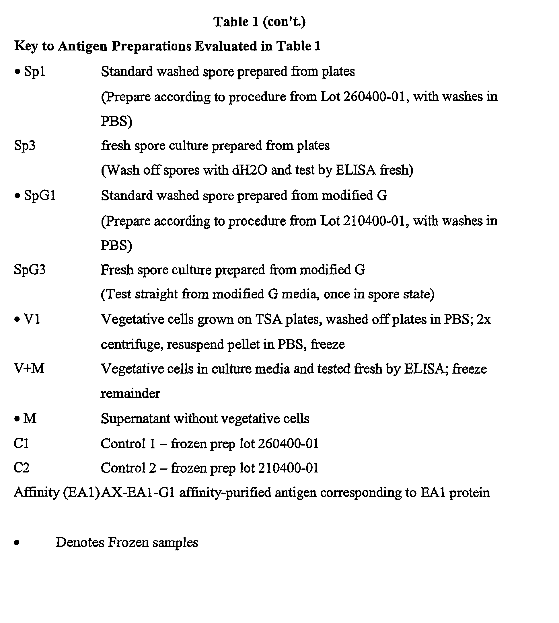
- Hybridoma cells were developed to B. anthracis spores by fusion of nonsecreting myeloma cells (SP2/0) with antibody-producing B-lymphocytes from the spleens of mice immunized with B. anthracis spores, in the presence of polyethylene glycol (PEG), according to standard hybridoma procedures. Cells were combined in a ratio of 3:1 (spleen:myeloma), and fused with PEG. Fused cells were plated, and cultured in 96-well cell culture grade plates. Fused cells were then selected by addition of HAT media [Iscove's Modified Dulbecco's Media (IMDM) with HAT supplement containing hypoxanthine, aminopterin, and thymidine]. These HAT supplements select for the fused hybridoma cells, and eliminate unfused or self-fused myeloma cells. Once clones appeared in the wells (usually 7-10 days after fusion), the culture supernatants were screened by ELISA for antibodies to B. anthracis spores. Positive antibody producing cells were subcloned by serial dilution, and plated at a cell concentration of three cells per well, and then further at one cell per three wells in a 96-well culture plate. This was performed with ten percent ORIGIN® Hybridoma Cloning Factor (HCF) in IMDM. Between each cloning step, culture supernatants were screened by ELISA for antibody production. Finalized clones were screened for isotype, and cryopreserved in liquid nitrogen. Two fusions were performed resulting in the generation of numerous monoclonal antibodies to Bacillus anthracis (Table 1).
- Cell supernatants were screened by direct ELISA. ELISA plates were coated with B. anthracis spore and vegetative preparation antigens as positive antigen, and bovine serum albumin (BSA) as negative antigen, diluted to an optimized concentration in PBS. Plates were incubated 18-24 hours at 4°C. Plates were washed four times with PBS. Cell supernatants were added to both positive and negative coated antigen wells, undiluted. Mouse sera from the immunized mice was added to plate at a dilution of 1:200, and serially diluted to an endpoint. This was included as a positive control. Plates were incubated at 37°C for one hour. Plates were washed four times with PBS. Horseradish peroxidase (HRP) conjugated goat anti mouse IgG + M + A (KPL) was added to all wells, and incubated at 37°C for one hour. Plates were washed four times with PBS. Substrate was added to plates and incubated at 37°C for 30 minutes. Plates were read for optical density at 280 nm, and evaluated for positive results. Cells producing the highest optical density readings, i.e., above 1.000 OD, were subcloned. After each subcloning, cell supernatants were screened for positive antibody. Finalized clones were tested for isotype using monoclonal antibody-based mouse Ig isotyping kit (catalog # 04017K; BD PharMingen). Three monoclonal antibodies (termed AX-EA1-G1, 8G4, and 9F5) were selected for their ability to uniquely detect B. anthracis and not cross-react with other closely related Bacillus species. Monoclonal antibody AX-EA1-G1 was deposited with the ATCC and accorded accession number PTA-2632, on October 26, 2000. The selection of these monoclonal antibodies was based on their strong reactivity against B. anthracis antigens and their negative reactivity against the closely related strains of B. thuringiensis (ATCC 33680, HD571, A1 Hakam, and commercial insecticide preparation from Dipel Dust), B. globigii and B. licheniformis (ATCC 25972) (Table 2, 3 and 4). In addition, these monoclonal antibodies were negative when tested against a selected number of other bacteria (Francisella tularensis and Yersinia pestis), purified proteins (ovalbumin and S. aureus enterotoxin B), and environmental components (red clay, gravel, and mulch) (Table 2, 3 and 4).
- To test for cross-reactivity, an antigen capture ELISA was performed. Plates were coated with rabbit anti-anthrax IgG antibody as positive capture, and normal rabbit IgG as negative capture antibody. Plates were incubated overnight at 4°C. Plates were washed four times with PBS and then blocked with dry skim milk buffer. Plates were incubated for one hour at 37°C, and washed four times with PBS. Antigens were added to both positive and negative antibody coated wells at concentrations determined for cross-reactivity analysis. Plates were incubated for one hour at 37°C, and washed four times with PBS. Monoclonal antibodies (Mabs) were added to the plate at optimized concentrations, as detector antibodies. Plates were incubated for one hour at 37°C, and washed four times with PBS. Anti-species conjugate was added to the plate. Plates were incubated for one hour at 37°C, and washed four times with PBS. Substrate solution was added to the plate, and incubated for 30 minutes at 37°C. Plates were read at 280 nm for optical density readings.
- An affinity column was made using the anthrax-specific monoclonal antibody AX-EA1-G1 complexed to the Immunopure Protein G IgG Orientation Kit (Pierce; Rockford, IL), according to manufacture's protocol. An anthrax spore antigen preparation was affinity purified over the column using the manufacturer's protocol.
- Affinity-purified anthrax antigens under went electrophoresis by SDS-PAGE on a 4-15% Tris-HCl polyacrylamide Ready Gel Precast Gel in a Mini-Protean 3 Electrophoresis Cell (Bio-Rad; Hercules, CA). Specifically, affinity-purified anthrax antigens (5.7 ug total), along with tubes containing molecular weight markers, were diluted in sample buffer (62.5 mM Tris-HCl pH 6.8, 2% SDS, 25% glycerol, 0.01 % Bromophenol blue), boiled for 2.5 minutes, loaded onto the 4-15% gel and under went electrophoresis at 200V for 30 minutes.
- The electroblotting procedure was performed according to the protocol posted on the Michigan State web site (http://gaea.bch.msu.edu/mssef/blotting.html) by the method of Matsudaira (J Biol Chem, 1987, 262:100035). Briefly, a 0.2 um PVDF membrane (Sequi-Blot PVDF Membrane for Protein Sequencing; Bio-Rad) was wet with methanol, soaked in CAPS/methanol buffer, electroblotted in a Mini Trans-Blot Electrophoresis Transfer Cell (Bio-Rad) at 50 V for one hour, according to manufacturer's instructions, in CAPS/methanol buffer.
- The blotted PVDF membrane was stained with 0.2% Amido Black in 40% methanol for 40 seconds and destained in dH2O. Two bands, of approximate molecular weight of 97 kD ("
Band 1") and 62 kD ("Band 2"), were visualized. - Monoclonal antibody AX-EA1-G1 was used to affinity purify the specific B. anthracis antigen that the antibody was detecting. The affinity-purified antigen(s) was separated on by SDS-PAGE and electroblotted onto a PVDF membrane. Two bands were visualized after staining at approximate molecular weights of 97 kD (Band 1) and 62 kD (Band 2); the membrane was sent to the Biotechnology Center of Utah State University for protein sequencing. The amino acid sequence was determined to be:
- Band 1: A G K Z F P Z V P A G H (SEQ ID NO 2)
- Band 2: D Z K Z N A Q A Y V T D (SEQ ID NO 3)
(Z = uncertain amino acid) - Using both of these amino acid sequences, a tblastn protein search of the Unfinished Microbial Genomes TIGR database of B. anthracis sequences was performed. An exact match was observed with the definitive amino acid sequences for Contig 1819. A BLAST search of GenBank using the nucleotide sequence of contig 1819 resulted in complete homology to the eag gene that codes for the EA1 protein of the B. anthracis S-layer. The amino acid position corresponding to the sequence of
Bands - To determine whether the monoclonal antibodies produced to B. anthracis compete for the same epitope(s), a competitive inhibition assay was performed. ELISA plates were coated with rabbit anti-anthrax IgG as positive capture antibody, and normal rabbit IgG as negative capture antibody. Plates were incubated overnight at 4°C. Plates were washed four times with PBS and then blocked with dry skim milk buffer. Plates were incubated for one hour at 37°C, and washed four times with PBS. Antigens were added to both positive and negative antibody coated wells at concentrations determined for inhibition analysis. Plates were incubated for one hour at 37°C, and washed four times with PBS. Three separate monoclonal antibodies were used in the competition at the detector antibody step. One Mab, labeled with biotin, was held constant while the other Mabs were unlabeled and combined separately at different concentrations, with the biotin labeled Mab. Each combination was prepared in a micro-tube rack, and then added to the plate at the same time. Plates were incubated for one hour at 37°C, and washed four times with PBS. Conjugated streptavidin was added to the plates and incubated for one hour at 37°C. Plates were washed and substrate solution added. Plates were incubated for 30 minutes, and read for optical density at 280 nm.
- Having determined that the monoclonal antibody AX-EA1-G1 reacts with the EA1 protein of B. anthracis, the other two monoclonal antibodies, 8G4 and 9F5, were tested against affinity-purified antigen and shown to also react with the EA1 antigen (Table 1). Therefore, all three monoclonal antibodies were shown to bind to the same EA1 protein. However, competitive inhibition analysis revealed that while 8G4 and 9F5 effectively compete for binding to the same epitope on the EA1 protein as each other, AX-EA1-G1 does not compete with the binding of 8G4 and 9F5 and therefore binds to a different epitope on the EA1 protein (Figure 2).



Claims (25)
- A product which is an antibody or reactive portion thereof which binds an EA 1 protein of B. anthracis, wherein said antibody or reactive portion thereof(1) specifically binds B. anthracis; and(2) is non-reactive against B. cereus or B. thuringiensis.
- The product of claim 1 which is an IgA, IgD, IgE, IgG or IgM.
- The product of claim 1 or claim 2 which binds to SEQ ID NO. 1 or an antigenic portion thereof.
- The product of any of claims 1 to 3 which is reactive against B. anthracis spores.
- The product of any of claims 1 to 4 which is reactive against B. anthracis vegetative cells.
- The product of any of claims 1 to 5 which is a monoclonal antibody.
- The product of claim 6, wherein said antibody is isolated from a hybridoma deposited with ATCC and accorded accession number PTA-2632.
- The product of any of claims 1 to 5 which is selected from: a murine antibody, a rabbit antibody, a rat antibody, a genetically engineered antibody, a recombinant antibody, a humanized antibody, a polyclonal antibody or an affinity-purified antibody.
- The product of any of claims 1 to 5 which is a Fab or Fv fragment.
- The product of any one of claims 1 to 9 wherein said antibody or reactive portion thereof is B. anthracis species specific.
- The product of any one of claims 1 to 10 wherein said antibody or reactive portion thereof is non-reactive against B. licheniformis.
- A hybridoma that produces the antibody of claim 6.
- The hybridoma of claim 12 which is derived from an animal selected from the group consisting of cattle, chickens, goats, guinea pigs, horses, mice, pigs, primates, rabbits, rats and sheep.
- The hybridoma of claim 12 or claim 13 which is deposited with ATCC and accorded accession number PTA-2632.
- A diagnostic kit comprising the product of any of claims 1 to 11.
- The diagnostic kit of claim 15 which incorporates a colloidal particle based lateral flow detection system.
- The diagnostic kit of claim 15 which incorporates a detection system selected from the group consisting of a carbon based lateral flow system; a fluorescent based assay system, a chemiluminescent system, an up converting phosphors system, a refractive indexed based detection system, a magnetic bead or latex bead system, and a micro array system.
- The diagnostic kit of claim 15, wherein the product is an antibody, or reactive portion thereof, that binds an EA1 antigen of B. anthracis spores and is reactive against spores of B. anthracis and is non-reactive against B. thuringiensis, and wherein said kit incorporates a colloidal particle based lateral flow detection system.
- The diagnostic kit of claim 18 wherein said antibody or reactive portion thereof is not reactive against B. licheniformis.
- A therapeutic agent comprising the product of any one of claims 1 to 11.
- The use of the product of any one of claims 1 to 11, for the manufacture of a medicament for treating, preventing or controlling B. anthracis infection.
- The product of any one of claims 1 to 11, in a detectably labelled form.
- A method of detecting B. anthracis in a sample, said method comprising
contacting a product of any one of claims 1 to 11 with a collected sample to form a complex between said product and B. anthracis in said sample, and
detecting said complex. - The method of claim 23 wherein the sample is collected from a surface or the environment.
- The method of claim 24 or claim 25 wherein said detecting is visual.
Applications Claiming Priority (3)
| Application Number | Priority Date | Filing Date | Title |
|---|---|---|---|
| US20050500P | 2000-04-28 | 2000-04-28 | |
| US200505P | 2000-04-28 | ||
| PCT/US2001/013648 WO2001083561A2 (en) | 2000-04-28 | 2001-04-30 | Anthrax-specific antigen, vaccines comprising said antigen, anthrax-specific antibodies, and their uses |
Publications (2)
| Publication Number | Publication Date |
|---|---|
| EP1280828A2 EP1280828A2 (en) | 2003-02-05 |
| EP1280828B1 true EP1280828B1 (en) | 2006-09-13 |
Family
ID=22742002
Family Applications (1)
| Application Number | Title | Priority Date | Filing Date |
|---|---|---|---|
| EP01932697A Expired - Lifetime EP1280828B1 (en) | 2000-04-28 | 2001-04-30 | Anthrax-specific antigen, vaccines comprising said antigen, anthrax-specific antibodies, and their uses |
Country Status (7)
| Country | Link |
|---|---|
| US (2) | US7618783B2 (en) |
| EP (1) | EP1280828B1 (en) |
| AT (1) | ATE339449T1 (en) |
| AU (1) | AU2001259204A1 (en) |
| CA (1) | CA2420287A1 (en) |
| DE (1) | DE60123036T2 (en) |
| WO (1) | WO2001083561A2 (en) |
Cited By (1)
| Publication number | Priority date | Publication date | Assignee | Title |
|---|---|---|---|---|
| CN105092853A (en) * | 2014-05-06 | 2015-11-25 | 中国检验检疫科学研究院 | Liquid chip for detecting various Bt proteins and detection method |
Families Citing this family (29)
| Publication number | Priority date | Publication date | Assignee | Title |
|---|---|---|---|---|
| TWI253471B (en) * | 2001-01-31 | 2006-04-21 | Food Industry Res & Dev Inst | Method for rapid identification of bacillus cereus |
| CA2446246A1 (en) * | 2001-05-03 | 2002-11-14 | Immunetics, Inc. | Systems and methods for detection of analytes in biological fluids |
| US7262019B2 (en) * | 2001-05-03 | 2007-08-28 | Immunetics, Inc. | System and methods for detection of Bacillus anthracis related analytes in biological fluids |
| US7306930B2 (en) | 2001-11-30 | 2007-12-11 | California Institute Of Technology | Method bacterial endospore quantification using lanthanide dipicolinate luminescence |
| GB0129776D0 (en) * | 2001-12-13 | 2002-01-30 | Sec Dep For Environment Food & | Assay device and method |
| US6927068B2 (en) * | 2002-01-30 | 2005-08-09 | The United States Of America As Represented By The Secretary Of The Navy | Rapid and non-invasive method to evaluate immunization status of a patient |
| EP1478912A4 (en) * | 2002-02-01 | 2007-03-21 | California Inst Of Techn | METHOD AND DEVICE FOR BIOLOGICAL ASSAYS OF BACTERIAL SPORES |
| JP4532121B2 (en) * | 2002-04-10 | 2010-08-25 | レスポンス バイオメディカル コーポレイション | Highly sensitive immunochromatographic assay |
| US7175992B2 (en) * | 2002-04-10 | 2007-02-13 | Response Biomedical Corporation | Sensitive immunochromatographic assay |
| US7601351B1 (en) | 2002-06-26 | 2009-10-13 | Human Genome Sciences, Inc. | Antibodies against protective antigen |
| US7611862B2 (en) | 2004-11-12 | 2009-11-03 | California Institute Of Technology | Method and apparatus for detecting and quantifying bacterial spores on a surface |
| US7608419B2 (en) | 2003-11-13 | 2009-10-27 | California Institute Of Technology | Method and apparatus for detecting and quantifying bacterial spores on a surface |
| US7563615B2 (en) | 2005-04-15 | 2009-07-21 | California Institute Of Technology | Apparatus and method for automated monitoring of airborne bacterial spores |
| CA2517198C (en) * | 2003-03-21 | 2016-06-21 | The Government Of The United States Of America As Represented By The Secretary Of The Department Of Health And Human Services, Centers For Disease Control And Prevention | Determining a concentration of a substance in a sample using a usable portion of a sigmoid curve |
| CA2526398C (en) * | 2003-05-21 | 2014-07-15 | Medarex, Inc. | Human monoclonal antibodies against bacillus anthracis protective antigen |
| US7928204B2 (en) * | 2004-05-12 | 2011-04-19 | Tetracore, Inc. | Spore specific antigen |
| CN101031587A (en) * | 2004-05-12 | 2007-09-05 | 泰树公司 | Spore specific antibodies |
| US20060115908A1 (en) * | 2004-11-30 | 2006-06-01 | Tetracore, Inc. | Multiplexed analyses of contaminant-laden gas in a particle impact collector |
| US8173871B2 (en) * | 2005-07-08 | 2012-05-08 | Universidad Nacional Autonoma De Mexico | Bacterial proteins with pesticidal activity |
| GB0514319D0 (en) * | 2005-07-13 | 2006-06-14 | Secr Defence | Antibodies for anthrax |
| US7794732B2 (en) * | 2006-05-12 | 2010-09-14 | Oklahoma Medical Research Foundation | Anthrax compositions and methods of use and production |
| US8420607B2 (en) * | 2006-06-30 | 2013-04-16 | University Of Georgia Research Foundation, Inc. | Anthrax carbohydrates, synthesis and uses thereof |
| AU2007349310B2 (en) | 2006-06-30 | 2013-03-14 | THE UNITED STATES OF AMERICA, as represented by THE SECRETARY OF THE DEPARTMENT OF HEALTH AND HUMAN SERVICES, CENTERS OF DISEASE CONTROL AND PREVENTION (WASHINGTON D.C.) | Anthrax carbohydrates,synthesis and uses thereof |
| EP2132571B1 (en) * | 2007-02-26 | 2014-09-03 | Response Biomedical Corporation | Comparative multiple analyte assay |
| US7935345B2 (en) | 2007-05-21 | 2011-05-03 | Children's Hospital & Research Center At Oakland | Monoclonal antibodies that specifically bind to and neutralize bacillus anthracis toxin, compositions, and methods of use |
| US8343495B2 (en) * | 2009-01-10 | 2013-01-01 | Auburn University | Equine antibodies against Bacillus anthracis for passive immunization and treatment |
| US9857370B2 (en) * | 2013-07-22 | 2018-01-02 | National Technology & Engineering Solutions Of Sandia, Llc | Amplification of biological targets via on-chip culture for biosensing |
| CN105203766B (en) * | 2015-09-29 | 2017-01-11 | 河南省科学院生物研究所有限责任公司 | Preparation method for pathogenic yersinia enterocolitica test strips |
| CN110498854B (en) * | 2019-09-28 | 2021-01-29 | 中国人民解放军陆军军医大学 | An antibody against Staphylococcus aureus enterotoxin B and its application |
Family Cites Families (6)
| Publication number | Priority date | Publication date | Assignee | Title |
|---|---|---|---|---|
| GB8426470D0 (en) | 1984-10-19 | 1984-11-28 | Technology Licence Co Ltd | Monoclonal antibodies |
| US5840312A (en) | 1991-05-02 | 1998-11-24 | Institut Pasteur | Recombinant Bacillus anthracis strains unable to produce the lethal factor protein or edema factor protein |
| US5895922A (en) * | 1996-03-19 | 1999-04-20 | Her Majesty The Queen In Right Of Canada, As Represented By The Minister Of National Defence | Fluorescent biological particle detection system |
| US6913756B1 (en) | 1998-04-29 | 2005-07-05 | The Uab Research Foundation | Monoclonal antibodies specific for anthrax and peptides derived from the antibodies thereof |
| AU4562599A (en) | 1998-06-12 | 1999-12-30 | New Horizons Diagnostics, Inc. | Colloidal colorimetric flow through and lateral flow assays utilizing soluble submicron particles |
| AU5287701A (en) | 2000-01-06 | 2001-07-16 | Biosite Diagnostics Incorporated | Assays for detection of bacillus anthracis |
-
2001
- 2001-04-30 US US09/844,281 patent/US7618783B2/en not_active Expired - Fee Related
- 2001-04-30 AU AU2001259204A patent/AU2001259204A1/en not_active Abandoned
- 2001-04-30 WO PCT/US2001/013648 patent/WO2001083561A2/en not_active Ceased
- 2001-04-30 EP EP01932697A patent/EP1280828B1/en not_active Expired - Lifetime
- 2001-04-30 CA CA002420287A patent/CA2420287A1/en not_active Abandoned
- 2001-04-30 DE DE60123036T patent/DE60123036T2/en not_active Expired - Lifetime
- 2001-04-30 AT AT01932697T patent/ATE339449T1/en not_active IP Right Cessation
-
2009
- 2009-11-16 US US12/619,489 patent/US20110200614A1/en not_active Abandoned
Cited By (1)
| Publication number | Priority date | Publication date | Assignee | Title |
|---|---|---|---|---|
| CN105092853A (en) * | 2014-05-06 | 2015-11-25 | 中国检验检疫科学研究院 | Liquid chip for detecting various Bt proteins and detection method |
Also Published As
| Publication number | Publication date |
|---|---|
| CA2420287A1 (en) | 2001-11-08 |
| ATE339449T1 (en) | 2006-10-15 |
| WO2001083561A3 (en) | 2002-05-30 |
| US20110200614A1 (en) | 2011-08-18 |
| WO2001083561A2 (en) | 2001-11-08 |
| AU2001259204A1 (en) | 2001-11-12 |
| US7618783B2 (en) | 2009-11-17 |
| DE60123036T2 (en) | 2007-04-05 |
| DE60123036D1 (en) | 2006-10-26 |
| US20020082386A1 (en) | 2002-06-27 |
| EP1280828A2 (en) | 2003-02-05 |
Similar Documents
| Publication | Publication Date | Title |
|---|---|---|
| US7618783B2 (en) | Anthrax specific antibodies | |
| Donohue-Rolfe et al. | Purification of Shiga toxin and Shiga-like toxins I and II by receptor analog affinity chromatography with immobilized P1 glycoprotein and production of cross-reactive monoclonal antibodies | |
| US8124107B2 (en) | Antibodies recognizing a highly expressed putative antigen of CA-MRSA and methods of use | |
| US20170183396A1 (en) | Ebola monoclonal antibodies | |
| US7879330B2 (en) | Antibodies against type A botulinum neurotoxin | |
| CN102021184A (en) | Antigenic polypeptides | |
| Miner et al. | Characterization of murine monoclonal antibodies to Escherichia coli J5 | |
| JP3747057B2 (en) | Bacterial stress protein | |
| CN104862283B (en) | The monoclonal antibody of a pair of of high specific high-affinity combination human muscle hemoglobin and its application | |
| KR20100103675A (en) | Antibody directed against pcrv | |
| MX2008013363A (en) | Methods and compositions based on shiga toxin type 1 protein. | |
| JPH05304990A (en) | Monoclonal antibody to mycoplasma pneumoniae | |
| RU2555530C2 (en) | METHOD OF IDENTIFYING POLYPEPTIDES AND PROTEINS OF H.parasuis | |
| CA2545714C (en) | Neutralizing human antibodies to anthrax toxin generated by recall technology | |
| CN115260306B (en) | Monoclonal antibodies targeting SARS-CoV-2 receptor binding motifs and their recognized antigen epitopes and applications | |
| Schou et al. | The interaction between pertussis toxin and 10 monoclonal antibodies | |
| US9310368B1 (en) | High affinity monoclonal antibodies for detection of Shiga toxin 2 (STX2) | |
| Usuwanthim et al. | Murine monoclonal antibodies neutral-izing the cytotoxic activity of diphtheria toxin | |
| US9513287B1 (en) | High affinity monoclonal antibodies for detection of shiga toxin 2 | |
| CN116425870B (en) | A monoclonal antibody 31A8 against the N protein of the novel coronavirus, its products and applications | |
| CN114891096B (en) | Anti-H1N1 influenza virus hemagglutinin protein monoclonal antibody ZJU11-03 and its application | |
| US7718779B2 (en) | Prophylactic and therapeutic monoclonal antibodies | |
| CN119192352B (en) | Neutralizing monoclonal antibodies against pseudorabies virus gD protein, hybridoma cells and their applications | |
| US9475864B2 (en) | Methods, compositions and vaccines relating to Neisseria meningitidis antibodies | |
| AU708879B2 (en) | Means for detection of bacteria of the species Taylorella equigenitalis and their biological applications |
Legal Events
| Date | Code | Title | Description |
|---|---|---|---|
| PUAI | Public reference made under article 153(3) epc to a published international application that has entered the european phase |
Free format text: ORIGINAL CODE: 0009012 |
|
| 17P | Request for examination filed |
Effective date: 20021126 |
|
| AK | Designated contracting states |
Designated state(s): AT BE CH CY DE DK ES FI FR GB GR IE IT LI LU MC NL PT SE TR |
|
| AX | Request for extension of the european patent |
Extension state: AL LT LV MK RO SI |
|
| 17Q | First examination report despatched |
Effective date: 20041004 |
|
| RAP1 | Party data changed (applicant data changed or rights of an application transferred) |
Owner name: TETRACORE, INC. |
|
| GRAP | Despatch of communication of intention to grant a patent |
Free format text: ORIGINAL CODE: EPIDOSNIGR1 |
|
| GRAS | Grant fee paid |
Free format text: ORIGINAL CODE: EPIDOSNIGR3 |
|
| GRAA | (expected) grant |
Free format text: ORIGINAL CODE: 0009210 |
|
| AK | Designated contracting states |
Kind code of ref document: B1 Designated state(s): AT BE CH CY DE DK ES FI FR GB GR IE IT LI LU MC NL PT SE TR |
|
| PG25 | Lapsed in a contracting state [announced via postgrant information from national office to epo] |
Ref country code: IT Free format text: LAPSE BECAUSE OF FAILURE TO SUBMIT A TRANSLATION OF THE DESCRIPTION OR TO PAY THE FEE WITHIN THE PRESCRIBED TIME-LIMIT;WARNING: LAPSES OF ITALIAN PATENTS WITH EFFECTIVE DATE BEFORE 2007 MAY HAVE OCCURRED AT ANY TIME BEFORE 2007. THE CORRECT EFFECTIVE DATE MAY BE DIFFERENT FROM THE ONE RECORDED. Effective date: 20060913 Ref country code: NL Free format text: LAPSE BECAUSE OF FAILURE TO SUBMIT A TRANSLATION OF THE DESCRIPTION OR TO PAY THE FEE WITHIN THE PRESCRIBED TIME-LIMIT Effective date: 20060913 Ref country code: CH Free format text: LAPSE BECAUSE OF FAILURE TO SUBMIT A TRANSLATION OF THE DESCRIPTION OR TO PAY THE FEE WITHIN THE PRESCRIBED TIME-LIMIT Effective date: 20060913 Ref country code: AT Free format text: LAPSE BECAUSE OF FAILURE TO SUBMIT A TRANSLATION OF THE DESCRIPTION OR TO PAY THE FEE WITHIN THE PRESCRIBED TIME-LIMIT Effective date: 20060913 Ref country code: BE Free format text: LAPSE BECAUSE OF FAILURE TO SUBMIT A TRANSLATION OF THE DESCRIPTION OR TO PAY THE FEE WITHIN THE PRESCRIBED TIME-LIMIT Effective date: 20060913 Ref country code: LI Free format text: LAPSE BECAUSE OF FAILURE TO SUBMIT A TRANSLATION OF THE DESCRIPTION OR TO PAY THE FEE WITHIN THE PRESCRIBED TIME-LIMIT Effective date: 20060913 |
|
| REG | Reference to a national code |
Ref country code: GB Ref legal event code: FG4D |
|
| REG | Reference to a national code |
Ref country code: CH Ref legal event code: EP |
|
| REG | Reference to a national code |
Ref country code: IE Ref legal event code: FG4D |
|
| REF | Corresponds to: |
Ref document number: 60123036 Country of ref document: DE Date of ref document: 20061026 Kind code of ref document: P |
|
| PG25 | Lapsed in a contracting state [announced via postgrant information from national office to epo] |
Ref country code: DK Free format text: LAPSE BECAUSE OF FAILURE TO SUBMIT A TRANSLATION OF THE DESCRIPTION OR TO PAY THE FEE WITHIN THE PRESCRIBED TIME-LIMIT Effective date: 20061213 |
|
| REG | Reference to a national code |
Ref country code: SE Ref legal event code: TRGR |
|
| PG25 | Lapsed in a contracting state [announced via postgrant information from national office to epo] |
Ref country code: ES Free format text: LAPSE BECAUSE OF FAILURE TO SUBMIT A TRANSLATION OF THE DESCRIPTION OR TO PAY THE FEE WITHIN THE PRESCRIBED TIME-LIMIT Effective date: 20061224 |
|
| PG25 | Lapsed in a contracting state [announced via postgrant information from national office to epo] |
Ref country code: PT Free format text: LAPSE BECAUSE OF FAILURE TO SUBMIT A TRANSLATION OF THE DESCRIPTION OR TO PAY THE FEE WITHIN THE PRESCRIBED TIME-LIMIT Effective date: 20070226 |
|
| NLV1 | Nl: lapsed or annulled due to failure to fulfill the requirements of art. 29p and 29m of the patents act | ||
| REG | Reference to a national code |
Ref country code: CH Ref legal event code: PL |
|
| ET | Fr: translation filed | ||
| PLBE | No opposition filed within time limit |
Free format text: ORIGINAL CODE: 0009261 |
|
| STAA | Information on the status of an ep patent application or granted ep patent |
Free format text: STATUS: NO OPPOSITION FILED WITHIN TIME LIMIT |
|
| 26N | No opposition filed |
Effective date: 20070614 |
|
| PG25 | Lapsed in a contracting state [announced via postgrant information from national office to epo] |
Ref country code: GR Free format text: LAPSE BECAUSE OF FAILURE TO SUBMIT A TRANSLATION OF THE DESCRIPTION OR TO PAY THE FEE WITHIN THE PRESCRIBED TIME-LIMIT Effective date: 20061214 |
|
| PG25 | Lapsed in a contracting state [announced via postgrant information from national office to epo] |
Ref country code: IE Free format text: LAPSE BECAUSE OF NON-PAYMENT OF DUE FEES Effective date: 20070430 |
|
| PG25 | Lapsed in a contracting state [announced via postgrant information from national office to epo] |
Ref country code: MC Free format text: LAPSE BECAUSE OF NON-PAYMENT OF DUE FEES Effective date: 20070430 |
|
| PG25 | Lapsed in a contracting state [announced via postgrant information from national office to epo] |
Ref country code: LU Free format text: LAPSE BECAUSE OF NON-PAYMENT OF DUE FEES Effective date: 20070430 Ref country code: CY Free format text: LAPSE BECAUSE OF FAILURE TO SUBMIT A TRANSLATION OF THE DESCRIPTION OR TO PAY THE FEE WITHIN THE PRESCRIBED TIME-LIMIT Effective date: 20060913 |
|
| PG25 | Lapsed in a contracting state [announced via postgrant information from national office to epo] |
Ref country code: TR Free format text: LAPSE BECAUSE OF FAILURE TO SUBMIT A TRANSLATION OF THE DESCRIPTION OR TO PAY THE FEE WITHIN THE PRESCRIBED TIME-LIMIT Effective date: 20060913 |
|
| REG | Reference to a national code |
Ref country code: FR Ref legal event code: PLFP Year of fee payment: 15 |
|
| REG | Reference to a national code |
Ref country code: DE Ref legal event code: R119 Ref document number: 60123036 Country of ref document: DE |
|
| REG | Reference to a national code |
Ref country code: DE Ref legal event code: R073 Ref document number: 60123036 Country of ref document: DE |
|
| PG25 | Lapsed in a contracting state [announced via postgrant information from national office to epo] |
Ref country code: DE Free format text: LAPSE BECAUSE OF NON-PAYMENT OF DUE FEES Effective date: 20151103 |
|
| PGFP | Annual fee paid to national office [announced via postgrant information from national office to epo] |
Ref country code: GB Payment date: 20151102 Year of fee payment: 15 Ref country code: FI Payment date: 20151102 Year of fee payment: 15 |
|
| PGFP | Annual fee paid to national office [announced via postgrant information from national office to epo] |
Ref country code: SE Payment date: 20151102 Year of fee payment: 15 Ref country code: FR Payment date: 20151102 Year of fee payment: 15 |
|
| REG | Reference to a national code |
Ref country code: DE Ref legal event code: R074 Ref document number: 60123036 Country of ref document: DE |
|
| PGFP | Annual fee paid to national office [announced via postgrant information from national office to epo] |
Ref country code: DE Payment date: 20151103 Year of fee payment: 15 |
|
| PGRI | Patent reinstated in contracting state [announced from national office to epo] |
Ref country code: DE Effective date: 20160310 |
|
| REG | Reference to a national code |
Ref country code: DE Ref legal event code: R082 Ref document number: 60123036 Country of ref document: DE Representative=s name: SSM SANDMAIR PATENTANWAELTE RECHTSANWALT PARTN, DE Ref country code: DE Ref legal event code: R082 Ref document number: 60123036 Country of ref document: DE Representative=s name: SCHWABE SANDMAIR MARX PATENTANWAELTE RECHTSANW, DE |
|
| REG | Reference to a national code |
Ref country code: DE Ref legal event code: R119 Ref document number: 60123036 Country of ref document: DE |
|
| GBPC | Gb: european patent ceased through non-payment of renewal fee |
Effective date: 20160430 |
|
| REG | Reference to a national code |
Ref country code: FR Ref legal event code: ST Effective date: 20161230 |
|
| PG25 | Lapsed in a contracting state [announced via postgrant information from national office to epo] |
Ref country code: FI Free format text: LAPSE BECAUSE OF NON-PAYMENT OF DUE FEES Effective date: 20160430 Ref country code: GB Free format text: LAPSE BECAUSE OF NON-PAYMENT OF DUE FEES Effective date: 20160430 Ref country code: FR Free format text: LAPSE BECAUSE OF NON-PAYMENT OF DUE FEES Effective date: 20160502 |
|
| PG25 | Lapsed in a contracting state [announced via postgrant information from national office to epo] |
Ref country code: SE Free format text: LAPSE BECAUSE OF NON-PAYMENT OF DUE FEES Effective date: 20160501 |
|
| PG25 | Lapsed in a contracting state [announced via postgrant information from national office to epo] |
Ref country code: DE Free format text: LAPSE BECAUSE OF NON-PAYMENT OF DUE FEES Effective date: 20161101 |