TECHNICAL FIELD
-
The present invention relates to a pharmaceutical preparation stabilizing an
antibody to parathyroid hormone related peptide and a pharmaceutical preparation for
injection.
BACKGROUND ART
-
Parathyroid hormone related peptide (hereinafter referred to as PTHrP) is a
protein produced by a tumor, which is a major causative agent of hypercalcemia. That
protein causes tumor-producing hypercalcemia (Humoral hypercalcemia of malignancy,
hereinafter referred to as HHM) by promoting calcium resorption in bone resorption and
uridiferous tubule. Although at present, a calcitonin or a bisphosphonate each which
has a bone resorption-inhibitory action, is used for HHM therapy, due to rapid progress
of HHM and worsening QOL (Quality of Life) of a terminal cancer patient, there is a
demand for development of an effective therapeutic agent according with the cause.
-
An antibody to parathyroid hormone related peptide (hereinafter referred to as
anti-PTHrP antibody) has an immediate therapeutic effect against HHM after
administration thereof, and thus it is excellent in comparison with a bisphosphonate
which requires several days until the therapeutic effect is observed. Further, the
antibody is useful as a therapeutic agent for cachexia observed in a terminal cancer
patient. (Japanese Patent Application Laying-Open (kokai) No. 11-80025)
DISCLOSURE OF THE INVENTION
-
In order to use the anti-PTHrP antibody as a therapeutic agent for diseases, it is
necessary to provide it as a stabilized preparation which can retain biological activity of
the anti-PTHrP antibody for a long period. Accordingly, it is an object of the present
invention to provide a stabilized pharmaceutical preparation of the anti-PTHrP antibody.
-
The inventors of the present invention prepared an anti-PTHrP antibody solution,
verified the influences of hydrogen ion concentration (pH) and buffer solution
concentration on physicochemical properties of the anti-PTHrP antibody, and succeeded
in producing a stabilized pharmaceutical preparation of the anti-PTHrP antibody.
-
In other words, the present invention provides a stabilized pharmaceutical
preparation of an anti-PTHrP antibody, wherein the anti-PTHrP antibody is dissolved in
a buffer solution containing at least one buffer selected from the group consisting of
acetic acid, citric acid, phosphoric acid, and salts thereof and is in the form of a solution
of pH 5 to 8.
-
Further, the present invention provides a stabilized composition of anti-PTHrP
antibody solution, wherein the anti-PTHrP antibody is dissolved in a buffer solution
containing at least one buffer selected from the group consisting of acetic acid, citric
acid, phosphoric acid, and salts thereof and is in the form of a solution of pH 5 to 8.
-
In detail, the present invention provides the composition of the antibody solution
wherein the composition of the antibody solution is a solution composition for bulk.
-
In more detail, the present invention provides the composition of the antibody
solution substantially free of stabilizers other than a buffer or an isotonizing agent.
-
In this description, "a buffer solution" means a solution having a buffer action
(that is, easing up the change of pH), and "a buffer" means a substance having a buffer
action.
-
The preparation or the composition may have a total concentration of the buffer
of 0.1 to 100 mmol/L, preferably 5 to 50 mmol/L.
-
To the preparation may be added an isotonizing agent such as sodium chloride
and glucose, so that the preparation essentially has the same osmotic pressure as human
blood. In general, a preferable osmotic pressure is approximately from 250 to 350
mOsm.
-
The anti-PTHrP antibody may be a monoclonal antibody, and this antibody is
preferably a human antibody, a humanized antibody, or a chimeric antibody.
-
Moreover, the inventors of the present invention prepared a pharmaceutical
preparation for injection containing the anti-PTHrP antibody solution, verified that pain
action at a time of administration is different depending on a kind of buffer solution, and
succeeded in producing a pharmaceutical preparation for injection which includes the
anti-PTHrP antibody and causes less pain.
-
Namely, the present invention provides a pharmaceutical preparation for injection
wherein the anti-PTHrP antibody is dissolved in a buffer solution containing a buffer
consisting of acetic acid and/or salts thereof.
-
In detail, the present invention provides the pharmaceutical preparation for
injection which is in the form of a solution of pH 5 to 8.
-
In more detail, the present invention provides the pharmaceutical preparation for
injection wherein the solution has a total concentration of the buffer of 0.1 to 100
mmol/L, preferably 5 to 50 mmol/L.
-
The anti-PTHrP antibody may be a monoclonal antibody, and this antibody
preferably be a human antibody, a humanized antibody, or a chimeric antibody.
-
Hereinbelow, the present invention will be illustrated in detail.
1. Anti-PTHrP antibody
-
The anti-PTHrP antibody used in the present invention may be any one as far as
it has the desired pharmacological effect, regardless of its source, type (monoclonal or
polyclonal) and configuration.
-
The anti-PTHrP antibody used in the present invention can be produced by any
known method as a polyclonal or monoclonal antibody. Preferably, the anti-PTHrP
antibody is a monoclonal antibody derived from a mammal. The mammal-derived
monoclonal antibody includes those produced from a hybridoma and those produced by
a genetic engineering technique from a host transformed with a recombinant expression
vector carrying a gene for the antibody. The antibody can bind to PTHrP to prevent the
binding of the PTHrP to a PTH/PTHrP receptor, thus blocking the signal transduction of
the PTHrP and consequently inhibiting the biological activity of the PTHrP.
-
A specific example of such antibody is #23-57-137-1 antibody which can be
produced with a hybridoma clone #23-57-137-1.
-
The hybridoma clone #23-57-137-1 has been designated "mouse-mouse
hybridoma #23-57-137-1" and deposited under the terms of the Budapest Treaty on
August 15, 1996 at the National Institute of Bioscience and Human-technology, Agency
of Industrial Science and Technology, Japan (1-3, Higashi 1-chome, Tsukuba-shi, Ibaraki,
Japan) under the accession No. FERM BP-5631.
2. Antibody-producing hybridoma
-
A monoclonal antibody-producing hybridoma can be produced as follows. That
is, PTHrP is used as an antigen for immunization in accordance with a conventional
immunization method. The resulting immunocytes are fused to known parent cells by a
conventional cell fusion method, and monoclonal antibody-producing cells are screened
from the fused cells by a conventional screening method.
-
First, a human PTHrP, which is used as an sensitizing antigen for producing the
antibody, is prepared by expressing the PTHrP gene/amino acid sequence disclosed in
Suva, L. J. et al., Science (1987) 237, 893. A nucleotide sequence encoding the PTHrP
is inserted into a known expression vector, and a suitable host cell is transformed with
the expression vector. The PTHrP protein is then isolated and purified from the
transformed host cell or from a culture supernatant of the transformed host cell by any
known method.
-
Then, the purified PTHrP protein is used as a sensitizing antigen. Alternatively,
a 34-amino acid peptide of the N-terminal region of the PTHrP may be chemically
synthesized as the sensitizing antigen.
-
The mammal to be immunized with the sensitizing antigen is not particularly
limited. However, the mammal is preferably selected taking into consideration of
compatibility with the patent cell used for cell fusion. Generally, a rodent (e.g., mouse,
rat, hamster), rabbit or monkey may be used.
-
The immunization of the mammal with the sensitizing antigen can be performed
in accordance with any known method, for example, by injecting the sensitizing antigen
to a mammal intraperitoneally or subcutaneously. More specifically, the sensitizing
antigen is properly diluted with or suspended to phosphate-buffered saline (PBS) or
physiological saline, the resulting dilution or suspension is then mixed with an
appropriate amount of a conventional adjuvant (e.g., Freund's complete adjuvant) to give
an emulsion. The emulsion is injected to a mammal several times at intervals of 4 to
21 days. For the immunization, the sensitizing antigen may be attached to a suitable
carrier.
-
After the immunization, the serum antibody level is checked. When the serum
antibody level is confirmed to reach a desired level, immunocytes are isolated from the
mammal and then subjected to cell fusion. A preferable immunocyte is a spleen cell.
-
The parent cell used for the cell fusion (i.e., the counterpart of the cell fusion
with the immunocyte) is a myeloma cell derived from a mammal. The myeloma cell is
of any known cell line, and, for example, P3 (P3x63Ag8.653) (J. Immnol. (1979) 123,
1548-1550), P3x63Ag8U.1 (Current Topics in Microbiology and Immunology (1978) 81,
1-7), NS-1 (Kohler, G. and Milstein, C. Eur. J. Immunol. (1976) 6, 511-519), MPC-11
(Margulies, D. H. et al., Cell (1976) 8, 405-415), SP2/0 (Shulman, M. et al., Nature
(1978) 276, 269-270), FO (de St. Groth, S. F. et al., J. Immunol. Methods (1980) 35, 1-21),
S194 (Trowbridge, I. S., J. Exp. Med. (1978) 148, 313-323) or R210 (Galfre, G. et
al., Nature (1979) 277, 131-133).
-
Cell fusion of the immunocyte to the myeloma cell is basically performed in
accordance with any known method, such as the method of Milstein et al. (Kohler, G.
and Milstein, C., Methods Enzymol. (1981) 73, 3-46).
-
More specifically, the cell fusion is performed, for example, in a conventional
nutrient culture medium in the presence of a cell fusion promoter. The cell fusion
promoter may be polyethylene glycol (PEG) or a Sendai virus (hemagglutinating virus
of Japan; HVJ). If desired, for the purpose of improving the fusion efficiency, an
additive such as dimethyl sulfoxide may be incorporated.
-
The ratio between the immunocytes and the myeloma cells for the cell fusion
may be any one. For example, the immunocytes are used in the amount 1-10 times
larger than the myeloma cells. The culture medium used for the cell fusion is, for
example, RPMI 1640 medium or MEM medium suitable for the growth of the above-mentioned
myeloma cell lines, or other medium conventionally used for the culture of
such cell lines. If desired, a serum supplement, such as feral calf serum (FCS), may be
added to the culture medium.
-
The cell fusion is performed by fully mixing given amounts of the immunocytes
and the myeloma cells in the culture medium, adding a PEG solution (e.g., mean
molecular weight: about 1000-6000) (which has been previously warmed to about 37°
C) to the mixture usually to a concentration of 30-60% (w/v), and then mixing the
resulting solution, thereby producing the desired fusion cells (i.e., hybridomas).
Subsequently, an appropriate culture medium is added to the culture solution
successively, and centrifuged to remove the supernatant. This procedure is repeated
several times to remove the cell fusion promoter or the like that are undesirable for the
growth of the hybridomas, from the culture medium.
-
The obtained hybridomas can be selected by culturing in a conventional selective
medium, such as hypoxanthine-aminopterin-thymidine (HAT) medium. The culturing
of the hybridomas in HAT medium is performed for the time of period enough to cause
the death of the cells other than the desired hybridomas (i.e., cells that fail to fuse),
usually for several days to several weeks. Subsequently, conventional limiting dilution
method is performed for screening and mono-cloning of the hybridomas that are
secreting the desired antibody.
-
As a method other than preparing the hybridomas by immunizing a non-human
mammal with the antigen as described above, a human lymphocyte may be sensitized
with PTHrP in vitro, and then subjected the sensitized lymphocyte to cell fusion to a
human-derived myeloma cell capable of infinite growth, thereby producing a human
antibody having a binding activity against the PTHrP (Japanese Patent Publication No.
1-59878). Alternatively, a human antibody against PTHrP may be prepared by
injecting PTHrP as an antigen to a transgenic animal that has the entire repertories of
human antibody genes to produce an anti-PTHrP antibody-producing cell, and then
immortalizing the cells, thus producing the human antibody from the immortalized cell
(International Patent Publication Nos. WO 94/25585, WO 93/12227, WO 92/03918 and
WO 94/02602).
-
The monoclonal antibody-producing hybridoma prepared as above can be
subcultured in a conventional culture medium and stored under liquid nitrogen for a long
time of period.
-
For the production of a monoclonal antibody from the hybridoma, a method
may be employed that involves culturing the hybridoma in accordance with a
conventional technique and collecting the monoclonal antibody from the culture
supernatant, or that involves injecting the hybridoma to a mammal compatible with the
hybridoma to grow the hybridoma in the mammal and collecting the hybridoma from the
ascites of the mammal. The former method is suitable for producing the antibody in
high purity, while the latter method is suitable for producing the antibody in a large
amount.
3. Recombinant antibody
-
In the present invention, a recombinant-type monoclonal antibody may be used,
which can be produced by cloning an antibody gene from the hybridoma, integrating the
antibody gene into a suitable vector, introducing the vector into a host, and then
producing the antibody from the host according to a conventional genetic recombination
technique (see, for example, Vandamme, A. M. et al., Eur. J. Biochem. (1990) 192, 767-775,
1990).
-
Specifically, mRNA encoding variable (V) region of an anti-PTHrP antibody is
isolated from the anti-PTHrP antibody-producing hybridoma. The isolation of the
mRNA is performed by preparing a total RNA by any known method, such as guanidium
ultracentrifugation method (Chirgwin, J. M. et al., Biochemistry (1979) 18, 5294-5299)
and AGPC method (Chomczynski, P. et al., Anal. Biochem. (1987) 162, 156-159), and
then producing the desired mRNA from the total RNA using mRNA Purification Kit
(Pharmacia) or the like. Alternatively, the mRNA may also be prepared directly using
QuickPrep mRNA Purification Kit (Pharmacia).
-
Next, cDNA for the antibody V-region is synthesized from the obtained mRNA
with a reverse transcriptase. The synthesis of the cDNA is performed using AMV
Reverse Transcriptase First-strand cDNA Synthesis Kit (Seikagaku Corporation) or the
like. The cDNA may also be synthesized and amplified by 5'-RACE method (Frohman,
M.A. et al., Proc. Natl. Acad. Sci. USA (1988) 85, 8998-9002; Belyavsky, A. et al.,
Nucleic Acids Res. (1989) 17, 2919-2932) using 5'-Ampli FINDER RACE Kit
(CLONETECH) in combination with PCR method, or the like.
-
A DNA fragment of interest is isolated and purified from the resulting PCR
product and then ligated to a vector DNA to obtain a recombinant vector. The
recombinant vector is introduced into a host such as E. coli, and a colony containing a
desired recombinant vector is selected. The nucleotide sequence of the DNA of
interest in the recombinant vector is confirmed by, for example, dideoxynucleotide chain
termination method.
-
Once DNA encoding the anti-PTHrP antibody V-region is obtained, the DNA is
integrated into an expression vector containing a DNA encoding a desired antibody
constant (C) region.
-
For the production of the anti-PTHrP antibody used in the present invention, the
antibody gene is integrated into an expression vector so that the antibody gene can be
expressed under the control of expression control regions (e.g., enhancer, promoter). A
host cell is transformed with the expression vector to express the antibody.
-
In the expression of the antibody gene, a DNA encoding heavy (H) chain and a
DNA encoding light (L) chain of the antibody may be integrated into separate
expression vectors, and then a host cell is co-transformed with the resulting recombinant
expression vectors. Alternatively, both the DNA encoding H-chain and the DNA
encoding L-chain of the antibody may be integrated together into a single expression
vector, and then a host cell may be transformed with the resulting recombinant
expression vector (WO 94/11523).
-
For the production of the recombinant antibody, besides the above-mentioned
host cells, a transgenic animal may also be used as a host. For example, the antibody
gene is inserted into a predetermined site of a gene encoding a protein inherently
produced in the milk of an animal (e.g., goat β-casein) to obtain a fusion gene. A
DNA fragment containing the antibody gene-introduced fusion gene is injected into an
embryo of a goat, and the embryo is then introduced into a female goat. The female
goat having the embryo therein bears a transgenic goat. The antibody of interest is
secreted in the milk from the transgenic goat or a progeny thereof. For the purpose of
increasing the amount of the antibody-containing milk from the transgenic goat, an
appropriate hormone may be administered to the transgenic goat (Ebert, K.M. et al.,
Bio/Technology (1994) 12, 699-702).
4. Modified antibody
-
In the present invention, for the purpose of reducing the heterogenisity against a
human body or the like, an artificially modified recombinant antibody may be used, such
as a chimeric antibody and a humanized antibody. These modified antibodies can be
prepared by the following known methods.
-
A chimeric antibody usable in the present invention can be prepared by ligating
the DNA encoding the antibody V-region prepared as set forth above to a DNA
encoding a human antibody C-region, integrating the ligation product into an expression
vector, and introducing the resulting recombinant expression vector into a host to
produce the chimeric antibody.
-
A humanized antibody is also referred to as a "reshaped human antibody", in
which the complementarity determining regions (CDRs) of an antibody of a non-human
mammal (e.g., a mouse) are grafted to those of a human antibody. The general genetic
recombination procedures for producing such humanized antibody are also known (EP
125023; WO 96/02576).
-
Specifically, a DNA sequence in which mouse antibody CDRs are ligated through
framework regions (FRs) of a human antibody is amplified by PCR method using
several oligonucleotides as primers which have been designed to have regions
overlapping to the terminal regions of the CDRs and the FRs. The resulting DNA is
ligated to a DNA encoding a human antibody C-region, and the ligation product is
integrated into an expression vector. The resulting recombinant expression vector is
introduced into a host, thereby producing the humanized antibody (EP 239044, WO
96/02576).
-
The FRs of the human antibody ligated through the CDRs are selected so that the
CDRs can form a suitable antigen binding site. If necessary, an amino acid(s) in the
FRs of the antibody V-region may be replaced so that the CDRs of the reshaped human
antibody can form a suitable antigen binding site (Sato, K. et al., Cancer Res. (1993) 53,
851-856).
-
The C-region of the chimeric or humanized antibody may be any human antibody
C-region, such as Cγ1, Cγ2, Cγ3 or Cγ4 for the H-chain, and Cκ or Cλ for the L-chain.
The human antibody C-region may be modified for the purpose of improving the stable
production of the antibody.
-
The chimeric antibody is composed of V-regions derived from a non-human
mammalian antibody and C-regions derived from a human antibody. The humanized
antibody is composed of CDRs derived from a non-human mammalian antibody and FRs
and C-regions derived from a human antibody. The humanized antibody is useful as an
active ingredient for the drug of the present invention, because the antigenicity of the
antibody against a human body is reduced.
-
A specific example of the humanized antibody usable in the present invention is
humanized #23-57-137-1 antibody; in which the CDRs are derived from mouse-derived
#23-57-137-1 antibody; the L-chain is composed of the CDRs ligated through three FRs
(FR1, FR2 and FR3) derived from human antibody HSU 03868 (GEN-BANK, Deftos, M.
et al., Scand. J. Immunol., 39, 95-103, 1994) and a FR (PR4) derived from human
antibody S25755 (NBRF-PDB); and the H-chain is composed of the CDRs ligated
through FRs derived from human antibody S31679 (NBRF-PDB, Cuisinier, A. M. et al.,
Eur. J. Immunol. 23, 110-118, 1993) in which a part of the amino acid residues in the
FRs is replaced so that the reshaped humanized antibody can exhibit an antigen-binding
activity.
-
The E. coli strains containing the plasmids having DNAs encoding the H-chain
and the L-chain of the humanized #23-57-137-1 antibody, respectively, are designated
Escherichia coli JM109 (hMBC1HcDNA/pUC19) (for H-chain) and Escherichia coli
JM109 (hMBC1Lqλ/pUC19) (for L-chain), respectively. These strains have been
deposited under the terms of the Budapest Treaty on August 15, 1996 at the National
Institute of Bioscience and Human-technology, Agency of Industrial Science and
Technology, Japan (1-3, Higashi 1-chome, Tsukuba-shi, Ibaraki, Japan), under the
accession No. FERM BP-5629 for Escherichia coli JM109 (hMBC1HcDNA/pUC19),
and under the accession No. FERM BP-5630 for Escherichia coli JM109
(hMBC1Lqλ/pUC19).
5. Antibody variants
-
The antibody used in the present invention may be a fragment thereof or a
modified form of the fragment, as long as it can bind to PTHrP and inhibit the activity of
the PTHrP. For example, the fragment of the antibody includes Fab, F(ab')2, Fv, or a
single chain Fv (scFv) composed of a H-chain Fv fragment and a L-chain Fv fragment
linked together through a suitable linker. Specifically, such antibody fragments can be
produced by cleaving the antibody with an enzyme (e.g., papain, pepsin) into antibody
fragments, or by constructing a gene encoding the antibody fragment and inserting the
gene into an expression vector and introducing the resulting recombinant expression
vector into a suitable host cell, thereby expressing the antibody fragment (see, for
example, Co, M. S., et al., J. Immunol. (1994), 152, 2968-2976; Better, M. & Horwitz, A.
H., Methods in Enzymology (1989), 178, 476-496, Academic Press, Inc.; Plueckthun, A.
& Skerra, A., Methods in Enzymology (1989) 178, 476-496, Academic Press, Inc.;
Lamoyi, E., Methods in Enzymology (1989) 121, 652-663; Rousseaux, J. et al., Methods
in Enzymology (1989) 121, 663-669; and Bird, R. E. et al., TIBTECH (1991) 9, 132-137).
-
The scFv can be produced by linking the H-chain V-region to the L-chain V-region
through a linker, preferably a peptide linker (Huston, J. S. et al., Proc. Natl. Acad.
Sci. USA (1988) 85, 5879-5883). The H-chain V-region and the L-chain V-region in
the scFv may be derived from any one of the antibodies described herein. The peptide
linker which binds the V-regions may be any single chain peptide, for example, of 12-19
amino acid residues.
-
The DNA encoding the scFv can be prepared by first amplifying a DNA encoding
the H-chain V-region and a DNA encoding the L-chain V-region of the antibody
separately using a DNA fragment encoding the entire region or a part of the H-chain that
includes the V-region and a DNA fragment encoding the entire region or a part of the L-chain
that includes the V-region as templates and primer pairs that define the terminal
ends of the DNA fragments; and then amplifying a DNA encoding the peptide linker
using a DNA fragment encoding the peptide linker as a template and a primer pair that
define the terminal ends of the DNA fragment so that each terminal end of the peptide
linker is ligated to the H-chain V-region and the L-chain V-region, respectively.
-
Once the DNA encoding the scFv is prepared, an expression vector carrying the
DNA and a host transformed with the expression vector can be prepared by conventional
methods. The scFv can be produced from the transformed host by a conventional
method.
-
The fragments of the antibody may be produced by preparing genes for the
fragments and expressing the genes in suitable hosts as described above. The antibody
fragments is also encompassed in the "antibody" of the present invention.
-
As a modified form of the above-mentioned antibodies, for example, anti-PTHrP
antibody conjugated to any molecule (e.g., polyethylene glycol) may also be used.
Such modified antibodies are also encompassed in the "antibody" of the present
invention. The modified antibodies can be prepared by chemical modifications of the
antibodies. The chemical modification techniques suitable for this purpose have
already been established in the art.
6. Expression and production of recombinant antibody or modified antibody
-
The antibody gene constructed as described above can be produced and expressed
by known methods. For the expression in a mammalian cell, a conventional useful
promoter, the antibody gene to be expressed and a poly(A) signal (located downstream
to the 3' end of the antibody gene) are operably linked. For example, as the useful
promoter/enhancer system, a human cytomegalovirus immediate early
promoter/enhancer system may be used.
-
Other promoter/enhancer systems usable in the expression of the antibody used in
the present invention include those derived from viruses (e.g., retrovirus, polyoma virus,
adenovirus and simian virus 40 (SV40)) and those derived from mammalian cells (e.g.,
human elongation factor 1 α (HEF1 α).
-
When SV40 promoter/enhancer system is used, the gene expression may be
performed readily by the method of Mulligan et al. (Nature (1979) 277, 108). When
HEFT a promoter/enhancer system is used, the gene expression may be performed
readily by the method of Mizushima et al. (Nucleic Acids Res. (1990) 18, 5322).
-
For the expression in E. coli, a conventional useful promoter, a signal sequence
for secreting the antibody of interest and the antibody gene may be operably linked.
As such a promoter, lacZ promoter or araB promoter may be used. When lacZ
promoter is used, the gene expression may be performed by the method of Ward et al.
(Nature (1098) 341, 544-546; FASBE J. (1992) 6, 2422-2427). When araB promoter is
used, the gene expression may be performed by the method of Better et al. (Better et al.,
Science (1988) 240, 1041-1043).
-
Regarding the signal sequence for secretion of the antibody, when the antibody of
interest is intended to be secreted in a periplasmic space of the E. coli, pelB signal
sequence (Lei, S. P. et al., J. Bacteriol. (1987) 169, 4379) may be used. The antibody
secreted into the periplasmic space is isolated and then refolded so that the antibody
takes an appropriate configuration for use.
-
Regarding the replication origin, those derived from viruses (e.g., SV40, polyoma
virus, adenovirus, bovine papilloma virus (BPV)) or the like may be used. In order to
increase the gene copy number in the host cell system, the expression vector may further
contain a selective marker gene, such as an aminoglycoside phosphotranferase (APH)
gene, a thymidine kinase (TK) gene, an E. coli xanthine-guanine
phosphoribosyltransferase (Ecogpt) gene and a dihydrofolate reductase (dhfr) gene.
-
For the production of the antibody used in the present invention, any expression
system such as eukaryotic and prokaryotic cell systems may be used. The eukaryotic
cell includes established cell lines of animals (e.g., mammals, insects, molds and fungi,
yeast). The prokaryotic cell includes bacterial cells such as E. coli cells.
-
It is preferable that the antibody used in the present invention be expressed in a
mammalian cell, such as a CHO, COS, myeloma, BHK, Vero or HeLa cell.
-
Next, the transformed host cell is cultured in vitro or in vivo to produce the
antibody of interest. The culturing of the host cell may be performed by any known
method. The culture medium usable herein may be DMEM, MEM, RPMI 1640 or
IMDM medium. The culture medium may contain a serum supplement, such as fetal
calf serum (FCS).
7. Isolation and purification of antibody
-
The antibody expressed and produced as described above may be isolated from
the cells or the host animal body and purified to uniformity. The isolation and
purification of the antibody used in the present invention may be performed on an
affinity column. Examples of a protein A column include Hyper D, POROS and
Sepharose F.F. (Pharmacia). The method is not particularly limited and other methods
conventionally used for the isolation and purification of an antibody may also be
employed. For example, various chromatographs using columns other than the above-mentioned
affinity column, filtration, ultrafiltration, salting out and dialysis may be used
singly or in combination to isolate and purify the antibody of interest (Antibodies A
Laboratory Manual. Ed. Harlow, David Lane, Cold Spring Harbor Laboratory, 1988).
8. Determination of the activities of the antibody
-
The determination of the antigen-binding activity (Antibodies A Laboratory
Manual, Ed. Harlow, David Lane, Cold Spring Harbor Laboratory, 1988) or the
inhibitory activity against a ligand receptor (Harada, A. et al., International Immunology
(1993) 5, 681-690) of the antibody used in the present invention may be performed by
any known methods.
-
The method for the determination of the antigen-binding activity of the anti-PTHrP
antibody used in the present invention may be BIACORE method (analytical
method using surface plasmon resonance), ELISA (enzyme-linked immunosorbent
assay), EIA (enzyme immunoassay), RIA (radioimmunoassay) or a fluorescent antibody.
For example, when enzyme immunoassay is employed, a sample solution containing the
anti-PTHrP antibody (e.g., a culture supernatant of anti-PTHrP antibody-producing cells,
or the anti-PTHrP antibody in a purified form) is added to a plate on which PTHrP (1-34)
is previously coated. A secondary antibody labeled with an enzyme (e.g., alkaline
phosphatase) is further added to the plate. The plate is incubated and washed. A
substrate for the enzyme (e.g., p-nitrophenylphosphoric acid) is added to the plate, and
the absorbance of the solution in the plate is measured to evaluate the antigen-binding
activity of the antibody.
-
To confirm the activity of the antibody used in the present invention, a
neutralizing activity of the antibody (e.g., anti-PTHrP antibody) may be determined.
9. Bulk material
-
The term "bulk material" refers to a composition containing an anti-PTHrP
antibody and an anti-PTHrP antibody composition obtained by isolating and purifying,
from a cell or a host animal, the antibody expressed and produced by the above method.
The bulk material obtained by isolation and purification includes a solvent such as a
buffer solution, used during the isolation and the purification. Further, a metal halide
or the like, such as sodium chloride, potassium chloride, and calcium chloride,
preferably sodium chloride, is added as an isotonizing agent to the purified anti-PTHrP
antibody solution composition of the present invention so that the composition
essentially has the same osmotic pressure as human blood. In general, a preferable
osmotic pressure is about 250 to 350 mOsm.
-
Additionally, at least one kind of the buffer selected from the group consisting of
acetic acid, citric acid, phosphoric acid, and salts thereof is added so that the
concentration becomes 0.1 to 100 mmol/L, preferably about 5 to 50 mmol/L, and the pH
is adjusted to 5 to 8, preferably 5.5 to 7.0, more preferably about 6.0, thereby preparing
the antibody solution composition for bulk.
-
The antibody solution composition for bulk is stored in the state of solution or is
frozen until it is used as pharmaceutical preparation, preferably in a frozen state.
-
The composition contains about 1 to 100 mg/mL of the antibody, and further may
contain a surfactant (e.g., Polysorbate 20, Polysorbate 80, Triton, sodium dodecyl sulfate,
sodium octyl glycoside, lauryl-, linoleyl-, or stearyl-sulfobetaine, lauryl-, myristyl-,
linoleyl- or stearyl-sarcosine, myristyl-, linoleyl-, or cetyl-betaine, lauroamidopropyl-,
cocamidopropyl-, linoleamidopropyl-, myristamidopropyl-, palmidopropyl-, or
isostearamidopropyl-betaine, myristamidopropyl-, palmidopropyl-, or
isostearamidopropyl-dimethylamine, sodium methyl cocoyl- or disodium methyl oleyl-taurate,
polyethylene glycol, polypropylene glycol, and a copolymer of ethylene glycol
or propylene glycol (Pluronics etc.)), sugar or a sugar alcohol (such as polyols of
sucrose, trehalose, glycerol, arabitol, xylitol, sorbitol, and mannitol), an amino acid such
as glutamic acid and histidine, or the like which becomes a cryoprotectant for reducing
fine particle formation of the antibody during freeze-thawing or which becomes a
lyoprotectant during freeze-drying. The concentration thereof may depend on the
concentration of the antibody and isotinicity of the pharmaceutical preparation.
-
It is preferred that the antibody solution composition for bulk, of the present
invention is stable at least for 2 years at 2 to 8 °C.
-
The solution composition for bulk is required to have a long-term preservability
and stability against physical stresses during transportation, and simultaneously, in a
certain embodiment, it is desirable not to add as little an additive, such as a stabilizer, as
possible so as to prepare a pharmaceutical preparation for an administration method, for
example subcutaneous administration, suitable for a patient to be treated by using this
composition.
-
Accordingly, it is preferred that the solution composition for bulk of the present
invention does not contain any stabilizer except a buffer, an isotonizing agent such as a
metal halide, a surfactant, sugar, or sugar alcohol. Further, it is preferred that the
solution composition for bulk of the present invention does not contain any stabilizer
except a buffer, an isotonizing agent such as a metal halide, a surfactant. It is most
preferred that the solution composition for bulk of the present invention does not contain
any stabilizer except a buffer and an isotonizing agent such as a metal halide.
10. Administration method and preparation
-
The anti-PTHrP antibody can be used as an active ingredient for the following: a
therapeutic agent for diseases caused by PTH or PTHrP (for example hypercalcemia,
hypercalcemia crisis, drug-resistant hypercalcemia, cachexia, a symptom caused by low
vasopressin concentration); a QOL improving agent for alleviating a symptom of a
disease caused by PTH or PTHrP; an alleviation agent for a central nervous system
disease caused by PTH or PTHrP; an alleviation agent for a disease caused by PTH or
PTHrP- cytokine cascade; a central nervous system regulator; a cytokine network
regulator. The anti-PTHrP antibody can be administered as a purpose for any one of or
a plurality of the above applications.
-
A drug containing the anti-PTHrP antibody as an active ingredient may be
administered orally or parenterally, preferably parenterally. The drug may take any
dosage form, such as a transpulmonary agent (e.g., an agent administered with the help
of a device such as a nebulizer), a nasogastric agent, a transdermic agent (e.g., ointment,
cream) or an injection. Examples of the injection include an intervenous injection (e.g.,
drops), an intramuscular injection, an intraperitoneal injection and a subcutaneous
injection for systemic or topical administration.
-
In particular, in the case of the preparation for injection, acetic acid and/or a salt
thereof may preferably be used as a buffer. When acetic acid and/or a salt thereof is
used as a buffer, a patient feels less pain at a time of administration of the injection
preparation.
-
Further, depending on the age or the condition of a patient, the route of
administration may be selected as appropriate. An effective single dose may be
selected within the range from 0.001 to 1000 mg per kg of the body weight.
Alternatively, the dose per patient may be selected in the range from 0.01 to 100000
mg/body, preferably 0.1 to 10000 mg/body, more preferably 0.5 to 1000 mg/body, much
more preferably 1 to 100 mg/body. However, the drug containing the anti-PTHrP
antibody of the present invention is not particularly limited to these ranges.
-
With respect to the timing of the administration, the drug may be administered to
a patient at any stage, including before or after the occurrence of the disease or symptom.
The drug may also be administered at the stage where the development of weight loss is
predicted in the patient.
-
The drug comprising the anti-PTHrP antibody as an active ingredient of the
present invention may be formulated by any conventional method (Remington's
Pharmaceutical Science, latest edition, Mark Publishing Company, Easton, USA). The
preparation may further comprise pharmaceutically acceptable carriers and additives.
-
Examples of such carriers and additives include water, pharmaceutically
acceptable organic solvents, amino acids, collagen, polyvinyl alcohol,
polyvinylpyrrolidone, carboxyvinyl polymer, sodium carboxymethyl cellulose, sodium
polyacrylate, alginate sodium, water soluble dextran, carboxymethyl starch sodium,
pectin, methylcellulose, ethylcellulose, xanthan gum, gum Arabic, casein, agar,
polyethylene glycol, diglycerine, glycerine, propylene glycol, Vaseline, paraffin, stearyl
alcohol, stearic acid, human serum albumin (HSA), mannitol, sorbitol, lactose, and
surfactants acceptable as pharmaceutical additives.
-
In the practical use, the additive is properly selected from the above members
either singly or in combination depending on (without limitation) the dosage form
employed. For example, for use as an injectable form, the anti-PTHrP antibody of the
purified form is dissolved in a solvent (e.g., physiological saline, a buffer, a grape sugar
solution) and then an adsorption-preventing agent (e.g., Polysorbate 80, Polysorbate 20,
a gelatin, human serum albumin) is added thereto. The therapeutic agent of the present
invention may also be in a re-constitutable freeze-dried form, which is dissolved before
use. For the formulation of the freeze-dried dosage form, an excipient such as a sugar
alcohol (e.g., mannitol, grape sugar) or a sugar may be incorporated,
-
In order to provide a stabilized pharmaceutical preparation of the anti-PTHrP
antibody, the anti-PTHrP antibody is dissolved in the buffer solution so that the solution
has a pH of 5 to 8. The buffer solution may be a mixture solution of an acid
(preferably a weak acid such as acetic acid, citric acid, and phosphoric acid) and a salt
thereof (preferably an alkali salt such as sodium salt, and potassium salt). The total
concentration of the buffer (for example, an acid and a salt thereof) in the preparation
may be from 0.1 to 100 mmol/L, preferably 5 to 50 mmol/L, more preferably 10 to 20
mmol/l. The buffer solution of acetic acid, citric acid, or phosphoric acid is prepared
by a generally known method (D. D. Perrin, B. Dempsey, "Selection and Application of
Buffer Solution" Kodansha Scientific).
-
An example of prescription for stabilized pharmaceutical preparation of the anti-PTHrP
antibody is shown as follows.
| anti-PTHrP antibody | 20∼100 mg |
| buffer | 5∼50 mmol/L |
| sodium chloride | 130∼150 mmol/L |
| total | 1∼5 mL(pH 5 to 8) |
-
Incidentally, this description incorporates all the contents of the specification
and/or drawings of Japanese Patent Application No. 11-375203, from which the priority
is claimed.
BRIEF DESCRIPTION OF THE DRAWINGS
-
- Fig. 1 is an electropherogram showing SDS-PAGE patterns before and after
acceleration test with respect to preparations at various pH values.
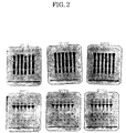
- Fig. 2 is an electropherogram showing SDS-PAGE patterns before and after
acceleration test with respect to preparations with various concentrations of buffer
solutions.
-
BEST MODE FOR CARRYING OUT THE INVENTION
-
Hereinbelow, the present invention will be described in greater detail with
reference to the following Reference Examples and Examples, which should not be
construed as limiting the technical scope of the invention.
[EXAMPLE 1] Effect of hydrogen ion concentration (pH)
-
The anti-PTHrP antibodies used in Examples 1 and 2 were humanized antibodies
(hereinafter referred to as "humanized antibody") prepared in accordance with
Reference Examples 1 to 4 described below. In addition, analytical methods and
analytical conditions for Examples 1 and 2 were as follows.
GPC-UV
-
- Stationary phase: 50 mmol/L of phosphoric acid buffer solution containing 300 mmol/L
of NaCl (pH 6.8)
- Flow rate: 0.5 mL/min
- Column: G-3000SWXL
- Detection: 280 nm
- Sample injection volume: 90 µg (inject 30 µl of 3 mg/ml)
-
BIACORE method (analytical method using surface plasmon resonance. BIACORE
manufactured by Pharmacia Biotech K. K.)
-
- 1) Reagent
NHS (N-hydroxysuccinic acid imido), EDC (N-ethyl-N'(3-dimethylamino
propyl)-carbodiimido hydrochloride), Ethanolamine: amine coupling kit (Biacore Co.)PDEA (2-(2-pyridinyldithio)ethaneamine hydrochloride): thiol coupling kit
(Biacore Co.), PTHrP (1-34+C): synthesized product (Sawady technology Co., Ltd.)
- 2) Sensor chip ligand: antigen PTHrP or antigen Protein A
- 3) Preparation of sample solution
- i) A sample solution was diluted with distilled water for injection to about 100 µg/mL,
and the concentration was determined by absorbance method at 280 nm.
- ii) preparation of unknown sample: with reference to the concentration determined by
the absorbance method, the unknown sample was diluted with HBS-EP buffer (Biacore
Co., Code #BR-1001-88) identical to one used at a time of BIACORE measurement and
20 µg/mL solution was prepared. All the HBS-EP buffers used for dilution were the
same as those used at a time of BIACORE measurement. Then, 20 µL of this solution
and 180 µL of HBS-EP buffer were mixed and thus obtained 2 µg/mL solution was
regarded as a unknown sample.
- iii) preparation of samples for calibration curve: about 100 µg/mL solution, as a
standard, was diluted to make not less than 5 different concentrations thereof.
-
-
Ion Exchange Chromatography (hereinafter referred to as IEC-UV)
- Column: PolyCAT A 4.6 × 250 mm
- Flow rate: 1.0 ml/min
- Detection wavelength: 280 nm
- Injection volume: about 30 µg
- Elution conditions:
- Solvent A: 50 mmol/L MES-NaOH (pH 6.1)
- Solvent B: 50 mmol/L MES-NaOH (pH 6.1), 500mmol/L NaCl
- SDS-PAGE (reduction, nonreduction / CBB, WB)
-
-
The sample and the pre-treatment solution were mixed with a ratio of 1:1, and
heated at 100 °C for 1 minute. Then, SDS-PAGE (Gradient Gel 10-15 was used) was
performed. A sample application volume was adjusted so that the concentration
became about 1 to 2 mg/ml. Staining and bleaching were conducted. The sample was
dipped in a bleaching solution containing 5 % of glycerine for not less than 30 minutes,
and dried.
-
Pre-treatment solution: 40 mmol/L Tris-HCl buffer solution (pH 8.0) containing
5 % SDS (for reduction treatment, 10 % 2-mercaptoethanol was contained)
Staining: 0.1 % CBB (PhastGel Blue R)
Molecular weight marker: SDS-PAGE Standards Broad Range (BIO-RAD/Cat.No.161-0317)
-
| Protein |
Mol.Wt. |
| Myosin, Rabbit Muscle |
200,000 |
| Galactosidase, E.coli |
116,250 |
| Phosphorylase b, Rabbit Muscle |
97,400 |
| Albumin, Bovine Serum |
66,200 |
| Ovalbumin, Chicken Egg |
45,000 |
| Carbonic Anhydrase, Bovine Erythrocytes |
31,000 |
| Trypsin Inhibitor, Soybean |
21,500 |
| Lysozyme , Chicken Egg |
14,400 |
| Aprotinin, Bovine Lung |
6,500 |
(1) Phosphoric acid buffer solution and citric acid buffer solution
-
A pharmaceutical preparation having the following composition was prepared
and an acceleration test was conducted.
- 1mg/mL of humanized antibody
- 100 mmol/L of citric acid/sodium citrate buffer solution (pH 4 to 5.5) or 100 mmol/L of
sodium phosphate buffer solution (pH 6 to 8)
-
-
The conditions for the acceleration test were as follows. The pharmaceutical
preparation was stored at 50 °C for from 1 week to 1 month, and during that period the
stability was checked. In general, for storage of the preparation at 2 to 8 °C, when the
stability can be retained at least at 25 °C for 6 months, at 30 °C for 1 month, or at 40 °C
for 1 month, the stability should be retained at 2 to 8 °C for 2 years. Furthermore, for
storage of the preparation at 25 or 30 °C, generally when the stability can be retained at
40 °C for 6 months, the stability should be retained at 25 °C for 2 years or at 30 °C for 2
years.
-
In Table 1, with respect to the preparation before and after the acceleration test,
there are described survival rate (%) of the humanized antibodies by GPC-UV,
remaining bioactivity rate (%) of the humanized antibodies by BIACORE, and main
peak survival rate (%) of the humanized antibody by IEC-UV.
(2) Acetic acid buffer solution
-
A pharmaceutical preparation having the following composition was prepared
and the acceleration test was conducted.
- 13 mg/mL of humanized antibody
- 20 mmol/L of acetic acid/sodium acetate buffer solution
- 150 mmol/L of sodium chloride
-
-
In Table 2, with respect to the preparation before and after the acceleration test,
there are described hydrogen ion concentration, UV
360nm, survival rate (%) of the
humanized antibodies by GPC-UV, remaining bioactivity rate (%) of the humanized
antibodies by BIACORE, and main peak survival rate (%) of the humanized antibody by
IEC-UV.
| (Effect of hydrogen ion concentration (pH)) |
| Samples | 01L | 02L | 03L | 04L | 05L |
| hydrogen ion concentration(pH) | before accel. Test | 4.94 | 5.40 | 6.01 | 6.52 | 6.92 |
| 50 °C - 1 month | 4.99 | 5.44 | 6.04 | 6.55 | 6.92 |
| UV360nm | 50 °C - month | 1.4458 | 0.1730 | 0.1038 | 0.0878 | 0.0821 |
| survival rate (%) of the humanized antibodies by GPC-UV | before accel. Test | 100 | 100 | 100 | 100 | 100 |
| 25 °C - 1 month | 96.8 | 97.0 | 98.6 | 98.5 | 96.6 |
| 25 °C - 3 months | 93.7 | 97.7 | 97.3 | 101.8 | 100.6 |
| 50 °C - 1 month | - | 82.5 | 91.4 | 92.2 | 90.8 |
| remaining bioactivity rate (%) of the humanized antibodies by BIACORE | before accel. test | PTHrP | 100 | 100 | 100 | 100 | 100 |
| Protein A | 100 | 100 | 100 | 100 | 100 |
| 50 °C - 1 month | PTHrP | - | 78.1 | 90.0 | 90.4 | 90.6 |
| Protein A | - | 79.1 | 94.0 | 95.1 | 94.8 |
| main peak survival rate (%) of the humanized antibody by IEC-UV | before accel. Test | 100 | 100 | 100 | 100 | 100 |
| 25 °C - 1 month | 103.4 | 85.6 | 100.9 | 96.6 | 107.5 |
| 25 °C - 1 month | 84.9 | 71.6 | 95.3 | 102.1 | 118.0 |
-
In addition, SDS-PAGE patterns of the humanized antibodies before and after
acceleration test are described in Fig. 1. In Fig. 1, the upper column shows results under
reduction condition, and the lower column shows results under nonreduction condition. The
left column shows patterns of the preparation before acceleration test, the center column
shows patterns of the preparation after acceleration test with the condition at 50 °C for 1 week,
and the right column shows patterns of the preparation after acceleration test with the
condition at 50 °C for 1 month. Furthermore, lanes from the left show a pattern of the
preparation at pH of 5.0, 5.5, 6.0, 6.5, and 7.0, respectively.
-
According to the analytical results of UV360nm, GPC-UV, BIACORE, and IEC-UV, the
humanized antibody was stable at pH of 6 to 7, and most stable at pH 6.
-
In view of the analytical results of SDS-PAGE, it was recognized that band on H-chain
was increased at pH of 7 and 6.5, and that low molecular decomposition products were
increased at pH of 5 and 5.5, and thus it was decided that an optimum pH value was 6.
[Example 2] Effect of concentration of buffer solution
(1) Acetic acid buffer solution
-
A pharmaceutical preparation having the following composition was prepared and an
acceleration test was conducted.
- 6.5 mg/mL of humanized antibody
- 10, 50, and 100 mmol/L of acetic acid/sodium acetate buffer solution (pH 6)
- 150 mmol/L of sodium chloride
-
-
In Table 3, with respect to the preparation before and after the acceleration test, the
following are described: hydrogen ion concentration, UV
360nm, survival rate (%) of the
humanized antibodies by GPC-UV, and remaining bioactivity rate (%) of the humanized
antibodies by BIACORE.
| (Effect of concentration of acetic acid buffer solution) |
| Samples | 06L | 07L | 08L |
| Concentration of buffer solution. [mmol/L] | 10 | 50 | 100 |
| hydrogen ion concentration (pH) | before accel. Test | 6.00 | 6.00 | 5.99 |
| 50 °C - 1 month | 6.04 | 6.01 | 6.00 |
| UV360nm | 50 °C - 1 month | 0.0401 | 0.0359 | 0.0353 |
| survival rate (%) of the humanized antibodies by GPC-UV | before accel. test | 100 | 100 | 100 |
| 25 °C - 1 month | 99.1 | 96.7 | 95.9 |
| 25 °C - 3 months | 95.8 | 97.1 | 96.7 |
| 50 °C - 1 month | 92.5 | 92.2 | 92.5 |
| remaining bioactivity rate (%) of the humanized antibodies by BIACORE | before accel. test | PTHrP | 100 | 100 | 100 |
| Protein A | 100 | 100 | 100 |
| 50 °C- 1 month | PTHrP | 90.4 | 90.2 | 90.4 |
| Protein A | 94.5 | 94.0 | 92.0 |
(2) Citric acid buffer solution
-
A pharmaceutical preparation having the following composition was prepared and the
acceleration test was conducted.
- 8 mg/mL of humanized antibody
- 10, 50, and 100 mmol/L of citric acid/sodium citrate buffer solution (pH 6)
- 150 mmol/L of sodium chloride
-
-
In Table 4 with respect to the preparation before and after the acceleration test, the
following are described: hydrogen ion concentration, UV
360nm, survival rate (%) of the
humanized antibodies by GPC-UV, and remaining bioactivity rate (%) of the humanized
antibodies by BIACORE.
| (Effect of concentration of citric acid buffer solution) |
| Samples | 09L | 10L | 11L |
| Concentration of buffer solution. [mmol/L] | 10 | 50 | 100 |
| hydrogen ion concentration (pH) | before accel. test | 6.10 | 6.01 | 6.03 |
| 50 °C - 1 month | 6.12 | 6.03 | 6.04 |
| UV360nm | 50 °C - 1 month | 0.0558 | 0.0597 | 0.0536 |
| survival rate (%) of the humanized antibodies by GPC-UV | before accel. test | 100 | 100 | 100 |
| 25 °C - 1 month | 96.2 | 97.1 | 96.0 |
| 25 °C - 3 months | 97.0 | 95.9 | 96.9 |
| 50 °C - 1 month | 92.5 | 91.0 | 92.5 |
| remaining bioactivity rate (%) of the humanized antibodies by BIACORE | before accel. test | PTHrP | 100 | 100 | 100 |
| Protein A | 100 | 100 | 100 |
| 50 °C - 1 month | PTHrP | 90.3 | 88.4 | 89.7 |
| Protein A | 93.6 | 90.0 | 92.1 |
-
In addition, SDS-PAGE patterns of the humanized antibodies before and after
acceleration test are described in Fig. 2. In Fig. 2, the upper column shows results under
reduction condition, and the lower column shows results under nonreduction condition. The
left column shows patterns of the preparation before acceleration test, the center column
shows patterns of the preparation after acceleration test with the condition at 50 °C for 1 week,
and the right column shows patterns of the preparation after acceleration test with the
condition at 50 °C for 1 month. Furthermore, lanes from the left show a pattern of the
preparation by using acetic acid buffer solution having a concentration of 10, 50, and 100
mmol/L and citric acid buffer solution having a concentration of 10, 50, 100 mmol/L,
respectively.
-
According to the analytical results of BIACORE, the humanized antibody was stable
by 10 to 50 mmol/L of acetic acid buffer solution and citric acid buffer solution, more stable
by 10 mmol/L of the buffer solutions thereof.
-
In view of the analytical results of SDS-PAGE, the amount of decomposition products
was smaller by citric acid rather than acetic acid, and with low concentration rather than high
concentration.
[Example 3]
-
In Example 3, a pharmaceutical preparation for injection was prepared, and the
preparation was reviewed concerning pain-easing effect. Incidentally, the anti-PTHrP
antibody used in this Example 3 was a humanized antibody prepared in the Reference
Examples 1 to 4 described later. (hereinafter this antibody is referred to as "humanized
antibody")
-
In this Example, 10 kinds of preparations for injection were prepared as shown in
Table 5.
| Preparation for injection | Buffer Solution | pH | humanized antibody |
| 1 | Acetic acid buffer solution 20mM | 5 | - |
| 2 | Acetic acid buffer solution 20mM | 6 | - |
| 3 | Acetic acid buffer solution 20mM | 7 | - |
| 4 | Acetic acid buffer solution 100mM | 6 | - |
| 5 | Citric acid buffer solution 20mM | 5 | - |
| 6 | Citric acid buffer solution 20mM | 6 | - |
| 7 | Citric acid buffer solution 20mM | 7 | - |
| 8 | Citric acid buffer solution 100mM | 6 | - |
| 9 | Acetic acid buffer solution 20mM | 6 | 13.0mg/ml |
| 10 | Citric acid buffer solution 20mM | 6 | 16.8mg/ml |
-
These preparations for injection were quasi-aseptically prepared, kept in cold storage,
and dispensed into a syringe for use at a time of administration.
Animal to be used and breeding environment
-
Rabbits were selected as the animal to be used, because their biological characteristics
have been well studied, a large number of homogeneous rabbits are available, the size of a
subcutaneous area around posterior auricular vein of a rabbit is suitable, as administration site,
for administration and examination, and a macroscopic observation on a detriment site is easy.
Male New Zealand white rabbits (Kbl:NZW) (12 week-old at purchase) were purchased from
Kitayama Labes Co., Ltd., and 7 days acclimation breeding was conducted. During the
acclimation breeding, the selection among them was made, considering general conditions
and weights thereof. Then, 22 rabbits were selected for use. At a time of administration
(13 week-old at administration), these animals had a weight of 2.6 to 3.3 kg.
-
These animals were each accommodated in a suspended aluminum cage having a three
dimensions of 350 mm width × 500 mm depth × 350 mm height in an animal room with the
conditions of temperature of 24 ± 2 °C, humidity of 55 ± 10 %, a light/dark cycle of 14 hours
(from 5:00 to 19:00) of light per day, and 14 to 16 times ventilations per hour. Solid diet
RC4 (available from Oriental Yeast Co., Ltd.) was served as feed through stainless steel feed
supplier by an uninterrupted feeding method, and tap water was served as drinking water
through an automatic water supply apparatus by an uninterrupted watering method. Both
were served for animal's intake without restriction. For individual identification, the number
of each animal was written on auricle (writing on not administration site but outside
therefrom) by an oil marker pen, and for cage identification, a cage card was attached thereto.
-
During the examination period, in the analysis of breeding room environment, feed,
and drinking water, there was recognized no defect which may cause any influence on the
examination system.
Setting of dose solution and group constitution
-
The dose volume of the injection preparations described in Table 5 was determined to
be 0.2 mL/(administration site) in accordance with a local disorder examination which had
already been conducted (Masao SUNAGA, Kazuhiro SHIMOMURA, Haruko KOIZUMI,
Local Irritation Examination of Gadoteridol on rabbit at perivenous subcutaneous and
intravenous administration, Preclin. Rep. Cent. Inst. Exp. Anim. 1992,18(1) :47-57.). Each
preparation for injection was administered to a group of 2 rabbits (4 auricle perivenous
subcutaneous sites), and one of each group was autopsied 2 days after the administration, and
the other was autopsied 4 days thereafter.
Route of Administration
-
As an administration site of the preparation for injection, on a posterior auricular vein
positioned almost at the center of auricle, a portion of the posterior auricular vein having less
bifurcation of small veins was selected. For administration, a disposable syringe and needle
(27G) were used. The needle was subcutaneously implanted toward a root of auricle along
the vein, and then a slow single dose was provided in about 3 seconds. Further, in order to
identify a needle implanting portion and a tip portion (injection portion) of the implanted
needle, a marking was made on the side of the implanting portion and tip portion by an oil
marker pen.
Observation of administration site (macroscopic findings)
-
Observations were made on the administration site and the vicinity thereof once a day
until 2 or 4 days had passed just after administration (counted from the date of next day of the
administration). Then, the observed changes in size (long and short diameters) were
measured by a vernier caliper (JIS Standard), obtaining the product of the long and short
diameters as an area.
Histopathological examination (histopathological findings)
-
After finishing the observation of the administration site during the period of 2 or 4
days just after the administration, body weight measurement was conducted for the
calculation of anesthetic drug volume. The animals were euthanized under anesthesia of
pentobarbital sodium (Nembutal available from Dainippon Pharmaceutical Co., Ltd) by
exsanguinating from abdominal aorta. In passing, the anesthetic drug was carefully
administered at posterior auricular vein of a root of auricle, and attention was paid not to
cause influence on the evaluation of administration sites. Further, photos were taken on
administration sites before the exsanguinations.
-
After collecting right and left auricles from a root of auricle to which the examination
substance was administered, the collected auricles were fixed with neutral buffered formalin
fixative. Thereafter, the examination substance-implanting portion of the right and left
auricles, including a blood vessel adjacent to the administration site, was cut out in vertical
direction to the run of the blood vessel, and paraffin embedded thin sectioning tissue
preparations thereof were made by a conventional method. Hematoxylin and eosin (HE)
stain was conducted thereon, and histopathological examination was conducted with a light
-
The irritancy according to histopathologial examination was judged based on the
following standard points.
- (1) Bleeding was not considered as an index for irritancy but a change of administration
technique.
- (2) A slight change of tissue, such as slight cell density increase or lacuna formation of the
administration sites, was not considered as an index for irritancy but a change of
administration technique.
- (3) In a case where there is other slight histological change, and vacuole formation of an
epidermal cornification cell and epidermis thickening are observed, but is not considered as
an index for irritancy. (These changes are considered to be expressed when there is a strong
hitological influence, and accordingly when other histological change is light, and vacuole
formation of an epidermal cornification cell and epidermis thickening are observed this is not
an index for irritancy.)
- (4) When inflammatory cell infiltration and edema are more than slight, this is an index for
irritancy.
- (5) With respect to epidermis thickening, when inflammatory cell infiltration and edema are
more than slight, this is an index for irritancy.
-
-
Then, biological evaluations were made on examination results, concerning difference
among the groups or time-series changes. The evaluation results are shown in Table 6.
-
As shown in Table 6, the solution of the humanized antibody (16.8 mg/Lm)/20
mmol/L of citric acid buffer solution (pH 6) provoked erythema and swelling by macroscopic
observation, and light inflammatory cell infiltration and light edema by histopathological
examination. Therefore, it was determined to have a local irritancy. In contract to this, it
was not recognized that the solution of the humanized antibody (13.0 mg/mL)/20 mmol/L of
acetic acid buffer solution (pH 6) caused any macroscopic finding or histopathological finding,
and thus it was determined to have no irritancy.
-
In view of the above results, it becomes clear that a pharmaceutical preparation for
injection having a pain-easing action can be obtained by using acetic acid as buffer.
[REFERENCE EXAMPLE 1]
Preparation of hybridomas producing anti-PTHrP (1-34) mouse monoclonal antibody
-
Hybridomas capable of producing a monoclonal antibody against human PTHrP (1-34)
(SEQ ID NO: 75), #23-57-154 and #23-57-137-1, were prepared as follows (see Sato, K.
et al., J. Bone Miner. Res. 8, 849-860, 1993). The amino acid sequence of the human PTHrP
(1-34) is shown in SEQ ID NO:75.
-
For use as an immunogen, PTHrP (1-34) (Peninsula) was conjugated with a carrier
protein thyroglobulin using carbodiimide (Dojinn). The thycloglobulin-conjugated PTHrP
(1-34) was dialyzed to obtain a solution having a protein concentration of 2 µg/ml. The
resulting solution was mixed with Freund's adjuvant (Difco) at a mixing ratio of 1:1 to give an
emulsion. This emulsion was injected to 16 female BALB/C mice 11 times subcutaneously
at the back or intraperitoneally at a dose level of 100 µg/mouse for each injection, thereby
immunizing the mice. For the priming immunization, Freund's complete adjuvant was used;
while for the boosting immunization, Freund's incomplete adjuvant was used.
-
Each of the immunized mice was determined for its antibody titer in the serum in the
following manner. That is, each of the mice was blood-drawn via its tail vein, and the anti-serum
is separated from the blood. The anti-serum was diluted with a RIA buffer and mixed
with 125I-labeled PTHrP (1-34) to determine the binding activity. The mice that were
confirmed to have a sufficiently increased titer were injected with PTHrP (1-34) without a
carrier protein intraperitoneally at a dose level of 50 µg/mouse for the final immunization.
-
Three days after the final immunization, the mouse was sacrificed and the spleen was
removed therefrom. The spleen cells were subjected to cell fusion with mouse myeloma cell
line P3x63Ag8U.1 in accordance with a conventional known method using 50% polyethylene
glycol 4000. The fused cells thus prepared were seeded to each well of eighty-five 96-well
plates at a density of 2 x 10⁴/well. Hybridomas were screened in HAT medium as follows.
-
The screening of hybridomas was performed by determining the presence of PTHrP-recognition
antibodies in the culture supernatant of the wells in which cell growth had been
observed in HAT medium, by solid phase RIA method. The hybridomas were collected
from the wells in which the binding ability to the PTHrP-recognition antibodies had been
confirmed. The hybridomas thus obtained was suspended into RPMI-1640 medium
containing 15% FCS supplemented with OPI-supplement (Sigma), followed by unification of
the hybridomas by limiting dilution method. Thus, two types of hybridoma clones, #23-57-154
and #23-57-137-1, could be obtained, both which had a high binding ability to PTHrP (1-34).
-
Hybridoma clone #23-57-137-1 was designated "mouse-mouse hybridoma #23-57-137-1",
and has been deposited under the terms of the Budapest Treaty on August 15, 1996 at
the National Institute of Bioscience and Human-technology, Agency of Industrial Science and
Technology, Japan (1-3, Higashi 1-chome, Tsukuba-shi, Ibaraki, Japan) under the accession
No. FERM BP-5631.
[REFERENCE EXAMPLE 2]
Cloning of DNAs encoding V-regions of mouse monoclonal antibody against human PTHrP
(1-34)
-
Cloning of DNAs encoding the V-regions of a mouse monoclonal antibody against
human PTHrP (1-34), #23-57-137-1, was performed in the following manner.
(1) Preparation of mRNA
-
mRNA from hybridoma #23-57-137-1 was prepared using Quick Prep mRNA
Purification Kit (Pharmacia Biotech). That is, cells of hybridoma #23-57-137-1 were fully
homogenized with an extraction buffer, and mRNA was isolated and purified therefrom on an
oligo(dT)-Cellulose Spun Column in accordance with the instructions included in the kit. The
resulting solution was subjected to ethanol precipitation to obtain the mRNA as a precipitate.
The mRNA precipitate was dissolved in an elution buffer.
(2) Production and amplification of cDNA for gene encoding mouse H-chain V-region
(i) Cloning of cDNA for #23-57-137-1 antibody H-chain V-region
-
A gene encoding H-chain V-region of the mouse monoclonal antibody against
human PTHrP was cloned by 5'-RACE method (Frohman, M. A. et al., Proc. Natl. Acad. Sci.
USA, 85, 8998-9002, 1988; Belyavsky, A. et al., Nucleic Acids Res. 17, 2919-2932, 1989).
The 5'-RACE method was performed using 5'-Ampli FINDER RACE Kit (CLONETECH) in
accordance with the instructions included in the kit. In this method, the primer used for
synthesis of cDNA was MHC2 primer (SEQ ID NO: 1) which is capable of hybridizing to
mouse H-chain C-region. The above-prepared mRNA (about 2 µg), which was a template
for the cDNA synthesis, was mixed with MHC2 primer (10 pmoles). The resulting mixture
was reacted with a reverse transcriptase at 52° C for 30 minuets to effect the reverse
transcription of the mRNA into cDNA.
-
The resulting reaction solution was added with 6N NaOH to hydrolyze any RNA
remaining therein (at 65° C for 30 min.) and then subjected to ethanol precipitation to isolate
and purify the cDNA as a precipitate. The purified cDNA was ligated to Ampli FINDER
Anchor (SEQ ID NO: 42) at the 5' end by reacting with T4 RNA ligase at 37° C for 6 hours
and additionally at room temperature for 16 hours. As the primers for amplification of the
cDNA by PCR method, Anchor primer (SEQ ID NO: 2) and MHC-G1 primer (SEQ ID NO:
3) (S.T. Jones, et al., Biotechnology, 9, 88, 1991) were used.
-
The PCR solution comprised (per 50 µl)10 mM Tris-HCl (pH 8.3), 50 mM KCI,
0.25 mM dNTPs (dATP, dGTP, dCTP, dTTP), 1.5 mM MgCl2, 2.5 units of TaKaRa Taq
(Takara Shuzo Co., Ltd.), 10 pmoles Anchor primer, and 1 µl of the reaction mixture of the
cDNA to which MHC-G1 primer and Ampli FINDER Anchor primer had been ligated, over
which mineral oil (50 µl) was layered. The PCR was performed on Thermal Cycler Model
480J (Perkin Elmer) for 30 cycles under the conditions: 94° C for 45 sec.; 60° C for 45 sec.;
and 72° C for 2 min.
(ii) Cloning of eDNA for #23-57-137-1 antibody L-chain V-region
-
A gene encoding L-chain V-region of the mouse monoclonal antibody against human
PTHrP was cloned by 5'-RACE method (Frohman, M. A. et al., Proc. Natl. Acad. Sci. USA,
85, 8998-9002, 1988; Belyavsky, A. et al., Nucleic Acids Res. 17, 2919-2932, 1989). The
5'-RACE method was performed using 5'-Ampli Finder RACE Kit (CLONETECH) in
accordance with the instructions included in the kit. In this method, oligo-dT primer was
used as the primer for synthesizing cDNA. The above-prepared mRNA (about 2 µg),
which was a template for the cDNA synthesis, was mixed with oligo-dT primer. The resulting
mixture was reacted with a reverse transcriptase at 52° C for 30 min. to effect the reverse
transcription of the mRNA into cDNA. The resulting reaction solution was added with 6N
NaOH to hydrolyze any RNA remaining therein (at 65° C for 30 min.). The resulting
solution was subjected to ethanol precipitation to isolate and purified the cDNA as a
precipitate. The cDNA thus synthesized was ligated to Ampli FINDER Anchor at the 5' end
by reacting with T4 RNA ligase at 37° C for 6 hours and additionally at room temperature
for 16 hours.
-
A PCR primer MLC (SEQ ID NO: 4) was designed based on the conserved sequence
of mouse L-chain λ chain C-region and then synthesized using 394 DNA/RNA Synthesizer
(ABI). The PCR solution comprised (per 100 µl) 10 mM Tris-HCI (pH 8.3), 50 mM KCI,
0.25 mM dNTPs (dATP, dGTP, dCTP, dTTP), 1.5 mM MgCl2, 2.5 units of AmpliTaq
(PERKIN ELMER), 50 pmoles of Anchor primer (SEQ ID NO: 2), and 1 µl of the reaction
mixture of the cDNA to which MLC (SEQ ID NO: 4) and Ampli FINDER Anchor were
ligated, over which mineral oil (50 µl) was layered. The PCR reaction was performed on
Thermal Cycler Model 480J (Perkin Elmer) for 35 cycles under the conditions: 94° C for 45
sec.; 60° C for 45 sec.; and 72° C for 2 min.
(3) Purification and fragmentation of PCR products
-
Each of the DNA fragments amplified by PCR method described above was
separated by agarose gel electrophoresis on a 3% Nu Sieve GTG agarose (FMC Bio.
Products). For each of the H-chain V-region and the L-chain V-region, an agarose gel
segment containing a DNA fragment of about 550 bp was excised from the gel. Each of the
gel segments was subjected to purification of the DNA fragment of interest using
GENECLEAN II Kit (BIO101 in accordance with the instructions included in the kit. The
purified DNA was precipitated with ethanol, and the DNA precipitate was dissolved in 20 µl
of a solution containing 10 mM Tris-HCl(pH 7.4) and 1 mM EDTA. An aliquot (1 µl) of
the DNA solution was digested with a restriction enzyme Xmal (New England Biolabs) at 37°
C for 1 hour and further digested with a restriction enzyme EcoRI (Takara Shuzo Co., Ltd.)
at 37° C for 1 hour. The digestion solution was extracted with phenol and chloroform and
then precipitated with ethanol to collect the DNA.
-
In this manner, two DNA fragments containing a gene encoding mouse H-chain V-region
and a gene encoding mouse L-chain V-region, respectively, were obtained, both which
had an EcoRI recognition sequence on the 5' end and an XmaI recognition sequence on the 3'
end.
-
The EcoRI-XmaI DNA fragments containing a gene encoding mouse H-chain V-region
and a gene encoding mouse L-chain V-region, respectively, were separately ligated to
pUC19 vector that had been digested with EcoRI and XmaI at 16° C for 1 hour using DNA
Ligation Kit ver.2 (Takara Shuzo Co., Ltd.) in accordance with the instructions included in the
kit. An aliquot (10 µl) of the ligation mixture was added to 100 µl of a solution
containing competent cells of E. coli, JM 109 (Nippon Gene Co., Ltd.). The cell mixture
was allowed to stand on ice for 15 min., at 42° C for 1 min. and additionally for 1 min. on ice.
The resulting cell mixture was added with 300 µl of SOC medium (Molecular Cloning: A
Laboratory Manual, Sambrook, et al., Cold Spring Harbor Laboratory Press, 1989) and then
incubated at 37 C for 30 min. The resulting cell solution was plated on LB agar medium
or 2xYT agar medium (Molecular Cloning: A Laboratory Manual, Sambrook, et al., Cold
Spring Harbor Laboratory Press, 1989) containing either 100 or 50 µg/ml of ampicillin, 0.1
mM of IPTG and 20 µg/ml of X-gal, and then incubated at 37° C overnight. In this
manner, E. coli transformants were prepared.
-
The transformants were cultured at 37° C overnight in 2 ml of LB or 2xYT medium
containing either 100 or 50 µg/ml of ampicillin. The cell fraction was applied to Plasmid
Extracter PI-100( (Kurabo Industries, Ltd.) or QIAprep Spin Plasmid Kit (QIAGEN) to give a
plasmid DNA. The plasmid DNA was sequenced as follows.
(4) Sequencing of genes encoding mouse antibody V-regions
-
The nucleotide sequence of the cDNA coding region carried on the plasmid was
determined in DNA Sequencer 373A (ABI; Perkin-Elmer) using Dye Terminator Cycle
Sequencing Kit (Perkin-Elmer). M13 Primer M4 (Takara Shuzo Co., Ltd.) (SEQ ID NO: 5)
and M13 Primer RV (Takara Shuzo Co., Ltd.) (SEQ ID NO: 6) were used as the primers for
sequencing, and the nucleotide sequence was confirmed in the both directions.
-
The plasmid containing a gene encoding mouse H-chain V-region derived from
hybridoma #23-57-137-1 was designated "MBC1H04", and the plasmid containing a gene
encoding mouse L-chain V-region derived from hybridoma #23-57-137-1 was designated
"MBC1L24". The nucleotide sequences (including the corresponding amino acids
sequences) of the gene encoding the mouse #23-57-137-1 antibody-derived H-chain V-region
in plasmid MBC1H04 and the gene encoding the mouse #23-57-137-1 antibody-derived L-chain
V-region in plasmid MBC1H24 were shown in SEQ. ID Nos: 57 and 65, respectively.
The amino acid sequences of the polypeptides for the H-chain V-region and the L-chain V-region
were shown in SEQ. ID NOs: 46 and 45, respectively.
-
The E. coli strain containing plasmid MBC1H04 and the E. coli strain containing
plasmid MBC1L24 were designated "Escherichia coli JM109 (MBC1H04)" and "Escherichia
coli JM 109 (MBC1L24)", respectively. These E. coli strains have been deposited under the
terms of the Budapest Treaty at the National Institute of Bioscience and Human-Technology,
Agency of Industrial Science and Technology, Japan (1-3, Higashi 1-chome, Tsukuba-shi,
Ibaraki, Japan) on August 15, 1996, under the Accession No. FERM BP-5628 for Escherichia
coli JM109 (MBC1H04) and FERM BP-5627 for Escherichia coli JM109 (MBC1L24),
respectively.
(5) Determination of CDRs of mouse monoclonal antibody
#23-57-137-1 against human PTHrP
-
The H-chain V-region and the L-chain V-region have general structures similar to
each other, each of which has four framework regions (FRs) linked through three
hypervariable regions (i.e., complementarity determining regions; CDRs). The amino acid
sequences of the FRs are relatively well conserved, while the amino acid sequence of the
CDRs have an extremely high variability (Kabat, E.A. et al., "Sequence of Proteins of
Immunological Interest", US Dept. Health and Human Services, 1983).
-
In view of these facts, the homology in amino acid between the V-regions of the
mouse monoclonal antibody against human PTHrP was determined with reference to the
database of amino acid sequences of antibodies established by Kabat et al. Thus, the CDRs
of the V-regions were determined as shown in Table 7.
-
The amino acid sequences of CDRs 1-3 in the L-chain V-region are shown in SEQ
ID Nos: 59 to 61, respectively; and the amino acid sequences of CDRs 1-3 in the H-chain V-region
are shown in SEQ ID Nos: 62 to 64, respectively.
| V-region | SEQ ID NO. | CDR1 | CDR2 | CDR3 |
| H-chain V-region | 57 | 31-35 | 50-66 | 99-107 |
| L-chain V-region | 65 | 23-34 | 50-60 | 93-105 |
[REFERENCE EXAMPLE 3] Construction of Chimeric Antibody
(1) Construction of chimeric antibody H-chain
(i) Construction of H-chain V-region
-
To ligate to an expression vector carrying a genomic DNA of human H-chain C-region
C γ 1, the cloned DNA encoding mouse H-chain V-region was modified by PCR
method. A backward primer MBC1-S1 (SEQ ID NO: 7) was designed to hybridize to a
DNA sequence encoding the 5' region of the leader sequence of the V-region and to have both
a Kozak consensus sequence (Kozak, M. et al., J. Mol. Biol., 196, 947-950, 1987) and a
HindIII-recognition sequence. A forward primer MBC1-a (SEQ ID NO: 8) was designed to
hybridize to a DNA sequence encoding the 3' region of the J region and to have both a donor
splice sequence and a BamHI-recognition sequence. The PCR reaction was performed using
TaKaRa Ex Taq (Takara Shuzo Co., Ltd.) and a buffer appended thereto. The PCR solution
comprised (per 50 µl) 0.07 µg of plasmid MBC1H04 as a template DNA, 50 pmoles of
MBC1-a and 50 pmoles of MBC1-S1 as primers, 2.5U of TaKaRa Ex Taq and 0.25 mM
dNTPs in the buffer, over which 50 µl of mineral oil was layered. The PCR was run for 30
cycles under the conditions: 94° C for 1 min.; 55° C for 1 min.; 72° C for 2 min. The
DNA fragments thus amplified by the PCR method were separated by agarose gel
electrophoresis on a 3% Nu Sieve GTG Agarose (FMC Bio. Products).
-
Then, an agarose gel segment containing a DNA fragment of 437 bp was excised,
and the DNA fragment was purified therefrom using GENECLEAN II Kit (BIO101) in
accordance with the instructions included in the kit. The purified DNA was collected by
ethanol precipitation, and then dissolved in 20 µl of a solution containing 10 mM Tris-HCl
(pH 7.4) and 1 mM EDTA. An aliquot (1 µl) of the resulting DNA solution was digested
with restriction enzymes BamHI and HindIII (Takara Shuzo Co., Ltd.) at 37 C for 1 hour.
The digestion solution was extracted with phenol and chloroform and then precipitated with
ethanol to collect the DNA of interest.
-
The obtained HindIII-BamHI DNA fragment, which containing a gene encoding the
mouse H-chain V-region, was subcloned into pUC19 vector that had been digested with
HindIII and BamHI. The resulting plasmid was sequenced on DNA Sequencer 373A
(Perkin-Elmer) using M13 Primer M4 and M13 Primer RV as primers and Dye Terminator
Cycle Sequencing Kit (Perkin-Elmer). As a result, a plasmid which carried a gene of correct
nucleotide sequence encoding the mouse H-chain V-region derived from hybridoma #23-57-137-1
and had a HindIII-recognition sequence and a Kozak sequence on its 5' region and a
BamHI-recognition sequence on its 3' region was obtained, which was designated
"MBC1H/pUC19".
(ii) Construction of H-chain V-region for preparation of cDNA-type of mouse-human
chimeric H-chain
-
To ligate to cDNA of the human H-chain C-region C γ 1, the DNA encoding the
mouse H-chain V-region constructed as described above was modified by PCR method. A
backward primer MBC1HVS2 (SEQ ID NO: 9) for the V-region was designed to cause the
replacement of the second amino acid (asparagine) of the sequence encoding the front part of
the leader sequence of the H-chain V-region by glycine and to have a Kozak consensus
sequence (Kozak, M. et al., J. Mol. Biol., 196, 947-950, 1987) and HindIII- and EcoRI-recognition
sequences. A forward primer MBC1HVR2 (SEQ ID NO: 10) for the H-chain V-region
was designed to hybridize to a DNA sequence encoding the 3' region of the J region, to
encoding the 5' region of the C-region and to have ApaI- and SmaI-recognition sequences.
-
The PCR reaction was performed using TaKaRa Ex Taq (Takara Shuzo Co., Ltd.)
and a buffer appended thereto. The PCR solution comprised (per 50 µl) 0.6 µg of plasmid
MBC1H/pUC19 as a template DNA, 50 pmoles of MBC1HVS2 and 50 pmoles of
MBC1HVR2 as primers, 2.5U of TaKaRa Ex Taq and 0.25 mM of dNTPs in the buffer, over
which 50 µl of mineral oil was layered. The PCR reaction was run for 30 cycles under the
conditions: 94° C for 1 min.; 55° C for 1 min.; 72° C for 1 min. The DNA fragments
amplified by the PCR reaction were separated by agarose gel electrophoresis on a 1 % Sea
Kem GTG Agarose (FMC Bio. Products). Then, an agarose gel segment containing a DNA
fragment of 456 bp was excised and the DNA fragment was purified therefrom using
GENECLEAN II Kit (BIO101) in accordance with the instructions included in the kit. The
purified DNA was precipitated with ethanol and then dissolved in 20 µl of a solution
containing 10 mM Tris-HCl (pH 7.4) and 1 mM EDTA.
-
The resulting DNA solution (1 µg) was digested with restriction enzymes EcoRI
and Smal (Takara Shuzo Co., Ltd.) at 37° C for 1 hour. The digestion solution was
extracted with phenol and chloroform and then precipitated with ethanol to collect the DNA.
The obtained EcoRI-SmaI DNA fragment, which containing a gene encoding the mouse H-chain
V-region, was subcloned into pUC19 vector that had been digested with EcoRI and
SmaI. The resulting plasmid was sequenced on DNA Sequencer 373A (Perkin-Elmer) using
M13 Primer M4 and M13 Primer RV, and Dye Terminator Cycle Sequencing Kit (Perkin-Elmer).
As a result, a plasmid which contained a gene of correct nucleotide sequence
encoding mouse H-chain V-region derived from hybridoma #23-57-137-1 and had EcoRI-
and HindIII-recognition sequences and a Kozak sequence on its 5 region and ApaI- and
SmaI-recognition sequences on its 3' region was obtained, which was designated
"MBC1Hv/pUC19".
(iii) Construction of expression vector for chimeric antibody H-chain
-
cDNA containing the DNA for human antibody H-chain C-region C γ 1 was
prepared as follows. mRNA was prepared from a CHO cell into which both an expression
vector DHFR- ΔE-RVh-PM-1-f (see WO 92/19759) encoding the genomic DNAs of
humanized PM1 antibody H-chain V-region and human antibody H-chain C-region IgG1 (N.
Takahashi et al., Cell 29, 671-679, 1982) and an expression vector RV1-PM1a (see WO
92/19759) encoding the genomic DNAs of humanized PM1 antibody L-chain V-region and
human antibody L-chain κ chain C-region had been introduced. Using the mRNA, cDNA
containing the humanized PM1 antibody H-chain V-region and the human antibody C-region
Cγ1 was cloned by RT-PCR method, and then subcloned into plasmid pUC19 at the HindIII-BamHI
site. After sequencing, a plasmid which had the correct nucleotide sequence was
obtained, which was designated "pRVh-PM1f-cDNA".
-
An expression vector DHFR-ΔE-RVh-PM-1-f in which both a HindIII site located
between SV40 promoter and a DHFR gene and an EcoRI site located between EF-1 α
promoter and a humanized PM1 antibody H-chain V-region gene had been deleted, was
prepared for the construction of an expression vector for cDNA containing the humanized
PM1 antibody H-chain V-region gene and the human antibody C-region Cγ1 gene.
-
The plasmid obtained (pRVh-PM1f-cDNA) was digested with BamHI, blunt-ended
with Klenow fragment, and further digested with HindIII, thereby obtaining a blunt-ended
HindIII-BamHI fragment. The blunt-ended HindIII-BamHI fragment was ligated to the
above-mentioned HindIII site- and EcoRI site-deleted expression vector DHFR-ΔE-RVh-PM1-f
that had been digested with HindIII and BamHI. Thus, an expression vector RVh-PM1f-cDNA
was constructed which contained cDNA encoding the humanized PM1 antibody
H-chain V-region and the human antibody C-region Cγ1.
-
The expression vector RVh-PM1f-cDNA containing the cDNA encoding the
humanized PM1 antibody H-chain V-region and the human antibody C-region Cγ1 was
digested with Apal and BamHI, and a DNA fragment containing the H-chain C-region was
collected therefrom. The resulting DNA fragment was introduced into the plasmid
MBC1Hv/pUC 19 that had been digested with ApaI and BamHI. The plasmid thus prepared
was designated "MBC1HcDNA/pUC19". This plasmid contained cDNA encoding the
mouse antibody H-chain V-region and the human antibody C-region Cγ1, and had EcoRI- and
HindIII-recognition sequences on its 5' region and a BamHI-recognition sequence on its 3'
region.
-
The plasmid MBC1HcDNA/pUC19 was digested with EcoRI and BamHI to give a
DNA fragment comprising a nucleotide sequence encoding the chimeric antibody H-chain.
The resulting DNA fragment was introduced into an expression vector pCOS1 that had been
digested with EcoRI and BamHI, thereby giving an expression vector for the chimeric
antibody, which was designated "MBC1HcDNA/pCOS1". Here, the expression vector
pCOS1 was constructed using HEF-PMh-gγ1 (see WO 92/19759) by deleting therefrom an
antibody gene by digestion with EcoRI and SmaI, and then ligating it to EcoRI-NotI-BamHI
Adaptor (Takara Shuzo Co., Ltd.).
-
For preparing a plasmid for the expression in a CHO cell, the plasmid
MBC1HcDNA/pUC19 was digested with EcoRI and BamHI to obtain a DNA fragment
containing a gene for the chimeric antibody H-chain. The DNA fragment was then
introduced into an expression plasmid pCHO1 that had been digested with EcoRI and BamHI
to give an expression plasmid for the chimeric antibody, which was designated
"MBC1HcDNA/pCHO1". Here, the expression vector pCHO1 was constructed using
DHFR-ΔE-rvH-PM1-f (see WO 92/19759) by deleting therefrom an antibody gene by
digestion with EcoRI and SmaI, and then ligating it to EcoRI-NotI-BamHI Adaptor (Takara
Shuzo Co., Ltd.).
(2) Construction of human L-chain C-region
(i) Preparation of cloning vector
-
To construct pUC19 vector containing a gene for human L-chain C-region, a HindIII
site-deleted pUC19 vector was prepared. pUC19 vector (2 µg) was digested in 20 µl of a
reaction solution containing 20 mM Tris-HCI (pH 8.5), 10 mM MgCl2, 1 mM DTT, 100 mM
KCl, 8 U of HindIII (Takara Shuzo Co., Ltd.) at 37° C for 1 hour. The resulting digestion
solution was extracted with phenol and chloroform, and then subjected to ethanol
precipitation to collect the DNA of interest.
-
The DNA collected was reacted in 50 µl of a reaction solution containing 50 mM
Tris-HCl (pH 7.5), 10 mM MgCl2, 1 mM DTT, 100 mM NaCI, 0.5 mM dNTPs and 6U of
Klenow fragment (GIBCO BRL) at room temperature for 20 min., thereby rendering the
terminal ends of the DNA blunt. This reaction mixture was extracted with phenol and
chloroform and then subjected to ethanol precipitation to collect the vector DNA.
-
The vector DNA thus collected was reacted in 10 µl of a reaction solution
containing 50 mM Tris-HCl (pH 7.6), 10 mM MgCl2, 1 mM ATP, 1 mM DTT, 5% (v/v)
polyethylene glycol-8000 and 0.5 U of T4 DNA ligase (GIBCO BRL) at 16 °C for 2 hours, to
cause self-ligation of the vector DNA. The reaction solution (5 µl) was added to 100 µl of a
solution containing competent cells of E. coli, JM109 (Nippon Gene Co., Ltd.), and the
resulting solution was allowed to stand on ice for 30 min., at 42 °C for 1 min., and
additionally on ice for 1 min. SOC culture medium (500 (µl) was added to the reaction
solution and then incubated at 37 °C for 1 hour. The resulting solution was plated on 2 × YT
agar medium (containing 50 µg/ml of ampicillin) on which X-gal and IPTG had been applied
(Molecular Cloning: A Laboratory Manual, Sambrook, et al., Cold Spring Harbor Laboratory
Press, 1989), and then cultured at 37 °C overnight, thereby obtaining a transformant.
-
The transformant was cultured in 2 × YT medium (20 ml) containing ampicillin (50
µg/ml) at 37 °C overnight. From the cell fraction of the culture medium, a plasmid DNA
was isolated and purified using Plasmid Mini Kit (QIAGEN) in accordance with the
instructions included in the kit. The purified plasmid was digested with HindIII. The
plasmid that was confirmed to have a HindIII site-deletion was designated "pUC19 ΔHindIII".
(ii) Construction of DNA encoding human L-chain λ chain C-region
-
Human antibody L-chain λ chain C-region is known to have at least four isotypes
including Mcg+Ke+Oz-, Mcg-Ke-Oz-, Mcg-Ke-Oz+ and Mcg-Ke+Oz- (P. Dariavach, et al., Proc.
Natl. Acad. Sci. USA, 84, 9074-9078, 1987). A search was made for a human antibody L-chain
λ chain C-region homologous to the #23-57-137-1 mouse L-chain λ chain C-region
from the EMBL database. As a result, it was found that the isotype Mcg+Ke+Oz- of the
human antibody L-chain λ chain (Accession No. X57819) (P. Dariavach, et al., Proc. Natl.
Acad. Sci. USA, 84, 9074-9078, 1987) showed the highest degree of homology to the #23-57-137-1
mouse L-chain λ chain C-region, with a 64.4% homology in terms of amino acid
sequence and a 73.4% homology in terms of nucleotide sequence.
-
Then, a gene encoding the human antibody L-chain λ chain C-region was
constructed by PCR method. The primers for the PCR were synthesized using 394 DNA/RNA
Synthesizer (ABI). The synthesized primers were as follows: HLAMB1 (SEQ ID NO: 11)
and HLAMB3 (SEQ ID NO: 13), both having a sense DNA sequence; and HLAMB2 (SEQ
ID NO: 12) and HLAMB4 (SEQ ID NO: 14), both having an antisense DNA sequence; each
primer containing a complementary sequence of 20-23 bp on the both terminal ends.
-
External primers HLAMBS (SEQ ID NO: 15) and HLAMBR (SEQ ID NO: 16) had
sequences homologous to the primers HLAMB1 and HLAMB4, respectively. HLAMBS
contained EcoRI-, HindIII- and B1nI-recognition sequences, and HLAMBR contained an
EcoRI-recognition sequence. In the first-round PCR reaction, the reactions between
HLAMB1 and HLAMB2 and between HLAMB3 and HLAMB4 were performed. After the
reactions were completed, both of the resulting PCR products were mixed in equivalent
quantities, and then assembled in the second-round PCR reaction. The reaction solution was
added with the external primers HLAMBS and HLAMBR. This reaction mixture was
subjected to the third-round PCR reaction to amplify the full length DNA.
-
Each PCR reaction was performed using TaKaRa Ex Taq (Takara Shuzo Co., Ltd.) in
accordance with the instructions included in the kit. In the first-round PCR reaction, 100 µl
of either a reaction solution containing 5 pmoles of HLAMB1, 0.5 pmole of HLAMB2 and
5U of TaKaRa Ex Taq (Takara Shuzo Co., Ltd.) or a reaction solution containing 0.5 pmole of
HLAMB3, 5 pmoles of HLAMB4 and 5U of TaKaRa Ex Taq (Takara Shuzo Co., Ltd.) was
used, over which 50 µl of mineral oil was layered. The PCR reaction was run for 5 cycles
under the conditions: 94° C for 1 min., 60° C for 1 min. and 72° C for 1 min.
-
In the second-round PCR reaction, a mixture of both the reaction solutions (50 µl
each) was used, over which 50 µl of mineral oil was layered. The PCR reaction was run
for 3 cycles under the conditions: 94° C for 1 min., 60° C for 1 min. and 72° C for 1 min.
-
In the third-round PCR reaction, the reaction solution to which the external primers
HLAMBS and HLAMBR (50 pmoles each) were added was used. The PCR reaction was
run for 30 cycles under the conditions: 94° C for 1 min., 60° C for 1 min. and 72° C for 1
min.
-
The DNA fragment obtained by the third-round PCR reaction was subjected to
electrophoresis on a 3% low-melting agarose gel (NuSieve GTG Agarose, FMC), and
separated and purified from the gel using GENECLEAN II Kit (BIO101) in accordance with
the instructions included in the kit.
-
The DNA fragment obtained was digested in a reaction solution (20 µl) containing
50 mM Tris-HCl (pH 7.5), 10 mM MgCl2, 1 mM DTT, 100 mM NaCl and 8U of EcoRI
(Takara Shuzo Co., Ltd.) at 37° C for 1 hour. The digestion solution was extracted with
phenol and chloroform, and the DNA was collected therefrom by the ethanol precipitation.
The DNA was dissolved in a solution (8 µl) containing 10 mM Tris-HCl (pH 7.4) and 1 mM
EDTA.
-
The above-prepared plasmid pUC19 ΔHindIII (0.8 µg) was digested with EcoRI
in the same manner as set forth above. The digestion solution was subjected to
phenol/chloroform extraction and then ethanol precipitation, thereby giving a digested
plasmid pUC19 ΔHindIII. The digested plasmid was reacted in a reaction solution (50 µ
I) containing 50 mM Tris-HCl (pH 9.0), 1 mM MgCl2 and alkaline phosphatase (E. coli C75;
Takara Shuzo Co., Ltd.) at 37° C for 30 min. to dephosphorylate (i.e., BAP-treat) the plasmid.
The reaction solution was subjected to phenol/chloroform extraction, and the DNA was
collected therefrom by ethanol precipitation. The DNA thus obtained was dissolved in a
solution (10 µl) containing 10 mM Tris-HCl (pH 7.4) and 1 mM EDTA.
-
The BAP-treated plasmid pUC19 ΔHindIII (1 µl) was ligated to the above-obtained
PCR product (4 µl) using DNA Ligation Kit Ver.2 (Takara Shuzo Co., Ltd.). The
resulting plasmid was introduced into a competent cell of E. coli, JM109, to give a
transformant. The transformant was cultured overnight in 2xYT medium (2 ml) containing
50 µg/ml of ampicillin. From the cell fraction, the plasmid was isolated using QIAprep
Spin Plasmid Kit (QIAGEN).
-
The plasmid obtained was sequenced for the cloned DNA part. The sequencing
was performed on 373A DNA Sequencer (ABI) using M13 Primer M4 and M13 Primer RV
(Takara Shuzo Co., Ltd.). As a result, it was found that the cloned DNA had a 12-bp
deletion therein. The plasmid was designated "C λ Δ/pUC19". Then, for making up for
the deleted part, primers HCLMS (SEQ ID NO: 17) and HCLMR (SEQ ID NO: 18) were
newly synthesized, and a DNA of correct sequence was reconstructed using these primers by
PCR method.
-
In the first-round PCR reaction, the plasmid C λ Δ/pUC19 having the DNA deletion
therein was used as a template, and the reaction was performed with each of the primer sets of
HLAMBS and HCLMS and HCLMS and HLAMB4. The PCR products were purified
separately. In the second-round PCR reaction, the PCR products were assembled together.
In the third-round PCR reaction, the reaction product of the second-round PCR reaction was
added with external primers HLAMBS and HLAMB4 and amplified to give the full length
DNA.
-
In the first-round PCR reaction, a reaction solution (100 µl) containing 0.1 µg of
C λ Δ/pUC19 as a template, either 50 pmoles of each of the primers HLAMBS and HCLMR
or 50 pmoles of each of the primers HCLMS and HLAMB4, and 5U of TaKaRa Ex Taq
(Takara Shuzo Co., Ltd.) was used, over which 50 µl of mineral oil was layered. The PCR
reaction was run for 30 cycles under the conditions: 94° C for 1 min., 60° C for 1 min. and
72° C for 1 min.
-
The PCR products of the first-round PCR reaction, HLAMBS-HCLMR (236 bp) and
HCLMS-HLAMB4 (147 bp), were subjected to electrophoresis separately on a 3% low-melting
agarose gel to isolate the DNA fragments. The DNA fragments were collected and
purified from the gels using GENECLEAN II Kit (BIO101). In the second-round PCR
reaction, 20 µl of a reaction solution containing 40 ng of each of the purified DNA
fragments and 1U of TaKaRa Ex Taq (Takara Shuzo Co., Ltd.) was used, over which 25 µl
of mineral oil was layered. The PCR reaction was run for 5 cycles under the conditions: 94
°C for 1 min., 60° C for 1 min. and 72° C for 1 min.
-
In the third-round PCR reaction, 100 µl of a reaction solution containing 2 µl of
the reaction solution obtained by the second-round PCR reaction, 50 pmoles of each of
external primers HLAMBS and HLAMB4 and 5U of TaKaRa Ex Taq (Takara Shuzo Co.,
Ltd.) was used, over which 50 µl of mineral oil was layered. The PCR reaction was run for
30 cycles under the conditions: 94° C for 1 min., 60° C for 1 min. and 72° C for 1 min.,
thereby obtaining a DNA fragment of 357 bp (the third PCR product). The DNA fragment
was subjected to electrophoresis on a 3% low-melting agarose gel to isolate the DNA
fragment. The resulting DNA fragment was collected and purified using GENECLEAN Kit
(BIO101).
-
An aliquot (0.1 µg) of the DNA fragment thus obtained was digested with EcoRI,
and then subcloned into plasmid pUC19 ΔHindIII that had been BAP-treated. The resulting
plasmid was introduced into a competent cell of E. coli, JM109, to form a transformant. The
transformant was cultured overnight in 2 ml of 2xYT medium containing 50 µg/ml of
ampicillin. From the cell fraction, the plasmid was isolated and purified using QIAprep Spin
Plasmid Kit (QIAGEN).
-
The purified plasmid was sequenced on 373A DNA Sequencer (ABI) using M13
Primer M4 and M13 Primer RV (Takara Shuzo Co., Ltd.). The plasmid that was confirmed
to have the correct nucleotide sequence without any deletion was designated "C λ /pUC19".
(iii) Construction of gene encoding human L-chain κ chain C-region
-
A DNA fragment encoding the L-chain κ, chain C-region was cloned from plasmid
HEF-PMlk-gk (WO 92/19759) by PCR method. A forward primer HKAPS (SEQ ID NO:
19) was designed to contain EcoRI-, HindIII and BlnI-recognition sequences, and a backward
primer HKAPA (SEQ ID NO: 20) was designed to contain an EcoRI-recognition sequence.
These primers were synthesized on 394 DNA/RNA Synthesizer (ABI).
-
A PCR reaction was performed using 100 µl of a reaction solution containing 0.1 µ
g of plasmid HEF-PM1k-gk as a template, 50 pmoles of each of primers HKAPS and HKAPA
and 5U of TaKaRa Ex Taq (Takara Shuzo Co., Ltd.), over which 50 µl of mineral oil was
layered. The PCR reaction was run for 30 cycles under the conditions: 94° C for 1 min., 60°
C for 1 min. and 72° C for 1 min., thereby giving a PCR product of 360 bp. The DNA
fragment was isolated and purified by electrophoresis on a 3% low-melting agarose, and then
collected and purified using GENECLEAN II Kit (BIO101).
-
The DNA fragment thus obtained was digested with EcoRI, and then cloned into
plasmid pUC19 (HindIII that had been BAP-treated. The resulting plasmid was introduced
into a competent cell of E. coli, JM109, to form a transformant. The transformant was
cultured overnight in 2 ml of 2xYT medium containing 50 µg/ml of ampicillin. From the
cell fraction, the plasmid was purified using QIAprep Spin Plasmid Kit (QIAGEN).
-
The purified plasmid was sequenced on 373A DNA Sequencer (ABI) using M13
Primer M4 and M13 Primer RV (Takara Shuzo Co., Ltd.). The plasmid that was confirmed
to have the correct nucleotide sequence was designated "C κ/pUC19".
(3) Construction of chimeric antibody L-chain expression vector
-
An expression vector for the chimeric #23-57-137-1 antibody L-chain was
constructed. A gene encoding #23-57-137-1L-chain V-region was ligated to the HindIII-B1nI
site (located just in front of the human antibody C-region) of each of the plasmids C λ
/pUC19 and C κ /pUC19, thereby obtaining pUC19 vectors that contained the DNAs
encoding the chimeric #23-57-137-1 antibody L-chain V-region and either of the L-chain λ
chain C-region or the L-chain κ region C-region, respectively. Each of the resulting
vectors was then digested with EcoRI to separate the gene for the chimeric antibody L-chain.
The gene was subcloned into HEF expression vector.
-
That is, a DNA fragment encoding #23-57-137-1 antibody L-chain V-region was
cloned from plasmid MBC1L24 by PCR method. Primers used in the PCR method were
separately synthesized using 394 DNA/RNA Synthesizer (ABI). A backward primer
MBCCHL1 (SEQ ID NO: 21) was designed to contain a HindIII-recognition sequence and a
Kozak sequence (Kozak, M. et al., J. Mol. Biol. 196, 947-950, 1987), and a forward primer
MBCCHL3 (SEQ ID NO: 22) was designed to contain BglII- and RcoRI-recognition
sequences.
-
The PCR reaction was performed using 100 µl of a reaction solution containing 10
mM Tris-HCl (pH 8.3), 50 mM KCl, 1.5 mM MgCl2, 0.2 mM dNTPs, 0.1 µg MBC1 L24, 50
pmoles of each of primers MBCCHL1 and MBCCHL3 and 1 µl of AmpliTaq (PERKIN
ELMER), over which 50 µl of mineral oil was layered. The PCR reaction was run for 30
cycles under the conditions: 94° C for 45 sec., 60° C for 45 sec. and 72° C for 2 min.
-
A PCR product of 444 bp was electrophoresed on a 3% low-melting agarose gel, and
collected and purified using GENECLEAN II Kit (BIO101). The purified PCR product was
dissolved in 20 µl of a solution containing 10 mM Tris-HCl (pH 7.4) and 1 mM EDTA.
The PCR product (1 µl) was digested in 20 µl of a reaction solution containing 10 mM
Tris-HCl (pH 7.5), 10 mM MgCl2, 1 mM DTT, 50 mM NaCl, 8U of HindIII (Takara Shuzo
Co., Ltd.) and 8U of EcoRI (Takara Shuzo Co., Ltd.) at 37° C for 1 hour. The digestion
solution was subjected to phenol/chloroform extraction, and the DNA of interest was
collected therefrom by ethanol precipitation. The DNA was dissolved in 8 µl of a solution
containing 10 mM Tris-HCl (pH 7.4) and 1 mM EDTA.
-
In the same manner, plasmid pUC19 (1 µg) was digested with HindIII and EcoRI,
and subjected to phenol/chloroform extraction and then ethanol precipitation. The obtained
digested plasmid was BAP-treated with alkaline phosphatase (E. coli C75; Takara Shuzo Co.,
Ltd.). The resulting reaction solution was extracted with phenol and chloroform, and the
DNA was collected therefrom by ethanol precipitation. The DNA was dissolved in 10 µl
of a solution containing 10 mM Tris-HCl (pH 7.4) and 1 mM EDTA.
-
The BAP-treated plasmid pUC19 (1 µl) was ligated to the above-obtained PCR
product (4 µl) using DNA Ligation Kit Ver. 2 (Takara Shuzo Co., Ltd.). The resulting
plasmid was introduced into a competent cell of E. coli, JM109 (Nippon Gene Co., Ltd.), in
the same manner as set forth above, to form a transformant. The transformant was plated on
2xYT agar medium containing 50 µg/ml of ampicillin and cultured at 37° C overnight.
The resulting transformant was then cultured at 37° C overnight in 2 ml of 2xYT medium
containing 50 µg/ml of ampicillin. From the cell fraction, the plasmid was purified using
QIAprep Spin Plasmid Kit (QIAGEN). After determining the nucleotide sequence, the
plasmid that was confirmed to have the correct nucleotide sequence was designated
"CHL/pUC19".
-
Each of plasmids C λ /pUC19 and C κ /pUC19 (1 µg each) was digested in 20 µl
of a reaction solution containing 20 mM Tris-HCl (pH 8.5), 10 mM MgCl2, 1 mM DTT, 100
mM KCl, 8U of HindIII (Takara Shuzo Co., Ltd.) and 2U of BlnI (Takara Shuzo Co., Ltd.) at
37° C for 1 hour. The digestion solution was extracted with phenol and chloroform, and the
DNA was collected therefrom by ethanol precipitation. The DNA was BAP-treated at 37 °
C for 30 min. The reaction solution was extracted with phenol and chloroform, and the
DNA was collected therefrom by ethanol precipitation. The DNA was dissolved in 10 µl
of a solution containing 10 mM Tris-HCl (pH 7.4) and 1 mM EDTA.
-
The plasmid CHL/pUC19 (8 µg) that contained DNA encoding #23-57-137-1L-chain
V-region was digested with HindIII and BlnI in the same manner as set forth above to
give a DNA fragment of 409 bp. The DNA fragment was electrophoresed on a 3% low-melting
agarose gel, and then collected and purified from the gel using GENECLEAN II Kit
(BIO101). The DNA was dissolved in 10 µl of a solution containing 10 mM Tris-HCl (pH
7.4) and 1 mM EDTA.
-
The DNA for L-chain V-region DNA (4 µl) was subcloned into 1 µl of each of
the BAP-treated plasmids C λ /pUC19 and C κ /pUC19, and then introduced into a competent
cell of E. coli, JM109, to form a transformant. The transformant was cultured overnight in 3
ml of 2xYT medium containing 50 µg/ml of ampicillin. From the cell fraction, the plasmid
was isolated and purified using QIAprep Spin Plasmid Kit (QIAGEN). The two plasmids
thus prepared were designated "MBC1L(λ)/pUC19" and "MBC1L(κ)/pUC19", respectively.
-
Each of plasmids MBC1L(λ)/pUC19 and MBC1L(κ)/pUC19 was digested with
EcoRI and then subjected to electrophoresis on a 3% low-melting agarose gel. A DNA
fragment of 743 bp was isolated and purified from the gel using GENECLEANII Kit
(BIO101), and then dissolved in 10 µl of a solution containing 10 mM Tris-HCl (pH 7.4)
and 1 mM EDTA.
-
An expression vector (plasmid HEF-PM1k-gk) (2.7 µg) was digested with EcoRI
and then extracted with phenol and chloroform, and the DNA was collected therefrom by
ethanol precipitation. The DNA fragment was BAP-treated, and then subjected to
electrophoresis on a 1 % low-melting agarose gel. From the gel, a DNA fragment of 6561 bp
was isolated and purified therefrom using GENECLEANII Kit (BIO101). The purified
DNA fragment was dissolved in 10 µl of a solution containing 10 mM Tris-HCI (pH 7.4)
and 1 mM EDTA.
-
BAP-treated HEF vector (2 µl) was ligated to an EcoRI fragment (3 µl) of each of
plasmid MBC1L(λ)/pUC19 and MBC1L(κ)/pUC19. The ligation product was introduced
into a competent cell of E. coli, JM109, to form a transformant. The transformant was
cultured in 2 ml of 2xYT medium containing 50 µg/ml of ampicillin. From the cell
fraction, the plasmid was purified using QIAprep Spin Plasmid Kit (QIAGEN).
-
The purified plasmid was digested in 20 µl of a reaction solution containing 20 mM
Tris-HCl (pH 8.5), 10 mM MgCl2, 1 mM DTT, 100 mM KCI, 8U of HindIII (Takara Shuzo
Co., Ltd.) and 2 U of PvuI (Takara Shuzo Co., Ltd.) at 37° C for 1 hour. This reaction gave
digestion fragments of 5104/2195 bp if the fragment was inserted in the correct orientation, or
gave digestion fragments of 4378/2926 bp if the fragment was inserted in the reverse
orientation. The plasmid that was confirmed to have the fragment in the correct orientation
was designated "MBC1L(λ)/neo" for plasmid MBC1L(λ)/pUC19 or "MBC1L(κ)/neo" for
plasmid MBC1L(κ)/pUC19.
(4) Transfection of COS-7 cell
-
To evaluate the antigen-binding activity and the neutralizing activity of the chimeric
antibodies, the expression plasmids prepared above were separately expressed transiently in a
COS-7 cell.
-
The transient expression of the chimeric antibodies was performed using each of the
combinations of plasmids MBC1HcDNA/pCOS1 and MBC1L (λ)/neo and plasmids
MBC1HcDNA/pCOS1 and MBC1L(κ)/neo, by co-tansfecting a COS-7 cell with the
plasmids by electroporation using Gene Pulser (Bio Rad). That is, the plasmids (10 µg
each) were added to a COS-7 cell suspension (0.8 ml; 1 x 10⁷ cells/ml) in PBS(-). The
resulting solution was applied with pulses at an electrostatic capacity of 1,500V and 2 µF to
cause electroporation. After 10 min. of recovery period at room temperature, the
electroporated cells were suspended in DMEM medium (GIBCO) containing 2% Ultra Low
IgG fetal calf serum (GIBCO), and then cultured using a 10-cm culture dish in a CO2
incubator. After culturing for 72 hours, a culture supernatant was collected and centrifuged
to remove cell debris, and was provided for use as a sample for the subsequent ELISA. In
this procedure, the purification of the chimeric antibody from the COS-7 cell culture
supernatant was performed using AffiGel Protein A MAPSII Kit (Bio Rad) in accordance
with the instructions included in the kit.
(5) ELISA
(i) Determination of antibody concentration
-
An ELISA plate for determining antibody concentration was prepared as follows.
Each well of a 96-well ELISA plate (Maxisorp, NUNC) was coated with 100 µl of a coating
buffer (0.1 M NaHCO3, 0.02% NaN3) supplemented with 1 µg/ml of goat anti-human IgG
antibody (TAGO), and then blocked with 200 µl of a dilution buffer [50 mM Tris-HCl, 1
mM MgC12, 0.1 M NaCl, 0.05% Tween 20,0.02% NaN3, 1% bovine serum albumin (BSA);
pH 7,2]. Each well of the plate was added with each of the serial dilutions of the COS-7 cell
culture supernatant in which each of the chimeric antibodies had been expressed, or added
with each of the serial dilutions of each of the chimeric antibodies per se in a purified form.
The plate was incubated at room temperature for 1 hour and washed with PBS-Tween 20.
Each well of the plate was then added with 100 µl of a solution of alkaline phosphatase-conjugated
goat anti-human IgG antibodies (TAGO). After the plate was incubated at room
temperature for 1 hour and washed with PBS-Tween 20, each well was added with 1mg/ml of
a substrate solution ("Sigma 104", p-nitrophenylphosphoric acid, SIGMA). The solution
was measured on its absorbance at 405 nm using Microplate Reader (Bio Rad) to determine
the antibody concentration. In this determination, Hu IgG1 λ Purified (The Binding Site)
was used as the standard substance.
(ii) Determination of antigen-binding ability
-
An ELISA plate for the determination of antigen-binding ability was prepared as
follows. Each well of a 96-well ELISA plate was coated with 100 µl of a coating buffer
supplemented with 1 µg/ml of human PTHrP (1-34) (Peptide Research Institute), and then
blocked with 200 µl of a dilution buffer. Each well was added with each of the serial
dilutions of the COS-7 cell culture supernatant in which each of the chimeric antibodies had
been expressed, or added with each of the serial dilutions of each of the chimeric antibodies
per se in a purified form. After the plate was incubated at room temperature and washed
with PBS-Tween 20, each well of the plate was added with 100 µl of a solution of alkaline
phosphatase-conjugated goat anti-human IgG antibodies (TAGO). After the plate was
incubated at room temperature and washed with PBS-Tween 20, each well of the plate was
added with 1 mg/ml of a substrate solution ("Sigma 104", p-nitrophenylphosphoric acid,
SIGMA). The solution was measured on its absorbance at 405 nm using Microplate Reader
(Bio Rad).
-
As a result, it was found that the chimeric antibodies had an ability to bind to human
PTHrP (1-34) and the cloned mouse antibody V-regions had the correct structures (FIG. 5).
It was also found that there was no difference in the ability to bind to PTHrP (1-34) between
the chimeric antibody with L-chain λ chain C-region and the chimeric antibody with L-chain
κ chain C-region. Therefore, the humanized antibody L-chain λ chain was used for
construction of the L-chain C-region of the humanized antibody.
(6) Establishment of CHO cell line capable of stable production of chimeric antibodies
-
To establish a cell line capable of producing the chimeric antibodies stably, the
above-prepared expression plasmids were introduced into CHO cells (DXB11).
-
For the establishment of a cell line capable of producing the chimeric antibodies
stably, either of the following combinations of the expression plasmids for CHO cell was
used: MBC1HcDNA/pCHO1 and MBC1L(λ)/neo; and MBC1HcDNA/pCHO1 and MBC1L(
κ)/neo. A CHO cell was co-transfected with the plasmids by electroporation using Gene
Pulser (Bio Rad) as follows. The expression vectors were separately cleaved with a
restriction enzyme PvuI to give linear DNAs. The resulting DNAs were extracted with
phenol and chloroform and collected by precipitation with ethanol. The plasmid DNAs thus
prepared were subjected to electroporation. That is, each of the plasmid DNAs (10 µg
each) was added to 0.8 ml of a cell suspension of CHO cells in PBS(-) (1x10⁷ cells/ml). The
resulting solution was applied with pulses at an electrostatic capacity of 1,500V and 25 µF.
After 10 min. of recovery period at room temperature, the electroporated cells were
suspended in MEM-α medium (GIBCO) containing 10% fetal calf serum (GIBCO). The
resulting suspension was cultured using three 96-well plates (Falcon) in a CO2 incubator.
On the day following the culturing being started, the medium was replaced by a selective
medium [ribonucleoside- or deoxyribonucleoside-free MEM-α medium (GIBCO)
containing 10% fetal calf serum (GIBCO) and 500 mg/ml of GENETICIN (G418Sulfate;
GIBCO)]. From the culture medium, cells into which the antibody gene was introduced
were selected. The selective medium is replaced by a fresh one. About two weeks after the
medium replacement, the cells were observed under a microscope. When a satisfactory cell
growth was observed, the amount of the antibodies produced was determined by ELISA as set
forth above. Among the cells, those cells which produced a larger amount of antibodies
were screened.
-
Then, the culturing of the established cell line capable of stable production of the
antibodies was scaled up in a roller bottle using ribonucleoside- or deoxyribonucleoside-free
MEM medium containing 2% Ultra Low IgG fetal calf serum. On day 3 and day 4 of the
culturing, the culture supernatant was collected and then filtered on a 0.2- µm filter
(Millipore) to remove cell debris therefrom.
-
Purification of the chimeric antibodies from the CHO cell culture supernatant was
performed using POROS Protein A Column (PerSeptive Biosystems) on ConSep LC100
(Millipore) in accordance with the instructions included in the kit. The purified chimeric
antibodies were provided for use as samples for the determination of neutralizing activity and
for the examination of therapeutic efficacy in hypercalcemic model animals. The
concentration and the antigen-binding activity of the purified chimeric antibodies were
determined using the same ELISA system as set forth above.
[REFERENCE EXAMPLE 4] Construction of humanized antibody
(1) Construction of humanized antibody H-chain
(i) Construction of humanized H-chain V-region
-
A humanized #23-57-137-1 antibody H-chain was produced by CDR-grafting
technique by means of PCR method. For the production of a humanized #23-57-137-1
antibody H-chain (version "a") having FRs derived from human antibody S31679 (NBRF-PDB;
Cuisinier, A. M. et al., Eur. J. Immunol., 23, 110-118, 1993), the following six PCR
primers were used: CDR-grafting primers: MBC1HGP1 (SEQ ID NO: 23) and MBC1HGP3
(SEQ ID NO: 24) (both containing a sense DNA sequence) and MBC1HGP2 (SEQ ID NO:
25) and MBC1HGP4 (SEQ ID NO: 26) (both containing an antisense DNA sequence), all of
which containing a 15-21 bp complementary sequence on both terminal ends thereof; and
external primers: MBC1HVS1 (SEQ ID NO: 27) and MBC1HVR1 (SEQ ID NO: 28) having
a homology to the CDR-grafting primers MBC1HGP1 and MBC1HGP4, respectively.
-
The CDR-grafting primers MBC1HGP1, MBC1HGP2, MBC1HGP3 and
MBC1HGP4 were separated on an urea-denatured polyacrylamide gel (Molecular Cloning: A
Laboratory Manual, Sambrook, et al., Cold Spring Harbor Laboratory Press, 1989), and
extracted therefrom by crush-and-soak method (Molecular Cloning: A Laboratory Manual,
Sambrook, et al., Cold Spring Harbor Laboratory Press, 1989) in the following manner.
-
Each of the CDR-grafting primers (1 nmole) was separated on a 6% denatured
polyacrylamide gel to give DNA fragments. From the resulting DNA fragments, a DNA
fragment having a desired length was identified on a silica gel thin plate by irradiation of UV
ray and then collected therefrom by crush-and-soak method. The resulting DNA was
dissolved in 20 µl of a solution containing 10 mM Tris-HCl (pH 7.4) and 1 mM EDTA.
The PCR reaction was performed using TaKaRa Ex Taq (Takara Shuzo Co., Ltd.). The PCR
reaction solution (100 µl) comprised 1 µl of each of the above-mentioned CDR-grafting
primers MBC1HGP1, MBC1HGP2, MBC1HGP3 and MBC1HGP4, 0.25 mM dNTPs and
2.5U of TaKaRa Ex Taq in the buffer. The PCR reaction was run for 5 cycles under the
conditions: 94° C for 1 min., 55° C for 1 min. and 72° C for 1 min. The resulting reaction
solution was added with the external primers MBC1HVS1 and MBC1HVR1 (50 pmoles
each). Using this reaction mixture, the PCR reaction was run for additional 30 cycles under
the same conditions. The DNA fragment thus amplified was separated by agarose gel
electrophoresis on a 4% Nu Sieve GTG agarose (FMC Bio. Products).
-
An agarose segment containing a DNA fragment of 421 bp was excised, and the
DNA fragment was purified therefrom using GENECLEANII Kit (BIO101) in accordance
with the instructions included in the kit. The DNA fragment thus purified was precipitated
with ethanol and then dissolved in 20 µl of a solution containing 10 mM Tris-HCI (pH 7.4)
and 1 mM EDTA. The resulting PCR reaction mixture was used for subcloning of the DNA
fragment into plasmid pUC19 that had been digested with BamHI and HindIII, and
subsequently the nucleotide sequence of the resulting plasmid was determined. A plasmid
having the correct nucleotide sequence was designated "hMBCHv/pUC19".
(ii) Construction of H-chain V-region of Humanized H-chain cDNA
-
To ligate to cDNA for humanized H-chain C-region C γ 1, the DNA for the
humanized H-chain V-region constructed in the above step was modified by PCR method.
For the PCR method, a backward primer MBC1HVS2 was designed to hybridize to the
sequence encoding the 5' region of the leader sequence for the V-region and to have a Kozak
consensus sequence (Kozak et al., J. Mol. Biol. 196, 947-950, 1987) and HindIII- and EcoRI-recognition
sequences; and a forward primer MBC1HVR2 was designed to hybridize to both
the DNA sequence encoding the 3' region of the J region and the DNA sequence encoding the
5' region of the C-region and to have ApaI- and SmaI-recognition sequences.
-
The PCR reaction was performed using TaKaRa Ex Taq (Takara Shuzo Co., Ltd.)
and a buffer appended thereto. The PCR reaction solution comprised 0.4 µg of
hMBCHv/pUC19 as a DNA template, 50 pmoles of each of MBC1HVS2 and MBC1HVR2 as
primers, 2.5U of TaKaRa Ex Taq and 0.25 mM dNTPs in the buffer. The PCR reaction was
run for 30 cycles under the conditions: 94° C for 1 min., 55° C for 1 min. and 72° C for 1
min. The DNA fragment thus amplified was separated by agarose gel electrophoresis on a
3% Nu Sieve GTG agarose (FMC Bio. Products).
-
A gel segment containing a DNA fragment of 456 bp was excised, and the DNA
fragment was purified therefrom using GENECLEANII Kit (BIO101) in accordance with the
instructions included in the kit. The DNA fragment thus purified was precipitated with
ethanol and then dissolved in 20 µl of a solution containing 10 mM Tris-HCl (pH 7.4) and 1
mM EDTA. The PCR reaction solution thus obtained was used for subcloning of the DNA
fragment into plasmid pUC19 that had been digested with EcoRI and SmaI, and then the
resulting plasmid was sequenced. As a result, a plasmid was obtained which contained a
DNA encoding mouse H-chain V-region derived from hybridoma #23-57-137-1 and also
contained EcoRI- and HindIII-recognition sequences and a Kozak sequence on the 5' region
and Apal- and SmaI-recognition sequences on the 3' region, which was designated
"hMBC1Hv/pUC19".
(2) Construction of expression vector for humanized antibody H-chain
-
Plasmid RVh-PM1f-cDNA carrying a cDNA sequence for hPM1 antibody H-chain
was digested with ApaI and BamHI to give a DNA fragment containing a DNA fragment
containing a DNA encoding the H-chain C-region. The DNA fragment was introduced into
plasmid hMBC1Hv/pUC19 that had been digested with ApaI and BamHI. The obtained
plasmid was designated "hMBC1HcDNA/pUC19". This plasmid contained both a DNA
encoding the humanized #23-57-137-1 antibody H-chain V-region and a DNA encoding the
human H-chain C-region C γ 1 and had EcoRI- and HindIII-recognition sequences on the 5'
region and a BamHI-recognition sequence on the 3' region. The nucleotide sequence and the
corresponding amino acid sequence of the humanized H-chain version "a" carried on the
plasmid hMBC1HcDNA/pUC19 are shown in SEQ ID NO: 58 and SEQ ID NO: 56,
respectively.
-
The plasmid hMBC1HcDNA/pUC19 was digested with EcoRI and BamHI to give a
DNA fragment containing a DNA encoding the H-chain. The DNA fragment was
introduced into expression plasmid pCOS1 that had been digested with EcoRI and BamHI.
As a result, an expression plasmid for a humanized antibody was obtained, which was
designated "hMBC1HcDNA/pCOS1".
-
To produce a plasmid used for expression in a CHO cell, plasmid
hMBC1HcDNA/pUC19 was digested with EcoRI and BamHI to give a DNA fragment
containing a DNA encoding the H-chain. The DNA fragment was introduced into
expression vector pCHO1 that had been digested with EcoRI and BamHI. As a result, an
expression plasmid for the humanized antibody was obtained, which was designated
"hMBC1HcDNA/pCHO1".
(3) Construction of L-chain hybrid V-region
(i) Preparation of FR1,2/FR3,4 hybrid antibody
-
A gene for the FR hybrid L-chain having both FRs from a humanized antibody and
FRs from a mouse (chimeric) antibody was constructed, and evaluated each region for the
humanization. In this step, a hybrid antibody having FR1 and FR2 both derived from a
human antibody and FR3 and FR4 both derived from a mouse antibody was prepared by
utilizing the AflII restriction site located on CDR2.
-
Plasmids MBC1L(λ)/neo and hMBC1L(γ)/neo (10 µg each) were separately
digested in 100 µl of a reaction solution containing 10 mM Tris-HCl (pH 7.5), 10 mM
MgCl2, 1 mM DTT, 50 mM NaCl, 0.01% (w/v) of BSA and 10 U of AflII (Takara Shuzo Co.,
Ltd.) at 37 °C for 1 hour. The reaction solutions were subjected to electrophoresis on a 2%
low-melting agarose gel, thereby giving DNA fragments of 6282 bp (referred to as "c1") and
1022 bp (referred to as "c2") from the plasmid MBC1L(λ)/neo or DNA fragments of 6282
bp (referred to as "h1" ) and 1022 bp (referred to as "h2") from the plasmid hMBC1L(λ)/neo.
These DNA fragments were collected and purified from the gels using GENECLEANII Kit
(BIO101).
-
Each of the c1 and h1 fragments (1 µg each) was BAP-treated. The DNA
fragment was extracted with phenol and chloroform, collected by ethanol precipitation, and
then dissolved in 10 µl of a solution containing 10 mM Tris-HCl (pH 7.4) and 1 mM EDTA.
-
The BAP-treated c1 and h1 DNA fragments (1 µl each) were ligated to the h2 and
c2 DNA fragments (4 µl 1 each), respectively, (at 4° C overnight). Each of the ligation
products was introduced into a competent cell of E. coli, JM109, to form a transformant.
The transformant was cultured in 2 ml of 2xYT medium containing 50 µg/ml of ampicillin.
From the cell fraction, the plasmid was purified using QIAprep Spin Plasmid Kit (QIAGEN).
-
The purified plasmid was digested in 20 µl of a reaction solution containing 10 mM
Tris-HCl (pH 7.5), 10 mM MgCl2, 1 mM DTT, and either 2U of ApaLI (Takara Shuzo Co.,
Ltd.) or 8U of BamHI (Takara Shuzo Co., Ltd.) and HindIII (Takara Shuzo Co., Ltd.) at 37°
C for 1 hour. It was expected that if the c1-h2 was ligated correctly, this digestion reaction
would give fragments of 5560/1246/498 bp (by the ApaLI digestion) or fragments of
7134/269 bp (by the BamHI/HindIII digestion). Based on this expectation, the desired
plasmids were identified.
-
The expression vector encoding the human FR1,2/mouse FR3,4 hybrid antibody L-chain
was designated "h/mMBC1L(λ)/neo". On the other hand, since a clone for the h1-c1
could not be obtained, recombination on a pUC vector was performed and then the resulting
recombinant product was cloned into a HEF vector. In this procedure, plasmid hMBC1La λ
/pUC19, which contained DNA encoding a humanized antibody L-chain V-region without
any amino acid replacements, and plasmid hMBC1Ld λ/pUC19, which contained a DNA
encoding a humanized antibody L-chain V-region with an amino acid replacement at the 91-position
amino acid tyrosine in FR3 (i.e., the 87th amino acid in accordance with The Kabat's
prescription) by isoleucine, were used as templates.
-
Plasmids MBC1L(λ)/pUC19, hMBC1Laλ/pUC19 and hMBC1Ldλ/pUC19 (10 µ
l each) were separately digested in 30 µl of a reaction solution containing 10 mM Tris-HCl
(pH 7.5), 10 mM MgCl2, 1 mM DTT, 50 mM NaCI, 0.01 % (w/v) of BSA, 16U of HindIII and
4U of AflII at 37° C for 1 hour. The reaction solutions were separately subjected to
electrophoresis on a 2% low-melting agarose gel, thereby giving a DNA fragment of 215 bp
from plasmid MBC1L(λ)/pUC19 (referred to as "c2"') and a DNA fragment of 3218 bp from
each of plasmids hMBC1Laλ/pUC19 and hMBC1Ldλ/pUC19 (referred to as "ha1"' and
"hd1"', respectively). These DNA fragments were collected and purified using
GENECLEANII Kit (BIO101).
-
Each of the ha1' and hd1' fragments was ligated to the c2' fragment and then
introduced into a competent cell of E. coli, JM109, to form a transformant. The
transformant was cultured in 2 ml of 2xYT medium containing 50 µg/ml of ampicillin.
From the cell fraction, the plasmid was purified using QIAprep Spin Plasmid Kit (QIAGEN).
The plasmids thus prepared were designated "m/hMBC1Laλ/pUC19" for the ha1' fragment-containing
plasmid and "m/hMBC1Ldλ/pUC19" for the hd1' fragment-containing plasmid.
-
Each of the plasmids m/hMBC1Laλ/pUC19 and m/hMBC1Ldλ/PUC19 was
digested with EcoRI. The DNA fragment of 743 bp was electrophoresed on a 2% low-melting
agarose gel, and then collected and purified therefrom using GENECLEANII Kit
(BIO101). The resulting DNA fragment was dissolved in 20 µl of a solution containing 10
mM Tris-HCl (pH 7.4) and 1 mM EDTA.
-
Each of the DNA fragments (4 µl each) was ligated to the above-obtained BAP-treated
HEF vector (1 µl). The ligation product was introduced into a competent cell of E.
coli, JM109, to form a transformant. The transformant was cultured in 2 ml of 2xYT
medium containing 50 µg/ml of ampicillin. From the cell fraction, the plasmid was
purified using QIAprep Spin Plasmid Kit (QIAGEN).
-
Each of the purified plasmids was digested in 20 µl of a reaction solution
containing 20 mM Tris-HCl (pH 8.5), 10 mM MgCl2, 1 mM DTT, 100 mM KCl, 8U of
HindIII (Takara Shuzo Co., Ltd.) and 2U of PvuI (Takara Shuzo Co., Ltd.) at 37 °C for 1 hour.
It was expected that if the DNA fragment was inserted in the plasmid in a correct orientation,
this digestion would give digestion fragments of 5104/2195 bp, whereas if the DNA fragment
is inserted in the plasmid in the reverse orientation, this digestion would give digestion
fragments of 4378/2926 bp. The plasmid DNA was identified based on the expectation.
The plasmids thus obtained were expression vectors encoding mouse FR1,2/human FR3,4
hybrid antibody L-chain, which were designated expression vectors "m/hMBC1Laλ/neo"
and "m/hMBC1Ldλ/neo", respectively.
(ii) Preparation of FR1/FR2 hybrid antibody
-
An FR1/FR2 hybrid antibody was prepared in the same manner as set forth above
utilizing a SnaBI restriction site located on CDR1.
-
Plasmids MBC1L(λ)/neo and h/mMBC1L(λ)/neo (10 µg each) were separately
digested in 20 µl of a reaction solution containing 10 mM Tris-HCl (pH 7.9), 10 mM MgCl2,
1 mM DTT, 50 mM NaCI, 0.01% (w/v) of BSA and 6U of SnaBI (Takara Shuzo Co., Ltd.) at
37 ° C for 1 hour. The resulting reaction solutions were further digested in 50 µl of a
reaction solution containing 20 mM Tris-HCl (pH 8.5), 10 mM MgCl2, 1 mM DTT, 100 mM
KC1, 0.01 % (w/v) of BSA and 6U of PvuI at 37° C for 1 hour.
-
The resulting reaction solutions were separately subjected to electrophoresis on a
1.5% low-melting agarose gel, thereby giving DNA fragments of 4955 bp (m1) and 2349 bp
(m2) from the plasmid MBC1L(λ)/neo and DNA fragments of 4955 bp (hm1) and 2349 bp
(hm2) from the plasmid h/mMBC1L(λ)/neo. These DNA fragments were collected and
purified from the gels using GENECLEANII Kit (BIO101). Each of the DNA fragments
obtained was dissolved in 40 µl of a solution containing 10 mM Tris-HCl (pH 7.4) and 1
mM EDTA.
-
The m1 and hm1 fragments (1 µl each) were ligated to the hm2 and m2 fragments
(4 µl each), respectively. Each of the resulting ligation products was introduced into a
competent cell of E. coli, JM109, to form a transformant. The transformant obtained was
cultured in 2 ml of 2xYT medium containing 50 µg/ml of ampicillin. From the cell
fraction, the plasmid was purified using QIAprep Spin Plasmid Kit QIAGEN).
-
Each of the purified plasmids was digested in 20 µl of a reaction solution
containing 10 mM Tris-HCl (pH 7.5), 10 mM MgCl2, 1 mM DTT and either 8U of ApaI
(Takara Shuzo Co., Ltd.) or 2U of ApaLI (Takara Shuzo Co., Ltd.) at 37° C for 1 hour.
-
It was expected that if the fragments were ligated correctly, the digestion reaction
would give a fragment of 7304 bp (by the ApaI digestion) or fragments of 5560/1246/498 bp
(by the ApaLI digestion) for m1-hm2, and would give fragments of 6538/766 bp (by the ApaI
digestion) or fragments of 3535/2025/1246/498 bp (by the ApaLI digestion) for hm1-m2.
Based on this expectation, the plasmids were identified. As a result, an expression vector
encoding a human FR1/mouse FR2,3,4 hybrid antibody L-chain (designated "hmmMBC1L(
λ )/neo") and an expression vector encoding a mouse FR1/human FR2/mouse FR3,4 hybrid
antibody L-chain (designated "mhmMBC1L(λ)/neo") were obtained.
(4) Construction of humanized antibody L-chain
-
A humanized #23-57-137-1 antibody L-chain was prepared by CDR-grafting
technique by means of PCR method. For the preparation of a humanized #23-57-137-1
antibody L-chain (version "a") that contained FR1, FR2 and FR3 derived from human
antibody HSU03868 (GEN-BANK, Deftos M. et al., Scand. J. Immunol., 39, 95-103, 1994)
and FR4 derived from human antibody 525755 (NBRF-PDB), six PCR primers were used.
-
The six primers were as follows: CDR-grafting primers MBC1LGP1 (SEQ ID NO:
29) and MBC1LGP3 (SEQ ID NO: 30), both having a sense DNA sequence, CDR-grafting
primers MBC1LGP2 (SEQ ID NO: 31) and MBC1LGP4 (SEQ ID NO: 32), both having an
antisense DNA sequence, all of which had a 15-21 bp complementary sequence on the both
terminal ends; and external primers MBC1LVS1 (SEQ ID NO: 33) and MBC1LVR1 (SEQ ID
NO: 34) having a homology to the CDR-grafting primers MBC1LGP1 and MBC1LGP4,
respectively.
-
The CDR-grafting primers MBC1LGP1, MBC1LGP2, MBC1LGP3 and
MBC1LGP4 were separated on a urea-denatured polyacrylamide gel (Molecular Cloning: A
Laboratory Manual, Sambrook et al., Cold Spring Harbor Laboratory Press, 1989) and
extracted therefrom by crush-and-soak method (Molecular Cloning: A Laboratory Manual,
Sambrook et al., Cold Spring Harbor Laboratory Press, 1989).
-
Each of the CDR-grafting primers (1 nmole each) was separated on a 6% denatured
polyacrylamide gel. The identification of the DNA fragment of a desired length was
performed on a silica gel thin plate by irradiation of UV ray. The desired DNA fragment
was collected from the gel by crush-and-soak method. The collected DNA fragment was
dissolved in 20 µl of a solution containing 10 mM Tris-HCl (pH 7.4) and 1 mM EDTA.
-
The PCR reaction was performed using TaKaRa Ex Taq (Takara Shuzo Co., Ltd.)
and a buffer appended thereto. The PCR reaction solution comprised (per 100 µl) 1 µl of
each of the CDR-grafting primers MBC1LGP1, MBC1LGP2, MBC1LGP3 and MBC1LGP4,
0.25 mM dNTPs, 2.5U of TaKaRa Ex Taq in the buffer. The PCR reaction was run for 5
cycles under the conditions: 94° C for 1 min., 55° C for 1 min. and 72° C for 1 min. The
resulting reaction mixture was added with 50 pmoles of each of the external primers
MBC1LVS1 and MBC1LVR1. Using this reaction mixture, the PCR reaction was run for
additional 30 cycles under the same conditions. The DNA fragment thus amplified was
separated by agarose gel electrophoresis on a 3% Nu Sieve GTG agarose (FMC Bio.
Products).
-
An agarose segment containing a DNA fragment of 421 bp was excised, and the
DNA fragment was purified therefrom using GENECLEANII Kit (BIO101) in accordance
with the instructions included in the kit. The PCR reaction mixture thus obtained was used
for subcloning of the DNA fragment into plasmid pUC19 that had been digested with BamHI
and HindIII. The resulting plasmid was sequenced. The plasmid thus prepared was
designated "hMBCL/pUC19". In this plasmid, however, the 104-position amino acid
(corresponding to the 96th amino acid in accordance with the Kabat's prescription) of CDR4
was replaced by arginine. For the correction of this amino acid to tyrosine, a correction
primer MBC1LGP10R (SEQ ID NO: 35) was designed and synthesized. The PCR reaction
was performed using TaKaRa Ex Taq (Takara Shuzo Co., Ltd.) and a buffer appended thereto.
The PCR reaction solution comprised (per 100 µl) 0.6 µg of the plasmid hMBCL/pUC19
as a template DNA, 50 pmoles of each of the primers MBC1LVS1 and MBC1LGP10R, 2.5U
of TaKaRa Ex Taq (Takara Shuzo Co., Ltd.) and 0.25 mM dNTPs in the buffer, over which
mineral oil (50 µl) was layered. The PCR reaction was run for 30 cycles under the
conditions: 94° C for 1 min., 55° C for 1 min. and 72° C for 1 min. The DNA fragment
thus amplified was separated by agarose gel electrophoresis on a 3% Nu Sieve GTG agarose
(FMC Bio. Products).
-
A gel segment containing a DNA fragment of 421 bp was excised, and the DNA
fragment was purified therefrom using GENECLEANII Kit (BIO101) in accordance with the
instructions included in the kit. The PCR reaction mixture thus prepared was used for
subcloning of the DNA fragment into plasmid pUC19 that had been digested with BamHI and
HindIII.
-
The plasmid was sequenced using M13 Primer M4 and M13 Primer RV. As a result,
it was confirmed that the plasmid had the correct sequence. The plasmid was then digested
with HindIII and BlnI, and a DNA fragment of 416 bp was separated by electrophoresis on a
1% agarose gel. The DNA fragment was purified using GENECLEANII Kit (BIO101) in
accordance with the instructions included in the kit, and then introduced into plasmid Cλ
/pUC19 that had been digested with HindIII and BlnI. The resulting plasmid was designated
"hMBC1Laλ/pUC19". This plasmid was digested with EcoRI to give a DNA fragment
encoding humanized L-chain. The DNA fragment was introduced into plasmid pCOS1 so
that the initiation codon for the humanized L-chain was located downstream to the EF1α
promoter. The plasmid thus obtained was designated "hMBC1Laλ/pCOS1". The DNA
sequence (including the corresponding amino acid sequence) of the humanized L-chain
version "a" is shown in SEQ ID NO: 66. The amino acid sequence of the version "a" is also
shown in SEQ ID NO: 47.
-
A humanized L-chain version "b" was prepared using mutagenesis by PCR method.
The version "b" was designed such that the 43-position amino acid glycine (corresponding to
the 43th amino acid in accordance with the Kabat's prescription) was replaced by proline and
the 49-position amino acid lysine (corresponding to the 49th amino acid accordance with the
Kabat's prescription) by aspartic acid in the version "a". The PCR reaction was performed
using plasmid hMBC1Laλ/pUC19 as a template and a mutagenic primer MBC1LGP5R
(SEQ ID NO: 36) and a primer MBC1LVS1. The DNA fragment obtained was digested
with BamHI and HindIII, and the digestion fragment was subcloned into the BamHI-HindIII
site of pUC19. After sequencing, the plasmid was digested with HindIII and Af1II, and the
resulting digestion fragment was ligated to plasmid hMBC1Laλ/pUC19 that had been
digested with HindIII and AflII.
-
The plasmid thus obtained was designated "hMBC1Lbλ/pUC19". This plasmid
was digested with EcoRI to give a DNA fragment containing a DNA encoding the humanized
L-chain. The DNA fragment was introduced into plasmid pCOS1 such that the initiation
codon for the humanized L-chain was located downstream to the EF1 α promoter. The
plasmid thus obtained was designated "hMBC1Lbλ/pCOS1".
-
A humanized L-chain version "c" was prepared using mutagenesis by PCR method.
The version "c" was designed such that the 84-position amino acid serine (corresponding to
the 80th amino acid in accordance with the Kabat's prescription) was replaced by proline.
The PCR reaction was performed using plasmid hMBC1Laλ/pUC19 as a template and a
mutagenic primer MBC1LGP6S (SEQ ID NO: 37) and a primer M13 Primer RV. The DNA
fragment obtained was digested with BamHI and HindIII and then subcloned into pUC19 that
had been digested with BamHI and HindIII.
-
After sequencing, the plasmid was digested with BstPI and Aor51HI, and the
resulting DNA fragment was ligated to plasmid hMBC1Laλ/pUC19 that had been digested
with BstPI and Aor51HI. The plasmid thus obtained was designated "hMBC1Lcλ/pUC19".
This plasmid was digested with EcoRI to give a DNA fragment containing a DNA encoding
the humanized L-chain. The fragment was introduced into the EcoRI site of plasmid pCOS1
such that the initiation codon for the humanized L-chain was located downstream to the EF1
α promoter. The plasmid thus obtained was designated "hMBC1Lcλ/pCOS1".
-
Humanized L-chain versions "d", "e" and "f" were also prepared using mutagenesis
by PCR method. The versions "d", "e" and "f" were designed such that the 91-position
amino acid tyrosine (corresponding to the 87th amino acid in accordance with the Kabat's
prescription) was replaced by isoleucine in the versions "a", "b" and "c", respectively. For
each of the versions "d", "e" and "f', a PCR reaction was performed using each of plasmid
hMBC1Laλ/pCOS1 (for version "d"), hMBC1Lbλ/pCOS1 (for version "e") and hMBC1Lc
λ/pCOS1 (for version "f"), respectively, as a template, a mutagenic primer MBC1LGP11R
(SEQ ID NO: 38) and a primer M-S1 (SEQ ID NO: 44). The DNA fragment thus obtained
was digested with BamHI and HindIII and then subcloned into pUC19 that had been digested
with BamHI and HindIII. After sequencing, the plasmid was digested with HindIII and BlnI,
and the resulting digestion fragment was ligated to plasmid Cλ/pUC19 that had been
digested with HindIII and BlnI.
-
The plasmids thus obtained were respectively designated "hMBC1Ldλ/pUC19" (for
version "d"), "hMBC1Leλ/pUC19" (for version "e") and "hMBC1Lfλ/pUC19" (for version
"f"). Each of these plasmids was digested with EcoRI to give a DNA fragment containing a
DNA encoding the humanized L-chain. The DNA fragment was introduced into the EcoRI
site of plasmid pCOS1 such that the initiation codon for the humanized L-chain was located
downstream to the EF1 α promoter of the plasmid. The plasmids thus obtained were
respectively designated "hMBC1Ldλ/pCOS1" (for version "d"), "hMBC1Leλ/pCOS1" (for
version "e") and "hMBC1Lfλ/pCOS1" (for version "f").
-
Humanized L-chain versions "g" and "h" were also prepared using mutagenesis by
PCR method. The versions "g" and "h" were designed such that the 36-position amino acid
histidine (corresponding to the 36th amino acid in accordance with the Kabat's prescription)
was replaced by tyrosine in the versions "a" and "d", respectively. The PCR reaction was
performed using a mutagenic primer MBC1LGP9R (SEQ ID NO: 39), M13 Primer RV and
plasmid hMBC1Laλ/pUC19 as a template. An additional PCR was performed using the
PCR product thus obtained and M13 Primer M4 as primers and plasmid hMBC1Laλ/pUC19
as a template. The DNA fragment obtained was digested with HindIII and BlnI and then
subcloned into plasmid Cλ/pUC19 that had been digested with HindIII and BlnI. Using
this plasmid as a template, a PCR reaction was performed using primers MBC1LGP13R
(SEQ ID NO: 40) and MBC1LVS1. The PCR fragment obtained was digested with ApaI
and HindIII and then introduced into either of plasmids hMBC1Laλ/pUC19 and hMBC1Ld
λ/pUC19 that had been digested with ApaI and HindIII. The plasmids obtained were
sequenced. Plasmids that were confirmed to contain the correct sequence were designated
"hMBC1Lgλ/pUC19" (for version "g") and "hMBC1Lhλ/pUC19" (for version "h"). Each
of these plasmids was digested with EcoRI to give a DNA fragment containing a DNA
encoding the humanized L-chain. The DNA fragment was introduced into the EcoRI site of
plasmid pCOS1 such that the initiation codon for the humanized L-chain was located
downstream to the EF1 α promoter. The plasmids thus obtained were respectively
designated "hMBC1Lgλ/pCOS1" (for version "g") and "hMBC1Lhλ/pCOS1" (for version
"h").
-
Humanized L-chain versions "i", "j", "k", "I", "m", "n" and "o" were also prepared
using mutagenesis by PCR method. The PCR reaction was performed using plasmid
hMBC1Laλ/pUC19 as a template and a mutagenic primer MBC1LGP14S (SEQ ID NO: 41)
and a primer V1RV (λ) (SEQ ID NO: 43). The resulting DNA fragment was digested with
Apal and BlnI and then subcloned into plasmid hMBC1Lgλ/pUC19 that had been digested
with ApaI and BlnI. The plasmid obtained was sequenced, and the clone into which the
mutation for each version was introduced was selected. The plasmid thus obtained was
designated "hMBC1Lxλ/pUC19 (x=i, j, k, l, m, n or o)". This plasmid was digested with
EcoRI to give a DNA fragment containing a DNA encoding the humanized L-chain. The
DNA fragment was introduced into the EcoRI site of plasmid pCOS1 such that the initiation
codon for the humanized L-chain was located downstream to the EF1 α promoter. The
plasmid thus obtained was designated "hMBC1Lxλ/pCOS1" (x = i, j, k, l, m, n or o). The
DNA sequences (including the corresponding amino acid sequences) of the versions "j", "l",
"m" and "o" are shown in SEQ ID NOs: 67, 68, 69 and 70, respectively. The amino acid
sequences of these versions are also shown in SEQ ID Nos: 48, 49, 50 and 51, respectively.
-
Humanized L-chain versions "p", "q", "r", "s" and "t" were designed such that the
87-position amino acid (tyrosine) was replaced by isoleucine in the versions "i", "j", "m", "l"
and "o", respectively. These versions were prepared utilizing an Aor51MI restriction site on
FR3 and replacing that site of each of the versions "i", "j", "m", "l" or "o" by that site of the
version "h". That is, an Aor51HI restriction fragment (514 bp) containing CDR3, a part of
FR3 and the entire FR4 were removed from an expression plasmid hMBC1Lxλ/pCOS1 (x =
i, j, m, l or o). To the removed site, an Aor51HI restriction fragment (514 bp) in the
expression plasmid hMBC1Lhλ/pCOS, which containing CDR3 and a part of FR3 and the
entire FR4, was ligated, so that the 91-position amino acid tyrosine (corresponding to the 87th
amino acid in accordance with the Kabat's prescription) was replaced by isoleucine. The
resulting plasmid was sequenced. A clone of each of the versions "i", "j", "m" "l" and "o" in
which 91-position amino acid tyrosine (corresponding to the 87th amino acid in accordance
with the Kabat's prescription) was replaced by isoleucine was selected. These modified
versions respectively corresponding to the versions "i", "j", "m" "l" and "o" were designated
versions "p", "q", "s", "r" and "t", respectively. The obtained plasmid was designated
"hMBC1Lxλ/pCOS1 (x =p, q, s, r or t). The DNA sequences (including the corresponding
amino acids) of the versions "q", "r", "s" and "t" are shown in SEQ ID Nos: 71, 72, 73 and 74,
respectively. The amino acid sequences of these versions are also shown in SEQ ID Nos: 52,
53, 54 and 55, respectively.
-
Plasmid hMBC1Lqλ/pCOS1 was digested with HindIII and EcoRI and then
subcloned into plasmid pUC19 that had been digested with HindIII and EcoRI. The plasmid
thus obtained was designated "hMBC1Lqλ/pUC19.
-
The positions of the replaced amino acids in the individual versions of the
humanized L-chain are shown in Table 8 below.
| Positions of replaced amino acid in sequence listings (amino acid numbering in accordance with the Kabat's prescription) |
| Versions | 36 | 43 | 45 | 47 | 49 | 80 | 87 |
| a |
| b | | P | | | D |
| c | | | | | | P |
| d | | | | | | | I |
| e | | P | | | D | | I |
| f | | | | | | P | I |
| g | Y |
| h | Y | | | | | | I |
| i | Y | | K |
| j | Y | | K | | D |
| k | Y | | K | V |
| l | Y | | K | V | D |
| m | Y | | | | D |
| n | Y | | | V |
| o | Y | | | V | D |
| p | Y | | K | | | | I |
| q | Y | | K | | D | | I |
| r | Y | | | | D | | I |
| s | Y | | K | V | D | | I |
| t | Y | | | V | D | | I |
In the Table, capital letters represent the following amino acids: Y: tyrosine; P: proline; K:
lysine, V: valine; D: aspartic acid; and I: isoleucine.
-
E. coli strains each containing plasmids hMBC1HcDNA/pUC19 and hMBC1Lqλ
/pUC19 were designated "Escherichia coli JM109 (hMBC1HcDNA/pUC19)" and
"Escherichia coli JM109 (hMBC1Lqλ/pUC19)", respectively, which have been deposited
under the terms of Budapest Treaty at the National Institute of Bioscience and Human-Technology,
Agency of Industrial Science and Technology, Japan, (1-3, Higashi 1-chome,
Tsukuba-shi, Ibaraki, Japan) on August 15, 1996, under the accession No. FERM BP-5629 for
Escherichia coli JM109 (hMBC1HcDNA/pUC19), and FERM BP-5630 for Escherichia coli
JM109 (hMBC1Lqλ/pUC19).
(5) Transfection into COS-7 cell
-
For the evaluation of the antigen-binding activity and the neutralizing activity of the
hybrid antibodies and the humanized #23-57-137-1 antibodies, the above-prepared expression
plasmids were expressed transiently in COS-7 cells. For the transient expression of the L-chain
hybrid antibodies, each of the following combinations of plasmids were co-transfected
into a COS-7 cell by electroporation using Gene Pulser (Bio Rad): hMBC1HcDNA/pCOS1
and h/mMBC1L(λ)/neo; hMBC1HcDNA/pCOS1 and m/hMBC1Laλ/neo;
hMBC1HcDNA/pCOS1 and m/hMBC1Ldλ/neo; hMBC1HcDNA/pCOS1 and
hmmMBC1L(λ)/neo; and hMBC1HcDNA/pCOS1 and mhmMBC1L(λ)/neo. That is, a
cell suspension (0.8 ml) of COS-7 cells in PBS(-) (1x10⁷ cells/ml) was added with each
combination of the plasmid DNAs (10 µg each). The resulting solution was applied with
pulses at an electrostatic capacity of 1,500V and 25 µF. After 10 min. of recovery period
at room temperature, the electroporated cells were suspended in DMEM medium containing
2% Ultra Low IgG fetal calf serum (GIBCO), and then cultured using a 10-cm culture dish in
a CO2 incubator. After culturing for 72 hours, a culture supernatant was collected and
centrifuged to remove cell debris. The solutions thus prepared were provided for use in the
ELISA below.
-
For the transient expression of the humanized #23-57-137-1 antibodies, plasmids of
hMBC1HcDNA/pCOS1 and hMBC1Lxλ/pCOS1 (x = a-t) were co-transfected into a COS-7
cell using Gene Pulser (Bio Rad) in the same manner as described for the hybrid antibodies
above. The culture supernatants were prepared and provided for use in the ELISA below.
-
The purification of the hybrid antibodies and the humanized antibodies from the
COS-7 cell culture supernatants was performed using AffiGel Protein A MAPSII Kit (Bio
Rad) in accordance with the instructions included in the kit.
(6) ELISA
(i) Determination of antibody concentration
-
An ELISA plate for determining antibody concentration was prepared as follows.
Each well of a 96-well ELISA plate (Maxisorp, NUNC) was coated with 100 µl of a coating
buffer (0.1 M NaHCO3, 0.02% NaN3) containing 1 µg/ml of goat anti-human IgG antibody
(TAGO) and then blocked with 200 µl of a dilution buffer [50 mM Tris-HCl, 1 mM MgCl2,
0.1 M NaCl, 0.05% Tween 20, 0.02% NaN3, 1% bovine serum albumin (BSA); pH 7.2].
Each of the wells was added with each of the serial dilutions of the COS cell culture
supernatant in which each of the hybrid antibodies and the humanized antibodies was
expressed, or added with each of the serial dilutions of each of the hybrid antibodies and
humanized antibodies in a purified form. The plate was incubated at room temperature for 1
hour and washed with PBS-Tween 20. Subsequently, each of the wells was added with 100
µl of alkaline phosphatase-conjugated goat anti-human IgG antibody (TAGO). The plate
was incubated at room temperature for 1 hour and washed with PBS-Tween 20.
Subsequently, each of the wells was added with 1 mg/ml of a substrate solution ("Sigma 104",
p-nitrophenylphosphoric acid, SIGMA). The solution in each well was measured on its
absorbance at 405 nm using Microplate Reader (Bio Rad) to determine the antibody
concentration. In this determination, Hu IgG1 λ Purified (The Binding Site) was used as
the standard substance.
(ii) Determination of antigen-binding ability
-
An ELISA plate for determining antigen-binding ability was prepared as follows.
Each well of a 96-well ELISA plate (Maxisorp, NUNC) was coated with 100 µl of a coating
buffer containing 1 µg/ml of human PTHrP (1-34) and then blocked with 200 µl of a
dilution buffer. Subsequently, each well was added with each of the serial dilutions of the
COS-7 cell culture supernatant in which each of the hybrid antibodies and humanized
antibodies was expressed, or added with each of the serial dilutions of each of the hybrid
antibodies and humanized antibodies in a purified form. The plate was incubated at room
temperature and washed with PBS-Tween 20. Subsequently, each well was added with 100
µl of alkaline phosphatase-conjugated goat anti-human IgG antibody (TAGO). The plate
was incubated at room temperature and washed with PBS-Tween 20. Subsequently, each
well was added with 1 mg/ml of a substrate solution ("Sigma 104", p-nitrophenylphosphoric
acid, SIGMA). The solution was measured on its absorbance at 405 nm using Microplate
Reader (Bio Rad).
(7) Confirmation of activities
(i) Evaluation of humanized H-chain
-
It was found that an antibody having both a humanized H-chain version "a" and a
chimeric L-chain exhibited the same level of PTHrP-binding activity as that of a chimeric
antibody. This result suggests that the version "a" achieves the humanization of the H-chain
V-region in the degree enough to evaluate the humanization. Therefore, the humanized H-chain
version "a" was provided for use as a humanized antibody H-chain in the following
experiments.
(ii) Activity of hybrid antibodies
(ii-a) FR1,2/FR3,4 hybrid antibody
-
When the L-chain was h/mMBC1L(λ), no antigen-binding activity was observed.
In contrast, when the L-chain was either m/hMBC1La λ or m/hMBC1Ld λ , the same level
of antigen-binding activity as that of the chimeric #23-57-137-1 antibody was observed (FIG.
7). These results suggest that FR3 and FR4 have no problem as humanized antibodies but
FR1 and FR2 contain amino acid residue(s) that need to be replaced.
(ii-b) FR1/FR2 hybrid antibody
-
When the L-chain was mhmMBC1L (λ), no antigen-binding activity was observed.
In contrast, when the L-chain was hmmMBC1L(λ), the same level of antigen-binding
activity as that of the chimeric #23-57-137-1 antibody was observed (FIG. 8). These results
suggest that FR1 has no problem as a humanized antibody but FR2 contains amino acid
residue(s) that need to be replaced.
(iii) Activity of humanized antibodies
-
The antigen-binding activity of the humanized antibodies having the L-chain
versions "a" to "t", respectively, were determined. As a result, it was found that the
humanized antibodies having the L-chain versions "j", "I" "m", "o", "q", "r", "s" and "t"
exhibited the same levels of PTHrP-binding activity as that of the chimeric antibody.
(8) Establishment of CHO cell line capable of stable production of antibody
-
For establishing a cell line capable of stable production of humanized antibodies,
each of the above-prepared expression plasmids was introduced into a CHO cell (DXB11).
-
That is, the establishment of a cell line capable of stable production of a humanized
antibody was performed using each of the following combinations of plasmids as expression
vectors for a CHO cell; hMBC1HcDNA/pCHO1 and hMBC1Lmλ/pCOS1;
hMBC1HcDNA/pCHO1 and hMBC1Lqλ/pCOS1; and hMBC1HcDNA/pCHO1 and
hMBC1Lrλ/pCOS1. The plasmids were co-transfected into a CHO cell by electroporation
using Gene Pulser (Bio Rad). Subsequently, the expression vectors were separately cleaved
with restriction enzyme PvuI to give linear DNA fragments. The resulting DNA fragments
were extracted with phenol and chloroform and then precipitated with ethanol. The DNA
fragments thus prepared were used in the subsequent electroporation. That is, the plasmid
DNA fragments (10 µg each) were added to 0.8 ml of a cell suspension of CHO cells in
PBS(-) (1x10⁷ cells/ml). The resulting solution was applied with pulses at an electrostatic
capacity of 1,500V and 25 µF. After 10 min. of recovery period at room temperature, the
cells thus treated were suspended in MEM-α medium (GIBCO) containing 10% fetal calf
serum (GIBCO), and then cultured in a CO2 incubator using 96-well plates (Falcon). On the
day following the culturing being started, the medium was replaced by ribonucleoside- or
deoxyribonucleoside-free MEM-α selective medium containing 10% fetal calf serum
(GIBCO) and 500 mg/ml of GENETICIN (G418Sulfate; GIBCO). From the culture
medium, cells into which the antibody gene was introduced were selected. The culture
medium was replaced by a fresh one. About two weeks after the medium replacement, the
cells were observed microscopically. When a satisfactory cell growth was observed, the
amount of the antibodies produced was determined by conventional ELISA for determination
of antibody concentration as set forth above. Among the cells, those cells which produced a
larger amount of antibodies were screened.
-
The culturing of the established cell line capable of stable production of antibodies
was scaled up in a roller bottle using a ribonucleoside- or deoxyribonucleoside-free MEM-α
medium containing 2% Ultra Low IgG fetal calf serum. On each of day 3 and day 4 of the
culturing, the culture supernatant was collected and filtered on a 0.2-µm filter (Millipore) to
remove cell debris therefrom. The purification of the humanized antibodies from the culture
supernatant of the CHO cells was performed using POROS Protein A Column (PerSeptive
Biosystems) on ConSep LC100 (Millipore) in accordance with the appended instructions.
The humanized antibodies were provided for use in the determination of neutralizing activity
and examination of pharmacological efficacy in hypercalcemic model animals. The
concentration and the antigen-binding activity of the purified humanized antibodies were
determined by the ELISA system as set forth above.
[REFERENCE EXAMPLE 5] Determination of neutralizing activity
-
The determination of neutralizing activity of the mouse antibodies, the chimeric
antibodies and the humanized antibodies was performed using rat myeloma cell line
ROS17/2.8-5 cells. The ROS17/2.8-5 cells were cultured in Ham'S F-12 medium (GIBCO)
containing 10% fetal calf serum (GIBCO) in a CO2 incubator. The ROS17/2.8-5 cells were
seeded into each well of a 96-well plate at a density of 104 cells/100 µl/well and cultured for
one day. After the culturing was completed, the culture medium was replaced by Ham'S F-12
medium (GIBCO) containing 4 mM Hydrocortisone and 10% fetal calf serum. After
culturing for three to four days, the cultured cells were washed with 260 µl of Ham'S F-12
medium (GIBCO), and then added with 80 µl of Ham's F-12 medium containing 1 mM
isobutyl-1-methyl xanthine (IBMX, SIGMA), 10% fetal calf serum and 10 mM HEPES.
The resulting mixture was incubated at 37 °C for 30 min.
-
The culture mediums of the mouse antibodies, the chimeric antibodies and the
humanized antibodies to be tested for neutralizing activity were previously diluted serially in
the following dilution series: [10 µg/ml, 3.3 µg/ml, 1.1 µg/ml and 0.37 µg/ml], [10 µg/ml, 2
µg/ml, 0.5 µg/ml and 0.01 µg/ml] and [10 µg/ml, 5 µg/ml, 1.25 µg/ml, 0.63 µg/ml and 0.31
µg/ml]. Each of the diluted antibody sample solutions was mixed with an equivalent amount
of 4 ng/ml of PTHrP (1-34). The resulting mixed solution (80 µl) was added to each well.
In each well, the final concentration of each antibody became a quarter of the above-mentioned
concentration of the antibody, and accordingly the concentration of PTHrP (1-34)
became 1 ng/ml. After the treatment at room temperature for 10 min., the culture
supernatant was removed and the residue was washed with PBS three times. Subsequently,
cAMP in the cells was extracted with 100 µl of a 0.3% HCl-95% ethanol and then evaporated
using a water jet aspirator to remove the HCl-ethanol. The residue was dissolved in 120 µl
of EIA buffer appended to cAMP EIA Kit (CAYMAN CHEMICAL'S) to extract the cAMP
therefrom. The cAMP was determined using cAMP EIA Kit (CAYMAN CHEMICAL'S) in
accordance with the instructions included in the kit. As a result, it was found that, among
the humanized antibodies having the same levels of antigen-binding activity as that of the
chimeric antibody, those antibodies having L-chain versions "q", "r", "s" and "t" (in which the
91-position tyrosine was replaced by isoleucine) exhibited the similar neutralizing activity to
that of the chimeric antibody, and that antibody having a L-chain version "q" exhibited the
strongest neutralizing activity.
Sequence Listing Free Text
-
- SEQ ID NO: 1 Synthesized DNA
- SEQ ID NO: 2 Synthesized DNA
- SEQ ID NO: 3 Synthesized DNA
- SEQ ID NO: 4 Synthesized DNA
- SEQ ID NO: 5 Synthesized DNA
- SEQ ID NO: 6 Synthesized DNA
- SEQ ID NO: 7 Synthesized DNA
- SEQ ID NO: 8 Synthesized DNA
- SEQ ID NO: 9 Synthesized DNA
- SEQ ID NO: 10 Synthesized DNA
- SEQ ID NO: 11 Synthesized DNA
- SEQ ID NO: 12 Synthesized DNA
- SEQ ID NO: 13 Synthesized DNA
- SEQ ID NO: 14 Synthesized DNA
- SEQ ID NO: 15 Synthesized DNA
- SEQ ID NO: 16 Synthesized DNA
- SEQ ID NO: 17 Synthesized DNA
- SEQ ID NO: 18 Synthesized DNA
- SEQ ID NO: 19 Synthesized DNA
- SEQ ID NO: 20 Synthesized DNA
- SEQ ID NO: 21 Synthesized DNA
- SEQ ID NO: 22 Synthesized DNA
- SEQ ID NO: 23 Synthesized DNA
- SEQ ID NO: 24 Synthesized DNA
- SEQ ID NO: 25 Synthesized DNA
- SEQ ID NO: 26 Synthesized DNA
- SEQ ID NO: 27 Synthesized DNA
- SEQ ID NO: 28 Synthesized DNA
- SEQ ID NO: 29 Synthesized DNA
- SEQ ID NO: 30 Synthesized DNA
- SEQ ID NO: 31 Synthesized DNA
- SEQ ID NO: 32 Synthesized DNA
- SEQ ID NO: 33 Synthesized DNA
- SEQ ID NO: 34 Synthesized DNA
- SEQ ID NO: 35 Synthesized DNA
- SEQ ID NO: 36 Synthesized DNA
- SEQ ID NO: 37 Synthesized DNA
- SEQ ID NO: 38 Synthesized DNA
- SEQ ID NO: 39 Synthesized DNA
- SEQ ID NO: 40 Synthesized DNA
- SEQ ID NO: 41 Synthesized DNA
- SEQ ID NO: 42 Synthesized DNA
- SEQ ID NO: 43 Synthesized DNA
- SEQ ID NO: 44 Synthesized DNA
-
-
All publications, patents and patent applications cited herein are incorporated by
reference in their entirety.
INDUSTRIAL APPLICABILITY
-
The present invention provides a stabilized pharmaceutical preparation of antibodies
against parathyroid hormone related peptides. In addition, the present invention provides a
pharmaceutical preparation for injection having a pain-easing action.