EP1252818A1 - Method of transferring mutant mitochondrial dna into genital cells - Google Patents
Method of transferring mutant mitochondrial dna into genital cells Download PDFInfo
- Publication number
- EP1252818A1 EP1252818A1 EP00985829A EP00985829A EP1252818A1 EP 1252818 A1 EP1252818 A1 EP 1252818A1 EP 00985829 A EP00985829 A EP 00985829A EP 00985829 A EP00985829 A EP 00985829A EP 1252818 A1 EP1252818 A1 EP 1252818A1
- Authority
- EP
- European Patent Office
- Prior art keywords
- mice
- mouse
- mtdna
- cells
- mitochondrial dna
- Prior art date
- Legal status (The legal status is an assumption and is not a legal conclusion. Google has not performed a legal analysis and makes no representation as to the accuracy of the status listed.)
- Withdrawn
Links
Images




Classifications
-
- C—CHEMISTRY; METALLURGY
- C12—BIOCHEMISTRY; BEER; SPIRITS; WINE; VINEGAR; MICROBIOLOGY; ENZYMOLOGY; MUTATION OR GENETIC ENGINEERING
- C12N—MICROORGANISMS OR ENZYMES; COMPOSITIONS THEREOF; PROPAGATING, PRESERVING, OR MAINTAINING MICROORGANISMS; MUTATION OR GENETIC ENGINEERING; CULTURE MEDIA
- C12N15/00—Mutation or genetic engineering; DNA or RNA concerning genetic engineering, vectors, e.g. plasmids, or their isolation, preparation or purification; Use of hosts therefor
- C12N15/09—Recombinant DNA-technology
- C12N15/87—Introduction of foreign genetic material using processes not otherwise provided for, e.g. co-transformation
- C12N15/88—Introduction of foreign genetic material using processes not otherwise provided for, e.g. co-transformation using microencapsulation, e.g. using amphiphile liposome vesicle
-
- A—HUMAN NECESSITIES
- A01—AGRICULTURE; FORESTRY; ANIMAL HUSBANDRY; HUNTING; TRAPPING; FISHING
- A01K—ANIMAL HUSBANDRY; AVICULTURE; APICULTURE; PISCICULTURE; FISHING; REARING OR BREEDING ANIMALS, NOT OTHERWISE PROVIDED FOR; NEW BREEDS OF ANIMALS
- A01K2217/00—Genetically modified animals
- A01K2217/05—Animals comprising random inserted nucleic acids (transgenic)
-
- C—CHEMISTRY; METALLURGY
- C12—BIOCHEMISTRY; BEER; SPIRITS; WINE; VINEGAR; MICROBIOLOGY; ENZYMOLOGY; MUTATION OR GENETIC ENGINEERING
- C12N—MICROORGANISMS OR ENZYMES; COMPOSITIONS THEREOF; PROPAGATING, PRESERVING, OR MAINTAINING MICROORGANISMS; MUTATION OR GENETIC ENGINEERING; CULTURE MEDIA
- C12N2517/00—Cells related to new breeds of animals
- C12N2517/04—Cells produced using nuclear transfer
Definitions
- the present invention relates to a method for introducing mutant mitochondrial DNA into a germ cell, and a mitochondrial DNA-knockout mouse which is produced by the method.
- mtDNA mitochondrial DNA
- the present invention has been achieved based on the above-mentioned technical background, and the purpose of the present invention is to provide a method to introduce mutant mtDNA into the mitochondria of germ cells.
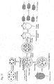
- mitochondria carrying mutant mtDNA can be introduced successfully into germ cells by fusing an enucleated cell carrying mutant mtDNA with a mtDNA-less cell to produce a cybrid, enucleating the cybrid, and then fusing the enucleated cybrid with a germ cell.
- the present invention provides a method for introducing mutant mtDNA into a germ cell, which comprises enucleating a cell carrying mutant mtDNA, fusing the enucleated cell with a mtDNA-less cell to produce a cybrid, enucleating the cybrid, and then fusing the enucleated cybrid with a germ cell.
- the method of the present invention for introducing mutant mtDNA into a germ cell comprises enucleating a cell carrying mutant mtDNA, fusing the enucleated cell with a mtDNA-less cell to produce a cybrid, enucleating the cybrid, and then fusing the enucleated cybrid with a germ cell.
- cell carrying mutant mtDNA that can be used herein is, but is not limited to, a cell carrying deletion mutant mtDNA4696.
- Cells carrying mutant mtDNA can be enucleated by ,for example, cytochalasin B treatment and high-speed centrifugation as described in Hayashi, J-I. et al., J. Biol. Chem 269, 19060-19066 (1994).
- mouse mtDNA-less cells that is, mouse ⁇ 0 cells that can be used herein include, but are not limited to, ⁇ 0 C2C12 cells.
- mouse ⁇ 0 cells we have established the mouse ⁇ 0 cells (for detail, see Inoue, K. et al., Isolation of mitochondrial DNA-less mouse cell lines and their application for trapping mouse synaptosomal mitochondrial DNA with deletion mutations. J. Biol. Chem. 272, 15510-15515; Inoue, K. et al., Isolation and characterization of mitochondrial DNA-less lines from various mammalian cell lines by application of an anticancer drug, ditercalinium. Biochem. Biophys. Res. Commun. 239, 257-260(1997)).
- Enucleated cells carrying mutant mtDNA can be fused with mtDNA-less cells by, for example, polyethylene glycol treatment as described in Hayashi, J-I. et al., J. Biol. Chem. 269, 19060-19066 (1944).
- Cybrids produced from the above fusion procedure can be enucleated in a manner similar to enucleation of cells carrying mutant mtDNA.
- pro-nuclear stage embryos can be used, but are not limited thereto.
- Cybrids and pro-nuclear stage embryos are fused by electrofusion.
- mice can be produced.
- embryos with the mtDNAs introduced therein are cultured upto the 4-cell stage in culture, and then the embryos are transplanted into the oviducts of female mice (pseudopregnant), thereby generating F0 mice.
- progeny mice can be generated by mating these F0 female mice with male mice having normal mtDNAs.
- ⁇ mtDNA cybrids carrying deletion mutant mtDNA
- Enucleated culture cells (NIH3T3 cells) were fused with mouse mtDNA-less cells (that is, ⁇ 0 cells) by polyethylene glycol (PEG) treatment, thereby producing 26 cybrid clones.
- PEG polyethylene glycol
- PCR was (PCR conditions: 30 cycles of 94 °C for 60 sec, 45 °C for 60 sec, and 45 °C for 120 sec) performed using the following primer set.
- ⁇ mtDNA contained in all but one of these clones were not sufficient to be detected by the Southern blotting. Only one clone, Cy4696, possessed 30% ⁇ mtDNA. The mutation was derived from the mouse culture cells.
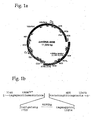
- ⁇ mtDNA4696 Nucleotide sequence analysis on PCR products revealed that ⁇ mtDNA carried by the cybrid clone Cy4696 (hereinafter, referred to as " ⁇ mtDNA4696) had 4696 bp deletion corresponding to nucleotide sequence 7759-12454 of normal mtDNA.
- the deleted region contained 6 tRNA genes arid 7 structural genes (Fig. 1a shows the gene map of ⁇ mtDNA4696). Unlike other mouse ⁇ mtDNA reported so far, the ⁇ mtDNA4696 contained no direct repeat around the region carrying the deletion (Fig. 1b shows the nucleotide sequence around the deleted region in the L strand of ⁇ mtDNA4696).
- ⁇ mtDNA 4696 in Cy4696 cybrid increased from 30% to 83% while culturing of the cells at 37 °C for 30 months, but increases exceeding this level have not been observed in longer period culturings. These behaviors were very similar to those observed for human cybrids in which ⁇ mtDNA that had been shown to be responsible for pathogenesis of a mitochondrial disease, Kearns-Sayre syndrome, had been introduced. Probably, the size of ⁇ mtDNA is small enough to be advantageous upon replication, so that it is predominant over the normal mtDNA during culturing for a long period of time.
- Cy4696 cybrids were enucleated by cytochalasin B treatment and high speed centrifuge, thereby producing several cytoplasts.
- theoviducts of the female mice were punctured, and then pro-nuclear stage embryos were collected.
- the collected embryos were transferred into CZB media, and then allowed to stand in the CZB media containing hyaluronidase (SIGMA), thereby removing cumulus cells.
- SIGMA hyaluronidase
- pulse stimulation (000 or 3500 V/cm, 10 ⁇ sec) was applied in the presence of alternating electric field (before and after fusion: 100 V/cm, 2 MHz, 30 sec) to fuse the cytoplasts with the embryos.
- alternating electric field (before and after fusion: 100 V/cm, 2 MHz, 30 sec)
- mice were produced using embryos carrying ⁇ mtDNA4696 prepared in Example 1.
- Example 1 1142 4-cell stage embryos (possessing ⁇ mtDNA4696) obtained in Example 1 were transferred into the oviducts of ICR female mice (8- to 12-weeks-old, pseudopregnant female) on day 1 after pseudo-pregnancy treatment.
- day 1 after treatment refers to the next day after mating (pseudo-pregnancy) with vasoligated male mice.
- Muscle biopsy was performed for the musculus tibialis anterior (M. tibialis anterior) of 31 F0 mice having ⁇ mtDNA4696, and then the proportion of ⁇ mtDNA4696 was measured by the Southern blotting. 24 mice were shown to have 5.7 to 41.8% ⁇ mtDNA4696 (The results are shown in Figure 2a).
- mtDNA inherits strictly maternally.
- mice had 27.4%, 38.0%, 47.0% and 48.7% ⁇ mtDNA4696, respectively.
- Seven F2 mice had 19.6%, 39.1%, 41.3%, 63.9%, 73.2%, 73.5% and 74.8% ⁇ mtDNA4696.
- mice possessing higher proportions of ⁇ mtDNA4696 could be obtained through successive breedings.
- mice Five male mice (21-weeks-old) having 47.3%, 70.0%, 79.0%, 82.8% and 84.8% ⁇ mtDNA4696 were selected by muscle biopsy from F2 mice. Two serial cryosections (10 ⁇ m and 20 ⁇ m in thickness) were prepared from the skeletal muscle (musculus soleus) of each mouse. These sections were used to examine whether there is a correlation between a reduction in COX activity and the proportion of ⁇ mtDNA4696.
- COX staining was performed by incubating the 10 ⁇ m section prepared from the musculus soleus of the mice carrying 84.8% ⁇ mtDNA4696 in a reaction solution (DAB 60mg, 0.1M acetate buffer (pH5.6) 27ml, 1% MnCl 2 (pH5.5) 3ml, 0.1% H 2 O 2 0.3ml) at 37°C for 30 min.
- DAB 60mg 0.1M acetate buffer (pH5.6) 27ml, 1% MnCl 2 (pH5.5) 3ml, 0.1% H 2 O 2 0.3ml
- cytoplasms of COX-negative fibers and COX-positive fibers were excised from the 20 ⁇ m section, and then the proportion of ⁇ mtDNA4696 in each cytoplasm was determined by quantitative PCR analysis using the following 3 primers:
- PCR conditions consist of 30 cycles of 94 °C for 30 sec, 55 °C for 30 sec, and 72 °C for 1 min for 1 st PCR and 10 cycles of 94 °C for 1 min, 48 °C for 1 min, and 72 °C for 3 min for 2 nd PCR.
- Mutant mtDNA ( ⁇ mtDNA4696) and normal mtDNA were respectively detected as 210 bp and 220 bp fragments.
- the proportions of mutant and normal mtDNA fragments contained in each fiber were quantified using the FM-BIO image analyzer (HITACHI).
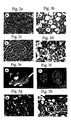
- Figure 3 shows the observed histological and morphological abnormalities.
- Fig. 3a and 3c show findings obtained from normal 21-weeks-old male mice (ICR, control).
- fiber samples numbered 1 to 7 possessed 95.6%, 90.0%, 86.5%, 81.9%, 82.2%, 83.2%, and 77.3% ⁇ mtDNA4696, respectively.
- COX-positive fiber samples (1, 2 and 3) possessed 90.7 ⁇ 3.8 % ⁇ mtDNA4696 on average, while COX-negative fiber samples (4, 5, 6 and 7) possessed 81.2 ⁇ 2.3 % on average.
- FIG. 3d shows the findings obtained for the mouse cardiac muscle possessing 84.8% ⁇ mtDNA4696.
- Figure 3f shows the kidney derived from the above mouse (right) and from a normal male mouse (left) at the same age (weeks-old).
- the mouse kidney possessing ⁇ mtDNA exhibited a granular surface (Fig. 3f, right) and possessed 88.2% ⁇ mtDNA4696.
- Figure 3h shows the findings on the renal tissue of a 24-weeks-old male mouse having 94.0% ⁇ mtDNA4696; and Fig. 3g shows the kidney of a normal male mouse at the same age (week-old).
- the mouse renal tissue possessing ⁇ mtDNA was observed to have significant cortical dilation of proximal and distal renal tubules, and partially contain a substance stainable with eosin.
- Peripheral blood was collected from tail of 12 male mice (12 weeks-old) having ⁇ mtDNA4696 to measure blood pyruvate levels (mg/dL) and lactate levels (mg/dL) before and after oral administration of glucose (1.5 g/kg body weight).
- ⁇ mtDNA4696 blood pyruvate levels and lactate levels before and after oral administration of glucose (1.5 g/kg body weight).
- Blood pyruvate levels and lactate levels were measured based on changes in concentration between NAD and NADH. The results are shown in Fig. 4a and 4b.
- peripheral blood was collected from five F2 male mice (21-weeks-old) possessing 47.3%, 70.0%, 79.0%, 82.8% and 84.8% ⁇ mtDNA4696 same as those in (5), and then blood urea nitrogen levels and creatinine levels were measured using DryChem (Fuji Film). These levels were compared with the levels obtained for the peripheral blood of mice having normal mtDNA. The results are shown in Figure 4c.
- mice having the greater proportions of ⁇ mtDNA4696 in the muscle tissue showed more increased peripheral blood lactate levels (In the figure, a diamond symbol (white) represents levels measured before glucose administration, and a diamond symbol (black) represents levels measured after the administration).
- mice having a high proportion of ⁇ mtDNA4696 showed significantly high blood urea nitrogen and creatinine levels, compared to mice having normal mtDNA.
- muscle fibers possessing 85% or less ⁇ mtDNA4696 exhibited normal mitochondrial respiratory functions can be described well by the presence of an interaction between mitochondria possessing normal mtDNA and mitochondria possessing ⁇ mtDNA4696 introduced therein in a fertilized egg which is a recipient of ⁇ mtDNA4696.
- the interaction supports the idea that we have previously proposed that mammalian mitochondria are functionally simple, and they can exchange their content as a result of their interaction.
- mice having a large amount of ⁇ mtDNA4696 introduced following only two generations because of the two reasons below: that somatic cell-derived mitochondria possessing ⁇ mtDNA4696 introduced into embryos can evade elimination from the embryos, and that ⁇ mtDNA4696 has an advantage in replicating more easily than normal mtDNA, thus ⁇ mtDNA4696 is transmitted more predominantly to the next generation compared to normal mtDNA.
- the toxicity of the mouse ⁇ mtDNA4696 provided by the present invention is less than that of human mtDNA with deletion mutation, but is almost the same as that of human mtDNA with point mutation.
- the ⁇ mtDNA4696 in the mouse of the present invention has a toxicity weaker than that of human ⁇ mtDNA; or mouse cells are resistant against energy loss, and can survive even when mouse egg cells, embryos and mitotic tissue contain a large amount of ⁇ mtDNA4696, thereby enabling the maternal inheritance of the mouse ⁇ mtDNA4696 and the accumulation of ⁇ mtDNA4696 in large amounts in mitotic tissues.
- chimera mice having chloramphenicol-resistant mtDNA in very small amount have been reported.
- chimera mice are generated by transferring female ES cells carrying a large amount of chloramphenicol-resistant mtDNA into normal blastocyst stage embryos.
- such chimera mice cannot be selected using chloramphenicol, because the proportion of chloramphenicol-resistant mtDNA would sharply decrease as the mtDNAs were not transmitted through germ cells to the succeeding generations.
- the present invention enables to provide an effective method for introducing mutant mtDNA into cells.
- the present invention also enables the production of mtDNA-knockout mice carrying ⁇ mtDNA4696 by using mutant mtDNA in the form of ⁇ mtDNA4696. Since this model mouse is useful in studying in detail the transmission of ⁇ mtDNA and the expression mechanism in various types of tissue, it can be used as a pathological model for mitochondrial diseases.
- the model mouse is also applicable to verify the hypothesis that the accumulation of mutant mtDNA may be responsible for aging and age-related disorders. We are planning to pursue this study after the mice grow older, because many mitochondrial diseases develop with age.
- the present invention can provide specimens at any age and at any stage of medically progressive conditions from any type of tissue possessing ⁇ mtDNA at various proportions. This may prove that mutations in mtDNA cause cryptogenic diseases.
- the present invention is also applicable to screening for a therapeutic agent and establishing gene therapies that are possible only with the model animal.
Landscapes
- Genetics & Genomics (AREA)
- Health & Medical Sciences (AREA)
- Engineering & Computer Science (AREA)
- Life Sciences & Earth Sciences (AREA)
- Chemical & Material Sciences (AREA)
- Wood Science & Technology (AREA)
- Organic Chemistry (AREA)
- Biotechnology (AREA)
- General Engineering & Computer Science (AREA)
- Biomedical Technology (AREA)
- Zoology (AREA)
- Bioinformatics & Cheminformatics (AREA)
- Molecular Biology (AREA)
- Microbiology (AREA)
- Plant Pathology (AREA)
- Physics & Mathematics (AREA)
- Biochemistry (AREA)
- General Health & Medical Sciences (AREA)
- Biophysics (AREA)
- Micro-Organisms Or Cultivation Processes Thereof (AREA)
- Measuring Or Testing Involving Enzymes Or Micro-Organisms (AREA)
Abstract
Description








The ΔmtDNA4696 in the mouse of the present invention has a toxicity weaker than that of human ΔmtDNA; or
mouse cells are resistant against energy loss, and can survive even when mouse egg cells, embryos and mitotic tissue contain a large amount of ΔmtDNA4696, thereby enabling the maternal inheritance of the mouse ΔmtDNA4696 and the accumulation of ΔmtDNA4696 in large amounts in mitotic tissues.


Claims (4)
- A method for introducing mutant mitochondrial DNA into a germ cell, which comprises fusing an enucleated cell possessing mutant mitochondrial DNA with a mitochondrial DNA-less cell to produce a cybrid, enucleating the cybrid, and fusing the cybrid with a germ cell.
- The method of claim 1, wherein the mutant mitochondrial DNA is a deletion mutant mitochondrial DNA4696.
- A mitochondrial DNA-knockout mouse, which is produced by transferring a germ cell produced by the method of claim 2 into a pseudopregnant mouse.
- The mitochondrial DNA-knockout mouse of claim 3, wherein the germ cell is a pro-nuclear stage embryo.
Applications Claiming Priority (3)
| Application Number | Priority Date | Filing Date | Title |
|---|---|---|---|
| JP2000009194A JP2001197845A (en) | 2000-01-18 | 2000-01-18 | Method for introducing mutant mitochondrial DNA into germ cells |
| JP2000009194 | 2000-01-18 | ||
| PCT/JP2000/009111 WO2001052642A1 (en) | 2000-01-18 | 2000-12-21 | Method of transferring mutant mitochondrial dna into genital cells |
Publications (2)
| Publication Number | Publication Date |
|---|---|
| EP1252818A1 true EP1252818A1 (en) | 2002-10-30 |
| EP1252818A4 EP1252818A4 (en) | 2004-04-21 |
Family
ID=18537373
Family Applications (1)
| Application Number | Title | Priority Date | Filing Date |
|---|---|---|---|
| EP00985829A Withdrawn EP1252818A4 (en) | 2000-01-18 | 2000-12-21 | METHOD FOR TRANSFERRING MUTOCHONDRIAL DNA MUTANT TO REPRODUCTIVE CELLS |
Country Status (4)
| Country | Link |
|---|---|
| US (1) | US20030159167A1 (en) |
| EP (1) | EP1252818A4 (en) |
| JP (1) | JP2001197845A (en) |
| WO (1) | WO2001052642A1 (en) |
Families Citing this family (4)
| Publication number | Priority date | Publication date | Assignee | Title |
|---|---|---|---|---|
| JP4250017B2 (en) * | 2002-05-15 | 2009-04-08 | エーザイ・アール・アンド・ディー・マネジメント株式会社 | Culturable mitochondrial cells exhibiting mitochondrial activity without a nucleus |
| JP2007274907A (en) * | 2006-04-03 | 2007-10-25 | Univ Of Tsukuba | Screening method for mitochondrial function activator |
| CN101818193B (en) * | 2009-11-13 | 2012-09-05 | 中国人民解放军总医院 | Sequence and kit for screening and testing deletion of mitochondrial DNA fragment |
| US8338178B2 (en) | 2010-10-28 | 2012-12-25 | The Invention Science Fund I, Llc | Mitochondrial enhancement of cells |
Family Cites Families (2)
| Publication number | Priority date | Publication date | Assignee | Title |
|---|---|---|---|---|
| US6215041B1 (en) * | 1997-01-10 | 2001-04-10 | University Of Mmassachusetts | Cloning using donor nuclei from a non-quiesecent somatic cells |
| AU8505998A (en) * | 1997-07-22 | 1999-02-16 | Uab Research Foundation | Transgenic transmitochondrial animals |
-
2000
- 2000-01-18 JP JP2000009194A patent/JP2001197845A/en active Pending
- 2000-12-21 US US10/181,347 patent/US20030159167A1/en not_active Abandoned
- 2000-12-21 WO PCT/JP2000/009111 patent/WO2001052642A1/en not_active Ceased
- 2000-12-21 EP EP00985829A patent/EP1252818A4/en not_active Withdrawn
Also Published As
| Publication number | Publication date |
|---|---|
| EP1252818A4 (en) | 2004-04-21 |
| US20030159167A1 (en) | 2003-08-21 |
| WO2001052642A1 (en) | 2001-07-26 |
| JP2001197845A (en) | 2001-07-24 |
Similar Documents
| Publication | Publication Date | Title |
|---|---|---|
| CN108660161B (en) | Method for preparing knockout animals without chimeric genes based on CRISPR/Cas9 technology | |
| US20180084767A1 (en) | Animal models for cardiomyopathy | |
| EP3381278B1 (en) | Method for preparing a canine model of atherosclerosis | |
| CN109706184B (en) | Method for establishing autism model dog | |
| US20220369608A1 (en) | Method for establishing diabetes disease model dog | |
| JP2019502400A (en) | Compositions and methods for preparing chimeric embryonic auxiliary organs | |
| CN108866100A (en) | A kind of efficient gene editing method | |
| Epstein et al. | Production of viable adult trisomy 17 reversible diploid mouse chimeras. | |
| JP2023116719A (en) | Disease model pig showing stable phenotype and method for producing the same | |
| CN115968834A (en) | Preparation method and application of RS1 point mutation mouse model | |
| CN119769472B (en) | Construction method and application of Prpf gene mutation-induced single dose insufficient RP (RP) mouse model | |
| US20030159167A1 (en) | Method of transferring mutant mitochondrial dna into genital cells | |
| CN118726371A (en) | A method for constructing a mouse model of hereditary retinal degeneration with humanized ABCA4 point mutation and its application | |
| KR102922515B1 (en) | An animal model for the retinal degeneration having IMPDH1 gene mutation and method for preparing the same | |
| WO2021190226A1 (en) | Application of single-base editing-mediated splicing repair in preparation and treatment of spinal muscular atrophy | |
| CN113801851B (en) | Somatic cell nuclear transfer method and application thereof | |
| JP7199741B2 (en) | Method for producing somatic cell clone animal of non-human primate | |
| CN118345116A (en) | A method for constructing a Huntington's disease mouse model | |
| CN119776361A (en) | A method for constructing a pPB-CAG-hORMDL3 mouse model and its application | |
| KR20250116237A (en) | An animal model for the retinal degeneration having PDE6B gene mutation and method for preparing the same | |
| CN120290567A (en) | Methods for constructing a mouse model of congenital heart disease | |
| CN116904513A (en) | Method for establishing SCN5A gene edited arrhythmia model dog | |
| CN114517212A (en) | Construction method and application of CRX gene deletion mutant cat | |
| CN119842827A (en) | Construction method and application of congenital cataract animal model | |
| Miller | Mammals need moms and dads |
Legal Events
| Date | Code | Title | Description |
|---|---|---|---|
| PUAI | Public reference made under article 153(3) epc to a published international application that has entered the european phase |
Free format text: ORIGINAL CODE: 0009012 |
|
| 17P | Request for examination filed |
Effective date: 20020816 |
|
| AK | Designated contracting states |
Kind code of ref document: A1 Designated state(s): AT BE CH CY DE DK ES FI FR GB GR IE IT LI LU MC NL PT SE TR |
|
| RAP1 | Party data changed (applicant data changed or rights of an application transferred) |
Owner name: INSTITUTE OF TSUKUBA LIAISON CO., LTD Owner name: SUMITOMO CHEMICAL COMPANY, LIMITED |
|
| A4 | Supplementary search report drawn up and despatched |
Effective date: 20040304 |
|
| RBV | Designated contracting states (corrected) |
Designated state(s): DE FR GB IT |
|
| RAP1 | Party data changed (applicant data changed or rights of an application transferred) |
Owner name: INSTITUTE OF TSUKUBA LIAISON CO., LTD Owner name: SUMITOMO CHEMICAL COMPANY, LIMITED |
|
| 17Q | First examination report despatched |
Effective date: 20060126 |
|
| STAA | Information on the status of an ep patent application or granted ep patent |
Free format text: STATUS: THE APPLICATION IS DEEMED TO BE WITHDRAWN |
|
| 18D | Application deemed to be withdrawn |
Effective date: 20070613 |