EP1246934B1 - Microbial growth variations - Google Patents
Microbial growth variations Download PDFInfo
- Publication number
- EP1246934B1 EP1246934B1 EP00985593A EP00985593A EP1246934B1 EP 1246934 B1 EP1246934 B1 EP 1246934B1 EP 00985593 A EP00985593 A EP 00985593A EP 00985593 A EP00985593 A EP 00985593A EP 1246934 B1 EP1246934 B1 EP 1246934B1
- Authority
- EP
- European Patent Office
- Prior art keywords
- growth
- population
- test
- microbes
- antibiotic
- Prior art date
- Legal status (The legal status is an assumption and is not a legal conclusion. Google has not performed a legal analysis and makes no representation as to the accuracy of the status listed.)
- Expired - Lifetime
Links
- 230000012010 growth Effects 0.000 title claims abstract description 64
- 230000000813 microbial effect Effects 0.000 title claims description 12
- 238000012360 testing method Methods 0.000 claims abstract description 67
- 238000000034 method Methods 0.000 claims abstract description 60
- 230000004044 response Effects 0.000 claims abstract description 24
- 235000015097 nutrients Nutrition 0.000 claims abstract description 8
- 230000003115 biocidal effect Effects 0.000 claims description 31
- 238000011534 incubation Methods 0.000 claims description 18
- 239000003242 anti bacterial agent Substances 0.000 claims description 12
- 239000002054 inoculum Substances 0.000 claims description 12
- 239000003814 drug Substances 0.000 claims description 8
- 229940079593 drug Drugs 0.000 claims description 7
- 239000000126 substance Substances 0.000 claims description 7
- 230000007773 growth pattern Effects 0.000 claims description 6
- 238000012544 monitoring process Methods 0.000 claims description 6
- 230000001332 colony forming effect Effects 0.000 claims description 4
- 230000000844 anti-bacterial effect Effects 0.000 claims description 3
- 230000001580 bacterial effect Effects 0.000 claims description 3
- 230000008859 change Effects 0.000 claims description 3
- 230000001594 aberrant effect Effects 0.000 claims description 2
- 230000009699 differential effect Effects 0.000 claims description 2
- 230000000737 periodic effect Effects 0.000 claims description 2
- 230000004069 differentiation Effects 0.000 claims 5
- 230000000063 preceeding effect Effects 0.000 claims 2
- 230000002349 favourable effect Effects 0.000 claims 1
- 230000009036 growth inhibition Effects 0.000 claims 1
- 230000008569 process Effects 0.000 abstract description 11
- 230000000845 anti-microbial effect Effects 0.000 abstract description 3
- 238000012009 microbiological test Methods 0.000 abstract description 3
- 235000013343 vitamin Nutrition 0.000 abstract description 3
- 239000011782 vitamin Substances 0.000 abstract description 3
- 229940088594 vitamin Drugs 0.000 abstract description 3
- 229930003231 vitamin Natural products 0.000 abstract description 3
- 150000001875 compounds Chemical class 0.000 abstract description 2
- 239000007952 growth promoter Substances 0.000 abstract description 2
- 230000035945 sensitivity Effects 0.000 description 12
- 238000001514 detection method Methods 0.000 description 11
- 229940088710 antibiotic agent Drugs 0.000 description 8
- 230000000694 effects Effects 0.000 description 7
- 102000006635 beta-lactamase Human genes 0.000 description 5
- 230000008261 resistance mechanism Effects 0.000 description 5
- 241000894007 species Species 0.000 description 5
- 108090000204 Dipeptidase 1 Proteins 0.000 description 4
- 229930182555 Penicillin Natural products 0.000 description 4
- 238000002829 antibacterial sensitivity test Methods 0.000 description 4
- 238000013459 approach Methods 0.000 description 4
- 230000008901 benefit Effects 0.000 description 4
- 238000002560 therapeutic procedure Methods 0.000 description 4
- 241000894006 Bacteria Species 0.000 description 3
- JGSARLDLIJGVTE-MBNYWOFBSA-N Penicillin G Chemical compound N([C@H]1[C@H]2SC([C@@H](N2C1=O)C(O)=O)(C)C)C(=O)CC1=CC=CC=C1 JGSARLDLIJGVTE-MBNYWOFBSA-N 0.000 description 3
- 241000191967 Staphylococcus aureus Species 0.000 description 3
- 238000005516 engineering process Methods 0.000 description 3
- 230000005764 inhibitory process Effects 0.000 description 3
- 239000012528 membrane Substances 0.000 description 3
- 229940049954 penicillin Drugs 0.000 description 3
- 229920001817 Agar Polymers 0.000 description 2
- 208000035473 Communicable disease Diseases 0.000 description 2
- ULGZDMOVFRHVEP-RWJQBGPGSA-N Erythromycin Chemical compound O([C@@H]1[C@@H](C)C(=O)O[C@@H]([C@@]([C@H](O)[C@@H](C)C(=O)[C@H](C)C[C@@](C)(O)[C@H](O[C@H]2[C@@H]([C@H](C[C@@H](C)O2)N(C)C)O)[C@H]1C)(C)O)CC)[C@H]1C[C@@](C)(OC)[C@@H](O)[C@H](C)O1 ULGZDMOVFRHVEP-RWJQBGPGSA-N 0.000 description 2
- 239000008272 agar Substances 0.000 description 2
- 230000004888 barrier function Effects 0.000 description 2
- 239000003795 chemical substances by application Substances 0.000 description 2
- 230000034373 developmental growth involved in morphogenesis Effects 0.000 description 2
- 238000010790 dilution Methods 0.000 description 2
- 239000012895 dilution Substances 0.000 description 2
- 238000001914 filtration Methods 0.000 description 2
- 239000007850 fluorescent dye Substances 0.000 description 2
- 230000001771 impaired effect Effects 0.000 description 2
- 230000006872 improvement Effects 0.000 description 2
- 230000002401 inhibitory effect Effects 0.000 description 2
- 230000007246 mechanism Effects 0.000 description 2
- 230000002906 microbiologic effect Effects 0.000 description 2
- 244000005700 microbiome Species 0.000 description 2
- 238000012986 modification Methods 0.000 description 2
- 230000004048 modification Effects 0.000 description 2
- 238000002360 preparation method Methods 0.000 description 2
- 230000035899 viability Effects 0.000 description 2
- QIVUCLWGARAQIO-OLIXTKCUSA-N (3s)-n-[(3s,5s,6r)-6-methyl-2-oxo-1-(2,2,2-trifluoroethyl)-5-(2,3,6-trifluorophenyl)piperidin-3-yl]-2-oxospiro[1h-pyrrolo[2,3-b]pyridine-3,6'-5,7-dihydrocyclopenta[b]pyridine]-3'-carboxamide Chemical compound C1([C@H]2[C@H](N(C(=O)[C@@H](NC(=O)C=3C=C4C[C@]5(CC4=NC=3)C3=CC=CN=C3NC5=O)C2)CC(F)(F)F)C)=C(F)C=CC(F)=C1F QIVUCLWGARAQIO-OLIXTKCUSA-N 0.000 description 1
- 238000012935 Averaging Methods 0.000 description 1
- 108020004256 Beta-lactamase Proteins 0.000 description 1
- 241000588748 Klebsiella Species 0.000 description 1
- RJQXTJLFIWVMTO-TYNCELHUSA-N Methicillin Chemical compound COC1=CC=CC(OC)=C1C(=O)N[C@@H]1C(=O)N2[C@@H](C(O)=O)C(C)(C)S[C@@H]21 RJQXTJLFIWVMTO-TYNCELHUSA-N 0.000 description 1
- 241000187479 Mycobacterium tuberculosis Species 0.000 description 1
- MKYBYDHXWVHEJW-UHFFFAOYSA-N N-[1-oxo-1-(2,4,6,7-tetrahydrotriazolo[4,5-c]pyridin-5-yl)propan-2-yl]-2-[[3-(trifluoromethoxy)phenyl]methylamino]pyrimidine-5-carboxamide Chemical compound O=C(C(C)NC(=O)C=1C=NC(=NC=1)NCC1=CC(=CC=C1)OC(F)(F)F)N1CC2=C(CC1)NN=N2 MKYBYDHXWVHEJW-UHFFFAOYSA-N 0.000 description 1
- 241000295644 Staphylococcaceae Species 0.000 description 1
- 241001505901 Streptococcus sp. 'group A' Species 0.000 description 1
- 108010059993 Vancomycin Proteins 0.000 description 1
- 230000009471 action Effects 0.000 description 1
- 230000006978 adaptation Effects 0.000 description 1
- 239000002390 adhesive tape Substances 0.000 description 1
- 229960004821 amikacin Drugs 0.000 description 1
- LKCWBDHBTVXHDL-RMDFUYIESA-N amikacin Chemical compound O([C@@H]1[C@@H](N)C[C@H]([C@@H]([C@H]1O)O[C@@H]1[C@@H]([C@@H](N)[C@H](O)[C@@H](CO)O1)O)NC(=O)[C@@H](O)CCN)[C@H]1O[C@H](CN)[C@@H](O)[C@H](O)[C@H]1O LKCWBDHBTVXHDL-RMDFUYIESA-N 0.000 description 1
- 229940126575 aminoglycoside Drugs 0.000 description 1
- 230000003321 amplification Effects 0.000 description 1
- 238000004458 analytical method Methods 0.000 description 1
- 230000002547 anomalous effect Effects 0.000 description 1
- 230000008485 antagonism Effects 0.000 description 1
- 238000009635 antibiotic susceptibility testing Methods 0.000 description 1
- 239000004599 antimicrobial Substances 0.000 description 1
- 238000011203 antimicrobial therapy Methods 0.000 description 1
- 238000000149 argon plasma sintering Methods 0.000 description 1
- 238000003491 array Methods 0.000 description 1
- 239000012298 atmosphere Substances 0.000 description 1
- 210000001124 body fluid Anatomy 0.000 description 1
- 239000010839 body fluid Substances 0.000 description 1
- 230000030833 cell death Effects 0.000 description 1
- 230000001413 cellular effect Effects 0.000 description 1
- 238000012512 characterization method Methods 0.000 description 1
- 239000003153 chemical reaction reagent Substances 0.000 description 1
- 239000003593 chromogenic compound Substances 0.000 description 1
- 230000000052 comparative effect Effects 0.000 description 1
- 238000012733 comparative method Methods 0.000 description 1
- 230000000093 cytochemical effect Effects 0.000 description 1
- 230000006378 damage Effects 0.000 description 1
- 230000009977 dual effect Effects 0.000 description 1
- 230000002255 enzymatic effect Effects 0.000 description 1
- 229960003276 erythromycin Drugs 0.000 description 1
- 230000002068 genetic effect Effects 0.000 description 1
- 239000003102 growth factor Substances 0.000 description 1
- 239000001963 growth medium Substances 0.000 description 1
- 238000009396 hybridization Methods 0.000 description 1
- 238000010348 incorporation Methods 0.000 description 1
- 208000015181 infectious disease Diseases 0.000 description 1
- 238000002955 isolation Methods 0.000 description 1
- 238000003771 laboratory diagnosis Methods 0.000 description 1
- 238000011005 laboratory method Methods 0.000 description 1
- 238000011068 loading method Methods 0.000 description 1
- 239000011159 matrix material Substances 0.000 description 1
- 238000005259 measurement Methods 0.000 description 1
- 239000013028 medium composition Substances 0.000 description 1
- 230000002503 metabolic effect Effects 0.000 description 1
- 229960003085 meticillin Drugs 0.000 description 1
- 239000000203 mixture Substances 0.000 description 1
- ZBGPYVZLYBDXKO-HILBYHGXSA-N netilmycin Chemical compound O([C@@H]1[C@@H](N)C[C@H]([C@@H]([C@H]1O)O[C@@H]1[C@]([C@H](NC)[C@@H](O)CO1)(C)O)NCC)[C@H]1OC(CN)=CC[C@H]1N ZBGPYVZLYBDXKO-HILBYHGXSA-N 0.000 description 1
- 238000003199 nucleic acid amplification method Methods 0.000 description 1
- 230000003287 optical effect Effects 0.000 description 1
- 244000052769 pathogen Species 0.000 description 1
- 150000002960 penicillins Chemical class 0.000 description 1
- 239000008363 phosphate buffer Substances 0.000 description 1
- 230000000704 physical effect Effects 0.000 description 1
- 238000003752 polymerase chain reaction Methods 0.000 description 1
- 239000013641 positive control Substances 0.000 description 1
- 230000000750 progressive effect Effects 0.000 description 1
- 230000035755 proliferation Effects 0.000 description 1
- 238000012124 rapid diagnostic test Methods 0.000 description 1
- 230000009467 reduction Effects 0.000 description 1
- 238000011896 sensitive detection Methods 0.000 description 1
- 238000001228 spectrum Methods 0.000 description 1
- 239000000725 suspension Substances 0.000 description 1
- 230000001225 therapeutic effect Effects 0.000 description 1
- 229940041007 third-generation cephalosporins Drugs 0.000 description 1
- 229960000707 tobramycin Drugs 0.000 description 1
- NLVFBUXFDBBNBW-PBSUHMDJSA-N tobramycin Chemical compound N[C@@H]1C[C@H](O)[C@@H](CN)O[C@@H]1O[C@H]1[C@H](O)[C@@H](O[C@@H]2[C@@H]([C@@H](N)[C@H](O)[C@@H](CO)O2)O)[C@H](N)C[C@@H]1N NLVFBUXFDBBNBW-PBSUHMDJSA-N 0.000 description 1
- 230000001052 transient effect Effects 0.000 description 1
- 229960003165 vancomycin Drugs 0.000 description 1
- MYPYJXKWCTUITO-LYRMYLQWSA-N vancomycin Chemical compound O([C@@H]1[C@@H](O)[C@H](O)[C@@H](CO)O[C@H]1OC1=C2C=C3C=C1OC1=CC=C(C=C1Cl)[C@@H](O)[C@H](C(N[C@@H](CC(N)=O)C(=O)N[C@H]3C(=O)N[C@H]1C(=O)N[C@H](C(N[C@@H](C3=CC(O)=CC(O)=C3C=3C(O)=CC=C1C=3)C(O)=O)=O)[C@H](O)C1=CC=C(C(=C1)Cl)O2)=O)NC(=O)[C@@H](CC(C)C)NC)[C@H]1C[C@](C)(N)[C@H](O)[C@H](C)O1 MYPYJXKWCTUITO-LYRMYLQWSA-N 0.000 description 1
- MYPYJXKWCTUITO-UHFFFAOYSA-N vancomycin Natural products O1C(C(=C2)Cl)=CC=C2C(O)C(C(NC(C2=CC(O)=CC(O)=C2C=2C(O)=CC=C3C=2)C(O)=O)=O)NC(=O)C3NC(=O)C2NC(=O)C(CC(N)=O)NC(=O)C(NC(=O)C(CC(C)C)NC)C(O)C(C=C3Cl)=CC=C3OC3=CC2=CC1=C3OC1OC(CO)C(O)C(O)C1OC1CC(C)(N)C(O)C(C)O1 MYPYJXKWCTUITO-UHFFFAOYSA-N 0.000 description 1
- 230000000007 visual effect Effects 0.000 description 1
- 150000003722 vitamin derivatives Chemical class 0.000 description 1
Images


Classifications
-
- C—CHEMISTRY; METALLURGY
- C12—BIOCHEMISTRY; BEER; SPIRITS; WINE; VINEGAR; MICROBIOLOGY; ENZYMOLOGY; MUTATION OR GENETIC ENGINEERING
- C12Q—MEASURING OR TESTING PROCESSES INVOLVING ENZYMES, NUCLEIC ACIDS OR MICROORGANISMS; COMPOSITIONS OR TEST PAPERS THEREFOR; PROCESSES OF PREPARING SUCH COMPOSITIONS; CONDITION-RESPONSIVE CONTROL IN MICROBIOLOGICAL OR ENZYMOLOGICAL PROCESSES
- C12Q1/00—Measuring or testing processes involving enzymes, nucleic acids or microorganisms; Compositions therefor; Processes of preparing such compositions
- C12Q1/02—Measuring or testing processes involving enzymes, nucleic acids or microorganisms; Compositions therefor; Processes of preparing such compositions involving viable microorganisms
- C12Q1/025—Measuring or testing processes involving enzymes, nucleic acids or microorganisms; Compositions therefor; Processes of preparing such compositions involving viable microorganisms for testing or evaluating the effect of chemical or biological compounds, e.g. drugs, cosmetics
Definitions
- This invention centres on microbiological test processes, in particular those involving testing of the influence of antimicrobial compounds, nutrients and vitamins or other growth promoters.
- the invention is concerned with lack of uniformity in response to such agents.
- the purpose of the present invention to provide a new test process whereby differences in growth responses are detected.
- the presence of any antibiotic resistant sub populations can be established within the duration of the test.
- Clinical laboratories often test microbes clinically isolated from patient and other sources, examining their sensitivity to antimicrobial agents and their metabolic requirements. Effective antibacterial therapy is fundamental to modern medicine, but equally the ability to treat infectious diseases is often taken for granted.
- SensititreTM although among the popular microdilution products, is limited to 18-hour operation. Two other systems are described in publications as dealing with these issues somewhat tangentially.
- the Bio-Merieux VitekTM is reported as incorporating an expert system to analyse atypical patterns and report the most likely resistance mechanisms present in an isolate, using the AntibiogramTM.
- the Baxter MicroScanTM is said to feature software to alert the pharmacy to patients on inappropriate or excessive therapeutic choices.
- Genotypic methods can provide invaluable information when it is not easily available by other means.
- Phenotypic methods should be inexpensive and simple, all mechanisms are detected and there is established data linking tests results to clinical outcome. These methods are far more flexible in the range of antibiotics used. Where growth rate determinations are a key element it is important to limit the impact of factors such as media composition, temperature and atmosphere composition.
- the main technical barrier is the improvement in the sensitivity of growth detection technology and differentiating resistant sub-populations.
- existing test systems are severely limited.
- Interpretative methods depend very heavily upon a substantial database on antibiotics and resistance which in turn calls for complex logic to apply information effectively.
- the phenotypic approach of measuring activity of antibiotics against cultures of clinical isolates remains necessary.
- Another purpose of the present invention is to report each growth response and emphasise any detectable heterogeneity arising for any reason.
- the ability to obtain useful early/rapid results, particularly in antibiotic sensitivity tests, has obvious clinical benefits.
- the detection of any low grade resistance enhances the quality of the result and reinforces the efficacy of the test process as a laboratory diagnosis.
- each test condition e.g. antibiotic, vitamin or nutrient concentration
- each well is inoculated with approx. 5 x 10 3 organisms [colony forming units c.f.u.]. Since these products demonstrate early/late anomalies there are apparently sufficient cells present to express the effect. Equally there are finite limits to the number available after colony selection from the primary plate. For these reasons it seems reasonable that any new test deals with approximately equivalent numbers, although there are advantages in increasing the numbers to enhance detection of atypical cells.
- micro-organisms can be expected to grow and thus increase in numbers. If, for example, an organism has a doubling time of 30 mins then after an initial delay [lag] each cell will divide to create two cells at this interval. Within the short term - say, 3 hours - a single cell will yield in excess of 50-100 cells. It follows that a larger number - say 100 cells - can in the same time span generate about 5 x 10 3 cells. In contrast after a protracted incubation organisms will reach a maximum number which can be supported by the available nutrients, regardless of the number from which the final population originated. It is important to note that the differential is only of value during the transient stages of early incubation. During incubation the response of organisms to an antibiotic will depend on concentration and mode of action. Growth patterns will generally fall into these categories:
- the growth responses can be compared and contrasted across all replicates [also on a time course basis by regular monitoring where available]. Any growth [increase in numbers] must be looked at in the context of all replicates. A general, gross increase across most replicates suggests the whole population is actively growing. In contrast a random pattern of small increases, in a proportion of replicates, suggests that such growth is arising from a low level distribution of resistant sub-population. This will only be evident where the main population is inhibited. When applied to MIC Determinations the 'uniform' isolate will provide an uncomplicated, consistent result, repeated numerous times in the sets of replicates. In contrast the culture with low level resistance will exhibit significant differential growth effects, related to the incidence and distribution of resistant cells across the test array.
- any growth must be looked at in the context of all replicates. A general, gross increase across most replicates suggests the whole population is actively growing. In contrast a random pattern of small increases, in a proportion of replicates, suggests that such growth is arising from a low level distribution of resistant sub-population
- the ability to follow and compare growth responses depends on sensitive detection methods. It is also of value to establish baselines by averaging multiple results in any given test situation. Comparative methods are able to pinpoint atypical responses using the majority as a control.
- the reduction in inoculum level reduces any tendency for the sub-population to be masked by the density of the main inoculum.
- the assessment of growth is essentially growth/no growth. During the early stages of incubation any growth is evident as a progressive increase in numbers. Each species will tend to grow at some maximum rate associated with the species itself and the conditions of the test. Any antibiotic present will tend to decrease the growth rate, usually in direct proportion to the concentration of antibiotic.
- a quantitative assessment of growth adds a further dimension to the test process. Comparison between a culture containing no antibiotic and those subjected to known levels provides additional information. The extent to which a culture is impaired is a valuable indicator as to the concentration of drug needed to achieve effective inhibition.
- test processes involving incubation of a culture become easier to monitor the longer growth takes place.
- the ability to increase in numbers provides amplification of results, conversely the greater the sensitivity of the microbial detection then the earlier results become available.
- the test process described within this invention provides a rapid diagnostic test of significant clinical value.
- the process described here takes into account low grade resistance, thus providing the clinician with accurate and comprehensive information.
- Observations of growth may be made between 6-12 hours. Observations of growth may be made between 18-24 hours. Periodic observations [semi-continuous], or continuous monitoring, may be carried out for at least 18-24 hours to automatically assess growth patterns.
- the basic principle of the current invention is the comparison of differential growth patterns, to detect and rank variations.
- Exploitation of the method would benefit from repackaging into purpose designed test arrays. Modifications and improvements will occur to those skilled in the art. In particular non-invasive, semi-continuous growth monitoring would simplify operation of the test system.
- the method can determine the presence or absence of drug resistant bacteria, for example, and therefore can also be used to monitor the changes in drug resistant bacteria over a period of time. By repeating the method and comparing the actual or relative numbers of the major and minor bacterial populations any changes in population groups can be tracked and the effectiveness of any treatment assessed.
- Penicillin Organisms Two cultures of Staphylococcus aureus, with one isolated from plates containing penicillin supplemented agar to provide cells of known ⁇ lactamase activity.
- Inoculum A target level of 10 2 cells/wells was selected to test an adequate number of organisms within each treatment. In the examples 2 - 4 the aim was to include an increasing proportion of resistant/ ⁇ lactamase positive cells. Preparation by dilutions at these levels will undoubtedly be prone to errors.
- Incubation Sealed plates incubated for 5 hours at 37°C [previously shown to provide significant growth]. At the end of incubation well contents [50 ⁇ l] were transferred to 200 ⁇ l using individual sterile pipette tips [i.e. one/well].
- a 100 ⁇ l sample of diluted well contents was plated and spread on culture plates. The growth, corresponding to the situation at 5 hours, was assessed after incubation of the culture plates overnight.
- a second 100 ⁇ l sample was filtered through a commercially available 0.2 ⁇ nuclepore membrane. This non-fluorescent, black filter was supported on a 5.0 ⁇ cellulosic membrane filter. Discrete 3.0mm diameter filter zones were created by attaching pre-punched black adhesive tape to the membranes. After filtration the zones were stained with 0.01% w/v AcriflavineTM in phosphate buffer at pH 7.2 [filtered immediately before use]. Direct observation, using an epifluorescence microscope resembles the DEFT method described by Pettifer GL Rodrigues UK [1982] J.Appl.Bact.53:323-329. Using a large numerical aperture x 45 objective it is relatively simple to rank the estimated density of growth.
- the viability test depicts growth responses in terms of organisms recovered and subsequently able to produce colonies on a secondary culture plate.
- Direct epifluorescence/filtration does constitute rapid detection but in this manual form is both cumbersome and labour intensive. Within these constraints both methods served to assign rankings to the growth response of every well. The two methods were adequate to indicate the presence of growth.
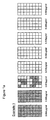
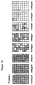
- Figure 1 shows the results of Examples 1-4. It can be seen that at very low antibiotic concentration [e.g. 0.004 ⁇ g/ml] all 32 replicate wells exhibit continued growth [dark shading]. With increasing concentration of antibiotic the population in some wells exhibits sensitivity [no shading], as seen at 0.015 ⁇ g/ml. In Example 1 there is no evidence of growth in wells containing antibiotic at levels higher than 0.015 ⁇ g/ml.
- antibiotic concentration e.g. 0.004 ⁇ g/ml
- Examples 2,3 and 4 an appreciable number of wells show low grade growth [light shading]. This is presumably attributable to the growth produced by a very small number [typically one] of resistant cells. Within a rapid timescale there will inevitably be a difference between the growth yielded by a whole inoculum multiplying compared to a few or even individual cells. In a longer period of incubation this differential is in fact lost. Examples 2,3 and 4 show increasing incidence of resistant cells; this corresponds to the proportions added during preparation of inocula. Dilution errors at these levels make it difficult to calculate actual ratios but the maximum tested here [Example4] probably corresponds to less than 10% of the population. The test examples demonstrate that, in Examples 2,3 and 4, there are resistant sub-populations in some wells which can be insensitive even at an antibiotic concentration of 0.250 ⁇ g/ml.
- the number of colony forming units in each replicate test is in the range 25-500, preferably 100-200.
- the number of replicates is between 20-100, typically about 50.
- the initial volume and/or initial concentration of organisms in the inoculum can be adjusted.
- the number of replicates can be increased above the number exemplified herein, in order to detect very low incidence rates for resistant cells.
Landscapes
- Chemical & Material Sciences (AREA)
- Engineering & Computer Science (AREA)
- Organic Chemistry (AREA)
- Life Sciences & Earth Sciences (AREA)
- Health & Medical Sciences (AREA)
- Proteomics, Peptides & Aminoacids (AREA)
- Bioinformatics & Cheminformatics (AREA)
- Zoology (AREA)
- Wood Science & Technology (AREA)
- Immunology (AREA)
- Toxicology (AREA)
- Biotechnology (AREA)
- Analytical Chemistry (AREA)
- Microbiology (AREA)
- Molecular Biology (AREA)
- Physics & Mathematics (AREA)
- Biophysics (AREA)
- Medicinal Chemistry (AREA)
- Biochemistry (AREA)
- General Engineering & Computer Science (AREA)
- General Health & Medical Sciences (AREA)
- Genetics & Genomics (AREA)
- Measuring Or Testing Involving Enzymes Or Micro-Organisms (AREA)
- Medicines Containing Material From Animals Or Micro-Organisms (AREA)
- Immobilizing And Processing Of Enzymes And Microorganisms (AREA)
Abstract
Description
Antibiotic: Penicillin
Organisms: Two cultures of Staphylococcus aureus, with one isolated from plates containing penicillin supplemented agar to provide cells of known β lactamase activity.
Inoculum: A target level of 102 cells/wells was selected to test an adequate number of organisms within each treatment. In the examples 2 - 4 the aim was to include an increasing proportion of resistant/β lactamase positive cells. Preparation by dilutions at these levels will undoubtedly be prone to errors.
Incubation: Sealed plates incubated for 5 hours at 37°C [previously shown to provide significant growth]. At the end of incubation well contents [50µl] were transferred to 200µl using individual sterile pipette tips [i.e. one/well].
Using a large numerical aperture x 45 objective it is relatively simple to rank the estimated density of growth.
The viability test depicts growth responses in terms of organisms recovered and subsequently able to produce colonies on a secondary culture plate. Direct epifluorescence/filtration does constitute rapid detection but in this manual form is both cumbersome and labour intensive. Within these constraints both methods served to assign rankings to the growth response of every well. The two methods were adequate to indicate the presence of growth.
Claims (15)
- A method for determining the influence of a test substance on microbial growth, which includes the steps of:a) taking a sample inoculum containing microbes whose growth response to the substance is to be tested, wherein the number and concentration of colony forming units of the microbe in the inoculum is approximately known;b) dividing the inoculum into a plurality of replicates so that the total number of c.f.u.'s is distributed between the replicates;c) contacting each of the replicates with the test substance under the same incubation conditions to provide replicate tests;d) observing the influence of the substance on the growth of the microbes at one or more times during the incubation period;e) differentiating the typical growth response of a majority of microbes constituting a main population group from a typical growth response of a minority of microbes constituting a minor sub-population group, the differentiation being on the basis of heterogeneity of response between respective replicate tests.
- A method according to claim 1 wherein the test substance is an antibiotic.
- A method according to claim 1 wherein the test substance is a bacterial nutrient.
- A method according to any preceeding claim wherein the growth response is selected from the group consisting of:I. growth rate changeII. bacteriostasisIII. bactericidal actionIV. total or partial growth inhibitionV. aberrant growth characteristics.
- A method according to claim 2 wherein the inoculum is distributed among replicate tests, at each antibiotic concentration, in such a way as to obtain a defined number of colony forming units in each and every replicate.
- A method according to claim 5 wherein the number in each test is in the range 25-500, preferably 100-200.
- A method according to claim 5 or 6 wherein the number of replicates is between 20-100, typically about 50.
- A method according to claim 1 wherein an observation of the growth is made at about 5 hours of incubation.
- A method according to claim 1 wherein observations of growth are made between 6-12 hours.
- A method according to claim 1 wherein observations of growth are made between 18-24 hours.
- A method according to claim 1 wherein periodic semi-continuous observations, or continuous monitoring, are carried out for at least 18-24 hours to automatically assess growth patterns.
- A method according to claim 11 wherein first results are presented as soon as significant differential effects are detected.
- A method according to claim 2, wherein the differentiation in growth response of the microbial populations indicates a population of drug resistant microbes.
- A method according to claim 3, wherein the differentiation in growth response of the microbial populations indicates a population of microbes in a favourable nutrient for growth.
- A method according to any of the preceeding claims, wherein the differentiation between the main population group and the minor sub-population group is determined and recorded, the method is repeated and the differentiation between the population groups is compared with one or more previous results to determine whether or not there has been any change in either or both microbial population.
Applications Claiming Priority (3)
| Application Number | Priority Date | Filing Date | Title |
|---|---|---|---|
| GB9929808 | 1999-12-16 | ||
| GBGB9929808.5A GB9929808D0 (en) | 1999-12-16 | 1999-12-16 | Microbial growth variations |
| PCT/GB2000/004842 WO2001044498A2 (en) | 1999-12-16 | 2000-12-15 | Microbial growth variations |
Publications (2)
| Publication Number | Publication Date |
|---|---|
| EP1246934A2 EP1246934A2 (en) | 2002-10-09 |
| EP1246934B1 true EP1246934B1 (en) | 2004-11-17 |
Family
ID=10866480
Family Applications (1)
| Application Number | Title | Priority Date | Filing Date |
|---|---|---|---|
| EP00985593A Expired - Lifetime EP1246934B1 (en) | 1999-12-16 | 2000-12-15 | Microbial growth variations |
Country Status (6)
| Country | Link |
|---|---|
| US (1) | US20030077689A1 (en) |
| EP (1) | EP1246934B1 (en) |
| AT (1) | ATE282712T1 (en) |
| DE (1) | DE60016035T2 (en) |
| GB (1) | GB9929808D0 (en) |
| WO (1) | WO2001044498A2 (en) |
Families Citing this family (1)
| Publication number | Priority date | Publication date | Assignee | Title |
|---|---|---|---|---|
| US20020025514A1 (en) * | 2000-07-05 | 2002-02-28 | Jaworski Deborah D. | High throughput assay |
Family Cites Families (4)
| Publication number | Priority date | Publication date | Assignee | Title |
|---|---|---|---|---|
| US4288543A (en) * | 1977-01-28 | 1981-09-08 | Pfizer Inc. | Method and apparatus for identifying microorganisms |
| US4236211A (en) * | 1978-09-15 | 1980-11-25 | Pfizer Inc. | Method and apparatus for determining the minimum concentration of antibiotic necessary to at least inhibit microorganism growth |
| FR2732693B1 (en) * | 1995-04-06 | 1997-05-09 | Bio Veto Tests Bvt | CONTAMINANT REVELATION INDICATOR AND METHOD OF APPLICATION TO THE PRODUCTION OF AN ANTIBIOGRAM DIRECTLY CARRIED OUT ON A SAMPLING |
| US6001586A (en) * | 1996-03-29 | 1999-12-14 | Genencor International, Inc. | Compartmentalization method for screening microorganisms |
-
1999
- 1999-12-16 GB GBGB9929808.5A patent/GB9929808D0/en not_active Ceased
-
2000
- 2000-12-15 WO PCT/GB2000/004842 patent/WO2001044498A2/en not_active Ceased
- 2000-12-15 US US10/149,790 patent/US20030077689A1/en not_active Abandoned
- 2000-12-15 DE DE60016035T patent/DE60016035T2/en not_active Expired - Lifetime
- 2000-12-15 EP EP00985593A patent/EP1246934B1/en not_active Expired - Lifetime
- 2000-12-15 AT AT00985593T patent/ATE282712T1/en not_active IP Right Cessation
Also Published As
| Publication number | Publication date |
|---|---|
| DE60016035D1 (en) | 2004-12-23 |
| EP1246934A2 (en) | 2002-10-09 |
| US20030077689A1 (en) | 2003-04-24 |
| DE60016035T2 (en) | 2005-11-10 |
| ATE282712T1 (en) | 2004-12-15 |
| WO2001044498A3 (en) | 2001-12-13 |
| GB9929808D0 (en) | 2000-02-09 |
| WO2001044498A2 (en) | 2001-06-21 |
Similar Documents
| Publication | Publication Date | Title |
|---|---|---|
| US6046021A (en) | Comparative phenotype analysis of two or more microorganisms using a plurality of substrates within a multiwell testing device | |
| US20110269130A1 (en) | Antibiotice Susceptibility Profiling Methods | |
| US6686173B2 (en) | Comparative phenotype analysis of two or more microorganisms using a plurality of substrates within a multiwell testing device | |
| Ryan et al. | Rapid assay for mycobacterial growth and antibiotic susceptibility using gel microdrop encapsulation | |
| Weaver et al. | Gel microdroplets: rapid detection and enumeration of individual microorganisms by their metabolic activity | |
| Abdulrazzaq et al. | Efficiency of hichrome Enterococcus faecium agar in the isolation of Enterococcus spp. and other associated bacterial genera from water | |
| WO1996023075A1 (en) | Screening for modulators of biomolecules | |
| Swenson et al. | Multilaboratory evaluation of screening methods for detection of high-level aminoglycoside resistance in enterococci. National Committee for Clinical Laboratory Standards Study Group on Enterococci | |
| US6436631B1 (en) | Comparative phenotype analysis for assessment of biologically active compounds such as antimicrobials | |
| US7087401B2 (en) | Culture medium and method for detecting thermonuclease-positive staphylococci | |
| CN110699421A (en) | Method for rapidly detecting drug-resistant phenotype of strain | |
| Ramotarac et al. | Comparison of phenotypic methods to identify enterococci intrinsically resistant to Vancomycin (VanC VRE)☆ | |
| US20030077689A1 (en) | Microbial growth variations | |
| JP7461935B2 (en) | Drug susceptibility testing methods | |
| US5759799A (en) | Marker for revealing contaminants and application method for performing an antibiogram carried out directly on a sample | |
| EP0632131B1 (en) | Method of examining with antibacterial and apparatus therefor | |
| Shaw et al. | Recent prevalence of clinical multidrug resistant Staphylococcus aureus in West Bengal | |
| WO1999041408A1 (en) | Microbial assay | |
| Nayak et al. | Influence of growth media on vancomycin resistance of Enterococcus isolates and correlation with resistance gene determinants | |
| Greenwood | Detection of antibiotic resistance in vitro | |
| Anwar et al. | Prevalence of diagnostically-discrepant Clostridioides difficile clinical specimens: insights from longitudinal surveillance | |
| Amsterdam | Principles of antibiotic testing in the laboratory | |
| US20190249218A1 (en) | Method, Medium and Kit for Detecting Multiple Aminoglycoside Resistance Including 16S rRNA Methylase Mediated Resistance in Gram-Negative Bacteria | |
| WO2001081920A2 (en) | Comparative phenotype analysis for assessment of biologically active compounds | |
| Notarnicola et al. | Misidentification of mucoid variants of Staphylococcus aureus by standard laboratory techniques |
Legal Events
| Date | Code | Title | Description |
|---|---|---|---|
| PUAI | Public reference made under article 153(3) epc to a published international application that has entered the european phase |
Free format text: ORIGINAL CODE: 0009012 |
|
| 17P | Request for examination filed |
Effective date: 20020715 |
|
| AK | Designated contracting states |
Kind code of ref document: A2 Designated state(s): AT BE CH CY DE DK ES FI FR GB GR IE IT LI LU MC NL PT SE TR |
|
| 17Q | First examination report despatched |
Effective date: 20030904 |
|
| GRAP | Despatch of communication of intention to grant a patent |
Free format text: ORIGINAL CODE: EPIDOSNIGR1 |
|
| GRAS | Grant fee paid |
Free format text: ORIGINAL CODE: EPIDOSNIGR3 |
|
| GRAA | (expected) grant |
Free format text: ORIGINAL CODE: 0009210 |
|
| AK | Designated contracting states |
Kind code of ref document: B1 Designated state(s): AT BE CH CY DE DK ES FI FR GB GR IE IT LI LU MC NL PT SE TR |
|
| PG25 | Lapsed in a contracting state [announced via postgrant information from national office to epo] |
Ref country code: IT Free format text: LAPSE BECAUSE OF FAILURE TO SUBMIT A TRANSLATION OF THE DESCRIPTION OR TO PAY THE FEE WITHIN THE PRESCRIBED TIME-LIMIT;WARNING: LAPSES OF ITALIAN PATENTS WITH EFFECTIVE DATE BEFORE 2007 MAY HAVE OCCURRED AT ANY TIME BEFORE 2007. THE CORRECT EFFECTIVE DATE MAY BE DIFFERENT FROM THE ONE RECORDED. Effective date: 20041117 Ref country code: CH Free format text: LAPSE BECAUSE OF FAILURE TO SUBMIT A TRANSLATION OF THE DESCRIPTION OR TO PAY THE FEE WITHIN THE PRESCRIBED TIME-LIMIT Effective date: 20041117 Ref country code: CY Free format text: LAPSE BECAUSE OF FAILURE TO SUBMIT A TRANSLATION OF THE DESCRIPTION OR TO PAY THE FEE WITHIN THE PRESCRIBED TIME-LIMIT Effective date: 20041117 Ref country code: LI Free format text: LAPSE BECAUSE OF FAILURE TO SUBMIT A TRANSLATION OF THE DESCRIPTION OR TO PAY THE FEE WITHIN THE PRESCRIBED TIME-LIMIT Effective date: 20041117 Ref country code: TR Free format text: LAPSE BECAUSE OF FAILURE TO SUBMIT A TRANSLATION OF THE DESCRIPTION OR TO PAY THE FEE WITHIN THE PRESCRIBED TIME-LIMIT Effective date: 20041117 Ref country code: BE Free format text: LAPSE BECAUSE OF FAILURE TO SUBMIT A TRANSLATION OF THE DESCRIPTION OR TO PAY THE FEE WITHIN THE PRESCRIBED TIME-LIMIT Effective date: 20041117 Ref country code: AT Free format text: LAPSE BECAUSE OF FAILURE TO SUBMIT A TRANSLATION OF THE DESCRIPTION OR TO PAY THE FEE WITHIN THE PRESCRIBED TIME-LIMIT Effective date: 20041117 |
|
| REG | Reference to a national code |
Ref country code: GB Ref legal event code: FG4D |
|
| REG | Reference to a national code |
Ref country code: CH Ref legal event code: EP |
|
| REG | Reference to a national code |
Ref country code: IE Ref legal event code: FG4D |
|
| REF | Corresponds to: |
Ref document number: 60016035 Country of ref document: DE Date of ref document: 20041223 Kind code of ref document: P |
|
| PG25 | Lapsed in a contracting state [announced via postgrant information from national office to epo] |
Ref country code: MC Free format text: LAPSE BECAUSE OF NON-PAYMENT OF DUE FEES Effective date: 20041231 |
|
| PG25 | Lapsed in a contracting state [announced via postgrant information from national office to epo] |
Ref country code: LU Free format text: LAPSE BECAUSE OF NON-PAYMENT OF DUE FEES Effective date: 20050117 |
|
| PG25 | Lapsed in a contracting state [announced via postgrant information from national office to epo] |
Ref country code: GR Free format text: LAPSE BECAUSE OF FAILURE TO SUBMIT A TRANSLATION OF THE DESCRIPTION OR TO PAY THE FEE WITHIN THE PRESCRIBED TIME-LIMIT Effective date: 20050217 |
|
| PG25 | Lapsed in a contracting state [announced via postgrant information from national office to epo] |
Ref country code: ES Free format text: LAPSE BECAUSE OF FAILURE TO SUBMIT A TRANSLATION OF THE DESCRIPTION OR TO PAY THE FEE WITHIN THE PRESCRIBED TIME-LIMIT Effective date: 20050228 |
|
| REG | Reference to a national code |
Ref country code: SE Ref legal event code: TRGR |
|
| REG | Reference to a national code |
Ref country code: DK Ref legal event code: T3 |
|
| REG | Reference to a national code |
Ref country code: CH Ref legal event code: PL |
|
| PLBE | No opposition filed within time limit |
Free format text: ORIGINAL CODE: 0009261 |
|
| STAA | Information on the status of an ep patent application or granted ep patent |
Free format text: STATUS: NO OPPOSITION FILED WITHIN TIME LIMIT |
|
| 26N | No opposition filed |
Effective date: 20050818 |
|
| ET | Fr: translation filed | ||
| PG25 | Lapsed in a contracting state [announced via postgrant information from national office to epo] |
Ref country code: PT Free format text: LAPSE BECAUSE OF NON-PAYMENT OF DUE FEES Effective date: 20050417 |
|
| PGFP | Annual fee paid to national office [announced via postgrant information from national office to epo] |
Ref country code: DK Payment date: 20131210 Year of fee payment: 14 |
|
| PGFP | Annual fee paid to national office [announced via postgrant information from national office to epo] |
Ref country code: IE Payment date: 20131210 Year of fee payment: 14 Ref country code: DE Payment date: 20131211 Year of fee payment: 14 Ref country code: SE Payment date: 20131211 Year of fee payment: 14 |
|
| PGFP | Annual fee paid to national office [announced via postgrant information from national office to epo] |
Ref country code: NL Payment date: 20131210 Year of fee payment: 14 Ref country code: FR Payment date: 20131209 Year of fee payment: 14 Ref country code: FI Payment date: 20131210 Year of fee payment: 14 |
|
| REG | Reference to a national code |
Ref country code: DE Ref legal event code: R119 Ref document number: 60016035 Country of ref document: DE |
|
| REG | Reference to a national code |
Ref country code: DK Ref legal event code: EBP Effective date: 20141231 |
|
| REG | Reference to a national code |
Ref country code: NL Ref legal event code: V1 Effective date: 20150701 |
|
| REG | Reference to a national code |
Ref country code: NL Ref legal event code: V1 Effective date: 20150701 |
|
| PG25 | Lapsed in a contracting state [announced via postgrant information from national office to epo] |
Ref country code: FI Free format text: LAPSE BECAUSE OF NON-PAYMENT OF DUE FEES Effective date: 20141215 Ref country code: SE Free format text: LAPSE BECAUSE OF NON-PAYMENT OF DUE FEES Effective date: 20141216 |
|
| PGFP | Annual fee paid to national office [announced via postgrant information from national office to epo] |
Ref country code: GB Payment date: 20150610 Year of fee payment: 15 |
|
| REG | Reference to a national code |
Ref country code: SE Ref legal event code: EUG |
|
| REG | Reference to a national code |
Ref country code: IE Ref legal event code: MM4A |
|
| REG | Reference to a national code |
Ref country code: FR Ref legal event code: ST Effective date: 20150831 |
|
| PG25 | Lapsed in a contracting state [announced via postgrant information from national office to epo] |
Ref country code: NL Free format text: LAPSE BECAUSE OF NON-PAYMENT OF DUE FEES Effective date: 20150701 |
|
| PG25 | Lapsed in a contracting state [announced via postgrant information from national office to epo] |
Ref country code: IE Free format text: LAPSE BECAUSE OF NON-PAYMENT OF DUE FEES Effective date: 20141215 Ref country code: DE Free format text: LAPSE BECAUSE OF NON-PAYMENT OF DUE FEES Effective date: 20150701 |
|
| PG25 | Lapsed in a contracting state [announced via postgrant information from national office to epo] |
Ref country code: FR Free format text: LAPSE BECAUSE OF NON-PAYMENT OF DUE FEES Effective date: 20141231 |
|
| PG25 | Lapsed in a contracting state [announced via postgrant information from national office to epo] |
Ref country code: DK Free format text: LAPSE BECAUSE OF NON-PAYMENT OF DUE FEES Effective date: 20141231 |
|
| GBPC | Gb: european patent ceased through non-payment of renewal fee |
Effective date: 20151215 |
|
| PG25 | Lapsed in a contracting state [announced via postgrant information from national office to epo] |
Ref country code: GB Free format text: LAPSE BECAUSE OF NON-PAYMENT OF DUE FEES Effective date: 20151215 |