EP1243923A1 - METHOD OF SCREENING REMEDY FOR CANCER WITH THE USE OF INTERACTION DOMAINS OF p53 AND MORTALIN - Google Patents
METHOD OF SCREENING REMEDY FOR CANCER WITH THE USE OF INTERACTION DOMAINS OF p53 AND MORTALIN Download PDFInfo
- Publication number
- EP1243923A1 EP1243923A1 EP00981763A EP00981763A EP1243923A1 EP 1243923 A1 EP1243923 A1 EP 1243923A1 EP 00981763 A EP00981763 A EP 00981763A EP 00981763 A EP00981763 A EP 00981763A EP 1243923 A1 EP1243923 A1 EP 1243923A1
- Authority
- EP
- European Patent Office
- Prior art keywords
- peptide
- amino acid
- protein
- human
- acid residues
- Prior art date
- Legal status (The legal status is an assumption and is not a legal conclusion. Google has not performed a legal analysis and makes no representation as to the accuracy of the status listed.)
- Granted
Links
Images


Classifications
-
- C—CHEMISTRY; METALLURGY
- C07—ORGANIC CHEMISTRY
- C07K—PEPTIDES
- C07K14/00—Peptides having more than 20 amino acids; Gastrins; Somatostatins; Melanotropins; Derivatives thereof
- C07K14/435—Peptides having more than 20 amino acids; Gastrins; Somatostatins; Melanotropins; Derivatives thereof from animals; from humans
- C07K14/46—Peptides having more than 20 amino acids; Gastrins; Somatostatins; Melanotropins; Derivatives thereof from animals; from humans from vertebrates
- C07K14/47—Peptides having more than 20 amino acids; Gastrins; Somatostatins; Melanotropins; Derivatives thereof from animals; from humans from vertebrates from mammals
- C07K14/4701—Peptides having more than 20 amino acids; Gastrins; Somatostatins; Melanotropins; Derivatives thereof from animals; from humans from vertebrates from mammals not used
- C07K14/4746—Peptides having more than 20 amino acids; Gastrins; Somatostatins; Melanotropins; Derivatives thereof from animals; from humans from vertebrates from mammals not used p53
-
- C—CHEMISTRY; METALLURGY
- C07—ORGANIC CHEMISTRY
- C07K—PEPTIDES
- C07K14/00—Peptides having more than 20 amino acids; Gastrins; Somatostatins; Melanotropins; Derivatives thereof
- C07K14/435—Peptides having more than 20 amino acids; Gastrins; Somatostatins; Melanotropins; Derivatives thereof from animals; from humans
- C07K14/46—Peptides having more than 20 amino acids; Gastrins; Somatostatins; Melanotropins; Derivatives thereof from animals; from humans from vertebrates
- C07K14/47—Peptides having more than 20 amino acids; Gastrins; Somatostatins; Melanotropins; Derivatives thereof from animals; from humans from vertebrates from mammals
- C07K14/4701—Peptides having more than 20 amino acids; Gastrins; Somatostatins; Melanotropins; Derivatives thereof from animals; from humans from vertebrates from mammals not used
- C07K14/4738—Cell cycle regulated proteins, e.g. cyclin, CDC, INK-CCR
-
- G—PHYSICS
- G01—MEASURING; TESTING
- G01N—INVESTIGATING OR ANALYSING MATERIALS BY DETERMINING THEIR CHEMICAL OR PHYSICAL PROPERTIES
- G01N33/00—Investigating or analysing materials by specific methods not covered by groups G01N1/00 - G01N31/00
- G01N33/48—Biological material, e.g. blood, urine; Haemocytometers
- G01N33/50—Chemical analysis of biological material, e.g. blood, urine; Testing involving biospecific ligand binding methods; Immunological testing
- G01N33/5005—Chemical analysis of biological material, e.g. blood, urine; Testing involving biospecific ligand binding methods; Immunological testing involving human or animal cells
- G01N33/5008—Chemical analysis of biological material, e.g. blood, urine; Testing involving biospecific ligand binding methods; Immunological testing involving human or animal cells for testing or evaluating the effect of chemical or biological compounds, e.g. drugs, cosmetics
- G01N33/5011—Chemical analysis of biological material, e.g. blood, urine; Testing involving biospecific ligand binding methods; Immunological testing involving human or animal cells for testing or evaluating the effect of chemical or biological compounds, e.g. drugs, cosmetics for testing antineoplastic activity
-
- G—PHYSICS
- G01—MEASURING; TESTING
- G01N—INVESTIGATING OR ANALYSING MATERIALS BY DETERMINING THEIR CHEMICAL OR PHYSICAL PROPERTIES
- G01N33/00—Investigating or analysing materials by specific methods not covered by groups G01N1/00 - G01N31/00
- G01N33/48—Biological material, e.g. blood, urine; Haemocytometers
- G01N33/50—Chemical analysis of biological material, e.g. blood, urine; Testing involving biospecific ligand binding methods; Immunological testing
- G01N33/53—Immunoassay; Biospecific binding assay; Materials therefor
- G01N33/575—Immunoassay; Biospecific binding assay; Materials therefor for cancer
- G01N33/5758—Immunoassay; Biospecific binding assay; Materials therefor for cancer involving compounds serving as markers for tumours, cancers or neoplasias, e.g. cellular determinants, receptors, heat shock/stress proteins, A-protein, oligosaccharides or metabolites
Definitions
- the present invention relates to methods of screening for therapeutic agents using interacting regions of p53 and mortalin.
- p53 is firmly established as one of the key tumor suppressor genes (Levine, A. J. (1997) Cell, 88, 323-331; Metz, T. et al.(1995) Cell, 82, 29-36). Functional inactivation of p53 is a very common characteristic of tumors (Harvey, D. M. and Levine, A. J. (1991) Genes Dev., 5, 2375-2385; Hainaut, P. et al. (1997) Nucleic Acids Res., 25, 151-157; Hollstein, M. et al. (1998) Mutat. Res., 405, 145-154; Moll, U. M. and Schramm, L. M. (1998) Critical Reviews In Oral Biol.
- the functional domains of p53 include an amino-terminus transactivation domain, a sequence-specific DNA binding domain, a carboxy-terminus oligomerization/tetramerization domain, and a regulatory domain (Harris, C. C. (1996) J. Natl. Cancer Inst., 88, 1442-1455). Accumulating evidence indicates that the activity of p53 is largely dependent on its conformation (Levine, A. J. (1997) Cell, 88, 323-331; Thomas, M. et al. (1999) Mol. Cell. Biol., 19, 1092-1100).
- the intracellular localization of p53 has been shown to be determined by nuclear localization signals; single amino acid residues, such as Leu 305 , Arg 306 and Ser 315 (Liang, S. H. and Clarke, M. F. (1999) Oncogene, 18, 2163-2166); nuclear export or import signals (Middeler, G. et al.(1997) Oncogene, 14, 1407-1417; Stommel, J. M. et al. (1999) EMBO J., 18,1660-1672); and interactions of p53 with other proteins, including mdm2 and members of the hsp70 family (Hansen, S. et al. (1996) J. Biol.
- Some mutant p53 proteins have been found in stable physical association with molecular chaperones, including hsp70, hsp90, cyclophilin 40, and p23 types that stabilize the conformation of the protein (Hinds, P. W. et al. (1987) Mol. Cell. Biol., 7, 2863-2869; Hainaut, P. and Milner, J. (1992) EMBO J., 11, 3513-3520; Lane, D. P. et al. (1993) Philos. Trans. R. So. Lond. B Biol. Sci., 339, 369-372; Merrick, B. A. et al. (1996) Biochim. Biophys. Acta, 13, 57-68; Whitesell, L.
- mutant proteins such as p53-273H and 281G
- hsc70 may facilitate cellular transformation by sequestering wild-type p53 protein (Hinds, P. W. et al. (1990) Cell Growth Differ., 1, 571-580).
- mot-2 a member of the hsp70 family, interacts with wild-type p53 (Wadhwa, R. et al. (1998) J. Biol. Chem., 273, 29586-29591).
- 1 Mortalin has also independently identified as PBP-74, mtHSP70, and Grp75; and has been assigned roles in antigen processing, in vivo nephrotoxicity, and radio-resistance in studies by other groups (Kaul, S. C. et al. (1998) Ind. J. Exp. Biol., 36, 345-352).
- Mot-1 that differs from mot-2 by only two amino acids in the carboxy-terminus and has pan-cytoplasmic cellular distribution in normal cells that did not show any overlap with p53 according to immunostaining assay. Mot-2, but not mot-1, was found to inactivate wild-type p53 (Wadhwa, R. et al, (1998) J. Biol. Chem., 273, 29586-29591).
- the object of the present invention is to identify regions contributing to the interaction between p53 and mortalin, to thereby provide methods of screening for drugs, particularly cancer therapeutic agents, by utilizing these regions.
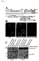
- the region of the p53 protein that interacts with mortalin was mapped to the C-terminal amino acid residues 312-352 of the p53 protein.
- a domain involved in cytoplasmic sequestration of the p53 protein is included in this region, which domain is thought to contribute to the inhibition of the migration of p53 protein into the nucleus as reported in breast cancer, neuroblastoma, and colon cancer.
- the above results illustrate a new mechanism by which cancer cells inactivate p53 protein, the product of a major cancer inhibitory gene.
- the present invention relates to methods for screening cancer therapeutic agents using the interacting regions of p53 and mortalin, and more specifically, the present invention provides:
- the present invention provides methods of screening for candidate compounds of cancer therapeutic agents by utilizing the interacting regions of p53 and mortalin protein.
- the screening of the present invention has various advantages over a screening that utilizes full-length proteins. For example, since peptides shorter than full-length proteins are used for the screening, the preparation is easier and more economical. In addition, stronger activities can be detected during the screening than in detected when the full-length proteins are used, which further enables more efficient screening. Moreover, development of drugs with high specificity is expected due to the use of interacting regions.
- One embodiment of the method for screening of the present invention uses the binding of the interacting regions of p53 and mortalin protein as an index. More specifically, the method comprises: (a) contacting a peptide comprising the amino acid residues 253 to 282 of the amino acid sequence of human mortalin protein with a peptide comprising the amino acid residues 312 to 352 of the amino acid sequence of human p53 in the presence of a test sample; (b) detecting the binding between these peptides; and (c) selecting the compound that weakens the binding between these peptides, in comparison to that occurring in the absence of the test sample.
- test sample may include, for example, cell culture supernatants, fermenting bacterial products, marine organism extracts, plant extracts, prokaryotic cell extracts, eukaryotic unicellular extracts, animal cell extracts or their libraries, purified or crude proteins, peptides, non-peptide compounds, synthetic low molecular weight compounds, and natural compounds.
- the mortalin peptide for use in the present invention, to be contacted with a test sample is not restricted in any way so long as it includes the amino acids residues 253 to 282 of the amino acid sequence of human mot-1 protein or human mot-2 protein (this region is common between human mot-1 protein and human mot-2 protein).
- the peptide comprises 600 amino acid residues or less of the amino acid residues of these proteins, more preferably 300 amino acid residues or less, and still more preferably 100 amino acid residues or less (e.g., 50 amino acid residues or less).
- the p53 peptide for use in the present invention so long as it includes the amino acid residues 312 to 352 of the amino acid sequence of human p53.
- the peptide comprises 300 amino acid residues or less of the amino acid residues of these proteins, more preferably 200 amino acid residues or less, and still more preferably 100 amino acid residues or less (e.g., 50 amino acid residues or less).
- at least one of the peptides used in the screening of the present invention is not a full-length protein. These peptides may be of natural origin, or may be prepared in accordance with known peptide synthesis methods.
- these peptides can be contacted with a test sample, for example, in the form of purified peptides, solubilized peptides, peptides bound to a carrier, or peptides fused to other peptides.
- peptides as recombinant peptides can be specifically carried out as described below.
- a gene encoding the target peptide is inserted into an exogenous gene expression vector, such as pSV2neo, pcDNA3, or pCD8, to express the gene in animal cells and so on.
- Any generally used promoters may be used for the expression including, for example, the SV40 early promoter (Rigby In Williamson (ed.), Genetic Engineering, Vol. 3, Academic Press, London, p. 83-141 (1982)), the EF-1 ⁇ promoter (Kim et al., Gene 91 , p.
- the CAG promoter Nawa et al., Gene 108 , p. 193-200 (1991)
- the RSV LTR promoter Cullen, Methods in Enzymology 152 , p. 684-704 (1987)
- the SR ⁇ promoter Takebe et al., Mol. Cell. Biol. 8 , p. 466 (1988)
- the CMV immediate early promoter Seed and Aruffo, Proc. Natl. Acad. Sci. USA 84 , p. 3365-3369 (1987)
- the SV40 late promoter Gheysen and Fiers, J. Mol. Appl. Genet. 1 , p. 385-394 (1982)
- the Adenovirus late promoter Kaufman et al., Mol. Cell. Biol. 9 , p. 946 (1989)
- the HSV TK promoter and so on.
- Examples of methods for introducing an exogenous gene into animal cells so as to allow for expression of the gene include the electroporation method (Chu, G. et al., Nucl. Acids Res. 15 , p. 1311-1326 (1987)), the calcium phosphate method (Chen, C. and Okayama, H., Mol. Cell. Biol. 7 , p. 2745-2752 (1987)), the DEAE dextran method (Lopata, M. A. et al., Nucl. Acids Res. 12 , p. 5707-5717 (1984); Sussman, D. J. and Milman, G., Mol. Cell. Biol. 4 , p.
- the peptides of the present invention can be expressed as fusion peptides, having a recognition site for a monoclonal antibody, by introducing a recognition site (epitope) for a monoclonal antibody with a pre-determined specificity into the N-terminus or C-terminus of a peptide of the present invention.
- epitope-antibody systems may be used (Experimental Medicine 13 , p. 85-90 (1995)).
- Vectors expressing fusion peptides for example, with ⁇ -galactosidase, maltose binding protein, glutathione S-transferase, green fluorescent protein (GFP), and such, via a multi-cloning site are commercially available.
- GFP green fluorescent protein
- epitopes such as polyhistidine (His-tag), influenza agglutinin HA, human c-myc, FLAG, vesicular stomatitis virus glycoprotein (VSV-GP), T7 gene 10 protein (T7-tag), human Herpes simplex virus glycoprotein (HSV-tag), and E-tag (epitope on a monoclonal phage); these epitopes can be used together with monoclonal antibodies that recognize the epitopes as an epitope-antibody system for screening proteins that bind with the peptides of the present invention (Experimental Medicine 13 , p. 85-90 (1995)).
- His-tag polyhistidine
- influenza agglutinin HA human c-myc
- FLAG vesicular stomatitis virus glycoprotein
- VSV-GP vesicular stomatitis virus glycoprotein
- T7-tag T7 gene 10 protein
- HSV-tag human Herpes simple
- the screening system provided by the present invention can be carried out as an in vitro assay system.
- Specific examples include in vitro assay systems carried out in a non-cellular system. More specifically, a screening that uses the binding of the above peptide pairs as an index can be carried out by binding either of the peptides to a support, and adding the other peptide and a test sample thereto, incubating the mixture followed by washing and detection or measurement of the binding of the other peptide to the peptide bound to the support.
- the peptides of the present invention may be used as purified or roughly purified peptides produced from cells that endogenously express the peptides, cells transformed with a DNA that encodes the peptides, and animals or plants transformed with a DNA that encodes the peptides.
- the peptides used in the present invention can be bound to a support.
- a support For example, first, one of the two peptides, which is purified or roughly purified, is bound to a support.
- the peptide may be bound to a support by conventional methods to fix peptides to a support.
- supports that may be used to bind the peptides include insoluble polysaccharides, such as agarose, dextran, and cellulose; synthetic resins, such as polystyrene, polyacrylamide, and silicon; and so on. More specifically, commercially available beads and plates manufactured from the above raw materials may also be used. For the use of beads, a column may be filled with the beads. Examples of plates that may be used include multi-well plates (96-well multi-well plates) and biosensor chips.
- peptides In order to bind peptides to a support, routine methods utilizing chemical bonding, physical adsorption, and such may be used.
- antibodies that specifically recognize the peptide can be bound to the support in advance, and then, the peptides may be bound to the support via the antibodies.
- binding of a peptide and a support can be also carried out by means of avidin/biotin binding.
- Binding between peptides is usually carried out in a buffer.
- buffers include phosphate buffer, Tris buffer, and so on.
- the condition for incubation include such conditions already well known in the art, for example, from 4°C to room temperature for 1 to 24 hours. Washing after incubation may be carried out with any solutions so long as the binding of peptides is not inhibited, and can be exemplified by buffer containing surfactants.
- An example of the surfactant is 0.05% Tween 20.
- specific binding and non-specific binding can be distinguished by incubating each of the peptides and the test sample under suitable conditions followed by washing. Either of the peptides may be bound to the support for selecting the desired compound. For example, when a peptide derived from mortalin protein is bound to a support, after fixing the peptide, a pre-mixed mixture of peptide derived from p53 protein and a test sample may be added to the support, or peptide derived from p53 protein may be added following the addition of a test sample.
- a pre-mixed mixture of peptide derived from mortalin protein and a test sample may be added to the fixed peptide, or peptide derived from mortalin protein may be added following the addition of a test sample. Then, the state of the binding between peptides can be evaluated by incubating the peptides and test sample added in the above order under suitable conditions.
- a control group may be adopted together with a "test" group, wherein test samples are contacted with the peptides.
- a negative control group that does not contain any test sample, a positive control group, or both may be used in the screening method of the present invention.
- the bound peptide may be either directly detected or measured quantitatively.
- the desired compound can be detected by comparing the result obtained for a negative control group containing no test sample and the result obtained for the group containing the test sample and/or the results obtained for a positive control group.
- results may be obtained as numerical values, and comparing those numerical values enables quantitative measurement of the activity of the desired compound.
- the desired compound may be detected by comparing the numerical value obtained with a negative control group containing no test sample with the numerical value obtained with a group including the test sample. If the resulting numerical values decrease in comparison with the negative control group, the test sample is judged to contain the desired compound.
- a quantitative measurement can be conducted by using a standard curve prepared based on numerical values obtained with a positive control group containing known amounts of compound that is known to inhibit the binding between the peptides.
- the activity of a compound to inhibit peptide binding is judged to be weak when large amount of peptides are bound; on the other hand, when the amount of bound peptide is small, the inhibitory activity of the compound to inhibit binding of the peptide is judged to be strong.
- a biosensor utilizing the surface plasmon resonance phenomenon can be used as means for detecting or measuring bound peptides.
- Biosensors utilizing the surface plasmon resonance phenomenon enable real-time observation of peptide-peptide interaction by generating a surface plasmon resonance signal, even with an extremely small amount of peptide and without requiring labeling of the peptides (e.g., BIAcore, Pharmacia).
- BIAcore e.g., Pharmacia
- the use of BIAcore or other biosensor enables real-time evaluation of the binding of the peptides of the present invention.
- one of the two peptides used in the screening of the present invention is fixed on a sensor chip; the other peptide is contacted to the sensor chip.
- the other peptide binding to the peptides on the sensor chip are detected as a change in the resonance signal.
- CM5 sensor chip (Biosensor) is activated to fix either the peptides derived from p53 or those derived from mortalin protein on the sensor chip. More specifically, the sensor chip is activated with aqueous EDC/NHS solution (containing 200 mM EDC (N-ethyl-N'-(3-dimethylaminopropyl)carbonate hydrochloride) and 50 mM NHS (N-hydroxysuccinimide)), and then, is washed with HBS buffer (containing 10 mM HEPES pH 7.4, 150 mM NaCl, 3.4 mM EDTA and 0.05% Tween 20).
- EDC N-ethyl-N'-(3-dimethylaminopropyl)carbonate hydrochloride
- NHS N-hydroxysuccinimide
- a suitable amount of the first peptide with interacting ability dissolved in HBS buffer is contacted with the sensor chip to fix the peptides thereon.
- residual activity group of the sensor chip is blocked with ethanolamine solution (1 M ethanolamine hydrochloride, pH 8.5). Then, the sensor chip is washed again with HBS buffer for the use in binding evaluation.
- a suitable amount of the second peptide (the other peptide of the two peptides used in the screening of the present invention, which was not bound to the sensor chip) dissolved in HBS buffer is injected.
- the amount of the second peptide having an ability to interact, bind, with the peptide fixed on the sensor chip is observed as an increase in the value of the resonance signal at the time of addition.
- a test sample is injected following the injection of the second peptide that interacts with the first peptide in the above binding evaluation system.
- a control group may be adopted together with the test group wherein a test sample is injected.
- the control group may include a negative control group that lacks the test sample, a positive control group that includes the test sample, or both.
- the bound peptide can be measured quantitatively as a degree of change in the resonance signal value.
- the desired compound can be detected and measured by comparing results obtained for a negative control group that contain no test sample with results obtained for a group that contain the test sample and/or results obtained for a positive control group.
- either of the two peptides may be labeled, and the label of the bound peptide may be used to detect or measure the bound peptide.
- the peptides are incubated with the test sample, and then, bound peptides are detected or measured according to the label after washing.
- a test sample and a labeled peptide is contacted with a peptide that is preferably bound to a support. After incubation and washing, the label of the peptide bound on the other peptide can be detected and measured.
- the peptides used in the present invention can be labeled by generally known methods.
- labeling substance includes radioisotopes, enzymes, fluorescent substances, biotin/avidin, and so on.
- Commercially available labeling substances can be used as such labeling substances.
- radioisotopes include 32 P, 33 P 131 I, 125 I, 3 H, 14 C, and 35 S.
- enzymes include alkaline phosphatase, horseradish peroxidase, ⁇ -galactosidase, ⁇ -glucosidase, etc.
- florescent substances include fluorescein isothiocyanate (FITC) and rhodamine.
- FITC fluorescein isothiocyanate
- rhodamine Such labeling substances are commercially available products, and can be used to label substances in accordance with known methods.
- the labeling can be carried out as described below. Namely, a solution containing either of the peptides is added to a plate and allowed to stand overnight. After washing the plate, blocking, for example, with BSA is performed to prevent non-specific peptide binding. After washing the plate again, a test sample and the other peptide, that which was not fixed on the plate yet has been labeled, is added to the plate. At the same time, a negative control group containing no test sample and/or a positive control group are provided and are also incubated. After the incubation, the plate is washed to detect or measure the bound peptide. The bound peptide may be detected or measured by liquid scintillation of a radioisotope.
- the label in the case where an enzyme is used as the label, a substrate of the enzyme is added, and the enzymatic change of the substrate, such as the generation of color, may be detected or measured using absorptiometer. Further, when a fluorescent substance is used as the label, the bound peptide may be detected or measured using fluorophotometer. Then, the desired compound can be determined by comparing these results with the numerical values obtained for a control group.
- antibodies specifically recognizing either of 'the two peptides can be used as means for detecting or measuring the bound peptide.
- the material is incubated and washed, and the bound peptide is detected or measured through a primary antibody that specifically recognizes the peptide.
- the test sample and the other peptide are contacted with the peptide fixed on a support.
- the bound peptide can be detected or measured with a primary antibody that specifically recognizes the bound peptide.
- the primary antibody is preferably labeled with a labeling substance.
- the method can be carried out as described below. Namely, a solution containing one of the two peptides is added to a plate and allowed to stand overnight. After washing the plate, blocking, for example, with BSA is conducted to prevent non-specific peptide binding. After washing the plate again, a test sample and the other peptide are added to the plate. At the same time, a negative control group containing no test sample and/or a positive control group are provided, and are also incubated.
- the bound peptide may be subjected to liquid scintillation.
- the label is an enzyme
- an enzyme substrate is added and the enzymatic change in the substrate, such as generation of color, may be detected or measured using absorptiometer.
- the bound peptide may be detected or measured using fluorophotometer. Then, the desired compound can be determined by comparing these results with the numerical values obtained for a control group.
- a primary antibody specifically recognizing peptides that are fused to the peptides used in the present invention can be utilized as means for detecting or measuring the bound peptide in the present invention.
- the two peptides used in the method of the present invention with a test sample and the other peptide (e.g., the second peptide)
- washing is conducted, and the second peptide that is bound to the first peptide is detected or measured by a primary antibody that specifically recognizes a peptide fused to the second peptide.
- the test sample and the second peptide are contacted with the first peptide preferably fixed on a support.
- the second peptide that is bound to the first peptide is detected or measured by a primary antibody that specifically recognizes the peptide fused to the second peptide.
- the primary antibody is preferably labeled with a labeling substance.
- the method is carried out as described below. Namely, a solution containing either of the two peptides (first peptide) used in the present method is added to a plate and allowed to stand overnight. After washing the plate, blocking, for example, with BSA is conducted to prevent non-specific peptide binding. After washing the plate again, a test sample and the other peptide (second peptide) fused with another peptide (third peptide) are added to the plate. At the same time, a negative control group containing no test sample and/or a positive control group are provided and are incubated.
- the plate is washed and the second peptide is detected or measured by a primary antibody that specifically recognizes the third peptide fused to the second peptide.
- the detection or measurement of the second peptide may be conducted by liquid scintillation when the primary antibody is labeled with a radioisotope.
- the label is an enzyme
- enzyme substrate is added and the enzymatic change of the substrate, such as the generation of color, may be detected or measured using absorptiometer.
- the second peptide may be detected or measured using fluorophotometer. Then, the desired compound can be determined by comparing these results with numerical values obtained for a control group.
- a primary antibody that specifically recognizes the peptides used in the present invention and a secondary antibody that specifically recognizes the primary antibody can be used as means for detecting or measuring the bound peptides.
- the mixture is incubated and washed, and the second peptide bound to the first peptide is detected or measured with primary antibodies that specifically recognize the second peptide and secondary antibodies that specifically recognize the primary antibody.
- the test sample and the second peptide are contacted with the first peptide preferably bound to a support.
- the second peptide bound to the first peptide can be detected or measured with a primary antibody that specifically recognizes the second peptide and a secondary antibody that specifically recognizes the primary antibody.
- the secondary antibody is preferably labeled with a labeling substance.
- the method can be carried out as described below. Namely, a solution containing either of the two peptides (first peptide) is added to a plate and allowed to stand overnight. After washing the plate, blocking, for example, with BSA, is performed to prevent non-specific peptide binding. After washing the plate again, a test sample and the other peptide (second peptide) are added to the plate. At the same time, a negative control group containing no test sample and/or a positive control group are provided and incubated.
- a primary antibody against the second peptide which has been added together with the test sample, is added after washing. After suitable incubation, the plate is washed, and a secondary antibody is added, which antibody specifically recognizes the primary antibody. After suitable incubation and washing of the plate, the bound peptide is detected or measured by the secondary antibody that specifically recognizes the primary antibody which, in turn, specifically recognizes the second peptide.
- the detection or measurement may be conducted by liquid scintillation when a radioisotope is used as the label.
- the label is an enzyme
- the enzyme substrate is added and the enzymatic change of the substrate, such as the generation of color, may be detected or measured using absorptiometer.
- the detection or measurement may be conducted using fluorophotometer. Then, the desired compound is selected by comparing these results with numerical values obtained for a control group.
- a primary antibody that specifically recognizes another peptide fused to one of the two peptides used in the present invention, and a secondary antibody that specifically recognizes the primary antibody can be used as means for detecting or measuring the peptide bound to the other peptide.
- the second peptide and the test sample are contacted with the first peptide that is preferably bound to a support.
- the second peptide that is bound to the first peptide can be detected or measured by a primary antibody that specifically recognizes the third peptide fused to the second peptide, and a secondary antibody that specifically recognizes the primary antibody.
- the secondary antibody is preferably labeled with a labeling substance.
- the method can be carried out as described below. Namely, a solution containing either of the two peptides (first peptide) is added to a plate and allowed to stand overnight. After washing the plate, blocking, for example, with BSA, is performed to prevent non-specific peptide binding. After washing the plate again, a test sample and the other peptide (second peptide) fused with another peptide (third peptide) are added to the plate. At the same time, a negative control group containing no test sample and/or a positive control group are provided and incubated.
- a primary antibody to the third peptide fused to the second peptide that has been added together with the test sample is added after washing. After suitable incubation, the plate is washed, and a secondary antibody is added, which antibody specifically recognizes the primary antibody. After suitably incubating and washing the plate again, the second peptide is detected or measured by a secondary antibody that specifically recognizes the primary antibody, wherein the primary antibody, in turn, specifically recognizes the third peptide fused to the second peptide.
- the detection or measurement may be performed by liquid scintillation wherein a radioisotope is used as the label.
- enzyme substrate is added and the enzymatic change in the substrate, such as the generation of color, may be detected or measured using absorptiometer.
- the bound second peptide may be detected or measured using fluorophotometer. The desired compound can then be determined by comparing these results with numerical values obtained for a control group.
- the detection or measurement is preferably carried out by Enzyme-Linked Immunoabsorbent Assay (ELISA) as described below.
- ELISA Enzyme-Linked Immunoabsorbent Assay
- a peptide (first peptide) of the peptide pairs used in the present method for example, any peptide fused with 6x His, is diluted with fixation buffer (0.1 M NaHCO 3 , 0.02% NaN 3 , pH 9.6).
- fixation buffer 0.1 M NaHCO 3 , 0.02% NaN 3 , pH 9.6
- a suitable amount of the diluted solution is then added to each well of a 96-well Immunoplate (Nunc) followed by incubation overnight at 4°C.
- washing buffer prepared to contain 0.05% Tween 20 in PBS
- washing buffer prepared to contain 0.05% Tween 20 in PBS
- 200 ⁇ l of a solution of 5% BSA (SIGMA) dissolved in PBS is added to perform blocking overnight at 4°C.
- second peptide of the present peptide pair diluted with dilution buffer (1% BSA, 0.5% Tween 20, and PBS), for example a peptide fused with FLAG, and a test sample are added and incubated for 1 hour at room temperature.
- dilution buffer 1% BSA, 0.5% Tween 20, and PBS
- alkaline phosphatase-labeled goat anti-mouse IgG antibody ZYMED
- 100 ⁇ l of alkaline phosphatase-labeled goat anti-mouse IgG antibody ZYMED
- 100 ⁇ l of color developing solution p-phenylphosphate (SIGMA) dissolved to a concentration of 1 mg/ml in substrate buffer (50 mM NaHCO 3 , 10 mM MgCl 2 , pH 9.8)
- substrate buffer 50 mM NaHCO 3 , 10 mM MgCl 2 , pH 9.8
- absorbance at 405 nm is measured using microplate reader (Model 3550, BIO-RAD). The desired compound can then be determined by comparing these results with numerical values obtained for a negative control group and/or positive control group.
- detection or measurement using an antibody of the present invention can be also conducted by using protein G or protein A in place of the secondary antibody.
- High throughput screening may be utilized for the screening method of the present invention. More specifically, high throughput screening can involve manually performing the procedure until the blocking step, and then automatically performing the subsequent reactions, with the aid of a robot, for example.
- the first peptide for example a peptide fused to 6x His, is diluted with fixation buffer (0.1 M NaHCO 3 , 0.02% NaN 3 , pH 9.6). A suitable amount of this diluted aqueous solution is then added to each well of a 96-well Immunoplate (Nunc) followed by incubation overnight at 4°C.
- fixation buffer 0.1 M NaHCO 3 , 0.02% NaN 3 , pH 9.6
- washing buffer prepared to contain 0.05% Tween 20 in PBS
- washing buffer prepared to contain 0.05% Tween 20 in PBS
- 200 ⁇ l of a solution of 5% BSA (SIGMA) dissolved in PBS is added to perform blocking overnight at 4°C.
- the blocked Immunoplate is placed in, for example, Biomek 2000 HTS system (Beckman), followed by running a system control program.
- injection and removal of solution into and from each well can be carried out by Biomek 2000 (Beckman) or Multipipette 96 well simultaneous injector (Sagian) as the injector.
- EL404 Microplate Washer Bio-Tek
- SPECTRAmax 250 plate reader Molecular Devices
- the program is set up to perform the following procedure. Namely, after washing each well three times with washing buffer, suitable amounts of test sample and the second peptide, for example; a peptide that is fused with another peptide, such as MBP (maltose bound protein), that are diluted with a dilution buffer (1% BSA, 0.5% Tween 20, and PBS) are added. At the same time, a negative control group containing no test sample and a positive control group are provided, and are incubated for 1 hour at room temperature.
- suitable amounts of test sample and the second peptide for example; a peptide that is fused with another peptide, such as MBP (maltose bound protein), that are diluted with a dilution buffer (1% BSA, 0.5% Tween 20, and PBS) are added.
- a negative control group containing no test sample and a positive control group are provided, and are incubated for 1 hour at room temperature.
- color developing solution p-nitrophenyl phosphate (SIGMA) dissolved to a concentration of 1 mg/ml in substrate buffer (50 mM NaHCO 3 , 10 mM MgCl 2 , pH 9.8)
- substrate buffer 50 mM NaHCO 3 , 10 mM MgCl 2 , pH 9.8
- absorbance at 405 nm may be measured using microplate reader or Biomek plate reader (Beckman/Molecular Devices). Then, the desired compound is determined by comparing these results with numerical values obtained for the control groups.
- Labeling substances include radioisotopes, enzymes, and fluorescent substances. Commercially available labeling substances can be used for these labeling substances. Examples of radioisotopes include 32 P, 33 P, 131 I, 125 I, 3 H, 14 C, and 35 S. Examples of enzymes include alkaline phosphatase, horseradish peroxidase, ⁇ -galactosidase, and ⁇ -glucosidase. Examples of fluorescent substances include fluorescein isothiocyanate (FITC) and rhodamine.
- FITC fluorescein isothiocyanate
- detection of peptide binding can be carried out by detecting and/or measuring changes in the amount of reporter gene expressed, whose expression is activated in response to the binding of the peptides.
- reporter genes include luciferase, ⁇ -galactosidase, HIS3 gene, chloramphenicol-acetyl transferase (CAT), and green fluorescence protein (GFP) gene.
- the peptides expressed in cells may be peptides fused with other peptides.
- the other peptide used for the fusion with the peptides expressed in cells may be any peptide so long as it can be used in the screening method of the present invention, it is preferably a transcription regulatory factor.
- DNAs encoding two different peptides (the binding of the two peptide is measured), respectively, wherein the peptides respectively are fused with subunits of a transcription regulatory factor that is a heterodimer known to activate transcription of a reporter gene by binding to a DNA, can be constructed and introduced into expression vectors to transform cells.
- the two peptides form a heteromer, i.e. the transcription regulatory factor, binding to the DNA to activate the reporter gene.
- the binding between the peptides is inhibited and as a result, the subunits of the transcription regulatory factor are unable to form the heteromer needed to induce transcription of the reporter gene.
- the desired compound can be detected or measured by determining the change in the amount of reporter gene expressed.
- a two-hybrid system (“Two-hybrid system” Fields, S., and Sternglanz, R., Trends. Genet. (1994) 10, 286-292) or three-hybrid system can be used.
- the hybrid system can be constructed by conventional methods or using commercially available kits.
- Examples of commercially available two-hybrid system kits include MATCHMAKER Two-Hybrid System, Mammalian MATCHMAKER Two-Hybrid Assay Kit (both made by CLONTECH), and HybriZAP Two-Hybrid Vector System (Stratagene).
- a two-hybrid system can be constructed as described below. Namely, a gene that codes for either of the two peptides (e.g., the first peptide) and a gene that codes for the DNA binding domain of LexA are linked to produce an expression vector.
- the expression plasmid is constructed by inserting the desired gene fragment into a yeast two-hybrid expression plasmid pBTM116 (Vojtek, A.B., et al., Cell (1993) 74, 205-214).
- an expression vector is prepared by linking a gene encoding the other peptide (e.g., the second peptide) to a gene that encodes the transcription activating domain of GAL4.
- Yeast two-hybrid expression plasmid pGAD10 (Clontech) can be used for the expression vector.
- another embodiment of the screening method of the present invention relates to a method that uses a construct containing the p53 responsive promoter and a reporter gene expressed in response to the activation of the promoter. More specifically, the method comprises the steps of:
- a vector that expresses human mortalin, a vector that expresses human p53, and a vector that expresses a reporter gene in response to the expression of human p53 are constructed and introduced into cells.
- the human mortalin expressed by the vector is not particularly restricted and any peptide can be used so long as the peptide includes the domain for interaction with p53 (i.e., amino acid residues 253 to 282).
- the peptide contains 600 amino acid residues or less of the amino acid residues of these proteins, more preferably 300 amino acid residues or less, and even more preferably 100 amino acid residues or less (for example, 50 amino acid residues or less).
- the human p53 expressed by the vector so long as it includes the domain that interacts with mortalin (i.e., amino acid residues 312 to 352).
- the peptide contains 300 amino acid residues or less of the amino acid residues of these proteins, more preferably 200 amino acid residues or less, and even more preferably 100 amino acid residues or less (for example, 50 amino acid residues or less).
- vectors can be constructed, for example, by inserting a DNA encoding these peptides into a mammalian expression vector, such as pSG5 or pcDNA3.
- construction of a vector wherein the reporter gene is functionally bound downstream of human p53 responsive promoter can be carried out by inserting human p53 responsive promoter into an expression vector comprising the reporter gene.
- the phrase "functionally bound” refers to the binding of a promoter and reporter gene to enable expression of the reporter gene in response to the activation of human p53 responsive promoter.
- human p53 responsive promoter that can be used in the present invention is described in the literature (El-Deiry, W.S., et al., (1993) Cell, 75, 817-825).
- any reporter gene may be used so long as its expression can be detected; examples include ⁇ -gal gene, the CAT gene, and the luciferase gene.
- preferable examples include MCF-7 cells and NIH3T3 cells.
- a test sample is contacted with the cells prepared as above.
- the test sample examples include cell culture supernatants, fermenting microorganism products, marine organism extracts, plant extracts, prokaryotic cell extracts, eukaryotic unicellilar extracts, animal cell extracts or their libraries, purified or crude proteins, peptides, non-peptide compounds, synthetic low molecular weight compounds, and naturally-occurring compounds.
- the activity of the reporter in the cells is detected. Detection of the reporter activity can be carried out corresponding to the type of reporter gene by methods known to those skilled in the art. As a result of detecting the reporter activity, if the reporter activity detected in the cells contacted with the test sample increases significantly as compared with the result obtained without a test sample (control), the used test compound serves as a candidate for a compound that inhibits the interaction between mortalin protein and p53.
- the present invention also relates to a kit for carrying out the above described screening method, which comprises: (a) a peptide comprising the amino acid residues 253 to 282 of the amino acid sequence of human mot-1 protein or human mot-2 protein; and (b) a peptide comprising the amino acid residues 312 to 352 of the amino acid sequence of human p53.
- the present invention also relates to a kit for carrying out the above described screening method, which comprises: (a) a vector comprising a DNA encoding a peptide comprising the amino acid residues 253 to 282 of the amino acid sequence of human mot-1 protein or human mot-2 protein; (b) a vector comprising a DNA encoding a peptide that comprises the amino acid residues 312 to 352 of the amino acid sequence of human p53, which peptide has the function to activate human p53 responsive promoter; and (c) a vector comprising a reporter gene functionally bound downstream of human p53 responsive promoter.
- the kit may additionally contain cells for introducing these vectors.
- the present invention also provides compounds isolated by the screening methods of the present invention and pharmaceutical applications thereof.
- p53 is established as a tumor suppressor gene (Levine, A.J. (1997) Cell, 88, 323-331; Metz, T. et al. (1995) Cell, 82, 29-36). Consequently, compounds isolated by the screening methods of the present invention serve as important candidates for cancer therapeutic agents.
- the compounds isolated by the screening methods of the present invention can be used as pharmaceutical agents, for example, for humans, mice, rats, guinea pigs, rabbits, chickens, cats, dogs, sheep, pigs, cows, monkeys, baboons, or chimpanzees, by directly administering the protein or the isolated compound itself, and they may be also administered in the form of a pharmaceutical composition by formulating in accordance with known pharmaceutical methods.
- a pharmaceutical composition may be formulated by mixing into a unit dose form required for typically observed pharmaceutical preparation by suitably combining with a pharmacologically acceptable carrier or medium, specific examples of which include sterilized water or physiological saline, vegetable oil, emulsifier, suspension agent, surfactant, stabilizer, flavoring, excipient, vehicle, antiseptic, binder, and so on. The amount of active ingredient in these preparations is adjusted so to obtain a suitable volume within a specified range.
- additives that can be mixed into tablets and capsules include binders, such as gelatin, cornstarch, gum tragacanth, and gum arabic; excipients, such as crystalline cellulose; swelling agents, such as cornstarch, gelatin, and alginic acid; lubricants, such as magnesium stearate; sweeteners, such as sucrose, lactose, and saccharin; and flavorings, such as peppermint, Gaultheria adenothrix oil, and cherry flavoring.
- binders such as gelatin, cornstarch, gum tragacanth, and gum arabic
- excipients such as crystalline cellulose
- swelling agents such as cornstarch, gelatin, and alginic acid
- lubricants such as magnesium stearate
- sweeteners such as sucrose, lactose, and saccharin
- flavorings such as peppermint, Gaultheria adenothrix oil, and cherry flavoring.
- Aseptic compositions for injection
- aqueous solutions suitable for injection include physiological saline, and isotonic liquids containing glucose or other adjuvants, such as D-sorbitol, D-mannose, D-mannitol, and sodium chloride, and these adjuvants may be used in combination with suitable dissolving assistants, examples of which include alcohols, such as ethanol; polyalcohols including propylene glycol and polyethylene glycol; and nonionic surfactants, such as Polysorbate 80 (TM) and HCO-50.
- suitable dissolving assistants examples of which include alcohols, such as ethanol; polyalcohols including propylene glycol and polyethylene glycol; and nonionic surfactants, such as Polysorbate 80 (TM) and HCO-50.
- oily liquids examples include sesame oil and soybean oil, and these may be used in combination with dissolving adjuvants, such as benzyl benzoate and benzyl alcohol.
- dissolving adjuvants such as benzyl benzoate and benzyl alcohol.
- buffers such as phosphate buffer and sodium acetate buffer
- analgesics such as procaine hydrochloride
- stabilizers such as benzyl alcohol and phenol
- antioxidants may also be blended.
- the prepared injection liquid is normally filled into suitable ampules.
- Administration to patients can be carried out by methods known to those skilled in the art, for example, by intraarterial injection, intravenous injection or subcutaneous injection; or intranasal, transbronchial, intramuscular, percutaneous or oral administration.
- the dosage varies depending on the body weight and age of the patient, administration method, and so on, an appropriate dosage can be suitably selected by a person skilled in the art.
- gene therapy may also be carried out by incorporating the DNA into a vector for gene therapy.
- the dosage and administration method vary according to the body weight, age, and symptoms of the patient, they can be suitably selected by a person skilled in the art.
- the dosage of the compound of the present invention varies according to the symptoms, in the case of oral administration, the dosage for adult (the body weight as 60 kg) typically ranges from about 0.1 to 100 mg per day, preferably about 1.0 to 50 mg per day, and more preferably about 1.0 to 20 mg per day.
- the single dosage in the form of an injection preparation for example, for an adult (a body weight of 60 kg) is normally about 0.01 to 30 mg per day, preferably about 0.1 to 20 mg per day, and more preferably about 0.1 to 10 mg per day; and the administration is preferably carried out by intravenous injection.
- an amount of the compound converted for the amount per 60 kg of body weight or the amount converted for the amount per body surface area can be administered.
- Figure 1 depicts photographs demonstrating the results of detecting in vitro binding of mot-1 and mot-2 to p53.
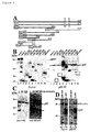
- Figure 2 depicts drawings ((B) - (D) are photographs) demonstrating the mapping of the p53 binding domain of mortalins.
- Figure 3 depicts illustration, graph, and photograph (panel C) demonstrating the result of in vivo p53-mediated reporter assays in the presence of various mortalin mutants.
- Figure 4 depicts an illustration and photographs (panels (B) and (C)) demonstrating the mortalin binding domain of p53.
- [ 35 S]-labeled p53 protein was obtained by subjecting to in vitro translation reaction using rabbit reticulocyte lysate (Stratagene) for 1 hour in the presence of L-[ 35 S]methionine. After separating the translation products by SDS-PAGE, the translation reaction products were confirmed by autoradiography.
- the in vitro translated p53 protein was purified by immunoprecipitation with anti-p53 antibodies (PAb 421, PAb 1620 (Calbiochem), and PAb 122 (Boehringer-Mannheim)), and was used in the subsequent in vitro binding assays described below.
- the resulting His-tagged recombinant mortalin proteins (0.5 to 1 ⁇ g) were mixed with the previously prepared in vitro translated [ 35 S]-labeled p53 protein in presence of either in 1 to 2 mg of bovine serum albumin or COS7 cell lysate in Nonidet P-40 solution (400 ⁇ l) , followed by the addition of anti-mortalin antibody (anti-mtHSP70, Affinity Bioreagents) or anti-p53 antibody (CM1, Novo Castra), and were allowed to incubate for 1 to 2 hours at 4°C.
- the formed immunocomplexes were mixed for 30 minutes with 20 ⁇ l Protein-A/G Sepharose (Gibco) and recovered by centrifugal separation.
- proteins eluted by heating the recovered Sepharose beads to 95°C in a sample buffer containing SDS was separated by SDS-PAGE and transferred to nitrocellulose membrane. Then, p53 protein was detected by autoradiography; mortalin and deletion mutants thereof were detected by Western blotting with anti-His antibody and ECL-chemiluminescence (Amersham).
- the region of the mortalin molecule involved in the binding with p53 was considered to be present within the region of amino acid residues 253-282.
- the present inventor next expressed the V5-tagged mortalin deletion mutants expressing amino acid residues 250-435, 252-679 (not shown), and 1-435 in COS 7 cells. Their presence in p58-immunocomplexes was analyzed by Western blotting with anti-V5 tag antibody ( Figure 2D).
- each of the mortalin deletion mutants was detected in the p53 immunocomplexes (as a single band for the fragment of amino acid residues 250-435, and as multiple bands (degraded products) for the fragment of amino acid residues 1-435).
- mot-1 protein did not have any effect on the ability of p53-dependent transcriptional activation, despite the structural difference of only two amino acid residues. Any of the deletions of each functional domain or the regions that were different between mot-1 and mot-2 conferred the inhibition of p53-dependent transcriptional activation similar to mot-2 protein to the mutant mortalin protein.
- Mutant V618M or R624G wherein one of the two amino acid residues that differed between mot-1 and mot-2 was made the same amino acid residue as mot-2, exhibited the inhibition of p53-dependent transcriptional activation similar to mot-2, and the effect was particularly strongly observed in mutant R624G.
- a definitely stronger ability to inhibit p53-dependent transcriptional activation as compared to mot-2 was also observed in the mutant, mut 12 ( ⁇ 233-251), wherein the EF hand and Hsp70 homologous domain continuing from EF hand had been deleted, among all the deletion mutants.
- the activity of mutant mut16 ( ⁇ 651-679), wherein the C terminus of the region containing the two amino acid residues that were different between mot-1 and mot-2 had been deleted, was comparable to the activity of mot-1.
- mot-1 or mut16 protein did not have a significant effect on the p53-dependent transcriptional activity similar to the transfection of mouse embryonic fibroblasts (Table 1)
- suppression of the p53-dependent transcriptional activation by mot-1 protein having the mutation V618M was significantly inhibited by intracellular injection of anti-mortalin neutralizing antibody (Table 1).
- mot-2 protein was suggested to prevent translocation of p53 to the nucleus through the interaction with p53 protein via the region of amino acid residues 253-282, thus inhibiting the transcriptional activation by p53.
- the substitution of two amino acids in mot-1 was suggested to inhibit the interaction with p53 protein by changes in the secondary structure of the protein or interaction with a different protein (mot-1-p53 binding inhibitory factor).
- methods of screening for cancer therapeutic agents which utilize the interacting region of p53 and mortalin.
- specific cancer therapeutic agents can be screened more efficiently and more economically.
Landscapes
- Health & Medical Sciences (AREA)
- Life Sciences & Earth Sciences (AREA)
- Chemical & Material Sciences (AREA)
- Engineering & Computer Science (AREA)
- Molecular Biology (AREA)
- Immunology (AREA)
- Biomedical Technology (AREA)
- Organic Chemistry (AREA)
- Hematology (AREA)
- Biochemistry (AREA)
- Urology & Nephrology (AREA)
- Medicinal Chemistry (AREA)
- General Health & Medical Sciences (AREA)
- Cell Biology (AREA)
- Toxicology (AREA)
- Microbiology (AREA)
- Genetics & Genomics (AREA)
- Physics & Mathematics (AREA)
- General Physics & Mathematics (AREA)
- Pathology (AREA)
- Food Science & Technology (AREA)
- Zoology (AREA)
- Gastroenterology & Hepatology (AREA)
- Biophysics (AREA)
- Analytical Chemistry (AREA)
- Proteomics, Peptides & Aminoacids (AREA)
- Biotechnology (AREA)
- Bioinformatics & Cheminformatics (AREA)
- Tropical Medicine & Parasitology (AREA)
- Investigating Or Analysing Biological Materials (AREA)
- Peptides Or Proteins (AREA)
- Measuring Or Testing Involving Enzymes Or Micro-Organisms (AREA)
Abstract
Description

Claims (7)
- A method of screening for candidate compounds of cancer therapeutic agents comprising the steps of:(a) contacting a peptide comprising amino acid residues 253 to 282 of an amino acid sequence of human mot-1 protein or human mot-2 protein with a peptide comprising amino acid residues 312 to 352 of an amino acid sequence of human p53 in the presence of a test sample;(b) detecting binding between said peptides; and(c) selecting the compound that weakens the binding between said peptides, in comparison to binding that occurs in the absence of the test sample.
- A method of screening for candidate compounds of cancer therapeutic agents comprising the steps of:(a) contacting a test sample with a cell transfected with the following vectors:(i) a vector containing a DNA encoding a peptide comprising amino acid residues 253 to 282 of an amino acid sequence of human mot-1 protein or human mot-2 protein;(ii) a vector containing a DNA encoding a peptide comprising amino acid residues 312 to 352 of an amino acid sequence of human p53, wherein said peptide activates human p53 responsive promoter; and(iii) a vector containing human p53 responsive promoter and a reporter gene functionally bound downstream thereof;(b) detecting the activity of said reporter gene within the cell; and(c) selecting the compound that increases the reporter expression, in comparison to that occurring in the absence of the test sample.
- A kit for screening candidate compounds of cancer therapeutic agents comprising:(a) a peptide comprising amino acid residues 253 to 282 of an amino acid sequence of human mot-1 protein or human mot-2 protein; and(b) a peptide comprising amino acid residues 312 to 352 of an amino acid sequence of human p53.
- A kit for screening candidate compounds of cancer therapeutic agents comprising:(a) a vector containing a DNA encoding a peptide comprising amino acid residues 253 to 282 of an amino acid sequence of human mot-1 protein or human mot-2 protein;(b) a vector containing a DNA encoding a peptide comprising amino acid residues 312 to 352 of an amino acid sequence of human p53, wherein said peptide activates human p53 responsive promoter; and(c) a vector containing human p53 responsive promoter and a reporter gene functionally bound downstream thereof.
- A compound isolated by the method of claim 1 or 2.
- A pharmaceutical composition comprising the compound of claim 5 as an active ingredient.
- The pharmaceutical composition of claim 6, wherein the composition is a cancer therapeutic agent.
Applications Claiming Priority (3)
| Application Number | Priority Date | Filing Date | Title |
|---|---|---|---|
| JP35754599 | 1999-12-16 | ||
| JP35754599 | 1999-12-16 | ||
| PCT/JP2000/008910 WO2001044807A1 (en) | 1999-12-16 | 2000-12-15 | METHOD OF SCREENING REMEDY FOR CANCER WITH THE USE OF INTERACTION DOMAINS OF p53 AND MORTALIN |
Publications (3)
| Publication Number | Publication Date |
|---|---|
| EP1243923A1 true EP1243923A1 (en) | 2002-09-25 |
| EP1243923A4 EP1243923A4 (en) | 2004-05-06 |
| EP1243923B1 EP1243923B1 (en) | 2006-03-08 |
Family
ID=18454687
Family Applications (1)
| Application Number | Title | Priority Date | Filing Date |
|---|---|---|---|
| EP00981763A Expired - Lifetime EP1243923B1 (en) | 1999-12-16 | 2000-12-15 | METHOD OF SCREENING REMEDY FOR CANCER WITH THE USE OF INTERACTION DOMAINS OF p53 AND MORTALIN |
Country Status (6)
| Country | Link |
|---|---|
| US (1) | US20030087263A1 (en) |
| EP (1) | EP1243923B1 (en) |
| AT (1) | ATE319996T1 (en) |
| AU (1) | AU1891801A (en) |
| DE (1) | DE60026576T2 (en) |
| WO (1) | WO2001044807A1 (en) |
Cited By (2)
| Publication number | Priority date | Publication date | Assignee | Title |
|---|---|---|---|---|
| EP1536230A1 (en) * | 2003-11-28 | 2005-06-01 | Deutsches Rheuma-Forschungszentrum Berlin | Flow cytometric, particle-based binding assay |
| US11105809B2 (en) * | 2012-10-09 | 2021-08-31 | Ramot At Tel-Aviv University Ltd. | Methods and kits for predicting prognosis of cancer using soluble mortalin in blood |
Families Citing this family (13)
| Publication number | Priority date | Publication date | Assignee | Title |
|---|---|---|---|---|
| WO2004079368A2 (en) | 2003-03-08 | 2004-09-16 | Auvation Ltd | Markers for colorectal cancer |
| JP2006089471A (en) | 2004-08-26 | 2006-04-06 | National Institute Of Advanced Industrial & Technology | Use of anti-mortalin 2 antibodies and functional nucleic acids in the treatment of cancer |
| US8889632B2 (en) | 2007-01-31 | 2014-11-18 | Dana-Farber Cancer Institute, Inc. | Stabilized p53 peptides and uses thereof |
| EP2508531B1 (en) | 2007-03-28 | 2016-10-19 | President and Fellows of Harvard College | Stitched polypeptides |
| SI2603600T1 (en) | 2010-08-13 | 2019-04-30 | Aileron Therapeutics, Inc. | Peptidomimetic macrocycles |
| TW201806968A (en) | 2011-10-18 | 2018-03-01 | 艾利倫治療公司 | Peptidomimetic macrocycles |
| HK1205454A1 (en) | 2012-02-15 | 2015-12-18 | Aileron Therapeutics, Inc. | Triazole-crosslinked and thioether-crosslinked peptidomimetic macrocycles |
| KR102112373B1 (en) | 2012-02-15 | 2020-05-18 | 에일러론 테라퓨틱스 인코포레이티드 | Peptidomimetic macrocycles |
| WO2014071241A1 (en) | 2012-11-01 | 2014-05-08 | Aileron Therapeutics, Inc. | Disubstituted amino acids and methods of preparation and use thereof |
| BR112017005598A2 (en) | 2014-09-24 | 2017-12-12 | Aileron Therapeutics Inc | peptidomimetic macrocycles and their uses |
| JP2018503595A (en) | 2014-09-24 | 2018-02-08 | エルロン・セラピューティクス・インコーポレイテッドAileron Therapeutics,Inc. | Peptidomimetic macrocycle and its preparation |
| WO2016154058A1 (en) | 2015-03-20 | 2016-09-29 | Aileron Therapeutics, Inc. | Peptidomimetic macrocycles and uses thereof |
| US10023613B2 (en) | 2015-09-10 | 2018-07-17 | Aileron Therapeutics, Inc. | Peptidomimetic macrocycles as modulators of MCL-1 |
Family Cites Families (3)
| Publication number | Priority date | Publication date | Assignee | Title |
|---|---|---|---|---|
| AU7445800A (en) * | 1999-09-27 | 2001-04-30 | Chugai Seiyaku Kabushiki Kaisha | Method of prolonging normal cell life span |
| EP1287116A2 (en) * | 2000-05-17 | 2003-03-05 | Geron Corporation | Neural progenitor cell populations |
| JP2001354564A (en) * | 2000-06-14 | 2001-12-25 | Chugai Bunshi Igaku Kenkyusho:Kk | An inhibitor of the interaction between mot-2 protein and p53 protein, comprising a cationic rhodocyanine dye derivative as an active ingredient |
-
2000
- 2000-12-15 DE DE60026576T patent/DE60026576T2/en not_active Expired - Fee Related
- 2000-12-15 EP EP00981763A patent/EP1243923B1/en not_active Expired - Lifetime
- 2000-12-15 AT AT00981763T patent/ATE319996T1/en not_active IP Right Cessation
- 2000-12-15 WO PCT/JP2000/008910 patent/WO2001044807A1/en not_active Ceased
- 2000-12-15 AU AU18918/01A patent/AU1891801A/en not_active Abandoned
- 2000-12-15 US US10/149,814 patent/US20030087263A1/en not_active Abandoned
Cited By (2)
| Publication number | Priority date | Publication date | Assignee | Title |
|---|---|---|---|---|
| EP1536230A1 (en) * | 2003-11-28 | 2005-06-01 | Deutsches Rheuma-Forschungszentrum Berlin | Flow cytometric, particle-based binding assay |
| US11105809B2 (en) * | 2012-10-09 | 2021-08-31 | Ramot At Tel-Aviv University Ltd. | Methods and kits for predicting prognosis of cancer using soluble mortalin in blood |
Also Published As
| Publication number | Publication date |
|---|---|
| DE60026576T2 (en) | 2007-01-18 |
| US20030087263A1 (en) | 2003-05-08 |
| DE60026576D1 (en) | 2006-05-04 |
| WO2001044807A1 (en) | 2001-06-21 |
| EP1243923A4 (en) | 2004-05-06 |
| ATE319996T1 (en) | 2006-03-15 |
| EP1243923B1 (en) | 2006-03-08 |
| AU1891801A (en) | 2001-06-25 |
Similar Documents
| Publication | Publication Date | Title |
|---|---|---|
| EP1243923B1 (en) | METHOD OF SCREENING REMEDY FOR CANCER WITH THE USE OF INTERACTION DOMAINS OF p53 AND MORTALIN | |
| Grönholm et al. | Homotypic and heterotypic interaction of the neurofibromatosis 2 tumor suppressor protein merlin and the ERM protein ezrin | |
| Morris et al. | Myosin VI binds to and localises with Dab2, potentially linking receptor‐mediated endocytosis and the actin cytoskeleton | |
| Chen et al. | F-actin and myosin II binding domains in supervillin | |
| US5411860A (en) | Amplification of human MDM2 gene in human tumors | |
| Dong et al. | The proteasome α-subunit XAPC7 interacts specifically with Rab7 and late endosomes | |
| Cuppen et al. | The zyxin-related protein TRIP6 interacts with PDZ motifs in the adaptor protein RIL and the protein tyrosine phosphatase PTP-BL | |
| Izawa et al. | ERBIN associates with p0071, an armadillo protein, at cell‐cell junctions of epithelial cells | |
| Peck et al. | Structure and function of human Vps20 and Snf7 proteins | |
| Drees et al. | Characterization of the interaction between zyxin and members of the Ena/vasodilator-stimulated phosphoprotein family of proteins | |
| JPH10506525A (en) | Inhibition of binding between MDM2 and p53 protein and its therapeutic application | |
| JP2004500807A (en) | Screening method for candidate drugs | |
| US20090155928A1 (en) | Modulating robo: ligand interactions | |
| US7176035B2 (en) | Protein-protein interactions and methods for identifying interacting proteins and the amino acid sequence at the site of interaction | |
| Van Ham et al. | Cloning and characterization of mCRIP2, a mouse LIM‐only protein that interacts with PDZ domain IV of PTP‐BL | |
| US20100136014A1 (en) | GIPs, a Family of Polypeptides with Transcription Factor Activity that Interact with Goodpasture Antigen Binding Protein | |
| US20060094101A1 (en) | Mk2 interacting proteins | |
| JP4638354B2 (en) | Gene encoding G protein-coupled receptor and gene product thereof | |
| US20030207791A1 (en) | Materials and methods relating to the treatment of leukaemias | |
| US20100125131A1 (en) | Gene Encoding A Guanine Nucleotide Exchange Factor And The Gene Product Thereof | |
| JPWO2001044807A1 (en) | Screening method for cancer therapeutic agents utilizing the interaction region between p53 and mortalin | |
| EP0856582A2 (en) | Inhibitor protein of transcription factors and its DNA | |
| US7214513B2 (en) | Transcriptional regulatory factor | |
| AU2416600A (en) | Exocytosis pathway proteins and methods of use | |
| JP2003501079A (en) | Gene encoding NADE (P75NTR-related cell death executive substance) and use thereof |
Legal Events
| Date | Code | Title | Description |
|---|---|---|---|
| PUAI | Public reference made under article 153(3) epc to a published international application that has entered the european phase |
Free format text: ORIGINAL CODE: 0009012 |
|
| 17P | Request for examination filed |
Effective date: 20020716 |
|
| AK | Designated contracting states |
Kind code of ref document: A1 Designated state(s): AT BE CH CY DE DK ES FI FR GB GR IE IT LI LU MC NL PT SE TR |
|
| AX | Request for extension of the european patent |
Free format text: AL;LT;LV;MK;RO;SI |
|
| A4 | Supplementary search report drawn up and despatched |
Effective date: 20040318 |
|
| 17Q | First examination report despatched |
Effective date: 20040708 |
|
| GRAP | Despatch of communication of intention to grant a patent |
Free format text: ORIGINAL CODE: EPIDOSNIGR1 |
|
| GRAS | Grant fee paid |
Free format text: ORIGINAL CODE: EPIDOSNIGR3 |
|
| GRAA | (expected) grant |
Free format text: ORIGINAL CODE: 0009210 |
|
| AK | Designated contracting states |
Kind code of ref document: B1 Designated state(s): AT BE CH CY DE DK ES FI FR GB GR IE IT LI LU MC NL PT SE TR |
|
| PG25 | Lapsed in a contracting state [announced via postgrant information from national office to epo] |
Ref country code: IT Free format text: LAPSE BECAUSE OF FAILURE TO SUBMIT A TRANSLATION OF THE DESCRIPTION OR TO PAY THE FEE WITHIN THE PRESCRIBED TIME-LIMIT;WARNING: LAPSES OF ITALIAN PATENTS WITH EFFECTIVE DATE BEFORE 2007 MAY HAVE OCCURRED AT ANY TIME BEFORE 2007. THE CORRECT EFFECTIVE DATE MAY BE DIFFERENT FROM THE ONE RECORDED. Effective date: 20060308 Ref country code: LI Free format text: LAPSE BECAUSE OF FAILURE TO SUBMIT A TRANSLATION OF THE DESCRIPTION OR TO PAY THE FEE WITHIN THE PRESCRIBED TIME-LIMIT Effective date: 20060308 Ref country code: AT Free format text: LAPSE BECAUSE OF FAILURE TO SUBMIT A TRANSLATION OF THE DESCRIPTION OR TO PAY THE FEE WITHIN THE PRESCRIBED TIME-LIMIT Effective date: 20060308 Ref country code: FI Free format text: LAPSE BECAUSE OF FAILURE TO SUBMIT A TRANSLATION OF THE DESCRIPTION OR TO PAY THE FEE WITHIN THE PRESCRIBED TIME-LIMIT Effective date: 20060308 Ref country code: CH Free format text: LAPSE BECAUSE OF FAILURE TO SUBMIT A TRANSLATION OF THE DESCRIPTION OR TO PAY THE FEE WITHIN THE PRESCRIBED TIME-LIMIT Effective date: 20060308 Ref country code: BE Free format text: LAPSE BECAUSE OF FAILURE TO SUBMIT A TRANSLATION OF THE DESCRIPTION OR TO PAY THE FEE WITHIN THE PRESCRIBED TIME-LIMIT Effective date: 20060308 Ref country code: NL Free format text: LAPSE BECAUSE OF FAILURE TO SUBMIT A TRANSLATION OF THE DESCRIPTION OR TO PAY THE FEE WITHIN THE PRESCRIBED TIME-LIMIT Effective date: 20060308 |
|
| REG | Reference to a national code |
Ref country code: GB Ref legal event code: FG4D |
|
| REG | Reference to a national code |
Ref country code: CH Ref legal event code: EP |
|
| REG | Reference to a national code |
Ref country code: IE Ref legal event code: FG4D |
|
| REF | Corresponds to: |
Ref document number: 60026576 Country of ref document: DE Date of ref document: 20060504 Kind code of ref document: P |
|
| PG25 | Lapsed in a contracting state [announced via postgrant information from national office to epo] |
Ref country code: DK Free format text: LAPSE BECAUSE OF FAILURE TO SUBMIT A TRANSLATION OF THE DESCRIPTION OR TO PAY THE FEE WITHIN THE PRESCRIBED TIME-LIMIT Effective date: 20060608 Ref country code: SE Free format text: LAPSE BECAUSE OF FAILURE TO SUBMIT A TRANSLATION OF THE DESCRIPTION OR TO PAY THE FEE WITHIN THE PRESCRIBED TIME-LIMIT Effective date: 20060608 |
|
| PG25 | Lapsed in a contracting state [announced via postgrant information from national office to epo] |
Ref country code: ES Free format text: LAPSE BECAUSE OF FAILURE TO SUBMIT A TRANSLATION OF THE DESCRIPTION OR TO PAY THE FEE WITHIN THE PRESCRIBED TIME-LIMIT Effective date: 20060619 |
|
| PG25 | Lapsed in a contracting state [announced via postgrant information from national office to epo] |
Ref country code: PT Free format text: LAPSE BECAUSE OF FAILURE TO SUBMIT A TRANSLATION OF THE DESCRIPTION OR TO PAY THE FEE WITHIN THE PRESCRIBED TIME-LIMIT Effective date: 20060808 |
|
| NLV1 | Nl: lapsed or annulled due to failure to fulfill the requirements of art. 29p and 29m of the patents act | ||
| ET | Fr: translation filed | ||
| REG | Reference to a national code |
Ref country code: CH Ref legal event code: PL |
|
| PG25 | Lapsed in a contracting state [announced via postgrant information from national office to epo] |
Ref country code: IE Free format text: LAPSE BECAUSE OF NON-PAYMENT OF DUE FEES Effective date: 20061215 |
|
| PGFP | Annual fee paid to national office [announced via postgrant information from national office to epo] |
Ref country code: DE Payment date: 20061218 Year of fee payment: 7 |
|
| PGFP | Annual fee paid to national office [announced via postgrant information from national office to epo] |
Ref country code: GB Payment date: 20061221 Year of fee payment: 7 |
|
| PG25 | Lapsed in a contracting state [announced via postgrant information from national office to epo] |
Ref country code: MC Free format text: LAPSE BECAUSE OF NON-PAYMENT OF DUE FEES Effective date: 20061231 |
|
| PLBE | No opposition filed within time limit |
Free format text: ORIGINAL CODE: 0009261 |
|
| STAA | Information on the status of an ep patent application or granted ep patent |
Free format text: STATUS: NO OPPOSITION FILED WITHIN TIME LIMIT |
|
| 26N | No opposition filed |
Effective date: 20061211 |
|
| PG25 | Lapsed in a contracting state [announced via postgrant information from national office to epo] |
Ref country code: GR Free format text: LAPSE BECAUSE OF FAILURE TO SUBMIT A TRANSLATION OF THE DESCRIPTION OR TO PAY THE FEE WITHIN THE PRESCRIBED TIME-LIMIT Effective date: 20060609 |
|
| PGFP | Annual fee paid to national office [announced via postgrant information from national office to epo] |
Ref country code: FR Payment date: 20061212 Year of fee payment: 7 |
|
| PG25 | Lapsed in a contracting state [announced via postgrant information from national office to epo] |
Ref country code: TR Free format text: LAPSE BECAUSE OF FAILURE TO SUBMIT A TRANSLATION OF THE DESCRIPTION OR TO PAY THE FEE WITHIN THE PRESCRIBED TIME-LIMIT Effective date: 20060308 Ref country code: LU Free format text: LAPSE BECAUSE OF NON-PAYMENT OF DUE FEES Effective date: 20061215 |
|
| GBPC | Gb: european patent ceased through non-payment of renewal fee |
Effective date: 20071215 |
|
| PG25 | Lapsed in a contracting state [announced via postgrant information from national office to epo] |
Ref country code: DE Free format text: LAPSE BECAUSE OF NON-PAYMENT OF DUE FEES Effective date: 20080701 |
|
| REG | Reference to a national code |
Ref country code: FR Ref legal event code: ST Effective date: 20081020 |
|
| PG25 | Lapsed in a contracting state [announced via postgrant information from national office to epo] |
Ref country code: CY Free format text: LAPSE BECAUSE OF FAILURE TO SUBMIT A TRANSLATION OF THE DESCRIPTION OR TO PAY THE FEE WITHIN THE PRESCRIBED TIME-LIMIT Effective date: 20060308 |
|
| PG25 | Lapsed in a contracting state [announced via postgrant information from national office to epo] |
Ref country code: GB Free format text: LAPSE BECAUSE OF NON-PAYMENT OF DUE FEES Effective date: 20071215 |
|
| PG25 | Lapsed in a contracting state [announced via postgrant information from national office to epo] |
Ref country code: FR Free format text: LAPSE BECAUSE OF NON-PAYMENT OF DUE FEES Effective date: 20071231 |