EP1243651A1 - Method of detecting sexual differentiation disruptor - Google Patents
Method of detecting sexual differentiation disruptor Download PDFInfo
- Publication number
- EP1243651A1 EP1243651A1 EP00985852A EP00985852A EP1243651A1 EP 1243651 A1 EP1243651 A1 EP 1243651A1 EP 00985852 A EP00985852 A EP 00985852A EP 00985852 A EP00985852 A EP 00985852A EP 1243651 A1 EP1243651 A1 EP 1243651A1
- Authority
- EP
- European Patent Office
- Prior art keywords
- gene
- medaka
- sex
- seq
- female
- Prior art date
- Legal status (The legal status is an assumption and is not a legal conclusion. Google has not performed a legal analysis and makes no representation as to the accuracy of the status listed.)
- Granted
Links
- 238000000034 method Methods 0.000 title claims description 67
- 230000001568 sexual effect Effects 0.000 title claims description 26
- 230000004069 differentiation Effects 0.000 title claims description 10
- 108090000623 proteins and genes Proteins 0.000 claims abstract description 151
- 241000276569 Oryzias latipes Species 0.000 claims description 45
- 230000000694 effects Effects 0.000 claims description 32
- 230000014509 gene expression Effects 0.000 claims description 32
- 230000012447 hatching Effects 0.000 claims description 21
- 108020004999 messenger RNA Proteins 0.000 claims description 14
- 239000002773 nucleotide Substances 0.000 claims description 11
- 125000003729 nucleotide group Chemical group 0.000 claims description 11
- 239000000049 pigment Substances 0.000 claims description 11
- 239000000598 endocrine disruptor Substances 0.000 claims description 10
- 231100000049 endocrine disruptor Toxicity 0.000 claims description 9
- 108091092195 Intron Proteins 0.000 claims description 8
- 108091034117 Oligonucleotide Proteins 0.000 claims description 8
- 108091028043 Nucleic acid sequence Proteins 0.000 claims description 7
- 241000276423 Fundulus heteroclitus Species 0.000 abstract 1
- 239000000523 sample Substances 0.000 description 40
- 239000000047 product Substances 0.000 description 28
- 108020004414 DNA Proteins 0.000 description 27
- 239000002299 complementary DNA Substances 0.000 description 22
- 108091032973 (ribonucleotides)n+m Proteins 0.000 description 14
- 238000009396 hybridization Methods 0.000 description 14
- XLYOFNOQVPJJNP-UHFFFAOYSA-N water Substances O XLYOFNOQVPJJNP-UHFFFAOYSA-N 0.000 description 14
- 235000013601 eggs Nutrition 0.000 description 12
- 238000012360 testing method Methods 0.000 description 12
- 239000000126 substance Substances 0.000 description 11
- 108020004635 Complementary DNA Proteins 0.000 description 10
- 230000003321 amplification Effects 0.000 description 10
- 238000006243 chemical reaction Methods 0.000 description 10
- 231100000507 endocrine disrupting Toxicity 0.000 description 10
- 238000003199 nucleic acid amplification method Methods 0.000 description 10
- 238000001514 detection method Methods 0.000 description 9
- IAZDPXIOMUYVGZ-UHFFFAOYSA-N Dimethylsulphoxide Chemical compound CS(C)=O IAZDPXIOMUYVGZ-UHFFFAOYSA-N 0.000 description 8
- 108020004707 nucleic acids Proteins 0.000 description 8
- 102000039446 nucleic acids Human genes 0.000 description 8
- 150000007523 nucleic acids Chemical class 0.000 description 8
- VOXZDWNPVJITMN-ZBRFXRBCSA-N 17β-estradiol Chemical compound OC1=CC=C2[C@H]3CC[C@](C)([C@H](CC4)O)[C@@H]4[C@@H]3CCC2=C1 VOXZDWNPVJITMN-ZBRFXRBCSA-N 0.000 description 7
- 235000019688 fish Nutrition 0.000 description 7
- 241000251468 Actinopterygii Species 0.000 description 6
- 230000004720 fertilization Effects 0.000 description 5
- 210000003765 sex chromosome Anatomy 0.000 description 5
- MJVIZWIQHKRBPI-UHFFFAOYSA-N 1-(aziridin-1-yl)but-3-en-2-ol Chemical compound C=CC(O)CN1CC1 MJVIZWIQHKRBPI-UHFFFAOYSA-N 0.000 description 4
- 108090000790 Enzymes Proteins 0.000 description 4
- 102000004190 Enzymes Human genes 0.000 description 4
- 230000002159 abnormal effect Effects 0.000 description 4
- 239000003153 chemical reaction reagent Substances 0.000 description 4
- 230000003247 decreasing effect Effects 0.000 description 4
- 210000004602 germ cell Anatomy 0.000 description 4
- UYTPUPDQBNUYGX-UHFFFAOYSA-N guanine Chemical compound O=C1NC(N)=NC2=C1N=CN2 UYTPUPDQBNUYGX-UHFFFAOYSA-N 0.000 description 4
- 229940088597 hormone Drugs 0.000 description 4
- 239000005556 hormone Substances 0.000 description 4
- 238000001727 in vivo Methods 0.000 description 4
- 230000035772 mutation Effects 0.000 description 4
- 238000003752 polymerase chain reaction Methods 0.000 description 4
- 102000040650 (ribonucleotides)n+m Human genes 0.000 description 3
- 230000004544 DNA amplification Effects 0.000 description 3
- 238000000018 DNA microarray Methods 0.000 description 3
- 108700024394 Exon Proteins 0.000 description 3
- 238000012408 PCR amplification Methods 0.000 description 3
- 238000003556 assay Methods 0.000 description 3
- 238000009395 breeding Methods 0.000 description 3
- 230000001488 breeding effect Effects 0.000 description 3
- 210000004027 cell Anatomy 0.000 description 3
- 210000000349 chromosome Anatomy 0.000 description 3
- OPTASPLRGRRNAP-UHFFFAOYSA-N cytosine Chemical group NC=1C=CNC(=O)N=1 OPTASPLRGRRNAP-UHFFFAOYSA-N 0.000 description 3
- 238000001962 electrophoresis Methods 0.000 description 3
- 230000007613 environmental effect Effects 0.000 description 3
- 229960005309 estradiol Drugs 0.000 description 3
- 239000012634 fragment Substances 0.000 description 3
- 238000012252 genetic analysis Methods 0.000 description 3
- 238000000338 in vitro Methods 0.000 description 3
- 238000011534 incubation Methods 0.000 description 3
- 238000012544 monitoring process Methods 0.000 description 3
- 238000002360 preparation method Methods 0.000 description 3
- 238000003757 reverse transcription PCR Methods 0.000 description 3
- 108700025491 zebrafish zp3b Proteins 0.000 description 3
- YBJHBAHKTGYVGT-ZKWXMUAHSA-N (+)-Biotin Chemical compound N1C(=O)N[C@@H]2[C@H](CCCCC(=O)O)SC[C@@H]21 YBJHBAHKTGYVGT-ZKWXMUAHSA-N 0.000 description 2
- 241000195649 Chlorella <Chlorellales> Species 0.000 description 2
- 241000195628 Chlorophyta Species 0.000 description 2
- 241000252233 Cyprinus carpio Species 0.000 description 2
- 102100037008 Factor in the germline alpha Human genes 0.000 description 2
- 101710123263 Factor in the germline alpha Proteins 0.000 description 2
- 206010021033 Hypomenorrhoea Diseases 0.000 description 2
- 241001465754 Metazoa Species 0.000 description 2
- 108020000284 NAD(P)H dehydrogenase (quinone) Proteins 0.000 description 2
- 241000276565 Oryziinae Species 0.000 description 2
- 108010044843 Peptide Initiation Factors Proteins 0.000 description 2
- 102000005877 Peptide Initiation Factors Human genes 0.000 description 2
- 102000028391 RNA cap binding Human genes 0.000 description 2
- 108091000106 RNA cap binding Proteins 0.000 description 2
- 241000700159 Rattus Species 0.000 description 2
- 102000040739 Secretory proteins Human genes 0.000 description 2
- 108091058545 Secretory proteins Proteins 0.000 description 2
- FAPWRFPIFSIZLT-UHFFFAOYSA-M Sodium chloride Chemical compound [Na+].[Cl-] FAPWRFPIFSIZLT-UHFFFAOYSA-M 0.000 description 2
- 238000002105 Southern blotting Methods 0.000 description 2
- 108091023040 Transcription factor Proteins 0.000 description 2
- 102000040945 Transcription factor Human genes 0.000 description 2
- 241000269370 Xenopus <genus> Species 0.000 description 2
- 238000007792 addition Methods 0.000 description 2
- 238000011161 development Methods 0.000 description 2
- 230000018109 developmental process Effects 0.000 description 2
- 108010037421 eIF-4 Proteins 0.000 description 2
- 230000002124 endocrine Effects 0.000 description 2
- 210000002149 gonad Anatomy 0.000 description 2
- 238000005462 in vivo assay Methods 0.000 description 2
- 239000000463 material Substances 0.000 description 2
- 238000002493 microarray Methods 0.000 description 2
- 239000000203 mixture Substances 0.000 description 2
- 238000007857 nested PCR Methods 0.000 description 2
- 230000031787 nutrient reservoir activity Effects 0.000 description 2
- 210000000287 oocyte Anatomy 0.000 description 2
- 102000004169 proteins and genes Human genes 0.000 description 2
- 239000008213 purified water Substances 0.000 description 2
- 230000001850 reproductive effect Effects 0.000 description 2
- 238000012216 screening Methods 0.000 description 2
- 230000035945 sensitivity Effects 0.000 description 2
- 239000002689 soil Substances 0.000 description 2
- 239000000243 solution Substances 0.000 description 2
- 239000002904 solvent Substances 0.000 description 2
- RWQNBRDOKXIBIV-UHFFFAOYSA-N thymine Chemical compound CC1=CNC(=O)NC1=O RWQNBRDOKXIBIV-UHFFFAOYSA-N 0.000 description 2
- 210000001519 tissue Anatomy 0.000 description 2
- 230000005945 translocation Effects 0.000 description 2
- 238000003911 water pollution Methods 0.000 description 2
- 239000001052 yellow pigment Substances 0.000 description 2
- GFFGJBXGBJISGV-UHFFFAOYSA-N Adenine Chemical compound NC1=NC=NC2=C1N=CN2 GFFGJBXGBJISGV-UHFFFAOYSA-N 0.000 description 1
- 229930024421 Adenine Natural products 0.000 description 1
- 241000269350 Anura Species 0.000 description 1
- 241000252229 Carassius auratus Species 0.000 description 1
- ZAMOUSCENKQFHK-UHFFFAOYSA-N Chlorine atom Chemical compound [Cl] ZAMOUSCENKQFHK-UHFFFAOYSA-N 0.000 description 1
- 102000004163 DNA-directed RNA polymerases Human genes 0.000 description 1
- 108090000626 DNA-directed RNA polymerases Proteins 0.000 description 1
- SHIBSTMRCDJXLN-UHFFFAOYSA-N Digoxigenin Natural products C1CC(C2C(C3(C)CCC(O)CC3CC2)CC2O)(O)C2(C)C1C1=CC(=O)OC1 SHIBSTMRCDJXLN-UHFFFAOYSA-N 0.000 description 1
- 201000009273 Endometriosis Diseases 0.000 description 1
- 241000282412 Homo Species 0.000 description 1
- 241000283973 Oryctolagus cuniculus Species 0.000 description 1
- 206010033128 Ovarian cancer Diseases 0.000 description 1
- 206010061535 Ovarian neoplasm Diseases 0.000 description 1
- 238000009004 PCR Kit Methods 0.000 description 1
- 206010060862 Prostate cancer Diseases 0.000 description 1
- 208000000236 Prostatic Neoplasms Diseases 0.000 description 1
- 208000002495 Uterine Neoplasms Diseases 0.000 description 1
- 108010090932 Vitellogenins Proteins 0.000 description 1
- 101710201545 ZP domain-containing protein Proteins 0.000 description 1
- JLCPHMBAVCMARE-UHFFFAOYSA-N [3-[[3-[[3-[[3-[[3-[[3-[[3-[[3-[[3-[[3-[[3-[[5-(2-amino-6-oxo-1H-purin-9-yl)-3-[[3-[[3-[[3-[[3-[[3-[[5-(2-amino-6-oxo-1H-purin-9-yl)-3-[[5-(2-amino-6-oxo-1H-purin-9-yl)-3-hydroxyoxolan-2-yl]methoxy-hydroxyphosphoryl]oxyoxolan-2-yl]methoxy-hydroxyphosphoryl]oxy-5-(5-methyl-2,4-dioxopyrimidin-1-yl)oxolan-2-yl]methoxy-hydroxyphosphoryl]oxy-5-(6-aminopurin-9-yl)oxolan-2-yl]methoxy-hydroxyphosphoryl]oxy-5-(6-aminopurin-9-yl)oxolan-2-yl]methoxy-hydroxyphosphoryl]oxy-5-(6-aminopurin-9-yl)oxolan-2-yl]methoxy-hydroxyphosphoryl]oxy-5-(6-aminopurin-9-yl)oxolan-2-yl]methoxy-hydroxyphosphoryl]oxyoxolan-2-yl]methoxy-hydroxyphosphoryl]oxy-5-(5-methyl-2,4-dioxopyrimidin-1-yl)oxolan-2-yl]methoxy-hydroxyphosphoryl]oxy-5-(4-amino-2-oxopyrimidin-1-yl)oxolan-2-yl]methoxy-hydroxyphosphoryl]oxy-5-(5-methyl-2,4-dioxopyrimidin-1-yl)oxolan-2-yl]methoxy-hydroxyphosphoryl]oxy-5-(5-methyl-2,4-dioxopyrimidin-1-yl)oxolan-2-yl]methoxy-hydroxyphosphoryl]oxy-5-(6-aminopurin-9-yl)oxolan-2-yl]methoxy-hydroxyphosphoryl]oxy-5-(6-aminopurin-9-yl)oxolan-2-yl]methoxy-hydroxyphosphoryl]oxy-5-(4-amino-2-oxopyrimidin-1-yl)oxolan-2-yl]methoxy-hydroxyphosphoryl]oxy-5-(4-amino-2-oxopyrimidin-1-yl)oxolan-2-yl]methoxy-hydroxyphosphoryl]oxy-5-(4-amino-2-oxopyrimidin-1-yl)oxolan-2-yl]methoxy-hydroxyphosphoryl]oxy-5-(6-aminopurin-9-yl)oxolan-2-yl]methoxy-hydroxyphosphoryl]oxy-5-(4-amino-2-oxopyrimidin-1-yl)oxolan-2-yl]methyl [5-(6-aminopurin-9-yl)-2-(hydroxymethyl)oxolan-3-yl] hydrogen phosphate Polymers Cc1cn(C2CC(OP(O)(=O)OCC3OC(CC3OP(O)(=O)OCC3OC(CC3O)n3cnc4c3nc(N)[nH]c4=O)n3cnc4c3nc(N)[nH]c4=O)C(COP(O)(=O)OC3CC(OC3COP(O)(=O)OC3CC(OC3COP(O)(=O)OC3CC(OC3COP(O)(=O)OC3CC(OC3COP(O)(=O)OC3CC(OC3COP(O)(=O)OC3CC(OC3COP(O)(=O)OC3CC(OC3COP(O)(=O)OC3CC(OC3COP(O)(=O)OC3CC(OC3COP(O)(=O)OC3CC(OC3COP(O)(=O)OC3CC(OC3COP(O)(=O)OC3CC(OC3COP(O)(=O)OC3CC(OC3COP(O)(=O)OC3CC(OC3COP(O)(=O)OC3CC(OC3COP(O)(=O)OC3CC(OC3COP(O)(=O)OC3CC(OC3CO)n3cnc4c(N)ncnc34)n3ccc(N)nc3=O)n3cnc4c(N)ncnc34)n3ccc(N)nc3=O)n3ccc(N)nc3=O)n3ccc(N)nc3=O)n3cnc4c(N)ncnc34)n3cnc4c(N)ncnc34)n3cc(C)c(=O)[nH]c3=O)n3cc(C)c(=O)[nH]c3=O)n3ccc(N)nc3=O)n3cc(C)c(=O)[nH]c3=O)n3cnc4c3nc(N)[nH]c4=O)n3cnc4c(N)ncnc34)n3cnc4c(N)ncnc34)n3cnc4c(N)ncnc34)n3cnc4c(N)ncnc34)O2)c(=O)[nH]c1=O JLCPHMBAVCMARE-UHFFFAOYSA-N 0.000 description 1
- 229960000643 adenine Drugs 0.000 description 1
- 239000011543 agarose gel Substances 0.000 description 1
- 238000004458 analytical method Methods 0.000 description 1
- 102000001307 androgen receptors Human genes 0.000 description 1
- 108010080146 androgen receptors Proteins 0.000 description 1
- 230000003388 anti-hormonal effect Effects 0.000 description 1
- 239000007864 aqueous solution Substances 0.000 description 1
- 230000015572 biosynthetic process Effects 0.000 description 1
- 229960002685 biotin Drugs 0.000 description 1
- 235000020958 biotin Nutrition 0.000 description 1
- 239000011616 biotin Substances 0.000 description 1
- 230000008859 change Effects 0.000 description 1
- 239000007795 chemical reaction product Substances 0.000 description 1
- 239000000460 chlorine Substances 0.000 description 1
- 229910052801 chlorine Inorganic materials 0.000 description 1
- 239000013611 chromosomal DNA Substances 0.000 description 1
- 230000000295 complement effect Effects 0.000 description 1
- 150000001875 compounds Chemical class 0.000 description 1
- 238000010276 construction Methods 0.000 description 1
- 230000013005 courtship behavior Effects 0.000 description 1
- 230000009089 cytolysis Effects 0.000 description 1
- 229940104302 cytosine Drugs 0.000 description 1
- 238000012217 deletion Methods 0.000 description 1
- 230000037430 deletion Effects 0.000 description 1
- 238000013461 design Methods 0.000 description 1
- QONQRTHLHBTMGP-UHFFFAOYSA-N digitoxigenin Natural products CC12CCC(C3(CCC(O)CC3CC3)C)C3C11OC1CC2C1=CC(=O)OC1 QONQRTHLHBTMGP-UHFFFAOYSA-N 0.000 description 1
- SHIBSTMRCDJXLN-KCZCNTNESA-N digoxigenin Chemical compound C1([C@@H]2[C@@]3([C@@](CC2)(O)[C@H]2[C@@H]([C@@]4(C)CC[C@H](O)C[C@H]4CC2)C[C@H]3O)C)=CC(=O)OC1 SHIBSTMRCDJXLN-KCZCNTNESA-N 0.000 description 1
- 239000012153 distilled water Substances 0.000 description 1
- 239000003814 drug Substances 0.000 description 1
- 210000002969 egg yolk Anatomy 0.000 description 1
- 210000002257 embryonic structure Anatomy 0.000 description 1
- 231100000613 environmental toxicology Toxicity 0.000 description 1
- 102000015694 estrogen receptors Human genes 0.000 description 1
- 108010038795 estrogen receptors Proteins 0.000 description 1
- ZMMJGEGLRURXTF-UHFFFAOYSA-N ethidium bromide Chemical compound [Br-].C12=CC(N)=CC=C2C2=CC=C(N)C=C2[N+](CC)=C1C1=CC=CC=C1 ZMMJGEGLRURXTF-UHFFFAOYSA-N 0.000 description 1
- 229960005542 ethidium bromide Drugs 0.000 description 1
- 238000000605 extraction Methods 0.000 description 1
- 239000013505 freshwater Substances 0.000 description 1
- 239000011521 glass Substances 0.000 description 1
- 230000036541 health Effects 0.000 description 1
- 229940094991 herring sperm dna Drugs 0.000 description 1
- 230000003054 hormonal effect Effects 0.000 description 1
- 238000009399 inbreeding Methods 0.000 description 1
- 208000000509 infertility Diseases 0.000 description 1
- 231100000535 infertility Toxicity 0.000 description 1
- 230000036512 infertility Effects 0.000 description 1
- 230000002401 inhibitory effect Effects 0.000 description 1
- 238000003780 insertion Methods 0.000 description 1
- 230000037431 insertion Effects 0.000 description 1
- 210000004164 leucophore Anatomy 0.000 description 1
- 239000003446 ligand Substances 0.000 description 1
- 238000004519 manufacturing process Methods 0.000 description 1
- 230000035800 maturation Effects 0.000 description 1
- 238000002844 melting Methods 0.000 description 1
- 230000008018 melting Effects 0.000 description 1
- 230000029052 metamorphosis Effects 0.000 description 1
- 230000011278 mitosis Effects 0.000 description 1
- 238000012986 modification Methods 0.000 description 1
- 230000004048 modification Effects 0.000 description 1
- 238000010369 molecular cloning Methods 0.000 description 1
- 239000008188 pellet Substances 0.000 description 1
- 239000013612 plasmid Substances 0.000 description 1
- 239000002243 precursor Substances 0.000 description 1
- 230000008569 process Effects 0.000 description 1
- 239000011541 reaction mixture Substances 0.000 description 1
- 102000005962 receptors Human genes 0.000 description 1
- 108020003175 receptors Proteins 0.000 description 1
- 230000010539 reproductive behavior Effects 0.000 description 1
- 238000011160 research Methods 0.000 description 1
- 108091008146 restriction endonucleases Proteins 0.000 description 1
- 150000003839 salts Chemical class 0.000 description 1
- 239000013535 sea water Substances 0.000 description 1
- 238000011896 sensitive detection Methods 0.000 description 1
- 230000008771 sex reversal Effects 0.000 description 1
- 239000011780 sodium chloride Substances 0.000 description 1
- NLJMYIDDQXHKNR-UHFFFAOYSA-K sodium citrate Chemical compound O.O.[Na+].[Na+].[Na+].[O-]C(=O)CC(O)(CC([O-])=O)C([O-])=O NLJMYIDDQXHKNR-UHFFFAOYSA-K 0.000 description 1
- 239000001509 sodium citrate Substances 0.000 description 1
- 239000007790 solid phase Substances 0.000 description 1
- 238000010186 staining Methods 0.000 description 1
- 238000006467 substitution reaction Methods 0.000 description 1
- 239000000758 substrate Substances 0.000 description 1
- 230000004083 survival effect Effects 0.000 description 1
- 238000003786 synthesis reaction Methods 0.000 description 1
- 239000008399 tap water Substances 0.000 description 1
- 235000020679 tap water Nutrition 0.000 description 1
- 229940113082 thymine Drugs 0.000 description 1
- 108090000721 thyroid hormone receptors Proteins 0.000 description 1
- 102000004217 thyroid hormone receptors Human genes 0.000 description 1
- 230000009772 tissue formation Effects 0.000 description 1
- 229910021642 ultra pure water Inorganic materials 0.000 description 1
- 239000012498 ultrapure water Substances 0.000 description 1
- 206010046766 uterine cancer Diseases 0.000 description 1
Classifications
-
- C—CHEMISTRY; METALLURGY
- C12—BIOCHEMISTRY; BEER; SPIRITS; WINE; VINEGAR; MICROBIOLOGY; ENZYMOLOGY; MUTATION OR GENETIC ENGINEERING
- C12N—MICROORGANISMS OR ENZYMES; COMPOSITIONS THEREOF; PROPAGATING, PRESERVING, OR MAINTAINING MICROORGANISMS; MUTATION OR GENETIC ENGINEERING; CULTURE MEDIA
- C12N15/00—Mutation or genetic engineering; DNA or RNA concerning genetic engineering, vectors, e.g. plasmids, or their isolation, preparation or purification; Use of hosts therefor
- C12N15/09—Recombinant DNA-technology
- C12N15/11—DNA or RNA fragments; Modified forms thereof; Non-coding nucleic acids having a biological activity
-
- C—CHEMISTRY; METALLURGY
- C07—ORGANIC CHEMISTRY
- C07K—PEPTIDES
- C07K14/00—Peptides having more than 20 amino acids; Gastrins; Somatostatins; Melanotropins; Derivatives thereof
- C07K14/435—Peptides having more than 20 amino acids; Gastrins; Somatostatins; Melanotropins; Derivatives thereof from animals; from humans
- C07K14/46—Peptides having more than 20 amino acids; Gastrins; Somatostatins; Melanotropins; Derivatives thereof from animals; from humans from vertebrates
- C07K14/461—Peptides having more than 20 amino acids; Gastrins; Somatostatins; Melanotropins; Derivatives thereof from animals; from humans from vertebrates from fish
Definitions
- the present invention relates to a method for rapidly assessing an endocrine-disrupting activity of a chemical substance as well as a series of techniques connected therewith in fields of medicine, pharmacology, environmental study and sitology.
- Endocrine disruptors (often called environmental hormones) collectively refer to chemical substances released into the environment for which hormone-like activities or anti-hormonal activities have been found. Altered reproductive potential (in particular, conversion of male into female), decreased reproductive potential, decreased hatchability, decreased survival rate of offspring, abnormal reproductive behavior and the like have been reported to be resulted from the influences of endocrine disruptors on the ecosystem of wild animals. Decreased number of sperms, endometriosis, infertility, ovarian cancer, uterine cancer, prostatic cancer and the like have been suspected to be resulted from the influences of endocrine disruptors on human health, although they have not been demonstrated.
- Known methods for determining endocrine disrupting activities are classified into two groups, i.e., in vitro methods and in vivo methods.
- Examples of the methods in the former group include a method in which an activity of binding to estrogen receptor, androgen receptor or thyroid hormone receptor is measured, and a method in which an activity of inhibiting a hormone synthesis enzyme system is measured.
- Examples of the methods in the latter group include a method in which production of various hormones and abnormal tissue formation in individuals at different postnatal days are determined, a method in which abnormal metamorphosis in a frog is determined, and a method in which abnormal maturation in a fish is determined (Analytical Chemistry, 70(15):528A-532A (1998)).
- the in vitro method is advantageous because it is sensitive, and it can be used to assay a number of test samples in a short time.
- it cannot be determined whether or not endocrine is actually disrupted using the in vitro method because it only determines an activity of binding to a receptor or the like.
- the actual influence on endocrine is directly examined using the in vivo method in which animals such as rats, frogs, fishes or the like are used.
- animals such as rats, frogs, fishes or the like are used.
- the in vivo method has drawbacks because, for example, its sensitivity is low, it requires complicated operation and, if a number of samples are to be examined, it requires a long time.
- a system in which conversion of male into female is monitored to assess an endocrine-disrupting activity has been constructed.
- the monitoring is carried out by determining the expression of a fish female-specific yolk precursor protein vitellogenin in males using an anti-vitellogenin antibody.
- an anti-vitellogenin antibody it is difficult to deal with a number of test samples at a time using the system.
- the sensitivity of the system in which adult fishes are used for the assessment is supposed to be lower than that of an assessment system in which fries are used because fries are relatively subject to disrupting activities.
- the system has further problems because it requires a wide space for breeding and a long breeding period.
- anti-vitellogenin antibodies specific for the respective fishes to be assessed are required for the system.
- Disruption of sexual differentiation can be examined by determining both the genotypic- sex and the phenotypic sex of an individual and comparing them each other.
- a system for assaying a disrupting activity in which sex reversal is used as an index has been proposed.
- a fry or an egg is bred while administering an environmental hormone thereto, and the phenotypic sex associated with secondary sex characteristics is then determined.
- the genotypic sex can be determined by using a PCR (Shinoyama, A.
- breeding for at least one month is required in order to determine the phenotypic sex based on the shape of a fin associated with secondary sex characteristics. Furthermore, skill is required for the determination. It is also difficult to deal with a number of test samples using this method. As described above, in fact, the in vivo assay requires a long time from the start of assessment, and it is difficult to assay a number of test samples at a time. However, such an in vivo assay is necessary for assessing chemical substances or water environment, or monitoring water pollution. In addition, construction of a rapid and accurate assay system has been desired.
- Examination of an endocrine-disrupting activity may provide an index for assessing the influence of a chemical substance on humans, for determining the influence on living bodies upon its release into the environment or on the ecosystem, or for monitoring water pollution.
- the prior art has drawbacks as described above. Thus, a sensitive and rapid method for determining an endocrine-disrupting activity has been desired.
- the present inventors have successfully isolated and identified genes expressed specifically in phenotypic females of medaka during early development for the first time.
- the present inventors have found that disruption of sexual differentiation can be examined by rapidly determining the phenotypic sex for a fry of medaka using the gene, and comparing the phenotypic sex with the genotypic sex.
- the present inventors have also found that an endocrine-disrupting activity of a sample can be rapidly determined using the method.
- the present inventors have successfully constructed a method for rapidly assessing endocrine-disrupting activities of chemical substances, samples of water environment (lakes, marshes, rivers, seas, etc.) or the like in vivo for a number of samples at a time. Thus, the present invention has been completed.
- the present invention is outlined as follows.
- the first aspect of the present invention relates to a gene expressed in a female-specific manner depending on its phenotypic sex, which has a nucleotide sequence selected from the group consisting of the nucleotide sequences of SEQ ID NOS:1 to 21. It also relates to a gene which hybridizes with said sequence under stringent conditions as well as a gene which has said sequence as well as an intron or introns being inserted.
- the second aspect of the present invention relates to a method for assessing a sexual differentiation-disrupting activity of a sample, the method comprising:
- the third aspect of the present invention relates to a method for detecting an endocrine disruptor, comprising assessing a sexual differentiation-disrupting activity by the method of the second aspect.
- the fourth aspect of the present invention relates to an oligonucleotide for detecting the gene of the first aspect.
- the fifth aspect of the present invention relates to a kit for assessing a sexual differentiation-disrupting activity by the method of the second aspect.
- the sixth aspect of the present invention relates to a kit for detecting an endocrine disruptor by the method of the third aspect.
- the present invention may be applied to any samples without limitation, including naturally occurring or artificially synthesized chemical compounds. Such a substance may be subjected to the method of the present invention in an isolated form or in a mixture.
- the method of the present invention is applicable to a sample from the environment such as river water, soil or the like.
- a medaka (Oryzias latipes) is used as an organism according to the method of the present invention. It is widely used as a material for studying genes because it is small and easy to handle, it releases a lot of eggs upon spawning, its generation time is short (about three months), and a genetically homogeneous strain can be established by inbreeding.
- a medaka can be bred in distilled water in a 96-well microplate for about one week after hatching. Since feeding is unnecessary, secondary factors such as a disrupting activity due to a bait can be excluded upon assessment.
- medaka there is no specific limitation concerning the medaka to be used according to the present invention.
- a wild type medaka or a medaka strain of which the genotypic sex is linked to pigment expression such as d-rR or Qurt may be used.
- Qurt is a medaka strain heterozygous for the leucophore free (lf) locus, which is closely linked to sex.
- a female (X lf /X lf ) individual of Qurt is colorless, whereas a male (X lf /Y + ) individual is yellow as a result of pigment expression. The yellow color can be observed for an egg. Therefore, Qurt can be preferably used according to the present invention because its genotypic sex can be readily determined by microscopically examining the egg without extracting the DNA (Zoological Science, 15:123-126, 1998).
- the genotypic sex can be determined by genetic analysis of sex chromosomes.
- the genotypic sex of medaka is fixed upon fertilization depending on the combination of sex chromosomes as follows: female in case of X/X; and male in case of X/Y.
- the genotypic sex can be determined by genetic analysis of sex chromosomes.
- the analysis can be carried out by hybridization using a probe that hybridizes with a gene on the sex chromosomes or a PCR using primers that can be used to amplify a gene on the sex chromosomes. If such a method is used to determine the genotypic sex, it is also necessary to prepare both DNA and RNA from an individual.
- RNA for determining the phenotypic sex as described below.
- the head portion of a fry is cut off.
- the remaining body portion which contains a gonad and the like is used to determine the expression of a gene that is specifically expressed depending on the phenotypic sex as described below.
- DNA extracted from the head portion is used to determine the genotypic sex.
- DNA and RNA may be prepared simultaneously using QIAGEN RNA/DNA System (QIAGEN).
- DNAs can be prepared simultaneously from a number of test samples in a 96-well microplate using DNeasy 96 Tissue Kit (QIAGEN).
- the difference in pigment expression specific for the genotypic sex can be recognized on the second day after fertilization.
- the genotypic sex can be determined for an egg by detecting the pigment without preparing DNA.
- a gene that is expressed in a medaka in a phenotypic sex-specific manner is used for determining the phenotypic sex according to the method of the present invention.
- a medaka gene expressed in a phenotypic female-specific manner is used.
- a gene that is specifically expressed within five days after hatching is preferable for assessment at an early stage.
- the gene may exist on a chromosome with an intron or introns being inserted.
- the present invention also encompasses such a gene having an intron or introns being inserted.
- examples of such genes include a gene having the nucleotide sequence of SEQ ID NO:22 (the sequence of SEQ ID NO:1 with introns being inserted) or a gene having the nucleotide sequence of SEQ ID NO:23 (the sequence of SEQ ID NO:8 with introns being inserted).
- the sexual differentiation of a medaka is first manifested as a phenomenon that the number of germ cells in a female is about twice as many as that in a male upon hatching, i.e., on about tenth day after fertilization (Satoh, N., Egami, N., J. Embryol. Exp. Morph., 28:385-395, 1972). This is because mitosis is initiated immediately after hatching in a portion of germ cells in a female whereas germ cells in a male do not divide until two months after hatching. It is possible to identify genes expressed in a female-specific manner at an early development stage using the difference in sexual differentiation in germ cell line as an index.
- Difference in gene expression between a male and a female of medaka can be examined using subtractive hybridization. Genotypic male and female are separated each other before hatching. RNAs are extracted from the both after hatching. Then, a gene expressed in a female-specific manner can be isolated using subtractive hybridization. If the medaka strain Qurt is used, genotypic male and female can be readily distinguished before hatching on the second day after fertilization using the expression of the pigment gene as an index.
- a genomic gene corresponding to each gene can be isolated by screening a genomic library according to a known method using the thus obtained gene as a probe.
- the gene can be detected using an oligonucleotide designed based on the nucleotide sequence of the gene.
- the oligonucleotides for detecting the gene according to the present invention include, but are not limited to, primers that can be used to amplify the gene or a portion thereof according a gene amplification method, and a probe that is hybridizable with the gene under stringent conditions.
- gene amplification methods examples include, but are not limited to, PCR, SDA, NASBA and ICAN (WO 00/56877).
- a primer or a probe can be designed at will based on the nucleotide sequence of the gene.
- a sequence is selected upon designing such that the primer or the probe does not form a secondary structure within the molecule, and attention is paid such that the melting temperature (Tm value) for the primer or the probe and the corresponding template is set at an appropriate temperature.
- the Tm value can be estimated, for example, as the sum of the product of the number of adenine and thymine (A+T) residues multiplied by 2(°C) and the product of the number of G+C residues multiplied by 4(°C), i.e., [(A+T) x 2 + (G+C) x 4].
- the chain length of the probe is preferably 15 bases or more, more preferably 18 bases or more in order to avoid nonspecific hybridization.
- a primer of 15 to 40 bases in length can be used.
- a primer of 17 to 30 bases in length can be preferably used.
- a primer such that the ratio of cytosine (C) and guanine (G) around the 3'-terminus becomes high.
- a commercially available software for primer designing such as OLIGOTM Primer Analysis software (Takara Shuzo) may be used for designing a primer.
- the primer or the probe may have a mutation such as deletion, substitution, insertion or addition of a nucleotide (or nucleotides) in a portion of the sequence as long as it can be used to detect the gene.
- a primer it is preferable not to include a mutation or, if any, to minimize mutations around the 3'-terminus of the primer because such a mutation greatly influences the efficiency of primer extension reaction.
- An appropriate sequence unrelated to the nucleotide sequence of the gene e.g., a promoter sequence recognized by an RNA polymerase
- the primer or the probe may be appropriately modified. Addition of a ligand such as biotin or digoxigenin, or a fluorescent substance to a primer or a probe facilitates the detection of the amplification reaction product.
- a ligand such as biotin or digoxigenin, or a fluorescent substance
- a product of a gene amplification reaction using the primers can be detected by subjecting a portion of the reaction mixture after the amplification reaction to electrophoresis, and then staining the DNAs with ethidium bromide.
- An amplification product can be detected without electrophoresis by utilizing hybridization. If a modified primer is used, a detection method suitable for the modification can be used.
- a kit containing the primer or the probe can be constructed and used for detecting the gene according to the present invention.
- Such a kit may contain a buffer or an enzyme to be used for an amplification reaction or hybridization. It may further contain a reagent to be used for preparation of a nucleic acid sample from cells or detection of an amplification product in order to make the detection more convenient.
- the expression can be detected by detecting the mRNA transcribed from the gene in RNA prepared from a medaka on the 1st to 5th day after hatching by Northern hybridization, RT-PCR or the like.
- RNA can be prepared from a medaka, for example, by directly treating the medaka individual using TRIzol reagent (Gibco-BRL) or the like.
- RNA and DNA can be simultaneously prepared using QIAGEN RNA/DNA System (QIAGEN).
- QIAGEN QIAGEN RNA/DNA System
- the DNA can be used for the determination of the genotypic sex.
- DNAs can be prepared simultaneously from a large number of test samples in a 96-well microplate using RNeasy 96 Kit (QIAGEN).
- a pair of primers for RT-PCR that can be used to amplify a region of mRNA transcribed from the gene of interest and that is designed such that the corresponding region in the gene contains an intron or introns being inserted.
- a combination of a primer F1 (SEQ ID NO:30) and a primer R1 (SEQ ID NO:31), a combination of a primer F2 (SEQ ID NO:32) and a primer R2 (SEQ ID NO:33) or the like can be used to detect Gene 5.
- the primers F1, R1, F2 and R2 have sequences in the exons 2, 8, 2 and 7, respectively.
- the size of the product amplified from the mRNA is about 730 bp, whereas the size of the product amplified from the genomic DNA is about 1.1 kbp. If the primers F2 and R2 are used, the size of the product amplified from the mRNA is about 590 bp, whereas the size of the product amplified from the genomic DNA is about 0.9 kbp. Thus, the product amplified from the mRNA can be clearly distinguished from the product amplified from the genomic DNA as a background.
- More sensitive detection can be accomplished using these primer pairs by carrying out a 1st PCR using the pair of primers F1 and R1 followed by a nested PCR using the pair of primers F2 and R2.
- a combination of a primer 863.3 (SEQ ID NO:24) and a primer 863.1 (SEQ ID NO:25), a combination of a primer 863.3 (SEQ ID NO:24) and a primer 1/15 (SEQ ID NO:26) or the like can be used to detect Gene 1.
- the primers 863.3, 863.1 and 1/15 have sequences in the exons 1, 3 and 4, respectively. If the primers 863.1 and 863.3 are used, the size of the product amplified from the mRNA is about 300 bp, whereas the size of the product amplified from the genomic DNA is about 1 kbp.
- the size of the product amplified from the mRNA is about 400 bp, whereas the size of the product amplified from the genomic DNA is about 4 kbp.
- the product amplified from the mRNA can be clearly distinguished from the product amplified from the genomic DNA as a background.
- a combination of a primer 6a (SEQ ID NO:27) and a primer 6b (SEQ ID NO:28), a combination of a primer 6a (SEQ ID NO:27) and a primer 8.3 (SEQ ID NO:29) or the like can be used to detect Gene 8.
- the primers 6a, 6b and 8.3 have sequences in the exons 3, 7 and 8, respectively. If the primers 6a and 6b are used, the size of the product amplified from the mRNA is about 530 bp, whereas the size of the product amplified from the genomic DNA is about 1.3 kbp.
- the size of the product amplified from the mRNA is about 880 bp, whereas the size of the product amplified from the genomic DNA is about 2.2 kbp.
- the product amplified from the mRNA can be clearly distinguished from the product amplified from the genomic DNA as a background.
- a probe for Northern hybridization that hybridizes with a region of mRNA transcribed from the gene of interest and that is designed such that the corresponding region in the gene contains an intron or introns being inserted.
- a DNA microarray is a material having nucleic acids being immobilized in which a number of different genes or DNA fragments are arrayed and immobilized on a solid phase substrate such as a slide glass.
- the DNA microarray is used to examine the existence of a nucleic acid in a nucleic acid sample that has a sequence complementary to the DNA immobilized on the microarray by contacting it with a nucleic acid sample (preferably a labeled nucleic acid sample) prepared from a sample for hybridization. Expression of plural genes specifically expressed depending on the phenotypic sex can be monitored at the same time using the microarray.
- the expression of the gene can be detected using an antibody to a protein translated from the gene.
- an antibody to a protein translated from the gene There is no specific limitation concerning the antibody used as long as it can recognize the protein expressed from the gene.
- a polyclonal antibody, a monoclonal antibody or the like prepared according to a known method may be used.
- a sexual differentiation-disrupting activity of a sample can be assessed by comparing the phenotypic sex with the genotypic sex both determined as described above.
- An endocrine-disrupting activity of a sample can be assessed according to this method.
- disruption of sexual differentiation may be represented by expression of a phenotypic female-specific gene in a genotypic male individual, or loss of expression of a phenotypic female-specific gene in a genotypic female individual.
- a sexual differentiation-disrupting activity of a sample can be assessed as follows. A medaka egg immediately after fertilization is bred in a test water sample. The genotypic sex and the phenotypic sex are determined for each individual as described above. An individual is determined to be influenced by an sexual differentiation-disrupting activity if the genotypic sex of the individual is different from the phenotypic sex of the same individual. The sexual differentiation-disrupting activity of the sample can be assessed by counting the number of such individuals.
- the relationship between the genotypic sex and the pigment expression may be reversed due to translocation of chromosome with the probability usually at several percentage or less.
- This problems can be solved by increasing the number of test samples. Translocation of chromosome is rarely observed for a medaka strain d-rR of which the genotypic sex is also linked to pigment expression. Thus, almost no reversion is observed for the relationship between the genotypic sex and the pigment expression if this medaka strain is used.
- kits for assessing a sexual differentiation-disrupting activity is one for assessing the activity using the method of the present invention.
- a kit containing an oligonucleotide that can be used to detect the gene of the present invention as described above is exemplified.
- Such a kit may contain a buffer or an enzyme to be used for an amplification reaction. It may further contain a reagent to be used for preparation of a nucleic acid sample from cells or detection of an amplification product in order to make the detection convenient.
- a kit for detecting an endocrine disruptor according to the present invention is one for detecting an endocrine disruptor using the method of the present invention.
- a kit containing an oligonucleotide that can be used to detect the gene of the present invention as described above is exemplified.
- Such a kit may contain a buffer or an enzyme to be used for an amplification reaction. It may further contain a reagent to be used for preparation of a nucleic acid sample from cells or detection of an amplification product in order to make the detection convenient.
- the phenotypic sex can be determined at an early stage by the fifth day after hatching according to the method of the present invention. Conventionally, the phenotypic sex could be determined only based on the shape change of a fin associated with secondary sex characteristics one month or more after hatching or the like. Furthermore, no special skill is required for the present invention. Thus, it is also possible to deal with a number of test samples according to the method of the present invention. The method is effective in rapidly and conveniently assessing endocrine-disrupting activities of naturally occurring or artificially synthesized chemical substances as well as samples from the environment such as river water or soil.
- a pair of mature Qurt medakas consisting of one male and one female was bred in 3 L of green water (purified water containing chlorella from green algae) at 25°C under conditions of a light period for 14 hours and a dark period for 10 hours. They were fed with TetraMin three to five times a day setting the amount of TetraMin such that it was consumed within three to five minutes, and then spawned. Embryos were genotypically classified into males and females by examining the expression of the yellow pigment gene as autofluorescence under a fluorescence microscope on the fourth day after spawning.
- mRNAs were prepared from samples at four stages (stage 37/39 (2-3 days before hatching), or 1, 5 or 30 day(s) after hatching) according the classification table of Iwamatsu (Iwamatsu, T., Zool. Sci., 11:825-839, 1994).
- cDNAs were prepared using an oligo(dT) primer and Copy Kit (Invitrogen). The cDNAs were cleaved with a restriction enzyme AluI, ligated to linkers (Wang, Z. and Brown, D., Proc. Natl. Acad. Sci. USA, 88:11505-11509, 1991), and amplified using PCRs. Subtractive hybridization was carried out using cDNAs from a male and a female at each stage.
- the remaining cDNAs were amplified using PCRs. This process of subtraction/PCR amplification was repeated three times.
- the amplified cDNAs for the males and the females at the respective stages were cloned into a plasmid to obtain eight cDNA libraries. Fragments inserted in clones selected at random from the respective libraries were isolated and subjected to Southern hybridization to screen for genes expressed in a sex-specific manner at each stage.
- the starting cDNAs and the cDNAs obtained after three rounds of subtraction/PCR amplification were used as probes for the Southern hybridization. No male-specific positive reaction was observed. Thus, no gene expressed in a male-specific manner could be isolated.
- Three clones and forty-seven clones were obtained from the 5th and 30th day female libraries, respectively, as clones that exhibit positive reactions in a female-specific manner.
- the fragments inserted in these clones were used as probes for hybridization with the cDNAs from the 1st, 5th or 30th day male or female (both the starting cDNAs and the cDNAs obtained after three rounds of subtraction/PCR amplification).
- the genes were classified into three groups, i.e., groups of genes expressed in females on the 1st, 5th or 30th day.
- Nucleotide sequences of two genes classified as those expressed in females on the 1st day (Genes 1 and 2) and nineteen genes classified as those expressed in females on the 5th day (Genes 3-21) were determined.
- the determined nucleotide sequences of Genes 1-21 are shown as SEQ ID NOS:1-21.
- Gene 1 (SEQ ID NO:1) - gene for FIG ⁇ , mouse transcription factor having a basic helix-loop-helix motif; Gene 2 (SEQ ID NO:2) - gene for eIF-4, human cap-binding subunit of elongation initiation factor; Gene 3 (SEQ ID NO:3) - gene encoding rabbit ZPA domain; Gene 4 (SEQ ID NO:4) - gene encoding goldfish ZPB domain; Gene 5 (SEQ ID NO:5) - gene encoding carp ZPC domain; Gene 6 (SEQ ID NO:6) - gene encoding zebra fish ZPC domain; Gene 7 (SEQ ID NO:7) - gene encoding carp ZPC domain; Gene 8 (SEQ ID NO:8) - gene encoding zebra fish ZPC domain; Gene 9 (SEQ ID NO:9) - gene encoding zebra fish ZPC domain; Gene 10 (SEQ ID NO:1) - gene for FIG ⁇ , mouse transcription factor having a basic helix-loop-helix motif;
- Pairs of mature Qurt medakas each consisting of one male and one female were bred in 3 L of green water (purified water containing chlorella from green algae) at 25°C under conditions of a light period for 14 hours and a dark period for 10 hours. They were fed with TetraMin three to five times a day setting the amount of TetraMin such that it was consumed within three to five minutes, and then spawned. Eggs resulted from five pairs were placed in a Petri dish immediately after spawning, separated each other using tweezers in tap water from which chlorine had been removed and washed. The water was replaced by a 1-ppb 17 ⁇ -estradiol (E2) aqueous solution containing 0.1% dimethyl sulfoxide.
- E2 1-ppb 17 ⁇ -estradiol
- the mixture was dispensed into wells of 96-well round bottom microplate (#3797, Corning) which had been extensively washed with ultrapure water such that each well contained one egg. After covering with a lid, the microplate was incubated in an incubator at 25°C. On the third day from the start of incubation, males and females were distinguished by examining the yellow pigment which is expressed in a male-specific manner as autofluorescence using a fluorescence microscope (Nikon). The incubation was further continued, and fries then hatched on the 7th to 9th day after spawning.
- RNAs were extracted from the fries using StrataPrep Total RNA Miniprep Kit (#400711, Stratagene). The test samples were soaked in a lysis solution attached to the kit, and homogenized in 1.5-mL tubes using a pellet mixer (Urin Seisakusho). Then, extraction was completed according to the manual attached to the kit. The extracted RNAs were subjected to TaKaRa One Step RNA PCR Kit (AMV) (Takara Shuzo).
- AMV TaKaRa One Step RNA PCR Kit
- 1st PCRs were carried out using a pair of primers F1 (SEQ ID NO:30) and R1 (SEQ ID NO:31) which is used to amplify a region of 729 bp in Gene 5 (SEQ ID NO:5), a gene expressed in a female-specific manner.
- nested PCRs were carried out using a pair of primers F2 (SEQ ID NO:32) and R2 (SEQ ID NO:33) which is used to amplify a region of 593 bp within the 729-bp region using 0.5 ⁇ L each of the products of the 1st PCRs as templates.
- the resulting amplification products were subjected to electrophoresis on 2% agarose gel.
- the present invention provides medaka genes expressed in a female-specific manner depending on the phenotypic sex for the first time.
- the present invention provides a rapid and convenient method for determining the phenotypic sex using the expression of the gene as an index.
- the phenotypic sex can be determined within five days after hatching according to the method of the present invention. Conventionally, the phenotypic sex could not be determined until one month after hatching or the like. In addition, determination can be carried out for a number of test samples in a short time according to the method of the preset invention.
- a sexual differentiation-disrupting activity of a sample can be determined rapidly and conveniently by administering a sample suspected to have a sexual differentiation-disrupting activity to a medaka, determining the phenotypic sex of the medaka according to the method of the present invention and comparing the result of the determination with the genotypic sex.
Landscapes
- Life Sciences & Earth Sciences (AREA)
- Health & Medical Sciences (AREA)
- Chemical & Material Sciences (AREA)
- Zoology (AREA)
- Genetics & Genomics (AREA)
- Organic Chemistry (AREA)
- Engineering & Computer Science (AREA)
- Molecular Biology (AREA)
- Biomedical Technology (AREA)
- Biochemistry (AREA)
- Biophysics (AREA)
- General Health & Medical Sciences (AREA)
- Proteomics, Peptides & Aminoacids (AREA)
- Bioinformatics & Cheminformatics (AREA)
- Gastroenterology & Hepatology (AREA)
- Toxicology (AREA)
- Medicinal Chemistry (AREA)
- Biotechnology (AREA)
- General Engineering & Computer Science (AREA)
- Wood Science & Technology (AREA)
- Physics & Mathematics (AREA)
- Microbiology (AREA)
- Plant Pathology (AREA)
- Measuring Or Testing Involving Enzymes Or Micro-Organisms (AREA)
- Measuring And Recording Apparatus For Diagnosis (AREA)
- Nitrogen And Oxygen Or Sulfur-Condensed Heterocyclic Ring Systems (AREA)
- Steroid Compounds (AREA)
Abstract
Description
- The present invention relates to a method for rapidly assessing an endocrine-disrupting activity of a chemical substance as well as a series of techniques connected therewith in fields of medicine, pharmacology, environmental study and sitology.
- Endocrine disruptors (often called environmental hormones) collectively refer to chemical substances released into the environment for which hormone-like activities or anti-hormonal activities have been found. Altered reproductive potential (in particular, conversion of male into female), decreased reproductive potential, decreased hatchability, decreased survival rate of offspring, abnormal reproductive behavior and the like have been reported to be resulted from the influences of endocrine disruptors on the ecosystem of wild animals. Decreased number of sperms, endometriosis, infertility, ovarian cancer, uterine cancer, prostatic cancer and the like have been suspected to be resulted from the influences of endocrine disruptors on human health, although they have not been demonstrated.
- Substances (or groups of substances) that are considered to cause endocrine disruption are reported in the interim report (July 1997) by "Exogenous Endocrine Disrupting Chemical Task Force" of Environment Agency. However, it is considered that the types of such substances would be further increased in the course of research and study in the future.
- Known methods for determining endocrine disrupting activities are classified into two groups, i.e., in vitro methods and in vivo methods. Examples of the methods in the former group include a method in which an activity of binding to estrogen receptor, androgen receptor or thyroid hormone receptor is measured, and a method in which an activity of inhibiting a hormone synthesis enzyme system is measured. Examples of the methods in the latter group include a method in which production of various hormones and abnormal tissue formation in individuals at different postnatal days are determined, a method in which abnormal metamorphosis in a frog is determined, and a method in which abnormal maturation in a fish is determined (Analytical Chemistry, 70(15):528A-532A (1998)).
- The in vitro method is advantageous because it is sensitive, and it can be used to assay a number of test samples in a short time. However, it cannot be determined whether or not endocrine is actually disrupted using the in vitro method because it only determines an activity of binding to a receptor or the like. On the other hand, the actual influence on endocrine is directly examined using the in vivo method in which animals such as rats, frogs, fishes or the like are used. Thus, such a method is necessary in order to determine the influence on a living body or the environment. However, the in vivo method has drawbacks because, for example, its sensitivity is low, it requires complicated operation and, if a number of samples are to be examined, it requires a long time.
- For example, a system in which conversion of male into female is monitored to assess an endocrine-disrupting activity has been constructed. The monitoring is carried out by determining the expression of a fish female-specific yolk precursor protein vitellogenin in males using an anti-vitellogenin antibody. However, it is difficult to deal with a number of test samples at a time using the system. The sensitivity of the system in which adult fishes are used for the assessment is supposed to be lower than that of an assessment system in which fries are used because fries are relatively subject to disrupting activities. The system has further problems because it requires a wide space for breeding and a long breeding period. In addition, anti-vitellogenin antibodies specific for the respective fishes to be assessed are required for the system.
- An assessment method in which the hatchability of eggs or the number of eggs spawned from an adult fish are used as indexes, and an assessment method in which courtship behavior or the like is monitored have been proposed (Lisa, D. et al., Environmental Toxicology and Chemistry, 17:49-57, 1998). However, it cannot be determined whether or not sexual differentiation is actually disrupted using such methods.
- Disruption of sexual differentiation can be examined by determining both the genotypic- sex and the phenotypic sex of an individual and comparing them each other. For example, a system for assaying a disrupting activity in which sex reversal is used as an index has been proposed. In the system, a fry or an egg is bred while administering an environmental hormone thereto, and the phenotypic sex associated with secondary sex characteristics is then determined. For example, in case of medaka, the genotypic sex can be determined by using a PCR (Shinoyama, A. et al., The Fish Biology Journal MEDAKA, 10:31-31, 1999), or a medaka strain of which the genotypic sex is linked to pigment expression, d-rR (Yamamoto, T., J. Exp. Zool., 123:571-594, 1958) or Qurt (Wada, H. et al., Zoological Science, 15:123-126, 1998). It is required to prepare a tissue section to microscopically examine a gonad in order to determine the phenotypic sex of a fry. Thus, it is difficult to deal with a number of test samples. Breeding for at least one month is required in order to determine the phenotypic sex based on the shape of a fin associated with secondary sex characteristics. Furthermore, skill is required for the determination. It is also difficult to deal with a number of test samples using this method. As described above, in fact, the in vivo assay requires a long time from the start of assessment, and it is difficult to assay a number of test samples at a time. However, such an in vivo assay is necessary for assessing chemical substances or water environment, or monitoring water pollution. In addition, construction of a rapid and accurate assay system has been desired.
- Examination of an endocrine-disrupting activity may provide an index for assessing the influence of a chemical substance on humans, for determining the influence on living bodies upon its release into the environment or on the ecosystem, or for monitoring water pollution. However, the prior art has drawbacks as described above. Thus, a sensitive and rapid method for determining an endocrine-disrupting activity has been desired.
- As a result of intensive studies, the present inventors have successfully isolated and identified genes expressed specifically in phenotypic females of medaka during early development for the first time. The present inventors have found that disruption of sexual differentiation can be examined by rapidly determining the phenotypic sex for a fry of medaka using the gene, and comparing the phenotypic sex with the genotypic sex. The present inventors have also found that an endocrine-disrupting activity of a sample can be rapidly determined using the method. The present inventors have successfully constructed a method for rapidly assessing endocrine-disrupting activities of chemical substances, samples of water environment (lakes, marshes, rivers, seas, etc.) or the like in vivo for a number of samples at a time. Thus, the present invention has been completed.
- The present invention is outlined as follows. The first aspect of the present invention relates to a gene expressed in a female-specific manner depending on its phenotypic sex, which has a nucleotide sequence selected from the group consisting of the nucleotide sequences of SEQ ID NOS:1 to 21. It also relates to a gene which hybridizes with said sequence under stringent conditions as well as a gene which has said sequence as well as an intron or introns being inserted.
- The second aspect of the present invention relates to a method for assessing a sexual differentiation-disrupting activity of a sample, the method comprising:
- (1) administering a sample to be assessed for its sexual differentiation-disrupting activity to a medaka;
- (2) determining the genotypic sex of the medaka;
- (3) determining the phenotypic sex of the medaka based on the expression of a female-specific gene; and
- (4) determining if the sexual differentiation is disrupted based on the results of steps (2) and (3).
-
- The third aspect of the present invention relates to a method for detecting an endocrine disruptor, comprising assessing a sexual differentiation-disrupting activity by the method of the second aspect.
- The fourth aspect of the present invention relates to an oligonucleotide for detecting the gene of the first aspect.
- The fifth aspect of the present invention relates to a kit for assessing a sexual differentiation-disrupting activity by the method of the second aspect.
- The sixth aspect of the present invention relates to a kit for detecting an endocrine disruptor by the method of the third aspect.
- The present invention is described in detail below.
- The present invention may be applied to any samples without limitation, including naturally occurring or artificially synthesized chemical compounds. Such a substance may be subjected to the method of the present invention in an isolated form or in a mixture. The method of the present invention is applicable to a sample from the environment such as river water, soil or the like.
- A medaka (Oryzias latipes) is used as an organism according to the method of the present invention. It is widely used as a material for studying genes because it is small and easy to handle, it releases a lot of eggs upon spawning, its generation time is short (about three months), and a genetically homogeneous strain can be established by inbreeding.
- A medaka can be bred in distilled water in a 96-well microplate for about one week after hatching. Since feeding is unnecessary, secondary factors such as a disrupting activity due to a bait can be excluded upon assessment.
- One can breed a medaka in water containing salt at various concentrations, including fresh water and seawater, at a wide range of temperatures from about 0 to about 30°C. Thus, it can be used for assessment assuming various environments.
- There is no specific limitation concerning the medaka to be used according to the present invention. A wild type medaka or a medaka strain of which the genotypic sex is linked to pigment expression such as d-rR or Qurt may be used.
- Qurt is a medaka strain heterozygous for the leucophore free (lf) locus, which is closely linked to sex. A female (Xlf/Xlf) individual of Qurt is colorless, whereas a male (Xlf/Y+) individual is yellow as a result of pigment expression. The yellow color can be observed for an egg. Therefore, Qurt can be preferably used according to the present invention because its genotypic sex can be readily determined by microscopically examining the egg without extracting the DNA (Zoological Science, 15:123-126, 1998).
- There is no specific limitation concerning the method for determining the genotypic sex according to the present invention. For example, the genotypic sex can be determined by genetic analysis of sex chromosomes.
- The genotypic sex of medaka is fixed upon fertilization depending on the combination of sex chromosomes as follows: female in case of X/X; and male in case of X/Y. Thus, the genotypic sex can be determined by genetic analysis of sex chromosomes. There is no specific limitation concerning the method for the genetic analysis. For example, the analysis can be carried out by hybridization using a probe that hybridizes with a gene on the sex chromosomes or a PCR using primers that can be used to amplify a gene on the sex chromosomes. If such a method is used to determine the genotypic sex, it is also necessary to prepare both DNA and RNA from an individual. This is because it is necessary to analyze the expression of a female-specific gene using RNA for determining the phenotypic sex as described below. In this case, the head portion of a fry is cut off. The remaining body portion which contains a gonad and the like is used to determine the expression of a gene that is specifically expressed depending on the phenotypic sex as described below. DNA extracted from the head portion is used to determine the genotypic sex. Alternatively, DNA and RNA may be prepared simultaneously using QIAGEN RNA/DNA System (QIAGEN). Furthermore, DNAs can be prepared simultaneously from a number of test samples in a 96-well microplate using DNeasy 96 Tissue Kit (QIAGEN).
- If the medaka strain Qurt is used, the difference in pigment expression specific for the genotypic sex can be recognized on the second day after fertilization. Thus, the genotypic sex can be determined for an egg by detecting the pigment without preparing DNA.
- A gene that is expressed in a medaka in a phenotypic sex-specific manner is used for determining the phenotypic sex according to the method of the present invention. For example, a medaka gene expressed in a phenotypic female-specific manner is used. A gene that is specifically expressed within five days after hatching is preferable for assessment at an early stage.
- Examples of such genes include the following:
- (1) FIGα, a transcription factor containing a basic helix-loop-helix motif;
- (2) eIF-4, a cap-binding subunit of an elongation initiation factor;
- (3) genes encoding ZP domain-containing proteins;
- (4) 42Sp50 and 42Sp43, genes encoding oocyte-specific RNA storage proteins;
- (5) quinone reductase gene; and
- (6) unknown genes encoding secretory proteins. Such genes are exemplified by ones having the sequences of SEQ ID NOS:1 to 21 or sequences that hybridize with said sequences under stringent conditions. Stringent hybridization conditions include, for example, those as described in T. Maniatis et al. (eds.), Molecular Cloning: A Laboratory Manual 2nd ed., Cold Spring Harbor Laboratory, 1989. Incubation with a probe at 65°C overnight in a solution containing 6 x SSC (1 x SSC: 0.15 M NaCl, 0.015 M sodium citrate, pH 7.0), 0.5% SDS, 5 x Denhardt's and 100 mg/ml herring sperm DNA exemplifies the conditions.
-
- The gene may exist on a chromosome with an intron or introns being inserted. The present invention also encompasses such a gene having an intron or introns being inserted. Examples of such genes include a gene having the nucleotide sequence of SEQ ID NO:22 (the sequence of SEQ ID NO:1 with introns being inserted) or a gene having the nucleotide sequence of SEQ ID NO:23 (the sequence of SEQ ID NO:8 with introns being inserted).
- For example, the above-mentioned genes can be isolated as follows.
- The sexual differentiation of a medaka is first manifested as a phenomenon that the number of germ cells in a female is about twice as many as that in a male upon hatching, i.e., on about tenth day after fertilization (Satoh, N., Egami, N., J. Embryol. Exp. Morph., 28:385-395, 1972). This is because mitosis is initiated immediately after hatching in a portion of germ cells in a female whereas germ cells in a male do not divide until two months after hatching. It is possible to identify genes expressed in a female-specific manner at an early development stage using the difference in sexual differentiation in germ cell line as an index. Difference in gene expression between a male and a female of medaka can be examined using subtractive hybridization. Genotypic male and female are separated each other before hatching. RNAs are extracted from the both after hatching. Then, a gene expressed in a female-specific manner can be isolated using subtractive hybridization. If the medaka strain Qurt is used, genotypic male and female can be readily distinguished before hatching on the second day after fertilization using the expression of the pigment gene as an index.
- A genomic gene corresponding to each gene can be isolated by screening a genomic library according to a known method using the thus obtained gene as a probe.
- The gene can be detected using an oligonucleotide designed based on the nucleotide sequence of the gene. The oligonucleotides for detecting the gene according to the present invention include, but are not limited to, primers that can be used to amplify the gene or a portion thereof according a gene amplification method, and a probe that is hybridizable with the gene under stringent conditions.
- Examples of gene amplification methods that can be used include, but are not limited to, PCR, SDA, NASBA and ICAN (WO 00/56877).
- A primer or a probe can be designed at will based on the nucleotide sequence of the gene. Of course, a sequence is selected upon designing such that the primer or the probe does not form a secondary structure within the molecule, and attention is paid such that the melting temperature (Tm value) for the primer or the probe and the corresponding template is set at an appropriate temperature.
- The Tm value of a primer or a probe can be determined, for example, according to the following equation:
- If the chain length of the primer or the probe is shorter than 18 bases, the Tm value can be estimated, for example, as the sum of the product of the number of adenine and thymine (A+T) residues multiplied by 2(°C) and the product of the number of G+C residues multiplied by 4(°C),
- Although is not intended to limit the present invention, the chain length of the probe is preferably 15 bases or more, more preferably 18 bases or more in order to avoid nonspecific hybridization.
- Although it is not intended to limit the present invention, for example, a primer of 15 to 40 bases in length can be used. In particular, a primer of 17 to 30 bases in length can be preferably used.
- Furthermore, it is desirable to design a primer such that the ratio of cytosine (C) and guanine (G) around the 3'-terminus becomes high. A commercially available software for primer designing such as OLIGO™ Primer Analysis software (Takara Shuzo) may be used for designing a primer.
- The primer or the probe may have a mutation such as deletion, substitution, insertion or addition of a nucleotide (or nucleotides) in a portion of the sequence as long as it can be used to detect the gene. In case of a primer, it is preferable not to include a mutation or, if any, to minimize mutations around the 3'-terminus of the primer because such a mutation greatly influences the efficiency of primer extension reaction. An appropriate sequence unrelated to the nucleotide sequence of the gene (e.g., a promoter sequence recognized by an RNA polymerase) may be added on the 5' side of the primer.
- Optionally, the primer or the probe may be appropriately modified. Addition of a ligand such as biotin or digoxigenin, or a fluorescent substance to a primer or a probe facilitates the detection of the amplification reaction product.
- A product of a gene amplification reaction using the primers can be detected by subjecting a portion of the reaction mixture after the amplification reaction to electrophoresis, and then staining the DNAs with ethidium bromide. An amplification product can be detected without electrophoresis by utilizing hybridization. If a modified primer is used, a detection method suitable for the modification can be used.
- A kit containing the primer or the probe can be constructed and used for detecting the gene according to the present invention. Such a kit may contain a buffer or an enzyme to be used for an amplification reaction or hybridization. It may further contain a reagent to be used for preparation of a nucleic acid sample from cells or detection of an amplification product in order to make the detection more convenient.
- There is no specific limitation concerning the method for detecting the expression of the gene. For example, the expression can be detected by detecting the mRNA transcribed from the gene in RNA prepared from a medaka on the 1st to 5th day after hatching by Northern hybridization, RT-PCR or the like.
- RNA can be prepared from a medaka, for example, by directly treating the medaka individual using TRIzol reagent (Gibco-BRL) or the like. Alternatively, RNA and DNA can be simultaneously prepared using QIAGEN RNA/DNA System (QIAGEN). In this case, the DNA can be used for the determination of the genotypic sex. Furthermore, DNAs can be prepared simultaneously from a large number of test samples in a 96-well microplate using RNeasy 96 Kit (QIAGEN).
- In order to exclude false positive results due to products amplified from a genomic DNA, it is desirable to use a pair of primers for RT-PCR that can be used to amplify a region of mRNA transcribed from the gene of interest and that is designed such that the corresponding region in the gene contains an intron or introns being inserted. For example, a combination of a primer F1 (SEQ ID NO:30) and a primer R1 (SEQ ID NO:31), a combination of a primer F2 (SEQ ID NO:32) and a primer R2 (SEQ ID NO:33) or the like can be used to detect Gene 5. The primers F1, R1, F2 and R2 have sequences in the exons 2, 8, 2 and 7, respectively.
- If the primers F1 and R1 are used, the size of the product amplified from the mRNA is about 730 bp, whereas the size of the product amplified from the genomic DNA is about 1.1 kbp. If the primers F2 and R2 are used, the size of the product amplified from the mRNA is about 590 bp, whereas the size of the product amplified from the genomic DNA is about 0.9 kbp. Thus, the product amplified from the mRNA can be clearly distinguished from the product amplified from the genomic DNA as a background.
- More sensitive detection can be accomplished using these primer pairs by carrying out a 1st PCR using the pair of primers F1 and R1 followed by a nested PCR using the pair of primers F2 and R2.
- A combination of a primer 863.3 (SEQ ID NO:24) and a primer 863.1 (SEQ ID NO:25), a combination of a primer 863.3 (SEQ ID NO:24) and a primer 1/15 (SEQ ID NO:26) or the like can be used to detect Gene 1. The primers 863.3, 863.1 and 1/15 have sequences in the exons 1, 3 and 4, respectively. If the primers 863.1 and 863.3 are used, the size of the product amplified from the mRNA is about 300 bp, whereas the size of the product amplified from the genomic DNA is about 1 kbp. If the primers 863.3 and 1/15 are used, the size of the product amplified from the mRNA is about 400 bp, whereas the size of the product amplified from the genomic DNA is about 4 kbp. Thus, the product amplified from the mRNA can be clearly distinguished from the product amplified from the genomic DNA as a background.
- A combination of a primer 6a (SEQ ID NO:27) and a primer 6b (SEQ ID NO:28), a combination of a primer 6a (SEQ ID NO:27) and a primer 8.3 (SEQ ID NO:29) or the like can be used to detect Gene 8. The primers 6a, 6b and 8.3 have sequences in the exons 3, 7 and 8, respectively. If the primers 6a and 6b are used, the size of the product amplified from the mRNA is about 530 bp, whereas the size of the product amplified from the genomic DNA is about 1.3 kbp. If the primers 6a and 8.3 are used, the size of the product amplified from the mRNA is about 880 bp, whereas the size of the product amplified from the genomic DNA is about 2.2 kbp. Thus, the product amplified from the mRNA can be clearly distinguished from the product amplified from the genomic DNA as a background.
- In order to exclude false positive results due to a genomic DNA, it is desirable to use a probe for Northern hybridization that hybridizes with a region of mRNA transcribed from the gene of interest and that is designed such that the corresponding region in the gene contains an intron or introns being inserted.
- The expression of the gene can be detected using a DNA microarray. A DNA microarray is a material having nucleic acids being immobilized in which a number of different genes or DNA fragments are arrayed and immobilized on a solid phase substrate such as a slide glass. The DNA microarray is used to examine the existence of a nucleic acid in a nucleic acid sample that has a sequence complementary to the DNA immobilized on the microarray by contacting it with a nucleic acid sample (preferably a labeled nucleic acid sample) prepared from a sample for hybridization. Expression of plural genes specifically expressed depending on the phenotypic sex can be monitored at the same time using the microarray.
- Also, the expression of the gene can be detected using an antibody to a protein translated from the gene. There is no specific limitation concerning the antibody used as long as it can recognize the protein expressed from the gene. A polyclonal antibody, a monoclonal antibody or the like prepared according to a known method may be used.
- A sexual differentiation-disrupting activity of a sample can be assessed by comparing the phenotypic sex with the genotypic sex both determined as described above. An endocrine-disrupting activity of a sample can be assessed according to this method. Although it is not intended to limit the present invention, disruption of sexual differentiation may be represented by expression of a phenotypic female-specific gene in a genotypic male individual, or loss of expression of a phenotypic female-specific gene in a genotypic female individual.
- For example, a sexual differentiation-disrupting activity of a sample can be assessed as follows. A medaka egg immediately after fertilization is bred in a test water sample. The genotypic sex and the phenotypic sex are determined for each individual as described above. An individual is determined to be influenced by an sexual differentiation-disrupting activity if the genotypic sex of the individual is different from the phenotypic sex of the same individual. The sexual differentiation-disrupting activity of the sample can be assessed by counting the number of such individuals. If a medaka strain Qurt of which the genotypic sex is linked to pigment expression is used, the relationship between the genotypic sex and the pigment expression may be reversed due to translocation of chromosome with the probability usually at several percentage or less. This problems can be solved by increasing the number of test samples. Translocation of chromosome is rarely observed for a medaka strain d-rR of which the genotypic sex is also linked to pigment expression. Thus, almost no reversion is observed for the relationship between the genotypic sex and the pigment expression if this medaka strain is used.
- A kit for assessing a sexual differentiation-disrupting activity according to the present invention is one for assessing the activity using the method of the present invention. Although it is not intended to limit the present invention, a kit containing an oligonucleotide that can be used to detect the gene of the present invention as described above is exemplified. Such a kit may contain a buffer or an enzyme to be used for an amplification reaction. It may further contain a reagent to be used for preparation of a nucleic acid sample from cells or detection of an amplification product in order to make the detection convenient.
- A kit for detecting an endocrine disruptor according to the present invention is one for detecting an endocrine disruptor using the method of the present invention. Although it is not intended to limit the present invention, a kit containing an oligonucleotide that can be used to detect the gene of the present invention as described above is exemplified. Such a kit may contain a buffer or an enzyme to be used for an amplification reaction. It may further contain a reagent to be used for preparation of a nucleic acid sample from cells or detection of an amplification product in order to make the detection convenient.
- The phenotypic sex can be determined at an early stage by the fifth day after hatching according to the method of the present invention. Conventionally, the phenotypic sex could be determined only based on the shape change of a fin associated with secondary sex characteristics one month or more after hatching or the like. Furthermore, no special skill is required for the present invention. Thus, it is also possible to deal with a number of test samples according to the method of the present invention. The method is effective in rapidly and conveniently assessing endocrine-disrupting activities of naturally occurring or artificially synthesized chemical substances as well as samples from the environment such as river water or soil.
- The following examples further illustrate the present invention in detail but are not to be construed to limit the scope thereof.
- A pair of mature Qurt medakas consisting of one male and one female (Zoological Science, 15:123-126, 1998) was bred in 3 L of green water (purified water containing chlorella from green algae) at 25°C under conditions of a light period for 14 hours and a dark period for 10 hours. They were fed with TetraMin three to five times a day setting the amount of TetraMin such that it was consumed within three to five minutes, and then spawned. Embryos were genotypically classified into males and females by examining the expression of the yellow pigment gene as autofluorescence under a fluorescence microscope on the fourth day after spawning. mRNAs were prepared from samples at four stages (stage 37/39 (2-3 days before hatching), or 1, 5 or 30 day(s) after hatching) according the classification table of Iwamatsu (Iwamatsu, T., Zool. Sci., 11:825-839, 1994). cDNAs were prepared using an oligo(dT) primer and Copy Kit (Invitrogen). The cDNAs were cleaved with a restriction enzyme AluI, ligated to linkers (Wang, Z. and Brown, D., Proc. Natl. Acad. Sci. USA, 88:11505-11509, 1991), and amplified using PCRs. Subtractive hybridization was carried out using cDNAs from a male and a female at each stage. The remaining cDNAs were amplified using PCRs. This process of subtraction/PCR amplification was repeated three times. The amplified cDNAs for the males and the females at the respective stages were cloned into a plasmid to obtain eight cDNA libraries. Fragments inserted in clones selected at random from the respective libraries were isolated and subjected to Southern hybridization to screen for genes expressed in a sex-specific manner at each stage. The starting cDNAs and the cDNAs obtained after three rounds of subtraction/PCR amplification were used as probes for the Southern hybridization. No male-specific positive reaction was observed. Thus, no gene expressed in a male-specific manner could be isolated. Three clones and forty-seven clones were obtained from the 5th and 30th day female libraries, respectively, as clones that exhibit positive reactions in a female-specific manner. The fragments inserted in these clones were used as probes for hybridization with the cDNAs from the 1st, 5th or 30th day male or female (both the starting cDNAs and the cDNAs obtained after three rounds of subtraction/PCR amplification). Based on the results of hybridization, the genes were classified into three groups, i.e., groups of genes expressed in females on the 1st, 5th or 30th day. Nucleotide sequences of two genes classified as those expressed in females on the 1st day (Genes 1 and 2) and nineteen genes classified as those expressed in females on the 5th day (Genes 3-21) were determined. The determined nucleotide sequences of Genes 1-21 are shown as SEQ ID NOS:1-21.
- Homology searches of database for the determined gene sequences revealed that the Genes shared homologies with known genes as follows: Gene 1 (SEQ ID NO:1) - gene for FIGα, mouse transcription factor having a basic helix-loop-helix motif; Gene 2 (SEQ ID NO:2) - gene for eIF-4, human cap-binding subunit of elongation initiation factor; Gene 3 (SEQ ID NO:3) - gene encoding rabbit ZPA domain; Gene 4 (SEQ ID NO:4) - gene encoding goldfish ZPB domain; Gene 5 (SEQ ID NO:5) - gene encoding carp ZPC domain; Gene 6 (SEQ ID NO:6) - gene encoding zebra fish ZPC domain; Gene 7 (SEQ ID NO:7) - gene encoding carp ZPC domain; Gene 8 (SEQ ID NO:8) - gene encoding zebra fish ZPC domain; Gene 9 (SEQ ID NO:9) - gene encoding zebra fish ZPC domain; Gene 10 (SEQ ID NO:10) - Xenopus 42Sp42 gene which encodes oocyte-specific RNA storage proteins; Gene 11 (SEQ ID NO:11) - Xenopus 42Sp50 gene; and Gene 12 (SEQ ID NO:12) - rat quinone reductase gene. Genes 13-21 (SEQ ID NOS:13-21) did not share homology with a known gene. It is supposed that they unknown genes that encode secretory proteins based on the sequence characteristics.
- Next, a genomic library was constructed using chromosomal DNA prepared from a medaka according to a known method. Screening was carried out using Gene 1 or 8 as a probe. Corresponding genomic genes were isolated and the nucleotide sequences were determined. Nucleotide sequences of Genomic Gene 22 (corresponding to Gene 1) and Genomic Gene 23 (corresponding to Gene 8) are shown as SEQ ID NOS:22 and 23.
- Pairs of mature Qurt medakas each consisting of one male and one female were bred in 3 L of green water (purified water containing chlorella from green algae) at 25°C under conditions of a light period for 14 hours and a dark period for 10 hours. They were fed with TetraMin three to five times a day setting the amount of TetraMin such that it was consumed within three to five minutes, and then spawned. Eggs resulted from five pairs were placed in a Petri dish immediately after spawning, separated each other using tweezers in tap water from which chlorine had been removed and washed. The water was replaced by a 1-ppb 17 β-estradiol (E2) aqueous solution containing 0.1% dimethyl sulfoxide. The mixture was dispensed into wells of 96-well round bottom microplate (#3797, Corning) which had been extensively washed with ultrapure water such that each well contained one egg. After covering with a lid, the microplate was incubated in an incubator at 25°C. On the third day from the start of incubation, males and females were distinguished by examining the yellow pigment which is expressed in a male-specific manner as autofluorescence using a fluorescence microscope (Nikon). The incubation was further continued, and fries then hatched on the 7th to 9th day after spawning. Fries were collected on the 12th day after spawning (the 3rd to 5th day after hatching), then soaked in RNAlater (#7021, Ambion) and stored at -80°C. RNAs were extracted from the fries using StrataPrep Total RNA Miniprep Kit (#400711, Stratagene). The test samples were soaked in a lysis solution attached to the kit, and homogenized in 1.5-mL tubes using a pellet mixer (Urin Seisakusho). Then, extraction was completed according to the manual attached to the kit. The extracted RNAs were subjected to TaKaRa One Step RNA PCR Kit (AMV) (Takara Shuzo). 1st PCRs were carried out using a pair of primers F1 (SEQ ID NO:30) and R1 (SEQ ID NO:31) which is used to amplify a region of 729 bp in Gene 5 (SEQ ID NO:5), a gene expressed in a female-specific manner. Next, nested PCRs were carried out using a pair of primers F2 (SEQ ID NO:32) and R2 (SEQ ID NO:33) which is used to amplify a region of 593 bp within the 729-bp region using 0.5 µL each of the products of the 1st PCRs as templates. The resulting amplification products were subjected to electrophoresis on 2% agarose gel. The results are shown in Table 1. In the table, the genotypic sex represents the result of microscopic examination, and the phenotypic sex is represented as the result of distinction between a female and a male using the RT-PCR.
Control group (treatment with solvent (water containing 0.1% DMSO)) Sample No. 1 2 3 4 5 6 7 8 Genotypic sex ♂ ♂ ♂ ♂ ♀ ♀ ♀ ♀ Expression of Gene 5 - - - - + + + + Treatment with 1 ppb 17 β-estradiol Sample No. 1 2 3 4 5 6 7 8 9 10 Genotypic sex ♂ ♂ ♂ ♂ ♂ ♀ ♀ ♀ ♀ ♀ Expression of Gene 5 - - - + + - - + + + - For the fries hatched from eggs treated with a solvent (water containing 0.1% DMSO) in the control group, the expression of the female-specific gene, Gene 5 was observed only for the genotypic females, and not for the genotypic males. Thus, the genotypic sex was consistent with the phenotypic sex. On the other hand, for the fries hatched from eggs treated with 1 ppb 17 β-estradiol, the expression of Gene 5 was observed for two out of the five genotypic males. Furthermore, the expression of Gene 6 was not observed for two out of the five genotypic females. These results show that the sexual differentiation was disrupted.
- The present invention provides medaka genes expressed in a female-specific manner depending on the phenotypic sex for the first time. The present invention provides a rapid and convenient method for determining the phenotypic sex using the expression of the gene as an index. The phenotypic sex can be determined within five days after hatching according to the method of the present invention. Conventionally, the phenotypic sex could not be determined until one month after hatching or the like. In addition, determination can be carried out for a number of test samples in a short time according to the method of the preset invention.
- A sexual differentiation-disrupting activity of a sample can be determined rapidly and conveniently by administering a sample suspected to have a sexual differentiation-disrupting activity to a medaka, determining the phenotypic sex of the medaka according to the method of the present invention and comparing the result of the determination with the genotypic sex.
-
- SEQ ID NO:1: cDNA for gene 1
- SEQ ID NO:2: cDNA for gene 2
- SEQ ID NO:3: cDNA for gene 3
- SEQ ID NO:4: cDNA for gene 4
- SEQ ID NO:5: cDNA for gene 5
- SEQ ID NO:6: cDNA for gene 6
- SEQ ID NO:7: cDNA for gene 7
- SEQ ID NO:8: cDNA for gene 8
- SEQ ID NO:9: cDNA for gene 9
- SEQ ID NO:10: cDNA for gene 10
- SEQ ID NO:11: cDNA for gene 11
- SEQ ID NO:12: cDNA for gene 12
- SEQ ID NO:13: cDNA for gene 13
- SEQ ID NO:14: cDNA for gene 14
- SEQ ID NO:15: cDNA for gene 15
- SEQ ID NO:16: cDNA for gene 16
- SEQ ID NO:17: cDNA for gene 17
- SEQ ID NO:18: cDNA for gene 18
- SEQ ID NO:19: cDNA for gene 19
- SEQ ID NO:20: cDNA for gene 20
- SEQ ID NO:21: cDNA for gene 21
- SEQ ID NO:22: Genomic DNA for gene 1
- SEQ ID NO:23: Genomic DNA for gene 8
- SEQ ID NO:24: PCR primer 863.3
- SEQ ID NO:25: PCR primer 863.1
- SEQ ID NO:26: PCR primer 1/15
- SEQ ID NO:27: PCR primer 6a
- SEQ ID NO:28: PCR primer 6b
- SEQ ID NO:29: PCR primer 8.3
- SEQ ID NO:30: PCR primer F1
- SEQ ID NO:31: PCR primer R1
- SEQ ID NO:32: PCR primer F2
- SEQ ID NO:33: PCR primer R2
-


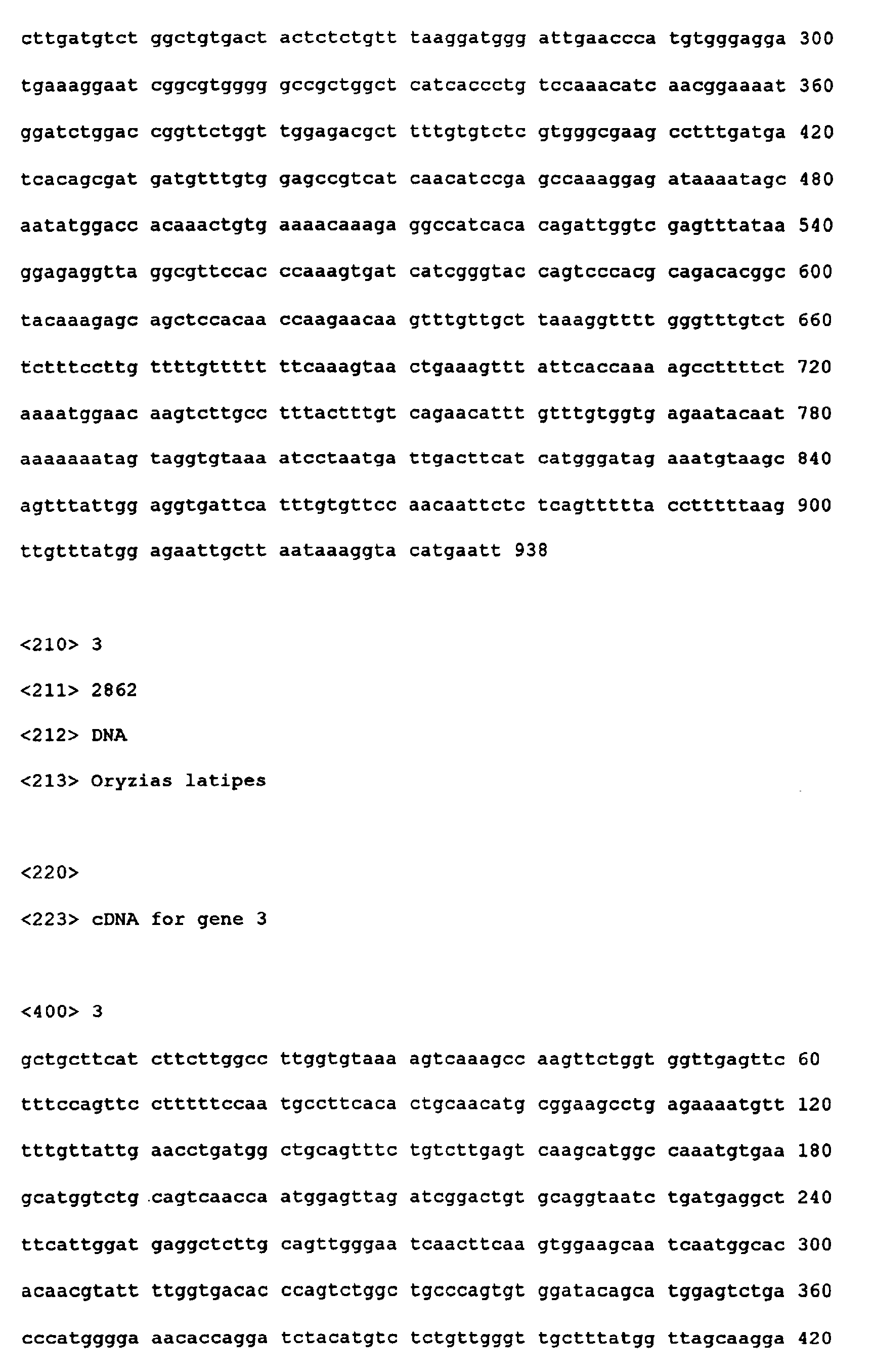
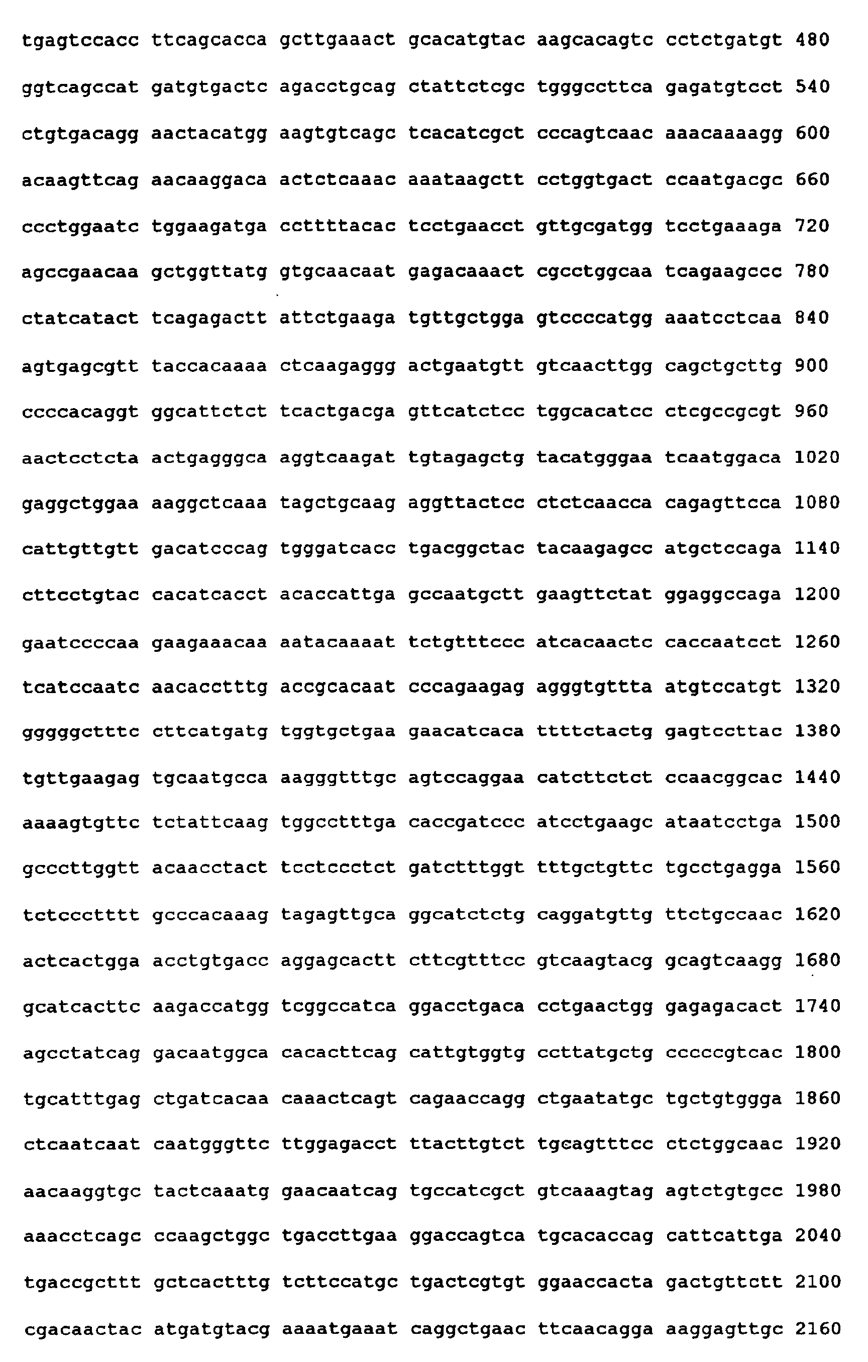


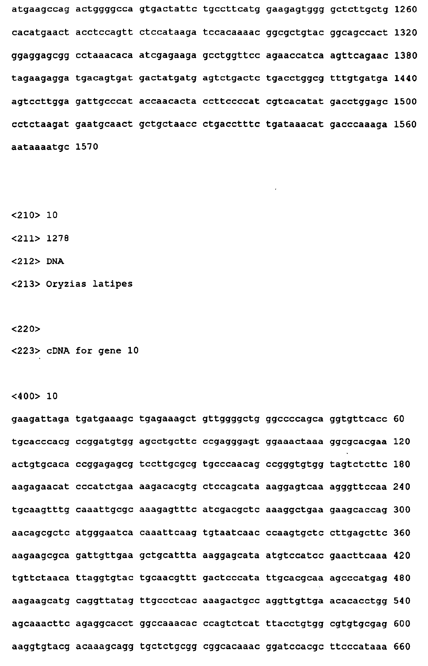
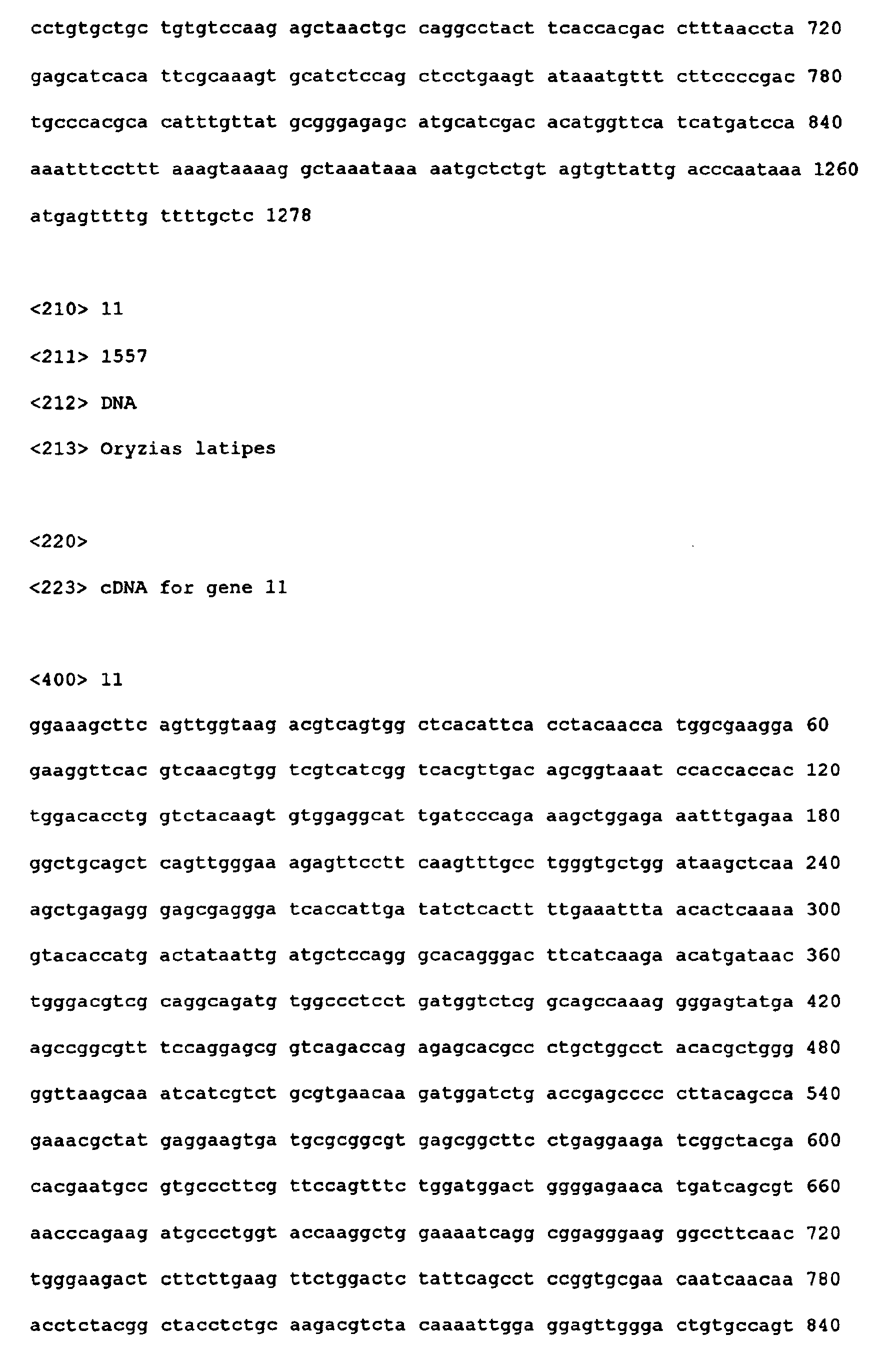
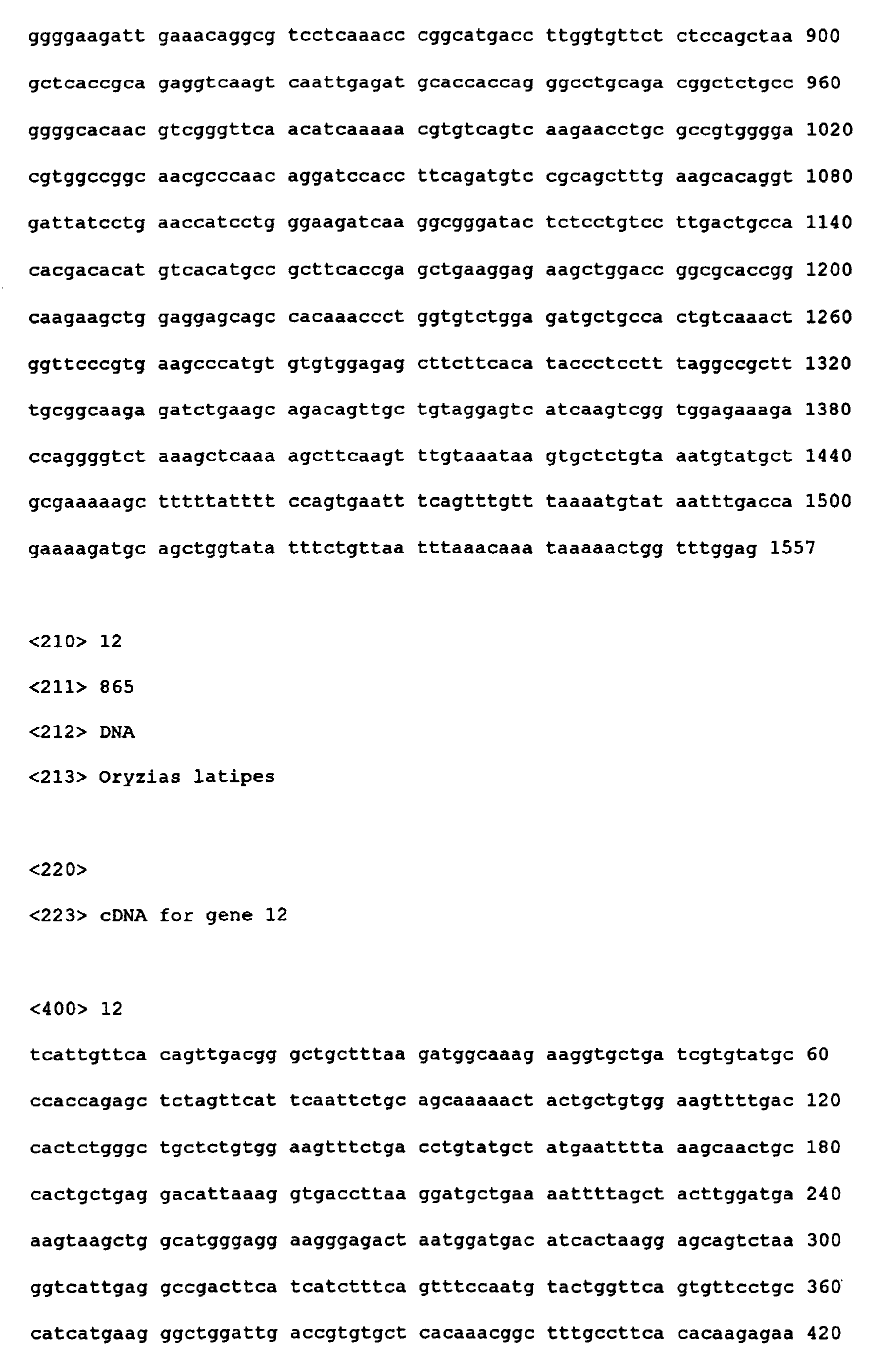


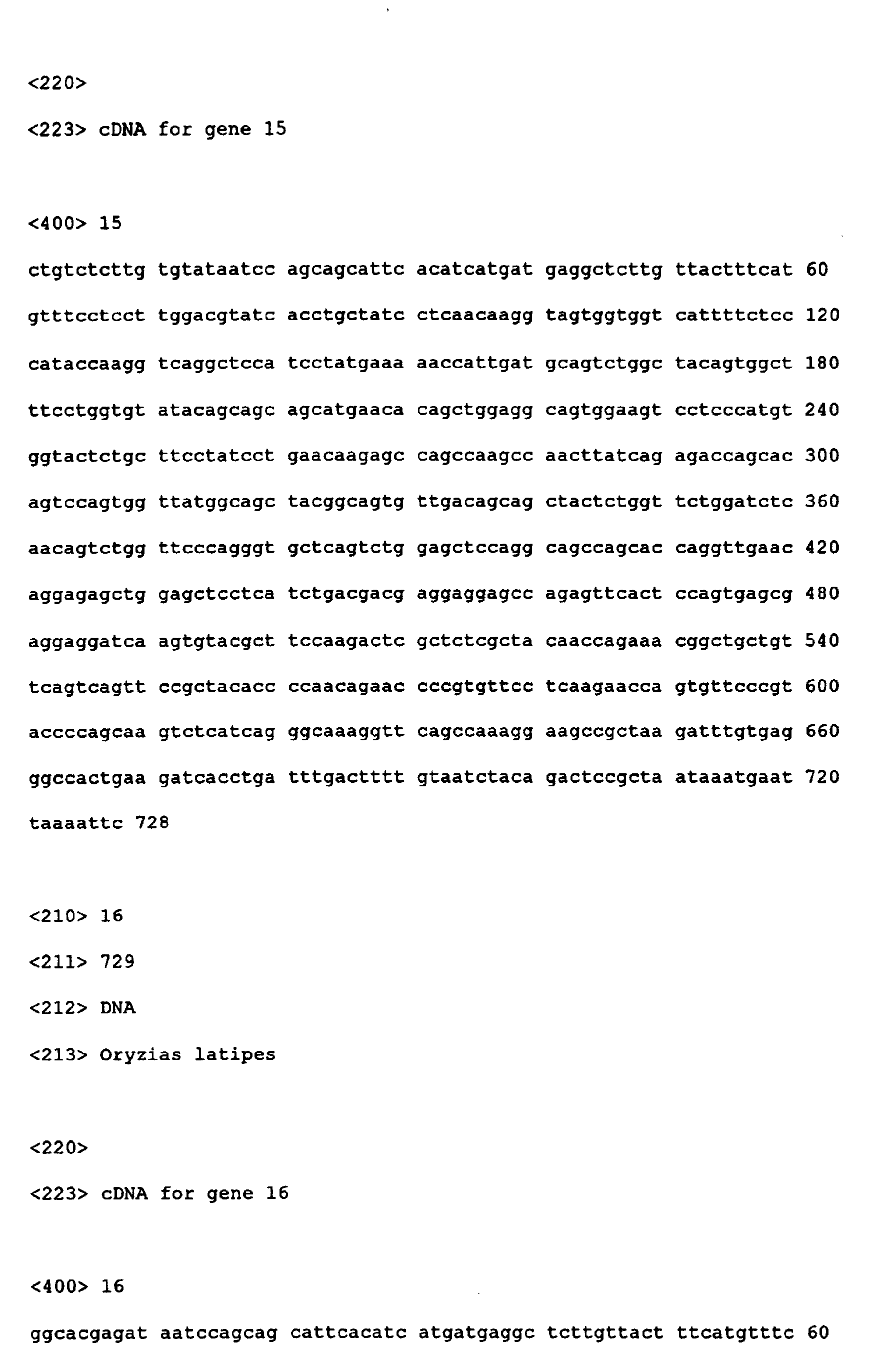


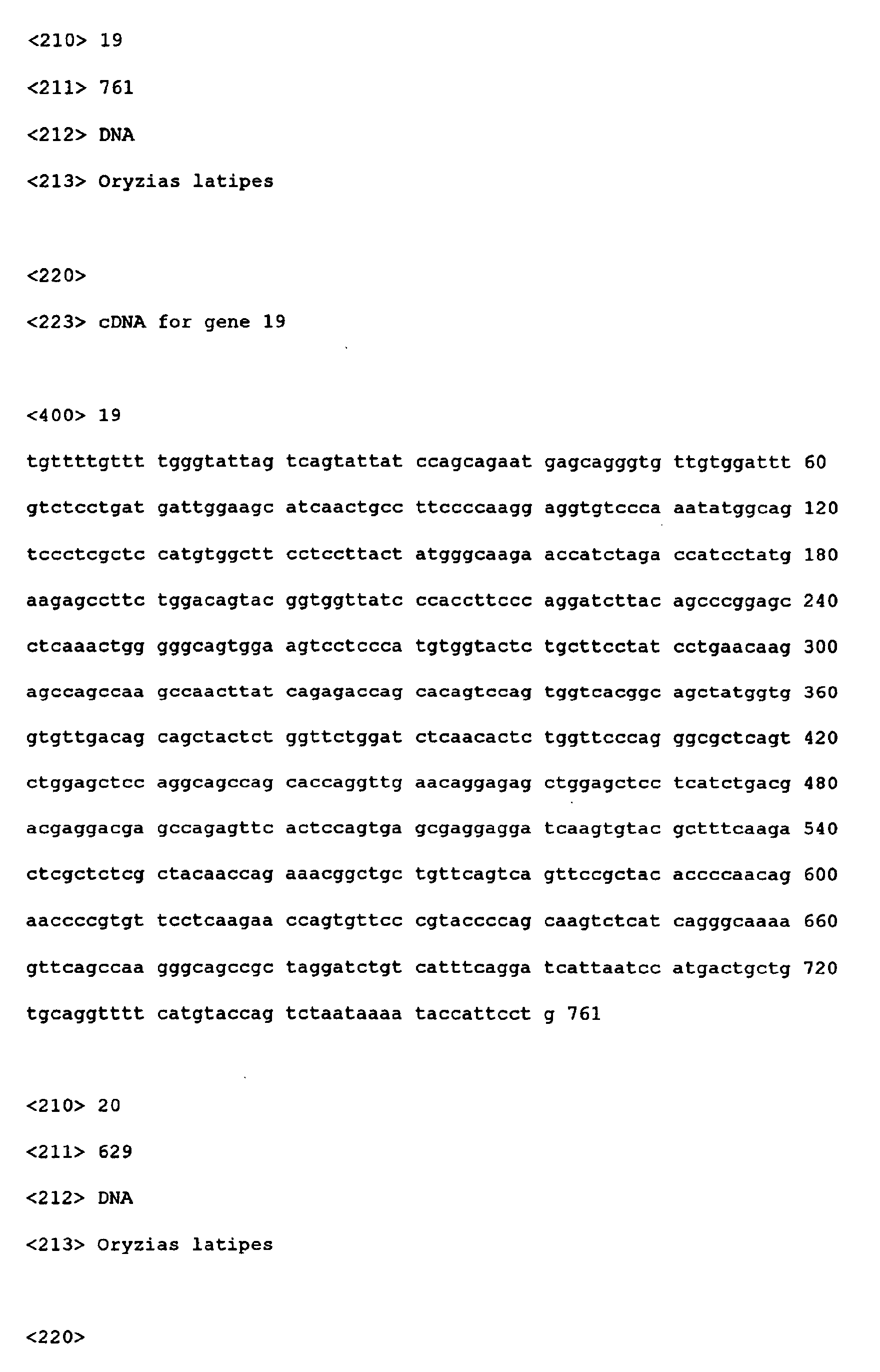
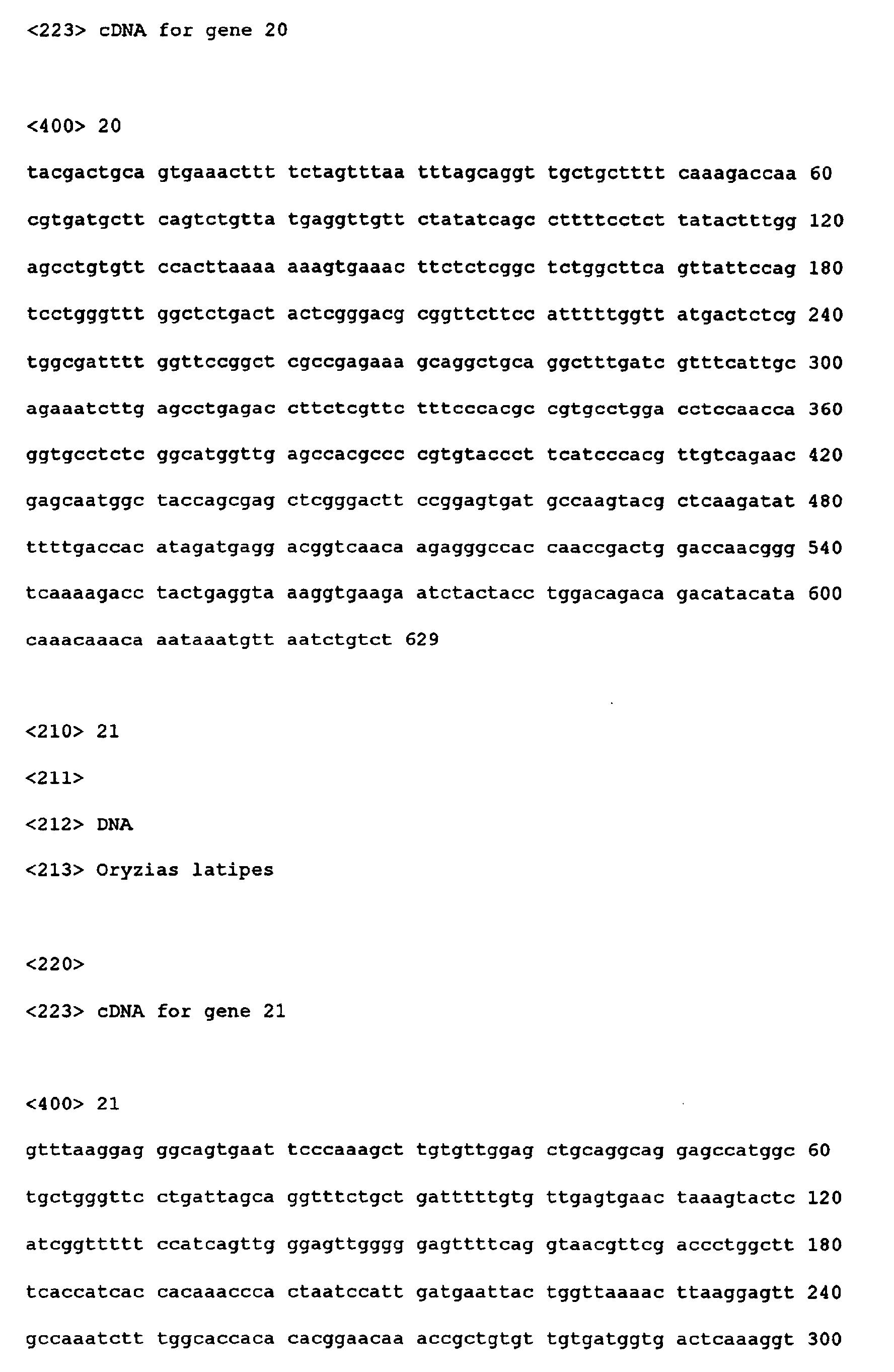


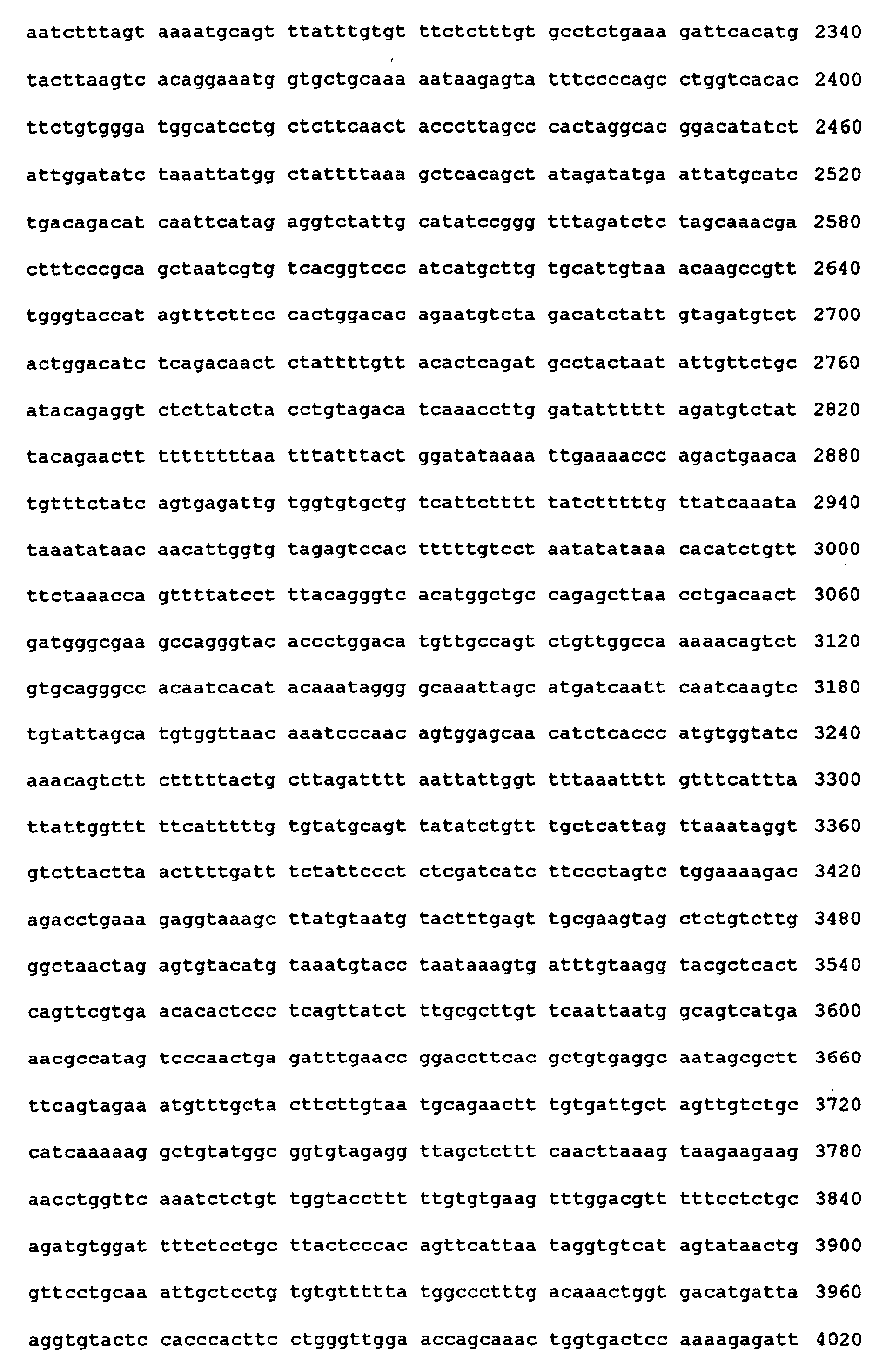


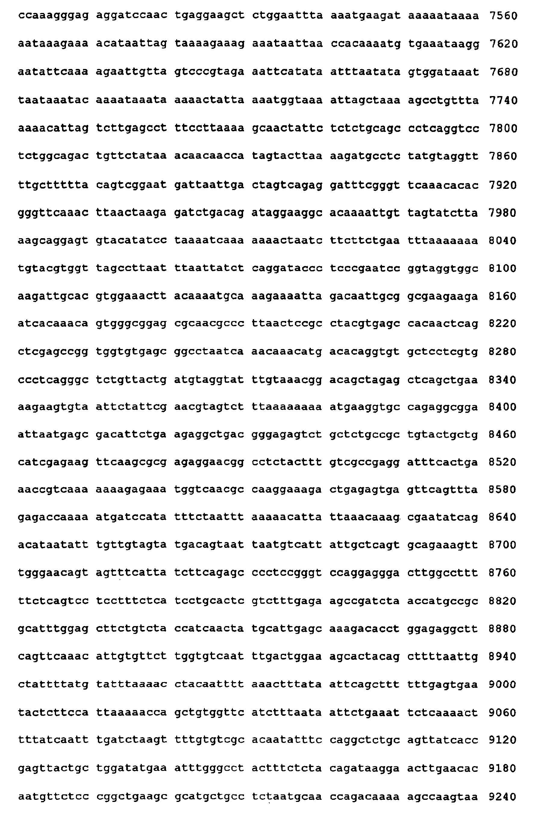

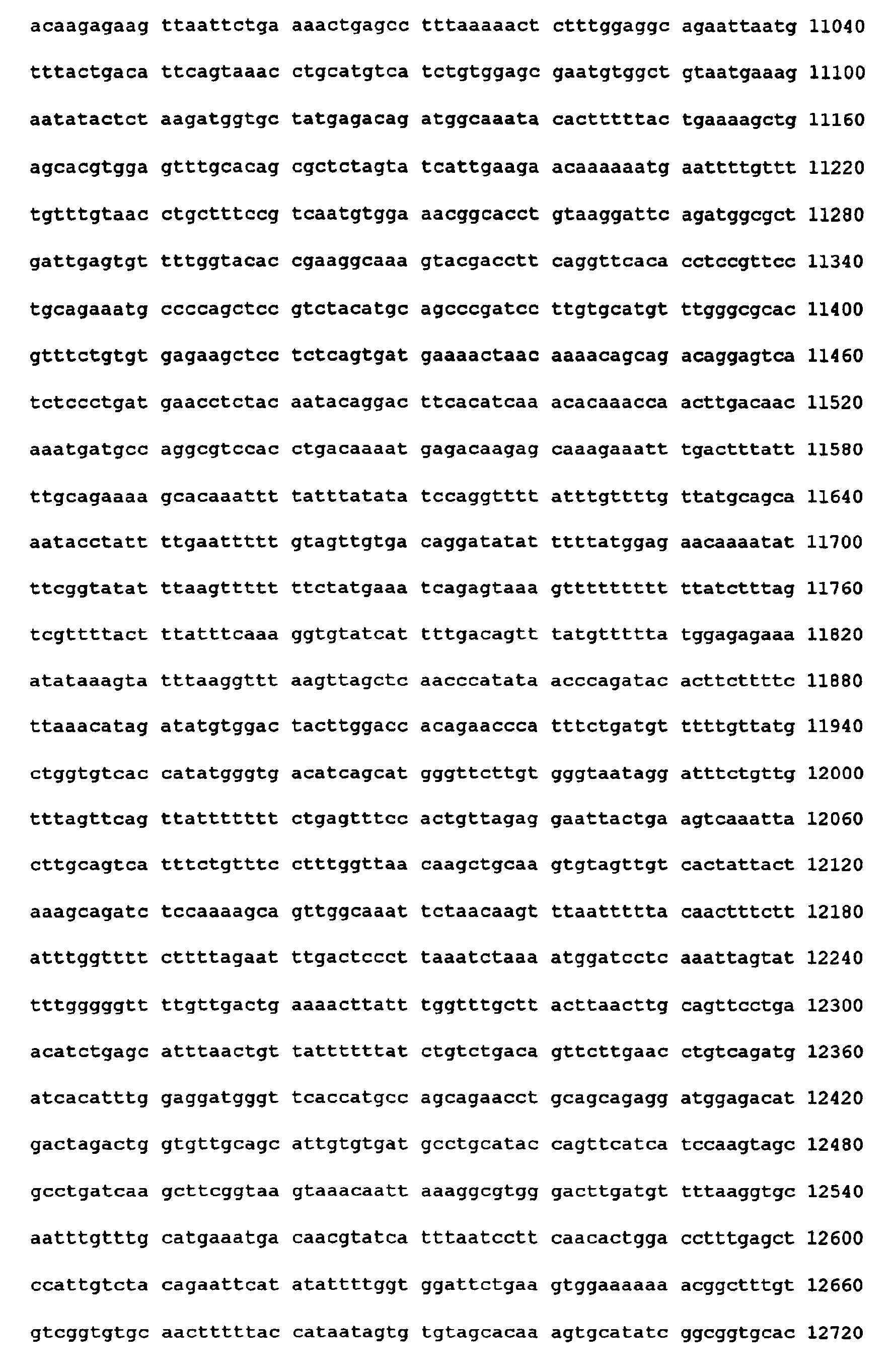

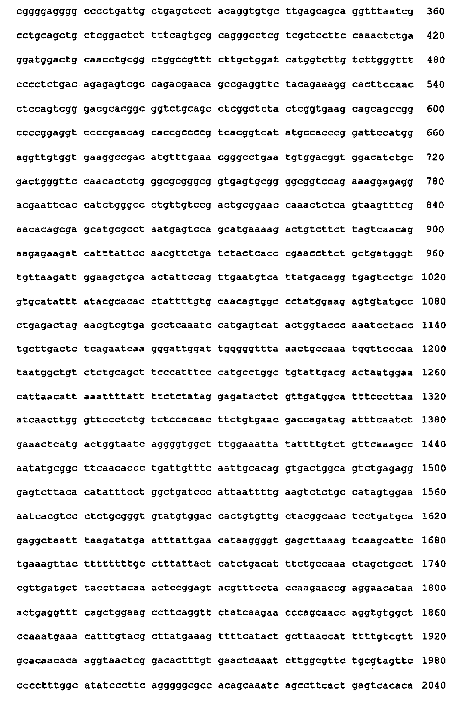


Claims (15)
- A medaka gene expressed in a female-specific manner depending on its phenotypic sex, which has a nucleotide sequence selected from the group consisting of the nucleotide sequences of SEQ ID NOS:1 to 21.
- A gene expressed in a female-specific manner depending on its phenotypic sex, which hybridizes with the gene defined by claim 1 under stringent conditions.
- A gene expressed in a female-specific manner depending on its phenotypic sex, which has a nucleotide sequence selected from the group consisting of the nucleotide sequences of SEQ ID NOS:1 to 21 as well as an intron or introns being inserted.
- The gene according to claim 3, which has a nucleotide sequence of SEQ ID NO:22 or 23.
- A method for assessing a sexual differentiation-disrupting activity of a sample, the method comprising:(1) administering a sample to be assessed for its sexual differentiation-disrupting activity to a medaka;(2) determining the genotypic sex of the medaka;(3) determining the phenotypic sex of the medaka based on the expression of a female-specific gene; and(4) determining if the sexual differentiation is disrupted based on the results of steps (2) and (3).
- The method according to claim 5, wherein the genotypic sex is determined using an egg of medaka before hatching or a fry of medaka within five days after hatching.
- The method according to claim 5 or 6, wherein the phenotypic sex is determined using a fry of medaka within five days after hatching.
- The method according to any one of claims 5 to 7, wherein the sample is administered to an egg of medaka before hatching or a fry of medaka within five days after hatching.
- The method according to any one of claims 5 to 8, wherein a medaka strain of which the genotypic sex is linked to pigment expression is used.
- The method according to any one of claims 5 to 9, wherein the phenotypic sex of the medaka is determined by examining the expression of the gene defined by any one of claims 1 to 4.
- The method according to claim 10, wherein the expression of the gene is examined using the generation of the mRNA for the gene as an index.
- A method for detecting an endocrine disruptor, comprising assessing a sexual differentiation-disrupting activity by the method defined by any one of claims 5 to 11.
- An oligonucleotide for detecting the gene defined by any one of claims 1 to 4.
- A kit for assessing a sexual differentiation-disrupting activity by the method defined by any one of claims 5 to 11, which contains the oligonucleotide defined by claim 13.
- A kit for detecting an endocrine disruptor by the method defined by claim 12, which contains the oligonucleotide defined by claim 13.
Applications Claiming Priority (3)
| Application Number | Priority Date | Filing Date | Title |
|---|---|---|---|
| JP37260299 | 1999-12-28 | ||
| JP37260299 | 1999-12-28 | ||
| PCT/JP2000/009170 WO2001048191A1 (en) | 1999-12-28 | 2000-12-25 | Method of detecting sexual differentiation disruptor |
Publications (3)
| Publication Number | Publication Date |
|---|---|
| EP1243651A1 true EP1243651A1 (en) | 2002-09-25 |
| EP1243651A4 EP1243651A4 (en) | 2005-01-26 |
| EP1243651B1 EP1243651B1 (en) | 2008-02-20 |
Family
ID=18500731
Family Applications (1)
| Application Number | Title | Priority Date | Filing Date |
|---|---|---|---|
| EP00985852A Expired - Lifetime EP1243651B1 (en) | 1999-12-28 | 2000-12-25 | Method of detecting sexual differentiation disruptor |
Country Status (10)
| Country | Link |
|---|---|
| US (1) | US20030162187A1 (en) |
| EP (1) | EP1243651B1 (en) |
| JP (1) | JP3910067B2 (en) |
| KR (1) | KR100687189B1 (en) |
| CN (1) | CN1214112C (en) |
| AT (1) | ATE386801T1 (en) |
| AU (1) | AU2223701A (en) |
| DE (1) | DE60038126T2 (en) |
| TW (1) | TWI289159B (en) |
| WO (1) | WO2001048191A1 (en) |
Family Cites Families (1)
| Publication number | Priority date | Publication date | Assignee | Title |
|---|---|---|---|---|
| US6001599A (en) * | 1992-11-09 | 1999-12-14 | Zonagen, Inc. | DNAs encoding mammalian ZPBs |
-
2000
- 2000-12-25 EP EP00985852A patent/EP1243651B1/en not_active Expired - Lifetime
- 2000-12-25 AU AU22237/01A patent/AU2223701A/en not_active Abandoned
- 2000-12-25 CN CNB008174342A patent/CN1214112C/en not_active Expired - Fee Related
- 2000-12-25 WO PCT/JP2000/009170 patent/WO2001048191A1/en not_active Ceased
- 2000-12-25 DE DE60038126T patent/DE60038126T2/en not_active Expired - Fee Related
- 2000-12-25 AT AT00985852T patent/ATE386801T1/en not_active IP Right Cessation
- 2000-12-25 US US10/169,157 patent/US20030162187A1/en not_active Abandoned
- 2000-12-25 JP JP2001548704A patent/JP3910067B2/en not_active Expired - Fee Related
- 2000-12-25 KR KR1020027006829A patent/KR100687189B1/en not_active Expired - Fee Related
- 2000-12-26 TW TW089127870A patent/TWI289159B/en not_active IP Right Cessation
Non-Patent Citations (9)
| Title |
|---|
| ARCAND-HOY LISA D ET AL: "Fish reproduction: An ecologically relevant indicator of endocrine disruption" ENVIRONMENTAL TOXICOLOGY AND CHEMISTRY, vol. 17, no. 1, January 1998 (1998-01), pages 49-57, XP008032487 ISSN: 0730-7268 * |
| BAROILLER J -F ET AL: "Endocrine and environmental aspects of sex differentiation in fish" CMLS CELLULAR AND MOLECULAR LIFE SCIENCES, vol. 55, no. 6-7, June 1999 (1999-06), pages 910-931, XP002287018 ISSN: 1420-682X * |
| DATABASE EMBL [Online] 18 June 1999 (1999-06-18), KANAMORI A: "Oryzias latipes FIGalpha mRNA, complete cds." XP002287020 Database accession no. AF128805 * |
| DATABASE EMBL [Online] 18 June 1999 (1999-06-18), KANAMORI A: "Oryzias latipes ZPC domain containing protein 1 mRNA, complete cds." XP002305973 Database accession no. AF128809 * |
| GRAY M A ET AL: "INDUCTION OF TESTIS-OVA IN JAPANESE MEDAKA (ORYZIAS LATIPES) EXPOSED TO P-NONYLPHENOL" ENVIRONMENTAL TOXICOLOGY AND CHEMISTRY, PERGAMON PRESS, US, vol. 16, no. 5, 1997, pages 1082-1086, XP002939716 ISSN: 0730-7268 * |
| KANAMORI A: "Systematic identification of genes expressed during early oogenesis in medaka." MOLECULAR REPRODUCTION AND DEVELOPMENT. JAN 2000, vol. 55, no. 1, January 2000 (2000-01), pages 31-36, XP008032480 ISSN: 1040-452X -& KANAMORI A: "Systematic identification of genes expressed during early oogenesis in medaka." MOLECULAR REPRODUCTION AND DEVELOPMENT, [Online] 6 December 1999 (1999-12-06), XP002287390 Retrieved from the Internet: URL:http://www3.interscience.wiley.com/cgi -bin/abstract/68000920/ABSTRACT> [retrieved on 2004-07-07] * |
| MURATA K ET AL: "Cloning of cDNA and estrogen-induced hepatic gene expression for choriogenin H, a precursor protein of the fish egg envelope (chorion)" PROCEEDINGS OF THE NATIONAL ACADEMY OF SCIENCES OF THE UNITED STATES OF AMERICA 1997 UNITED STATES, vol. 94, no. 5, 1997, pages 2050-2055, XP002287016 ISSN: 0027-8424 * |
| SCHOLZ S ET AL: "17-alpha-ethinylestradiol affects reproduction, sexual differentiation and aromatase gene expression of the medaka (Oryzias latipes)" AQUATIC TOXICOLOGY (AMSTERDAM), vol. 50, no. 4, October 2000 (2000-10), pages 363-373, XP002287017 ISSN: 0166-445X * |
| See also references of WO0148191A1 * |
Also Published As
| Publication number | Publication date |
|---|---|
| KR20020073135A (en) | 2002-09-19 |
| KR100687189B1 (en) | 2007-02-27 |
| ATE386801T1 (en) | 2008-03-15 |
| DE60038126D1 (en) | 2008-04-03 |
| WO2001048191A1 (en) | 2001-07-05 |
| CN1411507A (en) | 2003-04-16 |
| DE60038126T2 (en) | 2009-03-05 |
| US20030162187A1 (en) | 2003-08-28 |
| EP1243651A4 (en) | 2005-01-26 |
| CN1214112C (en) | 2005-08-10 |
| EP1243651B1 (en) | 2008-02-20 |
| AU2223701A (en) | 2001-07-09 |
| TWI289159B (en) | 2007-11-01 |
| JP3910067B2 (en) | 2007-04-25 |
Similar Documents
| Publication | Publication Date | Title |
|---|---|---|
| Yamaguchi et al. | Expression of the DMRT gene and its roles in early gonadal development of the Japanese pufferfish Takifugu rubripes | |
| Letelier et al. | A conserved Shh cis-regulatory module highlights a common developmental origin of unpaired and paired fins | |
| Tong et al. | Molecular cloning of zebrafish and medaka vitellogenin genes and comparison of their expression in response to 17β-estradiol | |
| Reynolds et al. | Translation of the synaptonemal complex component Sycp3 is enhanced in vivo by the germ cell specific regulator Dazl | |
| Cenik et al. | Maternal ribosomes are sufficient for tissue diversification during embryonic development in C. elegans | |
| Schroeder et al. | A novel candidate gene for temperature-dependent sex determination in the common snapping turtle | |
| Gautier et al. | The gsdf gene locus harbors evolutionary conserved and clustered genes preferentially expressed in fish previtellogenic oocytes | |
| Matsuda et al. | Evolution of the sex-determining gene in the teleostean genus Oryzias | |
| Luckenbach et al. | Aromatase cytochrome P450: cloning, intron variation, and ontogeny of gene expression in southern flounder (Paralichthys lethostigma) | |
| Nagasawa et al. | cDNA cloning and expression analysis of a vasa-like gene in Pacific bluefin tuna Thunnus orientalis | |
| Shen et al. | De novo transcriptome assembly and sex-biased gene expression in the gonads of Amur catfish (Silurus asotus) | |
| Thoemke et al. | Genome-wide analysis of sex-enriched gene expression during C. elegans larval development | |
| Ma et al. | Piwi1 is essential for gametogenesis in mollusk Chlamys farreri | |
| Zhang et al. | Embryonic exposure to fenbuconazole inhibits gametogenesis in adult zebrafish by targeting gonads not brain | |
| Xiao et al. | NYD-SP6, a novel gene potentially involved in regulating testicular development/spermatogenesis | |
| Wang et al. | Comparative studies on duplicated tdrd7 paralogs in teleosts: Molecular evolution caused neo-functionalization | |
| Bir et al. | High production of transfer RNAs identifies the presence of developing oocytes in ovaries and intersex testes of teleost fish | |
| Kanamori et al. | Comparative genomics approach to the expression of figα, one of the earliest marker genes of oocyte differentiation in medaka (Oryzias latipes) | |
| Alnafea et al. | Japanese medaka as a model for studying the relaxin family genes involved in neuroendocrine regulation: Insights from the expression of fish-specific rln3 and insl5 and rxfp3/4-type receptor paralogues | |
| Lawir et al. | A missense mutation in zbtb17 blocks the earliest steps of T cell differentiation in zebrafish | |
| EP1243651B1 (en) | Method of detecting sexual differentiation disruptor | |
| Sluder et al. | TheCaenorhabditis elegansOrphan Nuclear Hormone Receptor Genenhr-2Functions in Early Embryonic Development | |
| An et al. | Quantitative real-time RT-PCR for determination of vitellogenin mRNA in so-iuy mullet (Mugil soiuy) | |
| Lee et al. | Molecular characterization and tissue distribution of aryl hydrocarbon receptor nuclear translocator isoforms, ARNT1 and ARNT2, and identification of novel splice variants in common cormorant (Phalacrocorax carbo) | |
| Park et al. | The Caenorhabditis elegans XPA homolog of human XPA |
Legal Events
| Date | Code | Title | Description |
|---|---|---|---|
| PUAI | Public reference made under article 153(3) epc to a published international application that has entered the european phase |
Free format text: ORIGINAL CODE: 0009012 |
|
| 17P | Request for examination filed |
Effective date: 20020617 |
|
| AK | Designated contracting states |
Kind code of ref document: A1 Designated state(s): AT BE CH CY DE DK ES FI FR GB GR IE IT LI LU MC NL PT SE TR |
|
| AX | Request for extension of the european patent |
Free format text: AL;LT;LV;MK;RO;SI |
|
| RIN1 | Information on inventor provided before grant (corrected) |
Inventor name: CHONO, HIDETO Inventor name: KINOSHITA, MASATO Inventor name: TAKASHIMA, RYOKICHI Inventor name: KONDO, AKIHIRO, 101, HAMOPARESU-KUSATSU Inventor name: MIZUTANI, SHIGETOSHI Inventor name: KANAMORI, AKIRA Inventor name: KATO, IKUNOSHIN |
|
| RIC1 | Information provided on ipc code assigned before grant |
Ipc: 7C 12N 15/85 B Ipc: 7G 01N 33/00 B Ipc: 7C 12Q 1/68 B Ipc: 7C 12N 15/09 B Ipc: 7A 01K 67/00 B Ipc: 7C 07K 14/46 B Ipc: 7C 12N 15/12 B Ipc: 7A 01K 61/00 B Ipc: 7C 12N 15/11 A |
|
| A4 | Supplementary search report drawn up and despatched |
Effective date: 20041213 |
|
| 17Q | First examination report despatched |
Effective date: 20050718 |
|
| GRAP | Despatch of communication of intention to grant a patent |
Free format text: ORIGINAL CODE: EPIDOSNIGR1 |
|
| GRAS | Grant fee paid |
Free format text: ORIGINAL CODE: EPIDOSNIGR3 |
|
| GRAA | (expected) grant |
Free format text: ORIGINAL CODE: 0009210 |
|
| AK | Designated contracting states |
Kind code of ref document: B1 Designated state(s): AT BE CH CY DE DK ES FI FR GB GR IE IT LI LU MC NL PT SE TR |
|
| REG | Reference to a national code |
Ref country code: GB Ref legal event code: FG4D |
|
| REG | Reference to a national code |
Ref country code: CH Ref legal event code: EP |
|
| REG | Reference to a national code |
Ref country code: IE Ref legal event code: FG4D |
|
| REF | Corresponds to: |
Ref document number: 60038126 Country of ref document: DE Date of ref document: 20080403 Kind code of ref document: P |
|
| PG25 | Lapsed in a contracting state [announced via postgrant information from national office to epo] |
Ref country code: FI Free format text: LAPSE BECAUSE OF FAILURE TO SUBMIT A TRANSLATION OF THE DESCRIPTION OR TO PAY THE FEE WITHIN THE PRESCRIBED TIME-LIMIT Effective date: 20080220 Ref country code: ES Free format text: LAPSE BECAUSE OF FAILURE TO SUBMIT A TRANSLATION OF THE DESCRIPTION OR TO PAY THE FEE WITHIN THE PRESCRIBED TIME-LIMIT Effective date: 20080531 |
|
| NLV1 | Nl: lapsed or annulled due to failure to fulfill the requirements of art. 29p and 29m of the patents act | ||
| PG25 | Lapsed in a contracting state [announced via postgrant information from national office to epo] |
Ref country code: AT Free format text: LAPSE BECAUSE OF FAILURE TO SUBMIT A TRANSLATION OF THE DESCRIPTION OR TO PAY THE FEE WITHIN THE PRESCRIBED TIME-LIMIT Effective date: 20080220 |
|
| PG25 | Lapsed in a contracting state [announced via postgrant information from national office to epo] |
Ref country code: BE Free format text: LAPSE BECAUSE OF FAILURE TO SUBMIT A TRANSLATION OF THE DESCRIPTION OR TO PAY THE FEE WITHIN THE PRESCRIBED TIME-LIMIT Effective date: 20080220 |
|
| ET | Fr: translation filed | ||
| PG25 | Lapsed in a contracting state [announced via postgrant information from national office to epo] |
Ref country code: SE Free format text: LAPSE BECAUSE OF FAILURE TO SUBMIT A TRANSLATION OF THE DESCRIPTION OR TO PAY THE FEE WITHIN THE PRESCRIBED TIME-LIMIT Effective date: 20080520 Ref country code: DK Free format text: LAPSE BECAUSE OF FAILURE TO SUBMIT A TRANSLATION OF THE DESCRIPTION OR TO PAY THE FEE WITHIN THE PRESCRIBED TIME-LIMIT Effective date: 20080220 Ref country code: NL Free format text: LAPSE BECAUSE OF FAILURE TO SUBMIT A TRANSLATION OF THE DESCRIPTION OR TO PAY THE FEE WITHIN THE PRESCRIBED TIME-LIMIT Effective date: 20080220 Ref country code: PT Free format text: LAPSE BECAUSE OF FAILURE TO SUBMIT A TRANSLATION OF THE DESCRIPTION OR TO PAY THE FEE WITHIN THE PRESCRIBED TIME-LIMIT Effective date: 20080721 |
|
| PLBE | No opposition filed within time limit |
Free format text: ORIGINAL CODE: 0009261 |
|
| STAA | Information on the status of an ep patent application or granted ep patent |
Free format text: STATUS: NO OPPOSITION FILED WITHIN TIME LIMIT |
|
| 26N | No opposition filed |
Effective date: 20081121 |
|
| PG25 | Lapsed in a contracting state [announced via postgrant information from national office to epo] |
Ref country code: CY Free format text: LAPSE BECAUSE OF FAILURE TO SUBMIT A TRANSLATION OF THE DESCRIPTION OR TO PAY THE FEE WITHIN THE PRESCRIBED TIME-LIMIT Effective date: 20080220 Ref country code: MC Free format text: LAPSE BECAUSE OF NON-PAYMENT OF DUE FEES Effective date: 20081231 |
|
| REG | Reference to a national code |
Ref country code: CH Ref legal event code: PL |
|
| GBPC | Gb: european patent ceased through non-payment of renewal fee |
Effective date: 20081225 |
|
| PG25 | Lapsed in a contracting state [announced via postgrant information from national office to epo] |
Ref country code: IT Free format text: LAPSE BECAUSE OF FAILURE TO SUBMIT A TRANSLATION OF THE DESCRIPTION OR TO PAY THE FEE WITHIN THE PRESCRIBED TIME-LIMIT Effective date: 20080220 |
|
| REG | Reference to a national code |
Ref country code: FR Ref legal event code: ST Effective date: 20090831 |
|
| PG25 | Lapsed in a contracting state [announced via postgrant information from national office to epo] |
Ref country code: CH Free format text: LAPSE BECAUSE OF NON-PAYMENT OF DUE FEES Effective date: 20081231 Ref country code: LI Free format text: LAPSE BECAUSE OF NON-PAYMENT OF DUE FEES Effective date: 20081231 Ref country code: DE Free format text: LAPSE BECAUSE OF NON-PAYMENT OF DUE FEES Effective date: 20090701 Ref country code: IE Free format text: LAPSE BECAUSE OF NON-PAYMENT OF DUE FEES Effective date: 20081225 |
|
| PG25 | Lapsed in a contracting state [announced via postgrant information from national office to epo] |
Ref country code: GB Free format text: LAPSE BECAUSE OF NON-PAYMENT OF DUE FEES Effective date: 20081225 |
|
| PG25 | Lapsed in a contracting state [announced via postgrant information from national office to epo] |
Ref country code: FR Free format text: LAPSE BECAUSE OF NON-PAYMENT OF DUE FEES Effective date: 20081231 |
|
| PG25 | Lapsed in a contracting state [announced via postgrant information from national office to epo] |
Ref country code: LU Free format text: LAPSE BECAUSE OF NON-PAYMENT OF DUE FEES Effective date: 20081225 |
|
| PG25 | Lapsed in a contracting state [announced via postgrant information from national office to epo] |
Ref country code: TR Free format text: LAPSE BECAUSE OF FAILURE TO SUBMIT A TRANSLATION OF THE DESCRIPTION OR TO PAY THE FEE WITHIN THE PRESCRIBED TIME-LIMIT Effective date: 20080220 |
|
| PG25 | Lapsed in a contracting state [announced via postgrant information from national office to epo] |
Ref country code: GR Free format text: LAPSE BECAUSE OF FAILURE TO SUBMIT A TRANSLATION OF THE DESCRIPTION OR TO PAY THE FEE WITHIN THE PRESCRIBED TIME-LIMIT Effective date: 20080521 |