TECHNICAL FIELD
The present invention relates to derivatives of a
monoclonal antibody and the antibody fragment which
specifically react with ganglioside GD3 (hereinafter
referred to as "GD3"), and to a human CDR-grafted antibody
and the antibody fragment which specifically react with GD3.
Furthermore, the present invention relates to a DNA
sequence which encodes the derivatives, antibody or
antibody fragment. The present invention relates to a
vector comprising the DNA sequence and a cell transformed
with the vector. Moreover, the present invention relates
to a process for producing the derivatives, antibody and
antibody fragment using the transformed cell, and to a
diagnostic agent and a therapeutic agent for cancers using
the derivatives, antibody and antibody fragment.
BACKGROUND OF THE INVENTION
It is known that, when an antibody derived from a
non-human animal, e.g., a mouse antibody, is administered
to human, it is recognized as a foreign substance and thus
induces a human antibody against the mouse antibody in the
human body (human anti mouse antibody, hereinafter referred
to as "HAMA"). It is known that the HAMA reacts with the
administered mouse antibody to cause side effects (J. Clin.
Oncol., 2, 881 (1984); Blood, 65, 1349 (1985); J. Natl.
Cancer Inst., 80, 932 (1988); Proc. Natl. Acad. Sci. U.S.A.,
82, 1242 (1985)), to quicken disappearance of the
administered mouse antibody from blood (J. Nucl. Med., 26,
1011 (1985); Blood, 65, 1349 (1985); J. Natl. Cancer Inst.,
80, 937 (1988)) and to reduce therapeutic effects of the
mouse antibody (J. Immunol., 135, 1530 (1985); Cancer Res.,
46, 6489 (1986)).
In order to resolve these problems, attempts have
been made to genetic engineeringly convert an antibody
derived from a non-human animal into a humanized antibody
such as a human chimeric antibody or a human
complementarity determining region (hereinafter referred to
as "CDR")-grafted antibody. The human chimeric antibody is
an antibody in which its antibody variable region
(hereinafter referred to as "V region") is derived from a
non-human animal antibody and its constant region
(hereinafter referred to as "C region") is derived from a
human antibody (Proc. Natl. Acad. Sci. U.S.A., 81, 6851
(1984)). The human CDR-grafted antibody is an antibody in
which the amino acid sequence of CDR in the V region
derived from a non-human animal antibody is grafted to an
appropriate position of a human antibody (Nature, 321, 522
(1986)). These humanized antibodies have various
advantages in clinically applying to human, in comparison
with antibodies of non-human animals such as mouse
antibodies and the like. For example, it has been reported'
on the immunogenicity and stability in blood that, when a
human chimeric antibody is administered to human, its half-life
in blood increased about six times in comparison with
a mouse antibody (Proc. Natl. Acad. Sci. U.S.A., 86, 4220
(1989)). It has been reported that, in a test of a human
CDR-grafted antibody in monkeys, its immunogenicity is
reduced and its half-life in blood is increased four to
five times in comparison with a mouse antibody (J. Immunol.,
147, 1352 (1991)). That is, it is expected that humanized
antibodies have less side effects and their therapeutic
effects continue for a longer period of time than
antibodies of non-human animals. In addition, when
applications particularly as anti-tumor antibodies are
taken into consideration, higher cytotoxic activities such
as complement-dependent cytotoxicity (hereinafter referred
to as "CDC"), antibody-dependent cell-mediated cytotoxic
activity (hereinafter referred to as "ADCC") and the like
via an antibody Fc region (region in and after the hinge
region of an antibody heavy chain) are important for the
therapeutic effects. It has been reported that on such
cytotoxic activities, that the Fc region of human
antibodies is more superior in human to the Fc region of
antibodies of non-human animals, since the Fc region more
effectively activates human complement components and human
effector cells having Fc receptors of mononuclear cells,
macrophages and NK cells on the cell surface. For example,
it has been reported that when a mouse antibody against GD3
(hereinafter referred to as "anti-GD3 mouse antibody") is
converted into a human chimeric antibody having a human
antibody Fc region (hereinafter referred to as "anti-GD3
chimeric antibody"), its tumor cell growth-inhibitory
activity by human effector cells is increased (J. Immunol.,
144, 1382 (1990)), and similar results have been reported
on a human CDR-grafted antibody against CAMPATH-1 antigen
(Nature, 332, 323 (1988)).
These results clearly show that humanized
antibodies are preferred to non-human antibodies such as
mouse antibodies and the like, as antibodies for clinical
applications to human.
In addition, with the recent advance in protein
engineering and genetic engineering, antibody fragments
having a smaller molecular weight such as Fab, Fab',
F(ab')2, a single chain antibody (Science, 242, 423 (1988)),
a disulfide stabilized V region fragment (Molecular
Immunol., 32, 249 (1995)) and the like, can be produced.
Since these fragments have a smaller molecular weight than
whole antibody molecules, they are excellent in penetrating
ability into target tissues (Cancer Res., 52, 3402 (1992)).
It is considered that these fragments derived from
humanized antibodies are more desirable than those derived
from non-human animal antibodies such as mouse antibodies,
when used in clinical applications to human.
Ganglioside which is one of glycolipids containing
sialic acid is a molecule which constitutes animal cell
membrane and is constituted by a sugar chain as a
hydrophilic side chain and a sphingosine and fatty acid as
hydrophobic side chains. It is known that kinds and
expression levels of ganglioside vary depending on the cell
types, organ species, animal species and the like. It is
also known that the expression of ganglioside changes
quantitatively and qualitatively in the process of
malignant transformation of cells (Cancer Res., 45, 2405
(1985)). Particularly, the GD3 relating to the present
invention is present in an extremely small amount in normal
cells but in a large amount in cancer cells such as
melanoma, sarcoma, glioma, neuroblastoma and the like (Proc.
Natl. Acad. Sci. U.S.A., 77, 6114 (1980); J. Exp. Med., 155,
1133 (1982); Cancer Res., 45, 4401 (1985)), and an antibody
against GD3 (hereinafter referred to as "anti-GD3
antibody") is considered to be useful for the treatment of
these cancers (Melanoma Research, 1, S115 (1997)). Tests
on its administration to melanoma patients have already
been carried out using R24 as an anti-GD3 mouse antibody,
and its clinical effect was observed in apart of the
patients but not to an expected degree because of
considerably quick disappearance of the antibody from blood
due to induction of HAMA (Melanoma Research, 1, S115
(1997)). Under such circumstances, in order to achieve
more effective clinical applications of an anti-GD3
antibody to human, the present inventors have attempted to
produce an anti-GD3 chimeric antibody having similar
activity to that of a mouse antibody based on an anti-GD3
mouse antibody KM641 produced by the inventors, and have
succeeded in producing an anti-GD3 chimeric antibody KM871
(Japanese Published Unexamined Patent Application
No. 304989/93). It is expected that, in comparison with
the anti-GD3 mouse antibody, the anti-GD3 chimeric antibody
KM871 reduces immunogenicity in human, prolongs half-life
in blood and has a stronger anti-tumor effect. However, it
was found as a result of clinical tests on other anti-GD3
chimeric antibodies in human that allergy reactions are
induced due to induction of a human antibody against a V
reg'ion derived from a mouse antibody in the anti-GD3
chimeric antibodies (cancer J. From Scientific American, 3,
S121 (1997)). Even in the anti-GD3 chimeric antibody KM871
prepared by the inventors, it is necessary to await results
of clinical tests in human to know if similar reactions are
induced or not, and in order to reduce a possibility of
generating such a problem as low as possible, it is
desirable to produce a human CDR-grafted antibody against
GD3 (hereinafter referred to as "anti-GD3 CDR-grafted
antibody") which is considered to have lower immunogenicity
than those of anti-GD3 chimeric antibodies in human.
However, to date, there are no reports on the production of
an anti-GD3 CDR-grafted antibody.
with regard to antibody fragments, a single chain
antibody derived from an anti-GD2 mouse antibody has been
produced (Hybridoma, 16, 335 (1997)), but there are no
reports on the production of other antibody fragments.
With regard to antibody fragments derived from an anti-GD3
antibody, there are no reports on the production of
antibody fragments derived from a mouse antibody and a
humanized antibody. Thus, if an antibody fragment derived'
from a humanized antibody against GD3 (hereinafter referred
to as "anti-GD3 humanized antibody") can be produced, it is
expected that it has excellent target tissue
penetratability and the immunogenicity is reduced.
As has been described above, it is expected that a
humanized antibody and the antibody fragment have effect on
diagnostic and therapeutic methods by single use, but
further improvement of the effects has been examined by
using them in combination with other molecules. For
example, cytokine is used as one of these molecules.
Cytokine is a general term for various aqueous factors
which control intercellular mutual reactions in immune
interactions. As cytotoxic activities of antibodies such
as CDC activity, ADCC activity and the like are known and
the ADCC activity is effected by effector cells having Fc
receptors on the cell surface such as mononuclear cells,
macrophages, NK cells and the like (J. Immunol., 138, 1992
(1987)). Since various kinds of cytokine activate these
effector cells, their administration in combination with
antibodies has been carried out to improve ADCC activity
and the like of antibodies. For example, with regard to an
anti-GD2 mouse antibody 14.G2a and an anti-GD2 chimeric
antibody ch14.18, their administration to human in
combination with a cytokine human interleukin-2
(hereinafter referred to as "hIL-2") or a human granulocyte
macrophage colony-stimulating factor (hereinafter referred
to as "hGM-CSF") has been carried out (Cancer, 80, 317
(1997); Cancer J. From Scientific American, 3, S121 (1997)).
Regarding an anti-GD3 mouse antibody R24, combination.
therapy with various kinds of cytokine has also been
carried out (Cancer Res., 50, 7490 (1990), Proc. Am. Soc.
Clin. Oncol., 1186, 345 (1992), J. Biol. Response Mod., 9,
319 (1990), Proc. Am. Soc. Clin. Oncol., 1182, 344 (1992),
Proc. Am. Soc. Clin. Oncol., 1188, 346 (1992)). However,
the effects as to be expected are not found in these
combination therapies due to immunogenicity of mouse
antibodies or side effects derived from cytokine. Thus,
derivatives has been produced by conjugating an antibody or
the antibody fragment thereof with a radioisotope, a
protein (cytokine, toxin, enzyme or the like), a low
molecular weight agent and the like, chemically or genetic
engineeringly, and their clinical applications have been
examined (New Eng. J. Med., 329, 459 (1993), Anticancer
Res., 11, 1735 (1997), Blood, 78, 1173 (1991), J. Clin.
Oncol., 15, 723 (1997), Bioconjugate Chem., 1, 606 (1997),
Cancer, 61, 881 (1988), Jpn. J. Cancer Res., 85, 167 (1994),
Antibody Immunoconjugates and Radiopharmaceuticals, 3, 60
(1990), Surgery, 106, 533 (1989)). It is expected that
these derivatives can accumulate a radioisotope, a protein
(cytokine, toxin, enzyme or the like), a low molecular
weight agent or the like in the periphery of a target
tissue according to the binding specificity of antibodies
so that more effective diagnoses and treatments having less
side effects can be obtained. For example, with regard to
hIL-2 which showed a certain degree of anti-tumor effect
among cytokines used in the above combination therapy, its
fusion protein with an anti-GD2 chimeric antibody ch14.18
was produced genetic engineeringly, and it has been
reported that its antitumor effects in tests using mice
were superior to those of simultaneous administration of
anti-GD2 chimeric antibody ch14.18 and hIL-2 (Proc. Natl.
Acad. Sci. U.S.A., 89, 1428 (1992), Proc. Natl. Acad. Sci.
U.S.A., 91, 9626 (1994), Cancer Immunol. Immunother., 42,
88 (1996), Blood, 91, 1706 (1998)). However, with regard
to an anti-GD3 antibody, there are no reports on the
production of derivatives by conjugating it with a protein
(cytokine, toxin, enzyme or the like), a radioisotope, a
low molecular weight agent and the like. Accordingly, also
in the case of the anti-GD3 humanized antibody and the
antibody fragment of the present invention, if derivatives
conjugated with a radioisotope, a protein (toxin, enzyme or
the like), a low molecular weight agent and the like,
including fusion proteins with various kinds of cytokine,
can be produced, reduction of immunogenicity, less side
effects and more potent antitumor effects in the tumor
region can be expected when administered to the human body.
DISCLOSURE OF THE INVENTION
In the present invention, an amino acid sequence of
each CDR was identified from the amino acid sequences of H
chain and L chain V regions of the anti-GD3 mouse antibody
KM641 (Japanese Published Unexamined Patent Application
No. 304989/93), cDNAs encoding the H chain and L chain V
regions comprising the amino acid sequences of the CDRs of
KM641 and amino acid sequences of the framework regions
(hereinafter referred to as "FR") of human antibody H chain
and L chain V regions were cloned into an expression vector
for animal cell having cDNA encoding human antibody H chain
and L chain C regions, and then the thus constructed anti-GD3
CDR-grafted antibody expression vector was introduced
into an animal cell to produce a transformant KM8871 (FERM
BP-6790) which produces an anti-GD3 CDR-grafted antibody
KM8871 in a culture supernatant. Next, the anti-GD3 CDR-grafted
antibody KM8871 was purified from a culture
supernatant of the transformant KM8871 to find that the
anti-GD3 CDR-grafted antibody KM8871 shows an antigen
binding activity, an antigen binding specificity and a
strong cytotoxic activity against human cancer cell lines,
which are similar to those of the anti-GD3 chimeric
antibody KM871.
Furthermore, a cDNA was constructed by linking
'other cDNA encoding hIL-2 to the 3'-terminal of cDNA
encoding the H chain of an anti-GD3 antibody, particularly
anti-GD3 CDR-grafted antibody KM8871, and then the cDNA and
a cDNA encoding the L chain of KM8871 were cloned into an
expression vector for animal cell to construct an
expression vector of a fusion protein of anti-GD3 CDR-grafted
antibody KM8871 with hIL-2 (hereinafter referred to
as "KM8871-hIL-2"). A transformant KM8871hIL2 (FERM BP-6790)
which produces KM8871-hIL-2 in culture supernatant
was produced by introducing the KM8871-hIL-2 expression
vector into an animal cell. Furthermore, KM8871-hIL-2 was
purified from a culture supernatant of the transformant
KM8871hIL2 to find that KM8871-hIL-2 shows an antigen
binding activity and an antigen binding specificity, which
are similar to those of anti-GD3 CDR-grafted antibody
KM8871, and a growth supporting activity against cell lines
showing hIL-2-dependent growth, similar to that of hIL-2.
Thereafter, the present invention was accomplished by
finding that an activity of KM8871-hIL-2 in terms of the
cytotoxic activity measured using a human peripheral blood
mononuclear cell fraction is improved in comparison. with
that of anti-GD3 CDR-grafted antibody KM8871.
The present invention relates to the following (1)
to (66).
The present invention relates to derivatives of an
antibody, comprising a monoclonal antibody which
specifically reacts with ganglioside GD3 or the antibody
fragment thereof which is conjugated with a radioisotope, a
protein or a low molecular weight agent.
Examples of the monoclonal antibody include an
antibody produced by a hybridoma, a humanized antibody, a
human antibody and the like.
A hybridoma is a cell, which is obtained by
subjecting a B cell prepared by immunizing a non-human
mammal with an antigen to cell fusion with a myeloma cell
derived from a mouse or the like which produces a desired
monoclonal antibody having an antigen specificity and.
Examples of the humanized antibody include a human
chimeric antibody, a human CDR-grafted antibody and the
like.
A human chimeric antibody is an antibody which
comprises an H chain V region (hereinafter referred to as
"HV" or "VH") and an L chain V region (hereinafter referred
to as "LV" or "VL") of an antibody derived from a non-human
animal, an H chain C region (hereinafter referred to as
"CH") of a human antibody and an L chain C region
(hereinafter referred to as "CL") of a human antibody. As
the non-human animal, any animal such as mouse, rat,
hamster, rabbit or the like can be used, so long as a
hybridoma can be prepared therefrom.
The human chimeric antibody of the present
'invention can be produced by obtaining cDNAs encoding VH
and VL from a hybridoma which produces a monoclonal
antibody which specifically reacts with GD3, constructing a
human chimeric antibody expression vector by inserting them
into an expression vector for animal cell having genes
encoding human antibody CH and human antibody CL, and
expressing the expression vector by introducing it into an
animal cell.
As the CH of a human chimeric antibody, it may be
any region which belongs to human 'immunoglobulin
(hereinafter referred to as "hIg"), but those of hIgG class
are suitable and any one of subclasses belonging to hIgG
class, such as hIgG1, hIgG2, hIgG3 and hIgG4, can also be
used. Also, as the CL of a human chimeric antibody, it may
be any region which belongs to hIg, and those of κ class or
λ class can be used.
Examples of the anti-GD3 chimeric antibody include
KM871 described in Japanese Published Unexamined Patent
Application No. 304989/93.
A human CDR-grafted antibody is an antibody in
which amino acid sequences of CDRs in VH and VL derived
from a non-human animal antibody are grafted to appropriate
positions of VH and VL of a human antibody.
The anti-GD3 CDR-grafted antibody of the present
invention can be produced by constructing cDNAs encoding V
regions in which CDR sequences of VH and VL of an antibody
derived from a non-human animal which specifically reacts
with GD3 are grafted to CDR sequences of VH and VL of an
optional human antibody, constructing a human CDR-grafted
antibody expression vector by inserting them into an
expression vector for animal cell having genes encoding
human antibody CH and human antibody CL, and expressing the
human CDR-grafted antibody by introducing the expression
vector into an animal cell.
As the CH of the human CDR-grafted antibody of the
present invention, it may be any region which belongs to
hIg, but those of hIgG class are suitable and any one of
subclasses belonging to hIgG class, such as hIgG1, hIgG2,
hIgG3 and hIgG4, can also be used. Also, as the CL of the
human CDR-grafted antibody, it may be any region which
belongs to hIg, and those of κ class or λ class can be used.
Examples of the anti-GD3 CDR-grafted antibody
include antibody KM8871 in which VH of the antibody
comprises the amino acid sequence represented by SEQ ID
NO:9 and CH thereof comprises an amino acid sequence of a
human antibody IgG1 subclass, and VL of the antibody
comprises the amino acid sequence represented by SEQ ID
NO:54 and CL thereof comprises an amino acid sequence of a
human antibody κ class.
Originally, a human antibody is an antibody
naturally present in the human body. It also includes
antibodies obtained from a human antibody phage library and
a human antibody-producing transgenic animal which are
produced based on the recent advance in genetic engineering,
cell engineering and developmental engineering techniques.
With regard to the antibody present in the human
body, a lymphocyte capable of producing the antibody can be
cultured by isolating a human peripheral blood lymphocyte
and infecting it with EB virus or the like to thereby
immortalizing it, followed by cloning, and the antibody can
be purified from the culture mixture.
The human antibody phage library is a library in
which antibody fragments such as Fab, scFv and the like are
expressed on the phage surface by inserting an antibody
gene prepared from human B cell into a phage gene. From
the library, a phage expressing a desired antibody fragment
having an antigen binding activity can be -recovered, using
its activity to bind to an antigen-immobilized substrate as
the marker. The antibody fragment can be genetic
engineeringly converted further into a human antibody
molecule having two complete H chains and two complete L
chains.
A human antibody-producing transgenic non-human
animal is an animal in which a human antibody gene is
introducing into cells. Specifically, a human antibody-producing
transgenic animal can be produced by introducing
a human antibody gene into mouse ES cell, transplanting the
ES cell into an early stage embryo of other mouse and then
developing it. As the preparation method of a human
antibody from the human antibody-producing transgenic
animal, the-human antibody can be produced and accumulated
in a culture mixture by obtaining a human antibody-producing
hybridoma by a hybridoma production method
usually carried out in non-human mammals and then culturing
it.
Examples of the antibody fragment include Fab, Fab',
F(ab')2, scFv, dsFv, a peptide comprising CDR and the like.
An Fab is an antibody fragment having a molecular
weight of about 50,000 and antigen binding activity, in
which about a half of the N-terminal side of H chain and
the entire L chain, among fragments obtained by treating
IgG with a protease, papain (cut at the 224th position
amino acid residue of the H chain), are bound together
through a disulfide bond.
The Fab of the present invention can be obtained
treating an antibody which specifically reacts with GD3
with a protease, papain. Also, the Fab can be produced by
inserting DNA encoding Fab of the antibody into an
expression vector for procaryote or an expression vector
for eucaryote, and introducing the vector into a procaryote
or eucaryote to express the Fab.
An F(ab')2 is an antibody fragment having a
molecular weight of about 100,000 and antigen binding
activity, which is slightly larger than the Fab bound via a
disulfide bond of the hinge region, among fragments
obtained by treating IgG with a protease, pepsin (cut at
the 234th position amino acid residue of the H chain).
The F(ab')2 of the present invention can be
obtained treating an antibody which specifically reacts
with GD3 with a protease, pepsin. Also, the F(ab')2 can be
produced by binding Fab' described below via a thioether
bond or a disulfide bond.
An Fab' is an antibody fragment having a molecular
weight of about 50,000 and antigen binding activity, which
is obtained by cutting a disulfide bond of the hinge region
of the F(ab')2.
The Fab' of the present invention can be obtained
treating F(ab')2 which specifically reacts with GD3 with a
reducing agent, dithiothreitol. Also, the Fab' can be
produced by inserting DNA encoding Fab' fragment of the
antibody into an expression vector for procaryote or an
expression vector for eucaryote, and introducing the vector
into a procaryote or eucaryote to effect its expression.
An scFv is a VH-P-VL or VL-P-VH polypeptide in
which one chain VH and one chain VL are linked using an
appropriate peptide linker (hereinafter referred to as "P").
The VH and VL in the scFv used in the present invention may
be any antibody of an antibody produced by a hybridoma, a
humanized antibody and a human antibody.
The scFv of the present invention can be produced
by obtaining cDNA encoding the VH and VL of an antibody
which specifically reacts with GD3, constructing DNA
encoding scFv, inserting the DNA into an expression vector
for procaryote or an expression vector for eucaryote, and
then introducing the expression vector into a prokaryote or
eucaryote to express the scFv.
A dsFv is obtained by binding polypeptides in which
one amino acid residue of each of VH and VL is substituted
with a cysteine residue via a disulfide bond between the
cysteine residues. The amino acid residue to be
substituted with a cysteine residue can be selected based
on a three-dimensional structure estimation of the antibody
in accordance with the method shown by Reiter et al.
(Protein Engineering, 7: 697 (1994)). The VH and VL in the
dsFv of the present invention may be any antibody of an
antibody produced by a hybridoma, a humanized antibody and
a human antibody.
The dsFv of the present invention can be produced
by obtaining cDNA encoding the VH and VL of an antibody
capable of specifically reacting with GD3, constructing DNA
encoding dsFv, inserting the DNA into an expression vector
for procaryote or an expression vector for eucaryote, and
then introducing the expression vector into a prokaryote or
eucaryote to express the dsFv.
A peptide comprising CDR is constituted by at least
one region of H chain and L chain CDRs. Plural CDRs can be
bound directly or via an appropriate peptide linker.
The peptide comprising CDR of the present invention
can be produced by obtaining cDNA encoding the VH and VL of
an antibody capable of specifically reacting with GD3,
inserting the cDNA into an expression vector for prokaryote
or an expression vector for eucaryote, and then introducing
the expression vector into a prokaryote or eucaryote to
express the peptide comprising CDR.
The peptide comprising CDR can also be produced by
a chemical synthesis method such as Fmoc method
(fluorenylmethoxycarbonyl method) or tBoc method (t-butyloxycarbonyl
method).
The antibody derivatives of the present invention
can be produced by chemically conjugating a radioisotope, a
protein, a low molecular weight agent or the like to the N-terminal
side or C-terminal side of the H chain or L chain
of the antibody or the antibody fragment which specifically
reacts with GD3, to an appropriate substituent or side
chain of the antibody or the antibody fragment, or to a
sugar chain of the antibody or the antibody fragment
(Antibody Engineering Handbook, edited by Osamu Kanemitsu,
published by Chijin Shokan (1994)).
Also, the antibody derivatives can be genetically
produced by linking a DNA encoding an antibody or the
antibody fragment which specifically reacts with GD3 to
other DNA encoding a protein to be bound, inserting the DNA
into an expression vector, and introducing the expression
vector into a host cell.
Examples of the isotope include 131I, 125I and the
like, and they can be conjugated to an antibody by, e.g., a
chloramine T method.
Examples of the low molecular weight agent include
anticancer agents such as alkylating agent (e.g., nitrogen
mustard, cyclophosphamide, etc.), metabolic antagonist
(e.g., 5-fluorouracil, methotrexate, etc.), antibiotic
(e.g., daunomycin, bleomycin, mitomycin C, daunorubicin,
doxorubicin, etc.), plant alkaloid (e.g., vincristine,
vinblastine, vindesine, etc.), hormone drug (e.g.,
tamoxifen, dexamethasone, etc.) and the like (Clinical
Oncology, edited by Japanese Society of Clinical Oncology,
published by Cancer and Chemotherapy (1996)); antiinflammatory
agents such as steroid' agent (e.g.,
hydrocortisone, prednisone, etc.), non-steroidal drug (e.g.,
aspirin, indometacin, etc.), immunomodulator (e.g.,
aurothiomalate, penicillamine, etc.), immunosuppressing
agent (e.g., cyclophosphamide, azathioprine, etc.),
antihistaminic agent (e.g., chlorpheniramine maleate,
clemastine, etc.) and the like (Inflammation and Antiinflammatory
Therapy, Ishiyaku Shuppan (1982)); and the
like. Examples of the method for conjugating daunomycin to
an antibody include a method in which daunomycin and an
amino group of an antibody are conjugated via
glutaraldehyde, a method in which an amino group of
daunomycin and a carboxyl group of an antibody are
conjugated via a water-soluble carbodiimide, and the like.
As the protein, cytokine which activates
immunocompetent cells is preferred, and examples include
hIL-2, hGM-CSF, human macrophage colony-stimulating factor
(hereinafter referred to as "hM-CSF"), human interleukin 12
(hereinafter referred to as "hIL-12") and the like. Also,
in order to injure cancer cells directly, toxins such as
ricin, diphtheria toxin and the like can be used. For
example, a fusion antibody with a protein can be produced
by conjugating a cDNA encoding an antibody or the antibody
fragment to other cDNA encoding the protein, constructing a
DNA encoding the fusion antibody, inserting the DNA into an
expression vector for prokaryote or an expression vector
for eucaryote, and then introducing the expression vector
into a prokaryote or eucaryote to express the fusion
antibody.
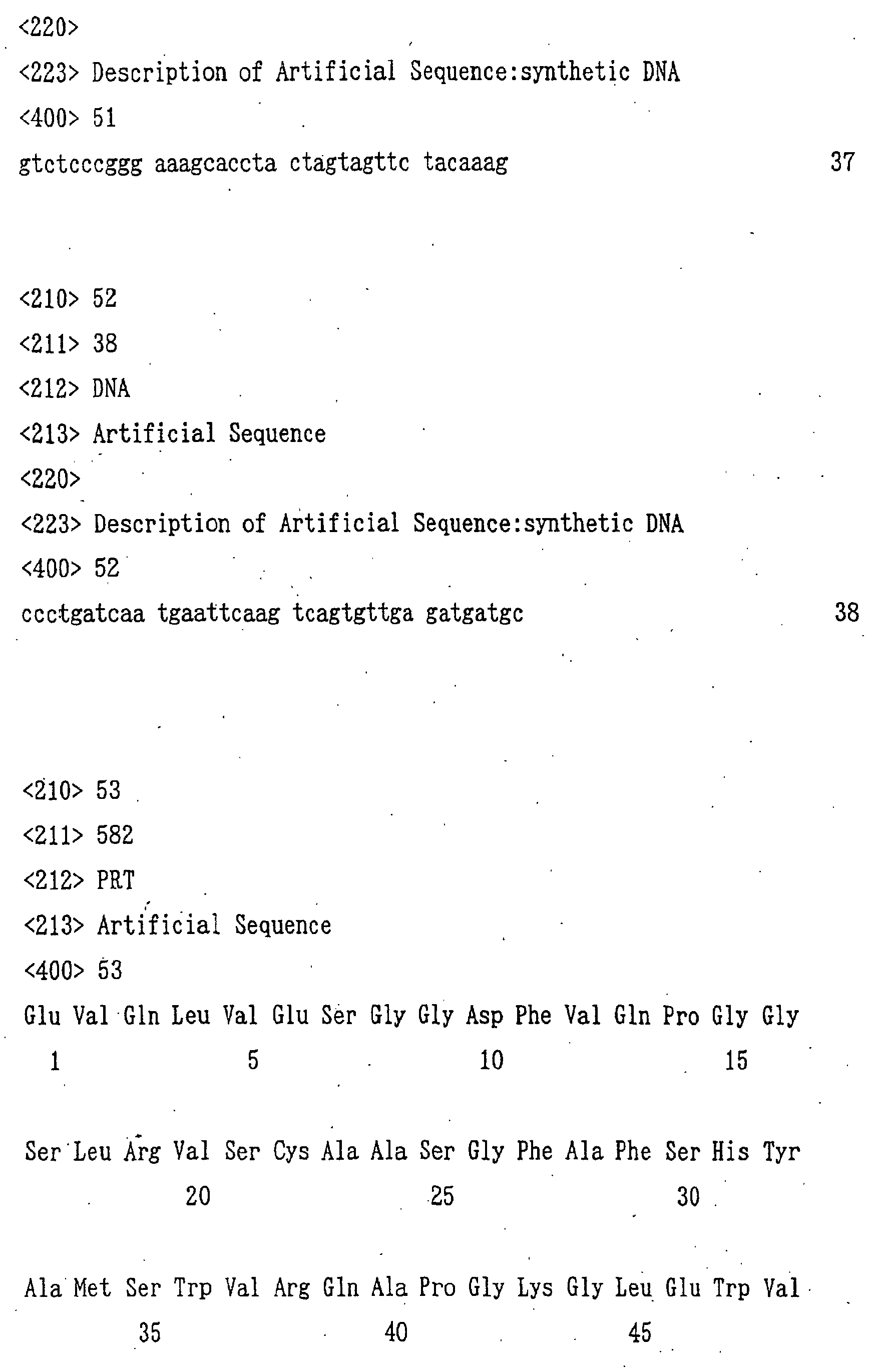
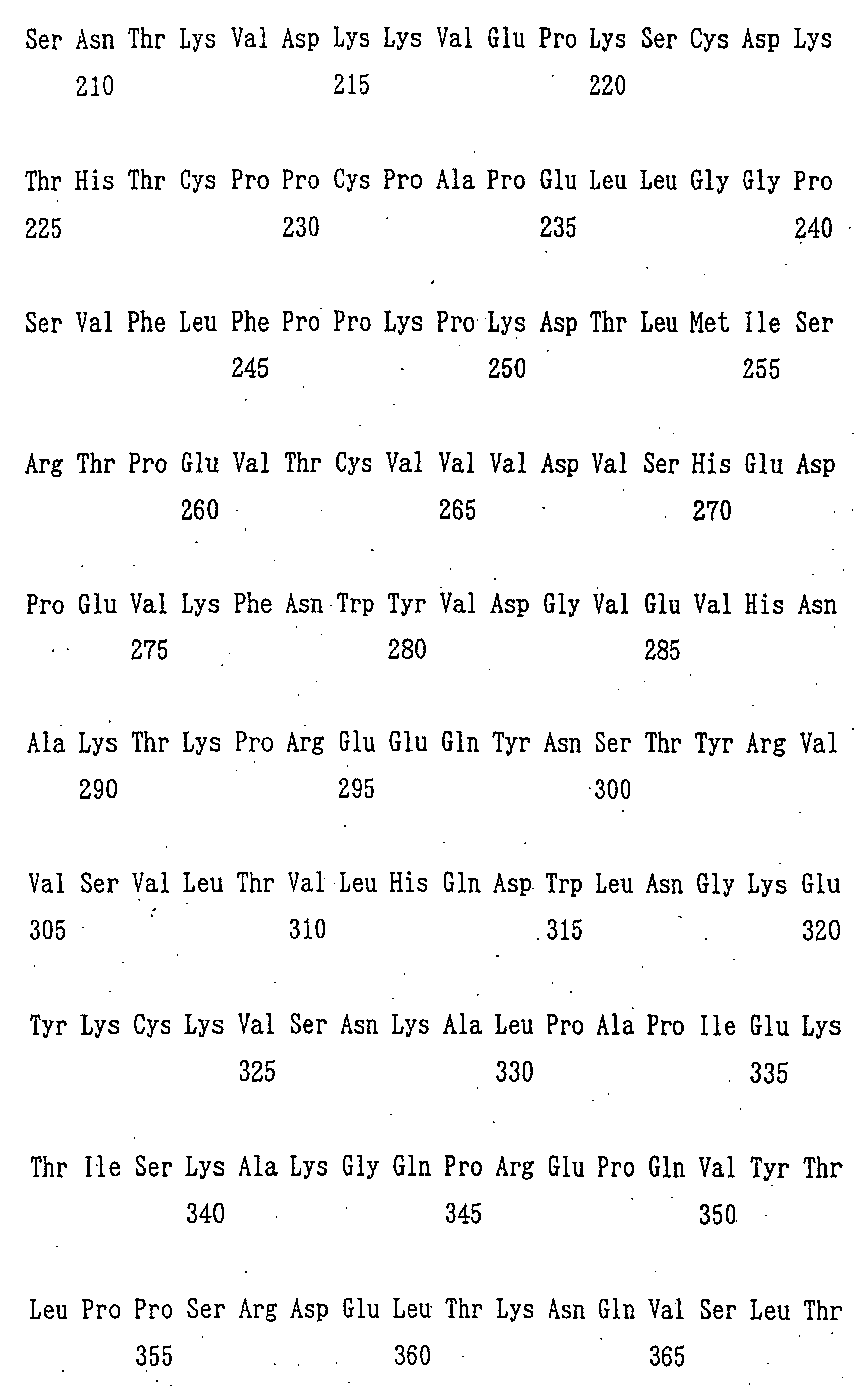
Examples of the fusion protein of the anti-GD3
human antibody with cytokine include a fusion protein of
anti-GD3 CDR-grafted antibody KM8871 with hIL-2 and a
fusion protein of anti-GD3 chimeric antibody KM871 with
hIL-2. Examples. of the fusion protein of anti-GD3 CDR-grafted
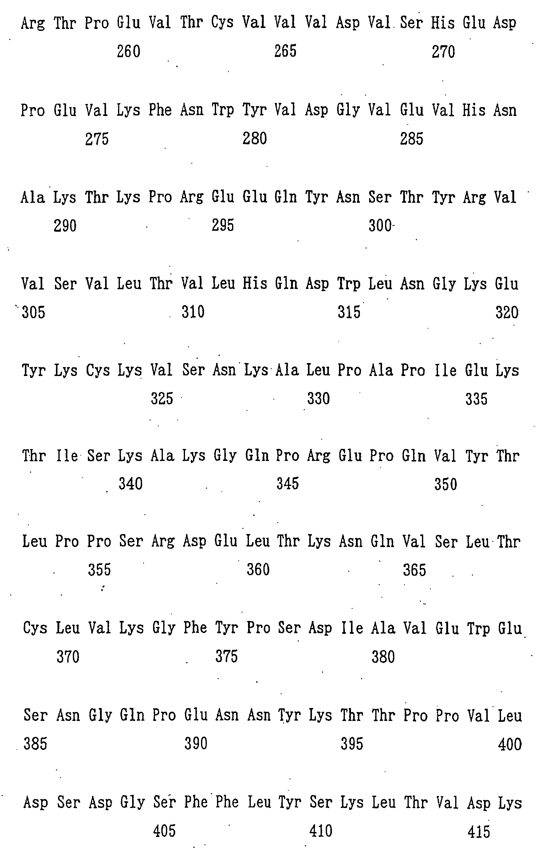
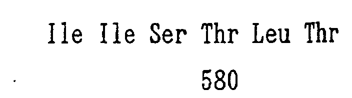
antibody KM8871 with hIL-2 include KM8871-hIL-2 in
which the H chain V region of the antibody conjugated with
hIL-2 has the amino acid sequence represented by SEQ ID
NO:53, the L chain V region of the antibody has the amino
acid sequence represented by SEQ ID NO:54 and the L chain C
region has an amino acid sequence of a human antibody κ
class. Examples of the fusion protein of anti-GD3 chimeric
antibody KM871 with hIL-2 include KM871-hIL-2 in which the
H chain V region of the antibody conjugated with hIL-2 has
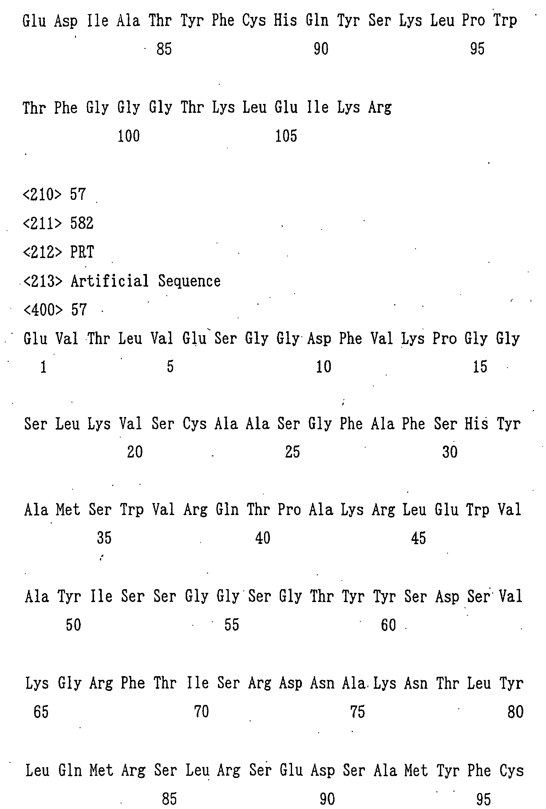
the amino acid sequence represented by SEQ ID NO:57, the L
chain V region of the antibody has the amino acid sequence
represented by SEQ ID NO:56 and the L chain C region has an
amino acid sequence of a human antibody κ class.
The methods for producing a humanized antibody
which specifically reacts with GD3 and derivatives of an
antibody or the antibody fragment which specifically reacts
with GD3.
BRIEF DESCRIPTION OF THE DRAWINGS
Fig. 1 is a drawing showing construction steps of
plasmid phKM641H.
Fig. 2 is a drawing showing construction steps of
plasmid phKM641L.
Fig. 3 is a drawing showing construction steps of
plasmid pT796H641L.
Fig. 4 is a drawing showing construction steps of
plasmid pBS641H.
Fig. 5 is a drawing showing construction steps of
plasmid pT641.
Fig. 6 is a drawing showing construction steps of
plasmid pT641HCDR.
Fig. 7 is a drawing showing construction steps of
plasmids pT641LCDR and pT641HLCDR.
Fig. 8 is a drawing showing activity evaluation of
an anti-GD3 chimeric antibody and an anti-GD3 CDR-grafted
antibody by their transient expression using plasmids pT641,
pT641HCDR, pT641LCDR and pT641HLCDR. The ordinate and the
abscissa represent the expression vector name and the
relative activity (%) when the activity of anti-GD3
chimeric antibody is defined as 100, respectively.
Fig. 9 is a drawing showing construction steps of
plasmid phKM641Lm-69.
Fig. 10 is a drawing showing construction steps of
plasmids pT641HLCDRNL, pT641HLCDRLm-1, pT641HLCDRLm-4,
pT641HLCDRLm-6, pT641HLCDRLm-7, pT641HLCDRLm-8,
pT641HLCDRLm-9 and pT641HLCDRLm-69.
Fig. 11 is a drawing showing activity evaluation of
anti-GD3 chimeric antibody and anti-GD3 CDR-grafted
antibody by their transient expression using plasmids pT641,
pT641HLCDR, pT641HLCDRNL, pT641HLCDRLm-1, pT641HLCDRLm-4,
pT641HLCDRLm-6, pT641HLCDRLm-7, pT641HLCDRLm-8,
pT641HLCDRLm-9 and pT641HLCDRLm-69. The ordinate and the
abscissa represent the expression vector name and the
relative activity (%) when the activity of anti-GD3
chimeric antibody is defined as 100, respectively.
Fig. 12 is a drawing showing construction steps of
plasmids pKANTEX641HLCDR, pKANTEX641HLCDRLm-9 and
pKANTEX641HLCDRLm-69.
Fig. 13 is a drawing showing SDS-PAGE (using a 4 to
15% gradient gel) electrophoresis patterns of purified
anti-GD3 CDR-grafted antibodies KM8869, KM8870 and KM8871.
The left side shows results of electrophoresis carried out
under non-reducing conditions, and the right side shows
those under reducing conditions. Lanes 1, 2, 3, 4, 5, 6, 7
and 8 show electrophoresis patterns of high molecular
weight markers, KM8869, KM8870, KM8871, low molecular
weight markers, KM8869, KM8870 and KM8871, respectively.
Fig. 14 is a drawing showing binding activity of
purified anti-GD3 chimeric antibody KM871 and purified
anti-GD3 CDR-grafted antibodies KM8869, KM8870 and KM8871
to GD3 measured by changing the antibody concentration.
The ordinate and the abscissa are the binding activity to
GD3 and the antibody concentration, respectively. " ○ ", "
", " □ " and " ▪ " show the activities of KM871, KM8869,
KM8870 and KM8871, respectively.
Fig. 15 is a drawing showing binding activity of
purified anti-GD3 chimeric antibody KM871 and purified
anti-GD3 CDR-grafted antibodies KM8869, KM8870 and KM8871
to GD3 measured by changing an amount of GD3 to be adsorbed
to a plate. The ordinate and the abscissa are the binding
activity to GD3 and the amount of GD3 adsorbed to the plate,
respectively. " ○ ", " ", " □ " and " ▪ " show the
activities of KM871, KM8869, KM8870 and KM8871,
respectively.
Fig. 16 is a drawing showing reactivity of purified
anti-GD3 chimeric antibody KM871 and purified anti-GD3 CDR-grafted
antibodies KM8869, KM8870 and KM8871 with various
gangliosides. The ordinate and the abscissa are the kind
of ganglioside and the binding activity, respectively.
AcGM2, GcGM2, AcGM3 and GcGM3 indicate N-acetylGM2, N-glycolylGM2,
N-acetylGM3 and N-glycolylGM3. " □ ", " □ ", "
□ " and " ▪ " show the reactivities of KM871, KM8869, KM8870
and KM8871, respectively.
Fig. 17 is a drawing showing reactivity of purified
anti-GD3 chimeric antibody KM871 and purified anti-GD3 CDR-grafted
antibodies KM8869, KM8870 and KM8871 with human
melanoma cell lines G-361 and SK-MEL-28. The ordinate and
the abscissa are the number of cells and the fluorescence
intensity, respectively. The graphs show reactivities of
control, KM871, KM8869, KM8870 and KM8871, respectively,
from the bottom.
Fig. 18 is a drawing showing CDC activity of
purified anti-GD3 chimeric antibody KM871 and purified
anti-GD3 CDR-grafted antibodies KM8869, KM8870 and KM8871
against human melanoma cell lines G-361 and SK-MEL-28. The
ordinate and the abscissa are the cytotoxic activity and
the antibody concentration, respectively. " □ ", " □ ", " D
" and " ▪ " " show the reactivities of KM871, KM8869, KM8870
and KM8871, respectively.
Fig. 19 is a drawing showing ADCC activity of
purified anti-GD3 chimeric antibody KM871 and purified
anti-GD3 CDR-grafted antibodies KM8869, KM8870 and KM8871
against human melanoma cell lines G-361 and SK-MEL-28. The
ordinate and the abscissa are the cytotoxic activity and
the antibody concentration, respectively. "□", " □", " D
" and " ▪ " show the reactivities of KM871, KM8869, KM8870
and KM8871, respectively.
Fig. 20 is a drawing showing construction steps of
plasmid pBSA-B.
Fig. 21 is a drawing showing construction steps of
plasmid pBSΔhCγ1-IL-2.
Fig. 22 is a drawing showing construction steps of
plasmid pBShCγ1-IL-2.
Fig. 23 is a drawing showing construction steps of
plasmid pKANTEX8871-hIL-2.
Fig. 24 is a drawing showing SDS-PAGE (using a 4 to
15% gradient gel) electrophoresis patterns of purified
anti-GD3 CDR-grafted antibody KM8871 and purified fusion
protein KM8871-hIL-2. The left side and the right side are
results of electrophoresis carried out under non-reducing
conditions, and those under reducing conditions,
respectively. Lanes 1, 2, 3, 4, 5 and 6 show
electrophoresis patterns of low molecular weight markers,
KM8871, KM8871-hIL-2, low molecular weight markers, KM8871
and KM8871-hIL-2, respectively.
Fig. 25 is a drawing showing binding activity of
purified anti-GD3 CDR-grafted antibody KM8871 and purified
fusion protein KM8871-hIL-2 to GD3 measured by changing the
antibody concentration. The ordinate and the abscissa are
the binding activity to GD3 and the antibody concentration.
"○" and "" show the activities of KM8871 and of KM8871-hIL-2,
respectively.
Fig. 26 is a drawing showing reactivity of purified
anti-GD3 CDR-grafted antibody KM8871 and purified fusion
protein KM8871-hIL-2 with various gangliosides. The
ordinate and the abscissa are the kind of ganglioside and
the binding activity. AcGM2, GcGM2, AcGM3 and GcGM3
indicate N-acetylGM2, N-glycolylGM2, N-acetylGM3 and N-glycolylGM3,
respectively. " □ " and " ▪ " show the
reactivities of KM8871 and KM8871-hIL-2, respectively.
Fig. 27 is a drawing showing reactivity of purified
anti-GD3 CDR-grafted antibody KM8871 and purified fusion
protein KM8871-hIL-2 with human melanoma cell line G-361.
The ordinate and the abscissa are the number of cells and
the fluorescence intensity, respectively. The drawings
show the reactivities of KM8871 and KM8871-hIL-2,
respectively, from the upper column.
Fig. 28 is a drawing showing growth-supporting
activity of hIL-2 and purified fusion protein KM8871-hIL-2
against hIL-1-dependent cell CTLL-2 measured by changing
concentration of each protein. The ordinate and the
abscissa are the growth-supporting activity and the protein
concentration, respectively. " ○ " and " " indicate the
activities of hIL-2 and KM8871-hIL-2, respectively.
Fig. 29 is a drawing showing results of the
measurement of the activation and accompanying cytotoxic
activity of human effector cells by purified anti-GD3 CDR-grafted
antibody KM8871 and purified fusion protein KM8871-hIL-2.
The ordinate and the abscissa are the cytotoxic
activity and the used protein, respectively. " □ " and " ▪
" indicate the activities of KM8871 and KM8871-hIL-2,
respectively.
Fig. 30 is a drawing showing construction steps of
plasmid pKANTEX871-hIL-2.
Fig. 31 is a drawing showing SDS-PAGE (using a 4 to
15% gradient gel) electrophoresis patterns of anti-GD3
chimeric antibody KM871 and purified fusion protein KM871-hIL-2.
The left side and the right side are results of
electrophoresis carried out under reducing conditions and
under non-reducing conditions, respectively. Lanes 1, 2, 3,
4, 5 and 6 show electrophoresis patterns of low molecular
weight markers, KM871, KM871-hIL-2, low molecular weight
markers, KM871 and KM871-hIL-2, respectively.
Fig. 32 is a drawing showing binding activity of
anti-GD3 chimeric antibody KM8871 and purified fusion
protein KM871-hIL-2 to GD3 measured by changing the
antibody concentration. The ordinate and the abscissa are
the binding activity to GD3 and the antibody concentration,
respectively. " " and " □ " show the activities of KM871
and KM871-hIL-2, respectively.
Fig. 33 is a drawing showing reactivity of anti-GD3
chimeric antibody KM871 and purified fusion protein KM871-hIL-2
with various gangliosides. The ordinate and the
abscissa are the kind of ganglioside and the binding
activity, respectively. AcGM2, GcGM2, AcGM3 and GcGM3
indicate N-acetylGM2, N-glycolylGM2, N-acetylGM3 and N-glycolylGM3,
respectively. " ▪ " and " □ " show the
reactivities of KM871 and KM871-hIL-2, respectively.
Fig. 34 is a drawing showing reactivity of anti-GD3
chimeric antibody KM871 and purified fusion protein KM871-hIL-2
with human melanoma cell lines G-361 and SK-MEL-28.
The ordinate and the abscissa are the number of cells and
the fluorescence intensity, respectively. The graphs show
the reactivities of KM871 to G361 cell, KM871-hIL-2 to G361
cell, KM871 to SK-MEL-28 and KM871-hIL-2 to SK-MEL-28,
respectively, from the upper column.
Fig. 35 is a drawing showing growth-supporting
activity of hIL-2 and purified fusion protein KM871-hIL-2
against hIL-2-dependent cell CTLL-2 measured by changing
concentration of each protein. The ordinate and the
abscissa are the growth-supporting activity and the protein
concentration, respectively. " " and " ○ " indicate the
activities of hIL-2 and KM871-hIL-2, respectively.
Fig. 36 is a drawing showing results of the
measurement of the activation and accompanying cytotoxic
activity of human effector cells by anti-GD3 chimeric
antibody KM871 and purified fusion protein KM871-hIL-2.
The ordinate and the abscissa are the cytotoxic activity
and the used protein, respectively. " ▪ " and " □ " indicate
activities of KM871 and KM871-hIL-2, respectively.
Fig. 37 is a drawing showing construction steps of
plasmid pKANTEXGD3.
Fig. 38 is a drawing showing reactivity of anti-GD3
chimeric antibody KM871 with GD3 synthase gene-introduced
B16 cell lines B16·29-10 and B16·8-3 and the parent line
B16·F10 cell. The ordinate and the abscissa are the number
of cells and the fluorescence intensity, respectively. The
drawings show the reactivities of B16·F10 cell, B16·29-10
and B16·8-3, respectively, from the upper column.
Fig. 39 is a drawing showing results of the
measurement of anti-metastatic effects of anti-GD3 chimeric
antibody KM871 and KM871-hIL-2 on BL/6 mice into which a
GD3-positive B16 cell line B16·29-10 is transplanted. The
ordinate and the bar of the graph show the number of
metastatic foci on the lung surface and the average value
of each group, respectively. No administration means a
control group in which the antibody is not administered.
" ⋄ ", " ○ " and " " indicate the numbers of lung-metastasized
foci in mice of the control group, the KM871-hIL-2-administered
group and the KM871-administered group,
respectively.
Fig. 40 is a drawing showing a result of the
measurement of anti-metastatic effects of anti-GD3 chimeric
antibody KM871 and KM871-hIL-2 on BL/6 mice into which a
GD3-positive B16 cell line B16·8-3 is transplanted. The
ordinate and the bar of the graph show the number of
metastatic foci on the lung surface and the average value
of each group, respectively. No administration means a
control group in which the antibody is not administered.
" ⋄ ", " ○ " and " " indicate the numbers of lung-metastasized
foci in mice of the control group, the KM871-hIL-2-administered
group and the KM871-administered group,
respectively.
Fig. 41 is a drawing showing results of the
measurement of anti-metastatic effects by combined
administration of anti-GD3 chimeric antibody KM871 and hIL-2
and administration of KM871-hIL-2 on BL/6 mice into which
a GD3-positive B16 cell line B16·29-10 is transplanted.
The ordinate and the bar of the graph show the number of
metastatic foci on the lung surface and the average value
of each group. No administration means a control group in
which the antibody is not administered. " ⋄", " ○ " and "
" indicate the number of lung-metastasized foci in mice of
the control group, the KM871-hIL-2-administered group and
the KM871/hIL-2 combined administered group, respectively.
Fig. 42 is a drawing showing results of the
measurement of growth inhibition effects of KM871-hIL-2 on
a solid tumor early stage model of BL/6 mice into which a
GD3-positive B16 cell line B16·29-10 is subcutaneously
transplanted. The ordinate and the abscissa of the graph
show the ratio of the average value of tumor volumes in
each group to the average value of tumor volumes in the
control group and days after commencement of the agent
administration, respectively. Dotted line, " □ ", " Δ " and
" ○ " indicate control, the KM871-administered group, the
hIL-2-administered group and the KM871-hIL-2-administered
group, respectively.
Fig. 43 is a drawing showing results of the
measurement of growth inhibition effects of KM871-hIL-2 on
a solid tumor advanced stage model of BL/6 mice into which
a GD3-positive B16 cell line B16·29-10 is subcutaneously
transplanted. The ordinate of the graph shows the ratio of
average value of V/V0 values in each group to average value
of V/V0 values in the control group. V/V0 indicates the
ratio of the tumor volume of each mouse on each measured
day to the tumor volume on the day of commencement of the'
administration. The abscissa shows days after commencement
of the agent administration. Dotted line, " Δ ", " □ ", " ▪
", " ○ " and " " indicate control, the hIL-2-administered
group, the KM871-administered group at 50 µg/day, the
KM871-administered group at 200 µg/day, the KM871-hIL-2-administered
group at 24 µg/day and KM871-hIL-2-administered
group at 60 µg/day.
Fig. 44 is a drawing showing results of the
measurement of growth inhibition effects by combined
administration of anti-GD3 chimeric antibody KM871 and
KM871-hIL-2 on a solid tumor advanced stage model of BL/6
mice into which a GD3-positive B16 cell line B16-29-10 is
subcutaneously transplanted. The ordinate of the graph
shows the ratio of the average value of V/V0 values in each
group to the average value of V/V0 values in the control
group. V/V0 indicates the ratio of tumor volume of each
mouse on each measured day to tumor volume on the day of
commencement of the administration. The abscissa shows
days after commencement of the agent administration.
Dotted line, " Δ ", " ▪ ", " ○ " and " " indicate control,
the hIL-2-administered group, the KM871-administered group,
the combined administration group of KM871 and KM871-hIL-2
at 15 µg/day and the combined administration group of KM871
and KM871-hIL-2 at 60 µg/day.
Fig. 45 is a drawing showing results of measurement
of the activation and accompanying enhance in cytotoxic
activity of BL/6 mouse spleen cells by hIL-2 or KM871-hIL-2.
The ordinate and the abscissa are the cytotoxic activity
and the concentration of KM871 used in the cytotoxic
activity measurement, respectively. Black bar, white bar,
gray bar and shaded bar indicate the activity when spleen
cells of non-administered control mice are used, the
activity when spleen cells of mice to which hIL-2 is
administered five times are used, the activity when spleen
cells of mice to which KM871-hIL-2 is administered once are
used and the activity when spleen cells of mice to which
KM871-hIL-2 is administered five times are used,
respectively.
Fig. 46 is a drawing showing the ratio of the
number of cells of each cell group in spleen cells of BL/6
mouse to which hIL-2 or KM871-hIL-2 is administered,
measured by an immunofluorescent method. The ordinate and
the abscissa are the ratio in spleen cells of each cell
group and each cell group, respectively. White bar, gray
bar, black bar and shaded bar indicate the ratio in spleen
cells of non-administered control mice, the ratio in spleen
cells of mice to which hIL-2 is administered five times,
the ratio in spleen cells of mice to which KM871-hIL-2 is
administered five times and the ratio in spleen cells of
mice to which KM871-hIL-2 is administered twice,
respectively.
Fig. 47 is a drawing showing a result of
measurement of cytotoxic activity by spleen cells when
KM871-hIL-2 is administered to BL/6 mice from which each of
NK cell, CD4-positive T cell and CD8-positive T cells are
depleted. The ordinate shows cytotoxic activity. White
'bar, black bar, gray bar and shaded bar indicate the
cytotoxic activity of mouse spleen cells which is not
subjected to the removing treatment, the cytotoxic activity
of NK cell-depleted mouse spleen cells, the cytotoxic
activity of CD4-positive T cell-depleted mouse spleen cells
and the cytotoxic activity of CD8-positive T cell-depleted
mouse spleen cells, respectively.
Fig. 48 is a drawing showing results of the
measurement of anti-metastatic effects of KM871-hIL-2 on
BL/6 mice in which a GD3-positive B16 cell line B16·29-10
is transplanted into mice from which each of NK cell, CD4-positive
T cell and CD8-positive T cell are depleted. The
ordinate shows the number of metastatic foci. " ○ ", " ",
horizontal bar and the value in the graph indicate the
number of metastatic foci of mouse lung, the number of
metastatic foci of KM871-hIL-2-administered mouse lung, the
average value of each group and the metastasis inhibition
ratio by KM871-hIL-2 administration.
BEST MODE FOR CARRYING OUT THE INVENTION
Examples of the present invention are shown below,
though the scope of the present invention is not restricted
thereby.
Example 1
Production of anti-GD3 CDR-grafted antibody:
1. Analysis of amino acid sequence of V region of anti-GD3
mouse antibody KM641
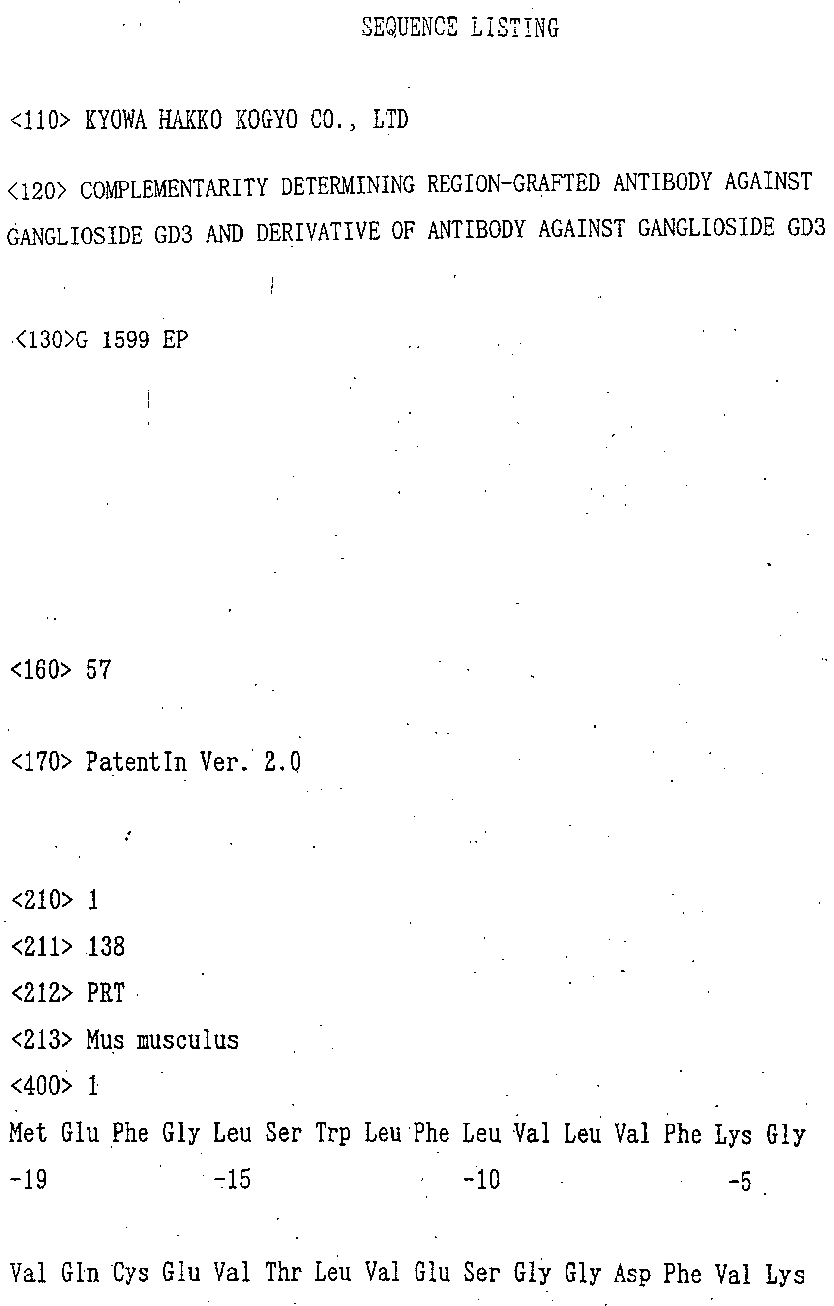
A full amino acid sequence of VH of anti-GD3 mouse
antibody KM641 disclosed in Japanese Published Unexamined
Patent Application No. 304989/93 is represented by SEQ ID
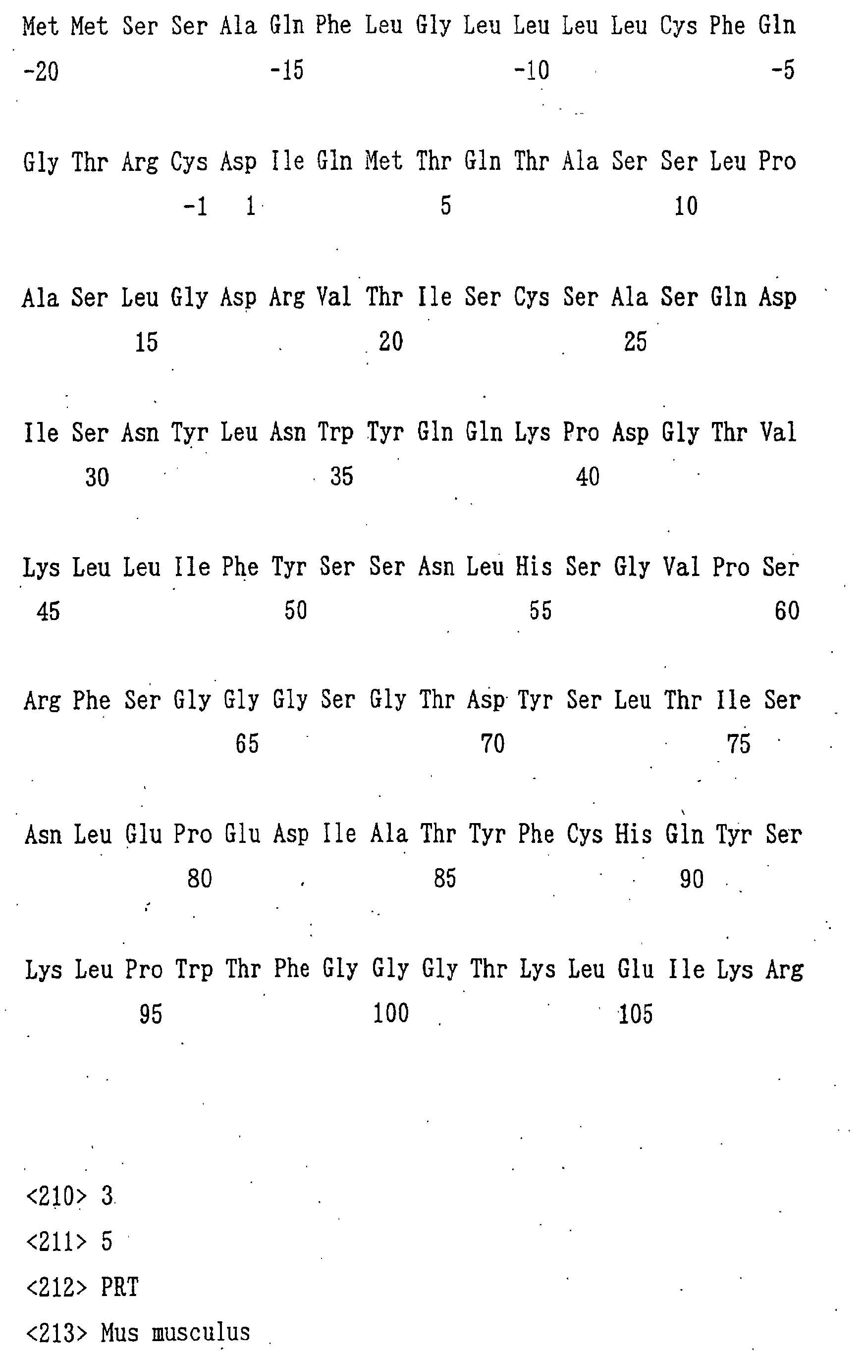
NO:1, and a full amino acid sequence of VL thereof is
represented by SEQ ID NO:2. Based on the comparison of
both sequences with analytical results of amino acid
sequences of known antibodies (Sequences of Proteins of
Immunological Interest) and N-terminal amino acid sequences
of H chain and L chain of purified anti-GD3 mouse antibody
KM641, it was confirmed that positions (-19) to (-1) in the
amino acid sequence of H chain and positions (-20) to (-1)
in the amino acid sequence of L chain are secretory signal
sequences. Full amino acid sequences of the secretory VH
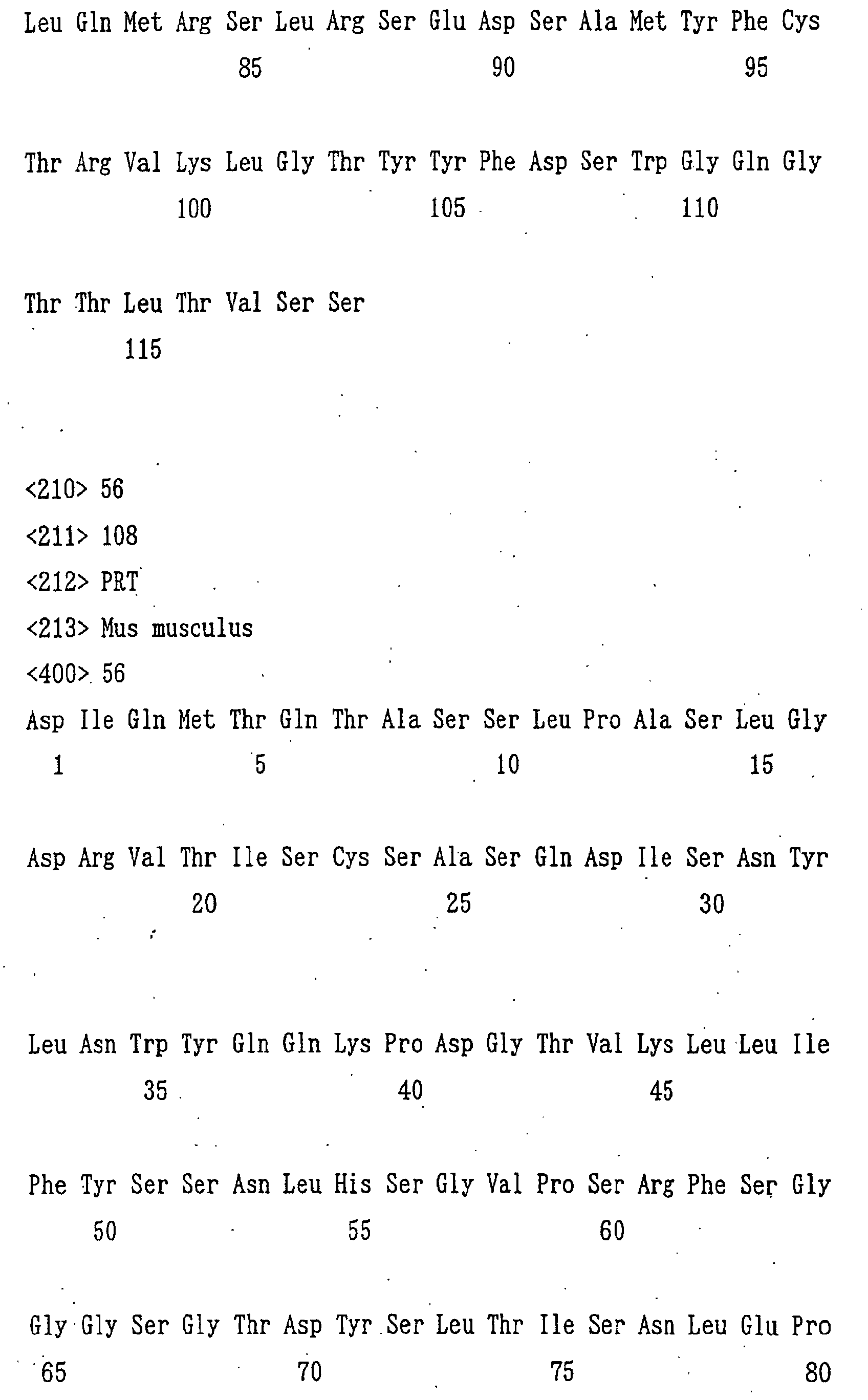
and VL are represented by SEQ ID NO:55 and SEQ ID NO:56,
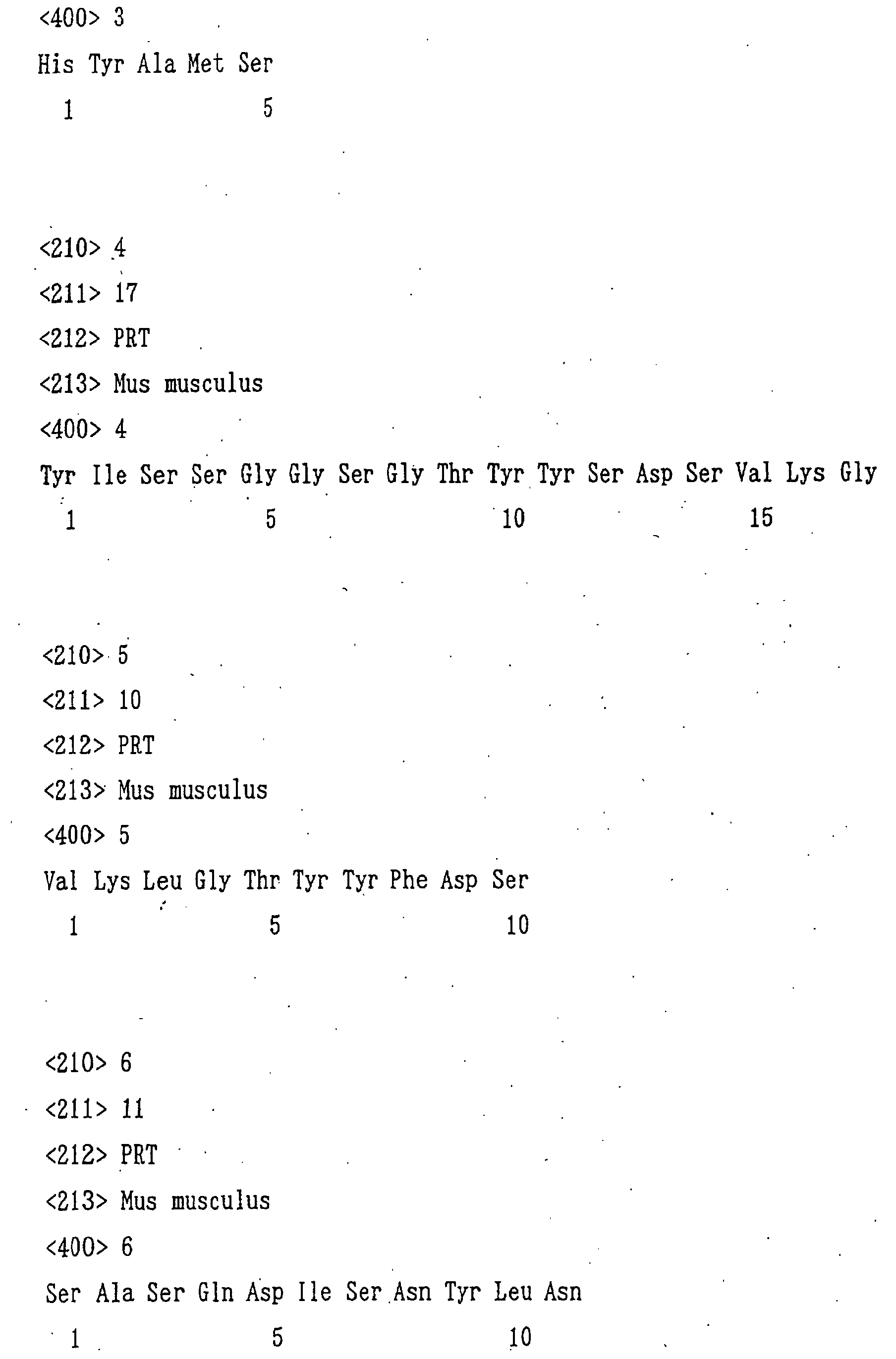
respectively. Also, it was found that CDRs 1, 2 and 3 of
VH have the amino acid sequences represented by SEQ ID
NOs:3, 4 and 5, and CDRs 1, 2 and 3 of VL have those
represented by SEQ ID NOs:6, 7 and 8.
2. Measurement of binding activity of antibody to various
ganglioside (ELISA)
The binding activity of antibodies to various
gangliosides was measured as follows.Each ganglioside (2 nmol) was dissolved in 2 ml of
ethanol solution containing 10 µg of
dipalmitoylphosphatidylcholine (manufactured by SIGMA) and
5 µg of cholesterol (manufactured by SIGMA). In each well
of a 96 well plate for ELISA use (manufactured by Greiner),
20 µl of the solution (becomes 20 pmol/well) or 20 µl of
the solution diluted with ethanol was respectively
dispensed, followed by air-drying, and then PBS containing
1% BSA (hereinafter referred to as "1% BSA-PBS") was added
in 100 µl/well and allowed to react at room temperature for
1 hour to thereby block remaining active groups. After
discarding 1% BSA-PBS, culture supernatants of
transformants or diluted solutions of humanized antibodies
were added in 50 µl/well and allowed to react at room
temperature for 1 hour. After the reaction, each well was
washed with PBS containing 0.05% Tween 20 (hereinafter
referred to as "Tween-PBS") and then a peroxidase-labeled
goat anti-human IgG(γ) antibody solution (manufactured by
Kirkegaard & Perry Laboratories) diluted 1,000 folds with
1% BSA-PBS was added as a secondary antibody solution in 50
µl/well and allowed to react at room temperature for 1 hour.
After the reaction and subsequent washing with Tween-PBS,
an ABTS substrate solution (a solution prepared by
dissolving 0.55 g of 2,2'-azinobis(3-ethylbenzothiazoline-6-sulfonic
acid) in 1 L of 0.1 M citrate buffer (pH 4.2)
and adding 1 µl/ml hydrogen peroxide just before the use)
was added in 50 µl/well for color development and the
absorbance at 415 nm (hereinafter referred to as "OD415")
was measured.
3. Construction of cDNAs encoding VH and VL of anti-GD3
CDR-grafted antibody
(1) Design of amino acid sequences of VH and VL of anti-GD3
CDR-grafted antibody
First, an amino acid sequence of VH of an anti-GD3
CDR-grafted antibody was designed as follows. In order to
graft the CDR amino acid sequences of VH of the anti-GD3
mouse antibody KM641 identified in the item 1 of Example 1,
an FR amino acid sequence of VH of a human antibody was
selected. Kabat et al. have classified the VH of various
known human antibodies into three subgroups (HSG I to III)
based on the homology of the amino acid sequences and then
reported consensus sequences among respective subgroups
(Sequences of Proteins of Immunological Interest). Since
these consensus sequences have a possibility that the
immunogenicity is reduced in human, it was decided to
design a VH amino acid sequence of an anti-GD3 CDR-grafted
antibody based on these consensus sequences. In order to
produce an anti-GD3 CDR-grafted antibody having higher
activity in designing it, it was decided to select an FR
amino acid sequence having the highest homology with the FR
amino acid sequence of VH of KM641, among FR amino acid
sequences of consensus sequences of the three subgroups of
VH of human antibodies. Results of the homology search are
shown in Table 1. As shown in Table 1, the FR amino acid
sequence of VH of KM641 showed the most high homology with
the subgroup III.
| Homology between FR amino acid sequence of consensus sequence of each subgroup of human antibody H chain V region and FR amino acid sequence of H chain V region of KM641 |
| HSG I | HSG II | HSG III |
| 62.1% | 56.3% | 78.2% |
Based on the above results, an amino acid sequence
HV.0 of VH of the anti-GD3 CDR-grafted antibody was
designed by grafting the CDR amino acid sequence of VH of
the anti-GD3 mouse antibody KM641 to an appropriate
position of the amino acid sequence of FR of the consensus
sequence of subgroup III of VH of the human antibody.Next, an amino acid sequence of VL of an anti-GD3
CDR-grafted antibody was designed as follows. In order to
graft the CDR amino acid sequence of VL of the anti-GD3
mouse antibody KM641 identified in the item 1 of Example 1,
an FR amino acid sequence of VL of a human antibody was
selected. Kabat et al. have classified the VL of various
known human antibodies into four subgroups (HSG I to IV)
based on the homology of the amino acid sequences and then
reported consensus sequences among respective subgroups
(Sequences of Proteins of Immunological Interest).
Accordingly, similar to the H chain, an FR amino acid
sequence having most high homology with the FR amino acid
sequence of VL of KM641 was selected from FR amino acid
sequences of consensus sequences of the four subgroups of
VL of human antibodies. Results of the homology search are
shown in Table 2. As shown in Table 2, the FR amino acid
sequence of VL of KM641 showed the most high homology with
the subgroup I.
| Homology between FR amino acid sequence of consensus sequence of each subgroup of human antibody L chain V region and FR amino acid sequence of L chain V region of KM641 |
| HSG I | HSG II | HSG III | HSG IV |
| 76.2% | 60.0% | 62.5% | 67.5% |
Based on the above results, an amino acid sequence
LV.0 of VL of the anti-GD3 CDR-grafted antibody was
designed by grafting the CDR amino acid sequence of VL of
the anti-GD3 mouse antibody KM641 to an appropriate
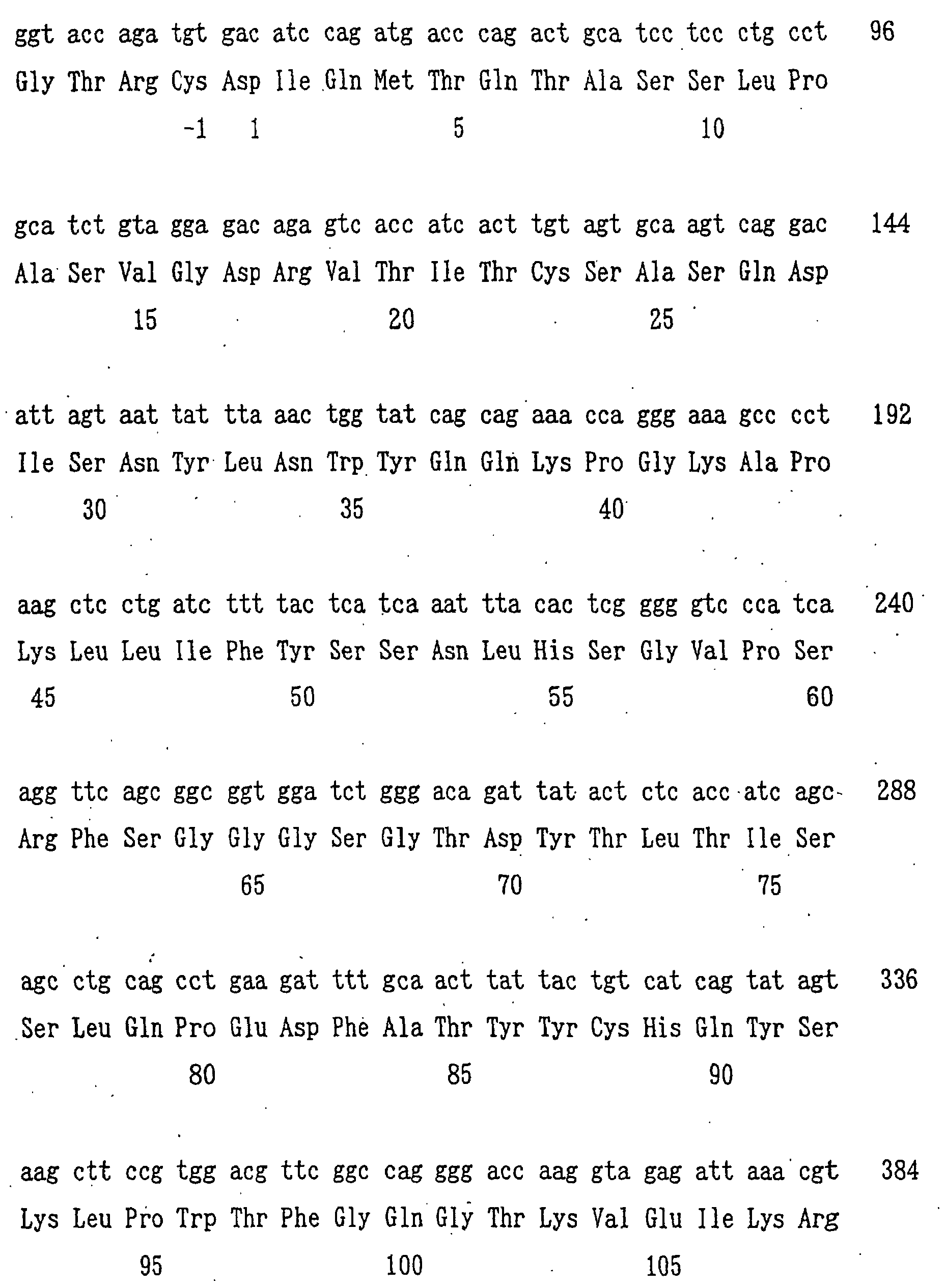
position of the amino acid sequence of FR of the consensus
sequence of subgroup I of VL of the human antibody.The thus designed amino acid sequence HV.0 of VH
and amino acid sequence LV.0 of VL of the anti-GD3 CDR-grafted
antibody are sequences in which only the CDR amino
acid sequence of the anti-GD3 mouse antibody is grafted to
the FR amino acid sequence of the selected human antibody.
In general, in the human CDR-grafted antibodies, the
antigen binding activity is reduced in many cases by
grafting of a mouse antibody CDR amino acid sequence alone.
Thus, in order to avoid the reduction of the antigen
binding activity of antibodies, among FR amino acid
residues different between a human antibody and a mouse
antibody, amino acid 'residues considered to be exerting
influences on the antigen binding activity are grafted
together with a CDR amino acid sequence. Accordingly,
identification of FR amino acid residues considered to have
influences on the activity was carried out.First, a three-dimensional structure of an antibody
V region comprised of the amino acid sequence HV.0 of VH
'and amino acid sequence LV.0 of VL of anti-GD3 CDR-grafted
antibody designed in the above (hereinafter referred to as
"HV0LV0") was constructed using computer-modeling. The
production of three-dimensional structure coordinates was
carried out using a software AbM (manufactured by Oxford
Molecular) and the display of three-dimensional structure
was carried out using a software Pro-Explore (manufactured
by Oxford Molecular) in accordance with the respective
manufacture's instructions. Also, a computer model of the
three-dimensional structure of the V region of anti-GD3
mouse antibody KM641 was constructed in the same manner.
In addition, a three-dimensional structure model of a
modified HV0LV having an amino acid sequence in which at
least one amino acid residue different from anti-GD3 mouse
antibody KM641 in the FR amino acid sequences of VH and VL
of HV0LV0 was substituted by the amino acid residues of
positions corresponding to the anti-GD3 mouse antibody
KM641 in order was also constructed. Three-dimensional
structures of V regions of the anti-GD3 mouse antibody
KM641, HV0LV0 and modified product were compared. As a
result, amino acid residues considered to have influences
on the antigen binding activity by changing three-dimensional
structure of the antigen-binding region were
selected from the FR amino acid residues of HV0LV0. As a
result of substituting the thus selected FR amino acid
residues of HV0LV0 by the amino acid residues found in the
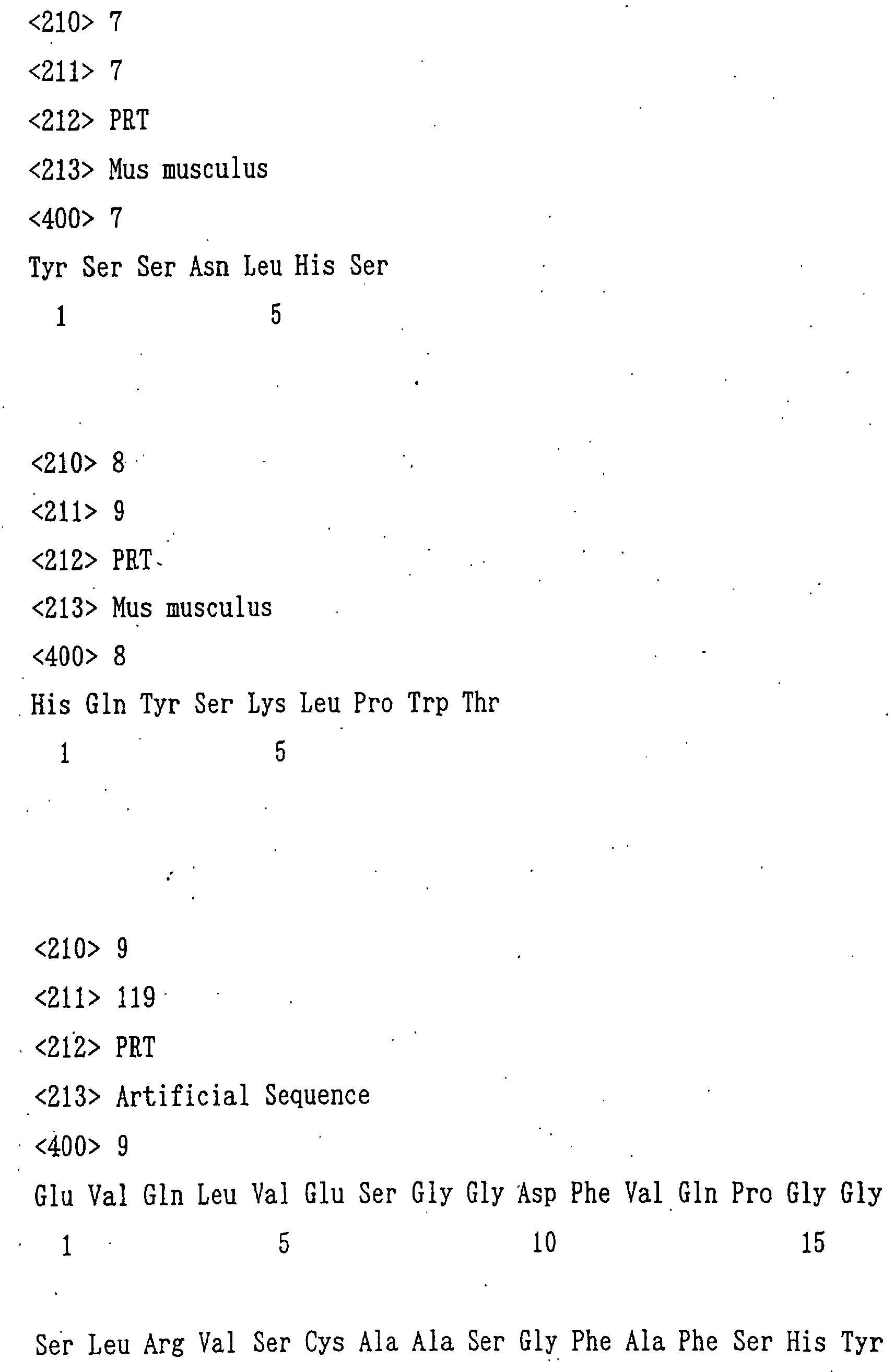
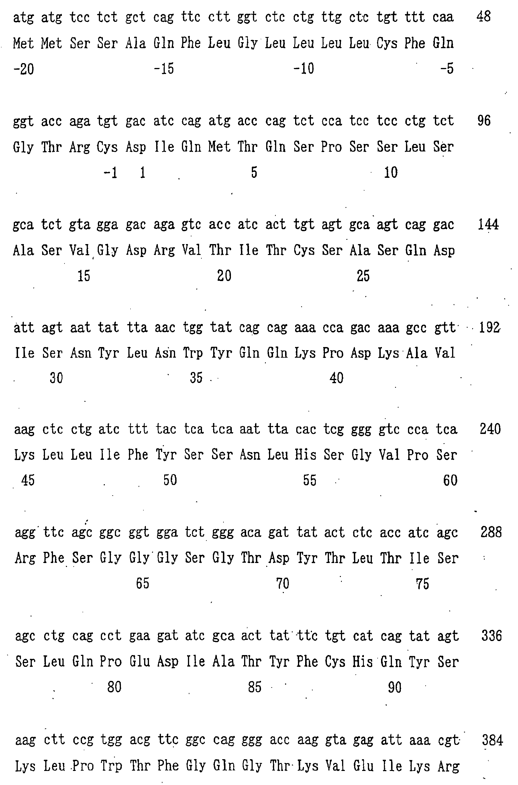
mouse antibody KM641, an amino acid sequence hKM641H of VH
of the anti-GD3 CDR-grafted antibody represented by SEQ ID
NO:9 and an amino acid sequence hKM641L of VL of the anti-GD3
CDR-grafted antibody represented by SEQ ID NO:10 were
designed. In the hKM641H, the 10th position Gly, the 11th
position Leu, the 20th position Leu, the 28th position Thr,
the 84th position Asn, the 91st position Thr, the 95th
position Tyr, the 97th position Ala and the 115th position
Val in the FR amino acid sequence of HV.0 were replaced by
Asp, Phe, Val, Ala, Arg, Ser, Phe, Thr and Leu,
respectively, as amino acid residues of positions
corresponding to the VH of anti-GD3 mouse antibody KM641.
In the hKM641L, the 49th position Tyr, the 65th position
Ser and the 71st position Phe in the FR amino acid sequence
of LV.0 were replaced by Tyr, Ser and Phe, respectively, as
amino acid residues of positions corresponding to the VL of
anti-GD3 mouse antibody KM641. (2) Construction of cDNA encoding VH of anti-GD3 CDR-grafted
antibody
A cDNA encoding the anti-GD3 CDR-grafted antibody
VH amino acid sequence hKM641H designed in the item 3(1) of
Example 1 was constructed as follows.First, the designed amino acid sequence was ligated
with the secretory signal sequence of H chain of anti-GD3
mouse antibody KM641 represented by SEQ ID NO:1 to produce
a full antibody amino acid sequence. Next, the amino acid
sequence is converted into genetic codons. When two or
more genetic codons were present for one amino acid residue,
corresponding genetic codon was determined by taking the
frequency of codon usage found in nucleotide sequences of
antibody genes into consideration (Sequences of Proteins of
Immunological Interest). A nucleotide sequence of cDNA
encoding the complete antibody V region amino acid sequence
was designed by ligating the thus determined genetic codons,
and nucleotide sequences of primer binding site for use in
the PCR amplification (including restriction enzyme
recognition sequences for cloning into a humanized antibody
expression vector) were added to the 5'-terminal and 3'-terminal.
The thus designed nucleotide sequence was
divided into a total of 6 nucleotide sequences from the 5'-terminal
side, each having about 100 bases (adjoining
nucleotide sequences are designed such that the termini
have an overlapping sequence of about 20 bases), and they
were synthesized in alternating order of the sense chain
and the antisense chain using an automatic DNA synthesizer
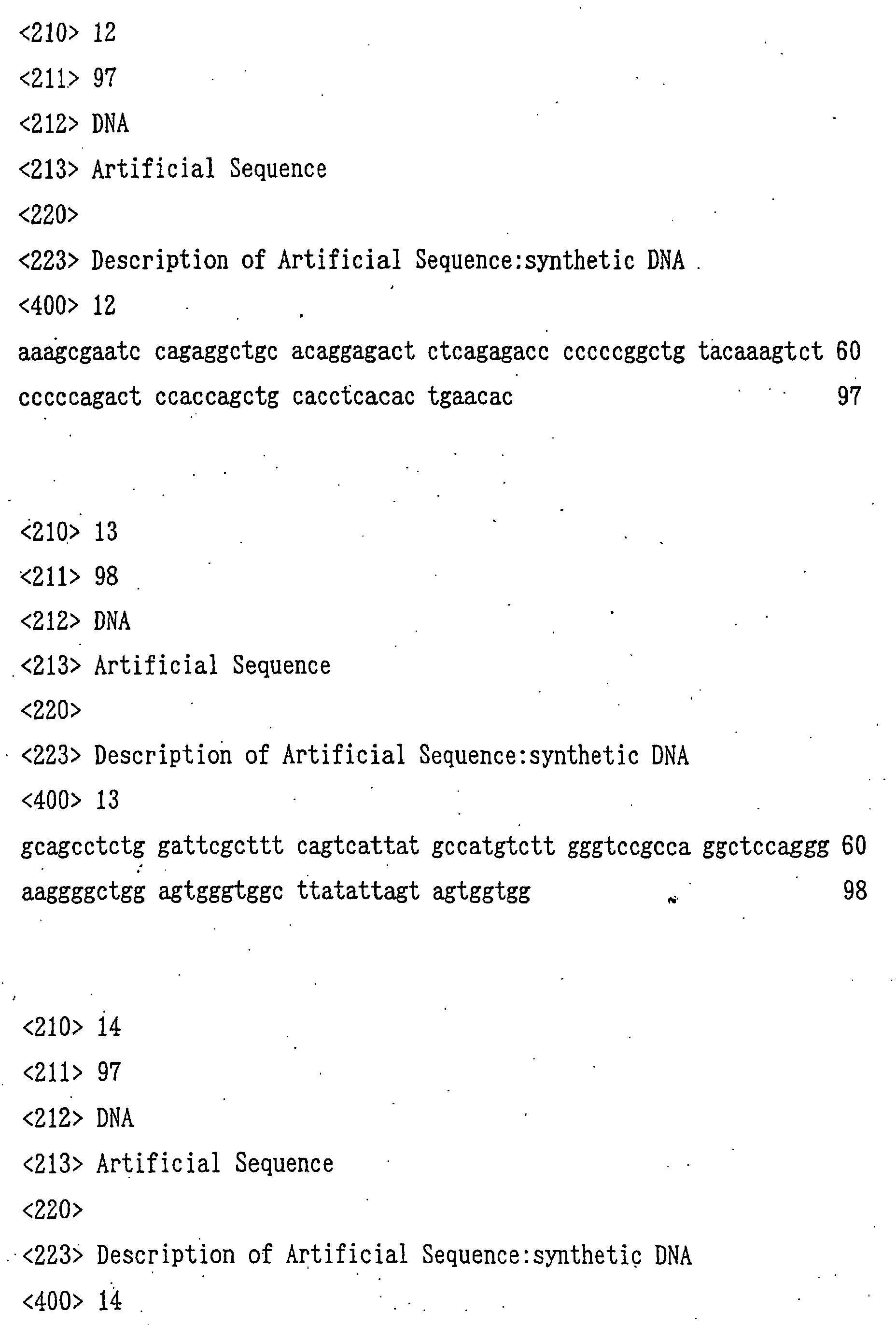
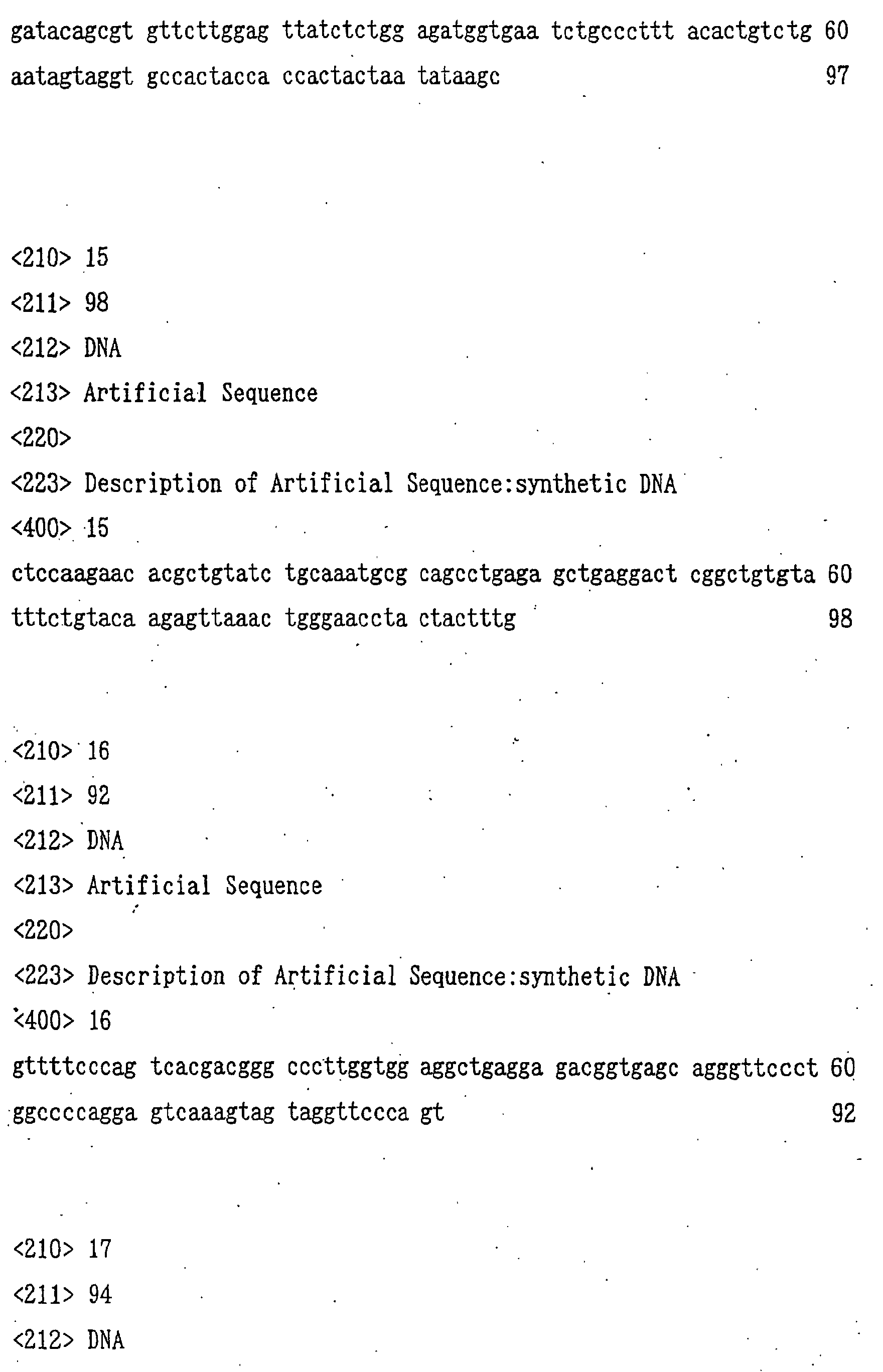
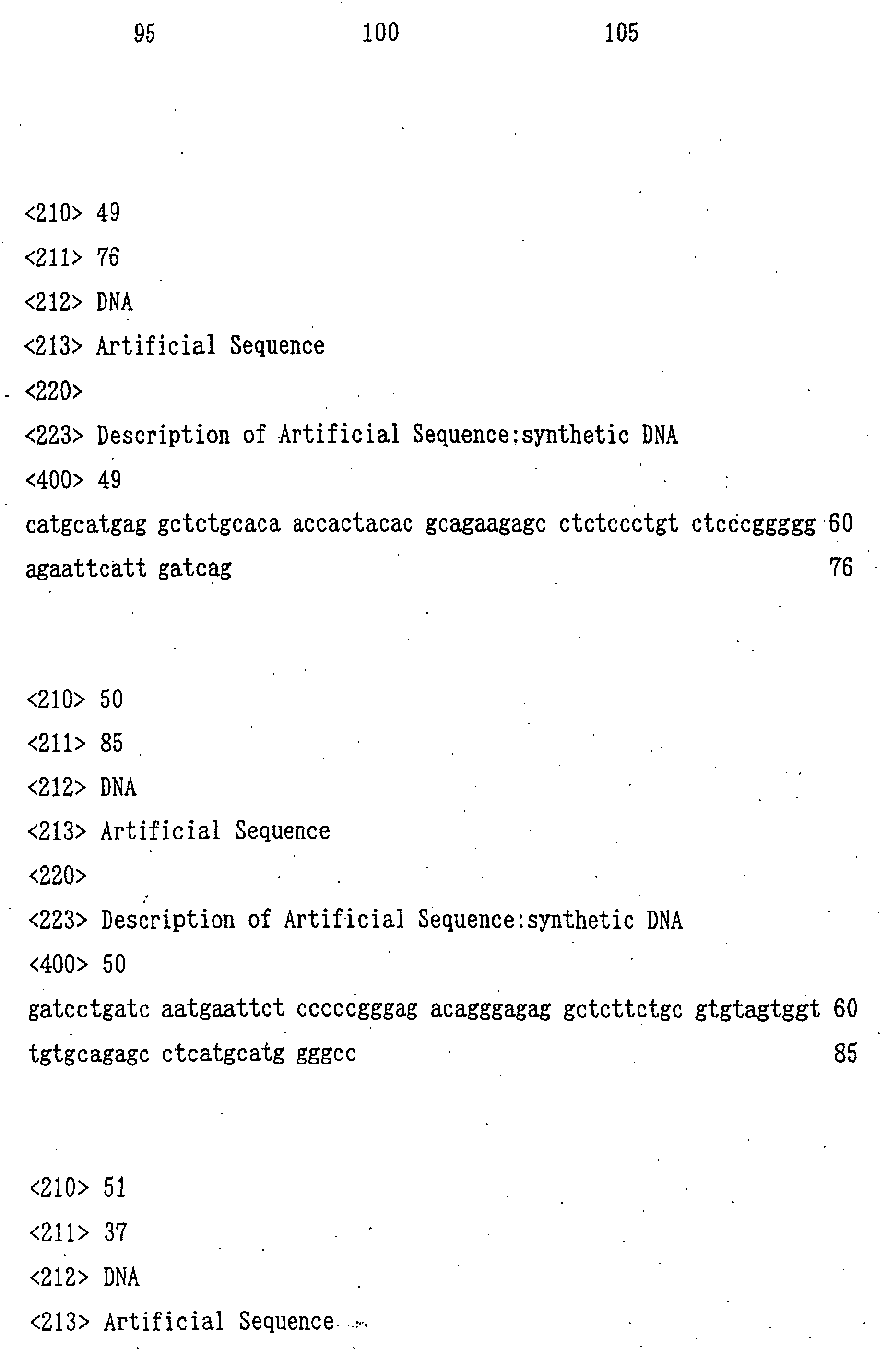
(380A, manufactured by Applied Biosystems).Specifically, 6 synthetic DNA fragments of SEQ ID
NO:11 to SEQ ID NO:16 were synthesized. Each DNA was added
to 50 µl of a buffer comprising 10 mM Tris-HCl (pH 8.3), 50
mM potassium chloride, 1.5 mM magnesium chloride, 0.001%
gelatin, 200 µM dNTPs, 0.5 µM M13 primer RV (manufactured
by Takara Shuzo), 0.5 µM M13 primer M4 (manufactured by
Takara Shuzo) and 2 units of TaKaRa Taq DNA polymerase
(manufactured by Takara Shuzo) to give a final
concentration of 0.1 µM, and the solution was covered with
50 µl of mineral oil and set to a DNA thermal cycler (PJ480,
manufactured by PERKIN ELMER) to carry out 30 cycles of the
reaction, each cycle including 2 minutes at 94°C, 2 minutes
at 55°C and 2 minutes at 72°C. The reaction solution was
purified using QIAquick PCR Purification Kit (manufactured
by QIAGEN) in accordance with the manufacture's
instructions and made into 30 µl of a buffer comprising 10
mM Tris-HCl (pH 7.5), 10 mM magnesium chloride and 1 mM DTT,
and 10 units of a restriction enzyme ApaI (manufactured by
Takara Shuzo) were further added thereto to carry out the
reaction at 37°C for 1 hour. The reaction solution was
precipitated with ethanol, added to 10 µl of a buffer
comprising 50 mM Tris-HCl (pH 7.5), 100 mM sodium chloride,
10 mM magnesium chloride, 1 mM DTT, 100 µg/ml BSA and 0.01%
Triton X-100, and 10 units of a restriction enzyme NotI
(manufactured by Takara Shuzo) were further added to carry
out the reaction at 37°C for 1 hour. The reaction solution
was fractionated by agarose gel electrophoresis to recover
about 0.2 µg of an ApaI-NotI fragment of about 0.44 kb
using QIAquick Gel Extraction Kit (manufactured by QIAGEN)
in accordance with the manufacture's instructions.Next, 3 µg of a plasmid pBluescript SK(-) was added
to 10 µl of a buffer comprising 10 mM Tris-HCl (pH 7.5), 10
mM magnesium chloride and 1 mM DTT, and 10 units of a
restriction enzyme ApaI (manufactured by Takara Shuzo) were
further added thereto to carry out the reaction at 37°C for
1 hour. The reaction solution was precipitated with
ethanol and added to 10 µl of a buffer comprising 50 mM
Tris-HCl (pH 7.5), 100 mM sodium chloride, 10 mM magnesium
chloride, 1 mM DTT, 100 µg/ml BSA and 0.01% Triton X-100,
and 10 units of a restriction enzyme NotI (manufactured by
Takara Shuzo) were further added thereto to carry out the
reaction at 37°C for 1 hour. The reaction solution was
fractionated by agarose gel electrophoresis to recover
about 2 µg of an ApaI-NotI fragment of about 2.95 kb using
QIAquick Gel Extraction Kit (manufactured by QIAGEN) in
accordance with the manufacture's instructions.Next, 0.1 µg of the ApaI-NotI fragment of the PCR
product of VH of anti-GD3 CDR-grafted antibody and 0.1 µg
of the ApaI-NotI fragment of the plasmid pBluescript SK(-),
both obtained in the above, were added to 20 µl in total
volume of sterile water and ligated using Ready-To-Go T4
DNA Ligase (manufactured by Pharmacia). Using the thus
obtained recombinant plasmid DNA solution, an Escherichia
coli line HB101 was transformed. Each plasmid DNA was
produced from 10 clones of the transformants, allowed to
react in accordance with the manufacture's instructions
attached to AutoRead Sequencing Kit (manufactured by
Pharmacia) and then subjected to electrophoresis using
A.L.F. DNA Sequencer (manufactured by Pharmacia) to
determine the nucleotide sequence, and as a result, the
plasmid phKM641H shown in Fig. 1 having the nucleotide
sequence of interest was obtained. (3) Construction of cDNA encoding VL of anti-GD3 CDR-grafted
antibody
A cDNA encoding the anti-GD3 CDR-grafted antibody
VL amino acid sequence hKM641L designed in the item 3(1) of
Example 1 was constructed as follows using the PCR similar
to the VH. In this case, the amino acid sequence of L
chain of anti-GD3 mouse antibody KM641 represented by SEQ
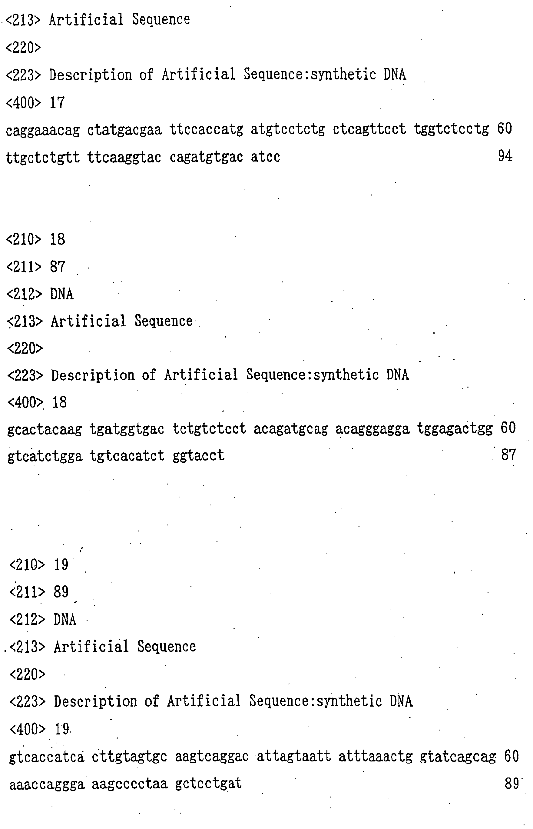
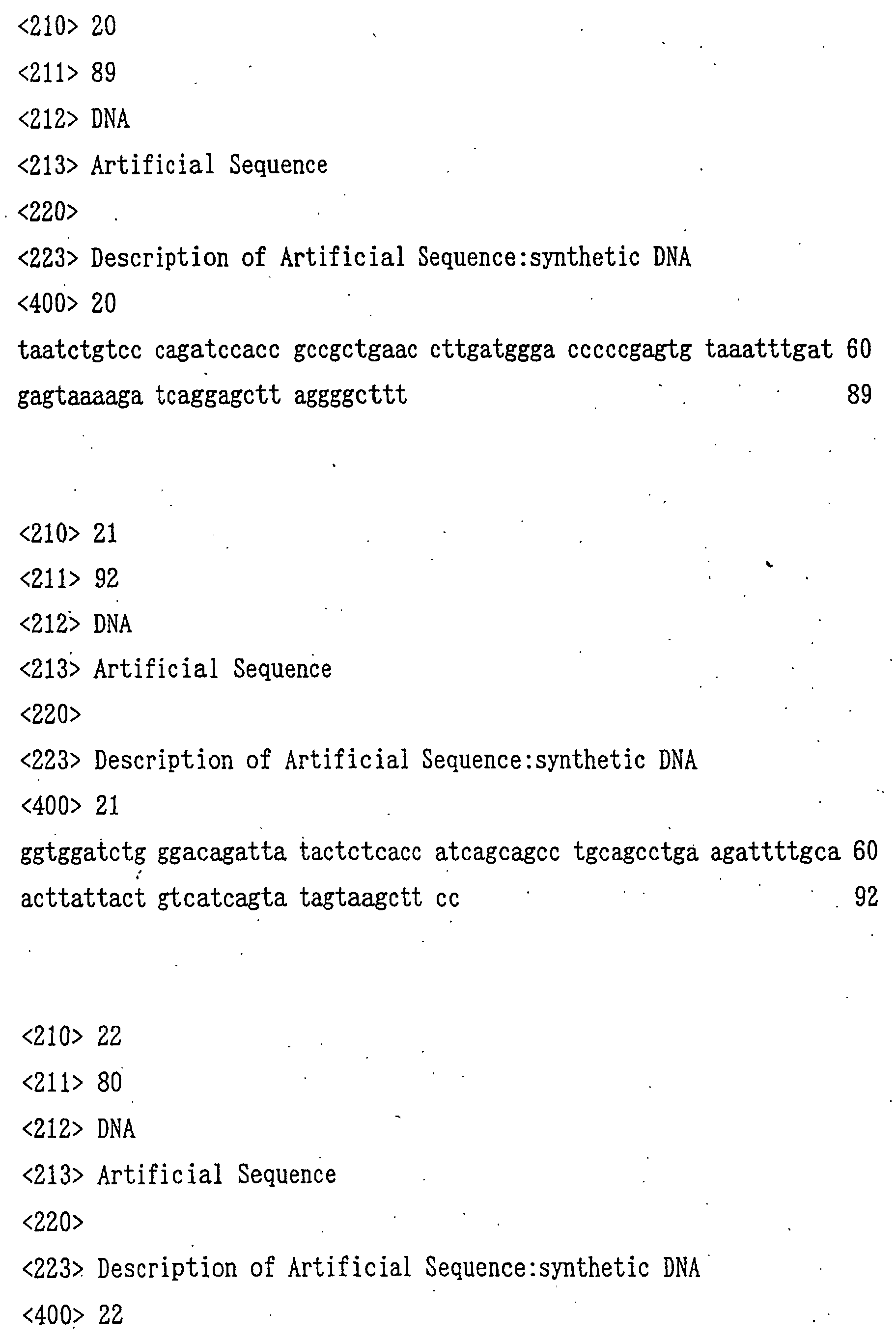
ID NO:2 was used as the secretory signal sequence.First, 6 synthetic DNA fragments of SEQ ID NO:17 to
SEQ ID NO:22 were synthesized using an automatic DNA
synthesizer (380A, manufactured by Applied Biosystems).
Each of the thus synthesized DNA fragments was added to 50
µl of a buffer comprising 10 mM Tris-HCl (pH 8.3), 50 mM
potassium chloride, 1.5 mM magnesium chloride, 0.001%
gelatin, 200 µM dNTPs, 0.5 µM M13 primer RV (manufactured
by Takara Shuzo), 0.5 µM M13 primer M4 (manufactured by
Takara Shuzo) and 2 units of TaKaRa Taq DNA polymerase
(manufactured by Takara Shuzo) to give a final
concentration of 0.1 µM, and the solution was covered with
50 µl of mineral oil and set to a DNA thermal cycler (PJ480,
manufactured by PERKIN ELMER) to carry out 30 cycles of the
reaction, each cycle including 2 minutes at 94°C, 2 minutes
at 55°C and 2 minutes at 72°C. The reaction solution was
purified using QIAquick PCR Purification Kit (manufactured
by QIAGEN) in accordance with the manufacture's
instructions and made into 30 µl of a buffer comprising 50
mM Tris-HCl (pH 7.5), 100 mM sodium chloride, 10 mM
magnesium chloride, 1 mM DTT and 100 µg/ml BSA, and 10
units of a restriction enzyme EcoRI (manufactured by Takara
Shuzo) and a restriction enzyme SplI (manufactured by
Takara Shuzo) were further added thereto to carry out the
reaction at 37°C for 1 hour. The reaction solution was
fractionated by agarose gel electrophoresis to recover
about 0.2 µg of an EcoRI- SplI fragment of about 0.39 kb
using QIAquick Gel Extraction Kit (manufactured by QIAGEN)
in accordance with the manufacture's instructions.Next, 3 µg of a plasmid pBSL3 described in Japanese
Published Unexamined Patent Application No. 257893/98 was
added to 10 µl of a buffer comprising 50 mM Tris-HCl (pH
7.5), 100 mM sodium chloride, 10 mM magnesium chloride, 1
mM DTT and 100 µg/ml BSA, and 10 units of a restriction
enzyme EcoRI (manufactured by Takara Shuzo) and a
restriction enzyme SplI (manufactured by Takara Shuzo) were
further added thereto to carry out the reaction at 37°C for
1 hour. The reaction solution was fractionated by agarose
gel electrophoresis to recover about 2 µg of an EcoRI-SplI
fragment of about 2.95 kb using QIAquick Gel Extraction Kit
(manufactured by QIAGEN) in accordance with the
manufacture's instructions.Next, 0.1 µg of the EcoRI-SplI fragment of the PCR
product of VL of anti-GD3 CDR-grafted antibody and 0.1 µg
of the EcoRI-SplI fragment of the plasmid pBSL3, both
obtained in the above, were added to 20 µl in total volume
of sterile water and ligated using Ready-To-Go T4 DNA
Ligase (manufactured by Pharmacia). Using the thus
obtained recombinant plasmid DNA solution, an E. coli line
HB101 was transformed. Each plasmid DNA was prepared from
10 clones of the transformants, allowed to react in
accordance with the manufacture's instructions attached to
AutoRead Sequencing Kit (manufactured by Pharmacia) and
then subjected to electrophoresis using A.L.F. DNA
Sequencer (manufactured by Pharmacia) to determine the
nucleotide sequence, and as a result, the plasmid phKM641L
shown in Fig. 2 having the nucleotide sequence of interest
was obtained.
4. Activity evaluation of anti-GD3 CDR-grafted antibody by
transient expression using animal cell
In order to carry out activity evaluation of anti-GD3
CDR-grafted antibody more quickly, transient expression
of anti-GD3 CDR-grafted antibody was carried out as follows
using COS-7 cell (ATCC CRL 1651).
(1) Construction of anti-GD3 chimeric antibody KM871
transient expression vector pT641
In order to use as a positive control in the
activity evaluation of anti-GD3 CDR-grafted antibody by
transient expression, a transient expression vector pT641
for anti-GD3 chimeric antibody KM871 was constructed as
follows.Since the efficiency of transient expression using
animal cells generally depends on the copy number of
introduced expression vector, it was considered that
smaller expression vector has higher expression efficiency.
Accordingly, the transient expression vector pT641 for
anti-GD3 chimeric antibody KM871 was constructed as follows
using the transient expression vector pT796 for anti-ganglioside
GM2 chimeric antibody KM966 described in
Japanese Published Unexamined Patent Application
No. 257893/98 and the plasmid pKM641HF1 having VH of anti-GD3
mouse antibody KM641 and plasmid pKM641LA2 having VL of
anti-GD3 mouse antibody KM641 described in Japanese
Published Unexamined Patent Application No. 304989/93. First, 3 µg of the plasmid pKM641LA2 having VL of
anti-GD3 mouse antibody KM641 described in Japanese
Published Unexamined Patent Application No. 304989/93 was
added to 10 µl of a buffer comprising 10 mM Tris-HCl (pH
7.5), 50 mM sodium chloride, 10 mM magnesium chloride and 1
mM DTT and the solution was further mixed with 10 units of
a restriction enzyme HindIII (manufactured by Takara Shuzo)
to carry out the reaction at 37°C for 1 hour. The reaction
solution was precipitated with ethanol and added to 10 µl
of a buffer comprising 50 mM Tris-HCl (pH 7.5), 100 mM
sodium chloride, 10 mM magnesium chloride and 1 mM DTT, and
10 units of a restriction enzyme EcoRI (manufactured by
Takara Shuzo) were further added to carry out the reaction
at 37°C for 1 hour. The reaction solution was fractionated
by agarose gel electrophoresis to recover about 0.2 µg of
an HindIII-EcoRI fragment of about 0.35 kb using QIAquick
Gel Extraction Kit (manufactured by QIAGEN) in accordance
with the manufacture's instructions.Next, 3 µg of the transient expression vector pT796
for anti-ganglioside GM2 chimeric antibody KM966 described
in Japanese Published Unexamined Patent Application
No. 257893/98 was added to 10 µl of a buffer comprising 50
mM Tris-HCl (pH 7.5), 100 mM sodium chloride, 10 mM
magnesium chloride, 1 mM DTT and 100 µg/ml BSA, and the
solution was further mixed with 10 units of a restriction
enzyme EcoRI (manufactured by Takara Shuzo) and a
restriction enzyme SplI (manufactured by Takara Shuzo) to
carry out the reaction at 37°C for 1 hour. The reaction
solution was fractionated by agarose gel electrophoresis to
recover about 2 µg of an EcoRI-SplI fragment of about 9.20
kb using QIAquick Gel Extraction Kit (manufactured by
QIAGEN) in accordance with the manufacture's instructions.Next, synthetic DNA fragments respectively having
the nucleotide sequences represented by SEQ ID NO:23 and
SEQ ID NO:24 were synthesized using an automatic DNA
synthesizer (380A, manufactured by Applied Biosystems). A
0.3 µg of each of the thus obtained synthetic DNA fragments
was added to 15 µl of sterile water and heated at 65°C for
5 minutes. After allowing the reaction solution to stand
at room temperature for 30 minutes, this was mixed with 2
µl of a 10 × buffer solution (500 mM Tris-HCl (pH 7.6), 100
mM magnesium chloride, 50 mM DTT) and 2 µl of 10 mM ATP and
further mixed with 10 units of T4 Polynucleotide Kinase
(manufactured by Takara Shuzo) to carry out the reaction at
37°C for 30 minutes to thereby phosphorylate the 5'-terminal.Next, 0.1 µg of the HindIII-EcoRI fragment derived
from the plasmid pKM641LA2, 0.1 µg of the EcoRI-SplI
fragment derived from the plasmid pT796 and 0.05 µg of the
phosphorylated synthetic DNA, obtained in the above, were
added to 20 µl in total volume of sterile water and ligated
using Ready-To-Go T4 DNA Ligase (manufactured by Pharmacia).
Using the thus obtained recombinant plasmid DNA solution,
an E. coli HB101 was transformed to obtain the plasmid
pT796H641L shown in Fig. 3. The thus obtained plasmid (10
µg) was allowed to react in accordance with the
manufacture's instructions attached to AutoRead Sequencing
Kit (manufactured by Pharmacia) and then subjected to an
electrophoresis using A.L.F. DNA Sequencer (manufactured by
Pharmacia) to determine the nucleotide sequence, and as a
result, it was confirmed that a plasmid into which the DNA
of interest was cloned was obtained.Next, 3 µg of a plasmid pBluescript SK(-)
(manufactured by Stratagene) was added to 10 µl of a buffer
comprising 10 mM Tris-HCl (pH 7.5), 50 mM sodium chloride,
10 mM magnesium chloride, 1 mM DTT and 100 µg/ml BSA and
the solution was further mixed with 10 units of a
restriction enzyme XbaI (manufactured by Takara Shuzo) to
carry out the reaction at 37°C for 1 hour. The reaction
solution was precipitated with ethanol, added to 30 µl of a
buffer comprising 30 mM sodium acetate (pH 5.0), 100 mM
sodium chloride, 1 mM zinc acetate and 5% glycerol, and
further mixed with 30 units of a modification enzyme Mung
Bean Nuclease (manufactured by Takara Shuzo) to carry out
the reaction at 25°C for 15 minutes. The reaction solution
was precipitated with ethanol, added to 10 µl of a buffer
comprising 10 mM Tris-HCl (pH 7.5), 10 mM magnesium
chloride and 1 mM DTT, and further mixed with 10 units of a
restriction enzyme ApaI (manufactured by Takara Shuzo) to
carry out the reaction at 37°C for 1 hour. The reaction
solution was fractionated by an agarose gel electrophoresis
to recover about 2 µg of a Blunt end-Apal fragment of about
2.95 kb using QIAquick Get Extraction Kit (manufactured by
QIAGEN) in accordance with the manufacture's instructions.Next, 3 µg of the plasmid pKM641HF1 was added to 10
µl of a buffer comprising 50 mM Tris-HCl (pH 7.5), 100 mM
sodium chloride, 10 mM magnesium chloride and 1 mM DTT, and
the solution was further mixed with 10 units of a
restriction enzyme EcoRI (manufactured by Takara Shuzo) to
carry out the reaction at 37°C for 1 hour. The reaction
solution was precipitated with ethanol, and the 5'
protruding end formed by the restriction enzyme digestion
was changed to blunt end using DNA Blunting Kit
(manufactured by Takara Shuzo). The reaction solution was
precipitated with ethanol, added to 10 µl of a buffer
comprising 10 mM Tris-HCl (pH 7.5), 10 mM magnesium
chloride and 1 mM DTT, and further mixed with 10 units of a
restriction enzyme ApaI (manufactured by Takara Shuzo) to
carry out the reaction at 37°C for 1 hour. The reaction
solution was fractionated by an agarose gel electrophoresis
to recover about 0.2 µg of a Blunt end-ApaI fragment of
about 0.44 kb using QIAquick Gel Extraction Kit
(manufactured by QIAGEN) in accordance with the
manufacture's instructions. Next, 0.1 µg of Blunt end-ApaI fragment derived
from the plasmid pBluescript SK(-) and 0.1 µg of the Blunt
end-ApaI fragment derived from the plasmid pKM641HF1
obtained in the above, were added to 20 µl in total volume
of sterile water and ligated using Ready-To-Go T4 DNA
Ligase (manufactured by Pharmacia). Using the thus
obtained recombinant plasmid DNA solution, an E. coli HB101
was transformed to obtain the plasmid pBS641H shown in
Fig. 4.Next, 3 µg of the plasmid pT796H641L obtained in
the above was added to 10 µl of a buffer comprising 10 mM
Tris-HCl, (pH 7.5), 10 mM magnesium chloride and 1 mM DTT
and the solution was further mixed with 10 units of a
restriction enzyme ApaI (manufactured by Takara Shuzo) to
carry out the reaction at 37°C for 1 hour. The reaction
solution was precipitated with ethanol, added to 10 µl of a
buffer comprising 50 mM Tris-HCl (pH 7.5), 100 mM sodium
chloride, 10 mM magnesium chloride, 1 mM DTT, 100 µg/ml BSA
and 0.01% Triton X-100, and further mixed with 10 units of
a restriction enzyme NotI (manufactured by Takara Shuzo) to
carry out the reaction at 37°C for 1 hour. The reaction
solution was fractionated by an agarose gel electrophoresis
to recover about 0.2 µg of an ApaI-NotI fragment of about
9.16 kb using QIAquick Gel Extraction Kit (manufactured by
QIAGEN) in accordance with the manufacture's instructions.Next, 3 µg of the plasmid pBS641H obtained in the
above was added to 10 µl of a buffer comprising 10 mM Tris-HCl
(pH 7.5), 10 mM magnesium chloride and 1 mM DTT and the
solution was further mixed with 10 units of a restriction
enzyme ApaI (manufactured by Takara Shuzo) to carry out the
reaction at 37°C for 1 hour. The reaction solution was
precipitated with ethanol, added to 10 µl of a buffer
comprising 50 mM Tris-HCl (pH 7.5), 100 mM sodium chloride,
10 mM magnesium chloride, 1 mM DTT, 100 µg/ml BSA and 0.01%
Triton X-100, and further mixed with 10 units of a
restriction enzyme NotI (manufactured by Takara Shuzo) to
carry out the reaction at 37°C for 1 hour. The reaction
solution was fractionated by an agarose gel electrophoresis
to recover about 2 µg of an ApaI-NotI fragment of about
0.44 kb using QIAquick Gel Extraction Kit (manufactured by
QIAGEN) in accordance with the manufacture's instructions.Next, 0.1 µg of the ApaI-NotI fragment derived from
the plasmid pT796H641L and 0.1 µg of the ApaI-NotI fragment
derived from the plasmid pBS641H, obtained in the above,
were added to 20 µl in total volume of sterile water and
ligated using Ready-To-Go T4 DNA Ligase (manufactured by
Pharmacia). Using the thus obtained recombinant plasmid
DNA solution, an E. coli HB101 was transformed to obtain
the plasmid pT641 shown in Fig. 5. (2) Construction of an anti-GD3 CDR-grafted antibody KM871
transient expression vector
A transient expression vector for anti-GD3 CDR-grafted
antibody was constructed as follows using the
transient expression vector pT641 for anti-GD3 chimeric
antibody described in the item 4(1) of Example 1 and the
plasmids phKM641H and phKM641L described in the items 3(2)
and(3) of Example 1.First, 3 µg of the plasmid phKM641H obtained in the
item 3(2) of Example 1 was added to 10 µl of a buffer
comprising 10 mM Tris-HCl (pH 7.5), 10 mM magnesium
chloride and 1 mM DTT and the solution was further mixed
with 10 units of a restriction enzyme ApaI (manufactured by
Takara Shuzo) to carry out the reaction at 37°C for 1 hour.
The reaction solution was precipitated with ethanol, added
to 10 µl of a buffer comprising 50 mM Tris-HCl (pH 7.5),
100 mM sodium chloride, 10 mM magnesium chloride, 1 mM DTT,
100 µg/ml BSA and 0.01% Triton X-100, and further mixed
with 10 units of a restriction enzyme NotI (manufactured by
Takara Shuzo) to carry out the reaction at 37°C for 1 hour.
The reaction solution was fractionated by an agarose gel
electrophoresis to recover about 0.2 µg of an ApaI-NotI
fragment of about 0.44 kb using QIAquick Gel Extraction Kit
(manufactured by QIAGEN) in accordance with the
manufacture's instructions.Next, 3 µg of the plasmid pT641 obtained in the
item 4(1) of Example 1 was added to 10 µl of a buffer
comprising 10 mM Tris-HCl (pH 7.5), 10 mM magnesium
chloride and 1 mM DTT, and the solution was further mixed
with 10 units of a restriction enzyme ApaI (manufactured by
Takara Shuzo) to carry out the reaction at 37°C for 1 hour.
The reaction solution was precipitated with ethanol, added
to 10 µl of a buffer comprising 50 mM Tris-HCl (pH 7.5),
100 mM sodium chloride, 10 mM magnesium chloride, 1 mM DTT,
100 µg/ml BSA and 0.01% Triton X-100, and further mixed
with 10 units of a restriction enzyme NotI (manufactured by
Takara Shuzo) to carry out the reaction at 37°C for 1 hour.
The. reaction solution was fractionated by an agarose gel
electrophoresis to recover about 2 µg of an ApaI-NotI
fragment of about 9.16 kb using QIAquick Gel Extraction Kit
(manufactured by QIAGEN) in accordance with the
manufacture's instructions.Next, 0.1 µg of the ApaI-NotI fragment derived from
the plasmid phKM641H and 0.1 µg of the ApaI-NotI fragment
derived from the plasmid pT641 were added to 20 µl in total
volume of sterile water and ligated using Ready-To-Go T4
DNA Ligase (manufactured by Pharmacia). Using the thus
obtained recombinant plasmid DNA solution, an E. coli HB101
was transformed to obtain the plasmid pT641HCDR shown in
Fig. 6.Next, 3 µg of the plasmid phKM641L obtained in the
item 3(3) of Example 1 was added to 10 µl of a buffer
comprising 50 mM Tris-HCl (pH 7.5), 100 mM sodium chloride,
10 mM magnesium chloride, 1 mM DTT and 100 µg/ml BSA, and
the solution was further mixed with 10 units of a
restriction enzyme EcoRI (manufactured by Takara Shuzo) and
a restriction enzyme SplI (manufactured by Takara Shuzo) to
carry out the reaction at 37°C for 1 hour. The reaction
solution was fractionated by an agarose gel electrophoresis
to recover about 0.2 µg of an EcoRI-SplI fragment of about
0.39 kb using QIAquick Gel Extraction Kit (manufactured by
QIAGEN) in accordance with the manufacture's instructions.Next, 3 µg of each of the plasmid pT641 obtained in
the item 4(1) of Example 1 and the plasmid pT641HCDR
obtained in the above was added to 10 µl of a buffer
comprising 50 mM Tris-HCl (pH 7.5), 100 mM sodium chloride,
10 mM magnesium chloride, 1 mM DTT and 100 µg/ml BSA, and
the solution was further mixed with 10 units of a
restriction enzyme EcoRI (manufactured by Takara Shuzo) and
a restriction enzyme SplI (manufactured by Takara Shuzo) to
carry out the reaction at 37°C for 1 hour. The reaction
solution was fractionated by an agarose gel electrophoresis
to recover about 2 µg of an EcoRI-SplI fragment of about
9.20 kb derived from each plasmid using QIAquick Gel
Extraction Kit (manufactured by QIAGEN) in accordance with
the manufacture's instructions.Next, 0.1 µg of the EcoRI-SplI fragment derived
from the plasmid phKM641L and 0.1 µg of the EcoRI-SplI
fragment derived from the plasmid pT641, obtained in the
above, were added to 20 µl in total volume of sterile water
and ligated using Ready-To-Go T4 DNA Ligase (manufactured
by Pharmacia). Also, 0.1 µg of the EcoRI-SplI fragment
derived from the plasmid phKM641L and 0.1 µg of the EcoRI-SplI
fragment derived from the plasmid pT641HCDR, obtained
in the above, were added to 20 µl in total volume of
sterile water and ligated using Ready-To-Go T4 DNA Ligase
(manufactured by Pharmacia). Using each of the thus
obtained recombinant plasmid DNA solutions, an E. coli
HB101 was transformed to obtain the plasmids pT641LCDR and
pT641HLCDR shown in Fig. 7. (3) Activity evaluation of anti-GD3 chimeric antibody and
anti-GD3 CDR-grafted antibody by transient expression using
animal cells
Transient expression of antibodies was carried out
using the transient expression vector pT641 for anti-GD3
chimeric antibody, the transient expression vector
pT641HLCDR for anti-GD3 CDR-grafted antibody and the
transient expression vectors pT641HCDR and pT641LCDR for
anti-GD3 hybrid antibodies having V regions of mouse
antibody and human CDR-grafted antibody, obtained in the
items 4(1) and (2) of Example 1.COS-7 cells (ATCC CRL 1651) were dispensed in 2 ml
at a density of 1×10⁵ cells/ml into a 6 well plate
(manufactured by Falcon) and cultured overnight at 37°C.
By adding 2 µg of each expression vector to 100 µl of OPTI-MEM
medium (manufactured by GIBCO BRL) and further adding a
solution in which 10 µl of LIPOFECTAMINE Reagent
(manufactured by GIBCO BRL) was added to 100 µl of the
OPTI-MEM medium, the reaction was carried out at room
temperature for 40 minutes to effect formation of a DNA-liposome
complex. The overnight-cultured COS-7 cells were
washed twice with 2 ml of the OPTI-MEM medium (manufactured
by GIBCO BRL), mixed with a solution prepared by adding 0.8
ml of OPTI-MEM medium to the complex-containing solution
and cultured at 37°C for 7 hours, the solution was
discarded and then the cells were mixed with 2 ml of 10%
FBS-containing DME medium (manufactured by GIBCO BRL) and
cultured at 37°C. After the introduction of each
expression vector, the culture supernatant was recovered 72
hours thereafter and its concentration operation was
carried out as occasion demands, and then binding activity
of the anti-GD3 humanized antibody for GD3 (manufactured by
DIA-IATRON) in the culture supernatant was measured by the
ELISA method described in the item 2 of Example 1, by
measuring concentration of the anti-GD3 humanized antibody
in the culture supernatant by the ELISA method described in
the item 4 which will be described later and calculating
the activity from the measured values as relative activity
(%) when the activity of the positive control anti-GD3
chimeric antibody is defined as 100. Results are shown in
Fig. 8. As shown in Fig. 8, the anti-GD3 hybrid antibody
derived from the transient expression vector pT641HCDR (VH
is derived from a human CDR-grafted antibody and VL is
derived from a chimeric antibody) showed almost the same
binding activity of that of the anti-GD3 hybrid antibody
derived from the transient expression vector pT641, but the
anti-GD3 hybrid antibody derived from the transient
expression vector pT641LCDR (VH is derived from a chimeric
antibody and VL is derived from a human CDR-grafted
antibody) showed a binding activity of about 50% of the
anti-GD3. chimeric antibody. In addition, the anti-GD3 CDR-grafted
antibody derived from the transient expression
vector pT641HLCDR showed a binding activity of only about
5% of the anti-GD3 chimeric antibody, showing considerable
decrease in the activity.Based on these results, binding activity of the
anti-GD3 CDR-grafted antibody having VH and VL designed in
the item 3(1) of Example 1 is considerably reduced and the
main cause of the activity reduction is considered to be
due to VL, but since about 50% of the activity is found by
its combination with the VH of anti-GD3 chimeric antibody,
it was suggested that there is a problem particularly at
the interaction region with VH. Accordingly, an attempt
was made to increase the antibody activity by further
modifying amino acid residues of the VL designed in the
item 3(1) of Example 1. (4) Measurement of the concentration of humanized antibody
in transient expression culture supernatant by ELISA method
A solution (50 µl) prepared by diluting a goat
anti-human IgG (γ-chain) antibody (manufactured by Medical
& Biological Laboratories) 400 folds with PBS was dispensed
in each well of a 96 well plate for ELISA use (manufactured
by Greiner) and allowed to react at 4°C overnight. After
discarding the antibody solution, 1% BSA-PBS was added in
100 µl/well and allowed to react at room temperature for 1
hour to thereby block the remaining active groups. After
discarding 1% BSA-PBS, the transient expression culture
supernatants obtained in the item 4(3) of Example 1 or
diluted solutions of the purified. anti-GD3 chimeric
antibody KM871 were added in 50 µl/well and allowed to
react at room temperature for 1 hour. After the reaction,
each well was washed with Tween-PBS and then a solution
prepared by diluting a peroxidase-labeled mouse anti-human
K L chain antibody (manufactured by Zymed) 500 folds with
PBS was added in 50 µl/well and allowed to react at room
temperature for 1 hour. After the reaction and subsequent
washing with Tween-PBS, an ABTS substrate solution (a
solution prepared by dissolving 0.55 g of 2,2'-azinobis(3-ethylbenzothiazoline-6-sulfonic
acid) in 1 L of 0.1 M
citrate buffer (pH 4.2) and adding. 1 µl/ml of hydrogen
peroxide just before the use) was added in 50 µl/well for
color development and OD415 was measured.
5. Increase of the binding activity by modifying amino acid
residues of VL of anti-GD3 CDR-grafted antibody
Increase of the binding activity by modifying amino
acid residues of VL of the anti-GD3 CDR-grafted antibody
designed in the item 3(1) of Example 1 was carried out as
follows.
(1) Modification of amino acid residues of VL of anti-GD3
CDR-grafted antibody and construction of cDNA encoding the
modified VL
First, amino acid residues which are amino acid
residues positioned in the interaction region of VH and VL
and amino acid residues considered to be exerting influence
on the three-dimensional structure of each CDR of VL and
are also different from the amino acid residues of mouse
antibody in the VL of human CDR-grafted antibody were
identified from computer models of the three-dimensional
structures of various antibodies constructed in the item
3(1) of Example 1. As a result, the 7th position Ser, the
8th position Pro, the 12th position Ser, the 41st position
Gly, the 44th position Pro, the 72nd position Thr, the 77th
position Ser, the 83rd position Phe and the 87th Tyr of
hKM641L as the amino acid sequence of VL of anti-GD3 CDR-grafted
antibody represented by SEQ ID NO:10 were
identified. By replacing these amino acid residues by
amino acid residues found in the mouse antibody, 8 VLs of
modified anti-GD3 CDR-grafted antibody were designed. That
is, the 8th position Pro, the 12th position Ser, the 44th
position Pro and the 87th position Tyr among the hKM641L
amino sequence were respectively replaced by Ala, Pro, Val
and Phe in the case of hKM641NL, and the 7th position Ser,
the 8th position Pro and the 12th position Ser among the
hKM641L amino sequence respectively by Thr, Ala and Pro in
the case of hKM641Lm-1, the 87th position Tyr among the
hKM641L amino sequence by Phe in the case of hKM641Lm-4,
the 41st position Gly and the 44th position Pro among the
hKM641L amino sequence respectively by Asp and Val in the
case of hKM641Lm-6, the 72nd position Thr, the 77th
position Ser and the 83rd position Phe among the hKM641L
amino sequence respectively by Ser, Asn and Ile in the case
of hKM641Lm-7, the 77th position Ser among the hKM641L
amino sequence by Asn in the case of hKM641Lm-8, the 83rd
position Phe and the 87th position Tyr among the hKM641L
amino sequence respectively by Ile and Phe in the case of
hKM641Lm-9, and the 41st position Gly, the 44th position
Pro and the 83rd position Phe among the hKM641L amino
sequence respectively by Asp, Val and Ile in the case of
hKM641Lm-69. Among these modified VLs, cDNA encoding each
of 6 modified VLs excluding hKM641NL and hKM641Lm-69 was
constructed as follows by PCR-aided mutagenesis. That is,
antisense chain and sense chain DNA primers were
synthesized using an automatic DNA synthesizer (380A,
manufactured by Applied Biosystems) for introducing
mutation, and a first PCR was carried out in accordance
with the method described in the item 3(2) of Example 1
using 1 ng of the plasmid phKM641L as the template and 0.5
µM in final concentration of M13 primer RV (manufactured by
Takara Shuzo) and the antisense chain DNA primer and M13
primer M4 (manufactured by Takara Shuzo) and the sense
chain DNA primer. Each of the reaction solutions was
purified using QIAquick PCR Purification Kit (manufactured
by QIAGEN) in accordance with the manufacture's
instructions by eluting with 20 µl of 10 mM Tris-HCl (pH
8.0), and then a second PCR was carried out using 5 µl of
each eluate in accordance with the method described in the
item 3(2) of Example 1. The reaction solution was purified
using QIAquick PCR Purification Kit (manufactured by
QIAGEN) in accordance with the manufacture's instructions
and made into 30 µl of a buffer comprising 50 mM Tris-HCl
(pH 7.5), 100 mM sodium chloride, 10 mM magnesium chloride,
1 mM DTT and 100 µg/ml BSA, and 10 units of a restriction
enzyme EcoRI (manufactured by Takara Shuzo) and a
restriction enzyme SplI (manufactured by Takara Shuzo) were
further added thereto to carry out the reaction at 37°C for
1 hour. The reaction solution was fractionated by agarose
gel electrophoresis to recover about 0.2 µg of an EcoRI-SplI
fragment of about 0.39 kb using QIAquick Gel
Extraction Kit (manufactured by QIAGEN) in accordance with
the manufacture's instructions.Next, 0.1 µg of the EcoRI-SplI fragment of the PCR
product of VL of the modified anti-GD3 CDR-grafted antibody
obtained in the above and 0.1 µg of the EcoRI-SplI fragment
of the plasmid pBSL3 obtained in the item 3(3) of Example 1
were added to 20 µl in total volume of sterile water and
ligated using Ready-To-Go T4 DNA Ligase (manufactured by
Pharmacia). Using the thus obtained recombinant plasmid
DNA solution, an E. coli HB101 was transformed. Each
plasmid DNA was produced from 10 clones of the
transformants, allowed to react in accordance with the
manufacture's instructions attached to AutoRead Sequencing
Kit (manufactured by Pharmacia) and then subjected to
electrophoresis using A.L.F. DNA Sequencer (manufactured by
Pharmacia) to determine the nucleotide sequence to thereby
obtain a plasmid having cDNA to which the intended
modification was applied.Specifically, a plasmid phKM641Lm-1 having the
nucleotide sequence represented by SEQ ID NO:27 was
obtained by carrying out a series of the above operations
using the synthetic DNA of SEQ ID NO:25 as the antisense
chain DNA primer and the synthetic DNA of SEQ ID NO:26 as
the sense chain DNA primer. The hKM641Lm-1 as an amino
acid sequence encoded by the nucleotide sequence is also
represented by SEQ ID NO:27.A plasmid phKM641Lm-4 having the nucleotide
sequence represented by SEQ ID NO:30 was obtained by
carrying out a series of the above operations using the
synthetic DNA of SEQ ID NO:28 as the antisense chain DNA
primer and the synthetic DNA of SEQ ID NO:29 as the sense
chain DNA primer. The hKM641Lm-4 as an amino acid sequence
encoded by the nucleotide sequence is also represented by
SEQ ID NO:30. A plasmid phKM641Lm-6 having the nucleotide
sequence represented by SEQ ID NO:33 was obtained by
carrying out a series of the above operations using the
synthetic DNA of SEQ ID NO:31 as the antisense chain DNA
primer and the synthetic DNA of SEQ ID NO:32 as the sense
chain DNA primer. The hKM641Lm-6 as an amino acid sequence
encoded by the nucleotide sequence is also represented by
SEQ ID NO:33.A plasmid phKM641Lm-7 having the nucleotide
sequence represented by SEQ ID NO:36 was obtained by
carrying out a series of the above operations using the
synthetic DNA of SEQ ID NO:34 as the antisense chain DNA
primer and the synthetic DNA of SEQ ID NO:35 as the sense
chain DNA primer. The hKM641Lm-7 as an amino acid sequence
encoded by the nucleotide sequence is also represented by
SEQ ID NO:36.A plasmid phKM641Lm-8 having the nucleotide
sequence represented by SEQ ID NO:39 was obtained by
carrying out a series of the above operations using the
synthetic DNA of SEQ ID NO:37 as the antisense chain DNA
primer and the synthetic DNA of SEQ ID NO:38 as the sense
chain DNA primer. The hKM641Lm-8 as an amino acid sequence
encoded by the nucleotide sequence is also represented by
SEQ ID NO:39.A plasmid phKM641Lm-9 having the nucleotide
sequence represented by SEQ ID NO:42 was obtained by
carrying out a series of the above operations using the
synthetic DNA of SEQ ID NO:40 as the antisense chain DNA
primer and the synthetic DNA of SEQ ID NO:41 as the sense
chain DNA primer. The hKM641Lm-9 as an amino acid sequence
encoded by the nucleotide sequence is also represented by
SEQ ID NO:42.Among the modified VLs, cDNA encoding the hKM641NL
was constructed by synthesizing 6 synthetic DNA fragments
of SEQ ID NOs:17, 22 and 43 to 46 using an automatic DNA
synthesizer (380A, manufactured by Applied Biosystems) and
carrying out the procedure described in the item 3(3) of
Example 1 using these fragments. As a result, a plasmid
phKM641NL having the nucleotide sequence represented by SEQ
ID NO:47 in which the modification of interest was effected
was obtained. The hKM641NL as an amino acid sequence
encoded by the nucleotide sequence is also represented by
SEQ ID NO:47.Among the modified VLs, cDNA encoding the hKM641Lm-69
was constructed as follows using the plasmids phKM641Lm-6
and phKM641Lm-9 having cDNA of modified VL obtained in
the above.First, 3 µg of the plasmid phKM641Lm-6 was added to
10 µl of a buffer comprising 50 mM Tris-HCl (pH 7.5), 100
mM of sodium chloride, 10 mM magnesium chloride and 1 mM
DTT and 10 units of restriction enzymes EcoRI (manufactured
by Takara Shuzo) and PstI (manufactured by Takara Shuzo)
were further added thereto to carry out the reaction at
37°C for 1 hour. The reaction solution was fractionated by
agarose gel electrophoresis to recover about 0.3 µg of an
EcoRI-PstI fragment of about 0.30 kb using QIAquick Gel
Extraction Kit (manufactured by QIAGEN) in accordance with
the manufacture's instructions.Next, 3 µg of the plasmid phKM641Lm-9 was added to
10 µl of a buffer comprising 50 mM Tris-HCl (pH 7.5), 100
mM of sodium chloride, 10 mM magnesium chloride and 1 mM
DTT and 10 units of restriction enzymes EcoRI (manufactured
by Takara Shuzo) and PstI (manufactured by Takara Shuzo)
were further added thereto to carry out the reaction at
37°C for 1 hour. The reaction solution was fractionated by
agarose gel electrophoresis to recover about 2 µg of an
EcoRI-PstI fragment of about 3.05 kb using QIAquick Gel
Extraction Kit (manufactured by QIAGEN) in accordance with
the manufacture's instructions. Next, 0.1 µg of the EcoRI-PstI fragment derived
from the plasmid phKM641Lm-6 and 0.1 µg of the EcoRI-PstI
fragment derived from the plasmid phKM641Lm-9, both
obtained in the above, were added to 20 µl in total volume
of sterile water and ligated using Ready-To-Go T4 DNA
Ligase (manufactured by Pharmacia). Using the thus
obtained recombinant plasmid DNA solution, an E. coli HB101
was transformed to obtain the plasmid phKM641Lm-69 shown in
Fig. 9. The reaction was carried out using 10 µg of the
thus obtained plasmid in accordance with the manufacture's
instructions attached to AutoRead Sequencing Kit
(manufactured by Pharmacia) and then the resulting mixture
was subjected to electrophoresis using A.L.F. DNA Sequencer
(manufactured by Pharmacia) to determine the nucleotide
sequence, and as a result, it was confirmed that it has the
nucleotide sequence represented by SEQ ID NO:48 in which
the modification of interest was carried out. The
hKM641Lm-69 as an amino acid sequence encoded by the
nucleotide sequence is also represented by SEQ ID NO:48. (2) Construction of transient expression vector for anti-GD3
CDR-grafted antibodies having modified VL
Transient expression vectors for anti-GD3 CDR-grafted
antibodies having various modified VLs were
constructed as follows using the plasmids having cDNA
molecules encoding various modified VLs, obtained in the
item 5(1) of Example 1, and the transient expression vector
pT641HCDR for anti-GD3 hybrid antibodies obtained in the
item 4(2) of Example 1.First, 3 µg of each of the plasmids phKM641NL,
phKM641Lm-1, phKM641Lm-4, phKM641Lm-6, phKM641Lm-7,
phKM641Lm-8, phKM641Lm-9 and phKM641Lm-69 obtained in the
item 5(1) of Example 1 was added to 10 µl of a buffer
comprising 50 mM Tris-HCl (pH 7.5), 100 mM of sodium
chloride, 10 mM magnesium chloride, 1 mM DTT and 100 µg/ml
of BSA, and 10 units of a restriction enzyme EcoRI
(manufactured by Takara Shuzo) and a restriction enzyme
SplI (manufactured by Takara Shuzo) were further added
thereto to carry out the reaction at 37°C for 1 hour. The
reaction solution was fractionated by agarose gel
electrophoresis to recover about 0.2 µg of an EcoRI-SplI
fragment of about 0.39 kb using QIAquick Gel Extraction Kit
(manufactured by QIAGEN) in accordance with the
manufacture's instructions.Next, 0.1 µg of each of the thus obtained EcoRI-SplI
fragments of various modified VLs and 0.1 µg of the
EcoRI-SplI fragment of the transient expression vector
pT641HCDR for anti-GD3 hybrid antibody obtained in the item
4(2) of Example 1, were added to 20 µl in total volume of
sterile water and ligated using Ready-To-Go T4 DNA Ligase
(manufactured by Pharmacia). Using the thus obtained
respective recombinant plasmid DNA solutions, an E. coli
HB101 was transformed to obtain the transient expression
vectors for anti-GD3 CDR-grafted antibodies having various
modified VLs, pT641HLCDRNL, pT641HLCDRLm-1, pT641HLCDRLm-4,
pT641HLCDRLm-6, pT641HLCDRLm-7, pT641HLCDRLm-8,
pT641HLCDRLm-9 and pT641HLCDRLm-69, shown in Fig. 10. (3) Evaluation of activity of anti-GD3 CDR-grafted antibody
having modified VLs by transient expression using animal
cell
Transient expression and evaluation of activity of
each antibody were carried out in accordance with the
method described in the item 4(3) of Example 1 using the
transient expression vector pT641 for ant-GD3 chimeric
antibody and the transient expression vector pT641HLCDR for
anti-GD3 CDR-grafted antibody, both obtained in the item
4(2) of Example 1, and the transient expression vectors,
pT641HLCDRNL, pT641HLCDRLm-1, pT641HLCDRLm-4, pT641HLCDRLm-6,
pT641HLCDRLm-7, pT641HLCDRLm-8, pT641HLCDRLm-9 and
pT641HLCDRLm-69, for anti-GD3 CDR-grafted antibodies having
various modified VLs, obtained in the item 5(2) of Example
1. Results are shown in Fig. 11. As shown in Fig. 11, the
modified anti-GD3 CDR-grafted antibodies derived from the
transient expression vectors, pT641HLCDRLm-6, pT641HLCDRLm-7,
pT641HLCDRLm-9 and pT641HLCDRLm-69, showed increased
binding activity in comparison with the anti-GD3 CDR-grafted
antibodies before modification, particularly, the
modified anti-GD3 CDR-grafted antibody derived from the
transient expression vector pT641HLCDRLm-69 showed its
binding activity of about 80% of the anti-GD3 chimeric
antibody. On the other hand, the modified anti-GD3 CDR-grafted
antibodies derived from other transient expression
vectors showed no increase in the binding activity in
comparison with the anti-GD3 CDR-grafted antibodies before
modification. Based on these results, it was found that,
among the modified amino acid residues of VL identified in
the item 5(1) of Example 1, the 41st position, 44th
position, 83rd position and 87th position amino acid
residues greatly contribute to the increase of binding
activity. It was also found that simultaneous substitution
of two amino acid residues of the 41st position and 44th
position or the 83rd position and 87th position contributes
to synergistic increase in the activity, and simultaneous
modification of four amino acid residues of the 41st
position, 44th position, 83rd position and 87th position
also contributes to synergistic increase in the activity.
Based on the computer models of three-dimensional
structures of V regions of various antibodies constructed
in the item 3(1) of Example 1, it was suggested that the
41st position and 44th position amino acid residues exert
influence on the interaction with VH, and the 83rd position
and 87th position amino acid residues on the three-dimensional
structure of CDR3 of VL, and it was considered
that three-dimensional structure of an antibody as a whole
is suitably maintained by modifying these amino acid
residues into amino acid residues found in a mouse antibody
and, as a result, its binding activity increases. This
information shows that it is necessary to take the
interaction regions of VH and VL also into consideration in
preparing human CDR-grafted antibodies and that it is
necessary to carry out various examinations on the
identification of these interaction regions based on the
information from three-dimensional structures of antibody V
regions. With regard to these interaction regions, it can
be easily analogize that they are different in individual
antibodies and it is difficult to find a general law at
present, so that it is necessary to carry out trial and
error in response to each antibody of interest.
6. Stable expression of anti-GD3 CDR-grafted antibody using
animal cell
Based on the results described in the item 5(3) of
Example 1, it was suggested that the anti-GD3 CDR-grafted
antibodies derived from transient expression vectors
pT641HLCDRLm-9 and pT641HLCDRLm-69 have about 20% and about
80% of binding activity for GD3, respectively, in
comparison with anti-GD3 chimeric antibodies. Accordingly,
in order to evaluate the activity of these anti-GD3 CDR-grafted
antibodies more in detail, transformed cell lines
capable of stably expressing ' anti-GD3 CDR-grafted
antibodies were obtained by the following method, and
purification of anti-GD3 CDR-grafted antibodies from
culture supernatants of the transformed cell lines was
carried out. Also, for comparison, stable expression and
purification of the anti-GD3 CDR-grafted antibody derived
from the transient expression vector pT641HLCDR were
carried out in the same manner.
(1) Construction of stable expression vector for anti-GD3
CDR-grafted antibody
Stable expression vectors were constructed by
introducing a resistance gene against an agent G418 and the
dhfr gene into various transient expression vectors
constructed in the item 5(2) of Example 1.First, 3 µg of the humanized antibody expression
vector pKANTEX93 described in WO 97/10354 was added to 10
µl of a buffer comprising 20 mM Tris-HCl (pH 8.5), 10 mM
magnesium chloride, 1 mM DTT and 100 mM potassium chloride,
and 10 units of a restriction enzyme BamHI (manufactured by
Takara Shuzo) and a restriction enzyme XhoI (manufactured
by Takara Shuzo) were further added thereto to carry out
the reaction at 37°C for 1 hour. The reaction solution was
fractionated by agarose gel electrophoresis to recover
about 1 µg of a BamHI-XhoI fragment of about 8.68 kb using
QIAquick Gel Extraction Kit (manufactured by QIAGEN) in
accordance with the manufacture's instructions.Next, 3 µg of each of the transient expression
vectors, pT641HLCDRLm-9, pT641HLCDRLm-69 and pT641HLCDR,
was added to 10 µl of a buffer comprising 20 mM Tris-HCl
(pH 8.5), 10 mM magnesium chloride, 1 mM DTT and 100 mM
potassium chloride, and 10 units of a restriction enzyme
BamHI (manufactured by Takara Shuzo), a restriction enzyme
XhoI (manufactured by Takara Shuzo) and a restriction
enzyme StuI (manufactured by Takara Shuzo) were further
added thereto to carry out the reaction at 37°C for 1 hour.
The reaction solution was fractionated by agarose gel
electrophoresis to recover about 1 µg of a BamHI-XhoI
fragment of about 4.90 kb using QIAquick Gel Extraction Kit
(manufactured by QIAGEN) in accordance with the
manufacture's instructions.Next, 0.1 µg of the BamHI-XhoI fragment derived
from the plasmid pKANTEX93 and 0.1 µg of the BamHI-XhoI
fragment derived from each of the transient expression
vectors, obtained in the above, were added to 20 µl in
total volume of sterile water and ligated using Ready-To-Go
T4 DNA Ligase (manufactured by Pharmacia). Using each of
the thus obtained recombinant plasmid DNA solutions, an E.
coli HB101 was transformed to obtain the anti-GD3 CDR-grafted
antibody stable expression vectors,
pKANTEX641HLCDRLm-9, pKANTEX641HLCDRLm-69 and
pKANTEX641HLCDR, shown in Fig. 12. (2) Stable expression of anti-GD3 CDR-grafted antibody
using animal cell
Using the various stable expression vectors for
anti-GD3 CDR-grafted antibody obtained in the item 6(1) of
Example 1, expression of anti-GD3 CDR-grafted antibody in
animal cells was carried out as follows.Each of the anti-GD3 CDR-grafted antibody
expression vectors (4 µg) was introduced into 4×10⁶ cells
of a rat myeloma cell line YB2/0 cell (ATCC CRL 1662) by
the electroporation method (Cytotechnology, 3, 133 (1990)),
and the cells were suspended in 40 ml of RPMI640-FBS(19)
(RPMI1640 medium containing 10% of fetal bovine serum
(FBS)) and dispensed in 200 µl/well into a 96 well
microtiter plate (manufactured by Sumitomo Bakelite).
After culturing at 37°C for 24 hours in a 5% CO2 incubator,
G418 was added to give a concentration of 0.5 mg/ml,
followed by culturing for 1 to 2 weeks. Culture
supernatants were recovered from wells in which colonies of
transformants showing G418-resistance were formed and
became confluent, and the antigen binding activity of
various anti-GD3 CDR-grafted antibodies in the supernatants
was measured by the ELISA shown in the item 2 of Example 1
(a peroxidase-labeled goat anti-human IgG(γ) antibody was
used as the secondary antibody).With regard to the transformants of wells in which
expression of anti-GD3 CDR-grafted antibodies was found in
the culture supernatants, in order to increase antibody
expression quantity making use of a dhfr gene amplification
system, they were suspended in the RPMI1640-FBS(10) medium
containing 0.5 mg/ml of G418 and 50 nM methotrexate
(hereinafter referred to as "MTX": manufactured by SIGMA)
as an inhibitor of the dhfr gene product dihydrofolate
reductase (hereinafter referred to as "DHFR") to give a
density of 1 to 2×10⁵ cells/ml, and dispensed in 2 ml into
a 24 well plate (manufactured by Greiner). By culturing at
37°C for 1 to 2 weeks in a 5% CO2 incubator, transformants
showing 50 nM MTX resistance were induced. When the
transformants became confluent, the antigen binding
activity of various anti-GD3 CDR-grafted antibodies in the
supernatants was measured by the ELISA shown in the item 2
of Example 1. With regard to the transformants of wells in
which expression of anti-GD3 CDR-grafted antibodies was
found in the culture supernatants, the MTX concentration
was increased to 100 nM and then to 200 nM by the method
similar to the above, and transformants capable of growing
in the RPMI1640-FBS(10) medium containing 0.5 mg/ml of G418
and 200 nM of MTX and of highly expressing anti-GD3 CDR-grafted
antibodies were finally obtained. With regard to
the thus obtained transformants, single cell isolation
(cloning) was carried out twice by limiting dilution
analysis. The transformed cell clones obtained by
introducing the stable expression vectors,
pKANTEX641HLCDRLm-9, pKANTEX641HLCDRLm-69 and
pKANTEX641HLCDR, were named KM8870, KM8871 and KM8869,
respectively, and expression levels of anti-GD3 CDR-grafted
antibody by transformed cell clones were about 5 µg/106
cells/24 hr, about 10 µg/106 cells/24 hr and about 30
µg/106 cells/24 hr, respectively. (3) Purification of anti-GD3 CDR-grafted antibody from
culture filtrate
Each of the transformed cell clones, KM8870, KM8871
and KM8869, obtained in the item 6(2) of Example 1 which
express various anti-GD3 CDR-grafted antibodies was
suspended in GIT medium (manufactured by Nippon
Pharmaceutical) containing 200 nM MTX to give a density of
1 to 2×10⁵ cells/ml, and dispensed in 200 ml into 175 cm2
flasks (manufactured by Greiner). The cells were cultured
at 37°C for 5 to 7 days until they became confluent and
then the culture supernatant was recovered. Each anti-GD3
CDR-grafted antibody was purified from the culture
supernatant using Prosep-A (manufactured by Bioprocessing)
column in accordance with the manufacture's instructions.
About 3 mg of anti-GD3 CDR-grafted antibody KM8870 was
obtained from 500 ml of the culture supernatant of KM8870,
and about 25 mg of anti-GD3 CDR-grafted antibody KM8871
from 1,600 ml of the culture supernatant of KM8871 and
about 65 mg of anti-GD3 CDR-grafted antibody KM8869 from
1,000 ml of the culture supernatant of KM8869. About 4 µg
of each of the thus obtained anti-GD3 CDR-grafted
antibodies was subjected to an electrophoresis in
accordance with the known method (Nature, 227, 680 (1970))
to examine its molecular weight and purification degree.
Results are shown in Fig. 13. As shown in Fig. 13,
molecular weight of each purified anti-GD3 CDR-grafted
antibody was about 150 kilodaltons (hereinafter referred to
as "Kd") under non-reducing conditions, and two bands of
about 50 Kd and about 25 Kd were found under reducing
conditions. The molecular weights almost coincided with
the molecular weights deduced from the cDNA nucleotide
sequences of H chain and L chain of the antibody (H chain:
about 49 Kd, L chain: about 24 Kd, whole molecule: about
146 Kd), and also coincided with the reports that the IgG
type antibody has a molecular weight of about 150 Kd under
non-reducing conditions and is degraded into H chains
having a molecular weight of about 50 Kd and L chains
having a molecular weight of about 25 Kd under reducing
conditions due to cutting of the disulfide bond
(hereinafter referred to as "S-S bond") in the molecule
(Antibodies: A Laboratory Manual, Monoclonal Antibodies:
Principles and Practice), so that it was confirmed that
each anti-GD3 CDR-grafted antibody was expressed as an
antibody molecule of a correct structure. Also, when N-terminal
amino acid sequences of the H chain and L chain of
each of the purified anti-GD3 CDR-grafted antibodies were
analyzed by Edman degradation using a protein sequencer
(470A, manufactured by Applied Biosystems), it was
confirmed that they coincide with the H chain and L chain
N-terminal amino acid sequences deduced from respective
cDNA nucleotide sequences.
7. Evaluation of activity of anti-GD3 CDR-grafted antibody
(1) Reactivity of anti-GD3 CDR-grafted antibody with GD3
(ELISA)
Reactivity of purified anti-GD3 CDR-grafted
antibodies KM8869, KM8870 and KM8871 with GD3 (manufactured
by DIA-IATRON) was measured by the ELISA shown in the item
2 of Example 1. Fig. 14 shows a result of the examination
of the reactivity carried out by fixing the amount of GD3
to be adsorbed to each well of a plate for ELISA to 20
pmol/well and changing the concentration of anti-GD3
chimeric antibody KM871 and anti-GD3 CDR-grafted antibodies
KM8869, KM8870 and KM8871 to be added. As shown in Fig. 14,
among the anti-GD3 CDR-grafted antibodies, KM8871 showed
the most highest binding activity which was about 1/2 of
the anti-GD3 chimeric antibody KM871. Fig. 15 shows a
result of the examination of the reactivity of a constant
concentration (10 µg/ml) of anti-GD3 chimeric antibody
KM871 and anti-GD3 CDR-grafted antibodies KM8869, KM8870
and KM8871, carried out by changing the amount of GD3 to be
adsorbed to each well of a plate for ELISA use. As shown
in Fig. 15, among the anti-GD3 CDR-grafted antibodies,
KM8871 showed the most highest binding activity which was
equivalent to the anti-GD3 chimeric antibody KM871.
Fig. 16 shows a result of the examination of the reactivity
of a constant concentration (10 µg/ml) of anti-GD3 chimeric
antibody KM871 and anti-GD3 CDR-grafted antibodies KM8869,
KM8870 and KM8871, carried out by changing the kind of
ganglioside to be adsorbed to each well of a plate for
ELISA use (adsorption amount: 20 pmol/well). A total of 11
kinds of ganglioside, GM1, N-acetylGM2 (manufactured by
Boehringer Mannheim, hereinafter referred to as "AcGM2"),
N-glycolylGM2 (hereinafter referred to as "GcGM2"), N-acetylGM3
(hereinafter referred to as "AcGM3"), N-glycolylGM3
(hereinafter referred to as "GcGM3"), GD1a and
GDlb (manufactured by DIA-IATRON), GD2 and GD3
(manufactured by DIA-IATRON), GQ1b (manufactured by DIA-IATRON)
and GT1b (manufactured by Funakoshi), were used.
In this case, GM1 and GD1a were purified from bovine brain,
and N-glycolylGM2 and N-glycolylGM3 from mouse liver, N-acetylGM3
from canine erythrocyte and GD2 from a human
neuroblastoma culture cell line IMR32 (ATCC CCL 127),
respectively in accordance with the known method (J. Biol.
Chem., 263, 10915 (1988)). As shown in Fig. 16, each of
the anti-GD3 CDR-grafted antibodies KM8869, KM8870 and
KM8871 bound strongly to GD3 and weakly to GQ1b similar to
the case of the anti-GD3 chimeric antibody KM871 but did
not bind to other gangliosides. Thus, it was shown that
they maintain the binding specificity of chimeric antibody
KM871. (2) Reactivity of anti-GD3 CDR-grafted antibody with human
cancer cell (immunofluorescent method)
Reactivity of purified anti-GD3 CDR-grafted
antibodies KM8869, KM8870 and KM8871 with human cancer
cells was measured as follows. That is, 1×10⁶ cells of
each of human melanoma culture cell lines G-361 (ATCC CRL
1424) and SK-MEL-28 (ATCC HTB72) were suspended in PBS, put
into microtubes and centrifuged (2,000 rpm for 2 minutes),
and the thus washed cells were stirred after adding 50 µl
of the anti-GD3 chimeric antibody KM871 or the anti-GD3
CDR-grafted antibody, KM8869, KM8870 or KM8871 (a solution
adjusted to 5 µg/ml with 1% BSA-PBS) and then allowed to
react at 4°C for 1 hour. After the reaction, followed by
centrifugation three times with PBS for washing, 20 µl of
rabbit anti-human IgG (H + L) F(ab')2 solution
(manufactured by Wako Pure chemical Industries, used by
diluting 30 times with 1% BSA-PBS) fluorescence-labeled
with fluorescein isothiocyanate (hereinafter referred to as
"FITC") was added thereto, followed by stirring, and then
the reaction was carried out at 4°C for 1 hour. After the
reaction, followed by centrifugation three times with PBS
for washing, the cells were again suspended in PBS to carry
out the analysis using a flow cytometer EPICS Elite
(manufactured by COULTER). As a control, the analysis was
carried out by the same procedure without adding antibodies.
Results are shown in Fig. 17. As shown in Fig. 17, all of
the anti-GD3 chimeric antibody KM871 and the anti-GD3 CDR-grafted
antibodies KM8869, KM8870 and KM8871 showed the
reactivity with both cell lines. Among the anti-GD3 CDR-grafted
antibodies, KM8871 showed the most strongest
reactivity. The above results show that the anti-GD3 CDR-grafted
antibodies are useful in the diagnosis, treatment
and the like of GD3-positive human tumors including
melanoma. (3) In vitro cytotoxic activity of anti-GD3 CDR-grafted
antibodies (CDC activity)
In order to evaluate in vitro cytotoxic activity of
the purified anti-GD3 CDR-grafted antibodies KM8869, KM8870
and KM8871, the CDC activity was measured by the following
method.
a. Preparation of target cell suspension
Each of human melanoma culture cell lines, G-361
(ATCC CRL 1424) and SK-MEL-28 (ATCC HTB72), was cultured in
RPMI1640-FBS(10) medium and adjusted to 5×10⁶ cells, and
the cells were isotope-labeled by adding 3.7 MBq equivalent
of a radioactive substance Na2 51CrO4 and carrying out the
reaction at 37°C for 1 hour. After the reaction, the cells
were washed three times by their suspension in RPMI1640-FBS(10)
medium and centrifugation, re-suspended in the
medium and then incubated in ice at 4°C for 30 minutes to
thereby spontaneously release the radioactive substance.
After centrifugation, the precipitate was adjusted to 1×10⁶
cells/ml by adding 5 ml of RPMI1640-FBS(10) medium and used
as the target cell suspension. b. Preparation of complement solution
Sera of three healthy persons were mixed and used
as the human complement source. At the time of its use, it
was diluted to 15% vol/vol with RPMI1640-FBS(10) medium and
used as the complement solution. c. Measurement of CDC activity
To each well of a 96 well U bottom plate
(manufactured by Falcon), 50 µl of the target cell
suspension prepared in "a" was added (5×10⁴ cells/well),
and then the anti-GD3 chimeric antibody KM871 or the anti-GD3
CDR-grafted antibody, KM8869, KM8870 or KM8871, was
added to give a final concentration of 0.05 to 50 µg/ml and
allowed to react at room temperature for 30 minutes. After
the reaction, the plate was centrifuged to remove the
supernatant, and 150 µl of the human complement solution
prepared in "b" was added thereto and allowed to react at
37°C for 1 hour. After centrifugation, the amount of 51Cr
released in the supernatant was measured by a γ-counter.
The amount of the spontaneously dissociated 51Cr was
calculated by carrying out the same procedure using the
medium alone instead of the antibody solution and
complement solution and measuring the amount of 51Cr in the
supernatant. Amount of the total dissociated 51Cr was
calculated by carrying out the same procedure by adding the
medium alone instead of the antibody solution, and 5 N
sodium hydroxide instead of the complement solution, and
measuring the amount of 51Cr in the supernatant. The CDC
activity was calculated by the following equation.
Results are shown in Fig. 18. As shown in Fig. 18,
it was found that all of the anti-GD3 chimeric antibody
KM871 and the anti-GD3 CDR-grafted antibodies KM8869,
KM8870 and KM8871 show the CDC activity for both cell lines,
particularly show remarkably strong cytotoxic activity for
the human melanoma culture cell line G-361. Among the
anti-GD3 CDR-grafted antibodies, KM8871 showed the most
stronger cytotoxic activity which was about 1/3 of the
anti-GD3 chimeric antibody KM871. (4) In vitro cytotoxic activity of anti-GD3 CDR-grafted
antibody (ADCC activity)
In order to evaluate in vitro cytotoxic activity of
the purified anti-GD3 CDR-grafted antibodies KM8869, KM8870
and KM8871, the ADCC activity was measured by the following
method.
a. Preparation of target cell suspension
Each of human melanoma culture cell lines, G-361
(ATCC CRL 1424) and SK-MEL-28 (ATCC HTB72), was cultured in
RPMI1640-FBS(10) medium and adjusted to 1×10⁶ cells, and
the cells were isotope-labeled by adding 1.85 MBq
equivalent of a radioactive substance Na2 51CrO4 and carrying
out the reaction at 37°C for 1 hour. After the reaction,
the cells were washed three times by their suspension in
RPMI1640-FBS(10) medium and centrifugation, re-suspended in
the medium and then incubated in ice at 4°C for 30 minutes
to thereby spontaneously release the radioactive substance.
After centrifugation, the precipitate was adjusted to 2×10⁵
cells/ml by adding 5 ml of RPMI1640-FBS(10) medium and used
as the target cell suspension. b. Preparation of effector cell suspension
Healthy human vein blood (50 ml) was collected, and
0.5 ml of heparin sodium (manufactured by Takeda
Pharmaceutical) was gently added thereto. The resulting
mixture was centrifuged (1,500 to 1,800 × g for 30 minutes)
using Polymorphprep (manufactured by Nycomed Pharma AS) in
accordance with the manufacture's instructions to separate
a mononuclear cell layer. The separated cells were three
times centrifuged (1,500 to 1,800 x g for 5 minutes) with
RPMI1640-FBS(10) medium for washing and then re-suspended
in the medium to give a density of 5×10⁶ cells/ml to be
used as the effector cell suspension. c. Measurement of ADCC activity
Into each well of a 96 well U bottom plate
(manufactured by Falcon), 50 µl of the target cell
suspension prepared in "a" was dispensed (1×10⁴ cells/well).
Next, 100 µl of the effector cell suspension prepared in
"b" was added (5×10⁵ cells/well, ratio of the effector
cells to the target cells becomes 50:1). Thereafter, each
of the anti-GD3 chimeric antibody KM871 and the anti-GD3
CDR-grafted antibodies KM8869, KM8870 and KM8871 was added
to give a final concentration of 0.05 to 50 µg/ml and
allowed to react at 37°C for 4 hours. After the reaction,
the plate was centrifuged and the amount of 51Cr in the
supernatant was measured by a γ-counter. The amount of the
spontaneously dissociated 51Cr was calculated by carrying
out the same procedure using the medium alone instead of
the effector cell suspension and antibody solution and
measuring the amount of 51Cr in the supernatant. The amount
of the total dissociated 51Cr was calculated by carrying
out the same procedure by adding the medium alone instead
of the antibody solution, and 5 N sodium hydroxide instead
of the effector cell suspension, and measuring the amount
of 51Cr in the supernatant. The ADCC activity was
calculated by the following equation.
Results are shown in Fig. 19. As shown in Fig. 19,
it was found that all of the anti-GD3 chimeric antibody
KM871 and the anti-GD3 CDR-grafted antibodies KM8869,
KM8870 and KM8871, show strong ADCC activity for both cell
lines. Among the anti-GD3 chimeric antibody KM871 and the
anti-GD3 CDR-grafted antibodies KM8869, KM8870 and KM8871,
difference in the cytotoxic activity was not found.The above results show that the anti-GD3 CDR-grafted
antibodies are useful in the diagnosis, treatment
and the like of GD3-positive human tumors similar to the
case of the anti-GD3 chimeric antibody KM871. In addition,
it is expected that immunogenicity of the anti-GD3 CDR-grafted
antibodies is reduced in human and the effect is
further prolonged.The transformed cell clone KM8871 capable of
producing KM8871 which showed the most high activity among
the anti-GD3 CDR-grafted antibodies has been deposited on
July 22, 1999, as FERM BP-6790 in National Institute of
Bioscience and Human Technology, Agency of Industrial
Science and Technology (Higashi 1-1-3, Tsukuba, Ibaraki,
Japan).
Example 2
Preparation of fusion protein of anti-GD3 CDR-grafted
antibody with human cytokine:
As an example of a fusion protein of anti-GD3 CDR-grafted
antibody with human cytokine, a fusion protein of
the anti-GD3 CDR-grafted antibody KM8871 with human IL-2,
KM8871-hIL-2, was prepared as follows and evaluation of the
activity was carried out.
Example 3
Production of fusion protein of anti-GD3 chimeric antibody
with human cytokine:
As an example of fusion protein of anti-GD3
chimeric antibody with human cytokine, a fusion protein of
the anti-GD3 chimeric antibody KM871 with human IL-2,
KM871-hIL-2, was prepared as follows to carry out
evaluation of the activity.
In accordance with the method described in the item
6(3) of Example 1, the transformed cell clone KM871hIL2
obtained in the item 1(2) of Example 3 which expresses
KM871-hIL-2 was cultured to obtain about 10.0 mg of
purified KM871-hIL-2 from about 3 L of the culture
supernatant. A result of SDS-PAGE of the purified KM871-hIL-2
is shown in Fig. 31. As shown in Fig. 31, molecular
weight of the purified KM871-hIL-2 was about 180 Kd under
non-reducing conditions, and two bands of about 65 Kd and
about 25 Kd were found under reducing conditions. These
molecular weights almost coincided with the molecular
weights deduced from the cDNA nucleotide sequences of H
chain of XM871-hIL-2 and hIL-2 and L chain (H chain and
hIL-2: about 64 Kd, L chain: about 24 Kd, whole molecule:
- about 176 Kd), and it was confirmed that the structure as
antibody molecule was maintained after fusion of hIL-2.
2. In vitro evaluation of KM871-hIL-2
(1) Reactivity of KM871-hIL-2 with GD3 (ELISA method)
Reactivity of the purified KM871-hIL-2 with GD3
(manufactured by DIA-IATRON) was measured in accordance
with the method described in the item 2 of Example 1. In
this case, a peroxidase-labeled goat anti-human IgG (H & L)
antibody (manufactured by American Qualex, used by diluting
3,000 times with 1% BSA-PBS) was used as the secondary
antibody solution. Fig. 32 shows results of the
examination of the reactivity carried out by fixing the
amount of GD3 to be adsorbed to each well of a plate for
ELISA to 20 pmol/well and changing the concentration of the
anti-GD3 chimeric antibody KM871 and KM871-hIL-2 to be
added. As shown in Fig. 32, it was found that KM871-hIL-2
has a GD3 binding activity similar to or higher than that
of the anti-GD3 chimeric antibody KM871. Fig. 33 shows
results of the examination of the reactivity of a constant
concentration (6.7 nM) of anti-GD3 chimeric antibody KM871
and KM871-hIL-2, carried out by changing the kinds of
ganglioside to be adsorbed to each well of a plate for
ELISA use (adsorption amount: 20 pmol/well). As shown in
Fig. 33, it was found that the KM871-hIL-2 strongly binds
to GD3 similar to the anti-GD3 chimeric antibody KM871, and
it was confirmed that the addition of hIL-2 does not have
large influence on the antigen specificity of KM871. (2) Reactivity of KM871-hIL-2 with cell surface GD3
(immunofluorescent method)
Reactivity of the purified KM871-hIL-2 with human
cancer cells was measured in accordance with the method
described in the item 7(2) of Example 1. Results are shown
in Fig. 34. As shown in Fig. 34, KM871-hIL-2 showed a
strong reactivity with the human melanoma culture cell line
G361 and SK-MEL-28, which was almost identical to that of
the anti-GD3 chimeric antibody KM871. The results show
that the KM871-hIL-2 is useful in the treatment and the
like of GD3-positive human tumors including melanoma. (3) Evaluation of hIL-2 activity of KM871-hIL-2
Activity of the purified KM871-hIL-2 as hIL-2 was
measured in accordance with the item 3(2) of Example 2, and
results are shown in Fig. 35. As shown in Fig. 35, KM871-hIL-2
showed a biological activity to support CTLL-2 growth
similar to that of hIL-2. The results show that the
activity of KM871-hIL-2 as hIL-2 is maintained after its
fusion with the anti-GD3 chimeric antibody KM871. (4) Activation of human lymphocyte by KM871-hIL-2
In order to evaluate activation of human lymphocyte
and accompanying enhancement of cytotoxic activity by the
hIL-2 moiety of KM871-hIL-2 in vitro, the cytotoxic
activity was measured by the following method in which the
method described in the item 7(4) of Example 1 was
partially changed.
a. Preparation of target cell suspension
In accordance with the method described in "a" of
the item 7(4) of Example 1, a human melanoma culture cell
line G-361 was adjusted to give a density of 2×10⁵ cells/ml
and used as the target cell suspension. b. Preparation of effector cell suspension
In accordance with the method described in "b" of
the item 7(4) of Example 1, mononuclear cells were
separated from venous blood of healthy person and re-suspended
to give a density of 1×10⁷ cells/ml to be used as
the effector cell suspension. c. Activation of lymphocyte
The effector cell suspension (50 µl) prepared in
"b" was dispensed into each well of a 96 well U bottom
plate (manufactured by Falcon) (5×10⁴ cells/well).
Furthermore, 50 µl of RPMI1640-FBS(10) medium to which the
anti-GD3 chimeric antibody KM871 or KM871-hIL-2 had been
added was added thereto to give a final concentration of 1
nM (molecular weight of KM871 was calculated as 150 Kd, and
molecular weight of KM871-hIL-2 as 180 Kd) and incubated at
37°C for 72 hours in a 5% CO2 incubator. d. Measurement of cytotoxic activity
The target cell suspension (50 µl) prepared in
"a" was added to each well of the plate prepared in
"c" (1×10⁴ cells/well). In this case, the ratio of the
effector cells to the target cells becomes 5:1.
Furthermore, 50 µl of RPMI1640-FBS(10) medium to which the
anti-GD3 chimeric antibody KM871 or KM871-hIL-2 had been
added was added thereto to give a final concentration of 1
nM to carry out the reaction at 37°C for 4 hours, and then
the plate was centrifuged and the amount of 51Cr in the
supernatant was measured by a γ-counter. The amount of the
spontaneously dissociated 51Cr was calculated by carrying
out the same procedure using the medium alone instead of
the effector cell suspension and antibody solution and
measuring the amount of 51Cr in the supernatant. The amount
of the total dissociated 51Cr was calculated by carrying
out the same procedure by adding the medium alone instead
of the antibody solution, and 1 N hydrochloric acid instead
of the effector cell suspension, and measuring the amount
of 51Cr in the supernatant. The cytotoxic activity was
calculated by the method described in "b" of the item 7(4)
of Example 1, and results are shown in Fig. 36. As shown
in Fig. 36, it was found that a cytotoxic activity induced
by KM871-hIL-2 was stronger than that induced by KM871.
Based on the results, it is expected that the KM871-hIL-2
will also enhance therapeutic effects of KM871 in the
clinical application of GD3-positive human tumors by the
activation of human lymphocyte via the hIL-2 moiety. 3. In vivo evaluation of KM871-hIL-2
(1) Production of B16 cell transfectants introduced with
GD3 synthase gene for mouse syngenic model
a. Construction of GD3 synthase gene stable expression
vector
A vector pAMo-GD3 (3 µg; WO 94/23020) for
expressing GD3 synthase gene in animal cell was added to 10
µl of a buffer comprising 10 mM Tris-HCl (pH 7.5), 50 mM
sodium chloride, 10 mM magnesium chloride and 1 mM DTT, and
10 units of a restriction enzyme HindIII (manufactured by
Takara Shuzo) were further added thereto to carry out the
reaction at 37°C for 1 hour. The reaction solution was
precipitated with ethanol and added to 10 µl of a buffer
comprising 50 mM Tris-HCl (pH 7.5), 100 mM sodium chloride,
10 mM magnesium chloride, 1 mM DTT, 100 µg/ml BSA and 0.01%
Triton X-100, and 10 units of a restriction enzyme NotI
(manufactured by Takara Shuzo) were further added thereto
to carry out the reaction at 37°C for 1 hour. The reaction
solution was precipitated with ethanol, and the 5'
protruding end formed by the restriction enzyme digestion
was changed to blunt end using DNA Blunting Kit
(manufactured by Takara Shuzo). The reaction solution was
fractionated by agarose gel electrophoresis to recover
about 2 µg of a blunt-ended HindIII-NotI fragment of about
2.1 kb using QIAquick Gel Extraction Kit (manufactured by
QIAGEN) in accordance with the manufacture's instructions.Next, 3 µg of the anti-GD3 chimeric antibody
expression vector pKANTEX641 was added to 10 µl of a buffer
comprising 50 mM Tris-HCl (pH 7.5), 100 mM sodium chloride,
10 mM magnesium chloride and 1 mM DTT, and 10 units of a
restriction enzyme EcoRI (manufactured by Takara Shuzo)
were further added thereto to carry out the reaction at
37°C for 1 hour. The reaction solution was precipitated
with ethanol, and the 5' protruding end formed by the
restriction enzyme digestion was changed to blunt end using
DNA Blunting Kit (manufactured by Takara Shuzo). The
reaction solution was precipitated with ethanol, added to
10 µl of a buffer comprising 50 mM Tris-HCl (pH 7.5) and 10
mM magnesium chloride, and the solution was further mixed
with 10 units of a 5'-terminal dephosphorylation enzyme,
calf intestine-derived alkaline phosphatase (manufactured
by Takara Shuzo), and allowed to react at 37°C for 1 hour
for dephosphorylation of the 5' end. The reaction solution
was subjected to phenol treatment and then fractionated by
agarose gel electrophoresis to recover about 2 µg of a
blunt end-dephosphorylated EcoRI-NruI fragment of about 9.4
kb using QIAquick Gel Extraction Kit (manufactured by
QIAGEN) in accordance with the manufacture's instructions.Next, 0.1 µg of the EcoRI-NruI fragment derived
from the plasmid pKANTEX641 and 0.1 µg of the HindIII-NotI
fragment derived from the plasmid pAMoGD3, both obtained in
the above, were added to 10 µl in total volume of sterile
water and ligated using DNA Ligation Kit Ver. 2
(manufactured by Takara Shuzo) in accordance with the
manufacture's instructions. Using the thus obtained
recombinant plasmid DNA solution, an E. coli DH5α was
transformed to obtain the GD3 synthase-stable expression
vector pKANTEXGD3 shown in Fig. 37. b. Introduction of GD3 synthase-expression vector into B16
cells
Using 4 µg of the GD3 synthase gene-stable
expression vector pKANTEXGD3 obtained in "a" of the item
3(1) of Example 3, B16·F10 cell (ATCC CRL 1581) was
transformed in accordance with the method described in the
item 6(2) of Example 1, and selection was carried out
finally by G418 (0.5 mg/ml) to obtain transfectant clones
B16·8-3 and B16·29-10. c. Analysis of GD3 expression level of GD3-expressing line
by immunofluorescent method
Each of 1×10⁶ cells of the GD3 synthase gene
transfectants B16·8-3 and B16·29-10 obtained in "b" of the
item 3(1) of Example 3 and the parent line B16·F10 cell
were suspended in PBS, put into microtubes and centrifuged
(2,000 rpm for 2 minutes), and the thus washed cells were
stirred after adding 50 µl of the anti-GD3 chimeric
antibody KM871 (a solution prepared by adjusting to 5 µg/ml
with 1% BSA-PBS, negative control was 1% BSA-PBS alone) and
then allowed to react at 4°C for 1 hour. After the
reaction, followed by centrifugation three times with PBS
for washing, the cells were mixed with 100 µl of biotin-labeled
goat anti-human IgG (H + L) solution (manufactured
by VECTOR, used by diluting 400 times with 1% BSA-PBS),
stirred and then allowed to react at 4°C for 1 hour. After
centrifugation three times with PBS for washing, 100 µl of
a streptoavidin-labeled Cy5 pigment solution (Streptavidin-RED670
manufactured by GIBCO, used by diluting 100 times
with 1% BSA-PBS) was added and stirred and then allowed to
react at 4°C for 1 hour. After the reaction, followed by
centrifugation three times with PBS for washing, the cells
were again suspended in PBS to carry out the analysis using
a flow cytometer EPICS Elite (manufactured by COULTER).
Results are shown in Fig. 38. As shown in Fig. 38, the
anti-GD3 chimeric antibody KM871 reacted only with B16·8-3
and B16·29-10 but did not show reactivity with the parent
line B16·F10 cells. The above results show that both of
the transfectants B16·8-3 and B16·29-10 express GD3 on the
surface and can be used as a model for measuring in vivo
antitumor effects of KM871-hIL-2 by transplanting them into
C57 BL/6 mouse as the origin of B16 cells. (2) Measurement of in vivo antitumor effects of KM871-hIL-2
a. Evaluation with mice transplanted with GD3-expressing
transformant B16·29-10
The GD3-expressing B16 cell B16·29-10 obtained in
"b" of the item 3(1) of Example 3 was suspended in PBS to
give a density of 2.5×10⁶ cells/ml, and 100 µl of the
suspension was injected into a C57 BL/6 Cr slc mouse
(female, 8 week-old, available from Japan SLC) through its
tail vein. Also, starting on the day of tumor
transplantation, 50 or 200 µg/day of KM871 or 20 or 50
µg/day of KM871-hIL-2 was administered intravenously once a
day continuously for 5 days. The lungs were excised on the
15th day after the tumor transplantation, and visible black
metastatic foci on the surface were counted, and results
are shown in Table 3 and Fig. 39.
| Group | The number of metastatic foci on the lung surface | Average value |
| Non-administered group | 147/136/106/72/70 | 106.2 |
| KM871-hIL-2 | 20 µg | 20/15/11/2/2/1 | 8.5 |
| KM871-hIL-2 | 50 µg | 15/12/5/2/0/0 | 5.7 |
| KM871 | 50 µg | 185/114/86/77 | 115.5 |
| KM871 | 200 µg | 113/87/86/86/77/55 | 84 |
As shown in Table 3 and Fig. 39, KM871-hIL-2 showed
clearly more potent anti-metastatic effect than that of
KM871 in the model mouse of the clone B16·29-10. b. Evaluation with mice transplanted with GD3-expressing
transformant B16·8-3
Using the GD3-expressing B16 cell B16·8-3 obtained
in "b" of the item 3(1) of Example 3, anti-metastatic
effect of KM871-hIL-2 was measured in accordance with the
method described in "a" of the item 3(2) of Example 3. The
dose was 40 or 100 µg/day for both KM871 and KM871-hIL-2.
Results are shown in Table 4 and Fig. 40.
| Group | The number of metastatic foci on the lung surface | Average value |
| Non-administered group | 170/128/113/66/42/16/0 | 76.4 |
| KM871-hIL-2 | 40 µg | 1/0/0 | 0.3 |
| KM871-hIL-2 | 100 µg | 3/0/0 | 1 |
| KM871 | 40 µg | 128/70/30 | 76 |
| KM871 | 100 µg | 26/20/17 | 21 |
As shown in Table 4 and Fig. 40, KM871-hIL-2 showed
more potent anti-metastatic effect than that of KM871 in
the model mouse of the clone B16·8-3. c. Comparison with combined administration of KM871 and
hIL-2
The GD3-expressing B16 cells B16·29-10 obtained in
"b" of the item 3(1) of Example 3 were suspended in PBS to
give a density of 2.5×10⁶ cells/ml, and 100 µl of the
suspension was injected into a C57 BL/6 Cr slc mouse
(female, 8 week-old, available from Japan SLC) through its
tail vein. Also, starting on the day of tumor
transplantation, 6 µg/day (0.0333 nmol) or 18 µg/day (0.1
nmol) of KM871-hIL-2 was administered intravenously once a
day continuously for 5 days. Using other groups, a mixed
solution of KM871 and hIL-2 (manufactured by Peprotech)
corresponding to the amount of the administered KM871-hIL-2
(5 µg (0.0333 nmol) of KM871 and 1 µg (0.0667 nmol) of hIL-2,
or 15 µg (0.1 nmol) of KM871 and 3 µg (0.2 nmol) of hIL-2)
was administered through the tail vein once a day
continuously for 5 days, starting on the day of tumor
transplantation. The lungs were excised on the 15th day
after the tumor transplantation, and visible black
metastatic foci on the surface were counted, and results
are shown in Table 5 and Fig. 41.
| Group | The number of metastatic foci on the lung surface | Average value |
| Non-administered group | 163/132/97/95/88/57 | 105.3 |
| KM871-hIL-2 | 6 µg | 26/26/17/12/8 | 17.8 |
| KM871-hIL-2 | 18 µg | 43/34/28/13/2 | 24 |
| KM871 + hIL-2 | 5 µg + 1 µg | 215/117/97/85/56 | 114 |
| KM871 + hIL-2 | 15 µg + 3 µg | 92/61/48/46/42 | 57.8 |
As shown in Table 5 and Fig. 41, KM871-hIL-2 showed
more potent anti-metastatic effect than that of the
combined administration of the equivalent amount of KM871
and hIL-2 in the model mouse of the clone B16·29-10. The
results show that the in vivo antitumor effect shown by
KM871-hIL-2 is not a simple additive effect of KM871 and
hIL-2 but a result of the efficient induction of antitumor
immunity due to activation of peripheral lymphocytes by the
hIL-2 moiety of KM871-hIL-2 accumulated in the tumor, thus
showing a possibility that KM871-hIL-2 will become a
therapeutic agent having higher therapeutic effect than
that of the conventional administration of hIL-2 alone or
its combination use with an antibody and also having
reduced side effects derived from hIL-2. d. Evaluation by solid tumor early model of B16·29-10 cell
The GD3-expressing B16 cell B16·29-10 obtained in
"b" of the item 3(1) of Example 3 was suspended in PBS to
give a density of 1×10⁷ cells/ml, and 50 µl of the
suspension was transplanted subcutaneously in C57 BL/6 mice
(male, 7 week-old, available from Charles River Japan). By
setting the following administration groups, each agent was
administered intravenously once a day continuously for 5
days, starting on the day of tumor transplantation.
- Non-administered group
- hIL-2: 10 µg/day (0.667 nmol)
- KM871: 50 µg/day (0.333 nmol)
- KM871-hIL-2: 60 µg/day (0.333 nmol)
The test was carried out using 5 animals for each
group. Each agent was prepared to give a concentration of
200 µl/animal by diluting with a citrate buffer. Five day
after the transplantation, the tumor diameter was measured
periodically using slide calipers, and the antitumor effect
was judged based on the ratio of the average value of tumor
volumes in each administered group to the average value of
tumor volumes in non-administered group and on the number
of survived days after commencement of the administration.
The tumor volume was calculated by the following equation
Tumor volume = (width)2 × length × 0.5The average value of tumor volumes in each group is
shown in Table 6, results of the ratio of the average value
of tumor volumes in each group to the average value of
tumor volumes in non-administered group are shown in Table
7 and Fig. 42, and results of the number of survived days
are shown in Table 8.
| Days after administration | Non-administered group | KM871 | hIL-2 | KM871-hIL-2 |
| 0 | 0 | 0 | 0 | 0 |
| 5 | 75 | 60 | 36 | 49 |
| 7 | 131 | 82 | 67 | 34 |
| 9 | 306 | 146 | 115 | 87 |
| 11 | 756 | 494 | 359 | 231 |
| 12 | 1325 | 939 | 489 | 332 |
| 14 | 2333 | 1701 | 1147 | 733 |
| 16 | 3656 | 3331 | 2268 | 1432 |
| 20 | 7799 | 8345 | 5765 | 3381 |
| Unit is mm3 |
| Days after administration | Non-administered group | KM871 | hIL-2 | KM871-hIL-2 |
| 0 | 1.000 | 1.000 | 1.000 | 1.000 |
| 5 | 1.000 | 0.796 | 0.484 | 0.656 |
| 7 | 1.000 | 0.624 | 0.510 | 0.261 |
| 9 | 1.000 | 0.477 | 0.376 | 0.285 |
| 11 | 1.000 | 0.654 | 0.475 | 0.305 |
| 12 | 1.000 | 0.709 | 0.369 | 0.251 |
| 14 | 1.000 | 0.729 | 0.492 | 0.314 |
| 16 | 1.000 | 0.911 | 0.620 | 0.392 |
| 20 | 1.000 | 1.070 | 0.739 | 0.434 |
| Group | Survived days after commencement of administration (day) | Average value (day) |
| Non-administered group | 24/27/27/30/30 | 27.6 |
| hIL-2 | 26/28/32/32/33 | 30.2 |
| KM871 | 27/30/30/32/35 | 30.8 |
| KM871-hIL-2 | 32/32/43/51/57 | 43.0 |
As shown in Tables 6, 7 and 8 and Fig. 42, KM871-hIL-2
showed more potent growth inhibition effect and life-prolonging
effect than those of the hIL-2 alone or antibody
alone. e. Evaluation by solid tumor advanced stage model of
B16·29-10 cell
The GD3-expressing B16 cell B16·29-10 was suspended
in PBS to a density of 1×10⁸ cells/ml, and 50 µl of the
suspension was transplanted subcutaneously in C57 BL/6 mice
(male, 7 week-old, available from Charles River Japan). By
setting the following administration groups, tumor volumes
on the 6th day after the tumor transplantation were
calculated by the measuring method described in "d" of the
item 3(2) of Example 3 to select individuals within the
range of from 10 to 80 mm3, and then each of the following
agents was administered intravenously once a day
continuously for 8 days.
- Non-administered group
- hIL-2: 10 µg/day (0.667 nmol)
- KM871: 50 µg/day (0.333 nmol)
- KM871: 200 µg/day (1.33 nmol)
- KM871-hIL-2: 24 µg/day (0.133 nmol)
- KM871-hIL-2: 60 µg/day (0.333 nmol)
The test was carried out using 3 animals only in
the KM871-hIL-2 60 µg/day administration group, and 5
animals for each of the other groups. Each agent was
prepared to a concentration of 200 µl/animal by diluting
with citrate buffer. Starting on the day of
transplantation, the tumor diameter was measured
periodically using slide calipers, and the antitumor effect
was judged based on the ratio of the average value of tumor
volumes on the measured day and tumor volumes on the day of
commencement of the administration in each mouse in each
treated groups (hereinafter referred to as "V/V0") to the
V/V0 in non-administered group and on the number of
survived days after commencement of the administration.Results of V/V0 in each group are shown in Table 9,
results of the ratio of the V/V0 in each group to the V/V0
in non-administered group are shown in Table 10 and Fig. 43,
and results of the number of survived days are shown in
Table 11.
| Days | Non-administered group | hIL-2 10 µg | KM871 50 µg | KM871 200 µg | KM871-hIL-2 24 µg | KM871-hIL-2 60 µg |
| 0 | 1.00 | 1.00 | 1.00 | 1.00 | 1.00 | 1.00 |
| 2 | 2.15 | 3.03 | 2.57 | 1.86 | 2.09 | 1.54 |
| 4 | 5.36 | 6.15 | 6.37 | 4.98 | 3.79 | 2.88 |
| 6 | 13.18 | 14.31 | 14.43 | 9.35 | 8.12 | 6.85 |
| 7 | 16.45 | 17.68 | 18.99 | 11.22 | 10.05 | 8.58 |
| 8 | 21.09 | 21.31 | 24.05 | 16.56 | 13.04 | 11.50 |
| 9 | 28.95 | 28.42 | 33.43 | 21.08 | 16.10 | 15.19 |
| 11 | 54.97 | 48.52 | 56.07 | 42.65 | 29.13 | 27.01 |
| 13 | 93.70 | 86.85 | 100.34 | 74.16 | 58.17 | 49.80 |
| Days | Non-administered group | hIL-2 10 µg | KM871 50 µg | KM871 200 µg | KM871-hIL-2 24 µg | KM871-hIL-2 60 µg |
| 0 | 1.000 | 1.000 | 1.000 | 1.000 | 1.000 | 1.000 |
| 2 | 1.000 | 1.406 | 1.196 | 0.862 | 0.973 | 0.718 |
| 4 | 1.000 | 1.147 | 1.187 | 0.928 | 0.706 | 0.537 |
| 6 | 1.000 | 1.086 | 1.096 | 0.710 | 0.616 | 0.520 |
| 7 | 1.000 | 1.075 | 1.155 | 0.682 | 0.611 | 0.522 |
| 8 | 1.000 | 1.011 | 1.141 | 0.785 | 0.619 | 0.545 |
| 9 | 1.000 | 0.982 | 1.155 | 0.728 | 0.556 | 0.525 |
| 11 | 1.000 | 0.883 | 1.020 | 0.776 | 0.530 | 0.491 |
| 13 | 1.000 | 0.927 | 1.071 | 6.791 | 0.621 | 0.531 |
| Group | Survived days after commencement of administration (day) | Average value (day) |
| Non-administered group | 15/23/23/24/34 | 23.8 |
| hIL-2 | | 15/15/18/27/29 | 20.8 |
| KM871 | 50 µg | 19/20/23/30/34 | 25.2 |
| KM871 | 200 µg | 18/19/23/27/34 | 24.2 |
| KM871-hIL-2 | 24 µg | 29/38/39/41/49 | 39.2 |
| KM871-hIL-2 | 60 µg | 29/32/37 | 32.7 |
As shown in Tables 9, 10 and 11 and Fig. 43, KM871-hIL-2
showed more potent growth inhibition effect and life-prolonging
effect than those of the hIL-2 alone or antibody
alone. f. Effect of combination use of KM871 and KM871-hIL-2 in
solid tumor advanced stage model of B16·29-10 cell
The GD3-expressing B16 cell B16·29-10 obtained in
"b" of the item 3(1) of Example 3 was suspended in PBS to
give a density of 1×10⁸ cells/ml, and 50 µl of the
suspension was transplanted subcutaneously in C57 BL/6 mice
(male, 7 week-old, available from Charles River Japan). By
setting the following administration groups, tumor volumes
on the 6th day after the tumor transplantation (0th day)
were calculated by the measuring method described in d. of
the item 3(2) of Example 3 to select individuals within the
range of from 10 to 100 mm3, and then each of the following
agents was administered intravenously.
- Non-administered group
- hIL-2: 10 µg/day x 5 days continuous
administration (0th to 4th days)
- KM871: 800 µg/day × single administration
(0th day)
- KM871 + KM871-hIL-2: 800 µg/day x single administration
(0th day) + 15 µg/day x 5 days
continuous administration (0th to
4th days)
- KM871 + KM871-hIL-2: 800 µg/day x single administration
(0th day) + 60 µg/day x 5 days
continuous administration (0th to
4th days)
The test was carried out using 5 animals for each
group. Each agent was prepared to give a concentration of
200 µl/animal by diluting with a citrate buffer. Starting
on the day of transplantation, the tumor diameter was
measured periodically using slide calipers, and the
antitumor effect was judged based on the ratio of the V/V0
in each treated group to the V/V0 in non-administered group
and on the number of survived days after commencement of
the administration. Results of V/V0 in each group are
shown in Table 12, results of the ratio of the V/V0 in each
group to the V/V0 in non-administered group are shown in
Table 13 and Fig. 44, and results of the number of survived
days are shown in Table 14.
| Days | Non-administered group | hIL-2 10 µg × 5 | KM871 800 µg × 1 | KM871 + KM871-hIL-2 800 µg × 1 + 15 µg × 5 | KM871 + KM871-hIL-2 800 µg × 1 + 60 µg × 5 |
| 0 | 1.00 | 1.00 | 1.00 | 1.00 | 1.00 |
| 2 | 3.09 | 2.76 | 1.81 | 1.68 | 1.49 |
| 4 | 8.54 | 4.10 | 4.10 | 2.56 | 2.68 |
| 6 | 17.48 | 9.96 | 14.17 | 6.42 | 6.03 |
| 8 | 46.84 | 20.63 | 25.08 | 11.62 | 10.58 |
| 10 | 63.99 | 33.09 | 49.33 | 22.98 | 17.65 |
| 12 | 104.89 | 57.06 | 75.47 | 38.46 | 27.76 |
| Days | Non-administered group | hIL-2 10 µg × 5 | KM871 800 µg × 1 | KM871 + KM871-hIL-2 800 µg × 1 + 15 µg × 5 | KM871 + KM871-hIL-2 800 µg × 1 + 60 µg × 5 |
| 0 | 1.00 | 1.00 | 1.00 | 1.00 | 1.00 |
| 2 | 1.00 | 0.89 | 0.59 | 0.54 | 0.48 |
| 4 | 1.00 | 0.48 | 0.48 | 0.30 | 0.31 |
| 6 | 1.00 | 0.57 | 0.81 | 0.37 | 0.34 |
| 8 | 1.00 | 0.44 | 0.54 | 0.25 | 0.23 |
| 10 | 1.00 | 0.52 | 0.77 | 0.36 | 0.28 |
| 12 | 1.00 | 0.54 | 0.72 | 0.37 | 0.26 |
| Group | Survived days after commencement of administration (day) | Average value (day) |
| Non-administered group | 9/12/16/16/29 | 16.4 |
| hIL-2 | | 17/17/24/32/36 | 25.2 |
| KM871 | | 8/16/17/26/31 | 19.6 |
| KM871 + KM871-hIL-2 | 15 µg | 17/24/29/32/43 | 29.7 |
| KM871 + KM871-hIL-2 | 60 µg | 29/29/36/36/43 | 34.6 |
As shown in Table 12, Table 13, Fig. 44 and Table
14, combined administration of KM871 and KM871-hIL-2 showed
more potent growth inhibition effect and life-prolonging
effect than those of the hIL-2 alone or antibody alone.
The results show a possibility that combined administration
of KM871-hIL-2 and KM871 will become an excellent
therapeutic method for GD3-positive cancers. 4. Analysis of in vivo antitumor mechanism of KM871-hIL-2
(1) Activation of mouse effector cell and enhancement of
cytotoxic activity by KM871-hIL-2
In order to evaluate activation of mouse effector
cell by the hIL-2 moiety of KM871-hIL-2 and accompanying
enhancement of cytotoxic activity, the cytotoxic activity
was measured in accordance with the method shown below.
a. Preparation of mouse effector cell suspension
The following administration groups were set using
C57 BL/6 mice (female, 8 week-old, Japan SLC), and each
agent was administered intravenously.
- Non-administered group
- hIL-2: 10 µg (0.667 nmol)/day × 5 days of continuous
administration (0th to 4th days)
- KM871-hIL-2: 60 µg (0.333 nmol)/day x single
administration (2nd day)
- KM871-hIL-2: 60 µg (0.333 nmol)/day x two administrations
(0th day and 2nd day)
After completion of all administrations on the 4th
day, the spleen was excised, mashed using a slide glass in
6 ml of RPMI1640-FBS(10) medium and then passed through a
70 µm diameter nylon mesh (manufactured by Falcon) to
prepare a suspension of splenocytes. A density separation
medium for mouse lymphocytes (5 ml), Lympholite-M, was put
into a 15 ml tube, overlaid gently with 5 ml of the thus
obtained splenocyte suspension and then centrifuged at room
temperature for 20 minutes at 1,000 × g. The lymphocyte
layer of the interface was recovered using a dropping
pipette, 10 ml of RPMI1640-FBS(10) medium was added thereto,
and the mixture was centrifuged (800 × g, 4°C, 10 minutes)
and then the supernatant was discarded for washing. After
repeating the washing procedure again, an effector cell
suspension was prepared to give a final density of 2×10⁷
cells/ml by adding RPMI1640-FBS(10) medium. b. Preparation of target cell suspension
In accordance with the method described in "a" of
the item 7(4) of Example 1, the GD3-expressing B16 cell
B16·29-10 obtained in "b" of the item 3(1) of Example 3 was
labeled with 51Cr to give 2×10⁵ cells/ml of a target cell
suspension. c. Measurement of cytotoxic activity
Each of 100 µl of the effector cell suspension
prepared in "a", 50 µl of the target cell suspension
prepared in "b" (1×10⁴ cells/well) and KM871 serially
diluted with RPMI1640-FBS(10) medium to give a final
concentration of 0, 0.05, 0.5, 5 or 50 µg/ml was added to
each well of a 96 well U bottom plate. In this case, the
ratio of the effector cells to the target cells becomes
200:1. After the reaction at 37°C for 10 hours, the plate
was centrifuged and the amount of 51Cr in the supernatant
was measured by a γ-counter. The amount of the
spontaneously dissociated 51Cr was calculated by carrying
out the same procedure using the medium alone instead of
the effector cell suspension and antibody solution and
measuring the amount of 51Cr in the supernatant. The amount
of the total dissociated 51Cr was calculated by carrying
out the same procedure by adding the medium alone instead
of the antibody solution, and 1 N hydrochloric acid instead
of the effector cell suspension, and measuring the amount
of 51Cr in the supernatant. The cytotoxic activity was
calculated by the method described in "b" of the item 7(4)
of Example 1. Results are shown in Fig. 45. As shown in
Fig. 45, it was found that KM871-hIL-2 induces the
cytotoxic activity higher than the 5 times administration
of hIL-2, by both of its single and two times
administrations. (2) Analysis of effector cells activated by KM871-hIL-2
(immunofluorescent method)
In order to measure influences of the
administration of KM871-hIL-2 on effector cells relating to
the antitumor effect in the mouse model, a flow cytometer
analysis was carried out by the following procedure. The
GD3-expressing B16 cell B16·29-10 obtained in b. of the
item 3(1) of Example 3 was suspended in PBS to a density of
1×10⁷ cells/ml, and 50 µl of the suspension was
transplanted under the ventral skin of a C57 BL/6 mouse
(male, 7 week-old, available from Charles River Japan). By
setting the following administration groups, each agent was
administered intravenously once a day continuously for 5
days, starting on the day of tumor transplantation.
- Non-administered group
- hIL-2: 10 µg/day (0.667 nmol)
- KM871-hIL-2: 60 µg/day (0.333 nmol)
Regarding KM871-hIL-2 (60 µg/day), a group in which
the frequency of administration was reduced to two times
(on the day of transplantation and the 3rd day after
transplantation) was also set. The test was carried out
using two animals for each group. After completion of all
administrations on the 4th day after transplantation, the
spleen was excised, mashed using a slide glass in 6 ml of
RPMI1640-FBS(10) medium and then passed through a 70 µm
diameter nylon mesh (manufactured by Falcon) to prepare a
suspension of splenocytes. A density separation medium for
mouse lymphocytes (5 ml), Lympholite-M, was put into a 15
ml tube, overlaid gently with 5 ml of the thus obtained
splenocyte suspension and then centrifuged at room
temperature for 20 minutes at 1,000 × g. The lymphocyte
layer of the interface was recovered using a dropping
pipette, 10 ml of RPMI1640-FBS(10) medium was added thereto,
and the mixture was centrifuged (800 × g, 4°C, 10 minutes)
and then the supernatant was discarded for washing. After
repeating the washing procedure again, a spleen lymphocyte
suspension was prepared to give a final density of 3×10⁷
cells/ml by adding 1% BSA-PBS containing 5% mouse serum
(manufactured by Chemicon). The lymphocyte suspension was
dispensed in 50 µl into a 96 well U bottom plate and each
of the following fluorescence-labeled antibodies was
further added thereto to carry out the reaction at 4°C for
1 hour.
- Phycoerythrin (hereinafter referred to as "PE")-
labeled rat anti-mouse CD4 antibody (manufactured by
CEDERLANE): 3 µl
- FITC-labeled rat anti-mouse CD8 antibody
(manufactured by Serotec): 5 µl
- FITC-labeled rat anti-mouse NK cell antibody
(manufactured by Serotec) : 15 µl
- FITC-labeled rat anti-mouse macrophage antibody
(manufactured by Serotec): 10 µl
- PE-labeled rat anti-mouse neutrophil antibody
(manufactured by Caltac): 5 µl
By analyzing using a flow cytometer EPICS ELITE
(manufactured by Coulter), the ratio of a group stained
with each antibody to the total intact cells was calculated,
and results are shown in Table 15 and Fig. 46. Each
numeral in the table shows the average value of two animals
in each group (unit: %).
| | CD4+T cell | CD8+T cell | NK cell | Macrophage | Neutrophil |
| No treatment | 28.5 | 21.7 | 3.71 | 0.643 | 7.63 |
| hIL-2 5 × administration | 21.3 | 20.7 | 16.2 | 0.448 | 13.9 |
| KM871-hIL-2 5 × administration | 16.1 | 20.9 | 18.6 | 0.392 | 7.26 |
| KM871-hIL-2 2 × administration | 27.2 | 28.8 | 17.0 | 0.498 | 6.06 |
As shown in Table 15 and Fig. 46, increase in the
ratio of NK cells in spleen lymphocytes was observed by the
administration of both hIL-2 and KM871-hIL-2. The results
show a possibility that the antitumor effect of KM871-hIL-2
is enhanced via the proliferation of NK cells. (3) Effector cell analysis of antitumor effect using in
vivo depletion method (in vitro cytotoxic activity)
Mechanism analysis of the antitumor effect of
KM871-hIL-2 was carried out by the method shown below,
using mice prepared by administering antibodies against NK
cell, CD4-positive T cell (helper T cell) and CD8-positive
T cell (cytotoxic T cell), as IL-2 responsive effector
cells in antitumor immunity, into mice to thereby deplete
respective cell group from the animal body. The GD3-expressing
B16 cell B16·29-10 obtained in "b" of the item
3(1) of Example 3 was suspended in PBS to a density of
5×10⁶ cells/ml, and 100 µl of the suspension was injected
intravenously in each of C57 BL/6 mice (male, 7 week-old,
available from Charles River Japan). Also, KM871-hIL-2 was
administered intravenously at a dose of 24 µg/day once a
day for continuous 5 days (0th day to 4th day) starting on
the day of tumor transplantation (0th day). At the same
time, no administration group was also set as a control
group. In addition, KM871-hIL-2 administration groups and
no administration groups were also set in the same manner
on mice into which each of anti-mouse NK1.1 antibody, anti-mouse
CD4 antibody and anti-mouse CD8 antibody was
administered by intraperitoneal injection at a dose of 0.5
mg/day three times (-3rd day, 1st day and 3rd day). Five
days after the tumor transplantation, the spleen was
excised and the cytotoxic activity was measured by the
method described in the item 4(1) of Example 3. In this
case, the concentration of KM871 at the time of assay was
fixed to 10 µg/ml, and the assay time was set to 4 hours.
Results are shown in Fig. 47. As shown in Fig. 47, it was
found that the main effector cell of the antitumor effect
of KM871-hIL-2 in the mice model is NK cell. (4) Effector cell analysis of antitumor effect using in
vivo depletion method (in vivo antitumor activity)
Mechanism analysis of the antitumor effect of
KM871-hIL-2 was carried out by the method shown below,
using mice prepared by administering antibodies against NK
cell, CD4-positive T cell (helper T cell) and CD8-positive
T cell (cytotoxic T cell), as IL-2 responsive effector
cells in antitumor immunity, into mice to thereby deplete
respective cell group from the animal body. The GD3-expressing
B16 cell B16·29-10 obtained in "b" of the item
3(1) of Example 3 was suspended in PBS to give a density of
5×10⁶ cells/ml, and 100 µl of the suspension was injected
intravenously in each of C57 BL/6 mice (male, 7 week-old,
manufactured by Charles River Japan). Also, KM871-hIL-2
was administered intravenously at a dose of 24 µg/day once
a day for continuous 5 days (0th day to 4th day) starting
on the day of tumor transplantation (0th day). At the same
time, no administration group was also set as a control
group. In addition, KM871-hIL-2 administration groups and
no administration groups were also set in the same manner
on mice into which each of anti-mouse NK1.1 antibody, anti-mouse
CD4 antibody and anti-mouse CD8 antibody were
administered by intraperitoneal injection at a dose of 0.5
mg/day four times (-3rd day, 1st day, 3rd day and 7th). On
the 15th day after tumor transplantation, the lungs were
excised and the visible black metastatic foci on the
surface were counted, and results are shown in Table 16 and
Fig. 48.
| Administration | Elimination | Number of metastatic foci | Average value | Metastasis inhibition ratio (%) |
| None | No | 302 | 274 | 269 | 234 | 177 | 176 | 173 | | 229.3 | 0.0 |
| NK cell | 443 | 430 | 389 | 380 | 373 | 361 | 359 | | 390.7 | 0.0 |
| CD4+T cell | 291 | 271 | 218 | 211 | 197 | 176 | 115 | 112 | 198.9 | 0.0 |
| CD8+T cell | 266 | 226 | 191 | 191 | 182 | 144 | 73 | | 181.9 | 0.0 |
| KM871-hIL-2 24 µg | No | 77 | 54 | 46 | 46 | 36 | | | | 51.8 | 77.4 |
| NK cell | 422 | 384 | 358 | 356 | 352 | | | | 374.4 | 4.2 |
| CD4+T cell | 164 | 134 | 103 | 95 | 47 | | | | 108.6 | 45.4 |
| CD8+T cell | 88 | 67 | 53 | 35 | 21 | | | | 52.8 | 71.0 |
As shown in Table 16 and Fig. 48, it was found that
the main effector cell of the antitumor effect of KM871-hIL-2
in the model animals is NK cell. Also, since partial
inhibition of the antitumor effect was found by the
elimination of CD4-positive T cell, a possibility was
suggested that CD4-positive T cell is indirectly concerned
in the antitumor effect of KM871-hIL-2 by activating NK
cell.
INDUSTRIAL APPLICABILITY
According to the present invention, a human CDR-grafted
antibody which specifically reacts with GD3 or the
antibody fragment thereof and derivatives of an antibody
against ganglioside GD3 or the antibody fragment thereof
are provided.
Free Text of Sequence Listings
SEQ ID NO:9-Explanation of artificial sequence: Amino acid
sequence obtained by synthetic DNA
SEQ ID NO:10-Explanation of artificial sequence: Amino acid
sequence obtained by synthetic DNA
SEQ ID NO:11-Explanation of artificial sequence: Synthetic
DNA
SEQ ID NO:12-Explanation of'artificial sequence: Synthetic
DNA
SEQ ID NO:13-Explanation of artificial sequence: Synthetic
DNA
SEQ ID NO:14-Explanation of artificial sequence: Synthetic
DNA
SEQ ID NO:15-Explanation of artificial sequence: Synthetic
DNA
SEQ ID NO:16-Explanation of artificial sequence: Synthetic
DNA
SEQ ID NO:17-Explanation of artificial'sequence: Synthetic
DNA
SEQ ID NO:18-Explanation of artificial sequence: Synthetic
DNA
SEQ ID NO:19-Explanation of artificial sequence: Synthetic
DNA
SEQ ID NO:20-Explanation of artificial sequence: Synthetic
DNA
SEQ ID NO:21-Explanation of artificial sequence: Synthetic
DNA
SEQ ID NO:22-Explanation of artificial sequence: Synthetic
DNA
SEQ ID NO:23-Explanation of artificial sequence: Synthetic
DNA
SEQ ID NO:24-Explanation of artificial sequence: Synthetic
DNA
SEQ ID NO:25-Explanation of artificial sequence: Synthetic
DNA
SEQ ID NO:26-Explanation of artificial sequence: Synthetic
DNA
SEQ ID NO:27-Explanation of artificial sequence: Synthetic
DNA
SEQ ID NO:28-Explanation of artificial sequence: Synthetic
DNA
SEQ ID NO:29-Explanation of artificial sequence: Synthetic
DNA
SEQ ID NO:30-Explanation of artificial sequence: Synthetic
DNA
SEQ ID NO:31-Explanation of artificial sequence: Synthetic
DNA
SEQ ID NO:32-Explanation of artificial sequence: Synthetic
DNA
SEQ ID NO:33-Explanation of artificial sequence: Synthetic
DNA
SEQ ID NO:34-Explanation of artificial sequence: Synthetic
DNA
SEQ ID NO:35-Explanation of artificial sequence: Synthetic
DNA
SEQ ID NO:36-Explanation of artificial sequence: Synthetic
DNA
SEQ ID NO:37-Explanation of artificial sequence: Synthetic
DNA
SEQ ID NO:38-Explanation of artificial sequence: Synthetic
DNA
SEQ ID NO:39-Explanation of artificial sequence: Synthetic
DNA
SEQ ID NO:40-Explanation of artificial sequence: Synthetic
DNA
SEQ ID NO:41-Explanation of artificial sequence: Synthetic
DNA
SEQ ID NO:42-Explanation of artificial sequence: Synthetic
DNA
SEQ ID NO:43-Explanation of artificial sequence: Synthetic
DNA
SEQ ID NO:44-Explanation of artificial sequence: Synthetic
DNA
SEQ ID NO:45-Explanation of. artificial sequence: Synthetic
DNA
SEQ ID NO:46-Explanation of artificial sequence: Synthetic
DNA
SEQ ID NO:47-Explanation of artificial sequence: Synthetic
DNA
SEQ ID NO:48-Explanation of artificial sequence: Synthetic
DNA
SEQ ID NO:49-Explanation of artificial sequence: Synthetic
DNA
SEQ ID NO:50-Explanation of artificial sequence: Synthetic
DNA
SEQ ID NO:51-Explanation of artificial sequence: Synthetic
DNA
SEQ ID NO:52-Explanation of artificial sequence: Synthetic
DNA
SEQ ID NO:53-Explanation of artificial sequence: Amino acid
sequence obtained by synthetic DNA
SEQ ID NO:54-Explanation of artificial sequence: Amino acid
sequence obtained by synthetic DNA
SEQ ID NO:57-Explanation of artificial sequence: Amino acid
sequence obtained by synthetic DNA