EP1235073A1 - cGMP-VISUALIZING PROBE AND METHOD OF DETECTING AND QUANTIFYING cGMP BY USING THE SAME - Google Patents
cGMP-VISUALIZING PROBE AND METHOD OF DETECTING AND QUANTIFYING cGMP BY USING THE SAME Download PDFInfo
- Publication number
- EP1235073A1 EP1235073A1 EP01943873A EP01943873A EP1235073A1 EP 1235073 A1 EP1235073 A1 EP 1235073A1 EP 01943873 A EP01943873 A EP 01943873A EP 01943873 A EP01943873 A EP 01943873A EP 1235073 A1 EP1235073 A1 EP 1235073A1
- Authority
- EP
- European Patent Office
- Prior art keywords
- cgmp
- cgy
- cells
- visualizing
- visualizing probe
- Prior art date
- Legal status (The legal status is an assumption and is not a legal conclusion. Google has not performed a legal analysis and makes no representation as to the accuracy of the status listed.)
- Granted
Links
Images




Classifications
-
- C—CHEMISTRY; METALLURGY
- C12—BIOCHEMISTRY; BEER; SPIRITS; WINE; VINEGAR; MICROBIOLOGY; ENZYMOLOGY; MUTATION OR GENETIC ENGINEERING
- C12Q—MEASURING OR TESTING PROCESSES INVOLVING ENZYMES, NUCLEIC ACIDS OR MICROORGANISMS; COMPOSITIONS OR TEST PAPERS THEREFOR; PROCESSES OF PREPARING SUCH COMPOSITIONS; CONDITION-RESPONSIVE CONTROL IN MICROBIOLOGICAL OR ENZYMOLOGICAL PROCESSES
- C12Q1/00—Measuring or testing processes involving enzymes, nucleic acids or microorganisms; Compositions therefor; Processes of preparing such compositions
- C12Q1/48—Measuring or testing processes involving enzymes, nucleic acids or microorganisms; Compositions therefor; Processes of preparing such compositions involving transferase
-
- C—CHEMISTRY; METALLURGY
- C12—BIOCHEMISTRY; BEER; SPIRITS; WINE; VINEGAR; MICROBIOLOGY; ENZYMOLOGY; MUTATION OR GENETIC ENGINEERING
- C12Q—MEASURING OR TESTING PROCESSES INVOLVING ENZYMES, NUCLEIC ACIDS OR MICROORGANISMS; COMPOSITIONS OR TEST PAPERS THEREFOR; PROCESSES OF PREPARING SUCH COMPOSITIONS; CONDITION-RESPONSIVE CONTROL IN MICROBIOLOGICAL OR ENZYMOLOGICAL PROCESSES
- C12Q1/00—Measuring or testing processes involving enzymes, nucleic acids or microorganisms; Compositions therefor; Processes of preparing such compositions
- C12Q1/34—Measuring or testing processes involving enzymes, nucleic acids or microorganisms; Compositions therefor; Processes of preparing such compositions involving hydrolase
- C12Q1/44—Measuring or testing processes involving enzymes, nucleic acids or microorganisms; Compositions therefor; Processes of preparing such compositions involving hydrolase involving esterase
-
- C—CHEMISTRY; METALLURGY
- C12—BIOCHEMISTRY; BEER; SPIRITS; WINE; VINEGAR; MICROBIOLOGY; ENZYMOLOGY; MUTATION OR GENETIC ENGINEERING
- C12Q—MEASURING OR TESTING PROCESSES INVOLVING ENZYMES, NUCLEIC ACIDS OR MICROORGANISMS; COMPOSITIONS OR TEST PAPERS THEREFOR; PROCESSES OF PREPARING SUCH COMPOSITIONS; CONDITION-RESPONSIVE CONTROL IN MICROBIOLOGICAL OR ENZYMOLOGICAL PROCESSES
- C12Q1/00—Measuring or testing processes involving enzymes, nucleic acids or microorganisms; Compositions therefor; Processes of preparing such compositions
- C12Q1/48—Measuring or testing processes involving enzymes, nucleic acids or microorganisms; Compositions therefor; Processes of preparing such compositions involving transferase
- C12Q1/485—Measuring or testing processes involving enzymes, nucleic acids or microorganisms; Compositions therefor; Processes of preparing such compositions involving transferase involving kinase
-
- G—PHYSICS
- G01—MEASURING; TESTING
- G01N—INVESTIGATING OR ANALYSING MATERIALS BY DETERMINING THEIR CHEMICAL OR PHYSICAL PROPERTIES
- G01N33/00—Investigating or analysing materials by specific methods not covered by groups G01N1/00 - G01N31/00
- G01N33/48—Biological material, e.g. blood, urine; Haemocytometers
- G01N33/50—Chemical analysis of biological material, e.g. blood, urine; Testing involving biospecific ligand binding methods; Immunological testing
- G01N33/53—Immunoassay; Biospecific binding assay; Materials therefor
- G01N33/536—Immunoassay; Biospecific binding assay; Materials therefor with immune complex formed in liquid phase
- G01N33/542—Immunoassay; Biospecific binding assay; Materials therefor with immune complex formed in liquid phase with steric inhibition or signal modification, e.g. fluorescent quenching
-
- G—PHYSICS
- G01—MEASURING; TESTING
- G01N—INVESTIGATING OR ANALYSING MATERIALS BY DETERMINING THEIR CHEMICAL OR PHYSICAL PROPERTIES
- G01N33/00—Investigating or analysing materials by specific methods not covered by groups G01N1/00 - G01N31/00
- G01N33/48—Biological material, e.g. blood, urine; Haemocytometers
- G01N33/50—Chemical analysis of biological material, e.g. blood, urine; Testing involving biospecific ligand binding methods; Immunological testing
- G01N33/53—Immunoassay; Biospecific binding assay; Materials therefor
- G01N33/573—Immunoassay; Biospecific binding assay; Materials therefor for enzymes or isoenzymes
- G01N33/5735—Immunoassay; Biospecific binding assay; Materials therefor for enzymes or isoenzymes co-enzymes or co-factors, e.g. NAD or ATP
-
- G—PHYSICS
- G01—MEASURING; TESTING
- G01N—INVESTIGATING OR ANALYSING MATERIALS BY DETERMINING THEIR CHEMICAL OR PHYSICAL PROPERTIES
- G01N33/00—Investigating or analysing materials by specific methods not covered by groups G01N1/00 - G01N31/00
- G01N33/48—Biological material, e.g. blood, urine; Haemocytometers
- G01N33/50—Chemical analysis of biological material, e.g. blood, urine; Testing involving biospecific ligand binding methods; Immunological testing
- G01N33/58—Chemical analysis of biological material, e.g. blood, urine; Testing involving biospecific ligand binding methods; Immunological testing involving labelled substances
- G01N33/582—Chemical analysis of biological material, e.g. blood, urine; Testing involving biospecific ligand binding methods; Immunological testing involving labelled substances with fluorescent label
-
- G—PHYSICS
- G01—MEASURING; TESTING
- G01N—INVESTIGATING OR ANALYSING MATERIALS BY DETERMINING THEIR CHEMICAL OR PHYSICAL PROPERTIES
- G01N2500/00—Screening for compounds of potential therapeutic value
- G01N2500/02—Screening involving studying the effect of compounds C on the interaction between interacting molecules A and B (e.g. A = enzyme and B = substrate for A, or A = receptor and B = ligand for the receptor)
-
- Y—GENERAL TAGGING OF NEW TECHNOLOGICAL DEVELOPMENTS; GENERAL TAGGING OF CROSS-SECTIONAL TECHNOLOGIES SPANNING OVER SEVERAL SECTIONS OF THE IPC; TECHNICAL SUBJECTS COVERED BY FORMER USPC CROSS-REFERENCE ART COLLECTIONS [XRACs] AND DIGESTS
- Y10—TECHNICAL SUBJECTS COVERED BY FORMER USPC
- Y10T—TECHNICAL SUBJECTS COVERED BY FORMER US CLASSIFICATION
- Y10T436/00—Chemistry: analytical and immunological testing
- Y10T436/14—Heterocyclic carbon compound [i.e., O, S, N, Se, Te, as only ring hetero atom]
- Y10T436/142222—Hetero-O [e.g., ascorbic acid, etc.]
- Y10T436/143333—Saccharide [e.g., DNA, etc.]
Definitions
- the invention of the present application relates to a visualizing probe for detecting and quantifying cGMP and a method of detecting and quantifying cGMP by using the same. More specifically, the present invention relates to a cGMP-visualizing probe which can specifically bind to cGMP, thereby generating an optical change to enable detection and quantification of cGMP, and a method of detecting and quantifying cGMP by using the same.
- Cyclic guanosinemonophosphate is an intracellular second messenger which acts as a signal molecule in various biochemical reaction processes in living body. So far it has been revealed that various physiological processes such as relaxation of vascular muscle cells, phototransduction in retina, epithelial electrolyte transport, bone growth, and neuronal activity are regulated by cGMP.
- the invention of the present application has been made in view of the circumstances as described above, and the object of the present invention is to solve the problem in the prior art and to provide a cGMP-visualizing probe, which enables the easy and highly accurate detection and quantification of cGMP, even in vivo , as well as a method of detecting and quantifying cGMP by using the same.
- the invention of the present application first provides a cGMP-visualizing probe.
- a cGMP-visualizing probe comprising a polypeptide that binds specifically to cGMP and two chromophores with different fluorescence wavelengths, which are each linked to the two terminals of the polypeptide.
- the invention of the present application provides the cGMP-visualizing probe, wherein the polypeptide that binds specifically to cGMP is a cGMP-binding protein.
- the invention of the present application provides the cGMP-visualizing probe, wherein the polypeptide that binds specifically to cGMP is cGMP-dependent kinase I ⁇ .
- the invention of the present application provides the cGMP-visualizing probe, wherein the chromophores are cyan fluorescent protein linked to the N-terminal of the polypeptide and yellow fluorescent protein linked to the C-terminal of the polypeptide.
- the invention of the present application provides a method for detecting and quantifying cGMP, which comprises making the cGMP-visualizing probe coexist with cGMP; and measuring the change in the fluorescence wavelength.
- the invention of the present application provides the method for detecting and quantifying cGMP, which comprises introducing a polynucleotide expressing the cGMP-visualizing probe into cells, thereby making the cGMP-visualizing probe coexist with cGMP.
- the invention of the present application provides the method for detecting and quantifying cGMP, which comprises introducing a polynucleotide expressing a cGMP-visualizing probe into cells and performing ontogenesis from the non-human animal totipotent cells, thereby making the cGMP-visualizing probe coexist with cGMP in every cell of the resultant animal or its offspring.
- the invention of the present application provides a non-human animal or offspring thereof, which is obtained by introducing a polynucleotide expressing a cGMP-visualizing probe into cells and performing ontogenisis from the non-human animal totipotent cells.
- the invention of the present application provides a method for screening a substance, which comprises introducing a test sample containing the substance into a non-human animal or offspring thereof, and quantifying cGMP in the cells of the non-human animal or offspring thereof.
- the cGMP-visualizing probe of the invention of the present application comprises two sites each having adifferent function. That is, this cGMP-visualizing probe comprises a site that selectively recognizes cGMP and binds specifically to cGMP, and coloring sites that transmit optical signals upon recognition of cGMP by the site that binds specifically to cGMP.
- cGMP-visualizing probe of this invention coexists with cGMP; the site that binds specifically to cGMP (cGMP-binding site) binds to cGMP, causing the change of configuration of the coloring sites, which may be detected as an optical change.
- the site for binding specifically to cGMP is, for example, a polypeptide such as various cGMP-binding proteins.
- the cGMP-binding proteins include cGMP-dependent protein kinase I ⁇ (PKG I ⁇ ).
- Mammalian PKG I ⁇ is composed of two identical monomers each having four types of functional domains (dimerization domain, autoinhibitory domain, cGMP-binding domain and catalytic domain) as shown in Fig. 1.
- the dimerization domain located at the N-terminus is composed of a leucine/isoleucine zipper motif.
- PKG I ⁇ displays a kinase inactive closed conformation, in which its catalytic center is occupied by an autoinhibitory domain.
- PKG I ⁇ Upon binding of PKG I ⁇ to cGMP, PKG I ⁇ displays an open conformation in which the autoinhibitory domain is removed from the catalytic center. Accordingly, if chromophores are linked to both terminals of PKG I ⁇ , an optical change will occur upon binding of PKG I ⁇ to cGMP, thus allowing the optical detection of the binding to cGMP.
- polypeptide binding specifically to cGMP is not limited to PKG I ⁇ or cGMP-binding proteins, and every possible synthetic or natural peptide chain may be used.
- the site transducing the molecular recognition to an optical change may be chosen from various chromophores.
- the chromophores should generate a change in wavelength highly accurately by responding to the change in the stereostructure resulting from the binding of the cGMP-binding site to cGMP.
- fluorescent chromophores including, as chromophores responding rapidly to a change in the stereostructure, those causing change in color by the generation of fluorescence resonance energy transfer (FRET).
- two fluorescent chromophores each having a different fluorescence wavelength are linked respectively to the two terminals of the polypeptide binding specifically to cGMP.
- fluorescent chromophores cyan fluorescent protein (CFP) i.e. a blue-shifted mutant of green fluorescent protein (GFP) and yellow fluorescent protein (YFP) i.e. a red-shifted mutant of GFP are preferably selected.
- CFP cyan fluorescent protein
- GFP green fluorescent protein
- YFP yellow fluorescent protein
- the cGMP-visualizing probe of the invention of the present application coexists with cGMP
- the cGMP-binding protein binds to cGMP to allow FRET to be generated by the fluorescent chromophores at the N- and C-terminals thereof, thus causing a change in fluorescence wavelength.
- cGMP may be detected by measuring such fluorescence change by a variety of conventional chemical and/or biochemical analysis techniques.
- the concentration of cGMP in a sample solution may also be quantified by previously calibrating the relationship between fluorescence intensity and cGMP concentration.
- various methods are applicable for the cGMP-visualizing probe described above to coexist with cGMP.
- a method wherein cells are disrupted, cGMP is eluted from the cells, and the cGMP-visualizing probe is added to the solution, to allow the cGMP-visualizing probe to be coexistent with cGMP maybe applied.
- the cGMP-visualizing probe is allowed to be coexistent with cGMP by such a method, cGMP can be detected and quantified in vitro.
- the cGMP-visualizing probe may also be made to coexist with cGMP.
- the expression vector a plasmid vector for expression in animal cells is preferable.
- the introduction of suchplasmid vectors into cells may be accomplished by known methods such as electroporation, calcium phosphate method, liposome method and DEAE-dextran method.
- CGMP and the cGMP-visualizing probe may coexist in the cells, thus enabling an in vivo method of detecting and quantifying cGMP without disrupting the cells.
- a polynucleotide expressing the cGMP-visualizing probe may be introduced into cells, and by ontogenesis, the non-human animal totipotent cells may be generated into an individual non-human animal, for the cGMP-visualizing probe to coexist with cGMP in every cell of the resultant animal or its offspring.
- a transgenic non-human animal having the cGMP-visualizing probe and CGMP coexisting in every cell thereof may be obtained in the invention of the present application by introducing a polynucleotide expressing the cGMP-visualizing probe into cells and developing the non-human animal totipotent cells into an individual.
- the transgenic non-human animal may be created by known preparative methods (for example, Proc. Natl. Acad. Sci. USA 77:7380-7384, 1980).
- the transgenic non-human animal obtained would have the cGMP-visualizing probe in every somatic cell, and may be used, for example, in the screening of various substances by introducing test substances such as pharmaceutical preparations or toxins to the body of said transgenic animal and measuring the concentration of cGMP in the cells and tissues.
- the cGMP-visualizing probe of the invention of the present application is genetically encoded; thus it may be optimized by genetic engineering means.
- the position of the cGMP-visualizing probe in the cell may be controlled by fusing it to a protein such as a signal sequence, guanylyl cyclase, a cyclic nucleotide-agonistic cation channel, and an anchoring protein for PKG I ⁇ .
- cyclic adenosine-phosphate (CAMP) visualizing probes may be developed based on knowledge on genetic engineering in relation to the binding domains of cyclic nucleotides.
- cyan fluorescent protein F64L/S65T/Y66W/N1461/M153T/V163A/N212K
- yellow fluorescent protein EYFP: S65G/V68L/Q69K/S72A/T203Y
- EGFP green fluorescent protein
- CGY-FL CGY construct having the full-length amino acid sequence of PKG I ⁇
- the following CGY constructs were prepared in an analogous method: a CGY construct (CGY-A12) wherein in the amino acids 1 to 47 in PKG I ⁇ , all of leucine, isoleucine and cysteine residues were converted into alanine residues, and a CGY construct (CGY-Del1) that is PKG I ⁇ ( ⁇ 1-47) wherein the amino acids 1 to 47 in PKG I ⁇ were deleted.
- CGY-Del2 a construct wherein the linker sequence (MDELKY) of SEQ ID NO: 1 was introduced between PKG I ⁇ ( ⁇ 1-47) and ECFP
- CGY-Del3 a construct wherein the linker sequence (YPYDVPDYAN) of SEQ ID NO: 2 was introduced between PKG I ⁇ ( ⁇ 1-47) and EYFP
- CGY-Del4 a construct wherein the linker sequence (MDELKY) of SEQ ID NO: 1 was introducedbetween PKG I ⁇ ( ⁇ 1-47) and ECFP and the linker sequence (YPYDVPDYAN) of SEQ ID NO: 2 was introduced between PKG I ⁇ ( ⁇ 1-47) and EYFP.
- CHO-K1 Chinese hamster ovary cells
- FCS fetal calf serum
- the resulting CHO-K1 cells were transfected by Lipofect AMINE 2000 reagent (Life Technologies) with CGY-FL, CGY-Dell and CGY-A12 expression vectors respectively.
- the CHO-K1 cells expressing each CGY were removed and spread onto glass-bottom culture dishes.
- Example 2 the culture liquid in Example 2 was replaced with Hank's balanced salt solution.
- the CHO-K1 cells were imaged at room temperature on a Carl Zeiss Axiovert 135 microscope with a cooled CCD camera MicroMAX (Roper Scientific Inc.) controlled by MetaPluor (Universal Imaging). The exposure time at 440 ⁇ 10 nm excitation was 50 ms. The fluorescence images were obtained through 480 ⁇ 15 nm and 535 ⁇ 12.5 nm filters with a 40 ⁇ oil-immersion objective (Carl Zeiss).
- Fig. 2(a) shows a microphotograph of CFP (480 nm) and YFP (535 nm) of the transfected cells. From this result, it was revealed that CGY-A12 was introduced into the cell nucleus.
- the leucine/isoleucine zipper motif in the dimerization domain of PKG I ⁇ may provide important information to the direct localization of kinase in the extranuclear compartment.
- the CGY-expressing CHO-K1 cells prepared in Example 1 were stimulated with 8-Br-cGMP known as a cell membrane-permeable and phosphodiesterase-resistant-analogue of cGMP, and the fluorescence was measured under a fluorescence microscope.
- Fig. 2(b) shows changes in emission ratios (480 ⁇ 15 nm to 535 ⁇ 12.5 nm) in the CGY-expressing CHO-K1 before and after the addition of 8-Br-cGMP (1 mM).
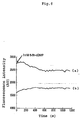
- Fig. 3 shows changes with time in emission ratios in the cell substrate.
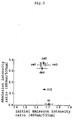
- CGY-Del1, CGY-Del2, CGY-Del3 and CGY-Del4 prepared in Example 1 were expressed in CHO-K1 cells in the same manner as in Example 2, and examined for changes in the emission intensities,
- HEK293 Human embryonic kidney cells (HEK293) were cultured in Dulbecco's modified eagle medium supplemented with 10 % FCS, 1 mM sodium pyruvate, and 0.1 mM nonessential amino acids at 37 °C in 5 % CO 2 , to give HEK293 cells.
- the resulting HEK293 cells were transfected by the same method as in Example 2 with CGY-Del1, CGY-Del2, CGY-Del3 and CGY-Del4 expression vectors, to give HEK293 cells expressing each type of CGY.
- the cGMP-visualizing probes were used to detect intracellular cGMP generated upon stimulation of the living cells with nitrogen monoxide (NO).
- Fig. 6 shows a photomicrograph of emissions induced by NOC-7 in the CGY-Del1-expressing HEK293 single cell.
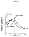
- Fig. 6 indicates that the emission ratio was rapidly decreased upon addition of 500 ⁇ M NOC-7 to the CGY-Del1-expressing HEK293 cells, and a subsequent increase in the emission ratio to nearly the initial level was observed.
- Fig. 7 shows changes in the NOC-7-induced emission ratio for the CGY-Dell-expressing HEK293 single cell.
- the emission ratio was first rapidly decreased followed by a slower increase in the ratio.
- the ratio was decreased again by adding 1 mM 8-Br-cGMP.
- zaprinast in place of 8-Br-cGMP, a similar re-increase was confirmed in the system where the emission ratio was recovered.
- the present invention provides a cGMP-visualizing probe that enables the easy detection and quantification of cGMP with high accurately even in vivo, as well as a method of detecting and quantifying cGMP by using the same.
Landscapes
- Life Sciences & Earth Sciences (AREA)
- Health & Medical Sciences (AREA)
- Chemical & Material Sciences (AREA)
- Engineering & Computer Science (AREA)
- Immunology (AREA)
- Organic Chemistry (AREA)
- Molecular Biology (AREA)
- Urology & Nephrology (AREA)
- Biotechnology (AREA)
- Proteomics, Peptides & Aminoacids (AREA)
- Microbiology (AREA)
- Biomedical Technology (AREA)
- Analytical Chemistry (AREA)
- Wood Science & Technology (AREA)
- Zoology (AREA)
- Biochemistry (AREA)
- Physics & Mathematics (AREA)
- Hematology (AREA)
- General Health & Medical Sciences (AREA)
- Bioinformatics & Cheminformatics (AREA)
- General Physics & Mathematics (AREA)
- Cell Biology (AREA)
- General Engineering & Computer Science (AREA)
- Biophysics (AREA)
- Food Science & Technology (AREA)
- Medicinal Chemistry (AREA)
- Genetics & Genomics (AREA)
- Pathology (AREA)
- Measuring Or Testing Involving Enzymes Or Micro-Organisms (AREA)
- Investigating Or Analysing Biological Materials (AREA)
- Investigating Or Analysing Materials By The Use Of Chemical Reactions (AREA)
- Investigating, Analyzing Materials By Fluorescence Or Luminescence (AREA)
- Enzymes And Modification Thereof (AREA)
- Micro-Organisms Or Cultivation Processes Thereof (AREA)
- Peptides Or Proteins (AREA)
Abstract
Description

Claims (9)
- A cGMP-visualizing probe, comprising:a polypeptide that binds specifically to cGMP; andtwo chromophores with different fluorescence wavelengths, which are each linked separately to the two terminals of the polypeptide.
- The cGMP-visualizing probe of claim 1, wherein the polypeptide that binds specifically to cGMP is a cGMP-binding protein.
- The cGMP-visualizing probe of claim 2, wherein the polypeptide that binds specifically to cGMP is cGMP-dependent kinase Iα.
- The cGMP-visualizing probe of any one of claims 1 to 3, wherein the chromophores are cyan fluorescent protein linked to the N-terminal of the polypeptide and yellow fluorescent protein linked to the C-terminal of the polypeptide.
- A method for detecting and quantifying cGMP, which comprises:making the cGMP-visualizing probe of any one of claims 1 to 4 coexist with cGMP; andmeasuring the change in the fluorescence wavelength.
- The method for detecting and quantifying cGMP of claim 5, which comprises introducing a polynucleotide expressing the cGMP-visualizing probe of any one of claims 1 to 4 into cells, whereby making the cGMP-visualizing probe coexist with cGMP.
- The method for detecting and quantifying cGMP of claim 5, which comprises:introducing a polynucleotide expressing a cGMP-visualizingprobe of any one of claims 1 to 4 into cells; andperforming ontogenisis from the non-human animal totipotent cells, thereby making the cGMP-visualizing probe coexist with cGMP in every cell of the resultant animal or its offspring.
- A non-human animal or offspring thereof, which is obtained by introducing a polynucleotide expressing a cGMP-visualizing probe of any one of claims 1 to 4 into cells and performing ontogenisis from the non-human animal totipotent cells.
- A method for screening a substance, which comprises introducing a test sample containing the substance into a non-human animal or offspring thereof of claim 8, and quantifying cGMP in the cells of the non-human animal or offspring thereof.
Applications Claiming Priority (3)
| Application Number | Priority Date | Filing Date | Title |
|---|---|---|---|
| JP2000202730A JP3643520B2 (en) | 2000-07-04 | 2000-07-04 | cGMP visualization probe and cGMP detection / quantification method using the same |
| JP2000202730 | 2000-07-04 | ||
| PCT/JP2001/005631 WO2002003069A1 (en) | 2000-07-04 | 2001-06-29 | cGMP-VISUALIZING PROBE AND METHOD OF DETECTING AND QUANTIFYING cGMP BY USING THE SAME |
Publications (3)
| Publication Number | Publication Date |
|---|---|
| EP1235073A1 true EP1235073A1 (en) | 2002-08-28 |
| EP1235073A4 EP1235073A4 (en) | 2004-06-09 |
| EP1235073B1 EP1235073B1 (en) | 2008-12-31 |
Family
ID=18700219
Family Applications (1)
| Application Number | Title | Priority Date | Filing Date |
|---|---|---|---|
| EP01943873A Expired - Lifetime EP1235073B1 (en) | 2000-07-04 | 2001-06-29 | cGMP-VISUALIZING PROBE AND METHOD OF DETECTING AND QUANTIFYING cGMP BY USING THE SAME |
Country Status (6)
| Country | Link |
|---|---|
| US (2) | US6924119B2 (en) |
| EP (1) | EP1235073B1 (en) |
| JP (1) | JP3643520B2 (en) |
| CA (1) | CA2384170C (en) |
| DE (1) | DE60137189D1 (en) |
| WO (1) | WO2002003069A1 (en) |
Cited By (2)
| Publication number | Priority date | Publication date | Assignee | Title |
|---|---|---|---|---|
| EP1767646A4 (en) * | 2004-05-21 | 2008-03-19 | Japan Science & Tech Agency | PROBE FOR DETECTION / DETERMINATION OF THE PRESENCE OF INOSITOL-1,4,5-TRIPHOSPHORIC ACID AND METHOD FOR DETECTION / DETERMINATION OF THE PRESENCE OF INOSITOL-1,4,5-TRIPHOSPHORIC ACID USING THE SAME PROBE |
| EP1780274A4 (en) * | 2004-07-05 | 2009-01-21 | Japan Science & Tech Agency | QUANTITATIVE DETECTION / DETERMINATION PROBE OF NITROGEN MONOXIDE, AND METHOD OF QUANTITATIVE DETECTION / DETERMINATION OF NITROGEN MONOXIDE USING THE SAME |
Families Citing this family (7)
| Publication number | Priority date | Publication date | Assignee | Title |
|---|---|---|---|---|
| WO2002090985A1 (en) * | 2001-05-07 | 2002-11-14 | Kiyoshi Nokihara | Peptide-immobilized baseboard and method of assaying target protein using the same |
| WO2006004216A1 (en) * | 2004-07-05 | 2006-01-12 | Japan Science And Technology Agency | Sensor cell for nitrogen monoxide detection and method of nitrogen monoxide detection and quantitative determination using the same |
| US8236523B2 (en) * | 2004-08-23 | 2012-08-07 | The Johns Hopkins University | Camp reporters and high throughput assays |
| FR2890446B1 (en) * | 2005-09-05 | 2008-04-18 | Cis Bio Internat Sa | METHOD FOR DETECTING INTRACELLULAR INTERACTION BETWEEN BIO-MOLECULES |
| US7982021B2 (en) * | 2006-07-14 | 2011-07-19 | The Johns Hopkins University | Nucleic acid molecules encoding emission ratiometric indicators of phosphoinositides |
| US8420327B2 (en) * | 2006-12-14 | 2013-04-16 | Georgia State University Research Foundation | Analyte sensors, methods for preparing and using such sensors, and methods of detecting analyte activity |
| WO2009055644A2 (en) * | 2007-10-24 | 2009-04-30 | California Institute Of Technology | Methods using the grueneberg ganglion chemosensory system |
Family Cites Families (2)
| Publication number | Priority date | Publication date | Assignee | Title |
|---|---|---|---|---|
| US6900304B2 (en) * | 1996-01-31 | 2005-05-31 | The Regents Of The University Of California | Emission ratiometric indicators of phosphorylation |
| US5998204A (en) * | 1997-03-14 | 1999-12-07 | The Regents Of The University Of California | Fluorescent protein sensors for detection of analytes |
-
2000
- 2000-07-04 JP JP2000202730A patent/JP3643520B2/en not_active Expired - Fee Related
-
2001
- 2001-06-29 DE DE60137189T patent/DE60137189D1/en not_active Expired - Lifetime
- 2001-06-29 CA CA002384170A patent/CA2384170C/en not_active Expired - Fee Related
- 2001-06-29 EP EP01943873A patent/EP1235073B1/en not_active Expired - Lifetime
- 2001-06-29 WO PCT/JP2001/005631 patent/WO2002003069A1/en not_active Ceased
- 2001-06-29 US US10/070,131 patent/US6924119B2/en not_active Expired - Fee Related
-
2005
- 2005-05-25 US US11/136,430 patent/US7329494B2/en not_active Expired - Fee Related
Cited By (2)
| Publication number | Priority date | Publication date | Assignee | Title |
|---|---|---|---|---|
| EP1767646A4 (en) * | 2004-05-21 | 2008-03-19 | Japan Science & Tech Agency | PROBE FOR DETECTION / DETERMINATION OF THE PRESENCE OF INOSITOL-1,4,5-TRIPHOSPHORIC ACID AND METHOD FOR DETECTION / DETERMINATION OF THE PRESENCE OF INOSITOL-1,4,5-TRIPHOSPHORIC ACID USING THE SAME PROBE |
| EP1780274A4 (en) * | 2004-07-05 | 2009-01-21 | Japan Science & Tech Agency | QUANTITATIVE DETECTION / DETERMINATION PROBE OF NITROGEN MONOXIDE, AND METHOD OF QUANTITATIVE DETECTION / DETERMINATION OF NITROGEN MONOXIDE USING THE SAME |
Also Published As
| Publication number | Publication date |
|---|---|
| CA2384170A1 (en) | 2002-01-10 |
| WO2002003069A1 (en) | 2002-01-10 |
| EP1235073B1 (en) | 2008-12-31 |
| CA2384170C (en) | 2006-02-14 |
| DE60137189D1 (en) | 2009-02-12 |
| US6924119B2 (en) | 2005-08-02 |
| EP1235073A4 (en) | 2004-06-09 |
| US20020137115A1 (en) | 2002-09-26 |
| US7329494B2 (en) | 2008-02-12 |
| US20060134655A1 (en) | 2006-06-22 |
| JP3643520B2 (en) | 2005-04-27 |
| JP2002017359A (en) | 2002-01-22 |
Similar Documents
| Publication | Publication Date | Title |
|---|---|---|
| US6900304B2 (en) | Emission ratiometric indicators of phosphorylation | |
| EP1088233B9 (en) | A bioluminescence resonance energy transfer (bret) system and its use | |
| US20070254373A1 (en) | Fragments of fluorescent proteins for protein fragment complementation assays | |
| EP2623514A1 (en) | Linker for unimolecular fret biosensor based on principle of fluorescence resonance energy transfer | |
| US8669074B2 (en) | Chimeric phosphorylation indicator | |
| AU2002312149A1 (en) | Emission ratiometric indicators of phosphorylation | |
| US6924119B2 (en) | cGMP-visualizing probe and a method of detecting and quantifying of cGMP by using the same | |
| US7855167B2 (en) | In vivo screening of protein-protein interactions with protein-fragment complementation assays | |
| US7282347B2 (en) | Method for extracting quantitative information relating to interactions between cellular components | |
| WO2002033102A1 (en) | Receptor mediated activation of heterotrimeric g-proteins | |
| Ward et al. | The uses of green fluorescent protein in mammalian cells | |
| EP1645881A1 (en) | Screening process for the detection and characterization of protein-protein-interactions in vivo by fluorescence cross correlation spectroscopy | |
| US20030143634A1 (en) | Method to detect interactions between cellular components in intact living cells, and to extract quantitative information relating to those interactions by fluorescence redistribution | |
| WO2003089627A1 (en) | Translocation dependent complementation for drug screening | |
| JP2022529592A (en) | Complementary system of split photoactive yellow protein and its use | |
| EP1624304B1 (en) | Probe for detecting and quantifying lipid second messenger and method of detecting and quantifying lipid second messenger using the same | |
| JP2006506950A (en) | Two green fluorescent protein fragments and their use in a method for detecting protein-protein interactions | |
| Rusanov et al. | Fluorescence resonance energy transfer between fluorescent proteins as powerful toolkits for in vivo studies | |
| US20060247419A1 (en) | Translocation dependent complementation for drug screening | |
| Prokhorov et al. | LETTERS EDITORIAL BOARD | |
| WO2005113792A1 (en) | Probe for detection/determination of inositol-1,4,5-triphosphoric acid and method of detecting/determining inositol-1,4,5-triphosphoric acid with the same |
Legal Events
| Date | Code | Title | Description |
|---|---|---|---|
| PUAI | Public reference made under article 153(3) epc to a published international application that has entered the european phase |
Free format text: ORIGINAL CODE: 0009012 |
|
| 17P | Request for examination filed |
Effective date: 20020308 |
|
| AK | Designated contracting states |
Kind code of ref document: A1 Designated state(s): AT BE CH CY DE DK ES FI FR GB GR IE IT LI LU MC NL PT SE TR |
|
| RBV | Designated contracting states (corrected) |
Designated state(s): CH DE FR GB LI NL SE |
|
| A4 | Supplementary search report drawn up and despatched |
Effective date: 20040426 |
|
| RIC1 | Information provided on ipc code assigned before grant |
Ipc: 7G 01N 33/58 A Ipc: 7G 01N 33/542 B Ipc: 7C 12N 15/62 B |
|
| RAP1 | Party data changed (applicant data changed or rights of an application transferred) |
Owner name: JAPAN SCIENCE AND TECHNOLOGY AGENCY |
|
| GRAP | Despatch of communication of intention to grant a patent |
Free format text: ORIGINAL CODE: EPIDOSNIGR1 |
|
| GRAS | Grant fee paid |
Free format text: ORIGINAL CODE: EPIDOSNIGR3 |
|
| GRAA | (expected) grant |
Free format text: ORIGINAL CODE: 0009210 |
|
| AK | Designated contracting states |
Kind code of ref document: B1 Designated state(s): CH DE FR GB LI NL SE |
|
| REG | Reference to a national code |
Ref country code: CH Ref legal event code: EP Ref country code: GB Ref legal event code: FG4D |
|
| REF | Corresponds to: |
Ref document number: 60137189 Country of ref document: DE Date of ref document: 20090212 Kind code of ref document: P |
|
| REG | Reference to a national code |
Ref country code: CH Ref legal event code: NV Representative=s name: BOHEST AG |
|
| REG | Reference to a national code |
Ref country code: SE Ref legal event code: TRGR |
|
| PLBE | No opposition filed within time limit |
Free format text: ORIGINAL CODE: 0009261 |
|
| STAA | Information on the status of an ep patent application or granted ep patent |
Free format text: STATUS: NO OPPOSITION FILED WITHIN TIME LIMIT |
|
| 26N | No opposition filed |
Effective date: 20091001 |
|
| REG | Reference to a national code |
Ref country code: CH Ref legal event code: PCAR Free format text: NEW ADDRESS: HOLBEINSTRASSE 36-38, 4051 BASEL (CH) |
|
| PGFP | Annual fee paid to national office [announced via postgrant information from national office to epo] |
Ref country code: GB Payment date: 20140513 Year of fee payment: 14 |
|
| PGFP | Annual fee paid to national office [announced via postgrant information from national office to epo] |
Ref country code: SE Payment date: 20140513 Year of fee payment: 14 Ref country code: CH Payment date: 20140513 Year of fee payment: 14 Ref country code: NL Payment date: 20140513 Year of fee payment: 14 Ref country code: DE Payment date: 20140513 Year of fee payment: 14 Ref country code: FR Payment date: 20140513 Year of fee payment: 14 |
|
| REG | Reference to a national code |
Ref country code: DE Ref legal event code: R119 Ref document number: 60137189 Country of ref document: DE |
|
| REG | Reference to a national code |
Ref country code: CH Ref legal event code: PL |
|
| REG | Reference to a national code |
Ref country code: SE Ref legal event code: EUG |
|
| GBPC | Gb: european patent ceased through non-payment of renewal fee |
Effective date: 20150629 |
|
| PG25 | Lapsed in a contracting state [announced via postgrant information from national office to epo] |
Ref country code: SE Free format text: LAPSE BECAUSE OF NON-PAYMENT OF DUE FEES Effective date: 20150630 |
|
| REG | Reference to a national code |
Ref country code: NL Ref legal event code: MM Effective date: 20150701 |
|
| REG | Reference to a national code |
Ref country code: FR Ref legal event code: ST Effective date: 20160229 |
|
| PG25 | Lapsed in a contracting state [announced via postgrant information from national office to epo] |
Ref country code: NL Free format text: LAPSE BECAUSE OF NON-PAYMENT OF DUE FEES Effective date: 20150701 Ref country code: GB Free format text: LAPSE BECAUSE OF NON-PAYMENT OF DUE FEES Effective date: 20150629 Ref country code: LI Free format text: LAPSE BECAUSE OF NON-PAYMENT OF DUE FEES Effective date: 20150630 Ref country code: CH Free format text: LAPSE BECAUSE OF NON-PAYMENT OF DUE FEES Effective date: 20150630 Ref country code: DE Free format text: LAPSE BECAUSE OF NON-PAYMENT OF DUE FEES Effective date: 20160101 |
|
| PG25 | Lapsed in a contracting state [announced via postgrant information from national office to epo] |
Ref country code: FR Free format text: LAPSE BECAUSE OF NON-PAYMENT OF DUE FEES Effective date: 20150630 |