EP1231910B1 - Pentamidine for treating cancer - Google Patents
Pentamidine for treating cancer Download PDFInfo
- Publication number
- EP1231910B1 EP1231910B1 EP00975725A EP00975725A EP1231910B1 EP 1231910 B1 EP1231910 B1 EP 1231910B1 EP 00975725 A EP00975725 A EP 00975725A EP 00975725 A EP00975725 A EP 00975725A EP 1231910 B1 EP1231910 B1 EP 1231910B1
- Authority
- EP
- European Patent Office
- Prior art keywords
- pentamidine
- endo
- exonuclease
- cancer
- cells
- Prior art date
- Legal status (The legal status is an assumption and is not a legal conclusion. Google has not performed a legal analysis and makes no representation as to the accuracy of the status listed.)
- Expired - Lifetime
Links
- 229960004448 pentamidine Drugs 0.000 title claims abstract description 135
- XDRYMKDFEDOLFX-UHFFFAOYSA-N pentamidine Chemical compound C1=CC(C(=N)N)=CC=C1OCCCCCOC1=CC=C(C(N)=N)C=C1 XDRYMKDFEDOLFX-UHFFFAOYSA-N 0.000 title claims abstract description 134
- 206010028980 Neoplasm Diseases 0.000 title claims abstract description 118
- 201000011510 cancer Diseases 0.000 title claims abstract description 67
- 108010051357 endoexonuclease Proteins 0.000 claims abstract description 56
- NWIBSHFKIJFRCO-WUDYKRTCSA-N Mytomycin Chemical compound C1N2C(C(C(C)=C(N)C3=O)=O)=C3[C@@H](COC(N)=O)[C@@]2(OC)[C@@H]2[C@H]1N2 NWIBSHFKIJFRCO-WUDYKRTCSA-N 0.000 claims abstract description 35
- 239000003814 drug Substances 0.000 claims abstract description 32
- 230000004614 tumor growth Effects 0.000 claims abstract description 28
- 229960004857 mitomycin Drugs 0.000 claims abstract description 20
- DQLATGHUWYMOKM-UHFFFAOYSA-L cisplatin Chemical compound N[Pt](N)(Cl)Cl DQLATGHUWYMOKM-UHFFFAOYSA-L 0.000 claims abstract description 18
- 229960004316 cisplatin Drugs 0.000 claims abstract description 18
- 239000008194 pharmaceutical composition Substances 0.000 claims abstract description 12
- 230000002401 inhibitory effect Effects 0.000 claims abstract description 11
- AOJJSUZBOXZQNB-TZSSRYMLSA-N Doxorubicin Chemical compound O([C@H]1C[C@@](O)(CC=2C(O)=C3C(=O)C=4C=CC=C(C=4C(=O)C3=C(O)C=21)OC)C(=O)CO)[C@H]1C[C@H](N)[C@H](O)[C@H](C)O1 AOJJSUZBOXZQNB-TZSSRYMLSA-N 0.000 claims description 28
- 239000003795 chemical substances by application Substances 0.000 claims description 24
- 230000035755 proliferation Effects 0.000 claims description 20
- VJJPUSNTGOMMGY-MRVIYFEKSA-N etoposide Chemical compound COC1=C(O)C(OC)=CC([C@@H]2C3=CC=4OCOC=4C=C3[C@@H](O[C@H]3[C@@H]([C@@H](O)[C@@H]4O[C@H](C)OC[C@H]4O3)O)[C@@H]3[C@@H]2C(OC3)=O)=C1 VJJPUSNTGOMMGY-MRVIYFEKSA-N 0.000 claims description 19
- 229960005420 etoposide Drugs 0.000 claims description 18
- 229940009456 adriamycin Drugs 0.000 claims description 14
- 238000002360 preparation method Methods 0.000 claims description 10
- 239000000693 micelle Substances 0.000 claims description 5
- 239000002502 liposome Substances 0.000 claims description 3
- 238000003745 diagnosis Methods 0.000 claims description 2
- -1 vesicles Substances 0.000 claims 1
- 230000000694 effects Effects 0.000 abstract description 47
- 150000001875 compounds Chemical class 0.000 abstract description 43
- 229940079593 drug Drugs 0.000 abstract description 28
- 238000000034 method Methods 0.000 abstract description 23
- 239000000203 mixture Substances 0.000 abstract description 15
- 238000011282 treatment Methods 0.000 abstract description 14
- 210000002966 serum Anatomy 0.000 abstract description 5
- 230000008439 repair process Effects 0.000 abstract description 4
- 231100000053 low toxicity Toxicity 0.000 abstract description 3
- 238000012544 monitoring process Methods 0.000 abstract description 2
- 230000002195 synergetic effect Effects 0.000 abstract 1
- 210000004027 cell Anatomy 0.000 description 107
- 241001465754 Metazoa Species 0.000 description 29
- 108020004414 DNA Proteins 0.000 description 27
- 238000002474 experimental method Methods 0.000 description 22
- FAPWRFPIFSIZLT-UHFFFAOYSA-M Sodium chloride Chemical compound [Na+].[Cl-] FAPWRFPIFSIZLT-UHFFFAOYSA-M 0.000 description 17
- 239000002246 antineoplastic agent Substances 0.000 description 17
- 239000012528 membrane Substances 0.000 description 17
- 241000699670 Mus sp. Species 0.000 description 16
- 239000000872 buffer Substances 0.000 description 16
- 230000001093 anti-cancer Effects 0.000 description 15
- 230000004083 survival effect Effects 0.000 description 15
- 238000001727 in vivo Methods 0.000 description 14
- 208000006552 Lewis Lung Carcinoma Diseases 0.000 description 13
- 206010027476 Metastases Diseases 0.000 description 11
- 239000007943 implant Substances 0.000 description 11
- 239000011780 sodium chloride Substances 0.000 description 10
- 239000000243 solution Substances 0.000 description 10
- 229940127089 cytotoxic agent Drugs 0.000 description 9
- 230000012010 growth Effects 0.000 description 9
- 210000004072 lung Anatomy 0.000 description 9
- 229920000642 polymer Polymers 0.000 description 9
- UPBAOYRENQEPJO-UHFFFAOYSA-N n-[5-[[5-[(3-amino-3-iminopropyl)carbamoyl]-1-methylpyrrol-3-yl]carbamoyl]-1-methylpyrrol-3-yl]-4-formamido-1-methylpyrrole-2-carboxamide Chemical compound CN1C=C(NC=O)C=C1C(=O)NC1=CN(C)C(C(=O)NC2=CN(C)C(C(=O)NCCC(N)=N)=C2)=C1 UPBAOYRENQEPJO-UHFFFAOYSA-N 0.000 description 8
- 108090000623 proteins and genes Proteins 0.000 description 8
- 241000282414 Homo sapiens Species 0.000 description 7
- 230000005782 double-strand break Effects 0.000 description 7
- 230000005764 inhibitory process Effects 0.000 description 7
- 210000001519 tissue Anatomy 0.000 description 7
- KCXVZYZYPLLWCC-UHFFFAOYSA-N EDTA Chemical compound OC(=O)CN(CC(O)=O)CCN(CC(O)=O)CC(O)=O KCXVZYZYPLLWCC-UHFFFAOYSA-N 0.000 description 6
- WSFSSNUMVMOOMR-UHFFFAOYSA-N Formaldehyde Chemical compound O=C WSFSSNUMVMOOMR-UHFFFAOYSA-N 0.000 description 6
- 108010001336 Horseradish Peroxidase Proteins 0.000 description 6
- 238000004113 cell culture Methods 0.000 description 6
- 229940044683 chemotherapy drug Drugs 0.000 description 6
- 208000002154 non-small cell lung carcinoma Diseases 0.000 description 6
- 235000018102 proteins Nutrition 0.000 description 6
- 102000004169 proteins and genes Human genes 0.000 description 6
- 230000002829 reductive effect Effects 0.000 description 6
- XLYOFNOQVPJJNP-UHFFFAOYSA-N water Substances O XLYOFNOQVPJJNP-UHFFFAOYSA-N 0.000 description 6
- QKNYBSVHEMOAJP-UHFFFAOYSA-N 2-amino-2-(hydroxymethyl)propane-1,3-diol;hydron;chloride Chemical compound Cl.OCC(N)(CO)CO QKNYBSVHEMOAJP-UHFFFAOYSA-N 0.000 description 5
- 102000004190 Enzymes Human genes 0.000 description 5
- 108090000790 Enzymes Proteins 0.000 description 5
- 206010027458 Metastases to lung Diseases 0.000 description 5
- 229930192392 Mitomycin Natural products 0.000 description 5
- 230000009286 beneficial effect Effects 0.000 description 5
- 230000037396 body weight Effects 0.000 description 5
- 210000000056 organ Anatomy 0.000 description 5
- 239000000843 powder Substances 0.000 description 5
- 230000005855 radiation Effects 0.000 description 5
- 210000004881 tumor cell Anatomy 0.000 description 5
- 238000005406 washing Methods 0.000 description 5
- 206010006187 Breast cancer Diseases 0.000 description 4
- 208000002151 Pleural effusion Diseases 0.000 description 4
- 230000009471 action Effects 0.000 description 4
- 208000009956 adenocarcinoma Diseases 0.000 description 4
- 230000000259 anti-tumor effect Effects 0.000 description 4
- 230000030833 cell death Effects 0.000 description 4
- 238000009643 clonogenic assay Methods 0.000 description 4
- 231100000096 clonogenic assay Toxicity 0.000 description 4
- XNYZHCFCZNMTFY-UHFFFAOYSA-N diminazene Chemical compound C1=CC(C(=N)N)=CC=C1N\N=N\C1=CC=C(C(N)=N)C=C1 XNYZHCFCZNMTFY-UHFFFAOYSA-N 0.000 description 4
- 238000009472 formulation Methods 0.000 description 4
- 239000007928 intraperitoneal injection Substances 0.000 description 4
- 208000003849 large cell carcinoma Diseases 0.000 description 4
- 230000007246 mechanism Effects 0.000 description 4
- 239000000546 pharmaceutical excipient Substances 0.000 description 4
- 230000005783 single-strand break Effects 0.000 description 4
- 235000020183 skimmed milk Nutrition 0.000 description 4
- 108010042747 stallimycin Proteins 0.000 description 4
- 229950009902 stallimycin Drugs 0.000 description 4
- 108091003079 Bovine Serum Albumin Proteins 0.000 description 3
- 208000026310 Breast neoplasm Diseases 0.000 description 3
- 230000033616 DNA repair Effects 0.000 description 3
- 230000004543 DNA replication Effects 0.000 description 3
- PEDCQBHIVMGVHV-UHFFFAOYSA-N Glycerine Chemical compound OCC(O)CO PEDCQBHIVMGVHV-UHFFFAOYSA-N 0.000 description 3
- ZMXDDKWLCZADIW-UHFFFAOYSA-N N,N-Dimethylformamide Chemical compound CN(C)C=O ZMXDDKWLCZADIW-UHFFFAOYSA-N 0.000 description 3
- 241000283973 Oryctolagus cuniculus Species 0.000 description 3
- 241000233872 Pneumocystis carinii Species 0.000 description 3
- 208000029742 colonic neoplasm Diseases 0.000 description 3
- 239000013078 crystal Substances 0.000 description 3
- 230000006378 damage Effects 0.000 description 3
- 239000012091 fetal bovine serum Substances 0.000 description 3
- 210000002950 fibroblast Anatomy 0.000 description 3
- 230000006870 function Effects 0.000 description 3
- 239000000499 gel Substances 0.000 description 3
- 230000006698 induction Effects 0.000 description 3
- 238000007912 intraperitoneal administration Methods 0.000 description 3
- 238000002955 isolation Methods 0.000 description 3
- 231100001231 less toxic Toxicity 0.000 description 3
- 239000000463 material Substances 0.000 description 3
- 238000005259 measurement Methods 0.000 description 3
- 230000001394 metastastic effect Effects 0.000 description 3
- 206010061289 metastatic neoplasm Diseases 0.000 description 3
- 238000010172 mouse model Methods 0.000 description 3
- 239000002953 phosphate buffered saline Substances 0.000 description 3
- 150000003839 salts Chemical class 0.000 description 3
- 229960005322 streptomycin Drugs 0.000 description 3
- 239000000725 suspension Substances 0.000 description 3
- 231100000419 toxicity Toxicity 0.000 description 3
- 230000001988 toxicity Effects 0.000 description 3
- 230000035899 viability Effects 0.000 description 3
- CPKVUHPKYQGHMW-UHFFFAOYSA-N 1-ethenylpyrrolidin-2-one;molecular iodine Chemical compound II.C=CN1CCCC1=O CPKVUHPKYQGHMW-UHFFFAOYSA-N 0.000 description 2
- IJGRMHOSHXDMSA-UHFFFAOYSA-N Atomic nitrogen Chemical compound N#N IJGRMHOSHXDMSA-UHFFFAOYSA-N 0.000 description 2
- 239000011547 Bouin solution Substances 0.000 description 2
- 206010009944 Colon cancer Diseases 0.000 description 2
- 201000008808 Fibrosarcoma Diseases 0.000 description 2
- GHASVSINZRGABV-UHFFFAOYSA-N Fluorouracil Chemical compound FC1=CNC(=O)NC1=O GHASVSINZRGABV-UHFFFAOYSA-N 0.000 description 2
- 241000282412 Homo Species 0.000 description 2
- TWRXJAOTZQYOKJ-UHFFFAOYSA-L Magnesium chloride Chemical compound [Mg+2].[Cl-].[Cl-] TWRXJAOTZQYOKJ-UHFFFAOYSA-L 0.000 description 2
- 241000699666 Mus <mouse, genus> Species 0.000 description 2
- 239000000020 Nitrocellulose Substances 0.000 description 2
- 101710163270 Nuclease Proteins 0.000 description 2
- 239000012980 RPMI-1640 medium Substances 0.000 description 2
- UIIMBOGNXHQVGW-UHFFFAOYSA-M Sodium bicarbonate Chemical compound [Na+].OC([O-])=O UIIMBOGNXHQVGW-UHFFFAOYSA-M 0.000 description 2
- 101710183280 Topoisomerase Proteins 0.000 description 2
- 102000004142 Trypsin Human genes 0.000 description 2
- 108090000631 Trypsin Proteins 0.000 description 2
- 230000002159 abnormal effect Effects 0.000 description 2
- 238000002835 absorbance Methods 0.000 description 2
- 230000002001 anti-metastasis Effects 0.000 description 2
- 238000003556 assay Methods 0.000 description 2
- 229940064804 betadine Drugs 0.000 description 2
- 210000004369 blood Anatomy 0.000 description 2
- 239000008280 blood Substances 0.000 description 2
- 230000005880 cancer cell killing Effects 0.000 description 2
- 230000005907 cancer growth Effects 0.000 description 2
- 230000004663 cell proliferation Effects 0.000 description 2
- 238000006243 chemical reaction Methods 0.000 description 2
- 230000003021 clonogenic effect Effects 0.000 description 2
- 231100000135 cytotoxicity Toxicity 0.000 description 2
- 230000003013 cytotoxicity Effects 0.000 description 2
- 238000010790 dilution Methods 0.000 description 2
- 239000012895 dilution Substances 0.000 description 2
- LOKCTEFSRHRXRJ-UHFFFAOYSA-I dipotassium trisodium dihydrogen phosphate hydrogen phosphate dichloride Chemical compound P(=O)(O)(O)[O-].[K+].P(=O)(O)([O-])[O-].[Na+].[Na+].[Cl-].[K+].[Cl-].[Na+] LOKCTEFSRHRXRJ-UHFFFAOYSA-I 0.000 description 2
- 239000012153 distilled water Substances 0.000 description 2
- 230000006846 excision repair Effects 0.000 description 2
- 230000029142 excretion Effects 0.000 description 2
- 229960002949 fluorouracil Drugs 0.000 description 2
- 239000003112 inhibitor Substances 0.000 description 2
- 239000007924 injection Substances 0.000 description 2
- 238000002347 injection Methods 0.000 description 2
- 238000001990 intravenous administration Methods 0.000 description 2
- 231100000518 lethal Toxicity 0.000 description 2
- 230000001665 lethal effect Effects 0.000 description 2
- 239000007788 liquid Substances 0.000 description 2
- 210000001165 lymph node Anatomy 0.000 description 2
- 239000003550 marker Substances 0.000 description 2
- 239000002609 medium Substances 0.000 description 2
- 238000012986 modification Methods 0.000 description 2
- 230000004048 modification Effects 0.000 description 2
- 229920001220 nitrocellulos Polymers 0.000 description 2
- 238000011275 oncology therapy Methods 0.000 description 2
- 229940127084 other anti-cancer agent Drugs 0.000 description 2
- 231100000915 pathological change Toxicity 0.000 description 2
- 230000036285 pathological change Effects 0.000 description 2
- 239000013610 patient sample Substances 0.000 description 2
- YBYRMVIVWMBXKQ-UHFFFAOYSA-N phenylmethanesulfonyl fluoride Chemical compound FS(=O)(=O)CC1=CC=CC=C1 YBYRMVIVWMBXKQ-UHFFFAOYSA-N 0.000 description 2
- 239000008055 phosphate buffer solution Substances 0.000 description 2
- 230000036470 plasma concentration Effects 0.000 description 2
- 238000011886 postmortem examination Methods 0.000 description 2
- 230000003389 potentiating effect Effects 0.000 description 2
- 230000008569 process Effects 0.000 description 2
- 230000002035 prolonged effect Effects 0.000 description 2
- 230000006798 recombination Effects 0.000 description 2
- 238000005215 recombination Methods 0.000 description 2
- 206010041823 squamous cell carcinoma Diseases 0.000 description 2
- 239000000126 substance Substances 0.000 description 2
- 239000000758 substrate Substances 0.000 description 2
- 239000006228 supernatant Substances 0.000 description 2
- 238000001356 surgical procedure Methods 0.000 description 2
- 230000001225 therapeutic effect Effects 0.000 description 2
- 239000012588 trypsin Substances 0.000 description 2
- 230000036325 urinary excretion Effects 0.000 description 2
- 231100000925 very toxic Toxicity 0.000 description 2
- OZDAOHVKBFBBMZ-UHFFFAOYSA-N 2-aminopentanedioic acid;hydrate Chemical compound O.OC(=O)C(N)CCC(O)=O OZDAOHVKBFBBMZ-UHFFFAOYSA-N 0.000 description 1
- 125000000972 4,5-dimethylthiazol-2-yl group Chemical group [H]C([H])([H])C1=C(N=C(*)S1)C([H])([H])[H] 0.000 description 1
- JAVCHNFPSHBZAF-UUPCJSQJSA-N 4-[5-(4-carbamimidoylphenoxy)pentoxy]benzenecarboximidamide;(2r,3s,4r,5r)-2,3,4,5,6-pentahydroxyhexanoic acid Chemical compound OC[C@@H](O)[C@@H](O)[C@H](O)[C@@H](O)C(O)=O.OC[C@@H](O)[C@@H](O)[C@H](O)[C@@H](O)C(O)=O.C1=CC(C(=N)N)=CC=C1OCCCCCOC1=CC=C(C(N)=N)C=C1 JAVCHNFPSHBZAF-UUPCJSQJSA-N 0.000 description 1
- SQGXXDIGSNISIQ-UHFFFAOYSA-D 4-[5-(4-carbamimidoylphenoxy)pentoxy]benzenecarboximidamide;hydron;platinum(2+);tetradecachloride Chemical compound [H+].[H+].[H+].[H+].[Cl-].[Cl-].[Cl-].[Cl-].[Cl-].[Cl-].[Cl-].[Cl-].[Cl-].[Cl-].[Cl-].[Cl-].[Cl-].[Cl-].[Pt+2].[Pt+2].[Pt+2].[Pt+2].[Pt+2].C1=CC(C(=N)N)=CC=C1OCCCCCOC1=CC=C(C(N)=N)C=C1.C1=CC(C(=N)N)=CC=C1OCCCCCOC1=CC=C(C(N)=N)C=C1.C1=CC(C(=N)N)=CC=C1OCCCCCOC1=CC=C(C(N)=N)C=C1 SQGXXDIGSNISIQ-UHFFFAOYSA-D 0.000 description 1
- LVSPDZAGCBEQAV-UHFFFAOYSA-N 4-chloronaphthalen-1-ol Chemical compound C1=CC=C2C(O)=CC=C(Cl)C2=C1 LVSPDZAGCBEQAV-UHFFFAOYSA-N 0.000 description 1
- 108010017384 Blood Proteins Proteins 0.000 description 1
- 102000004506 Blood Proteins Human genes 0.000 description 1
- 206010055113 Breast cancer metastatic Diseases 0.000 description 1
- CPELXLSAUQHCOX-UHFFFAOYSA-M Bromide Chemical compound [Br-] CPELXLSAUQHCOX-UHFFFAOYSA-M 0.000 description 1
- 201000009030 Carcinoma Diseases 0.000 description 1
- 241000282693 Cercopithecidae Species 0.000 description 1
- 108020005199 Dehydrogenases Proteins 0.000 description 1
- 102000016911 Deoxyribonucleases Human genes 0.000 description 1
- 108010053770 Deoxyribonucleases Proteins 0.000 description 1
- 239000006144 Dulbecco’s modified Eagle's medium Substances 0.000 description 1
- 238000002965 ELISA Methods 0.000 description 1
- LFQSCWFLJHTTHZ-UHFFFAOYSA-N Ethanol Chemical class CCO LFQSCWFLJHTTHZ-UHFFFAOYSA-N 0.000 description 1
- WQZGKKKJIJFFOK-GASJEMHNSA-N Glucose Natural products OC[C@H]1OC(O)[C@H](O)[C@@H](O)[C@@H]1O WQZGKKKJIJFFOK-GASJEMHNSA-N 0.000 description 1
- WHUUTDBJXJRKMK-UHFFFAOYSA-N Glutamic acid Natural products OC(=O)C(N)CCC(O)=O WHUUTDBJXJRKMK-UHFFFAOYSA-N 0.000 description 1
- 239000012981 Hank's balanced salt solution Substances 0.000 description 1
- 108060003951 Immunoglobulin Proteins 0.000 description 1
- 208000029523 Interstitial Lung disease Diseases 0.000 description 1
- PIWKPBJCKXDKJR-UHFFFAOYSA-N Isoflurane Chemical compound FC(F)OC(Cl)C(F)(F)F PIWKPBJCKXDKJR-UHFFFAOYSA-N 0.000 description 1
- CKLJMWTZIZZHCS-REOHCLBHSA-N L-aspartic acid Chemical compound OC(=O)[C@@H](N)CC(O)=O CKLJMWTZIZZHCS-REOHCLBHSA-N 0.000 description 1
- GUBGYTABKSRVRQ-QKKXKWKRSA-N Lactose Natural products OC[C@H]1O[C@@H](O[C@H]2[C@H](O)[C@@H](O)C(O)O[C@@H]2CO)[C@H](O)[C@@H](O)[C@H]1O GUBGYTABKSRVRQ-QKKXKWKRSA-N 0.000 description 1
- 206010058467 Lung neoplasm malignant Diseases 0.000 description 1
- 231100000002 MTT assay Toxicity 0.000 description 1
- 238000000134 MTT assay Methods 0.000 description 1
- 241000204031 Mycoplasma Species 0.000 description 1
- 108091028043 Nucleic acid sequence Proteins 0.000 description 1
- 108010047956 Nucleosomes Proteins 0.000 description 1
- 206010035664 Pneumonia Diseases 0.000 description 1
- 206010035742 Pneumonitis Diseases 0.000 description 1
- 241000288906 Primates Species 0.000 description 1
- 239000006146 Roswell Park Memorial Institute medium Substances 0.000 description 1
- 229920002684 Sepharose Polymers 0.000 description 1
- 108010071390 Serum Albumin Proteins 0.000 description 1
- 102000007562 Serum Albumin Human genes 0.000 description 1
- 208000000453 Skin Neoplasms Diseases 0.000 description 1
- 241000191967 Staphylococcus aureus Species 0.000 description 1
- 238000000692 Student's t-test Methods 0.000 description 1
- GLNADSQYFUSGOU-GPTZEZBUSA-J Trypan blue Chemical compound [Na+].[Na+].[Na+].[Na+].C1=C(S([O-])(=O)=O)C=C2C=C(S([O-])(=O)=O)C(/N=N/C3=CC=C(C=C3C)C=3C=C(C(=CC=3)\N=N\C=3C(=CC4=CC(=CC(N)=C4C=3O)S([O-])(=O)=O)S([O-])(=O)=O)C)=C(O)C2=C1N GLNADSQYFUSGOU-GPTZEZBUSA-J 0.000 description 1
- 241000223104 Trypanosoma Species 0.000 description 1
- 239000002253 acid Substances 0.000 description 1
- 230000007059 acute toxicity Effects 0.000 description 1
- 231100000403 acute toxicity Toxicity 0.000 description 1
- 239000000654 additive Substances 0.000 description 1
- 230000000843 anti-fungal effect Effects 0.000 description 1
- 230000002141 anti-parasite Effects 0.000 description 1
- 239000000427 antigen Substances 0.000 description 1
- 102000036639 antigens Human genes 0.000 description 1
- 108091007433 antigens Proteins 0.000 description 1
- 229940041181 antineoplastic drug Drugs 0.000 description 1
- 239000003963 antioxidant agent Substances 0.000 description 1
- 230000003078 antioxidant effect Effects 0.000 description 1
- 235000003704 aspartic acid Nutrition 0.000 description 1
- OQFSQFPPLPISGP-UHFFFAOYSA-N beta-carboxyaspartic acid Natural products OC(=O)C(N)C(C(O)=O)C(O)=O OQFSQFPPLPISGP-UHFFFAOYSA-N 0.000 description 1
- 238000006065 biodegradation reaction Methods 0.000 description 1
- 230000004071 biological effect Effects 0.000 description 1
- 230000015572 biosynthetic process Effects 0.000 description 1
- 235000010290 biphenyl Nutrition 0.000 description 1
- 239000004305 biphenyl Substances 0.000 description 1
- 125000006267 biphenyl group Chemical group 0.000 description 1
- 238000004159 blood analysis Methods 0.000 description 1
- 210000001124 body fluid Anatomy 0.000 description 1
- 239000010839 body fluid Substances 0.000 description 1
- 210000001185 bone marrow Anatomy 0.000 description 1
- 201000008274 breast adenocarcinoma Diseases 0.000 description 1
- 239000002775 capsule Substances 0.000 description 1
- 230000000747 cardiac effect Effects 0.000 description 1
- 230000015556 catabolic process Effects 0.000 description 1
- 230000032823 cell division Effects 0.000 description 1
- 230000010261 cell growth Effects 0.000 description 1
- 230000006037 cell lysis Effects 0.000 description 1
- 239000006285 cell suspension Substances 0.000 description 1
- 230000008859 change Effects 0.000 description 1
- 238000012512 characterization method Methods 0.000 description 1
- 239000003153 chemical reaction reagent Substances 0.000 description 1
- 238000002512 chemotherapy Methods 0.000 description 1
- 230000001684 chronic effect Effects 0.000 description 1
- 238000003776 cleavage reaction Methods 0.000 description 1
- 201000010897 colon adenocarcinoma Diseases 0.000 description 1
- 230000002860 competitive effect Effects 0.000 description 1
- 239000006071 cream Substances 0.000 description 1
- 210000004748 cultured cell Anatomy 0.000 description 1
- 238000011461 current therapy Methods 0.000 description 1
- 230000007423 decrease Effects 0.000 description 1
- 238000006731 degradation reaction Methods 0.000 description 1
- 230000003111 delayed effect Effects 0.000 description 1
- 230000001419 dependent effect Effects 0.000 description 1
- 238000011161 development Methods 0.000 description 1
- 230000018109 developmental process Effects 0.000 description 1
- 239000008121 dextrose Substances 0.000 description 1
- 238000002405 diagnostic procedure Methods 0.000 description 1
- 235000005911 diet Nutrition 0.000 description 1
- 230000037213 diet Effects 0.000 description 1
- 235000014113 dietary fatty acids Nutrition 0.000 description 1
- 201000010099 disease Diseases 0.000 description 1
- 208000037265 diseases, disorders, signs and symptoms Diseases 0.000 description 1
- 238000009826 distribution Methods 0.000 description 1
- 231100000673 dose–response relationship Toxicity 0.000 description 1
- 239000000890 drug combination Substances 0.000 description 1
- 239000000975 dye Substances 0.000 description 1
- 239000003995 emulsifying agent Substances 0.000 description 1
- 239000000839 emulsion Substances 0.000 description 1
- 238000006911 enzymatic reaction Methods 0.000 description 1
- 229940125532 enzyme inhibitor Drugs 0.000 description 1
- 239000002532 enzyme inhibitor Substances 0.000 description 1
- 235000019441 ethanol Nutrition 0.000 description 1
- 229930195729 fatty acid Natural products 0.000 description 1
- 239000000194 fatty acid Substances 0.000 description 1
- 150000004665 fatty acids Chemical class 0.000 description 1
- 239000000945 filler Substances 0.000 description 1
- 239000000796 flavoring agent Substances 0.000 description 1
- 235000019634 flavors Nutrition 0.000 description 1
- 229940084362 forane Drugs 0.000 description 1
- 238000004108 freeze drying Methods 0.000 description 1
- 239000007903 gelatin capsule Substances 0.000 description 1
- 239000011521 glass Substances 0.000 description 1
- 235000013922 glutamic acid Nutrition 0.000 description 1
- 231100001261 hazardous Toxicity 0.000 description 1
- 238000003505 heat denaturation Methods 0.000 description 1
- 125000001072 heteroaryl group Chemical group 0.000 description 1
- 210000005260 human cell Anatomy 0.000 description 1
- 238000003119 immunoblot Methods 0.000 description 1
- 102000018358 immunoglobulin Human genes 0.000 description 1
- 238000000338 in vitro Methods 0.000 description 1
- 238000011534 incubation Methods 0.000 description 1
- 239000000411 inducer Substances 0.000 description 1
- 230000001939 inductive effect Effects 0.000 description 1
- 208000015181 infectious disease Diseases 0.000 description 1
- 238000011081 inoculation Methods 0.000 description 1
- 238000007918 intramuscular administration Methods 0.000 description 1
- 210000003734 kidney Anatomy 0.000 description 1
- 239000008101 lactose Substances 0.000 description 1
- 239000012669 liquid formulation Substances 0.000 description 1
- 201000005202 lung cancer Diseases 0.000 description 1
- 208000020816 lung neoplasm Diseases 0.000 description 1
- 208000037841 lung tumor Diseases 0.000 description 1
- 239000012139 lysis buffer Substances 0.000 description 1
- 229910001629 magnesium chloride Inorganic materials 0.000 description 1
- 230000003211 malignant effect Effects 0.000 description 1
- 230000009401 metastasis Effects 0.000 description 1
- 230000002438 mitochondrial effect Effects 0.000 description 1
- 230000011278 mitosis Effects 0.000 description 1
- 210000003205 muscle Anatomy 0.000 description 1
- VMGAPWLDMVPYIA-HIDZBRGKSA-N n'-amino-n-iminomethanimidamide Chemical compound N\N=C\N=N VMGAPWLDMVPYIA-HIDZBRGKSA-N 0.000 description 1
- 229910052757 nitrogen Inorganic materials 0.000 description 1
- 231100000404 nontoxic agent Toxicity 0.000 description 1
- 210000001623 nucleosome Anatomy 0.000 description 1
- 239000002674 ointment Substances 0.000 description 1
- 239000006186 oral dosage form Substances 0.000 description 1
- 239000012188 paraffin wax Substances 0.000 description 1
- 244000045947 parasite Species 0.000 description 1
- 239000008188 pellet Substances 0.000 description 1
- 229960001624 pentamidine isethionate Drugs 0.000 description 1
- YBVNFKZSMZGRAD-UHFFFAOYSA-N pentamidine isethionate Chemical compound OCCS(O)(=O)=O.OCCS(O)(=O)=O.C1=CC(C(=N)N)=CC=C1OCCCCCOC1=CC=C(C(N)=N)C=C1 YBVNFKZSMZGRAD-UHFFFAOYSA-N 0.000 description 1
- 230000002085 persistent effect Effects 0.000 description 1
- ZUOUZKKEUPVFJK-UHFFFAOYSA-N phenylbenzene Natural products C1=CC=CC=C1C1=CC=CC=C1 ZUOUZKKEUPVFJK-UHFFFAOYSA-N 0.000 description 1
- 150000003904 phospholipids Chemical class 0.000 description 1
- 239000006187 pill Substances 0.000 description 1
- 238000007747 plating Methods 0.000 description 1
- 229920002401 polyacrylamide Polymers 0.000 description 1
- 229920001184 polypeptide Polymers 0.000 description 1
- 239000003755 preservative agent Substances 0.000 description 1
- 230000002335 preservative effect Effects 0.000 description 1
- 108090000765 processed proteins & peptides Proteins 0.000 description 1
- 102000004196 processed proteins & peptides Human genes 0.000 description 1
- 230000000644 propagated effect Effects 0.000 description 1
- 238000011321 prophylaxis Methods 0.000 description 1
- 238000004080 punching Methods 0.000 description 1
- BOLDJAUMGUJJKM-LSDHHAIUSA-N renifolin D Natural products CC(=C)[C@@H]1Cc2c(O)c(O)ccc2[C@H]1CC(=O)c3ccc(O)cc3O BOLDJAUMGUJJKM-LSDHHAIUSA-N 0.000 description 1
- 230000000241 respiratory effect Effects 0.000 description 1
- 230000004044 response Effects 0.000 description 1
- 239000000523 sample Substances 0.000 description 1
- 230000007017 scission Effects 0.000 description 1
- 238000000926 separation method Methods 0.000 description 1
- 230000003007 single stranded DNA break Effects 0.000 description 1
- 201000000849 skin cancer Diseases 0.000 description 1
- 210000001626 skin fibroblast Anatomy 0.000 description 1
- 235000017557 sodium bicarbonate Nutrition 0.000 description 1
- 229910000030 sodium bicarbonate Inorganic materials 0.000 description 1
- 239000007787 solid Substances 0.000 description 1
- 238000005063 solubilization Methods 0.000 description 1
- 230000007928 solubilization Effects 0.000 description 1
- 239000011537 solubilization buffer Substances 0.000 description 1
- 239000002904 solvent Substances 0.000 description 1
- 230000006641 stabilisation Effects 0.000 description 1
- 238000010186 staining Methods 0.000 description 1
- 238000011255 standard chemotherapy Methods 0.000 description 1
- 238000010561 standard procedure Methods 0.000 description 1
- 238000007619 statistical method Methods 0.000 description 1
- 238000003860 storage Methods 0.000 description 1
- 239000000829 suppository Substances 0.000 description 1
- 229910000811 surgical stainless steel Inorganic materials 0.000 description 1
- 239000010966 surgical stainless steel Substances 0.000 description 1
- 239000000375 suspending agent Substances 0.000 description 1
- 208000024891 symptom Diseases 0.000 description 1
- 239000003826 tablet Substances 0.000 description 1
- 239000007916 tablet composition Substances 0.000 description 1
- 238000012360 testing method Methods 0.000 description 1
- UEUXEKPTXMALOB-UHFFFAOYSA-J tetrasodium;2-[2-[bis(carboxylatomethyl)amino]ethyl-(carboxylatomethyl)amino]acetate Chemical compound [Na+].[Na+].[Na+].[Na+].[O-]C(=O)CN(CC([O-])=O)CCN(CC([O-])=O)CC([O-])=O UEUXEKPTXMALOB-UHFFFAOYSA-J 0.000 description 1
- 125000003831 tetrazolyl group Chemical group 0.000 description 1
- 238000002560 therapeutic procedure Methods 0.000 description 1
- 230000004797 therapeutic response Effects 0.000 description 1
- 230000017423 tissue regeneration Effects 0.000 description 1
- 230000000699 topical effect Effects 0.000 description 1
- 231100000167 toxic agent Toxicity 0.000 description 1
- 239000003440 toxic substance Substances 0.000 description 1
- 238000012546 transfer Methods 0.000 description 1
- 230000001052 transient effect Effects 0.000 description 1
- 201000002311 trypanosomiasis Diseases 0.000 description 1
- 239000003981 vehicle Substances 0.000 description 1
Images







Classifications
-
- A—HUMAN NECESSITIES
- A61—MEDICAL OR VETERINARY SCIENCE; HYGIENE
- A61K—PREPARATIONS FOR MEDICAL, DENTAL OR TOILETRY PURPOSES
- A61K45/00—Medicinal preparations containing active ingredients not provided for in groups A61K31/00 - A61K41/00
- A61K45/06—Mixtures of active ingredients without chemical characterisation, e.g. antiphlogistics and cardiaca
-
- A—HUMAN NECESSITIES
- A61—MEDICAL OR VETERINARY SCIENCE; HYGIENE
- A61K—PREPARATIONS FOR MEDICAL, DENTAL OR TOILETRY PURPOSES
- A61K31/00—Medicinal preparations containing organic active ingredients
- A61K31/13—Amines
- A61K31/155—Amidines (), e.g. guanidine (H2N—C(=NH)—NH2), isourea (N=C(OH)—NH2), isothiourea (—N=C(SH)—NH2)
-
- A—HUMAN NECESSITIES
- A61—MEDICAL OR VETERINARY SCIENCE; HYGIENE
- A61K—PREPARATIONS FOR MEDICAL, DENTAL OR TOILETRY PURPOSES
- A61K31/00—Medicinal preparations containing organic active ingredients
- A61K31/33—Heterocyclic compounds
- A61K31/335—Heterocyclic compounds having oxygen as the only ring hetero atom, e.g. fungichromin
- A61K31/337—Heterocyclic compounds having oxygen as the only ring hetero atom, e.g. fungichromin having four-membered rings, e.g. taxol
-
- A—HUMAN NECESSITIES
- A61—MEDICAL OR VETERINARY SCIENCE; HYGIENE
- A61K—PREPARATIONS FOR MEDICAL, DENTAL OR TOILETRY PURPOSES
- A61K33/00—Medicinal preparations containing inorganic active ingredients
- A61K33/24—Heavy metals; Compounds thereof
- A61K33/243—Platinum; Compounds thereof
-
- A—HUMAN NECESSITIES
- A61—MEDICAL OR VETERINARY SCIENCE; HYGIENE
- A61P—SPECIFIC THERAPEUTIC ACTIVITY OF CHEMICAL COMPOUNDS OR MEDICINAL PREPARATIONS
- A61P35/00—Antineoplastic agents
Definitions
- the present invention relates to chemotherapeutic agents for treating cancer.
- Cancer cells proliferate more rapidly than normal cells. The rate of mitosis and DNA replication is therefore significantly greater in cancer cells. Agents that inhibit DNA replication and recombination affect cancer cells more than normal cells.
- chemotherapeutic agents for treating cancer inhibit DNA replication by inducing DNA breaks.
- Some drugs such as mitomycin, induce DNA breaks in part by binding to the DNA itself.
- Other anticancer agents interfere with topoisomerase enzymes, which modify DNA structure. In doing so, they induce strand breaks. Normally the breaks are transient but in the presence of a topoisomerase enzyme inhibitor, such as etoposide, the breaks become longer lived and expose the DNA to permanent damage.
- pentamidine was used in bone marrow transfer patients suffering from Pneumocystis carinii pneumonitis.
- Pneumocystis carinii pneumonitis In Waalkes et al., patients with malignant disease complicated by diffuse interstitial pneumonia due to proved or suspected Pneumocystis carinii were given pentamidine, and the plasma levels and urinary excretion of pentamidine during therapy was studied. Following the intramuscular administration of pentamidine, plasma levels were low and urinary excretion prolonged. After a single intraperitoneal injection in mice, the tissue distribution levels and excretion pattern for pentamidine were determined at various time intervals. There was storage of the drug in tissues and excretion was delayed. The highest concentration of pentamidine was found in the kidney.
- the present invention relates to the discovery that cancer cells have higher concentrations of endo-exonuclease than normal cells.
- the proliferation of cancer cells and the growth of tumours can be inhibited by inhibiting endo-exonuclease activity.
- Cancer can also be diagnosed based on elevated concentrations of endo-exonuclease in cancer cells.
- the proliferation of cancer cells and tumour growth can therefore be inhibited by administering to a patient compounds that inhibit the activity of the endo-exonuclease.
- Compounds that inhibit the activity of endo-exonuclease can be provided in combination with conventional chemotherapeutics that cause breaks in DNA, to inhibit the proliferation of cancer cells and tumour growth.
- the proliferation of cancer cells and tumour growth can therefore be inhibited by administering to a patient a compound that inhibits the activity of endo-exonuclease in combination with conventional chemotherapeutic drugs that cause breaks in DNA.
- Compounds that inhibit the activity of endo-exonuclease can therefore be used in combination with agents that cause breaks in DNA to inhibit the proliferation of cancer cells and tumour growth.
- a pharmaceutical composition for inhibiting the proliferation of cancer cells and tumour growth that comprises pentamidine and a compound that induces breaks in DNA strands, selected from the group consisting of cisplatin, mitomycin C, adriamycin and etoposide.
- a use of pentamidine in the preparation of a medicament to inhibit the proliferation of cancer cells or to inhibit tumour growth in a patient is provided.
- pentamidine in combination with a compound that induces breaks in DNA strands selected from the group consisting of cisplatin, mitomycin C, adriamycin, etoposide and ionizing irradiation in the preparation of a medicament for inhibiting the proliferation of cancer cells or to inhibit tumour growth in a patient.
- an antibody to endo-exonuclease for the preparation of an agent for the diagnosis of cancer in a patient.
- OP refers to pentamidine
- Adr refers to adriamycin
- CDDP refers to cisplatinum
- the invention relates to the surprising discovery that endo-exonuclease plays an important role in cell proliferation. It is required for normal growth and tissue repair. Endo-exonuclease plays a special role in cells that proliferate rapidly such as cancer cells. This enzyme is found in high concentrations in cancer cells where it actually helps to maintain tumour growth. Endo-exonuclease functions by repairing breaks in DNA molecules, which carry all of the genes that make cells function. DNA breaks often occur during the cell division process and must be repaired if cell proliferation is to continue.
- Chemical compounds that inhibit the activity of endo-exonuclease have been found to inhibit the proliferation of cancer cells and tumour growth. Compounds that inhibit the activity of endo-exonuclease can also be used in combination with agents that cause DNA breaks, preferably double strand breaks in DNA. Combining these types of compounds or other agents provides a valuable tool for cancer therapy.
- the present invention relates to the unexpected result that pentamidine inhibits the activity of endo-exonuclease. It was previously known that pentamidine has anti-fungal activity. It has been found that pentamidine inhibits the activity of endo-exonuclease sufficiently to stop the growth of cancer cell lines in-vitro, Pentamidine also slows tumour growth in animals with very aggressive cancers. Cancer treatment with pentamidine is especially advantageous because pentamidine has low toxicity relative to standard chemotherapeutic agents. The side effects of pentamidine are often different to those of standard chemotherapeutic agents so that when pentamidine and those agents are used together, the side effect profile is potentially less hazardous.
- the invention also relates to the combination effect of using known compounds and other agents that cause single strand or double strand DNA breaks with pentamidine to inhibit the proliferation of cancer cells and tumour growth. It has been found that compounds and other agents that cause double stranded DNA breaks work especially well in combination with pentamidine. This inhibits the proliferation of cancer cells and tumour growth. Agents can cause double strand breaks directly or can cause single strand breaks that progress to double strand breaks. This is a common occurrence in biological systems.
- the invention also relates to the use of pentamidine to inhibit the action of endo-exonuclease to inhibit the proliferation of cancer cells and tumour growth.
- pentamidine to inhibit the action of endo-exonuclease to inhibit the proliferation of cancer cells and tumour growth.
- Agents that induce DNA breaks that are within the scope of the present invention include cisplatin, mitomycin C, adriamycin, etoposide and ionizing irradiation.
- compositions or mixtures of these compounds and other agents may be administered to patients which include humans and animals.
- Compositions include all pharmaceutical formulations of a compound and a compound in its pure state. Combinations include two or more compositions. This includes two or more different formulations of a compound such as a tablet formulation and a liquid formulation. Mixtures of two or more compounds in the same formulation are also within the scope of the invention.
- Compositions also include excipients such as micelles, vesicles and liposomes that enhance the therapeutic performance of the compound and other agents. The action of vesicles, micelles and liposomes includes improving the solubilization of the compounds and agents, improving their delivery to tumour cells, and interacting with tumour cells to make these cells more permeable to compounds and agents. Improving efficiency could improve treatment or allow equivalent results with reduced dosing and side-effects.
- the cell lines from human colon adenocarcinoma (HT29), human breast adenocarcinoma (MCF7) and human cervical epitheloid carcinoma (HeLa) were obtained from the American Type Culture Collection (ATCC) and have ATCC accession numbers HTB-38, HTB-22, and CCL-2 respectively.
- the normal primary cell, NHDF was obtained from Dr. Shirley Lehnert. These cells are normal human skin fibroblasts. The cells were grown in RPMI media supplemented with 10% FCS at 37°C in a humidified incubator with 5% CO 2 .
- the endo-exonuclease level in the cell lines was determined with Immunoblot method as described by Chow and Resnick (1987). Exponentially growing cells were boiled in lysis buffer (0.125 M Tris-HCl pH7.0, 20% glycerol, 4% SDS, 0.5 mM EDTA). The lysed cells were then centrifuged at 10,000 g for 10 min and 25 ⁇ l of the supernatant were electrophoresed on a 10% SDS-polyacrylamide gel (SDS-PAGE) according to the method described by Laemmli (1970). Proteins that had been separated on the SDS-PAGE gel were transferred electrophoretically to a nitrocellulose membrane.
- SDS-PAGE 10% SDS-polyacrylamide gel
- nitrocellulose membrane was then reacted with rabbit antiserum raised against the monkey CV-1 endo-exonuclease in buffer B (10 mM Tris-HCl, pH8.0, 1 mM EDTA, 150 mM NaCl) containing 0.5% skim-milk powder according to the method previously described Chow and Resnick (1988). After the membrane had been washed three times in buffer B for 15 min., protein A (a polypeptide isolated from staphylococcus aureus that binds to the Fc region of the immunoglobulin molecules without interacting at the antigen binding site) conjugated with horseradish peroxidase in buffer B containing 0.5% skim-milk powder was added to the membrane and incubated for 3 h at room temperature.
- buffer B 10 mM Tris-HCl, pH8.0, 1 mM EDTA, 150 mM NaCl
- protein A a polypeptide isolated from staphylococcus aureus that binds to the Fc region of the immunoglobulin molecules
- the membrane was subsequently washed with buffer B for 15 min. Positive signals were indicated by colour development of the substrate 4-chloro-1-naphthol at the corresponding protein position in the horseradish peroxidase enzymatic reaction. Relative amounts of positive signals were detected using a HP4c scanner and Light Tool Research software program.
- Cells were harvested from cell cultures using the standard protocol (Trypsin/EDTA). The cells (1000 to 5000 cells depending on cell type in 50 ⁇ l) were then plated and incubated overnight at 37°C before the addition of experimental reagents (i.e. the drug of interest), for the combination experiment, both drugs were added. After 2 days of incubation at 37°C, 10 ⁇ l of a 5 mg/ml solution of MTT was then added to all the experimental wells as well as the media control well. The plates were further incubated for 4 hours. A 100 ⁇ l of MTT solubilization buffer was added and the plates were incubated overnight at 37°C.
- experimental reagents i.e. the drug of interest
- Lewis Lung Carcinoma Cell Line and Cell Culture The Lewis lung carcinoma clone, M47, is a metastatic model. Lewis lung carcinoma cells were maintained in RPMI-1640 medium supplemented with fetal bovine serum and penicillin-streptomycin. For tumour induction, cells were washed three times with phosphate buffer solution. They were then re-suspended at a dilution of 1x10 6 cells/0.1ml. Only cells where viability was >95% were used for in vivo studies.
- mice The mouse strain used in this study was C57BL/10. After one-week of acclimatization, cells were transplanted into the mice subcutaneously, as a suspension of tumour cells. All animals were inoculated at the same site.
- tumours were administered by intraperitoneal (ip) injection every two days. Animals were subjected, on a daily basis, to general examination. Tumour growth was monitored over time. To determine the effect of drugs on tumour metastasis, the tumours were allowed to reach a size of 0.5-1.0 cm 3 . Mice were randomized into various groups and the drugs were then given by ip. At the end of each experiment, animals were sacrificed and autopsied. Tumours, organs or both were removed under sterile conditions. Tumours were weighed. Organs were examined for gross pathological changes and then fixed in formalin.
- RIF Radial-induced Fibrosarcoma
- RIF-1 The radiation-induced fibrosarcoma clone, RIF-1, is a solid tumour model.
- RIF-1 cells were maintained in DMEM medium supplemented with fetal bovine serum and penicillin-streptomycin.
- 2 x 10 5 cells were injected s.c. into the backs of mice from the C3H strain. Tumours appeared within 10 days and reached a volume of 94-130 mm 3 within 3 weeks.
- Poly (carboxyphenoxypropane-co-sebacic acid) or poly (CPP-SA) polymer implants containing the drug were prepared and implanted into the tumour according to the method described by Yapp et al. (1997). The same person measured the sizes of the tumours every two days until they reached 4 times the initial volume at the time of implant. The final volume was 400 mm 3 .
- a signal dose of gamma irradiation 60 Co, Theratron 780
- a dose rate of 1 Gy/min was delivered 24 hrs after implant of the drug- containing polymer.
- the human endo-exonuclease was isolated according to the method described by Liu and et al (1995).
- the cultured cells were detached with trypsin-EDTA and the cell suspensions were centrifuged at 4°C with a force of 700 g for 10 minutes.
- the cell pellets were washed twice with cold phosphate buffered saline (PBS).
- PBS cold phosphate buffered saline
- the cells were then resuspended and sonicated in 20 mM Tris-HCl, pH 7.5, containing 5 mM EDTA and 1 mM PMSF (buffer A).
- the resulting cell lysis suspensions were centrifuged at 4°C at 10,000 g for 15 min.
- the supernatants were then loaded onto an antibody-protein A-Sepharose affinity column, as previously described by Chow and Resnick (1987). After washing extensively with buffer A, (i.e. until the A 280 of the eluates were zero), the column was then eluted with buffer A containing 3.5 M MgCl 2 to elute the endo-exonuclease. The eluted endo-exonuclease was dialyzed extensively against buffer A with at least two changes of buffer and one change of distilled water. The endo-exonuclease was then concentrated by lyophilization.
- the nuclease activities were determined by measuring the release of acid soluble radioactivity from ⁇ - 32 P-labelled, heat-denatured single-strand pBR322 DNA according to the method described by Chow and Resnick (1983). One unit of activity was defined as the amount of deoxyribonuclease that renders 1 ⁇ g of DNA acid-soluble in 30 min at 37°C.
- the drugs were added to the endo-exonuclease prior to the start of the nuclease reaction. Table 1 shows the levels of the endo-exonuclease inhibition by various chemotherapeutic agents.
- Example 4 Cell survival in the presence of pentamidine using clonogenic assay
- Clonogenic measurement of cell survival was used to determine the initial effectiveness of pentamidine according to the method described above.
- the rates of survival in the presence of pentamidine of primary cells, MCF7 and HeLa cells using the clonogenic assay are shown in Figure 2 .
- the results shown in Figure 2 demonstrate that pentamidine preferentially attacks cancer cells in a dose dependent manner.
- the cancerous MCF7 and HeLa cell lines were compared with the human primary fibroblast cells.
- the survival rates of the cells were measured at different doses of pentamidine.
- Pentamidine began to kill the cancer cells at concentrations of 0.1mM and was lethal at a concentration of 10mM. Under these conditions, pentamidine had no effect on normal primary human cells.
- the dose dependence and the selectivity towards cancer cells show that pentamidine is a useful anticancer agent.
- the anticancer activities of pentamidine and a number of known anticancer agents are shown in Table 2.
- Table 2 Comparison of the LC 50 of Various Anti-cancer Agents on Cancer Cell-lines Cancer cell type Pentamidine (mM) Mitomycin C (mM) Etoposide (mM) Cisplatin (mM) 2 day 4 day 2 day 4 day 2 day 4 day 4 day H520 0.24 0.13 0.234 0.130 >34 >34 0.50 3 - H460 1.34 0.16 0.065 0.030 >34 >34 0.50 3 - H661 0.15 0.07 0.006 0.00 8 28 15.6 0.41 3 - MCF-7 0.15 0.08 0.034 0.013 1 1.1 0.49 3 - 0.024 3 1 3 HT29 0.27 0.06 0.008 0.008 0.7 0.4 0.48 3 - 0.024 3 0.7 3
- the cancer cell types are: H520 - NSCLC (Squamous carcinoma,primary tumour), H460 - NSCLC (Large
- LC 50 is the concentration of a drug or chemical that kills 50% of the cells.
- pentamidine is an anticancer agent.
- the data also show that pentamidine is more lethal to cells than etoposide but less so than mitomycin C.
- the effectiveness of pentamidine increases if the experiment is run over 4 days as opposed to 2. This suggests that naturally occurring strand breaks in DNA are relatively infrequent and that prolonged exposure to pentamidine is beneficial.
- the cancer cell types are: H520 - NSCLC (Squamous carcinoma, primary tumour), H460 - NSCLC (Large cell carcinoma, pleural effusion), H661 - NSCLC (Large cell carcinoma, lymph node), MCF-7 - Breast cancer (Adenocarcinoma, pleural effusion), HT29 - Colon cancer (Adenocarcinoma, primary tumour).
- Table 4 shows the effect of combining pentamidine with mitomycin C, etoposide and cisplatin.
- Table 4 LC 50 of Pentamidine On Cancer Cells When Used Alone Or In Combination With Other Anti-cancer Agents Cancer cell type Pentamidine (mM) 2 days Pentamidine (mM) with Mitomycin C (1.56 ⁇ M) 2 days Pentamidine (mM) with Etoposide (34 ⁇ M) 2 days Pentamidine (mM) With Cisplatin (0.025 ⁇ M) 2 days H661 0.15 0.0029 0.10 0.039 MCF-7 0.15 0.0029 0.049 0.082 HT29 0.27 0.0022 0.085 0.032 Length of exposure to mixture is indicated in days.
- Figure 3 shows that combining mitomycin C and etoposide with pentamidine is 50 to over 1,000 times more efficient at killing of cancer cells than using mitomycin C and etoposide alone.
- [Pentamidine] o is the LC 50 dose of Pentamidine when used alone while [Pentamidine] c is the LC 50 dose required in the combination experiment.
- P represents either Mitomycin or Etoposide and the subscripts "o” and “c”, refer respectively to the experiment when the materials were used alone and in the combination experiment.
- Figure 4 shows that combining cisplatin and pentamidine leads to an even more profound increase in efficiency of killing of cancer cells.
- the combination of cisplatin with pentamidine is up to 16,000 times more efficient than using cisplatin alone.
- This surprising increase is consistent with the known mechanisms of action of the chemotherapeutic agents. Mitomycin C and etoposide achieve cell death through a complex mechanism involving single strand breaks. Relatively few of these single strand breaks progress to double strand breaks.
- cisplatin operates by a mechanism that ultimately induces double strand breaks. Endo-exonuclease repairs double strand breaks.
- pentamidine to a chemotherapy treatment allows the concentrations of the chemotherapeutic agents to be reduced without any loss of efficiency. It also enhances the efficiency of treatment.
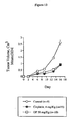
- Figure 5 shows the results of a preliminary experiment where mice with fairly large (100 mm 3 ) fibroblast (RIF) tumours (a cell line derived from skin cancer) received tumour implants of a biodegradeable polymer containing either saline, pentamidine or 5-fluorouracil, a standard anticancer agent. Pentamidine was intermediate in its efficacy at slowing tumour growth between the saline control and 5-fluorouracil. The result is positive because the solid tumours were already well established and the dose of pentamidine had not been optimized.
- RIF fibroblast
- the polymer implant system is a convenient way of administering the drug of interest. Biodegradation of the polymer causes the drug to be released. However, degradation is complete after three or four days after which no more drug is available. Despite these limitations, pentamidine was shown to be effective in the period when the drug was available.
- Figure 6 shows the results of a similar experiment using a polymer implant to deliver pentamidine.
- the experiment was carried out on mice with fibroblast (RIF) tumours that were also treated with radiation (24 hours after implant) shortly after the tumours had reached a size of 100 mm 3 .
- the results in Figure 6 show that the beneficial effects of the radiation treatment had worn off by day 12 after treatment.
- animals treated with a combination of radiation and pentamidine had no significant tumour growth for a much longer period.
- Pentamidine was delivered via a polymer implant and was therefore consumed after three or four days. Nevertheless, the beneficial effects of its action were quite persistent.
- the test mice showed no obvious signs of any side effects due to the use of pentamidine.
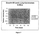
- Figure 7 shows the effectiveness of pentamidine as an anticancer agent when used against the Lewis lung carcinoma primary tumour model.
- Pentamidine was delivered by daily injection. The results show that pentamidine was as effective in inhibiting the cancer growth as cisplatinum, a compound that is currently used for the treatment of lung cancer.
- Table 5 The Effect of Pentamidine on Lung Metastases in Lewis Lung Carcinoma Mouse Model Compound Number of Metastases /Lung Blank 34 ⁇ 3 Pentamidine (25 mg/kg) 23 ⁇ 3 Pentamidine (50 mg/kg) 10 ⁇ 2
- Example 8 In vivo animal experiments
- Pentamidine was supplied. The solution was made by dissolving the pentamidine in sterile distilled water. The pentamidine solution was aliquoted and stored at -20°C upon receipt. Immediately prior to use, drug stock was quickly thawed, kept at 4°C and protected against light until administration. Cisplatin and adriamycin were provided. These drugs were prepared as indicated for the clinical preparation. The saline solution (0.9%) sodium chloride was stored at 4°C.
- the Lewis lung carcinoma clone, M47 with a high metastatic potential to the lung was used. Tumours induced by M47 have been well characterized in relation to their growth rates and response to standard chemotherapy drugs.
- the cell used was confirmed to be free of mycoplasma. Cells were maintained in RPMI-1640 medium supplemented with 10% fetal bovine serum and 1 % penicillin-streptomycin, under 5% CO 2 . Cells were then, propagated and stocks of the same passages were established and stored in liquid nitrogen. Oncozyme studies were done with the same stock of cells and same passage number.
- cells were grown to 70% confluence in complete medium and then collected using trypsin-EDTA solution [0.05% trypsin 0.53 mM EDTA-4Na in H B SS without Ca++. Mg++, and NaHCO3; Cellgro no. 25-052-Li]. Cells were then centrifuged and washed three times with phosphate buffer solution [D-PBS, Ca++ and Mg++ free; Cellgro no. 21-031-LV], and resuspended at a dilution of 0.1 to 1x10 6 cells/0.1 ml. Viability was examined by trypan blue staining and only cells in which the viability was >95% were used for in vivo studies.
- the mouse strain used in this study is C57BL/10 from Charles River Inc. Animals were housed 5 per cage and were fed a diet of animal chow and water ad libitum. After one week acclimatization, LLC cells were transplanted subcutaneously, as a suspension of tumour cells [2-5x10 5 viable cells per 0.1 ml], in the axillary region of the right flank. All animals were inoculated at the same site. Animals were subjected, on a daily basis, to general examination. Tumour growth was monitored every second or third day using calipers.
- mice were subjected to surgery to remove the primary tumour.
- the mice were lightly anesthetized with Forane.
- the skin overlying the tumour was cleaned with betadine and ethanols, in a laminar flow hood.
- a small skin incision (0.5-1cm) was made using a sterile scalpel, and the tumour was carefully separated from the normal tissues (skin and muscle).
- Lewis Lung carcinoma cells (at early stage of growth; 1-3 weeks) are a well localized tumour and separation was easy to achieve without any significant damage to normal tissues.
- the tumour was removed, weighed and in some cases fixed for histopathology purposes.
- the wound was closed with surgical stainless steel clips (Autoclips; 9mm; Clay Adams, Inc. Parsippany, NJ). This site was further disinfected with betadine and the animal was housed as described earlier.
- mice were randomized after surgery into a group of 5 per cage. Cages were randomly assigned to specific experimental groups. The mice were then labelled by numbers using the "ear punching" method. Mice were checked on a daily basis to ensure the absence of infection. Animals with discomfort were sacrificed immediately. For each experiment, an additional extra spared group of control mice was included to determine the optimal timing for sacrifice in order to obtain a significant number of well localized lung metastases. This group was subjected to the same experimental procedure as group 1 with the exception of drug treatment. Based on this group, a period of approximately two weeks after removal of the primary tumour was found to result in an average of 25-35 nodules.
- Pentamidine and chemotherapy drugs were given as described in the results. Control animals were given the same volume of saline solution [0.9% sodium chloride]. The dose of each drug was normalized to body weight per animal.
- the pentamidine and cisplatinum were injected by intraperitoneal injections. Adriamycin was injected intravenously.
- the pentamidine and chemotherapy drugs were also delivered to the animals at different times. They were given every second day for a total of 5 times. It is also possible to inject the pentamidine and all of the chemotherapy drugs intravenously and contemporaneously. This method and regimen of administration can lead to a different combination efficiency.
- mice were sacrificed by dislocation and autopsied. Tumours, organs or both were removed under sterile conditions [using a laminar flow hood]. Tumours were weighed. Organs (5 per group) were examined for gross pathological changes and then fixed in 10% formalin. Lungs were fixed in 10% Bouin's fixative diluted in a formalin solution, and lung surface metastases were counted using a stereomicroscope at 4x magnification or a magnifying-glass, and in some cases lungs were embedded in paraffin wax according to standard procedures. Embedded tissues were used to confirm metastases and further examine histopathological changes.
- blood was taken from 3-5 animals per group by cardiac ponction. Blood was collected in heparinized tubes and analyzed.
- the two-tailed Student T-test was used to compare statistical significance among various groups.
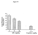
- pentamidine at a dose of 50mg/Kg inhibits lung metastases by more than 50% in comparison to saline-treated control (p ⁇ 0.001), as shown in Figure 16 .
- a pentamidine dose of 50mg/Kg was found to be the most active (p ⁇ 0.01), while doses of 10-25 mg/Kg have no significant effect. Microscopic examination showed clearly that the number of metastatic nodes was clearly reduced in pentamidine treated animals and the sizes of nodes were smaller, in comparison to the saline-treated group.
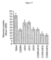
- a combination of 50mg/Kg/ip pentamidine and 4mg/kg/ip cisplatin showed an enhanced effect as shown in Figure 17 .
- a combination of 50mg/Kg/ip pentamidine and 5mg/kg adriamycin/iv showed some beneficial effect, as shown in Figure 20 .
- compositions of the above compounds are used to treat patients having cancer.
- Vehicles for delivering the compounds of the present invention to target tissues throughout the human body include saline and D5W (5% dextrose and water).
- Excipients used for the preparation of oral dosage forms of the compounds of the present invention include additives such as a buffer, solubilizer, suspending agent, emulsifying agent, viscosity controlling agent, flavor, lactose filler, antioxidant, preservative or dye.
- excipients for parenteral and other administration include serum albumin, glutamic or aspartic acid, phospholipids and fatty acids.
- the preferred formulation is in liquid form stored in a vial or an intravenous bag.
- the compounds of the present invention may also be formulated in solid or semisolid form, for example pills, tablets, creams, ointments, powders, emulsions, gelatin capsules, capsules, suppositories, gels or membranes.
- compositions of the invention may also be conjugated to transport molecules or included in transport modalities such as vesicles and micelles to facilitate transport of the molecules. Methods for the preparation of pharmaceutically acceptable compositions that can be administered to patients are known in the art.
- compositions of the invention may also be conjugated to transport molecules, monoclonal antibodies or transport modalities such as vesicles and micelles that preferentially target cancer cells or that potentiate cancer cells to receive drugs.
- compositions including the compounds of the present invention can be administered to humans or animals. Dosages to be administered depend on individual patient condition, indication of the drug, physical and chemical stability of the drug, toxicity, the desired effect and on the chosen route of administration (Robert Rakel, ed., Conn's Current Therapy (1995, W.B. Saunders Company, USA)). These pharmaceutical compositions are used to treat cancer.
- Serum from cancer patients were spotted onto nitocellulose membrane and were probed with a rabbit antiserum raised against the endo-exonuclease according to the method described by Liu et al (1995).
- a sample of the endo-exonuclease protein is spotted onto a membrane substrate.
- a solution of rabbit polyclonal antibodies added to the membrane onto which samples have been spotted. The antibodies bind to the protein.
- a second solutions of a commercially available anti-rabbit antibody or protein A is added that is conjugated with horseradish peroxidase (hrp).
- hrp horseradish peroxidase
- 4-chloro-1-napthol is finally added to the membrane. This reacts with the conjugated hrp to produce a blue colour.
- the intensity of the colour is proportional to the amount of endo-exonuclease present.
- the serum proteins were spotted onto the membrane using the Bio-Rad slot-blot apparatus. The membrane was then rinsed with 10mM Tris-HCl, pH8.0 containing 1 mM EDTA. After washing, the membrane was incubated with anti-endo-exonuclease antibody in buffer B (10 mM Tris-HCl, pH8.0, 1 mM EDTA, 150 mM NaCl) containing 1% skim milk powder.
Landscapes
- Health & Medical Sciences (AREA)
- Veterinary Medicine (AREA)
- Chemical & Material Sciences (AREA)
- Medicinal Chemistry (AREA)
- Pharmacology & Pharmacy (AREA)
- Life Sciences & Earth Sciences (AREA)
- Animal Behavior & Ethology (AREA)
- General Health & Medical Sciences (AREA)
- Public Health (AREA)
- Epidemiology (AREA)
- Inorganic Chemistry (AREA)
- Chemical Kinetics & Catalysis (AREA)
- General Chemical & Material Sciences (AREA)
- Nuclear Medicine, Radiotherapy & Molecular Imaging (AREA)
- Organic Chemistry (AREA)
- Pharmaceuticals Containing Other Organic And Inorganic Compounds (AREA)
- Acyclic And Carbocyclic Compounds In Medicinal Compositions (AREA)
- Medicines That Contain Protein Lipid Enzymes And Other Medicines (AREA)
- Medicines Containing Antibodies Or Antigens For Use As Internal Diagnostic Agents (AREA)
Abstract
Description
- The present invention relates to chemotherapeutic agents for treating cancer.
- Cancer cells proliferate more rapidly than normal cells. The rate of mitosis and DNA replication is therefore significantly greater in cancer cells. Agents that inhibit DNA replication and recombination affect cancer cells more than normal cells.
- Many chemotherapeutic agents for treating cancer inhibit DNA replication by inducing DNA breaks. Some drugs, such as mitomycin, induce DNA breaks in part by binding to the DNA itself. Other anticancer agents interfere with topoisomerase enzymes, which modify DNA structure. In doing so, they induce strand breaks. Normally the breaks are transient but in the presence of a topoisomerase enzyme inhibitor, such as etoposide, the breaks become longer lived and expose the DNA to permanent damage.
- Living organisms repair DNA by a variety of mechanisms including an excision-repair system. Enzymes that mediate excision-repair cut out the damaged DNA. They then replace the damaged DNA sequences with the correct sequences. This repair system lessens the efficiency of cancer therapies that are dependant on chemotherapeutics that induce DNA breaks. The loss in efficiency necessitates the use of high concentrations of DNA-breaking chemotherapeutics in order to obtain a satisfactory inhibition of cancer proliferation. These chemotherapeutics are very toxic and have damaging side effects. The need to use high concentrations is a significant drawback.
- It has been suggested that endo-exonuclease may function in DNA repair and recombination.
US patent 5,324,830 to Resnick et. al. describes the isolation of a DNA segment that codes for an endo-exonuclease, RhoNuc from S. cerevisiae.US patent 5,489,524 describes the characterization of a gene for mammalian endo-exonuclease and the isolation of primate endo-exonuclease. However, it has not been previously suggested that inhibiting endo-exonuclease activity would be effective for inhibiting the DNA repair process or the proliferation of cancer cells. - In Pérez et al., the binding of Pt-pentamidine (platinum-pentamidine) was shown to cause strong stabilisation of the double helix of nucleosomal DNA to heat denaturation. Also, in nucleosome:Pt-pentamidine complexes the nucleosomal denatured DNA is able to reassociate at 71°C.
- In
US patent 5,204,352 , compounds were provided that exhibited a high level of anti-parasitic activity, specifically against parasites of the genus Trypanosoma. The compounds comprised aryl-substituted quaternary heteroaromatic salts. It was suggested the compounds of this patent would be competitive with pentamidine in the treatment of trypanosomiasis.US patent 5,084,480 describes salts of pentamidine, formulations containing such salts and the use of such salts for treatment of prophylaxis or Pneumocystis carinii. Pentamidine gluconate and lactonate were described as having high water solubility while providing potentially less toxic alternatives to pentamidine isethionate. - In Tuan et al., pentamidine was used in bone marrow transfer patients suffering from Pneumocystis carinii pneumonitis. In Waalkes et al., patients with malignant disease complicated by diffuse interstitial pneumonia due to proved or suspected Pneumocystis carinii were given pentamidine, and the plasma levels and urinary excretion of pentamidine during therapy was studied. Following the intramuscular administration of pentamidine, plasma levels were low and urinary excretion prolonged. After a single intraperitoneal injection in mice, the tissue distribution levels and excretion pattern for pentamidine were determined at various time intervals. There was storage of the drug in tissues and excretion was delayed. The highest concentration of pentamidine was found in the kidney.
- There is a need for compounds that inhibit the proliferation of cancer cells that are less toxic than conventional chemotherapeutics. There is further need for compounds that inhibit DNA repair in order to inhibit the proliferation of cancer cells. There is further need for compounds that can be used in combination with conventional chemotherapeutics to improve the efficiency of cancer treatment. There is a further need for such compounds to be used in combination with conventional chemotherapeutics so that the combination permits the use of lower dosages of chemotherapeutics to cancer patients without loss of therapeutic efficiency.
- The present invention relates to the discovery that cancer cells have higher concentrations of endo-exonuclease than normal cells. The proliferation of cancer cells and the growth of tumours can be inhibited by inhibiting endo-exonuclease activity. Cancer can also be diagnosed based on elevated concentrations of endo-exonuclease in cancer cells.
- The proliferation of cancer cells and tumour growth can therefore be inhibited by administering to a patient compounds that inhibit the activity of the endo-exonuclease.
- Compounds that inhibit the activity of endo-exonuclease can be provided in combination with conventional chemotherapeutics that cause breaks in DNA, to inhibit the proliferation of cancer cells and tumour growth. The proliferation of cancer cells and tumour growth can therefore be inhibited by administering to a patient a compound that inhibits the activity of endo-exonuclease in combination with conventional chemotherapeutic drugs that cause breaks in DNA. Compounds that inhibit the activity of endo-exonuclease can therefore be used in combination with agents that cause breaks in DNA to inhibit the proliferation of cancer cells and tumour growth.
- According to one aspect of the present invention, there is provided a pharmaceutical composition for inhibiting the proliferation of cancer cells and tumour growth that comprises pentamidine and a compound that induces breaks in DNA strands, selected from the group consisting of cisplatin, mitomycin C, adriamycin and etoposide.
- According to one aspect of the present invention, there is provided a use of pentamidine in the preparation of a medicament to inhibit the proliferation of cancer cells or to inhibit tumour growth in a patient.
- According to another aspect of the present invention there is provided the use of pentamidine in combination with a compound that induces breaks in DNA strands selected from the group consisting of cisplatin, mitomycin C, adriamycin, etoposide and ionizing irradiation in the preparation of a medicament for inhibiting the proliferation of cancer cells or to inhibit tumour growth in a patient.
- According to yet another aspect of the present invention there is provided the use of an antibody to endo-exonuclease for the preparation of an agent for the diagnosis of cancer in a patient.
-
-
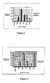
Figure 1 is a bar graph showing the level of endo-exonuclease in various cell lines; -
Figure 2 is a graph showing the survival of various cells in presence of different amounts of pentamidine using a clonogenic assay; -
Figure 3 is a bar graph showing the combination effect of different drugs (mitomycin C + pentamidine, etoposide + pentamidine) on cell death; -
Figure 4 is a bar graph showing the combination effect of cisplatin and pentamidine on cell death; -
Figure 5 is a graph showing the effect of a polymer implant of pentamidine on tumour growth in the RIF tumour mouse model; -
Figure 6 is a graph showing the combination effect of polymer implant of pentamidine and irradiation on tumour growth in RIF tumour mouse model; -
Figure 7 is a graph showing the effect of pentamidine on the growth of primary tumour in the Lewis lung carcinoma model in mice; -
Figure 8 is a graph showing the toxicity of pentamidine given by an intraperitoneal route; -
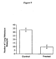
Figure 9 is a graph showing the anti-cancer effect of pentamidine in vivo; -
Figure 10 is a graph showing the anti-cancer effect of pentamidine and cisplatinum both individually and in combination in vivo; -
Figure 11 is a graph showing the anti-cancer effect of pentamidine and cisplatinum both individually and in combination in vivo; -
Figure 12 is a graph showing the anti-cancer effect of pentamidine and cisplatinum both individually and in combination in vivo; -
Figure 13 is a graph showing the effect of pentamidine on the growth of Lewis lung carcinoma primary tumours; -
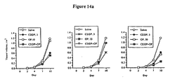
Figure 14a is graph showing the anti-cancer effect of pentamidine and cisplatinum both individually and in combination in vivo; -
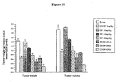
Figure 14b is a bar graph showing the anti-cancer effect of pentamidine and cisplatinum both individually and in combination in vivo; -
Figure 15 is a bar graph showing the anti-cancer effect of pentamidine and cisplatinum both individually and in combination in vivo; -
Figure 16 is a bar graph showing the effect of pentamidine on the incidence of Lewis lung carcinoma induced lung metasteses; -
Figure 17 a bar graph showing the anti-cancer effect of pentamidine and cisplatinum both individually and in combination in vivo; -
Figure 18 is a graph showing the anti-cancer effect of pentamidine and adriamycin in vivo; -
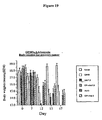
Figure 19 is a bar graph showing the effect of pentamidine and adriamycin on body weight for primary tumour in vivo; and -
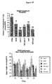
Figure 20 is bar graphs showing the antimetastatic effect of pentamidine and adriamycin in vivo; - For the purposes of the figures, OP refers to pentamidine, Adr refers to adriamycin and CDDP refers to cisplatinum.
- The invention relates to the surprising discovery that endo-exonuclease plays an important role in cell proliferation. It is required for normal growth and tissue repair. Endo-exonuclease plays a special role in cells that proliferate rapidly such as cancer cells. This enzyme is found in high concentrations in cancer cells where it actually helps to maintain tumour growth. Endo-exonuclease functions by repairing breaks in DNA molecules, which carry all of the genes that make cells function. DNA breaks often occur during the cell division process and must be repaired if cell proliferation is to continue.
- Chemical compounds that inhibit the activity of endo-exonuclease have been found to inhibit the proliferation of cancer cells and tumour growth. Compounds that inhibit the activity of endo-exonuclease can also be used in combination with agents that cause DNA breaks, preferably double strand breaks in DNA. Combining these types of compounds or other agents provides a valuable tool for cancer therapy.
- The present invention relates to the unexpected result that pentamidine inhibits the activity of endo-exonuclease. It was previously known that pentamidine has anti-fungal activity. It has been found that pentamidine inhibits the activity of endo-exonuclease sufficiently to stop the growth of cancer cell lines in-vitro, Pentamidine also slows tumour growth in animals with very aggressive cancers. Cancer treatment with pentamidine is especially advantageous because pentamidine has low toxicity relative to standard chemotherapeutic agents. The side effects of pentamidine are often different to those of standard chemotherapeutic agents so that when pentamidine and those agents are used together, the side effect profile is potentially less hazardous.
- The invention also relates to the combination effect of using known compounds and other agents that cause single strand or double strand DNA breaks with pentamidine to inhibit the proliferation of cancer cells and tumour growth. It has been found that compounds and other agents that cause double stranded DNA breaks work especially well in combination with pentamidine. This inhibits the proliferation of cancer cells and tumour growth. Agents can cause double strand breaks directly or can cause single strand breaks that progress to double strand breaks. This is a common occurrence in biological systems.
- The invention also relates to the use of pentamidine to inhibit the action of endo-exonuclease to inhibit the proliferation of cancer cells and tumour growth. One could also use this compound to inhibit tumour growth either alone or in combination with known drugs that cause DNA breaks. Agents that induce DNA breaks that are within the scope of the present invention include cisplatin, mitomycin C, adriamycin, etoposide and ionizing irradiation.
- Compositions or mixtures of these compounds and other agents may be administered to patients which include humans and animals. Compositions include all pharmaceutical formulations of a compound and a compound in its pure state. Combinations include two or more compositions. This includes two or more different formulations of a compound such as a tablet formulation and a liquid formulation. Mixtures of two or more compounds in the same formulation are also within the scope of the invention. Compositions also include excipients such as micelles, vesicles and liposomes that enhance the therapeutic performance of the compound and other agents. The action of vesicles, micelles and liposomes includes improving the solubilization of the compounds and agents, improving their delivery to tumour cells, and interacting with tumour cells to make these cells more permeable to compounds and agents. Improving efficiency could improve treatment or allow equivalent results with reduced dosing and side-effects.
- The cell lines from human colon adenocarcinoma (HT29), human breast adenocarcinoma (MCF7) and human cervical epitheloid carcinoma (HeLa) were obtained from the American Type Culture Collection (ATCC) and have ATCC accession numbers HTB-38, HTB-22, and CCL-2 respectively. The normal primary cell, NHDF, was obtained from Dr. Shirley Lehnert. These cells are normal human skin fibroblasts. The cells were grown in RPMI media supplemented with 10% FCS at 37°C in a humidified incubator with 5% CO2.
- The endo-exonuclease level in the cell lines was determined with Immunoblot method as described by Chow and Resnick (1987). Exponentially growing cells were boiled in lysis buffer (0.125 M Tris-HCl pH7.0, 20% glycerol, 4% SDS, 0.5 mM EDTA). The lysed cells were then centrifuged at 10,000 g for 10 min and 25 µl of the supernatant were electrophoresed on a 10% SDS-polyacrylamide gel (SDS-PAGE) according to the method described by Laemmli (1970). Proteins that had been separated on the SDS-PAGE gel were transferred electrophoretically to a nitrocellulose membrane. The nitrocellulose membrane was then reacted with rabbit antiserum raised against the monkey CV-1 endo-exonuclease in buffer B (10 mM Tris-HCl, pH8.0, 1 mM EDTA, 150 mM NaCl) containing 0.5% skim-milk powder according to the method previously described Chow and Resnick (1988). After the membrane had been washed three times in buffer B for 15 min., protein A (a polypeptide isolated from staphylococcus aureus that binds to the Fc region of the immunoglobulin molecules without interacting at the antigen binding site) conjugated with horseradish peroxidase in buffer B containing 0.5% skim-milk powder was added to the membrane and incubated for 3 h at room temperature. The membrane was subsequently washed with buffer B for 15 min. Positive signals were indicated by colour development of the substrate 4-chloro-1-naphthol at the corresponding protein position in the horseradish peroxidase enzymatic reaction. Relative amounts of positive signals were detected using a HP4c scanner and Light Tool Research software program.
- Based on this method, the endo-exonuclease levels in normal cells and the HT29, MCF-7 and HeLa cell lines were calculated. The results presented in
Figure 1 show that the level of the endo-exonuclease is much higher in cancer cells than in normal cells. The results suggest that inhibition of the enzyme should provide a means of preferentially attacking cancer cells. In addition, the results suggest that measurement of enzyme concentrations in body fluids or tissues provides a means of detecting cancer and of monitoring its progress. - Cell survival was determined according to the following methods:
- Cell Survival - Clonogenic assay: Clonogenic measurement of cell survival was used to determine the initial effectiveness of pentamidine according to the method described in Sadekova et al. (1997). In this method, logarithmically phase cells (range from 1000 to 3000 cells/ 50mm depending on plating efficiency) were seeded onto cell culture plates together with various drug concentrations (ranging from 0.2 µM to 20 mM). After 1 week of growth, cell colonies were stained with crystal violet and the numbers of colonies were counted.
- Cell Survival - MTT assay: The MTT (3-[4,5-Dimethylthiazol-2-yl]-2,5 diphenyl tertrazolim bromide) method of determining cell growth/cytotoxicity offers a convenient alternative to determine cell survival. MTT is a tetrazolium salt cleaved by mitochondrial dehydrogenases of living cells. Cleavage converts yellow, water soluble MTT to an insoluble, purple formazan crystal. The crystals can be solubilized with a 50% N,N-dimethylformamide (vol/vol), 20% SDS (wt/vol) solution (pH4.7), and absorbance determined at a wavelength of 570 nm. Dead cells will not cleave MTT and uncleaved MTT is not detectable at this wavelength. The amount of MTT that is cleaved increases with increasing cell numbers, and decreases as a result of cell cytotoxicity (Niks and Otto 1990, Hussain et al. 1993).
- Cells were harvested from cell cultures using the standard protocol (Trypsin/EDTA). The cells (1000 to 5000 cells depending on cell type in 50µl) were then plated and incubated overnight at 37°C before the addition of experimental reagents (i.e. the drug of interest), for the combination experiment, both drugs were added. After 2 days of incubation at 37°C, 10 µl of a 5 mg/ml solution of MTT was then added to all the experimental wells as well as the media control well. The plates were further incubated for 4 hours. A 100 µl of MTT solubilization buffer was added and the plates were incubated overnight at 37°C. The plates were then read on the ELISA plate reader with absorbance at 570 nm and a reference at 630 nm.
Lewis Lung Carcinoma Cell Line and Cell Culture: The Lewis lung carcinoma clone, M47, is a metastatic model. Lewis lung carcinoma cells were maintained in RPMI-1640 medium supplemented with fetal bovine serum and penicillin-streptomycin. For tumour induction, cells were washed three times with phosphate buffer solution. They were then re-suspended at a dilution of 1x10⁶ cells/0.1ml. Only cells where viability was >95% were used for in vivo studies. - The mouse strain used in this study was C57BL/10. After one-week of acclimatization, cells were transplanted into the mice subcutaneously, as a suspension of tumour cells. All animals were inoculated at the same site.
- To measure the effect of drugs on the primary tumour, drug solutions were administered by intraperitoneal (ip) injection every two days. Animals were subjected, on a daily basis, to general examination. Tumour growth was monitored over time. To determine the effect of drugs on tumour metastasis, the tumours were allowed to reach a size of 0.5-1.0 cm3. Mice were randomized into various groups and the drugs were then given by ip. At the end of each experiment, animals were sacrificed and autopsied. Tumours, organs or both were removed under sterile conditions. Tumours were weighed. Organs were examined for gross pathological changes and then fixed in formalin. Lungs were fixed in Bouin's fixative and lung surface metastases were counted using a stereomicroscope.
RIF (Radiation-induced Fibrosarcoma) Cell Line and Cell Culture: The radiation-induced fibrosarcoma clone, RIF-1, is a solid tumour model. RIF-1 cells were maintained in DMEM medium supplemented with fetal bovine serum and penicillin-streptomycin. For tumour induction, 2 x 10⁵ cells were injected s.c. into the backs of mice from the C3H strain. Tumours appeared within 10 days and reached a volume of 94-130 mm3 within 3 weeks. Poly (carboxyphenoxypropane-co-sebacic acid) or poly (CPP-SA) polymer implants containing the drug were prepared and implanted into the tumour according to the method described by Yapp et al. (1997). The same person measured the sizes of the tumours every two days until they reached 4 times the initial volume at the time of implant. The final volume was 400 mm3. - For the combination experiment, a signal dose of gamma irradiation (60Co, Theratron 780) at a dose rate of 1 Gy/min was delivered 24 hrs after implant of the drug- containing polymer.
- The human endo-exonuclease was isolated according to the method described by Liu and et al (1995). The cultured cells were detached with trypsin-EDTA and the cell suspensions were centrifuged at 4°C with a force of 700 g for 10 minutes. The cell pellets were washed twice with cold phosphate buffered saline (PBS). The cells were then resuspended and sonicated in 20 mM Tris-HCl, pH 7.5, containing 5 mM EDTA and 1 mM PMSF (buffer A). The resulting cell lysis suspensions were centrifuged at 4°C at 10,000 g for 15 min. The supernatants were then loaded onto an antibody-protein A-Sepharose affinity column, as previously described by Chow and Resnick (1987). After washing extensively with buffer A, (i.e. until the A280 of the eluates were zero), the column was then eluted with buffer A containing 3.5 M MgCl2 to elute the endo-exonuclease. The eluted endo-exonuclease was dialyzed extensively against buffer A with at least two changes of buffer and one change of distilled water. The endo-exonuclease was then concentrated by lyophilization.
- The nuclease activities were determined by measuring the release of acid soluble radioactivity from γ-32P-labelled, heat-denatured single-strand pBR322 DNA according to the method described by Chow and Resnick (1983). One unit of activity was defined as the amount of deoxyribonuclease that renders 1 µg of DNA acid-soluble in 30 min at 37°C. For the inhibition assay with the drugs, the drugs were added to the endo-exonuclease prior to the start of the nuclease reaction. Table 1 shows the levels of the endo-exonuclease inhibition by various chemotherapeutic agents.
Table 1: Inhibition of Endo-exonuclease Activity by Chemotherapeutic Agents Chemotherapeutic Agent Percent of Inhibition Pentamidine (25 µM) 37% Pentamidine (50 µM) 50% Pentamidine (100 µM) 100% Distamycin A (38 µM) 30% Berenil (2mM) 17% Mitomycin C (50 µM) 0% Etoposide (VP-16) (50 µM) 0% - Clonogenic measurement of cell survival was used to determine the initial effectiveness of pentamidine according to the method described above.
- The rates of survival in the presence of pentamidine of primary cells, MCF7 and HeLa cells using the clonogenic assay are shown in
Figure 2 . The results shown inFigure 2 demonstrate that pentamidine preferentially attacks cancer cells in a dose dependent manner. The cancerous MCF7 and HeLa cell lines were compared with the human primary fibroblast cells. The survival rates of the cells were measured at different doses of pentamidine. Pentamidine began to kill the cancer cells at concentrations of 0.1mM and was lethal at a concentration of 10mM. Under these conditions, pentamidine had no effect on normal primary human cells. The dose dependence and the selectivity towards cancer cells show that pentamidine is a useful anticancer agent. - The anticancer activities of pentamidine and a number of known anticancer agents are shown in Table 2.
Table 2: Comparison of the LC50 of Various Anti-cancer Agents on Cancer Cell-lines Cancer cell type Pentamidine (mM) Mitomycin C (mM) Etoposide (mM) Cisplatin (mM) 2 day 4 day 2 day 4 day 2 day 4 day 2 day 4 day H520 0.24 0.13 0.234 0.130 >34 >34 0.503 - H460 1.34 0.16 0.065 0.030 >34 >34 0.503 - H661 0.15 0.07 0.006 0.00 8 28 15.6 0.413 - MCF-7 0.15 0.08 0.034 0.013 1 1.1 0.493 - 0.0243 13 HT29 0.27 0.06 0.008 0.008 0.7 0.4 0.483 - 0.0243 0.73 numeral 3 was obtained from the National Cancer Institute. - LC50 is the concentration of a drug or chemical that kills 50% of the cells. The results show that pentamidine is an anticancer agent. The data also show that pentamidine is more lethal to cells than etoposide but less so than mitomycin C. The effectiveness of pentamidine increases if the experiment is run over 4 days as opposed to 2. This suggests that naturally occurring strand breaks in DNA are relatively infrequent and that prolonged exposure to pentamidine is beneficial.
- The clinical use of these agents depends upon the balance between anticancer activity and harmful side effects. Thus a relatively non-toxic agent, which can be given in high concentration may be more effective than a more aggressive but toxic agent which can only be tolerated in very small doses. Based on known clinical data, pentamidine has low toxicity.
- The anticancer activities of pentamidine and distamycin A and berenil are shown in Table 3.
Table 3: Comparison of LC50 of Pentamidine, Distamycin A, and Berenil on Cancer Cell-Lines Cancer cell type Pentamidine(mM) Distamycin A (mM) Berenil (mM) 2 day 2 day 2 day H520 0.24 >2.0 >4.0 H460 1.34 >2.0 >4.0 H661 0.15 >2.0 >4.0 MCF-7 0.15 1.52 3.0 HT29 0.27 >2.0 >4.0 - The cancer cell types are: H520 - NSCLC (Squamous carcinoma, primary tumour), H460 - NSCLC (Large cell carcinoma, pleural effusion), H661 - NSCLC (Large cell carcinoma, lymph node), MCF-7 - Breast cancer (Adenocarcinoma, pleural effusion), HT29 - Colon cancer (Adenocarcinoma, primary tumour).
- The length of time in days that the cells are exposed to the compound is indicated in Table 3.
- These results show that these inhibitors of endo-exonuclease have anti-cancer activity.
- The data in Table 4 shows the effect of combining pentamidine with mitomycin C, etoposide and cisplatin.
Table 4: LC50 of Pentamidine On Cancer Cells When Used Alone Or In Combination With Other Anti-cancer Agents Cancer cell type Pentamidine (mM)
2 daysPentamidine (mM) with Mitomycin C (1.56 µM)
2 daysPentamidine (mM) with Etoposide (34 µM)
2 daysPentamidine (mM) With Cisplatin (0.025 µM)
2 daysH661 0.15 0.0029 0.10 0.039 MCF-7 0.15 0.0029 0.049 0.082 HT29 0.27 0.0022 0.085 0.032 Length of exposure to mixture is indicated in days. - Comparison of the data in Tables 2 and 4 shows that the use of pentamidine in combination with mitomycin reduces concentrations of these drugs needed to bring about cell death. The same applies to pentamidine and etoposide. The magnitude of the effect suggests that the use of pentamidine in combination with mitomycin and etoposide leads to very efficient destruction of cancer cells. This allows for the delivery of much less toxic doses of anticancer drugs such as mitomycin and etoposide.
-
Figure 3 shows that combining mitomycin C and etoposide with pentamidine is 50 to over 1,000 times more efficient at killing of cancer cells than using mitomycin C and etoposide alone. - We have defined the efficiency of the combination as follows:

- In this equation [Pentamidine]o is the LC50 dose of Pentamidine when used alone while [Pentamidine]c is the LC50 dose required in the combination experiment. "P" represents either Mitomycin or Etoposide and the subscripts "o" and "c", refer respectively to the experiment when the materials were used alone and in the combination experiment.
-
Figure 4 shows that combining cisplatin and pentamidine leads to an even more profound increase in efficiency of killing of cancer cells. The combination of cisplatin with pentamidine is up to 16,000 times more efficient than using cisplatin alone. This surprising increase is consistent with the known mechanisms of action of the chemotherapeutic agents. Mitomycin C and etoposide achieve cell death through a complex mechanism involving single strand breaks. Relatively few of these single strand breaks progress to double strand breaks. By contrast, cisplatin operates by a mechanism that ultimately induces double strand breaks. Endo-exonuclease repairs double strand breaks. These results demonstrate that in cell culture, the inhibition of endo-exonuclease with pentamidine increases the efficiency of the anticancer activity of agents that induce double strand breaks much more than that of agents that induce single strand breaks. - The addition of pentamidine to a chemotherapy treatment allows the concentrations of the chemotherapeutic agents to be reduced without any loss of efficiency. It also enhances the efficiency of treatment.
-
Figure 5 shows the results of a preliminary experiment where mice with fairly large (100 mm3) fibroblast (RIF) tumours (a cell line derived from skin cancer) received tumour implants of a biodegradeable polymer containing either saline, pentamidine or 5-fluorouracil, a standard anticancer agent. Pentamidine was intermediate in its efficacy at slowing tumour growth between the saline control and 5-fluorouracil. The result is positive because the solid tumours were already well established and the dose of pentamidine had not been optimized. - The polymer implant system is a convenient way of administering the drug of interest. Biodegradation of the polymer causes the drug to be released. However, degradation is complete after three or four days after which no more drug is available. Despite these limitations, pentamidine was shown to be effective in the period when the drug was available.
-
Figure 6 shows the results of a similar experiment using a polymer implant to deliver pentamidine. The experiment was carried out on mice with fibroblast (RIF) tumours that were also treated with radiation (24 hours after implant) shortly after the tumours had reached a size of 100 mm3. The results inFigure 6 show that the beneficial effects of the radiation treatment had worn off byday 12 after treatment. However, animals treated with a combination of radiation and pentamidine had no significant tumour growth for a much longer period. Pentamidine was delivered via a polymer implant and was therefore consumed after three or four days. Nevertheless, the beneficial effects of its action were quite persistent. The test mice showed no obvious signs of any side effects due to the use of pentamidine. -
Figure 7 shows the effectiveness of pentamidine as an anticancer agent when used against the Lewis lung carcinoma primary tumour model. Pentamidine was delivered by daily injection. The results show that pentamidine was as effective in inhibiting the cancer growth as cisplatinum, a compound that is currently used for the treatment of lung cancer. - The lung tumour implants in the Lewis lung carcinoma form secondary tumours by metastases. The effect of pentamidine on the incidence of these lung metastases was studied in a separate study. In post-mortem examinations, the mice lung metastases were counted. Pentamidine reduced metastases in a dose dependant manner by a factor of three with the highest dosage tested. The results from these post-mortem examinations are set out in Table 5.
Table 5: The Effect of Pentamidine on Lung Metastases in Lewis Lung Carcinoma Mouse Model Compound Number of Metastases /Lung Blank 34±3 Pentamidine (25 mg/kg) 23±3 Pentamidine (50 mg/kg) 10±2 - Pentamidine was supplied. The solution was made by dissolving the pentamidine in sterile distilled water. The pentamidine solution was aliquoted and stored at -20°C upon receipt. Immediately prior to use, drug stock was quickly thawed, kept at 4°C and protected against light until administration. Cisplatin and adriamycin were provided. These drugs were prepared as indicated for the clinical preparation. The saline solution (0.9%) sodium chloride was stored at 4°C.
- The Lewis lung carcinoma clone, M47, with a high metastatic potential to the lung was used. Tumours induced by M47 have been well characterized in relation to their growth rates and response to standard chemotherapy drugs. The cell used was confirmed to be free of mycoplasma. Cells were maintained in RPMI-1640 medium supplemented with 10% fetal bovine serum and 1 % penicillin-streptomycin, under 5% CO2. Cells were then, propagated and stocks of the same passages were established and stored in liquid nitrogen. Oncozyme studies were done with the same stock of cells and same passage number.
- For tumour induction, cells were grown to 70% confluence in complete medium and then collected using trypsin-EDTA solution [0.05% trypsin 0.53 mM EDTA-4Na in HBSS without Ca++. Mg++, and NaHCO3; Cellgro no. 25-052-Li]. Cells were then centrifuged and washed three times with phosphate buffer solution [D-PBS, Ca++ and Mg++ free; Cellgro no. 21-031-LV], and resuspended at a dilution of 0.1 to 1x10⁶ cells/0.1 ml. Viability was examined by trypan blue staining and only cells in which the viability was >95% were used for in vivo studies.
- The mouse strain used in this study is C57BL/10 from Charles River Inc. Animals were housed 5 per cage and were fed a diet of animal chow and water ad libitum. After one week acclimatization, LLC cells were transplanted subcutaneously, as a suspension of tumour cells [2-5x10⁵ viable cells per 0.1 ml], in the axillary region of the right flank. All animals were inoculated at the same site. Animals were subjected, on a daily basis, to general examination. Tumour growth was monitored every second or third day using calipers. Parameters measured were: tumour measured along the longest axis (length) and the perpendicular shortest axis (width) and the relative tumour volume (in cm3) was calculated by the formula: [length (cm) x (width cm)2] (approximately 10-15 days), mice were randomized into one of the following groups:
- Animals were subjected to surgery to remove the primary tumour. The mice were lightly anesthetized with Forane. The skin overlying the tumour was cleaned with betadine and ethanols, in a laminar flow hood. A small skin incision (0.5-1cm) was made using a sterile scalpel, and the tumour was carefully separated from the normal tissues (skin and muscle). Lewis Lung carcinoma cells (at early stage of growth; 1-3 weeks) are a well localized tumour and separation was easy to achieve without any significant damage to normal tissues. The tumour was removed, weighed and in some cases fixed for histopathology purposes. The wound was closed with surgical stainless steel clips (Autoclips; 9mm; Clay Adams, Inc. Parsippany, NJ). This site was further disinfected with betadine and the animal was housed as described earlier.
- In this group, mice were randomized after surgery into a group of 5 per cage. Cages were randomly assigned to specific experimental groups. The mice were then labelled by numbers using the "ear punching" method. Mice were checked on a daily basis to ensure the absence of infection. Animals with discomfort were sacrificed immediately. For each experiment, an additional extra spared group of control mice was included to determine the optimal timing for sacrifice in order to obtain a significant number of well localized lung metastases. This group was subjected to the same experimental procedure as
group 1 with the exception of drug treatment. Based on this group, a period of approximately two weeks after removal of the primary tumour was found to result in an average of 25-35 nodules. - The conditions for this group were identical to the group used for the experiments on metastases with the exception that the primary tumour was not removed, and the animals were maintained until the tumours reached a large size that justified animal sacrifice, or the animals manifested a discomfort that justified their sacrifice (reduced mobility, severe respiratory symptoms, etc.).
- Pentamidine and chemotherapy drugs were given as described in the results. Control animals were given the same volume of saline solution [0.9% sodium chloride]. The dose of each drug was normalized to body weight per animal.
- The pentamidine and cisplatinum were injected by intraperitoneal injections. Adriamycin was injected intravenously. The pentamidine and chemotherapy drugs were also delivered to the animals at different times. They were given every second day for a total of 5 times. It is also possible to inject the pentamidine and all of the chemotherapy drugs intravenously and contemporaneously. This method and regimen of administration can lead to a different combination efficiency.
- At the end of each experiment ( a total of 5-8 weeks), animals were sacrificed by dislocation and autopsied. Tumours, organs or both were removed under sterile conditions [using a laminar flow hood]. Tumours were weighed. Organs (5 per group) were examined for gross pathological changes and then fixed in 10% formalin. Lungs were fixed in 10% Bouin's fixative diluted in a formalin solution, and lung surface metastases were counted using a stereomicroscope at 4x magnification or a magnifying-glass, and in some cases lungs were embedded in paraffin wax according to standard procedures. Embedded tissues were used to confirm metastases and further examine histopathological changes.
- For some experiments involving drug combinations, blood was taken from 3-5 animals per group by cardiac ponction. Blood was collected in heparinized tubes and analyzed.
- The two-tailed Student T-test was used to compare statistical significance among various groups.
- A preliminary study was conducted on non-tumour bearing mice to examine the maximal tolerable doses of pentamidine that can be used for antitumour studies. Three intraperitoneal injections (
day 1,day 3, and day 5) were tested of 25, 50, 100 and 200mg/Kg of body weight respectively. All animals receiving 100mg/Kg of body weight died because of acute toxicity as shown inFigure 8 . This was observed even after using IP injection. Doses of 25 and 50 mg/Kg of body weight were tolerated with no apparent side effects. Therefore, doses of less than or equal to 50 mg/Kg were used to examine the biological activity of pentamidine. - Several independent experiments were used to examine the antitumour properties of pentamidine on the growth of LLC primary tumour. These experiments indicate that the most active dose is 50 mg/Kg (p<0,01), as shown in
Figures 10 to 15 . The antitumour effect of pentamidine was very clear on the last day of tumour growth (day 14-16), as shown inFigure 10 . Of note, pentamidine was as active as cisplatin at a dose of 3-4mg/Kg/ip (Figure 13 ). Also, in an experiment shown inFigures 14a and14b , it is important to note that where pentamidine was combined with 3mg/Kg cisplatin, some animals showed a complete regression of the tumours. However, these animals were not kept for a longer period of time to ensure that there was no tumour regrowth. Combinations of 50mg/Kg of pentamidine and adriamycin showed some beneficial effect (Figure 18 ) but because adriamycin is very toxic at the highest dose tested (7.5mg/Kg/iv, 100% mortality (Fig 19 )), while the minimal dose of 5mg/Kg has a potent antitumour effect as did pentamidine, lower doses may be required to further increase the combination efficiency. - In the preliminary experiment, it was observed that pentamidine at a dose of 50mg/Kg inhibits lung metastases by more than 50% in comparison to saline-treated control (p<0.001), as shown in
Figure 16 . This was further confirmed in subsequent experiments. A pentamidine dose of 50mg/Kg was found to be the most active (p<0.01), while doses of 10-25 mg/Kg have no significant effect. Microscopic examination showed clearly that the number of metastatic nodes was clearly reduced in pentamidine treated animals and the sizes of nodes were smaller, in comparison to the saline-treated group. A combination of 50mg/Kg/ip pentamidine and 4mg/kg/ip cisplatin showed an enhanced effect as shown inFigure 17 . In a similar manner, a combination of 50mg/Kg/ip pentamidine and 5mg/kg adriamycin/iv showed some beneficial effect, as shown inFigure 20 . - This study indicates that pentamidine inhibits Lewis Lung Carcinoma tumour growth at tolerable doses after chronic intraperitoneal administration.
- An anti-metastatic effect was clearly observed in a dose-effect dependent manner in groups where the primary tumour was removed after it has reached a size of 0.5 to 1cm3. The highest tolerated dose of 50 mg/kg was the most active. Macroscopic examination reveals that the numbers of lung nodes were reduced and, when present, were smaller in pentamidine treated groups compared to controls.
- Combination of pentamidine and chemotherapy drugs clearly improves the therapeutic response in light of the data obtained.
- Pharmaceutical compositions of the above compounds are used to treat patients having cancer. Vehicles for delivering the compounds of the present invention to target tissues throughout the human body include saline and D5W (5% dextrose and water). Excipients used for the preparation of oral dosage forms of the compounds of the present invention include additives such as a buffer, solubilizer, suspending agent, emulsifying agent, viscosity controlling agent, flavor, lactose filler, antioxidant, preservative or dye. There are preferred excipients for parenteral and other administration. These excipients include serum albumin, glutamic or aspartic acid, phospholipids and fatty acids.
- The preferred formulation is in liquid form stored in a vial or an intravenous bag. The compounds of the present invention may also be formulated in solid or semisolid form, for example pills, tablets, creams, ointments, powders, emulsions, gelatin capsules, capsules, suppositories, gels or membranes.
- The preferred route of administration is intravenous. Other acceptable routes of administration include oral, topical, rectal, parenteral (injectable), local, inhalant and epidural administration. The compositions of the invention may also be conjugated to transport molecules or included in transport modalities such as vesicles and micelles to facilitate transport of the molecules. Methods for the preparation of pharmaceutically acceptable compositions that can be administered to patients are known in the art.
- The compositions of the invention may also be conjugated to transport molecules, monoclonal antibodies or transport modalities such as vesicles and micelles that preferentially target cancer cells or that potentiate cancer cells to receive drugs.
- Pharmaceutical compositions including the compounds of the present invention can be administered to humans or animals. Dosages to be administered depend on individual patient condition, indication of the drug, physical and chemical stability of the drug, toxicity, the desired effect and on the chosen route of administration (Robert Rakel, ed., Conn's Current Therapy (1995, W.B. Saunders Company, USA)). These pharmaceutical compositions are used to treat cancer.
- Serum from cancer patients were spotted onto nitocellulose membrane and were probed with a rabbit antiserum raised against the endo-exonuclease according to the method described by Liu et al (1995). In this method, a sample of the endo-exonuclease protein is spotted onto a membrane substrate. A solution of rabbit polyclonal antibodies added to the membrane onto which samples have been spotted. The antibodies bind to the protein. After washing, a second solutions of a commercially available anti-rabbit antibody or protein A is added that is conjugated with horseradish peroxidase (hrp). After washing, 4-chloro-1-napthol is finally added to the membrane. This reacts with the conjugated hrp to produce a blue colour. The intensity of the colour is proportional to the amount of endo-exonuclease present. Briefly, the serum proteins were spotted onto the membrane using the Bio-Rad slot-blot apparatus. The membrane was then rinsed with 10mM Tris-HCl, pH8.0 containing 1 mM EDTA. After washing, the membrane was incubated with anti-endo-exonuclease antibody in buffer B (10 mM Tris-HCl, pH8.0, 1 mM EDTA, 150 mM NaCl) containing 1% skim milk powder. After the membrane had been washed three times in buffer B for 15 min., commercially available anti-rabbit-Igg conjugated with horseradish peroxidase or protein A conjugated with horseradish peroxidase in buffer B containing 1% skim-milk powder was added to the membrane and incubated for 3h at room temperature. The membrane was subsequently washed with buffer B for 15 min., buffer containing 1 M NaCl for 30 min., and buffer B again for 15 min. After washing 4-chloro-1-naphtol solution was added to the membrane. Reaction with any horseradish peroxidase present produced a blue colour.
- With serum samples from 37 cancer patients (breast cancer metastases) of known history, a correlation between survival and the level of endo-exonuclease was found. With a cut point for high endo-exonuclease that was used of 5.5, the group of patients that had a mean survival of 38.91 months had low endo-exonuclease level whereas the group of patients that had a mean survival of 10.43 months had high endo-exonuclease levels. The p value of 0.02 indicated a high statistical significance for the result. Furthermore, virtually all the cancer patients were detected with abnormal levels of endo-exonuclease (above the value detected with cancer-free individuals) whereas the standard cancer diagnostic marker, CEA, only tested positive on 25% of serum samples from the patients.
- The study indicated that the level of endo-exonuclease has a good correlation with the length of survival in patients with metastatic breast cancer.
- An abnormal level of endo-exonuclease was detected in almost all the patient samples whereas the standard cancer diagnostic marker, CEA, gave positive results in only approximately 25% of the same patient samples.
- Although the invention has been described with preferred embodiments, it is to be understood that modifications may be resorted to as will be apparent to those skilled in the art. Such modifications and variations are to be considered within the purview and scope of the present invention.
-
- Chow, T. Y.-K., and Resnick, M. A. (1983) The identification of a deoxyribonuclease controlled by the RAD52 gene of Saccharomyces cerevisiae. In Friedberg, E. C. and Bridges, B. A. (eds), Cellular Responses to DNA Damages. Alan R. Liss, New York, pp. 447-455.
- Chow, T. Y-K., and Resnick, M. A. (1987) Purification and characterization of an endo-exonuclease activity of yeast that requires a functional RAD52 gene. J. Biol. Chem., 262, 17659-17667,
- Chow, T. Y-K., and Resnick, M. A. (1988) An endo-exonuclease activity of yeast that requires a functional RAD52 gene. Mol. Gen. Genet. 211, 41-48.
- Hussain, R. F., Nouri, A. M. E., and Oliver, R. T. D. (1993) A new approach for measurement of cytotoxicity using colorimetric assay. J. Immunol. Methods. 160, 89-96.
- Liu, G., Lehnert, S., and Chow, T. Y.-K. (1995) Mammalian endo-exonuclease activity and its level in various radiation sensitive cell lines. .
- Niks, M., and Otto, M. (1990) Towards an optimized MTT assay. J. Immunol. Methods. 130, 149-151.
- Pérez, J. M. et al., (1993) Binding of Pt-pentamidine to nucleosomal DNA. Studies of the antiproliferative activity of the drug against human cancer cells. Chemico-Biological Interactions 89 64-72.
- Sadekova, S., Lehnert, S., and Chow, T. Y.-K. (1997) Induction of PBP74/mortalin/Grp75, a member of the hsp70 family, by low doses of ionizing radiation: a possible role in the induced radioresistance. Int. J. Radiat. Biol. 72, 653660.
- Tuan, I.-Z., et al., (1992) Pneumocystis carinii pneumonitis following bone marrow transplantation. Bone Marrow Transplantation 10: 268-272.
- Waalkes, T. P., et al., (1970) Pentamidine: Clinical pharmacologic correlations in man and mice. Clinical Pharmacology and Therapeutics, 11 (4) 505-512.
- Yapp, D. T- T., Lloyd, D. K., Zhu, J., and Lehnert, S.M. (1997) Tumour treatment by sustained intratumoural release of cisplatin : effects of drug alone and combined with radiation. Int. J. Rad. Oncol. 39, 497-504.
Claims (7)
- A pharmaceutical composition suitable for inhibiting the proliferation of cancer cells or tumour growth comprising pentamidine and an agent that induces breaks in DNA strands selected from the group consisting of cisplatin, mitomycin C, adriamycin and etoposide.
- A pharmaceutical composition according to claim 1 wherein said pentamidine or said agent is conjugated to a transport agent for delivery to cancer cells.
- A pharmaceutical composition according to claim 2 wherein the transport agent is selected from the group consisting of micelles, vesicles, liposomes and monoclonal antibodies.
- Use of pentamidine in combination with an agent that induces breaks in DNA strands selected from the group consisting of cisplatin, mitomycin C, adriamycin and etoposide for the preparation of a medicament for inhibiting the proliferation of cancer cells or to inhibit tumour growth in a patient.
- Use of pentamidine in the preparation of a medicament to inhibit the proliferation of cancer cells or to inhibit tumour growth in a patient.
- Use of an antibody to endo-exonuclease for the preparation of an agent for the diagnosis of cancer in a patient.
- Pentamidine for use in inhibiting the proliferation of cancer cells or tumour growth in combination with ionizing irradiation.
Priority Applications (1)
| Application Number | Priority Date | Filing Date | Title |
|---|---|---|---|
| CY20091100855T CY1110503T1 (en) | 1999-11-16 | 2009-08-11 | PENTAMIDIN FOR CANCER TREATMENT |
Applications Claiming Priority (3)
| Application Number | Priority Date | Filing Date | Title |
|---|---|---|---|
| US16568899P | 1999-11-16 | 1999-11-16 | |
| US165688P | 1999-11-16 | ||
| PCT/CA2000/001355 WO2001035935A2 (en) | 1999-11-16 | 2000-11-16 | Inhibitors of endo-exonuclease activity such as pentamidine for the treatment of cancer |
Publications (2)
| Publication Number | Publication Date |
|---|---|
| EP1231910A2 EP1231910A2 (en) | 2002-08-21 |
| EP1231910B1 true EP1231910B1 (en) | 2009-05-20 |
Family
ID=22600019
Family Applications (1)
| Application Number | Title | Priority Date | Filing Date |
|---|---|---|---|
| EP00975725A Expired - Lifetime EP1231910B1 (en) | 1999-11-16 | 2000-11-16 | Pentamidine for treating cancer |
Country Status (12)
| Country | Link |
|---|---|
| US (2) | US20060276548A1 (en) |
| EP (1) | EP1231910B1 (en) |
| JP (1) | JP2003515534A (en) |
| AT (1) | ATE431734T1 (en) |
| AU (1) | AU780538B2 (en) |
| CA (1) | CA2388674C (en) |
| CY (1) | CY1110503T1 (en) |
| DE (1) | DE60042245D1 (en) |
| DK (1) | DK1231910T3 (en) |
| ES (1) | ES2327313T3 (en) |
| PT (1) | PT1231910E (en) |
| WO (1) | WO2001035935A2 (en) |
Families Citing this family (18)
| Publication number | Priority date | Publication date | Assignee | Title |
|---|---|---|---|---|
| WO2001097794A2 (en) * | 2000-06-21 | 2001-12-27 | Georgetown University | Inhibitors of matriptase for the treatment of cancer |
| US6569853B1 (en) | 2000-11-06 | 2003-05-27 | Combinatorx, Incorporated | Combinations of chlorpromazine and pentamidine for the treatment of neoplastic disorders |
| US6693125B2 (en) | 2001-01-24 | 2004-02-17 | Combinatorx Incorporated | Combinations of drugs (e.g., a benzimidazole and pentamidine) for the treatment of neoplastic disorders |
| US8053477B2 (en) * | 2002-03-29 | 2011-11-08 | University Of Maryland, Baltimore | Inhibitors of the S100-p53 protein-protein interaction and method of inhibiting cancer employing the same |
| ITMI20021455A1 (en) * | 2002-07-02 | 2004-01-02 | Ugo Raffaello Citernesi | PHOSPHOLIPIDIC FORMULATIONS OF LEXITROPSINS THEIR PREPARATION AND THERAPEUTIC USE |
| US20050054708A1 (en) * | 2003-07-28 | 2005-03-10 | Nichols Matthew James | Combinations of drugs for the treatment of neoplasms |
| CA2561513C (en) | 2004-03-29 | 2019-02-26 | University Of South Florida | Effective treatment of tumors and cancer with triciribine and related compounds |
| US20100028339A1 (en) | 2004-03-29 | 2010-02-04 | Cheng Jin Q | Compositions including triciribine and trastuzumab and methods of use thereof |
| US20100173864A1 (en) | 2004-03-29 | 2010-07-08 | Cheng Jin Q | Compositions including triciribine and one or more platinum compounds and methods of use thereof |
| US20100009929A1 (en) | 2004-03-29 | 2010-01-14 | Cheng Jin Q | Compositions including triciribine and bortezomib and derivatives thereof and methods of use thereof |
| US20110008327A1 (en) | 2004-03-29 | 2011-01-13 | Cheng Jin Q | Compositions including triciribine and epidermal growth factor receptor inhibitor compounds or salts thereof and methods of use thereof |
| US20100009928A1 (en) | 2004-03-29 | 2010-01-14 | Cheng Jin Q | Compositions including triciribine and taxanes and methods of use thereof |
| EP1832293A4 (en) * | 2004-12-10 | 2009-01-07 | Tmrc Co Ltd | Remedy for cancer metastasis and cancer metastasis inhibitor |
| WO2009086485A1 (en) | 2007-12-28 | 2009-07-09 | Server Technology, Inc. | Power distribution, management, and monitoring systems and methods |
| EP2424516A4 (en) * | 2009-05-01 | 2014-04-02 | Oncozyme Pharma Inc | Pentamidine combinations for treating cancer |
| WO2014008592A1 (en) * | 2012-07-13 | 2014-01-16 | Oncozyme Pharma Inc. | Combinations comprising diamidine compounds and parp inhibitors for use iν treating cancer |
| US11103468B2 (en) | 2017-11-16 | 2021-08-31 | Montdorex Inc. | Mono- and di-amidine endo-exonuclease inhibitors and methods for inhibiting endo-exonuclease activity |
| CN111671904B (en) * | 2020-04-15 | 2022-12-06 | 四川大学华西第二医院 | A drug containing the function of inhibiting endonuclease and its anti-tumor application |
Citations (1)
| Publication number | Priority date | Publication date | Assignee | Title |
|---|---|---|---|---|
| US5204352A (en) * | 1987-09-29 | 1993-04-20 | The United States Of America As Represented By The Secretary Of The Army | Compounds exhibiting anti-parasitic activity and a method for their use |
Family Cites Families (31)
| Publication number | Priority date | Publication date | Assignee | Title |
|---|---|---|---|---|
| DE2145528C3 (en) * | 1971-09-11 | 1981-12-17 | Heinrich Mack Nachf., 7918 Illertissen | Exonuclease and cytostatic agent containing it |
| EP0257406B2 (en) * | 1986-08-06 | 1998-08-12 | Ajinomoto Co., Inc. | Recombinant B-cell differentiation factor |
| JP2770970B2 (en) * | 1987-04-09 | 1998-07-02 | フアイソンズ・ピーエルシー | Pharmaceutical composition containing pentamidine |
| US5204113A (en) * | 1987-04-09 | 1993-04-20 | Fisons Plc | Pharmaceutical compositions containing pentamidine |
| US5166140A (en) * | 1987-05-05 | 1992-11-24 | City Of Hope | Use of certain nucleoside analogs to attenuate cancer cell resistance to DNA damaging chemotherapy |
| US5585363A (en) * | 1987-05-05 | 1996-12-17 | City Of Hope | Circumvention of human tumor drug resistance |
| US4856528A (en) * | 1987-06-26 | 1989-08-15 | John Hopkins University | Tumor volume determination |
| US5084480A (en) * | 1987-11-06 | 1992-01-28 | Fujisawa Usa, Inc. | Pentamidine salts useful in the treatment of pneumocystis carinii pneumonia |
| US5422120A (en) * | 1988-05-30 | 1995-06-06 | Depotech Corporation | Heterovesicular liposomes |
| MX16687A (en) * | 1988-07-07 | 1994-01-31 | Ciba Geigy Ag | BIARILE COMPOUNDS AND PROCEDURE FOR THE PREPARATION. |
| US5037758A (en) * | 1989-01-11 | 1991-08-06 | Her Majesty The Queen In Right Of Canada, As Represented By The National Research Council Of Canada | Enhanced production of biosurfactant through the use of a mutated B subtilis strain |
| GB8903438D0 (en) * | 1989-02-15 | 1989-04-05 | May & Baker Ltd | New compositions of matter |
| US5352581A (en) * | 1989-03-24 | 1994-10-04 | United States/National Institutes Of Health | Sensitive yeast genetic system for identifying agents causing double-stranded DNA damage |
| WO1991009954A1 (en) * | 1989-12-27 | 1991-07-11 | Resnick Michael A | Novel system for isolating and producing new genes, gene products and dna sequences |
| US5489524A (en) * | 1991-03-26 | 1996-02-06 | The United States Of America As Represented By The Department Of Health And Human Services | Chimeric protein that has a human Rho motif and deoxyribonuclease activity |
| US5324830A (en) * | 1991-03-26 | 1994-06-28 | United States Of America | Chimeric protein that has a human RHo Motif and deoxyribonuclease activity |
| US5283238A (en) * | 1992-04-24 | 1994-02-01 | Immtech International, Inc. | Methods of treating cancer using modified C-reactive protein |
| US5723288A (en) * | 1994-05-06 | 1998-03-03 | The University Of North Carolina At Chapel Hill | Method of fluorescent detection of nucleic acids and cytoskeleton elements using bis-dicationic aryl furans, and kits useful therefor |
| US5874283A (en) * | 1995-05-30 | 1999-02-23 | John Joseph Harrington | Mammalian flap-specific endonuclease |
| US5698556A (en) * | 1995-06-07 | 1997-12-16 | Chan; Carcy L. | Methotrexate analogs and methods of using same |
| US5643935A (en) * | 1995-06-07 | 1997-07-01 | The University Of North Carolina At Chapel Hill | Method of combatting infectious diseases using dicationic bis-benzimidazoles |
| US5916779A (en) * | 1995-09-21 | 1999-06-29 | Becton, Dickinson And Company | Strand displacement amplification of RNA targets |
| US5935982A (en) * | 1997-02-28 | 1999-08-10 | The University Of North Carolina At Chapel Hill | Methods of treating retroviral infection and compounds useful therefor |
| US5977311A (en) * | 1997-09-23 | 1999-11-02 | Curagen Corporation | 53BP2 complexes |
| KR100631766B1 (en) * | 1997-10-07 | 2006-10-09 | 오노 야꾸힝 고교 가부시키가이샤 | Polypeptides, cDNAs encoding the polypeptides and uses thereof |
| US7115665B1 (en) * | 2000-11-16 | 2006-10-03 | Onocozyme Pharma, Inc. | Inhibitors of endo-exonuclease activity for treating cancer |
| EP1263942B1 (en) * | 2000-03-08 | 2013-11-06 | Novozymes A/S | Variants with altered properties |
| US6569853B1 (en) * | 2000-11-06 | 2003-05-27 | Combinatorx, Incorporated | Combinations of chlorpromazine and pentamidine for the treatment of neoplastic disorders |
| US6693125B2 (en) * | 2001-01-24 | 2004-02-17 | Combinatorx Incorporated | Combinations of drugs (e.g., a benzimidazole and pentamidine) for the treatment of neoplastic disorders |
| US20040010045A1 (en) * | 2001-09-07 | 2004-01-15 | Taolin Yi | Therapeutic compositions comprised of pentamidine and methods of using same to treat cancer |
| US8053477B2 (en) * | 2002-03-29 | 2011-11-08 | University Of Maryland, Baltimore | Inhibitors of the S100-p53 protein-protein interaction and method of inhibiting cancer employing the same |
-
2000
- 2000-11-16 DK DK00975725T patent/DK1231910T3/en active
- 2000-11-16 EP EP00975725A patent/EP1231910B1/en not_active Expired - Lifetime
- 2000-11-16 DE DE60042245T patent/DE60042245D1/en not_active Expired - Lifetime
- 2000-11-16 JP JP2001537928A patent/JP2003515534A/en active Pending
- 2000-11-16 PT PT00975725T patent/PT1231910E/en unknown
- 2000-11-16 ES ES00975725T patent/ES2327313T3/en not_active Expired - Lifetime
- 2000-11-16 AT AT00975725T patent/ATE431734T1/en active
- 2000-11-16 AU AU13765/01A patent/AU780538B2/en not_active Ceased
- 2000-11-16 CA CA002388674A patent/CA2388674C/en not_active Expired - Fee Related
- 2000-11-16 WO PCT/CA2000/001355 patent/WO2001035935A2/en not_active Ceased
-
2006
- 2006-05-25 US US11/420,358 patent/US20060276548A1/en not_active Abandoned
-
2008
- 2008-08-27 US US12/199,603 patent/US20090068094A1/en not_active Abandoned
-
2009
- 2009-08-11 CY CY20091100855T patent/CY1110503T1/en unknown
Patent Citations (1)
| Publication number | Priority date | Publication date | Assignee | Title |
|---|---|---|---|---|
| US5204352A (en) * | 1987-09-29 | 1993-04-20 | The United States Of America As Represented By The Secretary Of The Army | Compounds exhibiting anti-parasitic activity and a method for their use |
Non-Patent Citations (1)
| Title |
|---|
| DIMARCO A. ET AL: "Experimental studies on distamycin A - a new antibiotic with cytotoxic activity", CANCER CHEMOTHERAPY REPORTS, no. 18, May 1962 (1962-05-01), pages 15 - 19, XP009045455 * |
Also Published As
| Publication number | Publication date |
|---|---|
| US20060276548A1 (en) | 2006-12-07 |
| AU780538B2 (en) | 2005-03-24 |
| ATE431734T1 (en) | 2009-06-15 |
| PT1231910E (en) | 2009-08-06 |
| DK1231910T3 (en) | 2009-08-31 |
| US20090068094A1 (en) | 2009-03-12 |
| EP1231910A2 (en) | 2002-08-21 |
| CA2388674C (en) | 2005-01-25 |
| DE60042245D1 (en) | 2009-07-02 |
| WO2001035935A3 (en) | 2002-01-03 |
| CA2388674A1 (en) | 2001-05-25 |
| ES2327313T3 (en) | 2009-10-28 |
| JP2003515534A (en) | 2003-05-07 |
| WO2001035935A2 (en) | 2001-05-25 |
| WO2001035935B1 (en) | 2002-02-07 |
| AU1376501A (en) | 2001-05-30 |
| CY1110503T1 (en) | 2015-04-29 |
Similar Documents
| Publication | Publication Date | Title |
|---|---|---|
| US20090068094A1 (en) | Inhibitors of endo-exonuclease activity for treating cancer | |
| US5541232A (en) | Treatment of multidrug resistant diseases | |
| Kim et al. | Experimental studies on biochemical modulation targeting topoisomerase I and II in human tumor xenografts in nude mice | |
| Torti et al. | The biology and treatment of superficial bladder cancer. | |
| Hotte et al. | Phase I trial of UCN-01 in combination with topotecan in patients with advanced solid cancers: a Princess Margaret Hospital Phase II Consortium study | |
| EP2034835B1 (en) | Non-toxic anti-cancer drug combining ascorbate, magnesium and a naphthoquinone | |
| US6689787B1 (en) | Method of using an integrin antagonist and radiation therapy as combination therapy in the treatment of neoplasia | |
| Motzer et al. | Phase I/II trial of dexverapamil plus vinblastine for patients with advanced renal cell carcinoma. | |
| JP7047148B2 (en) | CERDULATINIB for the treatment of myeloma | |
| KR20040108528A (en) | Small Molecule Antagonists of BCL2 Family Proteins | |
| KR20180121571A (en) | Combination therapy with LIV1-ADC and chemotherapeutic agents | |
| WO2019109074A1 (en) | Mebendazole cancer therapies and methods of use | |
| Cinatl et al. | Bovine seminal ribonuclease selectively kills human multidrug-resistant neuroblastoma cells via induction of apoptosis. | |
| KR101841924B1 (en) | Suppression of cancer metastasis | |
| WO1997027848A1 (en) | SENSITIZATION OF HER2/neu OVER-EXPRESSING CANCER CELLS TO CHEMOTHERAPEUTIC DRUGS | |
| Ji et al. | Synergistic effect of honokiol and 5‐fluorouracil on apoptosis of oral squamous cell carcinoma cells | |
| CN113329772B (en) | Combination Therapy of Chemotherapy and Recombinant Sclerotin | |
| US7115665B1 (en) | Inhibitors of endo-exonuclease activity for treating cancer | |
| US20220241294A1 (en) | Bisfluoroalkyl-1,4-benzodiazepinone compounds for treating notch-activated breast cancer | |
| EP4197525A1 (en) | Combination of drugs for the treatment of cancer | |
| CN111542325A (en) | Compositions for the treatment of malignancies and precancerous conditions, methods of their use, and methods of making medicaments | |
| US20250186438A1 (en) | Treatment of clear cell renal cell carcinoma | |
| CA2483352A1 (en) | Inhibitors of endo-exonuclease activity for treating cancer | |
| JP2011500580A (en) | Thrombopoietin receptor agonist (TpoRA) that kills acute human myeloid leukemia cells | |
| US20250268892A1 (en) | Treatment of cancer using aripiprazole and anticancer agent |
Legal Events
| Date | Code | Title | Description |
|---|---|---|---|
| PUAI | Public reference made under article 153(3) epc to a published international application that has entered the european phase |
Free format text: ORIGINAL CODE: 0009012 |
|
| 17P | Request for examination filed |
Effective date: 20020614 |
|
| AK | Designated contracting states |
Kind code of ref document: A2 Designated state(s): AT BE CH CY DE DK ES FI FR GB GR IE IT LI LU MC NL PT SE TR |
|
| AX | Request for extension of the european patent |
Free format text: AL;LT;LV;MK;RO;SI |
|
| 17Q | First examination report despatched |
Effective date: 20030904 |
|
| GRAP | Despatch of communication of intention to grant a patent |
Free format text: ORIGINAL CODE: EPIDOSNIGR1 |
|
| RIC1 | Information provided on ipc code assigned before grant |
Ipc: A61P 35/00 20060101ALI20080702BHEP Ipc: A61K 39/395 20060101ALI20080702BHEP Ipc: A61K 33/24 20060101ALI20080702BHEP Ipc: A61K 31/404 20060101ALI20080702BHEP Ipc: A61K 31/70 20060101ALI20080702BHEP Ipc: A61K 31/155 20060101AFI20080702BHEP |
|
| RTI1 | Title (correction) |
Free format text: PENTAMIDINE FOR TREATING CANCER |
|
| GRAS | Grant fee paid |
Free format text: ORIGINAL CODE: EPIDOSNIGR3 |
|
| GRAA | (expected) grant |
Free format text: ORIGINAL CODE: 0009210 |
|
| AK | Designated contracting states |
Kind code of ref document: B1 Designated state(s): AT BE CH CY DE DK ES FI FR GB GR IE IT LI LU MC NL PT SE TR |
|
| REG | Reference to a national code |
Ref country code: GB Ref legal event code: FG4D |
|
| REG | Reference to a national code |
Ref country code: CH Ref legal event code: EP |
|
| REG | Reference to a national code |
Ref country code: IE Ref legal event code: FG4D |
|
| REF | Corresponds to: |
Ref document number: 60042245 Country of ref document: DE Date of ref document: 20090702 Kind code of ref document: P |
|
| REG | Reference to a national code |
Ref country code: PT Ref legal event code: SC4A Free format text: AVAILABILITY OF NATIONAL TRANSLATION Effective date: 20090730 |
|
| REG | Reference to a national code |
Ref country code: CH Ref legal event code: NV Representative=s name: SERVOPATENT GMBH |
|
| REG | Reference to a national code |
Ref country code: SE Ref legal event code: TRGR |
|
| REG | Reference to a national code |
Ref country code: DK Ref legal event code: T3 |
|
| REG | Reference to a national code |
Ref country code: GR Ref legal event code: EP Ref document number: 20090402108 Country of ref document: GR |
|
| REG | Reference to a national code |
Ref country code: ES Ref legal event code: FG2A Ref document number: 2327313 Country of ref document: ES Kind code of ref document: T3 |
|
| REG | Reference to a national code |
Ref country code: CH Ref legal event code: PFA Owner name: ONCOZYME PHARMA INC. Free format text: ONCOZYME PHARMA INC.#6252 DES GALLERIES D'ANJOU BLVD.#ANJOU, QUEBEC H1M 1V8 (CA) -TRANSFER TO- ONCOZYME PHARMA INC.#555 RENE-LEVESQUE BOULEVARD WEST 9TH FLOOR#MONTREAL, QC H2Z 1B1 (CA) |
|
| PLBE | No opposition filed within time limit |
Free format text: ORIGINAL CODE: 0009261 |
|
| STAA | Information on the status of an ep patent application or granted ep patent |
Free format text: STATUS: NO OPPOSITION FILED WITHIN TIME LIMIT |
|
| 26N | No opposition filed |
Effective date: 20100223 |
|
| PG25 | Lapsed in a contracting state [announced via postgrant information from national office to epo] |
Ref country code: MC Free format text: LAPSE BECAUSE OF NON-PAYMENT OF DUE FEES Effective date: 20091130 |
|
| PGFP | Annual fee paid to national office [announced via postgrant information from national office to epo] |
Ref country code: DK Payment date: 20141128 Year of fee payment: 15 |
|
| PGFP | Annual fee paid to national office [announced via postgrant information from national office to epo] |
Ref country code: ES Payment date: 20141112 Year of fee payment: 15 Ref country code: GB Payment date: 20141112 Year of fee payment: 15 Ref country code: CH Payment date: 20141113 Year of fee payment: 15 Ref country code: GR Payment date: 20141112 Year of fee payment: 15 Ref country code: FI Payment date: 20141117 Year of fee payment: 15 Ref country code: DE Payment date: 20141127 Year of fee payment: 15 Ref country code: SE Payment date: 20141128 Year of fee payment: 15 Ref country code: TR Payment date: 20141114 Year of fee payment: 15 Ref country code: LU Payment date: 20141202 Year of fee payment: 15 Ref country code: IE Payment date: 20141113 Year of fee payment: 15 Ref country code: MC Payment date: 20141126 Year of fee payment: 15 |
|
| PGFP | Annual fee paid to national office [announced via postgrant information from national office to epo] |
Ref country code: PT Payment date: 20141110 Year of fee payment: 15 Ref country code: FR Payment date: 20141121 Year of fee payment: 15 Ref country code: NL Payment date: 20141110 Year of fee payment: 15 Ref country code: AT Payment date: 20141126 Year of fee payment: 15 |
|
| PGFP | Annual fee paid to national office [announced via postgrant information from national office to epo] |
Ref country code: IT Payment date: 20141125 Year of fee payment: 15 |
|
| PGFP | Annual fee paid to national office [announced via postgrant information from national office to epo] |
Ref country code: BE Payment date: 20141127 Year of fee payment: 15 |
|
| PGFP | Annual fee paid to national office [announced via postgrant information from national office to epo] |
Ref country code: CY Payment date: 20141114 Year of fee payment: 15 |
|
| REG | Reference to a national code |
Ref country code: PT Ref legal event code: MM4A Free format text: LAPSE DUE TO NON-PAYMENT OF FEES Effective date: 20160516 |
|
| REG | Reference to a national code |
Ref country code: DE Ref legal event code: R119 Ref document number: 60042245 Country of ref document: DE |
|
| REG | Reference to a national code |
Ref country code: DK Ref legal event code: EBP Effective date: 20151130 |
|
| PG25 | Lapsed in a contracting state [announced via postgrant information from national office to epo] |
Ref country code: MC Free format text: LAPSE BECAUSE OF NON-PAYMENT OF DUE FEES Effective date: 20151130 Ref country code: LU Free format text: LAPSE BECAUSE OF NON-PAYMENT OF DUE FEES Effective date: 20151116 |
|
| REG | Reference to a national code |
Ref country code: CH Ref legal event code: PL |
|
| REG | Reference to a national code |
Ref country code: AT Ref legal event code: MM01 Ref document number: 431734 Country of ref document: AT Kind code of ref document: T Effective date: 20151116 |
|
| GBPC | Gb: european patent ceased through non-payment of renewal fee |
Effective date: 20151116 |
|
| PG25 | Lapsed in a contracting state [announced via postgrant information from national office to epo] |
Ref country code: LI Free format text: LAPSE BECAUSE OF NON-PAYMENT OF DUE FEES Effective date: 20151130 Ref country code: IT Free format text: LAPSE BECAUSE OF NON-PAYMENT OF DUE FEES Effective date: 20151116 Ref country code: GR Free format text: LAPSE BECAUSE OF NON-PAYMENT OF DUE FEES Effective date: 20160602 Ref country code: CH Free format text: LAPSE BECAUSE OF NON-PAYMENT OF DUE FEES Effective date: 20151130 Ref country code: CY Free format text: LAPSE BECAUSE OF NON-PAYMENT OF DUE FEES Effective date: 20151116 |
|
| REG | Reference to a national code |
Ref country code: NL Ref legal event code: MM Effective date: 20151201 |
|
| REG | Reference to a national code |
Ref country code: IE Ref legal event code: MM4A |
|
| REG | Reference to a national code |
Ref country code: FR Ref legal event code: ST Effective date: 20160729 |
|
| PG25 | Lapsed in a contracting state [announced via postgrant information from national office to epo] |
Ref country code: PT Free format text: LAPSE BECAUSE OF NON-PAYMENT OF DUE FEES Effective date: 20160516 Ref country code: AT Free format text: LAPSE BECAUSE OF NON-PAYMENT OF DUE FEES Effective date: 20151116 Ref country code: SE Free format text: LAPSE BECAUSE OF NON-PAYMENT OF DUE FEES Effective date: 20151117 |
|
| PG25 | Lapsed in a contracting state [announced via postgrant information from national office to epo] |
Ref country code: NL Free format text: LAPSE BECAUSE OF NON-PAYMENT OF DUE FEES Effective date: 20151201 |
|
| REG | Reference to a national code |
Ref country code: GR Ref legal event code: ML Ref document number: 20090402108 Country of ref document: GR Effective date: 20160602 |
|
| PG25 | Lapsed in a contracting state [announced via postgrant information from national office to epo] |
Ref country code: DE Free format text: LAPSE BECAUSE OF NON-PAYMENT OF DUE FEES Effective date: 20160601 Ref country code: GB Free format text: LAPSE BECAUSE OF NON-PAYMENT OF DUE FEES Effective date: 20151116 Ref country code: IE Free format text: LAPSE BECAUSE OF NON-PAYMENT OF DUE FEES Effective date: 20151116 Ref country code: DK Free format text: LAPSE BECAUSE OF NON-PAYMENT OF DUE FEES Effective date: 20151130 |
|
| PG25 | Lapsed in a contracting state [announced via postgrant information from national office to epo] |
Ref country code: FR Free format text: LAPSE BECAUSE OF NON-PAYMENT OF DUE FEES Effective date: 20151130 |
|
| REG | Reference to a national code |
Ref country code: ES Ref legal event code: FD2A Effective date: 20161229 |
|
| PG25 | Lapsed in a contracting state [announced via postgrant information from national office to epo] |
Ref country code: ES Free format text: LAPSE BECAUSE OF NON-PAYMENT OF DUE FEES Effective date: 20151117 |
|
| PG25 | Lapsed in a contracting state [announced via postgrant information from national office to epo] |
Ref country code: FI Free format text: LAPSE BECAUSE OF NON-PAYMENT OF DUE FEES Effective date: 20151116 |
|
| PG25 | Lapsed in a contracting state [announced via postgrant information from national office to epo] |
Ref country code: BE Free format text: LAPSE BECAUSE OF NON-PAYMENT OF DUE FEES Effective date: 20151130 |
|
| PG25 | Lapsed in a contracting state [announced via postgrant information from national office to epo] |
Ref country code: TR Free format text: LAPSE BECAUSE OF NON-PAYMENT OF DUE FEES Effective date: 20151116 |