Technical Field
The present invention relates to a method of detecting
and quantitating a microorganism having a specific function
and its gene from natural environment, novel 16SrRNA gene data,
and use thereof.
Background of the Invention
Petroleum-degrading microorganisms inhabit in the sea.
In an oil-polluted sea area where an oil spill accident has
occurred, they act to degrade the oil and purify the sea into
its natural state with a lapse of time. Hitherto, various
treatment methods and techniques for the oil have been
developed through elucidation of physico-chemical factors
that are responsible for natural purification (self-purification)
of the oil (R.P.J. Swannell, K. Lee and M.
Mcdonagh, Microbiol. Rev., 60: 342-365, 1996). However, the
constitution and fluctuation of the microbial community or
consortium, responsible for natural purification has scarcely
been elucidated. In this state, it is very difficult to assess
the effectiveness or universality of the treatment technique.
There is an attempt to promote cleanup of the polluted sea by
addition of microbial inocula which can efficiently degrade
petroleum under culture conditions, though such an
artificially added microbial inocula has not yet been assessed
sufficiently on its safety and effectiveness in natural
environment.
It is required a great deal of labor and time to monitor
pollutant-degrading microorganisms in an environment polluted
with petroleum or harmful chemicals, because the degrading
microorganism has to be counted by a culture method, and as
well the microorganism has to be isolated from environmental
samples by an enrichment culture or plate culture method, and
the petroleum in the culture broth of isolates has to be
analyzed to assess its biodegradability. It is also very
important that among microorganisms inhabiting the natural
environment, those that currently can be isolated and cultured
as mentioned above are only 1% or less. Accordingly, in the
conventional isolation and cultivation methods described
above for monitoring petroleum- or harmful chemical-degrading
bacteria, only approximately 1% of microorganisms in the target
field can be monitored, and it is not possible to monitor most
of microorganisms hard to be isolated involved in degradation
of petroleum or harmful chemicals in a polluted field.
Recently, bioremediation utilizing a microorganism that
can degrade such harmful chemicals is in the development stage
for cleanup of soil or ground water polluted with harmful
chemicals such as PCB, trichloroethylene, etc., in addition
to petroleum [Tohru Kodama et al., edit. "Small living
creatures preserving the earth - Environmental microorganisms
and bioremediation", Gihodo Press, 1995; Osami Yagi,
Application of Bioremediation to the aquatic environment,
under the supervision of Japan's Fisheries Association
[Yuzaburo Ishida and Akinori Hino, edit.]: Restoration of
Environment by Biological Function - Possibility of
Bioremediation in Fisheries, Koseisha Koseikaku, p. 9-21,
1996]. However, behavior of microbial community and degrading
microorganisms in the process of bioremediation have not
generally been elucidated.
The object of the present invention is to provide a method
for elucidating a microorganism degrading pollutants such as
petroleum or harmful chemicals which hold a predominant
position in the field environment polluted with petroleum and
harmful chemicals. Also provided are materials and the
methods for detecting a microorganism degrading pollutants
such as petroleum or harmful chemicals, which is essential to
proceed the development of environmental cleanup and
restoration techniques utilizing a microorganism, and
microorganism producing a useful substance such as enzymes from
the natural environment.
Disclosure of Invention
The present inventors have developed a novel method for
monitoring a petroleum-degrading bacteria which comprises
detecting and quantitating said bacterium from a sample of a
petroleum-polluted field at the level of genes using a combined
technique comprising microplate MPN, direct PCR, and DNA
sequencing, and then analyzing a dominant level of the
petroleum-degrading bacteria through comparing the total
number of the microorganism by means of a direct counting
technique using a microscope. According to this method, it
is possible to overcome the past problems since no isolation
of petroleum-degrading bacteria is necessary and monitoring
is possible for petroleum-degrading bacteria even if they were
hardly isolable (not easy to be isolated). Moreover,
according to the above-mentioned method, without using a plate
culture method, a microorganism with specific function such
as microorganisms degrading pollutants such as harmful
chemicals or microorganism producing useful substances such
as enzymes, is detected and quantitated at the molecular or
cellular level. Thus, the invention is useful as a novel
monitoring method or a novel screening method of microorganism
producing useful substances applicable to the analysis of a
dominant level and behavior of these microorganisms with
specific function in an environment.
In addition, the present inventors have succeeded in
elucidating the molecular-phylogenic characteristics from the
nucleotide sequence data of 16S rDNA of the petroleum-degrading
bacterium, which holds a predominant position in the field
environment, by determining nucleotide sequence of the 16S rDNA
of a microorganism holding a predominant position in the
oil-polluted sea water which was collected on January 15, 1997,
by the above methods after the oil spill accident by the tanker
Nakhodka, which occurred on January 2, 1997 in the Sea of Japan.
The inventors have also succeeded in developing DNA probes by
which the above microorganism can be labeled specifically,
based on the nucleotide sequence data thus obtained, because
microorganisms can be detected at the molecular and cellular
level without using any culture method.
The present invention was completed on the basis of these
findings.
The invention is outlined as follows.
The invention provides a method for detecting and
quantitating a microorganism having a specific function and
its gene from the natural environment, which comprises: 1) the
step of subjecting a microorganism-containing samples
collected from the natural environment to serial dilution, then
incubating it under the conditions which the microorganism
having a specific function can grow, and then counting the grown
microorganism having a specific function, and in parallel with
these operations, counting the total number of microorganisms
in the above microorganism-containing samples, and
simultaneously counting the total number of heterotrophic
bacteria, and estimating the dominant level of the
microorganism having a specific function in the natural
environment from the ratio of the number of the microorganism
having a specific function to the total number of
microorganisms and/or of heterotrophic bacteria; 2) the step
of extracting DNA from the microorganism in a culture broth
of the highest dilution ratio at which the growth of the
microorganism is judged as positive, and amplifying specific
gene regions using said DNA as templates, followed by cloning;
3) the step of examining the difference of the gene regions
thus cloned, and determining the nucleotide sequences thereof;
and 4) the step of identifying the microorganism having a
specific function inhabiting in the natural environment from
the nucleotide sequences data thus determined. In this method,
it is essential to count the total number of microorganisms
in order to know the size of a microbial community (the size
of population) in a sample from the target natural environment.
This counting has a very important significance in the step
of estimating the dominant level of a specifically functional
microorganism in the sample.
Herein, the "natural environment" refers to the
biosphere on the earth, including hydrosphere such as sea and
lakes and marshes, rivers, waste water treatment environment;
geosphere such as soil, underground, ground under the bottom
of the sea; and atmosphere such as the surface over the earth,
that is, all of the microorganism-inhabitable environment on
the earth.
The "microorganism having a specific function" refers
to a microorganism inhabiting in the natural environment, of
which the function has been characterized by means of culture
or genetic analyses. For example, such a microorganism is
exemplified by those which can degrade harmful chemicals such
as petroleum, organochlorine compounds, e.g., trichloroethylene,
PCB, dioxin, etc., endocrine disrupting
substances (alkylphenol, bisphenol A, phthalic acid ester,
etc.), organomercury, cyanogen compounds, organotin compounds,
and the like, and which can produce useful substances such as
degradative enzymes, e.g., chitinase, lipase, cellulase,
xylanase, lignin-degrading enzyme, etc., or a variety of
antibiotics.
The "gene of a microorganism having a specific function"
means genetic information that is possessed by the above-mentioned
microorganism having a specific function. For
example, nucleotide sequence data on which the microorganism
having a specific function can be characterized in genes
involved in expression of the function and taxonomic standard
genes such as ribosomal RNA gene (rDNA), topoisomerase gene,
etc., are exemplified.
"Detection and quantification of a microorganism having
a specific function and its gene" means that the above-mentioned
microorganism having a specific function or its gene
is specifically detected and quantified (counted). When the
microorganism having a specific function is detected and
quantified, it can be detected specifically on the basis of
the presence of qrowth depending on the availability of
substrates used in the culture. Thus, serial dilution of the
sample prior to the incubation allows counting
(quantification) of the microorganism. Also, qualitative
analyses (identification of microorganisms and detection of
specific genes) are allowed by analyzing the genes or cells
of the microorganisms taken out from the culture broth which
permitted counting.
That is, the gene is extracted and its nucleotide
sequence is decoded with a sequencer to analyze the molecular
phylogeny and homology, by which the identification of the
microorganism and the detection of specific gene can be
attained. Such serial-dilution-sorting-out culture reduce a
multiplication error which becomes a problem of the usual PCR
and permit an exact and quantitative analysis of the gene. In
addition, a DNA/RNA probe or antibody specific to the
microorganism having a specific function allows detection of
the presence of said microorganism at a cellular level or
molecular level (in a case of a sample prepared by disruption
of the cells) using a detection technique through hybridization
or a labeled material (fluorescence, enzyme, etc.) on the
antibody. Therefore, in the course of such qualitative
analysis, when the presence of a target material such as the
nucleotide sequence of gene or an antigen in the sample is
confirmed, the presence: can be quantified by counting the
number of the microorganism having the target material in the
same way as in the above-mentioned microorganism having a
specific function.
In the above-mentioned method, first, a
microorganism-containing samples collected from the natural
environment is serially diluted and incubated under the
conditions in which the microorganism having a specific
function can grow.
The "microorganism-containing samples collected from
the natural environment" refers to that collected from the
microorganism-inhabiting biosphere on the earth, for example,
a sample such as water, soil, bottom mud, or rock collected
from atmosphere, sea, lakes and marshes, rivers, soil,
underground, ground under the bottom of the sea, etc. , as well
as a sample in which microorganisms coexist or symbiotically
live, such as microalga, macroalga, zooplankton, various
animals and plants, etc. are exemplified.
Serial dilution of the microorganism-containing sample
may be achieved, for example, as follows. When the
microorganism is supposed to coexist or symbiotically live with
other organisms or form a large conglomerate, the sample is
first applied, for example, to treatment with a vortex mixer
for several minutes or to ultrasonic washing for about 1 minute
prior to the serial dilution. When the sample is solid, this
is carried out in a sterilized initial diluent. This operation
is made with observation and confirmation of dispersion under
a microscope to freely disperse the microorganisms in the
sample. Subsequently, the microorganism-containing sample is
serially diluted with a sterilized diluent. For example, when
the sample is diluted on a 10-fold serial dilution on the 10
ml scale, a sterilized diluent (for example, artificial or
natural sea water in a case of a sample derived from the sea;
distilled water in a fresh-water sample; physiological saline
in a sample derived from animals or plants; or a culture medium
used in respective culture) 9 ml each is distributed into
sterilized test tubes, into which the microorganism-containing
sample 1 ml each is added and shaken/agitated well.
This is defined as the primary dilution sample (dilution ratio:
101-fold, 10-1-fold sample). Subsequently, 1 ml of this primary
dilution sample is added to 9 ml of fresh sterilized diluent
and shaken/agitated well. This is defined as the secondary
dilution sample (dilution ratio: 102-fold, 10-2-fold sample).
This operation is then repeated n times to give the n-order
dilution samples (dilution ratio: 10n-fold, 10-n-fold sample).
Thus, in 10-fold serial dilution, the ratio of the sample to
the diluent may be in 1:9.
The respective serially diluted solutions are incubated
under the conditions in which the microorganism having a
specific function grows. For example, when the microorganism
is a petroleum- or harmful chemical substance-degrading
microorganism, the respective serially diluted solutions may
be incubated in a culture medium containing petroleum or
harmful chemicals as carbon sources. Herein, the "culture"
or "incubation" means growing and multiplying microorganisms
existing in the serially diluted solution, and may be carried
out under the conditions where the temperature, humidity, pH,
the medium components such as the kind and amount of nutrients
are controlled.
After termination of the culture, the number of the
multiplied microorganism having a specific function is counted.
The counting of the microorganism may be achieved, for example,
by the MPN method using a liquid medium (Ushio Shimizu:
Measurement of the Number of General Heterotrophic Bacteria
by the MPN method; An Investigation Manual for Coastal
Environment II - Water Quality and Microorganisms; Edited by
Japan's Society of Oceanography, Koseisha Koseikaku, p. 281,
1990) and/or by the plate counting method using a solid medium
solidified with agar or silica gel. In this connection, when
a general aerobic microorganism is targeted as a microorganism
having a specific function, the culture medium may be prepared
according to the usual method without gas replacement to
achieve the culture. Alternatively, in the culture medium or
the test tubes or vessels in which the medium is placed, air
is replaced with nitrogen gas, etc., and the culture is carried
out under conditions containing no oxygen. Thus, it is
possible to target various anaerobic microorganisms for
counting. Among these methods, the MPN method is preferred
since the detection sensitivity is higher than that of the other
methods.
More specifically, 3 or 5 incubation tubes containing
9 ml of the culture medium are provided for the respective
serial dilutions of the above sample. Depending on the number
of microorganism in the sample, approximately 4- to 6-fold
diluted sample is provided. When the number of the
microorganism is expected to be large, the sample diluted in
advance may be used in counting. In the MPN method, the
dilution ratio becomes 10 times. To these 3 or 5 incubation
tubes, 1 ml each of the serially diluted samples containing
the above microorganism is added. These tubes are incubated
at an optimum temperature for the microorganism, for example,
20°C for a certain period of time, and the growth of the
microorganism may be judged by turbidity of the culture medium.
From the results, the number of the microorganism in the sample
is calculated according to the MPN counting table (3-tube or
5-tube method).
When the number of a petroleum-degrading or harmful
chemical-degrading bacterium is counted as a microorganism
having a specific function by means of MPN, the counting can
be achieved by adding petroleum or a harmful chemical substance
as the sole carbon source to the above-mentioned culture medium.
In a case of microorganisms producing a useful enzyme such as
chitinase, the number can be counted by employing a substrate
for the target enzyme (e.g., chitin for chitinase) as carbon
source of the culture medium.
In the above method, though the growth of microorganism
is usually judged based on the presence of turbidity of the
microorganism caused by growth, it can also be judged by means
of change of the medium composition with growth (e.g., decrease
of pH or substrate concentration, increase of the product,
etc.). In the sample of high dilution ratio, growth of the
microorganism or change of the medium composition is sometimes
poor depending on the medium components or culture conditions
employed. In such a case, it is desired to observe the presence
of the microorganism in the sample of culture medium directly
under a microscope for judgement. The direct observation
under a microscope may be carried out according to the direct
microscopic counting method (vide infra). In this case,
however, since the purpose is to judge the presence of the
microorganism in the sample, it is sufficient if the
significant number of the cells can be detected and confirmed
in comparison with that of the control to which no sample is
added.
Among the above-mentioned samples of which the growth
is judged positive to a specific substrate, that of the highest
dilution ratio contains theoretically 1 - 9 cells as the cell
number of the microorganism(s) having a specific function
immediately after the dilution, i.e., before starting the
incubation. The number of the kind of microorganism(s)
contained is also 1 - 9. Accordingly, even though isolation
by a conventional technique using an agar plate medium is
impossible, in the liquid culture medium of the highest
dilution ratio at which the growth has been judged positive,
there is much more in number of the microorganism(s) by the
growth. When this sample is intended to analyze at a DNA level,
it is an advantage that erroneous problems can be reduced at
the time of conducting PCR. In practice, it is an advantage
that a gene analysis technique (vide infra) permits estimation
of the kind of microorganism(s) efficiently in high precision,
and at the same time affords the data on the cell number and
the kind of the microorganism(s) existing in the collected
initial sample.
In parallel with a series of the above-mentioned
procedures including serial dilution, incubation and counting
of the microorganism(s) having a specific function, the total
number of the microorganisms in the above microorganism-containing
sample is counted, and concurrently the total number
of heterotrophic microorganisms are counted.
The "total number of the microorganisms in the
microorganism-containing sample" refers to the total cell
number of the microorganisms existing in the sample, and
practically it is represented as the cell density (the cell
number per unit volume).
The total number of the microorganisms may be counted,
for example, directly by using a hemocytometric plate on which
the cells are fixed with, e.g., formalin, without staining,
or by using a flow cytometer for counting the cells stained
with various DNA fluorochromes. More precisely, it can be done
by a direct microscopic counting method.
Among these methods, the direct microscopic counting
method is the most reliable technique as a method for counting
the total number of the environmental microorganisms, wherein
as a method using a DNA specific staining agent, an acridine
orange method (J.E. Hobbie et al., Appl. Environ. Microbiol.,
33: 1225-1228, 1977) or a DAPI method (K.G. Porter and Y.S.
Feig, Limmol. Oceanogr., 25: 943-948, 1980). When the cells
are not stained, there is a high risk that particles other than
microorganisms abundantly existing in the environmental
sample would be counted. Moreover, when the counting is
achieved mechanically after staining, there is a high
possibility that suspectedly stained particles would be
counted because the microbial cells are not recognized directly
by staining. Accordingly, the direct microscopic counting
method is most preferred in view of convenience and
reliability.
The following indicates a specific working example of
a direct microscopic counting method. The sample immediately
after collection is fixed with addition of neutral formalin
(approximately 2% final concentration) and preserved in a
refrigerator. The sample is divided into small portions, to
each of which is added a DAPI (4',6-diamidino-2-phenylindole)
solution at the final concentration of 0.5-5 µg/ml to stain
the cells for about 5 minutes. The stained microbial cells
are then filtered through a Nuclepore filter of 0.2µm in pore
size stained with a dark pigment to collect on the filter. This
filter is removed with a pair of forceps and placed on a slide
glass on which one drop of emulsion oil for a fluorescent
microscope has been placed in advance, so that the filter
surface becomes upwards. Then, one drop of the emulsion oil
is dropped on the filter, on which a cover glass is placed.
Thus prepared sample on which additional one drop of emulsion
oil is placed is observed and counted under an epi fluorescence
microscope of an oil immersion type. The total number of
microorganisms may be calculated from the following equation.
Total number of microorganisms (cells/ml) = (Mean cell number
per square × Filtration area on the filter)/(Filtrated amount
of the aqueous sample (ml) × The area of square
Herein, the square means the size of a square grid visual
field observed through a micrometer which is inserted into the
eye lens.
The "heterotrophic microorganisms" refer to
microorganisms for which organic compounds produced by other
organisms are essential as cellular components during growth.
The counting of the total number of heterotrophic
microorganisms may be carried out, for example, by the agar
plate method using a solid culture medium or by the MPN method
using a liquid medium. When the microorganism having a
specific function is counted by the MPN method, it is
appropriate to count the heterotrophic microorganisms by the
MPN method, too. When an aqueous sample from the general
natural environment is subjected to the counting of the
heterotrophic microorganisms, the MPN method is better than
the plate method in view of the detection sensitivity since
the viable count of microorganisms is usually larger in the
former method than in the latter.
Specifically, when the heterotrophic microorganisms are
counted by the MPN method, a sample is incubated in a culture
medium for the heterotrophic microorganisms (e.g., 1/2TZ
medium as shown in Example 1) for a certain period of time as
mentioned above. The growth of the microorganism is judged
from turbidity of the culture medium, and in the subsequent
procedure the total number of heterotrophic microorganisms in
the sample is calculated from the resultant data referring to
the MPN counting table.
Subsequently, the dominant level of the microorganism
having a specific function in the natural environment is
estimated from the ration of the microorganism having a
specific function to the total number of microorganisms and/or
the total number of heterotrophic microorganisms.
Specifically, the same samples collected from the same natural
environment at the same time are used as the target samples.
The dominant level may be estimated by using as a denominator
the total number of the microorganisms obtained by the direct
microscopic counting method and as a numerator the number of
the microorganism having a specific function obtained by the
MPN method. In this situation, theoretically the number of
the microorganism having a specific function exceeds never the
total number of the microorganisms counted by recognition of
the respective cells, and the dominant level can always be
estimated within a range of 0-100%, accordingly.
Alternatively, in place of the total number of microorganisms,
the total number of heterotrophic microorganisms obtained by
the MPN method may be put in the denominator to obtain the ratio
of the predominantly existing microorganism having a specific
function to the total number of heterotrophic microorganisms.
Moreover, plural samples are collected from the same natural
environment at the same time, and the number of the
microorganisms is measured for the respective samples. Thus,
the statistical treatment such as calculation of the mean
values and standard deviation can be made in order to improve
reliability of the dominant level of the microorganism having
a specific function in the target natural environment.
In addition, from the microorganism in the culture medium
of the highest dilution ratio at which the growth of the
microorganism having a specific function is judged positive,
DNA are extracted.
The extraction of DNAs from microorganisms may be carried
out according to the known methods (e.g., a method as described
in M.G. Murray and W.F. Thompson, Nucleic Acids Research, 8:
4321-4325, 1980).
The specific gene domains are then multiplied and cloned
using the extracted DNA as a template.
The "specific gene domains" include, for example, 16S
rDNA as well as ribosomal RNA genes such as 5S, 18S, 23S rDNA,
topoiomerase genes, elongation factor gene, genes for carbon
dioxide-fixation enzymes, and specifically functional genes
for enzymes involved in the substrate specificity of the
microorganisms.
The multiplication and cloning of the specific gene
domains may be carried out according to the known methods (e.g.,
for the multiplication, a method as described in J. Sambrook
et al., Molecular cloning: a laboratory manual, second edition,
Cold Spring Harbor Laboratory Press, New York, pp. 14.1-14.35,
1989; for the cloning, a method as described in D. Kaufman and
G. Evans, Bio Techniques, 9: 304-406, 1990). In practice,
several ten samples of clones are selected at random, and it
is confirmed whether the samples contain the full length of
the target specific gene domain by a gel electrophoretic
analysis. For example, when the full length of the multiplied
specific gene domain is 2 kbp, it can be confirmed with a band
around 2 kbp formed on the gel electrophoresis.
Subsequently, difference among the cloned gene domains
is examined, and their nucleotide sequence is determined.
Specifically, the clone samples obtained as mentioned
above, desirably 30 or more samples, are digested with some
restriction enzymes such as EcoRI, ApaI, XbaI, etc., and
subjected to a gel electrophoretic analysis to examine the
difference of polymorphism (RFLP) patterns in the length of
fragments digested with the restriction enzymes. At this
stage, since the samples that have shown the different RFLP
pattern are considered to have different nucleotide sequence
data, the clone samples are roughly divided into some groups
to exclude the same clone samples as much as possible for
selection. Their nucleotide sequence is then determined with
a DNA sequencer. In addition to the selection of the samples
by the RFLP pattern analysis, they can also be examined in
advance on the difference by a hybridization analysis using
DNA probes (in this case, homology of the nucleotide sequence
targeted by the probes). Thus, the samples of which the
difference is large (low homology) can be selected as the
targets for the nucleotide sequence analysis. As mentioned
above, in the process of selection by the technique of the
invention, theoretically at most 9 groups appear. If more than
9 samples of clones appear and if the difference is diverse,
it could be considered that there is high possibility of
misjudgment in the serial dilution/incubation counting,
though possibility of base substitution in virtue of the
enzymes which are possessed by the microorganism used in the
cloning step cannot be denied. Thus, the samples of the higher
dilution ratio can be used as the targets for the detection
of predominant microorganisms.
Finally, the microorganism having a specific function
inhabiting in the natural environment can be identified from
the nucleotide sequence thus determined. Specifically, the
nucleotide sequence data of some representative species over
every taxon groups are obtained from the database of Gene Bank,
EMBL or DDBJ, and subjected to the multiple alignment treatment
using such a program as Clustal W (version 1.7, Des Higgins,
1997) or Se-Al (version 1, Andrew Rambaut, 1996)(rearrangement
in which the insertion or deletion between the plural
nucleotide sequences targeted for analysis is removed to form
an assembly of the sequences showing the presence of reciprocal
substitution). Then, they are subjected to the molecular
phylogeny and homology analysis as mentioned below, and judged
and identified which microorganism with specific function is
most closely related to the known species from the molecular
phylogenic relationship and homogenicity of the nucleotide
sequence.
The microorganisms thus identified exist in samples
collected from the natural environment. As mentioned above,
however, if they are detected and identified in the highly
diluted samples, a probability that they have existed
predominantly in the natural environment would be very high.
Moreover, it will be possible to indicate the dominant level
as % more scientifically by determining in advance the total
number of microorganisms or of heterotrophic microorganisms
in the same samples collected from the natural environment as
mentioned above. For example, in a sample collected in the
oil-polluted field, a petroleum-degrading bacterium
predominantly existing in the field is identified in the method
as mentioned above and the number of it is compared to that
of the total number of microorganisms or the total number of
heterotrophic microorganisms to indicate its dominant level
in %.
Herein, the "identification" refers to only the process
of a molecular phylogenic analysis based on the nucleotide
sequence data in the modern classification or identification
method targeting for culturable microorganisms, wherein the
molecular phylogenic identity or relationship is demonstrated
in the gene of the microorganisms to be targeted for analysis
and that of the relative microorganisms. As the molecular
phylogenic analysis, the neighborhood-joining method (NJ
method; N. Saitou and M. Nei, Mol. Biol. Evol., 4: 406-425,
1987) and/or the maximum-parsimony method (MP method; W.M.
Fitch, Syst. Zool., 20: 406-419, 1971), maximum likelihood
method (ML method; J. Felsenstein, J. Mol. Evol., 17: 368-376,
1981), etc., may be employed. In practice, the analysis
by the NJ method can be achieved using a software of Clustal
W (version 1.7, Des Higgins, 1997) or PHYLIP (J. Felsenstein,
version 3.57c, 1995), and the MP analysis is done by that of
PAUP* (D.L. Swofford, test version 4.0d63), the ML analysis
is done by that of MOLPHY (version 2.3b; J. Adachi and M.
Hasegawa, Computer science monographs, no. 28. Tokyo Institute
of Statistical Mathematics, 1996). As a result of these
molecular phylogenic analyses, when some of the clones obtained
form a cluster of operational taxonomic unit quite different
from that of the known species on the phylogenetic tree (when
the bootstrap value indicating a probability of the
reproducibility of divergence is less than 70-80% as an
empirical standard), they are judged as a molecular
phylogenically new group, and can be considered to be possibly
a taxonomically new species.
In order to improve the precision of the identification
in the above method of the invention, it is appropriate to
include a step of homology analysis, etc. In this homology
analysis, the regions of which the respective nucleotide
sequences have been determined and are comparable each other
are used among the target nucleotide sequence regions.
Preferably, the number of the nucleotides in the whole sequence
of the target gene is put in the denominator, and the number
of the nucleotides that are completely identical therein is
put in the numerator. Thus, the homology can be calculated
as %, for example, using the Genentyx-MAC 8.0 analysis software
(Genetic Information Processing Software Co.). Particularly,
this is advantageous in view that it has been proposed as an
international standard that, when the homology in the whole
regions of 16S rDNA as targets is 97% or more, they are estimated
as identical species each other (E. Stackebrandt and B.M.
Goebel, Int. J. Syst. Bacteriol. 44: 846-849, 1994). On the
other hand, even though the species is estimated as identical
on the molecular phylogenic analysis and the homology analysis ,
when the homology estimated by the present process is less than
100%, it suggests that there are a certain variation of the
nucleotide sequence and its subspecies in the same species.
This means that these can become important reference data on
secondary work in which a species-specific nucleotide sequence
is selected from the new nucleotide sequence data or the
existing data on the nucleotide sequence database to design
the probe.
The microorganisms having a specific function may be
those capable of degrading a specific chemical substance, for
example, petroleum and petroleum components, organochlorine
compounds such as trichloroethylene, PCB, dioxin, etc.,
endocrine disputing substances (alkylphenol used in
detergents, bisphenol A as raw material for polycarbonate
resins, phthalic acid ester as plasticizer for plastics, etc.),
harmful chemicals such as organomercury, cyanogen compounds,
organotin compounds, and the like. The major hydrocarbons
contained in petroleum (in the broad sense, usually called
crude oil) are roughly classified into saturated hydrocarbons
and aromatic hydrocarbons depending on their chemical
structure. The former is further classified into paraffins
including n-paraffins and branched paraffins, and
cycloparaffins (naphthene) including monocyclic and
polycyclic cycloparaffins. The latter is classified into
monocyclic and polycyclic aromatic hydrocarbons (Takanori
Higashihara, Gekkan Kaiyo (Monthly Journal of Sea), Vol.30,
no.10, 613-621, 1998). In general, such microorganisms
degrading hydrocarbons contained in crude oil are called
petroleum-degrading microorganisms (J.G. Leahy and R.R.
Colwell, Microbiol. Rev., 54: 305-315, 1990; R.M. Atlas and
R.Bartha, Adv. Microbiol. Ecol., 12: 287-338, 1992).
The microorganisms having a specific function may be
those producing a particular useful enzyme, for example,
chitinase, lipase, cellulase, xylanase, lignin-degrading
enzyme, and the like.
According to the above-mentioned method, it is possible
to evaluate the function of microbial community in the natural
environment by analyzing the succession of the microorganisms
existing predominantly in the natural environment. Herein,
the "function of microbial community" refers to the function
performed as whole by the multiple types of microorganisms
inhabiting in the target natural environment. Herein, the
term species of microorganisms preferably refer to those of
the ordinary classification unit, but it may be one functional
unit in an experiment, for example, a functional unit for a
PCB-degrading bacterial group. Therefore, the function
possessed by only one microorganism having a specific function
is not called the function of microbial community.
Additionally, the interaction between different species of
microorganisms in a microbial community is considered to be
as strong as it may be called symbiosis, or it is considered
merely existing, but it is difficult to specify individually
all of them. So, the total function by the whole microorganisms
found in the sample is called the function of microbial
community.
In practice, there is no microorganism that alone has
all of the so far known microbial functions. The
microorganisms found therein have respectively a peculiar
niche (ecological position) on various factors influencing on
the growth such as nutrient or oxygen, and most of them are
considered to have a weak mutual relationship. The existence
of this function of a microbial community can easily be
estimated from the fact that no similar growth is observed under
the conditions using the same non-sterilized natural seawater,
for example, even in the microorganism which grows well under
conditions using sterilized seawater.
With respect to decomposition of petroleum, in addition
to microorganisms degrading aliphatic hydrocarbons and
aromatic hydrocarbons, many other microorganisms are involved
in the further process of degradation of intermediate
metabolites of these hydrocarbons to carbon dioxide.
Additionally, it is considered that other microorganisms
coexist and supply a trace amount of essential nutrients such
as vitamins to these microorganisms. Thus, it can be
considered that multiple types of microorganisms work in
cooperation to convert petroleum into inorganic and non-toxic
materials. In this case, when certain degradation products
can serve as growth inhibitors, it is considered that
coexistence of a microorganism that can utilize the degradation
products would increase the efficiency of degradation. Thus,
the microbial community as whole would manifest a high
degrading function.
Specifically, the dominant level(s) of one or multiple
types of microorganism(s) having a specific function in a
microbial community, for example, aerobic hydrocarbon-degrading
bacteria, PCB-degrading bacteria, protein-degrading
bacteria, glucose-assimilating bacteria, and
anaerobic sulfate-reducing bacteria or methanogenic bacteria,
can be examined by aforementioned method. Thus, succession
of each microorganism having a specific function can be
analyzed through investigation of the changes over time in the
dominant level. As a result, for example, when the dominant
level of a sulfate-reducing bacterium rises in the coastal
surface water at some time, it can be evaluated that the whole
microbial population has potentially high sulfate-reducing
ability at that time. It is estimated as a cause that such
a bacterium has been transferred from the ordinary anaerobic
inhabiting environment prior to collection of the sample due
to factors such as fling-up of sediments on the sea bottom by
a typhoon or tectonic movement, or inflow of anaerobic waste
water.
Moreover, the polluted environment can be analyzed and
evaluated according to the above-mentioned method. For
example, when the target microorganism having a specific
function is a heterotrophic bacterium predominantly existing
specifically in waste water of a certain pulp factory, if its
dominant level increases at that time, the environment could
be judged to be polluted with this waste water in high
probability. If a change of the dominant level is monitored
for a long period in order to grasp a periodic or seasonal change
of the succession, it will be possible to estimate whether its
change is an unexpected incident or whether its load is an
artificial event.
In addition, the environment polluted with harmful
chemicals can be analyzed and evaluated according to the
above-mentioned method. For example, when the target
microorganism having a specific function is a PCB-degrading
bacterium, if its dominant level increases at that time, the
environment could be judged to be polluted with PCB in high
probability, and the microbial community as the whole could
be judged increasing its PCB-degrading function. Moreover,
if a change of the dominant level is monitored for a long period
in order to grasp a periodic or seasonal change of the
succession, it will be possible to estimate whether its change
is an unexpected incident or whether its load is an artificial
event, e.g., inflow of industrial waste water.
In addition, the oil-polluted environment can be
analyzed and evaluated according to the above-mentioned method.
For example, when the target microorganism having a specific
function is that resulting from petroleum components, e.g.,
a tetradecane- or anthracene-degrading bacterium, if its
dominant level increases at that time, the environment could
be judged to be polluted with petroleum in high probability,
and the microbial community as the whole could be judged
increasing its petroleum-degrading function. Moreover, if a
change of the dominant level is monitored for a long period
in order to grasp aperiodic or seasonal change of the
succession, it will be possible to estimate whether its change
is an unexpected incident or whether its load is an artificial
event, e.g., a ship accident.
The above-mentioned method can be applied to detection,
quantification and screening of microorganisms producing
useful substances such as useful enzymes, antibiotics, etc.
For example, chitin, lipids, lignin, cellulose, xylan, etc.,
used as a carbon source in a culture medium are effective in
growth of the microorganisms producing enzymes which degrade
said carbon sources (e.g., chitinase, lipase, cellulase,
xylanase, etc.), of which the gene can be detected and
quantified. By growing a microorganism on a ordinary liquid
culture medium composed of peptone and yeast extract, etc.,
it is possible to examine whether the product of said
microorganism contains a useful substance such as antibiotics
in order to detect and quantify the microorganism producing
a useful substance.
Recently, the nucleotide sequences of the genes
producing said useful substances such as enzymes have been
elucidated and their database established. The present method,
accordingly, allows a search of useful substances by
elucidating a gene information of the microorganism existing
predominantly in the sample and comparing the resulting
nucleotide sequence with that of the existing gene database
relative to a useful substance such as an enzyme. Moreover,
it is also possible to grasp quantitatively the number of the
microorganism that has a nucleotide sequence of the gene
producing an objective useful substance such as an enzyme.
This is very important for detection, quantification and
screening of a microorganism producing a useful substance from
nature.
In detection, quantification and screening of
microorganisms producing useful substances such as enzymes.,
hitherto, a great deal of labor was required in order to isolate
many microorganisms from various samples as isolation source,
wherein the samples were subjected to an enrichment culture
or agar plate method on a culture medium containing an enzyme
substrate such as cellulose, chitin, as a sole carbon source,
and the isolate was then examined for productivity of the useful
substance.
As described above, the invention provides a method for
allowing quantitative detection and analysis of
microorganisms having the nucleotide sequence of gene
producing a useful substance such as an enzyme from a microbial
community in the natural environment, even though the
microorganism is difficult to isolate by a plate culture method.
The method of the present invention, accordingly, is more
efficient and convenient than the so far known methods, and
very profitable as a method for detection, quantification and
screening of microorganisms producing a useful substance,
which have not been targeted because of difficult isolation
in plate culture.
According to the above-mentioned method, the gene data
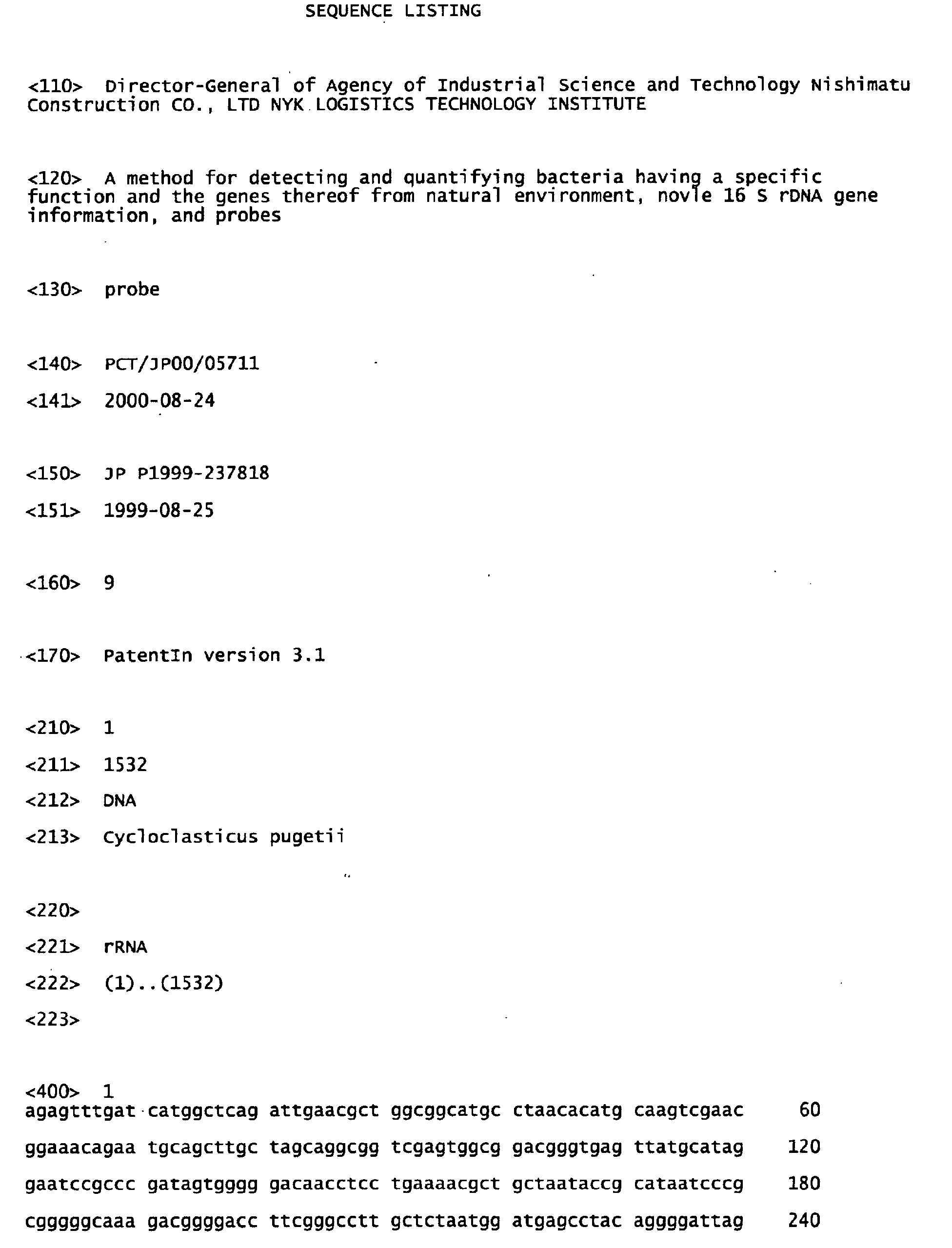
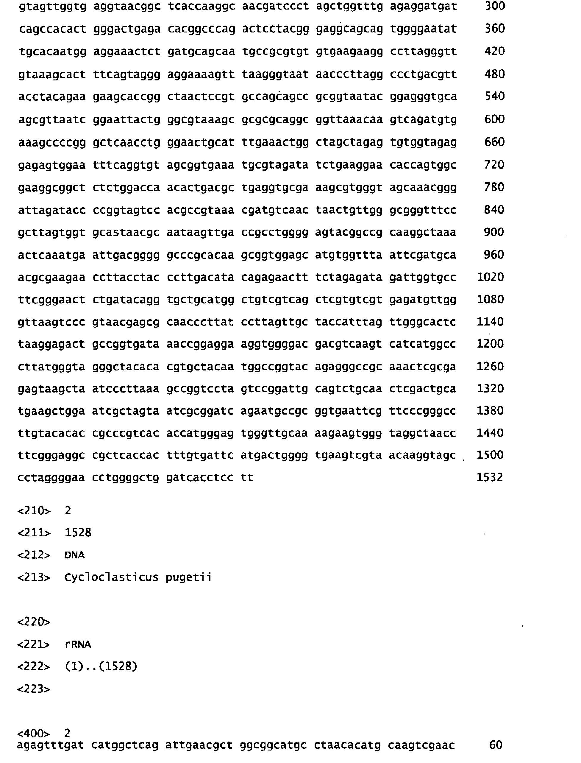
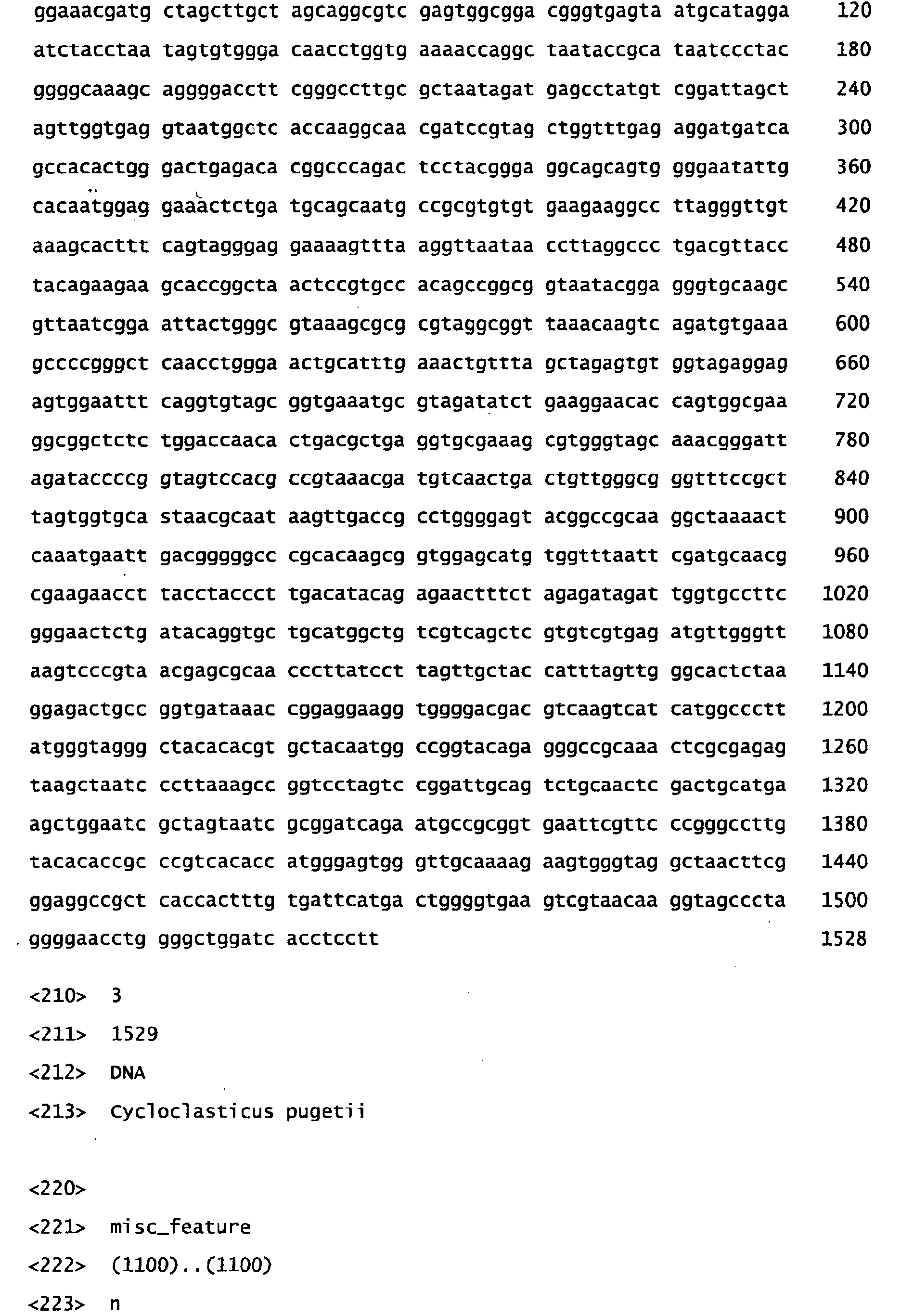
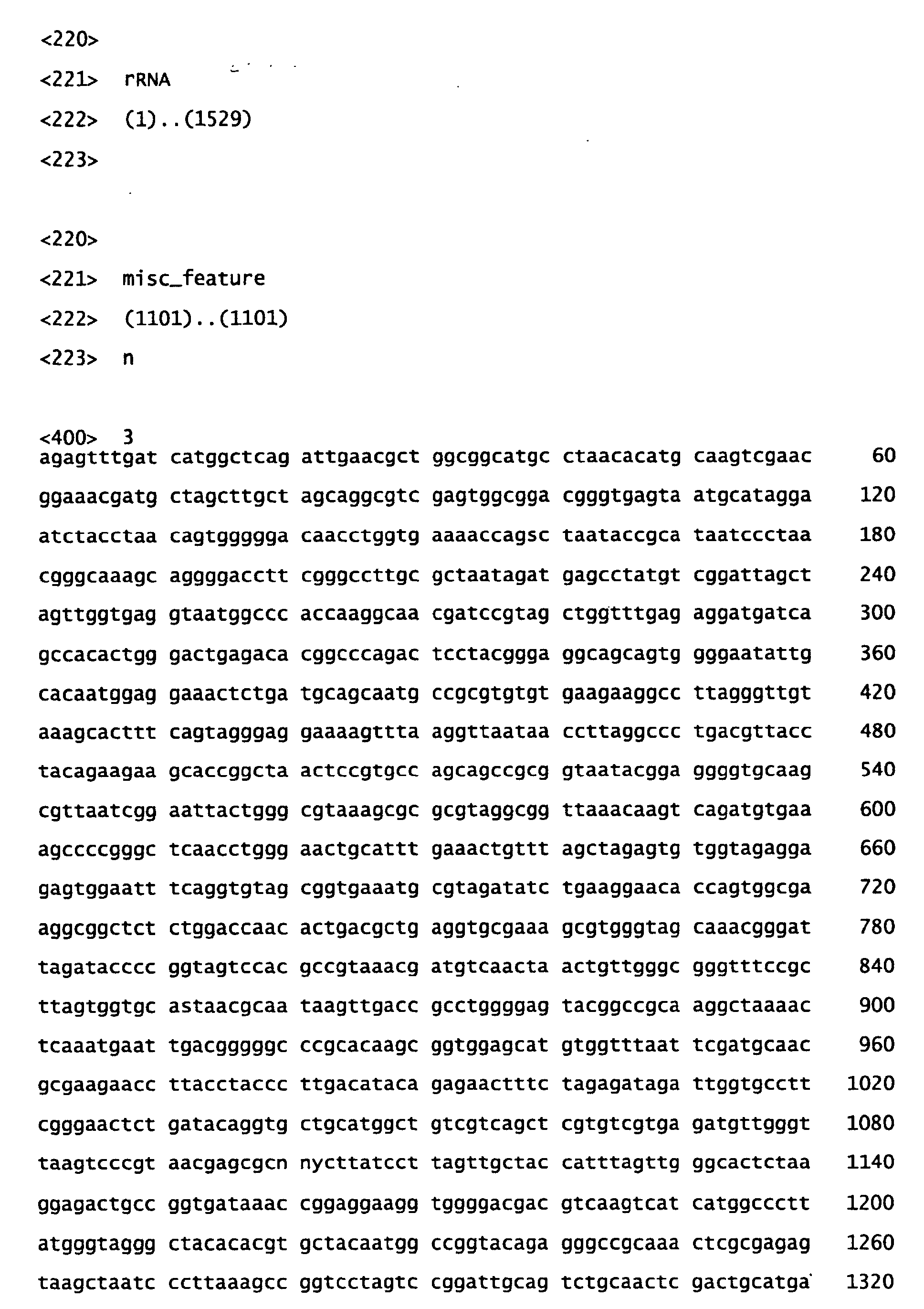
of 16S rDNA having a nucleotide sequence of SEQ ID NOs: 1 -
4 was obtained from the surface seawater of the coastal region
of Mikuni-cho, Fukui Pref., at which a large quantity of heavy
oil was cast ashore by an oil spill accident which occurred
at the Sea of Japan on January 2, 1997. From this gene data,
it was found that the petroleum-degrading bacterium existing
predominantly in that environment was Cyloclasticus pugetii
or its closely relative species. That is, as described in
Example below, a petroleum-degrading bacterium that existed
predominantly in polluted site was selected from a microbial
population in the oil-polluted sea area where the oil spill
accident has occurred by the MNP counting method with liquid
culture medium containing C-heavy oil as the sole carbon source.
Then, a DNA was extracted from the cultured cells of the
selected petroleum-degrading bacterium, which DNA was used as
a template to carry out PCR with a proper primer (for example,
a primer corresponding to the conserved region of 16S rDNA).
The PCR liquid product was then subjected to electrophoresis
to give 16S rDNAs having SEQ ID NOs: 1 - 4, of which the
nucleotide sequences were determined with an autosequencer.
Using this gene data, the petroleum-degrading bacterium
exisiting predominantly in the area was analyzed by the
above-mentioned molecular phylogenic or homology method and
identified to be Cyloclasticus pugetii or its closely relative
species.
On the basis of the nucleotide sequence data of SEQ ID
NOs: 1 - 4, an RNA and DNA probes can be designed so as to be
applicable to a variety of utilities. The nucleotide sequence
and length of the probe may be selected optionally depending
on the utilities. For example, by a FISH (fluorescence in situ
hybridization) method, in order to detect or quantitate a
microorganism belonging to the genus
Cycloclasticus in a sample
(for example, water collected from the sea, river, lake and
marsh in the area polluted with oil), particularly,
Cyloclasticus pugetii or its closely relative petroleum-degrading
bacterium, or to screen a microorganism belonging
to the genus
Cycloclasticus, particularly,
Cyloclasticus
pugetii or its closely relative petroleum-degrading bacterium,
from many microbial flora, it is appropriate to design a probe
which has 10-50 bases, preferably 15-25 bases in length,
corresponding to the region selected from the nucleotide nos.
823-853 of the nucleotide sequences of SEQ ID NOs: 1 - 4 (in
the nucleotide sequence of 16S rDNA in
Escherichia coli, the
region of nucleotide nos. 829-866 from the 5'-terminal position
(numbering system)). The followings are exemplified as the
probes.
(wherein *(number) indicates the position(numbering system)
from the 5'-terminal in the 16S rDNA nucleotide sequence of
Escherichia coli (H.F. Noller and C.R. Woese, Science, 212:
403-411, 1981))
The nucleotide sequence of the probe (3) is derived from
the nucleotide sequences of the probes (1) and (2) by lateral
conjugation. In this case, however, it should be cautious
because there is a possibility of self-conjugation of the
probes.
The probes may be synthesized in the known method, for
example, the phosphoramide method or triester method.
Alternatively, they may be synthesized by means of a DNA
autosynthesizer.
The probes may be labeled with an isotope (32P, 35S, etc.),
fluorescent dye (biotin/avidin, digoxigenin/antidigoxigenin-rhodamine,
fluorescein-isothiocyanate (FITC),
Lucifer Yellow CH, rhodamine 123, acridine orange, pyronin Y,
ethidium bromide, propidium iodide, ethidium homodimer,
BOBO-1, POPO-1, TOTO-1, YOYO-1, carboxyfluorescein diacetate
(CFDA), fluorescein diacetate (FDA), carboxyfluorescein
diacetate-acetoxymethyl ester (CFDA-AM), 5-cyano-2,30-ditolyl
tetrazolium chloride (CTC), tetramethylrhodamine
isothiocyanate (TRITC), sulforhodamine 101 acid chloride
(Texas Red), Cy3, Cy5, Cy7, 2-hydroxy-3-naphthoic acid-2'-phenylanilide
phosphate (HNPP), etc.), antigenic substance
such as digoxigenin, and the like. When labeled with an
antigenic substance, the probe may be detected by
chemiluminescence or fluorescence generated through enzymatic
decomposition of a substrate in an enzyme immunoassay
technique.
Using the RNA or DNA probes of the invention, microorganisms
belonging to the genus Cycloclasticus, particularly
a petroleum-degrading bacterium Cyloclasticus pugetii or its
closely relative species can be detected or quantitated or
screened by a variety of hybridization methods (Southern blot
method, Northern blot method, colony hybridization method, dot
hybridization method, in situ hybridization (e.g., FISH method,
etc.), and the like.
The followings describe as an example a method for
detecting and quantitating a petroleum-degrading bacterium
from the seawater collected at the site of an oil spill accident,
using the DNA probe of the invention. The seawater is collected
from the sea area of an oil spillage accident, and the
microorganisms contained in the seawater are fixed on a filter
(pore size 0.2µm) and hybridized with a DNA probe having the
nucleotide sequence of SEQ ID NO: 5 labeled with a fluorescent
dye. After washing out the probe, the bacterium
(petroleum-degrading bacterium) which has hybridized with the
DNA probe is detected and counted under a fluorescence
microscope.
Using the DNA probe of the invention, a microorganism
belonging to the genus Cycloclasticus, particularly a
petroleum-degrading bacterium Cyloclasticus pugetii or its
closely relative species can be screened from many microbial
population by means of a colony hybridization technique, dot
blot hybridization technique, flow cytometry, and the like.
In addition, it is possible to identify a microorganism
belonging to the genus Cycloclasticus by means of DNA/DNA or
DNA/RNA hybridization using an RNA or DNA probe which was
designed based on the nucleotide sequences of SEQ ID NOs: 1-4,
or the homology with the nucleotide sequence data of SEQ ID
NOs: 1-4. Herein, "identification" refers to a homology
calculation between the nucleotide sequences when the
nucleotide sequence of the approximately whole region of the
target gene was analyzed as in SEQ ID NOs: 1-4. And, when a
probe having the taxonomically specific nucleotide sequence
designed as described above (in this case, the nucleotide
sequence specific to a microorganism belonging to the genus
Cycloclasticus, particularly Cyloclasticus pugetii or its
closely relative species) is used, it means a hybridization
test between the nucleotide sequences contained in the target
gene sample.
The calculation of homology is made as follows. The
number of the nucleotide in the sequence of the target gene,
preferably of the whole region in which the individual
nucleotide sequences have been determined and are comparable
each other, is put in the denominator, and the number of the
nucleotide that are completely identical therein is put in the
numerator. Thus, the homology can be calculated as %, for
example, using the Genentyx-MAC 8.0 analysis software (Genetic
Information Processing Software Co.).
The identification of a petroleum-degrading bacterium
belonging to the genus Cycloclasticus based on the homology
of the nucleotide sequence of SEQ ID NOs: 1-4 may be made
according to the ordinary standard for judgement of a species
from the nucleotide sequence of 16S rDNA. That is, when the
homology is 97% or less on the approximately whole region of
16S rDNA, it may be judged to be a different species tentatively
(E. Stackebrandt and B.M. Goebel, Int. J. Syst. Bacteriol.,
44: 846-849, 1994).
For example, the homology of the nucleotide sequences
of 16S rDNA in a certain microorganism is examined in comparison
with those of SEQ ID NOs: 1-4 for calculation with an analysis
software such as Genentyx-MAC 8.0 (vide supra). As a result,
when the value is 97% or more, it is judged to be the same species,
and when less than 97%, judged a different species.
In some classified groups, however, even though the
homology is 97% or more, they have been judged different species.
For example, it is known that the homology between Vibrio
cholerae and Vibrio mimicus is 98.9-99.4% (Shimizu and
Tsukamoto, Classification and Identification of Marine
Bacteria, Marine Microorganisms and Biotechnology, Edited by
Ushio Shimizu. Gihodo Press, pp.1-24, 1991). In addition, in
a study for marine bacteria of the Vibrio family, it has been
estimated that a group having 99.3% or more of the homology
is within the range of one species, and a group having 97% or
more is within one genus (Shimizu and Tsukamoto, vide supra).
Accordingly, it should be considered that the identification
of the homology based on the standard of 97% or more is tentative,
and the relationship between the species and the homology in
the target taxon should be examined in advance prior to
application of this standard.
Specifically, since only one species of Cycloclasticus
pugetii has been accepted in the genus Cycloclasticus bacteria,
the homology of 97% tentatively accepts the identification that
the bacterium is of said species. As clearly seen from the
above example of the genus Vibrio, in said genus for which
further study is required, the standard of about 97% leaves
still more the possibility that said bacterium might belong
to a different species.
The petroleum-degrading bacteria of the genus
Cycloclasticus may be identified as follows by means of DNA/DNA
or DNA/RNA hybridization using the RNA or DNA probe.
The hybridization technique used in the identification
includes the dot blot hybridization technique (H.E.N. Bergmans
and W.Gaastra, New Nucleic Acid Techniques (J.M. Walker ed.);
Methods in Molecular Biology, vol. 4, Clifton, Humana press,
pp. 385-390, 1983), colony hybridization technique (M.
Grustein and D.S. Hogness, Proc. Nat. Acad. Sci. USA, 72:
3961-3965, 1975), Southern blot hybridization technique (E.M.
Southern, J. Mol. Biol., 98: 503-507, 1975), fluorescent in
situ hybridization (also called whole cell hybridization)
technique (R.I. Amann et al., Microbiol. Rev., 59: 143-169,
1995), and the like. The DNA/RNA sample used in these
hybridization analyses may be prepared from microorganisms
according to the known methods as described in the respective
literatures.
Even though any of the hybridization techniques is used,
it is important that strict hybridization conditions between
the probe used and the sample of standard strain having the
target gene nucleotide sequence should be determined in advance,
for example, according to the method as describe by D.A. Stahl
and R. Amann (Development and application of nucleic acid
probes; In: Nucleic acid techniques in bacterial systematics;
E. Stackebrandt and M. Goodfellow (ed.); John Wiley and Sons,
West Sussex, pp. 205-248, 1991), wherein the conditions such
as temperature, the concentration of formamide in the buffer
solution, etc. are estimated, and ideally when the
complementarity does not satisfy 100%, the conditions under
which the probe is liberated in the process of hybridization
and during the subsequent washing should be determined
experimentally.
Under the hybridization conditions thus determined,
hybridization is made for an unknown sample, using a target
sample of genes from the target standard strains as described
above or from the standard strains taxonomically far different
from them. As a result, the intensity of radioactivity,
fluorescence, or chemiluminescence (in a detection system of
antigen-antibody-enzyme, the system can be selected by
changing the enzyme substrate) generated by a radioactive
element or fluorescent substance or antigen (in this case,
utilizing the activity of an enzyme bound to the corresponding
antibody) directly labeled to the probe, is measured and
compared to that of the target sample. When the resulting
intensity of hybridization of the unknown sample is equal to
that of the target standard strain and recognized a great
difference with a taxonomically far different strain (ideally,
when the detected intensity is below the measuring limit), the
unknown sample is judged to be the same species as or defined
to be its closely relative species to the target microorganism.
In addition, for example, a species can be identified
by PCR using the nucleotide sequences (DNA fragments) of the
above probes as primers. That is, the target microbial cells
to be identified are lysed, to which is added the DNA fragment
having the nucleotide sequence of the above probe as a primer
and multiplied by PCR. The PCR product is analyzed by
electrophoresis, and if multiplication of 16S rDNA is confirmed,
the target bacterium would have the gene portion complementary
to the DNA fragment used. That is, the target bacterium is
defined to be the same species as that having 16S rDNA
containing the nucleotide sequence of SEQ ID NO: 1.
The microorganism of the genus Cycloclasticus detected,
screened or identified according to the method of the invention,
particularly Cyloclasticus pugetii or its closely relative
petroleum-degrading bacterium, may be utilized as a microbial
preparation (for example, petroleum-degrading microbial
preparation for bioremediation agent).
In addition, the use of 16S DNA or the nucleotide sequence
data of the probes of the invention allows elucidation of the
behavior of petroleum-degrading bacteria in natural
environment or under the conditions of oil treatment. It is
also effective in development of an environmental repairing
technology (bioremediation technology) such as promotion of
the efficiency of oil degradation in oil-polluted sea area by
addition of inorganic nutrients or by seeding of the
petroleum-degrading bacteria.
The present specification includes the content as
described in the specification and/or drawings of Japanese
Patent Application No. 11-237818 that is a priority application
of the present application.
Best Mode for Carrying Out the Invention
The invention will be described specifically by the
following examples. These examples, however, serve to
illustrate but are not intended to limit the scope of the
invention.
Example 1
(1) Investigation of petroleum-degrading microorganisms in
the sea area of a heavy oil spill accident
The surface seawater of the coastal region of Mikuni-cho,
Fukui Pref., at which a large quantity of heavy oil was cast
ashore by an oil spill accident which occurred at the Sea of
Japan on January 2, 1997, was collected on January 15
immediately after the pollution. This was filtered through
a plankton net (about 30µm in mesh), and the petroleum-degrading
microorganisms in the surface seawater of the
polluted area were counted by a microplate MPN (M-MPN) counting
method. The M-MPN counting was carried out as follows:
One point eight ml each of 1/10NP medium (100 mg NH4NO3, 2 mg
ferric citrate, 2 mg K2HPO4, 800 ml aged seawater, 200 ml
distilled water; pH 7.8) was added to wells on a 24-well
microplate. Then, after seawater sample was diluted on
10-fold in each well containing the medium on this microplate,
10 µl each of n-tetradecane, kerosene or C-heavy oil as the
sole carbon source was added to those wells. Diluted seawater
sample in the well was incubated at 20°C. This MPN counting
was carried out in triplicate. The growth was judged by
comparing turbidity of the medium or change of the floating
oil in each well with that in the control wells without inocula.
In parallel, the total number of microorganisms was counted
basically according to the Porter and Feig method (K.G. Porter
and Y.S. Feig, Limnol. Oceanogr., 25: 943-948, 1980), as well
as the number of heterotrophic bacteria was counted according
to the M-MPN counting using a 1/2TZ medium (2.5 g polypeptone,
0.5 g Bacto-Yeast extract, 4.77 g HEPES, 900 ml Kester's
artificial seawater, 100 ml distilled water; pH 7.5).
Moreover, the surface seawater was collected according to the
season from January 15, 1997 immediately after the accident
for about 1 year. And the total number of microorganisms in
the seawater was counted by the direct microscopic counting
method, and the number of the heterotrophic bacteria or
petroleum-degrading bacteria was examined by the MPN method.
As a result, it was found that the total number of
microorganisms in the oil spill sea area was kept at a level
of 105 cells/ml order over 1 year and there was no considerable
variation, but the number of the petroleum-degrading bacteria
was markedly changed. That is, the number of the n-tetradecane-,
kerosene- or C-heavy oil-degrading bacteria was
such a high value as about 103-104 MPN/ml (the ratio (dominant
level) of the number of the degrading bacteria to the total
number of microorganisms: 1-10%), but it was markedly reduced
to 10-103 MPN/ml (dominant level: 0.01-1%) after the lapse of
about 2 months at the same area. On the other hand, the number
of the heterotrophic bacteria was 104 MPN/ml immediately after
occurrence of the pollution, and about 2 months later
approximately the same value, 104-105 MPN/ml. From these
results, it was judged that the microbial community immediately
after the oil spill accident had much higher degrading function
to oil components than in an ordinary state even at a lower
water temperature of 12-13°C in winter.
(2) Selection of the petroleum-degrading bacteria
predominantly existing in the oil spill sea area
Using the surface seawater collected on January 15, 1997
immediately after the above-mentioned accident, a sample
containing a petroleum-degrading microorganism was collected
from the well (a sample of growth-positive front) of the highest
serial dilution ratio at which the growth of the microorganism
was judged positive among the wells containing a 1/10NP medium
which was added C-heavy oil and used in the M-MPN counting.
This sample of microorganism is a liquid culture medium derived
from the microbial community in the oil spill sea area by
diluting up to the limit by 10-fold serial dilution. Therefore,
the microorganism therein is considered the most predominant
one in the oil spill sea area.
(3) Extraction of DNA from the selected petroleum-degrading
microorganism
The sample of growth-positive front was centrifuged to
collect the cells, which were suspended in 567 µl of TE buffer
(10mM Tris-HCl, 1mM EDTA, pH 8.0). To this suspension was added
30 µl of 0.01% sodium dodecyl sulfate (SDS) and 3 µl of 20 mg/ml
proteinase K solution, and the mixture was incubated at 37°C
for 1 hour. Then, 100 µl of 5M NaCl was added, and the mixture
was stirred well and incubated at 65°C for 10 minutes. After
addition of 700 µl of chloroform/isoamyl alcohol mixture (24:1),
the mixture was gently stirred and centrifuged to collect the
supernatant (aqueous layer), to which was added an equivalent
volume of phenol/chloroform/isoamyl alcohol mixture (20:24:1).
The mixture was gently stirred and contrifuged to collect the
supernatant (aqueous layer), to which was added 0.6 volume part
of isopropanol, and DNA was collected by centrifugation. The
DNA was washed with 70% ethanol (-20°C), dried and dissolved
in 100 µl of TE buffer.
(4) Cloning
Using the DNA recovered and purified in (3) as a template
and the primers 27f and 1525r corresponding to the conserved
region of 16S rRNA, 16S rRNA was amplified by PCR.
This PCR product (16S rDNA), after confirmation of the
presence by electrophoresis, was subjected to cloning with
Original TA Cloning(r) kit (Invitrogen Co.).
(5) RFLP (restriction fragment length polymorphism) analysis
First, 40 clones were selected at random, and it was
confirmed whether the full length of 16S rRNA was inserted into
these clones. As a result, it was found that the full length
of 16S rRNA was inserted into 17 clones. Next, these 17 clones
were grouped by an RFLP (restriction fragment length
polymorphism) analysis using 5 different kinds of restriction
enzymes (EcoRI, HindIII, SalI, XbaI and RsaI) in order to grasp
a rough tendency of population of petroleum-degrading bacteria
which was considered to have the predominance in the oil-polluted
sea area. As a result, 1 to 3 fragment patterns were
obtained on the respective restriction enzymes. Finally,
these clones were classified into 4 groups from the respective
fragment patterns by the 5 kinds of restriction enzymes.
(6) Full sequencing
From the 4 groups classified by the RFLP analysis in (5),
every one clone, totally 4 clones (CHO11-1-1, CHO11-1-2,
CHO11-1-4, CHO11-1-34) were selected, and the whole nucleotide
sequence of 16S rRNA was determined.
The sequence was determined with an auto-sequencer
(ALFred DNA Sequencer, Pharmacia AB) using an RNA sequence kit
(Thermo Sequenase TM fluorescent labeled primer cycle
sequencing kit with 7-deaza-dGTP, Amersham Co.), and the
nucleotide sequence of 16S rDNA was determined.
(7) Molecular phylogenic analysis
From the nucleotide sequence data of 16S rDNA of thus
determined 4 clones, the presence of microorganisms having data
similar to them was examined as preliminary search using a
search engine Ribosomal Database Project (RDP) II (Illinois
University). Subsequently, the similar sequences as well as
the sequences of the representative strains of the respective
taxon were obtained (download) from the database (GenBank, EMBL,
DDBJ, etc.), and subjected to the multiple alignment treatment
with the nucleotide sequence of 16S rDNA of the 4 clones using
such a program as Clustal W (vide supra) or Se-Al (vide supra).
For this alignment data (about 1500 nucleotide of sequence),
then, the molecular phylogenic analysis by the NJ method (vide
supra) and the bootstrap analysis by 100 repetitions were
carried out using the PHYLIP program (vide supra).
As a result, it was found that the petroleum-degrading
microorganism which existed predominantly in the surface
seawater immediately after the oil spillage accident in the
Sea of Japan on January 1997 is a bacterium belonging to the
γ-subgroup of Proteobacteria and closely related to
Cycloclasticus pugetii (C. pugetii). In addition, as a result
of a homology analysis for the nearly full length of the
nucleotide sequence of 16S rDNA, it was found that the 3 clones
(CHO11-1-2, CHO11-1-4, CHO11-1-34) among the 4 clones have
about 99% of homology (99.2%, 98.8%, 99.1%, respectively) to
that of C. pugetii (GenBank No. U12624), while the other one
clone (CHO11-1-1) showed lower homology (97.2%). In these 4
clones, accordingly, it was estimated that the sequences were
derived from the same species as C. pugetii or its closely
related species. The nucleotide sequences of 16S rDNA of these
4 clones (CHO11-1-1, CHO11-1-2, CHO11-1-4, CHO11-1-34) which
were judged to be the same species as C. pugetii or its closely
related species are shown in SEQ ID NOs: 1-4.
Cycloclasticus pugetii which was judged to be closely
related to the above 4 clones was recently reported as a
microorganism which was isolated from the sediment on the
bottom of the sea polluted by polychlorinated biphenyl at the
coastal region of the Pacific Ocean in USA and which had an
aromatic hydrocarbon degrading capacity (S.E. Dyksterhouse et
al., Int. J. Syst. Bacteriol. 45: 116-123, 1995). Therefore,
the standard strain of this species (ATCC 51542) was obtained
to examine. As a result, it was found that this microorganism
is characterized in that it grows only on a culture medium
containing such a specific organic substance as biphenyl and
merely produces small colonies on plate culture involving such
a substance, and is difficult to isolate on a usually plate
culture medium for heterotrophic bacteria.
On the other hand, the nucleotide sequence data of 16S
rDNA of the present invention was first discovered from the
microbial sample collected from the oil-polluted seawater
immediately after an oil spillage accident. And the sample
has a quite different characteristic in the view that it was
not collected from the sediment on the bottom of the sea nor
from the sea area polluted by polychlorinated biphenyl. As
mentioned above, this is supposed to be caused by that isolation
of C. pugetii or its closely related species is very difficult.
In the future, however, they will possibly be detected widely
in the sea area of the world by such a method as described in
the present invention.
Since the nucleotide sequences (SEQ ID NOs: 1-4) of 16S
rDNA were obtained from an oil-polluted field through serially
diluted liquid culture media containing C-heavy oil as the sole
carbon source, they can be considered to be the nucleotide
sequence data of the gene resulting from the microorganism
which degrades hydrocarbons in C-heavy oil to grow. As
mentioned above, it was shown that the microorganism that has
said sequence data is molecular-phylogenically closely
related to Cycloclasticus pugetii that has a capacity degrading
aromatic hydrocarbons and two have a high homology each other.
Therefore, microorganisms which have the nucleotide sequences
of SEQ ID NOs: 1-4 and C. pugetii or its closely related
microorganisms can be considered to be bacteria which have a
function degrading hardly degradable polycyclic aromatic
hydrocarbons. This means that the gene data of SEQ ID NOs:
1-4 are quite effective in examining the microbial flora at
an oil spill field, or in analyzing the state of oil pollution
or oil degradation based on the microbial flora or distribution
density of the microorganisms having said gene data.
That is, recalcitrant polycyclic aromatic hydrocarbons
in spilled crude oil or heavy oil remain unchanged in the
polluted sea area for a longer period than readily decomposable
aliphatic hydrocarbons. Therefore, the nucleotide sequence
data of SEQ ID NOs : 1-4 resulting from microorganisms that might
possibly degrade hardly decomposable polycyclic aromatic
hydrocarbons to grow can be utilized as an effective indicator
in monitoring the process of repairing and purifying oil
pollution at the actual oil-polluted coastal area or polluted
field. In addition, the nucleotide sequence data of SEQ ID
NOs: 1-4, though supposed to be derived from Cycloclasticus
pugetii or its closely related microorganisms, dose not have
100% homology with the known sequences but new sequence data.
They, accordingly, are estimated very useful in preparing some
probes specific to the species or groups of this taxon.
Moreover, it became clear that a series of the
above-mentioned techniques, i.e., microplate MPN/direct
PCR/sequencing techniques, are very effective in analyzing
petroleum-degrading microorganisms predominantly existing in
the sea area.
Example 2
(1) Selection of species specific sequences for Cycloclasticus
pugetii (Probe design)
Using the results of the molecular phylogenic analysis
performed in Example 1, the sequences specific to
C. pugetii
were selected taking the high-order structure of 16S rRNAs into
account (the probes were designed). The followings indicate
2 kinds of the probes thus designed (Cyclopug 829-846 and
Cyclopug 847-866).
Wherein * indicates the position (numbering system) from the
5' -terminal in the 16S rDNA nucleotide sequence of
Escherichia
coli.
(2) Probe check by the database
In order to confirm the specificity of the probes
designed in (1), a probe-match was searched using the database
(Ribosomal Database Project II, RDP). A search engine
attached to RDP was used to search the miss match sequence under
a condition of 2 nucleotides. As a result, it was confirmed
that the two probes as designed matched only with 6 strains
of the genus
Cycloclasticus on RDP and were completely
identical with no miss match base. In addition, both probes
were confirmed not to self-conjugate. Among these 6 strains
of the genus
Cycloclasticus, two are
C. pugetii, and the
remaining four strains are unidentified or unaccepted strains
for
Cycloclasticus sp. For reference, the source of the 6
strains of the genus
Cycloclasticus which matched on RDP is
shown in Table 1.
| Source of the genus Cycloclasticus bacteria registered on RDP |
| Source | DataBase accession no. | Reference |
| Cycloclasticus pugetii str. PS-1(T)ATCC51542 | U12624 | 1 |
| Cycloclasticus pugetii str. PS-1(T)ATCC51542 | L34955 | 1 |
| Cycloclasticus sp. N3-PA321 | U57920 | 2 |
| Cycloclasticus sp. G | AF093002 | 3 |
| Cycloclasticus sp. E | AF093003 | 3 |
| Cycloclasticus sp. W | AF093004 | 3 |
| Cycloclasticus sp. (oligotrophus) | AF148215 | 4 |
| 1. DYKSTERHOUSE et al., Int. J. Syst. Bacteriol., 43: 116-123, 1995 |
| 2. GEISELBECHT et al., Appl. Environ. Microbiol., 62: 3344-3349, 1996 |
| 3. GEISELBECHT et al., Appl. Environ. Microbiol., 64: 4703-4710, 1998 |
| 4. BUTTON et al., Appl. Environ. Microbiol., 64: 4467-4476,
1998 |
Herein, as the nucleotide sequence data of 16S rDNA of
the genus Cycloclasticus, 6 strains in 7 cases are recorded
on the RDP database. In this database, the species which was
reported in IJSB (International Journal of Systematic
Bacteriology) and IJSEM (International Journal of Systematic
and Evolutionary Microbiology) and accepted formally is only
C. pugetii, i.e., one genus and one species at present. In
Table 1, C. oligotrophus is an unaccepted species, and
difference from C. pugetii is not clear (the homology between
both sequences is 99.3%).
From the above fact, this time it became clear that the
two probes which were designed from the nucleotide sequences
of 16S rDNA of microorganisms derived from an oil-polluted
environment have a very high specificity for Cycloclasticus
pugetii or its closely related species.
Example 3
It was confirmed that the probes thus designed hybridize
specifically with the standard strain of Cycloclasticus
pugetii ATCC51542 by the fluorescence in situ hybridization
(FISH) method.
(1) Synthesis and labeling of probes
Probes Cyclopug 829-846 and Cyclopug 847-866 of which
the specificity was confirmed by means of the database in
Example 2 were synthesized with a DNA synthesizer and purified
in a conventional manner. These probes Cyclopug 829-846 and
Cyclopug 847-866 were labeled at the 5'-terminal with
tetramethylrhodamine isothiocyanate (TRITC).
(2) Incubation and fixation of the strains provided
The standard strain of Cycloclasticus pugetii ATCC51542
was obtained from American Type Culture Collection (ATCC) and
incubated in a 1/2TZ medium (vide supra) containing polychlorinated
biphenyl at 20°C. As controls, 4 types of the
standard strains, Pseudomonas aeruginosa (P. aeruginosa),
Bacillus marinus (B. marinus), Vibrio parahaemolyticus (V.
parahaemolyticus) and Psychrobacter immobilis (Psy.
immobilis) were incubated under the conditions, i.e., culture
medium and temperature, recommended by the depositary
authority of the strains. The respectively cultured cells
were fixed with 3% (w/v) paraformaldehyde/3xPBS (24.0 g NaCl,
0.6 g KCl, 4.32 g Na2HPO4, 0.72 g KH2PO4) solution (pH 7.2-7.4).
The cells were collected by centrifugation and washed with
3xPBS buffer. In addition, as a control probe, probe EUB338
(R.I. Amann et al., J. Bacteriol. 172: 762-770, 1990) specific
to the Bacteria domain in the molecular phylogeny was provided.
(3) Hybridization
C. pugetii and 4 types of the control strains
preliminarily treated in (2) respectively were dropped on
gelatin-coated slide glasses (0.1% gelatin, 0.01% KCr(SO4)2),
and dried at room temperature to immobilized thereon. Then,
the slide glasses were dehydrated with 50, 80 and 100% ethanol.
On the samples immobilized on the slide glasses, 15 µl
of a hybridization buffer (0.9M NaCl, sodium sulfate buffer
[pH 7.2], 0.5% sodium dodecyl sulfate [SDS], 5mM EDTA 1mg/ml
Denhalt solution × 10 Poly(A)) was dropped, and the probe was
added at a concentration of 5 ng/µl and stored for hybridization
in a hybrichamber (humid state) at 45°C for 4.5 hours. After
completion of the hybridization, the surface of the slide
glasses was washed with 5 ml of a washing buffer (50mM sodium
phosphate buffer [pH 7.0], 0.1% SDS, 0.9M NaCl). The glasses
were immersed in 50 ml of the washing buffer at 42°C for 30
minutes, then washed with distilled water, and dried under air.
Then, 1 - 5 µg/ml of 4',6-diamidino-2-phenylindole (DAPI) was
added on the samples, and stained at room temperature for 5
minutes. After washing with distilled water and air-drying,
an anti-fading agent was dropped thereon, and covered with
slide glasses for sealing. The slide glasses were observed
under a fluorescence microscope (Zeiss, Oberkochen, Germany)
under UV excitation, B excitation and G excitation.
Table 2 shows the results. In the probes Cyclopug
829-846 and Cyclopug 847-866 as designed this time, DAPI was
bound universally to the DNAs in the respective cells by UV
excitation, so that blue fluorescence resulting from DAPI could
be observed in C.
pugetii and in the 4 types of the control
strains (
P. aeruginosa, B. marinus, V. parahaemolyticus and
Phy. immobilis). When the same field of vision was observed
under G excitation, however, only
C. pugetii emitted red
fluorescence caused by TRITC labeled at the 5'-terminal of the
probe because it had the sequence complementary to the probes
Cyclopug 829-846 and Cyclopug 847-866.
| Effectiveness of the DNA probes specific toCycloclasticus pugetii or its closely related species |
| Standard Strain Tested | Probe Tested |
| | Cyclopug 829-846 | Cyclopug 847-866 | EUB338 |
| Cycloclasticus pugetii | ○ | ○ | ○ |
| Pseudomonas aeruginosa | × | × | ○ |
| Bacillus marinus | × | × | ○ |
| Vibrio parahaemolyticus | × | × | ○ |
| Psychrobacter immobilis | × | × | ○ |
○: Indicating occurrence of hybridization and emission of fluorescence in observation under a microscope
X: Indicating no occurrence of hybridization and unsuccessful observation under a microscope |
On the other hand, in Probe EUB338 specific to Bacteria
domain, C. pugetii and the 4 types of the control strains, all
emitted green fluorescence caused by FITC which was labeled
at the 5'-terminal of EUB338 even under observation by B
excitation.
From the above fact, this time it was demonstrated that
the two probes that have been designed from the nucleotide
sequences of 16S rDNA of a microorganism isolated from an
oil-polluted environment are practically very effective in
specifically detecting Cycloclasticus pugetii or its closely
related species without obstruction caused by the higher-order
structure.
All of publications, patents and patent applications
cited in the present specification are herein incorporated by
reference.
Industrial Applicability
According to the invention, a technique for elucidating
microorganisms existing predominantly in the natural
environment (for example, a field environment polluted with
petroleum, etc.) was provided.
The nucleotide sequence data of 16S rDNA analyzed by the
technique of the invention are considered to be derived from
a naturally occurring petroleum-degrading microorganism
having a function predominant in an oil-polluted environment.
Therefore, the nucleotide sequence data of the DNA probes which
have been designed to allow the specific detection of the above
microorganism based on the gene and gene data are very useful
in elucidating the behavior of petroleum-degrading
microorganisms in nature or under the conditions of oil
treatment. The data are also effective in development of an
environmental repairing technology (bioremediation
technology) such as promotion of the efficiency of oil
degradation in the polluted sea area by addition of inorganic
nutrients or by seeding of the petroleum-degrading
microorganisms.
According to the invention, a method for elucidating the
behavior of microorganisms predominant in the natural
environment (for example, environment polluted with petroleum
or harmful chemicals) or degrading microorganisms in the
process of bioremediation was provided.
Moreover, according to the invention, a method for
selecting a microorganism or a microbial population having a
specific function by liquid culture not through plate culture
method for isolation of the microorganism is provided. Also
provided is a method for analyzing the function and
phylogenetic position of the microorganism at a gene level.
The present invention, accordingly, is useful in detection,
quantification and screening of microorganisms having said
degrading function as well as so far hardly isolated
microorganisms producing useful substances such as enzymes.
SEQ ID Table Free Text
SEQ ID NOS: 1-4 show the nucleotide sequences of 16S
rDNAs.
SEQ ID NOS: 5-7 show the nucleotide sequences of the
probes.
SEQ ID NOS: 8 and 9 show the nucleotide sequences of the
primers.