EP1229784B1 - An improved method for production of porcine clone embryos via somatic cell nuclear transfer - Google Patents
An improved method for production of porcine clone embryos via somatic cell nuclear transfer Download PDFInfo
- Publication number
- EP1229784B1 EP1229784B1 EP00981846A EP00981846A EP1229784B1 EP 1229784 B1 EP1229784 B1 EP 1229784B1 EP 00981846 A EP00981846 A EP 00981846A EP 00981846 A EP00981846 A EP 00981846A EP 1229784 B1 EP1229784 B1 EP 1229784B1
- Authority
- EP
- European Patent Office
- Prior art keywords
- porcine
- embryos
- cells
- oocytes
- nuclear transfer
- Prior art date
- Legal status (The legal status is an assumption and is not a legal conclusion. Google has not performed a legal analysis and makes no representation as to the accuracy of the status listed.)
- Expired - Lifetime
Links
- 210000002257 embryonic structure Anatomy 0.000 title claims description 99
- 238000004519 manufacturing process Methods 0.000 title claims description 15
- 238000010374 somatic cell nuclear transfer Methods 0.000 title description 8
- 210000000287 oocyte Anatomy 0.000 claims description 80
- 210000004027 cell Anatomy 0.000 claims description 53
- 210000001082 somatic cell Anatomy 0.000 claims description 40
- 210000004940 nucleus Anatomy 0.000 claims description 31
- 238000000338 in vitro Methods 0.000 claims description 29
- 238000000034 method Methods 0.000 claims description 28
- 238000012546 transfer Methods 0.000 claims description 24
- 210000002950 fibroblast Anatomy 0.000 claims description 23
- 230000000638 stimulation Effects 0.000 claims description 17
- 241000282887 Suidae Species 0.000 claims description 13
- 210000001771 cumulus cell Anatomy 0.000 claims description 10
- 230000007159 enucleation Effects 0.000 claims description 10
- 241000282898 Sus scrofa Species 0.000 claims description 6
- 210000003754 fetus Anatomy 0.000 claims description 4
- 230000008569 process Effects 0.000 claims description 4
- 210000001519 tissue Anatomy 0.000 claims description 3
- 241000124008 Mammalia Species 0.000 claims description 2
- 230000003213 activating effect Effects 0.000 claims description 2
- 210000003855 cell nucleus Anatomy 0.000 claims description 2
- 241000282890 Sus Species 0.000 claims 1
- 210000002459 blastocyst Anatomy 0.000 description 33
- 239000002609 medium Substances 0.000 description 33
- 230000004913 activation Effects 0.000 description 31
- 235000013601 eggs Nutrition 0.000 description 22
- 238000011161 development Methods 0.000 description 19
- 230000018109 developmental process Effects 0.000 description 19
- 241001465754 Metazoa Species 0.000 description 16
- 108091003079 Bovine Serum Albumin Proteins 0.000 description 10
- 238000011282 treatment Methods 0.000 description 10
- 230000004927 fusion Effects 0.000 description 9
- 230000009261 transgenic effect Effects 0.000 description 9
- 241000283690 Bos taurus Species 0.000 description 8
- 229940098773 bovine serum albumin Drugs 0.000 description 8
- 238000003776 cleavage reaction Methods 0.000 description 8
- 230000007017 scission Effects 0.000 description 8
- BVIAOQMSVZHOJM-UHFFFAOYSA-N N(6),N(6)-dimethyladenine Chemical compound CN(C)C1=NC=NC2=C1N=CN2 BVIAOQMSVZHOJM-UHFFFAOYSA-N 0.000 description 7
- 238000010367 cloning Methods 0.000 description 7
- 230000001605 fetal effect Effects 0.000 description 7
- 230000001776 parthenogenetic effect Effects 0.000 description 7
- IAZDPXIOMUYVGZ-UHFFFAOYSA-N Dimethylsulphoxide Chemical compound CS(C)=O IAZDPXIOMUYVGZ-UHFFFAOYSA-N 0.000 description 6
- 239000012591 Dulbecco’s Phosphate Buffered Saline Substances 0.000 description 6
- 238000002474 experimental method Methods 0.000 description 6
- 241001494479 Pecora Species 0.000 description 5
- LOKCTEFSRHRXRJ-UHFFFAOYSA-I dipotassium trisodium dihydrogen phosphate hydrogen phosphate dichloride Chemical compound P(=O)(O)(O)[O-].[K+].P(=O)(O)([O-])[O-].[Na+].[Na+].[Cl-].[K+].[Cl-].[Na+] LOKCTEFSRHRXRJ-UHFFFAOYSA-I 0.000 description 5
- 239000012091 fetal bovine serum Substances 0.000 description 5
- 238000001727 in vivo Methods 0.000 description 5
- 239000002953 phosphate buffered saline Substances 0.000 description 5
- 241000894007 species Species 0.000 description 5
- PRDFBSVERLRRMY-UHFFFAOYSA-N 2'-(4-ethoxyphenyl)-5-(4-methylpiperazin-1-yl)-2,5'-bibenzimidazole Chemical compound C1=CC(OCC)=CC=C1C1=NC2=CC=C(C=3NC4=CC(=CC=C4N=3)N3CCN(C)CC3)C=C2N1 PRDFBSVERLRRMY-UHFFFAOYSA-N 0.000 description 4
- TWRXJAOTZQYOKJ-UHFFFAOYSA-L Magnesium chloride Chemical compound [Mg+2].[Cl-].[Cl-] TWRXJAOTZQYOKJ-UHFFFAOYSA-L 0.000 description 4
- 241000699670 Mus sp. Species 0.000 description 4
- 230000007910 cell fusion Effects 0.000 description 4
- 239000013043 chemical agent Substances 0.000 description 4
- YPHMISFOHDHNIV-FSZOTQKASA-N cycloheximide Chemical compound C1[C@@H](C)C[C@H](C)C(=O)[C@@H]1[C@H](O)CC1CC(=O)NC(=O)C1 YPHMISFOHDHNIV-FSZOTQKASA-N 0.000 description 4
- 210000000805 cytoplasm Anatomy 0.000 description 4
- 230000000694 effects Effects 0.000 description 4
- 230000035800 maturation Effects 0.000 description 4
- UCSJYZPVAKXKNQ-HZYVHMACSA-N streptomycin Chemical compound CN[C@H]1[C@H](O)[C@@H](O)[C@H](CO)O[C@H]1O[C@@H]1[C@](C=O)(O)[C@H](C)O[C@H]1O[C@@H]1[C@@H](NC(N)=N)[C@H](O)[C@@H](NC(N)=N)[C@H](O)[C@H]1O UCSJYZPVAKXKNQ-HZYVHMACSA-N 0.000 description 4
- UXVMQQNJUSDDNG-UHFFFAOYSA-L Calcium chloride Chemical compound [Cl-].[Cl-].[Ca+2] UXVMQQNJUSDDNG-UHFFFAOYSA-L 0.000 description 3
- 241000283707 Capra Species 0.000 description 3
- 108010062540 Chorionic Gonadotropin Proteins 0.000 description 3
- 102000011022 Chorionic Gonadotropin Human genes 0.000 description 3
- LFQSCWFLJHTTHZ-UHFFFAOYSA-N Ethanol Chemical compound CCO LFQSCWFLJHTTHZ-UHFFFAOYSA-N 0.000 description 3
- WSFSSNUMVMOOMR-UHFFFAOYSA-N Formaldehyde Chemical compound O=C WSFSSNUMVMOOMR-UHFFFAOYSA-N 0.000 description 3
- PEDCQBHIVMGVHV-UHFFFAOYSA-N Glycerine Chemical compound OCC(O)CO PEDCQBHIVMGVHV-UHFFFAOYSA-N 0.000 description 3
- 229920000209 Hexadimethrine bromide Polymers 0.000 description 3
- 108010003272 Hyaluronate lyase Proteins 0.000 description 3
- 102000001974 Hyaluronidases Human genes 0.000 description 3
- FAPWRFPIFSIZLT-UHFFFAOYSA-M Sodium chloride Chemical compound [Na+].[Cl-] FAPWRFPIFSIZLT-UHFFFAOYSA-M 0.000 description 3
- GBOGMAARMMDZGR-UHFFFAOYSA-N UNPD149280 Natural products N1C(=O)C23OC(=O)C=CC(O)CCCC(C)CC=CC3C(O)C(=C)C(C)C2C1CC1=CC=CC=C1 GBOGMAARMMDZGR-UHFFFAOYSA-N 0.000 description 3
- 241000700605 Viruses Species 0.000 description 3
- 239000001110 calcium chloride Substances 0.000 description 3
- 229910001628 calcium chloride Inorganic materials 0.000 description 3
- GBOGMAARMMDZGR-JREHFAHYSA-N cytochalasin B Natural products C[C@H]1CCC[C@@H](O)C=CC(=O)O[C@@]23[C@H](C=CC1)[C@H](O)C(=C)[C@@H](C)[C@@H]2[C@H](Cc4ccccc4)NC3=O GBOGMAARMMDZGR-JREHFAHYSA-N 0.000 description 3
- GBOGMAARMMDZGR-TYHYBEHESA-N cytochalasin B Chemical compound C([C@H]1[C@@H]2[C@@H](C([C@@H](O)[C@@H]3/C=C/C[C@H](C)CCC[C@@H](O)/C=C/C(=O)O[C@@]23C(=O)N1)=C)C)C1=CC=CC=C1 GBOGMAARMMDZGR-TYHYBEHESA-N 0.000 description 3
- 230000004720 fertilization Effects 0.000 description 3
- 229940084986 human chorionic gonadotropin Drugs 0.000 description 3
- 229960002773 hyaluronidase Drugs 0.000 description 3
- 208000015181 infectious disease Diseases 0.000 description 3
- 238000002347 injection Methods 0.000 description 3
- 239000007924 injection Substances 0.000 description 3
- 210000001161 mammalian embryo Anatomy 0.000 description 3
- 210000001672 ovary Anatomy 0.000 description 3
- 210000004508 polar body Anatomy 0.000 description 3
- 238000007634 remodeling Methods 0.000 description 3
- 230000008672 reprogramming Effects 0.000 description 3
- 239000000243 solution Substances 0.000 description 3
- RTKIYNMVFMVABJ-UHFFFAOYSA-L thimerosal Chemical compound [Na+].CC[Hg]SC1=CC=CC=C1C([O-])=O RTKIYNMVFMVABJ-UHFFFAOYSA-L 0.000 description 3
- 229940033663 thimerosal Drugs 0.000 description 3
- 238000001890 transfection Methods 0.000 description 3
- LENZDBCJOHFCAS-UHFFFAOYSA-N tris Chemical class OCC(N)(CO)CO LENZDBCJOHFCAS-UHFFFAOYSA-N 0.000 description 3
- JKMHFZQWWAIEOD-UHFFFAOYSA-N 2-[4-(2-hydroxyethyl)piperazin-1-yl]ethanesulfonic acid Chemical compound OCC[NH+]1CCN(CCS([O-])(=O)=O)CC1 JKMHFZQWWAIEOD-UHFFFAOYSA-N 0.000 description 2
- 101500028876 Bos taurus Neurotensin Proteins 0.000 description 2
- FBPFZTCFMRRESA-KVTDHHQDSA-N D-Mannitol Chemical compound OC[C@@H](O)[C@@H](O)[C@H](O)[C@H](O)CO FBPFZTCFMRRESA-KVTDHHQDSA-N 0.000 description 2
- 239000006144 Dulbecco’s modified Eagle's medium Substances 0.000 description 2
- 101150066002 GFP gene Proteins 0.000 description 2
- 102000006771 Gonadotropins Human genes 0.000 description 2
- 108010086677 Gonadotropins Proteins 0.000 description 2
- 229930195725 Mannitol Natural products 0.000 description 2
- 239000005662 Paraffin oil Substances 0.000 description 2
- 229930182555 Penicillin Natural products 0.000 description 2
- JGSARLDLIJGVTE-MBNYWOFBSA-N Penicillin G Chemical compound N([C@H]1[C@H]2SC([C@@H](N2C1=O)C(O)=O)(C)C)C(=O)CC1=CC=CC=C1 JGSARLDLIJGVTE-MBNYWOFBSA-N 0.000 description 2
- PXIPVTKHYLBLMZ-UHFFFAOYSA-N Sodium azide Chemical compound [Na+].[N-]=[N+]=[N-] PXIPVTKHYLBLMZ-UHFFFAOYSA-N 0.000 description 2
- 108700019146 Transgenes Proteins 0.000 description 2
- 239000007983 Tris buffer Substances 0.000 description 2
- 102000004142 Trypsin Human genes 0.000 description 2
- 108090000631 Trypsin Proteins 0.000 description 2
- 239000012190 activator Substances 0.000 description 2
- 230000015572 biosynthetic process Effects 0.000 description 2
- RYYVLZVUVIJVGH-UHFFFAOYSA-N caffeine Chemical compound CN1C(=O)N(C)C(=O)C2=C1N=CN2C RYYVLZVUVIJVGH-UHFFFAOYSA-N 0.000 description 2
- 239000003710 calcium ionophore Substances 0.000 description 2
- 239000006143 cell culture medium Substances 0.000 description 2
- 238000005119 centrifugation Methods 0.000 description 2
- VHJLVAABSRFDPM-QWWZWVQMSA-N dithiothreitol Chemical compound SC[C@@H](O)[C@H](O)CS VHJLVAABSRFDPM-QWWZWVQMSA-N 0.000 description 2
- 238000001317 epifluorescence microscopy Methods 0.000 description 2
- CJAONIOAQZUHPN-KKLWWLSJSA-N ethyl 12-[[2-[(2r,3r)-3-[2-[(12-ethoxy-12-oxododecyl)-methylamino]-2-oxoethoxy]butan-2-yl]oxyacetyl]-methylamino]dodecanoate Chemical compound CCOC(=O)CCCCCCCCCCCN(C)C(=O)CO[C@H](C)[C@@H](C)OCC(=O)N(C)CCCCCCCCCCCC(=O)OCC CJAONIOAQZUHPN-KKLWWLSJSA-N 0.000 description 2
- 210000001733 follicular fluid Anatomy 0.000 description 2
- 239000002622 gonadotropin Substances 0.000 description 2
- 229940088597 hormone Drugs 0.000 description 2
- 239000005556 hormone Substances 0.000 description 2
- 238000002513 implantation Methods 0.000 description 2
- 239000002555 ionophore Substances 0.000 description 2
- 244000144972 livestock Species 0.000 description 2
- 229910001629 magnesium chloride Inorganic materials 0.000 description 2
- 239000000594 mannitol Substances 0.000 description 2
- 235000010355 mannitol Nutrition 0.000 description 2
- 230000031864 metaphase Effects 0.000 description 2
- 239000000203 mixture Substances 0.000 description 2
- 239000012120 mounting media Substances 0.000 description 2
- 210000003101 oviduct Anatomy 0.000 description 2
- 238000012856 packing Methods 0.000 description 2
- 229940049954 penicillin Drugs 0.000 description 2
- 210000000582 semen Anatomy 0.000 description 2
- 239000011780 sodium chloride Substances 0.000 description 2
- DAEPDZWVDSPTHF-UHFFFAOYSA-M sodium pyruvate Chemical compound [Na+].CC(=O)C([O-])=O DAEPDZWVDSPTHF-UHFFFAOYSA-M 0.000 description 2
- 229960005322 streptomycin Drugs 0.000 description 2
- 239000012588 trypsin Substances 0.000 description 2
- 210000004340 zona pellucida Anatomy 0.000 description 2
- 102000007469 Actins Human genes 0.000 description 1
- 108010085238 Actins Proteins 0.000 description 1
- KCXVZYZYPLLWCC-UHFFFAOYSA-N EDTA Chemical compound OC(=O)CN(CC(O)=O)CCN(CC(O)=O)CC(O)=O KCXVZYZYPLLWCC-UHFFFAOYSA-N 0.000 description 1
- 229930182566 Gentamicin Natural products 0.000 description 1
- CEAZRRDELHUEMR-URQXQFDESA-N Gentamicin Chemical compound O1[C@H](C(C)NC)CC[C@@H](N)[C@H]1O[C@H]1[C@H](O)[C@@H](O[C@@H]2[C@@H]([C@@H](NC)[C@@](C)(O)CO2)O)[C@H](N)C[C@@H]1N CEAZRRDELHUEMR-URQXQFDESA-N 0.000 description 1
- 241000713813 Gibbon ape leukemia virus Species 0.000 description 1
- WQZGKKKJIJFFOK-GASJEMHNSA-N Glucose Natural products OC[C@H]1OC(O)[C@H](O)[C@@H](O)[C@@H]1O WQZGKKKJIJFFOK-GASJEMHNSA-N 0.000 description 1
- 239000007995 HEPES buffer Substances 0.000 description 1
- 238000010867 Hoechst staining Methods 0.000 description 1
- LPHGQDQBBGAPDZ-UHFFFAOYSA-N Isocaffeine Natural products CN1C(=O)N(C)C(=O)C2=C1N(C)C=N2 LPHGQDQBBGAPDZ-UHFFFAOYSA-N 0.000 description 1
- JVTAAEKCZFNVCJ-UHFFFAOYSA-M Lactate Chemical compound CC(O)C([O-])=O JVTAAEKCZFNVCJ-UHFFFAOYSA-M 0.000 description 1
- 206010038997 Retroviral infections Diseases 0.000 description 1
- 241000282894 Sus scrofa domesticus Species 0.000 description 1
- 208000027418 Wounds and injury Diseases 0.000 description 1
- 239000003242 anti bacterial agent Substances 0.000 description 1
- 229940088710 antibiotic agent Drugs 0.000 description 1
- IYNDLOXRXUOGIU-LQDWTQKMSA-M benzylpenicillin potassium Chemical compound [K+].N([C@H]1[C@H]2SC([C@@H](N2C1=O)C([O-])=O)(C)C)C(=O)CC1=CC=CC=C1 IYNDLOXRXUOGIU-LQDWTQKMSA-M 0.000 description 1
- 230000029803 blastocyst development Effects 0.000 description 1
- 229960001948 caffeine Drugs 0.000 description 1
- VJEONQKOZGKCAK-UHFFFAOYSA-N caffeine Natural products CN1C(=O)N(C)C(=O)C2=C1C=CN2C VJEONQKOZGKCAK-UHFFFAOYSA-N 0.000 description 1
- 239000006285 cell suspension Substances 0.000 description 1
- 239000003795 chemical substances by application Substances 0.000 description 1
- 210000000349 chromosome Anatomy 0.000 description 1
- 238000007796 conventional method Methods 0.000 description 1
- 230000014507 cortical granule exocytosis Effects 0.000 description 1
- 238000012258 culturing Methods 0.000 description 1
- XUJNEKJLAYXESH-UHFFFAOYSA-N cysteine Natural products SCC(N)C(O)=O XUJNEKJLAYXESH-UHFFFAOYSA-N 0.000 description 1
- 235000018417 cysteine Nutrition 0.000 description 1
- 230000001086 cytosolic effect Effects 0.000 description 1
- 230000006378 damage Effects 0.000 description 1
- 230000006866 deterioration Effects 0.000 description 1
- 238000010586 diagram Methods 0.000 description 1
- 235000014113 dietary fatty acids Nutrition 0.000 description 1
- 238000002224 dissection Methods 0.000 description 1
- 230000013020 embryo development Effects 0.000 description 1
- 210000002308 embryonic cell Anatomy 0.000 description 1
- 238000005516 engineering process Methods 0.000 description 1
- 210000000981 epithelium Anatomy 0.000 description 1
- 230000001747 exhibiting effect Effects 0.000 description 1
- 238000001125 extrusion Methods 0.000 description 1
- 239000000194 fatty acid Substances 0.000 description 1
- 229930195729 fatty acid Natural products 0.000 description 1
- 150000004665 fatty acids Chemical class 0.000 description 1
- 210000004905 finger nail Anatomy 0.000 description 1
- 238000011010 flushing procedure Methods 0.000 description 1
- 239000012458 free base Substances 0.000 description 1
- 238000012239 gene modification Methods 0.000 description 1
- 230000005017 genetic modification Effects 0.000 description 1
- 235000013617 genetically modified food Nutrition 0.000 description 1
- 239000011521 glass Substances 0.000 description 1
- 239000008103 glucose Substances 0.000 description 1
- 239000001963 growth medium Substances 0.000 description 1
- 230000012447 hatching Effects 0.000 description 1
- XLYOFNOQVPJJNP-ZSJDYOACSA-N heavy water Substances [2H]O[2H] XLYOFNOQVPJJNP-ZSJDYOACSA-N 0.000 description 1
- 238000009396 hybridization Methods 0.000 description 1
- 230000006872 improvement Effects 0.000 description 1
- 238000011534 incubation Methods 0.000 description 1
- 208000014674 injury Diseases 0.000 description 1
- 238000003780 insertion Methods 0.000 description 1
- 230000037431 insertion Effects 0.000 description 1
- 210000000936 intestine Anatomy 0.000 description 1
- PGHMRUGBZOYCAA-UHFFFAOYSA-N ionomycin Natural products O1C(CC(O)C(C)C(O)C(C)C=CCC(C)CC(C)C(O)=CC(=O)C(C)CC(C)CC(CCC(O)=O)C)CCC1(C)C1OC(C)(C(C)O)CC1 PGHMRUGBZOYCAA-UHFFFAOYSA-N 0.000 description 1
- PGHMRUGBZOYCAA-ADZNBVRBSA-N ionomycin Chemical compound O1[C@H](C[C@H](O)[C@H](C)[C@H](O)[C@H](C)/C=C/C[C@@H](C)C[C@@H](C)C(/O)=C/C(=O)[C@@H](C)C[C@@H](C)C[C@@H](CCC(O)=O)C)CC[C@@]1(C)[C@@H]1O[C@](C)([C@@H](C)O)CC1 PGHMRUGBZOYCAA-ADZNBVRBSA-N 0.000 description 1
- 230000000236 ionophoric effect Effects 0.000 description 1
- 229940059904 light mineral oil Drugs 0.000 description 1
- 210000004185 liver Anatomy 0.000 description 1
- 230000007246 mechanism Effects 0.000 description 1
- 230000001404 mediated effect Effects 0.000 description 1
- 238000000520 microinjection Methods 0.000 description 1
- 239000008188 pellet Substances 0.000 description 1
- 239000013612 plasmid Substances 0.000 description 1
- 230000003389 potentiating effect Effects 0.000 description 1
- 230000035935 pregnancy Effects 0.000 description 1
- 238000002360 preparation method Methods 0.000 description 1
- 230000002035 prolonged effect Effects 0.000 description 1
- 230000008929 regeneration Effects 0.000 description 1
- 238000011069 regeneration method Methods 0.000 description 1
- 238000011160 research Methods 0.000 description 1
- 210000002966 serum Anatomy 0.000 description 1
- 210000004927 skin cell Anatomy 0.000 description 1
- 229940054269 sodium pyruvate Drugs 0.000 description 1
- 210000004872 soft tissue Anatomy 0.000 description 1
- 238000010186 staining Methods 0.000 description 1
- QTENRWWVYAAPBI-YCRXJPFRSA-N streptomycin sulfate Chemical compound OS(O)(=O)=O.OS(O)(=O)=O.OS(O)(=O)=O.CN[C@H]1[C@H](O)[C@@H](O)[C@H](CO)O[C@H]1O[C@@H]1[C@](C=O)(O)[C@H](C)O[C@H]1O[C@@H]1[C@@H](N=C(N)N)[C@H](O)[C@@H](N=C(N)N)[C@H](O)[C@H]1O.CN[C@H]1[C@H](O)[C@@H](O)[C@H](CO)O[C@H]1O[C@@H]1[C@](C=O)(O)[C@H](C)O[C@H]1O[C@@H]1[C@@H](N=C(N)N)[C@H](O)[C@@H](N=C(N)N)[C@H](O)[C@H]1O QTENRWWVYAAPBI-YCRXJPFRSA-N 0.000 description 1
- 239000000126 substance Substances 0.000 description 1
- 239000003774 sulfhydryl reagent Substances 0.000 description 1
- 239000006228 supernatant Substances 0.000 description 1
- 239000013589 supplement Substances 0.000 description 1
- 238000002054 transplantation Methods 0.000 description 1
- VBEQCZHXXJYVRD-GACYYNSASA-N uroanthelone Chemical compound C([C@@H](C(=O)N[C@H](C(=O)N[C@@H](CS)C(=O)N[C@@H](CC(N)=O)C(=O)N[C@@H](CS)C(=O)N[C@H](C(=O)N[C@@H]([C@@H](C)CC)C(=O)NCC(=O)N[C@@H](CC=1C=CC(O)=CC=1)C(=O)N[C@@H](CO)C(=O)NCC(=O)N[C@@H](CC(O)=O)C(=O)N[C@@H](CCCNC(N)=N)C(=O)N[C@@H](CS)C(=O)N[C@@H](CCC(N)=O)C(=O)N[C@@H]([C@@H](C)O)C(=O)N[C@@H](CCCNC(N)=N)C(=O)N[C@@H](CC(O)=O)C(=O)N[C@@H](CC(C)C)C(=O)N[C@@H](CCCNC(N)=N)C(=O)N[C@@H](CC=1C2=CC=CC=C2NC=1)C(=O)N[C@@H](CC=1C2=CC=CC=C2NC=1)C(=O)N[C@@H](CCC(O)=O)C(=O)N[C@@H](CC(C)C)C(=O)N[C@@H](CCCNC(N)=N)C(O)=O)C(C)C)[C@@H](C)O)NC(=O)[C@H](CO)NC(=O)[C@H](CC(O)=O)NC(=O)[C@H](CC(C)C)NC(=O)[C@H](CO)NC(=O)[C@H](CCC(O)=O)NC(=O)[C@@H](NC(=O)[C@H](CC=1NC=NC=1)NC(=O)[C@H](CCSC)NC(=O)[C@H](CS)NC(=O)[C@@H](NC(=O)CNC(=O)CNC(=O)[C@H](CC(N)=O)NC(=O)[C@H](CC(C)C)NC(=O)[C@H](CS)NC(=O)[C@H](CC=1C=CC(O)=CC=1)NC(=O)CNC(=O)[C@H](CC(O)=O)NC(=O)[C@H](CC=1C=CC(O)=CC=1)NC(=O)[C@H](CO)NC(=O)[C@H](CO)NC(=O)[C@H]1N(CCC1)C(=O)[C@H](CS)NC(=O)CNC(=O)[C@H]1N(CCC1)C(=O)[C@H](CC=1C=CC(O)=CC=1)NC(=O)[C@H](CO)NC(=O)[C@@H](N)CC(N)=O)C(C)C)[C@@H](C)CC)C1=CC=C(O)C=C1 VBEQCZHXXJYVRD-GACYYNSASA-N 0.000 description 1
- 230000035899 viability Effects 0.000 description 1
- 238000003260 vortexing Methods 0.000 description 1
- 238000005406 washing Methods 0.000 description 1
Images

Classifications
-
- C—CHEMISTRY; METALLURGY
- C12—BIOCHEMISTRY; BEER; SPIRITS; WINE; VINEGAR; MICROBIOLOGY; ENZYMOLOGY; MUTATION OR GENETIC ENGINEERING
- C12N—MICROORGANISMS OR ENZYMES; COMPOSITIONS THEREOF; PROPAGATING, PRESERVING, OR MAINTAINING MICROORGANISMS; MUTATION OR GENETIC ENGINEERING; CULTURE MEDIA
- C12N5/00—Undifferentiated human, animal or plant cells, e.g. cell lines; Tissues; Cultivation or maintenance thereof; Culture media therefor
- C12N5/06—Animal cells or tissues; Human cells or tissues
- C12N5/0602—Vertebrate cells
- C12N5/0603—Embryonic cells ; Embryoid bodies
-
- C—CHEMISTRY; METALLURGY
- C12—BIOCHEMISTRY; BEER; SPIRITS; WINE; VINEGAR; MICROBIOLOGY; ENZYMOLOGY; MUTATION OR GENETIC ENGINEERING
- C12N—MICROORGANISMS OR ENZYMES; COMPOSITIONS THEREOF; PROPAGATING, PRESERVING, OR MAINTAINING MICROORGANISMS; MUTATION OR GENETIC ENGINEERING; CULTURE MEDIA
- C12N15/00—Mutation or genetic engineering; DNA or RNA concerning genetic engineering, vectors, e.g. plasmids, or their isolation, preparation or purification; Use of hosts therefor
- C12N15/09—Recombinant DNA-technology
- C12N15/87—Introduction of foreign genetic material using processes not otherwise provided for, e.g. co-transformation
- C12N15/873—Techniques for producing new embryos, e.g. nuclear transfer, manipulation of totipotent cells or production of chimeric embryos
Definitions
- the present invention relates to a production method of clone embryos by nuclear transfer, and more particularly, to an improved method for mass production of clone embryos by a novel somatic cell nuclear transfer technique which comprises a fusion between an enucleated recipient oocyte and a somatic cell or a somatic cell nucleus by electric stimulation, an oocyte reconstruction, and an activation of said reconstructed oocytes by secondary electric stimulation.
- Electric stimulation has been also known as a potent activator of porcine oocytes (Jolliff and Prather, Biol. Reprod., 56:544-548 (1997); Wang et al., Mol. Reprod. Dev., 51:346-353 (1998)).
- porcine NT eggs with fibroblast cells were activated and fused simultaneously by electric stimulation, a few embryos were developed to blastocysts (Du et al., Theriogenology, 51:201 (1999)).
- the present invention relates to an improved method for mass production of porcine clone embryos by somatic cell nuclear transfer comprising a series of steps of (a) enucleation of recipient oocytes; (b) in vitro culture of nuclei-donating somatic cells; (c) injection of the somatic cells into the enucleated oocytes; (d) electrofusion of each enucleated oocyte with a donor somatic cell; (e) activation of the reconstructed embryos by secondary electric stimulation; (f) in vitro culture of NT embryos and transfer of the NT embryos to recipient pigs.
- the activation of reconstructed oocytes is executed after the transfer of somatic cell nuclei into recipient cytoplasts.
- Several combined chemical treatments such as calcium ionophore/6-DMAP or ethanol/cycloheximide/cytochalasin B, have been used to activate the bovine NT oocytes with somatic cells.
- porcine NT oocytes showed relatively lower rate of activation when porcine NT oocytes were treated with A23187 alone or with the co-agent of A23187/6-DMAP.
- porcine oocytes showed lower activation rates when treated with a single chemical agent while the porcine oocytes developed into blastocysts when treated with thimerosal/DTT, a combined chemical agent, although their developmental rates were low.
- porcine NT eggs induced by different activation times after electrofusion were examined.
- porcine NT embryos activated 1 to 2 h after fusion showed an improved development as compared to those exposed to either shorter periods (0 and 0.5 h) or longer periods (4 and 6 h). This may be due to the difference in timing of genomic activation among different species.
- the activation of embryonic genome usually occurs at a 4-cell stage in pigs, whereas it occurs during an 8- to 16-cell stage in sheep and cattle (Kopscny, Reprod. Nutr. Dev., 29:589-600 (1989)).
- the activation time of reconstructed porcine oocytes after electrofusion will play a crucial role in producing viable embryos after somatic cell nuclear transfer.
- the time required for the remodeling or reprogramming of donor nuclei in recipient cytoplasts after fusion may differ from species to species.
- NT embryos with somatic cells may be different depending on cell types in mammals (Kato et al., Science, 282:2095-2098 (1999), Zakhartchenko et al., Mol. Reprod. Dev., 54:264-272 (1999)).
- Both cumulus and fetal fibroblast cells used as donor nuclei in the present invention supported the development of NT porcine embryos to the pre-implantation stage and this implies that the activation system of the present invention in nuclear transfer can employ various cell types.
- Approximately 50 COC were cultured in each well of a 4-well multidish with 500 ⁇ l of maturation medium, respectively, (Nunc, Roskilde, Denmark) which were kept at 39°C, 5% CO 2 in air. After 20 to 22 h of culture, the oocytes were washed 3 times with TL-Hepes medium and further cultured in the maturation medium without supplement of hormones of pregnant mare's serum gonadotropin (PMSG) and human chorionic gonadotropin (hCG) for 22 h. The medium was covered with warm paraffin oil (light mineral oil) and equilibrated at 39°C, 5% CO 2 in air for at least 2 h prior to use.
- PMSG pregnant mare's serum gonadotropin
- hCG human chorionic gonadotropin
- COC cumulus cells
- TL-HEPES medium supplemented with 0.1% (w/v) hyaluronidase to disperse cumulus cells.
- Cumulus cells were precipitated by centrifugation at 700 ⁇ g on a microcentrifuge (Vision Scientific Co., Korea) for 5min.
- the cell pellet was resuspended in Ca 2+ , Mg 2+ -free phosphate buffered saline (DPBS(-)), and the cell suspension was kept at room temperature for up to 2 h before use as donor nuclei.
- DPBS(-) Mg 2+ -free phosphate buffered saline
- the cells were suspended, adjusted to a final concentration of 2 ⁇ 10 6 cells /ml and then cultured in 10 ml of the culture medium in 175 cm 2 tissue culture flask (Nunc, Roskilde, Denmark) at 39°C, 5% CO 2 in air.
- the fetal fibroblast cells were passaged 3 times before using as a source of donor nuclei.
- the virus-containing medium was harvested from virus-producing cells, which had been fed with non-selection medium the previous day. Following 1 day of culture after infection, the infected cells were trypsinized and split in non-selection medium. In both transfection and infection, G418 selection medium (600 ⁇ g/ml) was added on the following day after splitting. The transfected cells were passaged 14 times before using as a source of nucleus donors.
- the oocytes were transferred to 500 ⁇ l of TL-Hepes medium supplemented with 0.1% hyaluronidase and free of cumulus cells by vortexing for 3 min.
- the zona pellucida of oocytes was partially dissected using a fine glass needle as described in Tsunoda et al. (J. Exp. Zool., 240:119-125 (1986)).
- the activated eggs were cultured in 50 ⁇ l drops of NCSU 23 medium supplemented with 4 mg/ml BSA at 39°C, 5% CO 2 in air. After 72 h of culture, embryos at cleavage stage were selected. Forty to fifty cleaved embryos were cultured together in 50 ⁇ l drops of NCSU23 medium supplemented with 10% FBS at 39°C, 5% CO 2 in air for 3 days. After 6 days of culture, blastocyst formation was observed, and cell numbers of blastocysts were counted on a fluorescent microscope following Hoechst 33342 (2.5 ⁇ g/ml) staining.
- NT embryos (11.6 ⁇ 2.9%, 20/187) activated 2 h after electrofusion developed to the blastocyst stage, as compared to those of 4 h (6.6 ⁇ 2.3%, 11/190) and 6 h (8.1 ⁇ 3.3%, 14/184) groups.
- Mean cell numbers of NT embryos in 2, 4 and 6 h groups were 30.4 ⁇ 10.4, 24.6 ⁇ 10.1 and 16.5 ⁇ 7.4, respectively.
- a significant difference was observed between 2 h and 6 h groups (P ⁇ (1.05).
- developmental rates of the embryos blastocyst stage were 2.1% (2/97), 3.2% (2/85) and 10.4% (11/106), respectively.
- the developmental competence of NT embryos with fetal porcine fibroblast cells was compared to that of parthenogenetic and IVF-derived porcine embryos. And also, developmental potential of NT porcine embryos with cumulus or fibroblast cells was compared to that of IVF-derived embryos.
- NT embryos using fibroblast cells showed a lower developmental rate (9.4 ⁇ (0.9%, 20/289) to the blastocyst stage than IVF-derived (21.4 ⁇ 1.9%, 43/201) or parthenogenetic embryos (22.4 ⁇ 7.2%, 43/189) (P ⁇ 0.01), although there was no significant difference in the cleavage rate of the embryos among experimental groups (Table 4).
- most blastocysts (16/20) from NT embryo were hatched ( Figure 1), although the mean number of nuclei was smaller than that of IVF-derived blastocysts ( Figure 2).
- porcine oocytes where nuclear transfer was performed using cumulus (NT ⁇ ) or/and fibroblast cells (NT ⁇ ), was compared to that of IVF-derived embryos (Table 5). Fusion rates of enucleated oocytes reconstructed with cumulus or fibroblast cells after electrofusion were 67% (257/374) or 69% (273/396), respectively. Thus, there was no distinct difference in the cleavage rate of NT embryos between 2 donor cell types. NT embryos from both cell types, however, showed a lower developmental rate to the blastocyst stage than that of IVF-derived embryos (P ⁇ 0.01).
- porcine NT embryos with transfected cells develop to blastocysts and express the transgenes during early development.
- Fibroblasts expressing GFP gene were transferred into enucleated oocytes and the reconstructed oocytes were fused by a single DC pulse of 160 V/mm for 30 ⁇ sec. The fused embryos were activated and then cultured as described in EXAMPLE 2.
- GFP expression in NT embryos was observed under an epifluorescence microscopy (Olympus). Developmental potential of porcine NT oocytes with transfected cells was compared with that of non-transfected cells.
Landscapes
- Health & Medical Sciences (AREA)
- Life Sciences & Earth Sciences (AREA)
- Engineering & Computer Science (AREA)
- Genetics & Genomics (AREA)
- Biomedical Technology (AREA)
- Bioinformatics & Cheminformatics (AREA)
- Wood Science & Technology (AREA)
- Organic Chemistry (AREA)
- Biotechnology (AREA)
- Chemical & Material Sciences (AREA)
- Zoology (AREA)
- General Engineering & Computer Science (AREA)
- General Health & Medical Sciences (AREA)
- Microbiology (AREA)
- Developmental Biology & Embryology (AREA)
- Biochemistry (AREA)
- Plant Pathology (AREA)
- Molecular Biology (AREA)
- Physics & Mathematics (AREA)
- Biophysics (AREA)
- Gynecology & Obstetrics (AREA)
- Reproductive Health (AREA)
- Cell Biology (AREA)
- Micro-Organisms Or Cultivation Processes Thereof (AREA)
Description
- The present invention relates to a production method of clone embryos by nuclear transfer, and more particularly, to an improved method for mass production of clone embryos by a novel somatic cell nuclear transfer technique which comprises a fusion between an enucleated recipient oocyte and a somatic cell or a somatic cell nucleus by electric stimulation, an oocyte reconstruction, and an activation of said reconstructed oocytes by secondary electric stimulation.
- The traditional way of improving traits of a given animal species such as livestock animals had been heavily relied on the hybridization between animals exhibiting superior phenotypes. However, this method has not been considered very advantageous because it generally requires a considerable amount of time to generate genetically superior offspring thus impeding fast regeneration of selected animals. Since the introduction of a new scientific method called nuclear transfer (NT) which utilizes somatic cells of a given animal, however, cloning animals in sheep (Schnieke et al., Science, 278:2130-2133 (1997); Wilmut et al., Nature, 385:810-813 (1997)), goats (Baguisi et al., Nature Biotechnology, 17:456-461 (1999)), mice (Wakayama et al., Nature, 394:369-374 (1998)), cattle (Cibelli et al., Science, 280:1256-1258 (1998); Kato et al., Science, 282:2095-2098 (1998); Wells et al., Biol. Reprod., 60:996-1005 (1999)) and pigs (Onishi et al., Science, 289:1188-1190 (2000); Polejaeva et al., Nature, 407:505-509 (2000)) have been successfully performed. In fact, a variety of somatic cells from cumulus or oviduct (Wakayama et al., Nature, 394:369-374 (1998); Kato et al., Science, 282:2095-2098 (1998)), granulosa (Wells et al., Biol. Reprod., 60:996-1005 (1999)), mammary epithelium (Wilmut et al., Nature, 385:810-813 (1997)), fetal fibroblast (Schnieke et al., Science, 278:2130-2133 (1997); Baguisi et al., Nature Biotechnology, 17:456-461 (1999); Cibelli et al., Science, 280:1256-1258 (1998)) and ear skin cells (Zakhartchenko et al., Mol. Reprod. Dev., 54:264-272 (1999)) have been used as good sources of nuclei donors.
- Nevertheless, despite incessant trials, the nuclear transfer technique using somatic cells in pigs had not been successful for a long time (Du et al., Theriogenology, 51:201 (1999); Tao et al., Cloning, 1:55-62 (1999)), and there has been only a line of report on a piglet born from a NT embryo with 4-cell nucleus (Prather et al., Biol. Reprod., 41:414-418 (1989)). Therefore, cloning pigs using somatic cells had been regarded as a very difficult task to accomplish until recent years when a few lines of successful achievements were reported (Onishi et al., Science, 289:1188-1190 (2000); Polejaeva et al., Nature, 407:505-509 (2000)) where the researches were conducted using in vivo-derived oocytes as recipient cytoplasts.
- lonophore A23187 has been known as a good universal activator for mammalian eggs (Steinhardt et al., Nature, 252:41-43 (1974)) because it can induce cortical granule exocytosis, second polar body extrusion, and pronuclear formation in oocytes of several animal species (Marcus, Mol. Reprod. Dev., 16:1384-1391 (1990); Liu et al., Mol. Reprod. Dev., 49:298-307 (1998); Wang et al., Mol. Reprod. Dev., 51:346-353 (1998). Porcine oocytes treated with the ionophore were also shown to form pronuclei and parthenogenetically develop to blastocyst stage (Funahashi et al., Biol. Reprod., 50:1072-1077 (1994); Wang et al., Biol. Reprod., 60:1020-1028 (1999)).
- In general, combined chemical agents such as Ca-ionophore/6-DMAP (Cibelli et al., Science, 280:1256-1258 (1998); De Sousa et al., Cloning, 1:63-69 (1999)), ionomycin/6-DMAP (Wells et al., Biol. Reprod., 60:996-1005 (1999)) or cycloheximide/cytochalasin B (Zakhartchenko et al., J. Reprod. Fertil., 115:325-331 (1999)), have been used to activate bovine oocytes reconstructed with somatic cells. In pigs, some reconstructed eggs activated by exposing them to thimerosal/dithiothreitol were shown to develop into presumptive blastocysts (Tao et al., Cloning, 1:55-62 (1999)).
- Electric stimulation has been also known as a potent activator of porcine oocytes (Jolliff and Prather, Biol. Reprod., 56:544-548 (1997); Wang et al., Mol. Reprod. Dev., 51:346-353 (1998)). When porcine NT eggs with fibroblast cells were activated and fused simultaneously by electric stimulation, a few embryos were developed to blastocysts (Du et al., Theriogenology, 51:201 (1999)).
- In nuclear transfer using embryonic or somatic cells, it is very common to observe that a portion of the oocyte cytoplasm is removed during enucleation process. The resulting cytoplasm with a reduced volume is then ascribed to subsequent deterioration of the developmental potential as well as the decrease in cell numbers of NT embryos in cattle (Peura et al., Mol. Reprod. Dev., 50:185-191 (1998)). Bovine NT embryos injected with fibroblast cells showed a relatively low developmental rate by showing only 12% of the embryos under the experiment reaching blastocyst stage (Cibelli et al., Science, 280:1256-1258 (1998)), whereas the embryos injected with mural or cumulus granulosa cells resulted in a relatively higher in vitro development rate by showing about 50% of the embryos reaching the blastocyst stage (Kato et al., Science, 282:2095-2098 (1998); Wells et al., Biol. Reprod., 60:996-1005 (1999)). The difference in these two different sets of experiments suggests that each different cell type as a nucleus donor may have a different developmental potential, however, the details of the developmental competence of NT porcine embryos in vitro have not been clearly answered yet and still need to be further elucidated.
- Somatic cell nuclear transfer has become a powerful tool in producing transgenic animals, especially transgenic livestock animals. Cloned transgenic animals have been also produced from transfected fibroblast cells (Schnieke et al., Science, 278:2130-2133 (1997); Cibelli et al., Science, 280:1256-1258 (1998)). However, it should be also noted that genetic modifications on somatic cells is a prerequisite in generating these transgenic pigs by using the nuclear transfer technique mentioned in the above. Despite the recent success achieved in sheep, cattle, mice, goats and pigs using a variety of somatic cell types as nuclear donors, the efficiency of somatic cell nuclear transfer is still very low showing only less than 2%. Therefore, there has been a long-awaited need for the development of a more advanced nuclear transfer technique for more extensive applications to animal industry and biomedical areas.
- WO-9830683 discloses a nuclear transfer with differentiated fetal and adult donor cells. The method of nuclear transfer implies the transplantation of donor cell nuclei into enucleated oocytes of the same species as the donor cell.
- WO-9937143 describes the full term development of animals from enucleated oocytes reconstituted with adult somatic cell nuclei. An adult somatic cell nucleus is inserted in a recipient enucleated oocyte by microinjection and the oocyte is activated prior to, during, or up to 6 hours after insertion of the nucleus by, inter alia, electroactivation.
- WO-9707668 dicloses a method of reconstituting a diploid nucleus into an oocyte which is arrested in the metaphase of the second meiotic division.
- The object of the present invention is therefore to provide an improved method for mass production of porcine clone embryos via nuclear transfer of somatic cells. As a way to improve the relatively low in vitro developmental rates of cloned oocytes generated according to somatic cell nuclear transfer methods, the inventors of the present invention developed a new method of production of porcine clone embryos, wherein matured enucleated porcine oocytes are fused with nuclei derived from cultured somatic cells by electric stimulation, allowed to be remodeled for 1 ∼ 2 h, and then further activated by secondary electric stimulation to increase the viability of said embryos and mediate continuous in vitro development. The present invention then will be able to help to set up a concrete way toward the establishment of implantation in cloning pigs.
-
- Figure 1 shows a schematic diagram of the production procedures of porcine clone embryos according to the present invention.
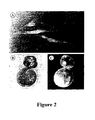
- Figure 2 illustrates embodiments of the invention for creating transgenic cell lines and cloned transgenic embryos. Expression of GFP in fibroblast cells (A) and NT blastocyst (C). The same NT embryo (B) was viewed under light microscope.
-
- The present invention relates to an improved method for mass production of porcine clone embryos by somatic cell nuclear transfer comprising a series of steps of (a) enucleation of recipient oocytes; (b) in vitro culture of nuclei-donating somatic cells; (c) injection of the somatic cells into the enucleated oocytes; (d) electrofusion of each enucleated oocyte with a donor somatic cell; (e) activation of the reconstructed embryos by secondary electric stimulation; (f) in vitro culture of NT embryos and transfer of the NT embryos to recipient pigs.
- The present invention can be described in more detail as follows.
- First, the present invention is characterized by having an 1∼2 h of oocyte reconstruction period following the oocyte enucleation and the fusion of said oocytes with somatic cells by electric stimulation. In general, it is recommended to use mammalian somatic cells as nuclei-donors except those from humans, and more preferably are fibroblast cells or cumulus cells taken from animals such as pigs, cows, sheep and mice. In the present invention, recipient oocytes were collected from prepubertal porcine ovaries, and immature oocytes were incubated to be matured in vitro. Then, only those matured oocytes having first polar bodies were selected and subsequently enucleated to be used. Single donor somatic cells were individually injected into each enucleated oocyte, exposed to primary electric stimulation of 160 V/mm of electric pulse for 30 µsec to induce the fusion between the cytoplasm of each enucleated oocyte and each nucleus of donor somatic cells, respectively. Thus obtained oocytes were then allowed to reconstruct for 1∼2 h by culturing in phosphate buffered saline (PBS) containing fetal bovine serum.
- The above reconstructed oocytes are then subject to secondary electric stimulation to be activated, wherein said oocytes are exposed to 120∼150 V/mm of electric pulse for 30 µsec. If the electric stimulation is below 120 V/mm the activation rate becomes low while if it exceeds 150 V/mm the risk for oocyte injury will be increased. These oocytes are then cultured in vitro for further development. In the conventional nuclear transfer methods, wherein only one electric stimulation is applied to enucleated oocytes, in vitro developmental rate usually results in having less than 5%; in contrast, the present invention by introducing secondary electric stimulation could markedly improve the developmental rate of said oocytes up to 11.6 %, i.e., 130 % increase as compared to those by conventional methods.
- The activation of reconstructed oocytes is executed after the transfer of somatic cell nuclei into recipient cytoplasts. Several combined chemical treatments, such as calcium ionophore/6-DMAP or ethanol/cycloheximide/cytochalasin B, have been used to activate the bovine NT oocytes with somatic cells. However, they showed relatively lower rate of activation when porcine NT oocytes were treated with A23187 alone or with the co-agent of A23187/6-DMAP. In general, porcine oocytes showed lower activation rates when treated with a single chemical agent while the porcine oocytes developed into blastocysts when treated with thimerosal/DTT, a combined chemical agent, although their developmental rates were low. In another report, the activation by sulfhydryl reagents showed a low cleavage rate and thus resulting in a subsequently low developmental rate of porcine NT oocytes that could reach the blastocyst. According to the present invention, porcine NT embryos activated by electric stimulation showed a relatively higher developmental rate than those treated with chemical agents.
- The developmental abilities of porcine NT eggs induced by different level of electric voltages after electrofusion were examined and the result showed that electric pulse (120V/mm DC pulse) is very effective on activation of porcine NT embryos with somatic cells.
- Approximately 70% of reconstructed embryos using pig fibroblasts failed to divide when activated with thimerosal/dithiothreitol treatment (Tao et al., Cloning, 1:55-62 (1999)). In the present invention, the average cleavage rates of porcine NT eggs activated by a single electric stimulus were similar to those of parthenogenetic and in vitro-derived fertilization-derived (IVF) embryos. Thus, this invention demonstrates that a single electric stimulus is sufficient to mediate the development of porcine NT embryos using somatic cells.
Optimal activation time of reconstructed eggs with somatic cells after electrofusion is provided in this invention. In cattle, NT embryos activated at 4 to 8 h after fusion of donor cells with MII cytoplasts showed an improvement in embryonic development (Stice et al., Biol. Reprod., 54:100-110 (1996); Wells et al., Reprod. Fertil. Dev., 10:369-378 (1998)). A prolonged exposure of transferred nuclei to oocyte cytoplasmic factors also facilitated nuclear remodeling and reprogramming (DiBerardino et al., Science, 224:946-952 (1984); Ware et al., Gamete Res., 22:265-275 (1989)). Therefore, it suggests that allowing a lag time for the reconstructed oocytes to stand for a few hours prior to activation will lead to a better facilitation of the nuclear remodeling or reprogramming albeit the mechanism itself has not been clearly understood yet. To determine the conditions for an optimal activation of oocytes, the developmental abilities of porcine NT eggs induced by different activation times after electrofusion were examined. In the present invention, the porcine NT embryos activated 1 to 2 h after fusion showed an improved development as compared to those exposed to either shorter periods (0 and 0.5 h) or longer periods (4 and 6 h). This may be due to the difference in timing of genomic activation among different species. The activation of embryonic genome usually occurs at a 4-cell stage in pigs, whereas it occurs during an 8- to 16-cell stage in sheep and cattle (Kopscny, Reprod. Nutr. Dev., 29:589-600 (1989)). Thus, it is suggested that the activation time of reconstructed porcine oocytes after electrofusion will play a crucial role in producing viable embryos after somatic cell nuclear transfer. Further, it is also suggested that the time required for the remodeling or reprogramming of donor nuclei in recipient cytoplasts after fusion may differ from species to species. - Successful production of porcine cloned embryos from in vitro matured oocytes after nuclear transfer is also disclosed in the present invention. Most cloned animals such as sheep, goats, mice and pigs except cattle have been developed from NT embryos produced by transfer of nuclei of somatic cells into in vivo-derived oocytes after enucleation. In vivo-derived oocytes are obtained by flushing oviducts of animals superovulated by treating with gonadotropin hormones, which differ among species. However, the numbers of in vivo-derived oocytes are generally limited to a certain extent because the procedures are laborious and time-consuming. Alternatively, a lot of oocytes as recipient cytoplasts can be prepared in vitro. Immature oocytes of good quality are obtained from slaughterhouse-derived ovaries, matured in vitro and used as recipient cytoplasts after enucleation. Recently, there was a report that in vivo-derived oocytes could generate clone pigs(Onishi et al., Science, 289:1188-1190 (2000); Polejaeva et al., Nature, 407:505-509 (2000)). However, this invention provides a method for producing porcine NT embryos using in vitro-derived oocytes after somatic cell nuclear transfer.
- It has been suggested that developmental ability of NT embryos with somatic cells may be different depending on cell types in mammals (Kato et al., Science, 282:2095-2098 (1999), Zakhartchenko et al., Mol. Reprod. Dev., 54:264-272 (1999)). Both cumulus and fetal fibroblast cells used as donor nuclei in the present invention supported the development of NT porcine embryos to the pre-implantation stage and this implies that the activation system of the present invention in nuclear transfer can employ various cell types.
- In the present invention, NT embryos with transfected cells had similar developmental competence as compared to those with non-transfected donor cells. Moreover, transgenes were strongly expressed during early stage of development in all the NT embryos with transfected cells and this strengthens the possibility of generating transgenic embryos or transgenic animals from NT embryos with transfected cells after nuclear transfer.
- Prepubertal porcine ovaries were collected from a local slaughterhouse and transported to a laboratory and kept at 25-30°C in 0.9% (w/v) saline supplemented with 75 µg/ml potassium penicillin G and 50 µg/ml streptomycin sulphate until use. Cumulus-oocyte complexes (COC) were obtained from porcine follicles with a diameter of 3 to 6 mm using an 18-gauge needle connected to a 10 ml disposable syringe. The COC were washed 3 times with Tyrode's lactate (TL)-Hepes medium. Approximately 50 COC were cultured in each well of a 4-well multidish with 500 µl of maturation medium, respectively, (Nunc, Roskilde, Denmark) which were kept at 39°C, 5% CO2 in air. After 20 to 22 h of culture, the oocytes were washed 3 times with TL-Hepes medium and further cultured in the maturation medium without supplement of hormones of pregnant mare's serum gonadotropin (PMSG) and human chorionic gonadotropin (hCG) for 22 h. The medium was covered with warm paraffin oil (light mineral oil) and equilibrated at 39°C, 5% CO2 in air for at least 2 h prior to use. The medium used for oocyte maturation was bovine serum albumin (BSA) free North Carolina State University (NCSU) 23 medium supplemented with 10% (v/v) porcine follicular fluid, 0.57 mM cysteine, 25 µg/ml gentamycin, 10 ng/ml epidermal growth factor (EGF), 10 IU/ml PMSG and 10 IU/ml hCG. Porcine follicular fluid was collected from follicles of 3-6 mm in diameter, centrifuged at 1,600 x g for 30 min at 4°C, filtered through 1.2 µm syringe filters and stored in aliquots at -20°C until use.
The basic medium used for IVF was essentially the same as described by Abeydeera and Day (Biol. Reprod., 57:729-734 (1997)). This IVF medium, designated as modified Tris-buffered medium (mTBM), consists of 113.1 mM NaCl, 3 mM KCl, 7.5 mM CaCl2 2H2O, 20 mM Tris (crystallized free base; Fisher Scientific, Fair Lawn, NJ), 11 mM glucose, 5 mM sodium pyruvate and no antibiotics. Fresh porcine semen was kindly supplied from Darby Pig Al Center (Anseong, Korea) once a week and kept at 17°C for 5 days. The above semen was washed 3 times by centrifugation with Dulbecco's phosphate buffered saline (DPBS) supplemented with 1 mg/ml BSA (Fraction V), 100 µg/ml penicillin and 75 µg/ml streptomycin. At the end of the wash, the spermatozoa were resuspended in Tris-buffered medium (pH 7.8). After the completion of IVM, cumulus cells of oocytes were removed by treatment with 0.1% (w/v) hyaluronidase in NCSU 23 medium. The denuded oocytes were washed 3 times with the mTBM supplemented with 2.5 mM caffeine and 4 mg/ml BSA and placed into a 50 µl of mTBM under paraffin oil. Fifty microliter of diluted spermatozoa was added to 50 µl of fertilization medium containing oocytes to give a final sperm concentration of 5 × 10⁵ cells/ml. The oocytes were coincubated with spermatozoa for 6 h at 39°C in an atmosphere of 5% CO2 in air. - To prepare cumulus cells, COC were collected from a single porcine ovarv. After 46 h of culture for IVM, COC were treated in TL-HEPES medium supplemented with 0.1% (w/v) hyaluronidase to disperse cumulus cells. Cumulus cells were precipitated by centrifugation at 700 × g on a microcentrifuge (Vision Scientific Co., Korea) for 5min. The cell pellet was resuspended in Ca2+, Mg2+-free phosphate buffered saline (DPBS(-)), and the cell suspension was kept at room temperature for up to 2 h before use as donor nuclei. Fetal fibroblast cells were isolated from fetuses at day 40 of gestation in Korean native swine (Sus scrofa domesticus L.). Briefly, porcine fetuses were washed 3 times with DPBS(-). The heads of fetuses were removed using iris scissors, and soft tissues from liver and intestine were discarded by scooping out with two watch-maker's forceps. After washing twice with DPBS(-), the carcass was minced with a surgical blade on an 100 mm culture dish. The minced tissues were incubated in 10 ml of 0.25% (w/v) trypsin/3.65 mM EDTA solution at 39°C for 30 min. Trypsin activity was inhibited by adding an equal volume of cell culture medium supplemented with 15% fetal bovine serum (FBS; Gibco, Life Technologies Inc., Grand Island, NY). The cell culture medium was composed of Dulbecco's modified Eagle's medium (DMEM; Gibco), 15% FBS, 1,000 units of penicillin and 1,000 µg/ml of streptomycin (Gibco). After vigorous pipetting, the supernatant was centrifuged at 150 × g for 5 min. The cells were suspended, adjusted to a final concentration of 2 × 10⁶ cells /ml and then cultured in 10 ml of the culture medium in 175 cm2 tissue culture flask (Nunc, Roskilde, Denmark) at 39°C, 5% CO2 in air. The fetal fibroblast cells were passaged 3 times before using as a source of donor nuclei.
- Polybrene/dimethyl sulfoxide (DMSO)-mediated transfection (Kawai and Nishizawa, Mol. Cell Biol., 4:1172-1174 (1984)) was performed as follows: 10 µg of plasmid pLNβ-eGFP DNA in 1 ml medium supplemented with 30 µg/ml polybrene (Aldrich, Milwaukee, WI, USA) was added to Gibbon Ape leukemia virus-based PG13 packing cells plated the previous day (5 X 10³ cells/60 mm dish). After 6 h of incubation at 37°C in an atmosphere of 5% CO2, the DNA-medium mixture was aspirated, and packing cells were shocked by adding 2 ml of medium containing 25% DMSO and then incubated for 1 min. Following 3 washes with the medium, cells were fed with 5 ml of medium and incubated overnight before splitting the transfected cells. Infection of fetal fibroblast target cells was performed following the modified procedure of Miller and Rosman (Biotechniques, 7:980-982 (1989)); 3 ml of virus-containing medium filtered through a 0.22 µm pore-size filter, and polybrene (5 µg/ml of final concentration) was added to target cells which were plated the previous day. Exposure of the target cells to the mixture was allowed only 1 hr. The virus-containing medium was harvested from virus-producing cells, which had been fed with non-selection medium the previous day. Following 1 day of culture after infection, the infected cells were trypsinized and split in non-selection medium. In both transfection and infection, G418 selection medium (600 µg/ml) was added on the following day after splitting. The transfected cells were passaged 14 times before using as a source of nucleus donors.
- After 44 to 46 h of IVM, the oocytes were transferred to 500 µl of TL-Hepes medium supplemented with 0.1% hyaluronidase and free of cumulus cells by vortexing for 3 min. The zona pellucida of oocytes was partially dissected using a fine glass needle as described in Tsunoda et al. (J. Exp. Zool., 240:119-125 (1986)). Oocyte manipulation, including enucleation and cell injection, was performed by using a micromanipulator equipped with an inverted microscope (Leitz, Ernst Leitz Wetzlar GmbH, Germany). The medium used for oocyte manipulation was DPBS(-) containing 7.5 µg/ml cytochalasin B. The first polar body and partial cytoplasm presumptively containing metaphase 11 chromosomes were removed together by using a micropipette with an inner diameter of 20 µm. After the enucleation, oocytes were stained with 5 µM bisbenzimide (Hoechst 33342) for 5 min and observed under an epifluorescent microscope (Olympus, Tokyo, Japan).
- Single fibroblast cells were individually transferred into each enucleated oocyte. Small cells with smooth surface were used as a source of nucleus donors. Reconstructed embryos were equilibrated in a 50 µl drop of cell fusion medium for 10 to 20 sec, and transferred to a fusion chamber with two electrodes 1 mm apart overlaid with cell fusion medium. The cell fusion medium consisted of 0.3 M mannitol, 0.5 mM Hepes, 0.01% BSA, 0.1 mM CaCl2 and 0.1 mM MgCl2. Cell fusion was induced with a single DC pulse of 160 V/mm for 30 µsec using a BTX Electro-cell Manipulator 2001 (BTX, San Diego, CA, USA). After the electrofusion, embryos were activated by a 5 sec pulse of 3 V/mm AC followed by a 30 µsec pulse of 120 V/mm DC.
- Parthenogenetic activation of pig oocytes was carried out as described previously (Wang et al., Mol. Reprod. Dev., 51:346-353 (1998)). Briefly, cumulus-free oocytes were equilibrated for 1 min in 0.3 M mannitol solution supplemented with 0.5 mM Hepes, 0.01% BSA (fatty acid free), 0.01 mM CaCl2 and 0.01 mM MgCl2, and transferred to a chamber consisting of two electrodes 1 mm apart which was overlaid with activation solution. Oocytes were exposed to a single AC pulse of 3 V/mm for 5 µsec followed by a single DC pulse of 180 V/ mm for 30 µsec.
- In all experiments, the activated eggs were cultured in 50 µl drops of NCSU 23 medium supplemented with 4 mg/ml BSA at 39°C, 5% CO2 in air. After 72 h of culture, embryos at cleavage stage were selected. Forty to fifty cleaved embryos were cultured together in 50 µl drops of NCSU23 medium supplemented with 10% FBS at 39°C, 5% CO2 in air for 3 days. After 6 days of culture, blastocyst formation was observed, and cell numbers of blastocysts were counted on a fluorescent microscope following Hoechst 33342 (2.5 µg/ml) staining.
- All blastocysts developed from NT embryos using somatic cells were counted for nuclei after Hoechst staining. Briefly, the embryos were fixed with 1% formaldehyde in PBS for 10 min at room temperature and then placed in a drop of mounting medium on a slide. The mounting medium consisted of 25% (v/v) glycerol in PBS containing 2.5 mg/ml sodium azide and 2.5 µg/ml Hoechst 33342. A cover slip was placed on the top of the embryos and the edge of the slide was sealed with fingernail polish. The nuclei numbers were counted on an epifluorescent microscope (Olympus, Japan).
- This invention is explained in more detail based on the following Examples but they should not be construed as limiting the scope of this invention.
- It was examined whether different activation treatments after electrofusion affect in vitro development of NT porcine embryos to blastocyst stage. At 2 to 3 h after electrofusion, approximately one-third of reconstructed embryos were simultaneously allocated to 3 different activation groups; 1) embryos exposed to a 5 sec pulse at 3 V/mm AC followed by a 30 µsec pulse at 120 V/mm DC, 2) embryos activated by treatment with calcium ionophore (A23187, 5 µM for 5 min), and 3) embryos treated with A23187 (5 µM for 5 min) and then incubated in NCSU23 medium supplemented with 2 mM 6-DMAP for 4 h. After activation treatments, the reconstructed embryos from each group were washed 4 times and cultured in NCSU23 medium containing 4 mg/ml BSA. As shown in Table 1, differences were detected in the cleavage rate (58.1±13.9, 60.7±6.3 and 74.9±7.5%) and the developmental rate to blastocysts (2.2±2.8, 2.2±1.5 and 11.0±4.1%) among the groups (P<0.05). However, no significant difference in the mean cell number (20.5±3.5, 26.6±4.5 and 26.3±10.3) of blastocysts was observed. The results demonstrate that electric pulse could induce a higher in vitro development of NT embryos than treatments with A23187 alone or A23187/6-DMAP.
Effect of different activation treatments on in vitro development of NT porcine embryos with cumulus cells Treatment No. of eggs examined % of eggs cleaved % of blastocysts Cell number
(Mean±SD)A23187 128 58.1±13.9 (71) 2.2±2.8 (2) 20.5±3.5 A23187+6-DMAP 121 60.7±6.3 (75) 2.2±1.5 (3) 26.6±4.5 Electric pulse 129 74.9±7.5 (95) 11.0±4.1 (13) 26.3±10.3 - The effect of activation voltage on in vitro development of NT embryos to the blastocyst stage was examined. At 2-3 h after electrofusion, the reconstructed embryos were activated by a pulse of 3 V/mm AC for 5 sec followed by a pulse of 120 or 150 V/mm DC for 30 µsec. There was no significant difference in the cleavage rate between the two groups. The rate of blastocyst development (11.6±1.6%, 14/121) of NT embryos in field strength of 120 V/mm was significantly high as compared to that of 150 V/mm group (6.5±2.3%, 8/121) (P<0.05). However, mean cell numbers of blastocysts were similar between the two groups (29.5±11.3 and 27.6±10.5, respectively) (P>0.05). For subsequent experiments, the activation was performed with a single 120 V/mm DC pulse (Table 2). The experiment was carried out to determine the optimal activation timing after electrofusion (Table 3). It was also investigated if the timing of activation after electrofusion affects the developmental ability of NT embryos. Approximately one-third of fused eggs were activated by a pulse of 3 V/mm AC for 5 sec followed by a single pulse of 120 V/mm DC for 30 µsec on different time (2, 4 or 6 h) after electrofusion. The cleavage rate of NT embryos in 2 h and 4 h groups was higher than that of 6 h group. A higher proportion of NT embryos (11.6±2.9%, 20/187) activated 2 h after electrofusion developed to the blastocyst stage, as compared to those of 4 h (6.6±2.3%, 11/190) and 6 h (8.1±3.3%, 14/184) groups. Mean cell numbers of NT embryos in 2, 4 and 6 h groups were 30.4±10.4, 24.6±10.1 and 16.5±7.4, respectively. A significant difference was observed between 2 h and 6 h groups (P<(1.05). When reconstructed embryos were activated at 0, 30 min and 1 h after electrofusion, in subsequent experiments, developmental rates of the embryos blastocyst stage were 2.1% (2/97), 3.2% (2/85) and 10.4% (11/106), respectively. These results suggest that activation timing after electrofusion can affect developmental potential of porcine NT eggs.
Effect of activation voltage on in vitro development of porcine NT embryos with somatic cells Group
(V/mm DC)No. of eggs examined of eggs cleaved of blastocysts No. of Nuclei
(Mean±SD)120 121 71.4±10.9 (86) 11.6±1.6 (14) 29.5±11.3 150 121 63.7±8.1 (78) 6.5±2.3 (8) 27.6±10.5 In vitro development of porcine NT embryos activated at different times after electrofusion Activation
timeNo. of eggs examined % of eggs cleaved % of blastocysts No. of Nuclei
(Mean±5D)2 h 187 71.8±13.2 (140) 21.6±2.9 (20) 30.4±10.4 4 h 190 76.5±6.8 (140) 6.6±23 (11) 24.6±10.1 6 h 184 59.4±4.9 (110) 8.1±3.3 (14) 16.5±7.4 - The developmental competence of NT embryos with fetal porcine fibroblast cells was compared to that of parthenogenetic and IVF-derived porcine embryos. And also, developmental potential of NT porcine embryos with cumulus or fibroblast cells was compared to that of IVF-derived embryos.
- NT embryos using fibroblast cells showed a lower developmental rate (9.4±(0.9%, 20/289) to the blastocyst stage than IVF-derived (21.4±1.9%, 43/201) or parthenogenetic embryos (22.4±7.2%, 43/189) (P<0.01), although there was no significant difference in the cleavage rate of the embryos among experimental groups (Table 4). After 6 days of culture, most blastocysts (16/20) from NT embryo were hatched (Figure 1), although the mean number of nuclei was smaller than that of IVF-derived blastocysts (Figure 2). The reason for the early hatching of NT blastocysts seems to be due to the partial dissection of zona pellucida during the enucleation process. The mean cell number of NT embryos (28.9±11.5, ranging from 14 to 61, n=18) was smaller than that of IVF-derived blastocysts (36.2±9.7, ranging from 18 to 67, n=22) (P<0.05), while it was similar to that of parthenogenetic embryos (29.9±12.1, ranging from 13 to 50, n=24) (Table 4). These results represent that the porcine NT embryos using fibroblast cells could develop to blastocysts in vitro, although they had a lower developmental competence when compared to that of IVF-derived embryos.
- Developmental potential of porcine oocytes, where nuclear transfer was performed using cumulus (NT†) or/and fibroblast cells (NT‡ ), was compared to that of IVF-derived embryos (Table 5). Fusion rates of enucleated oocytes reconstructed with cumulus or fibroblast cells after electrofusion were 67% (257/374) or 69% (273/396), respectively. Thus, there was no distinct difference in the cleavage rate of NT embryos between 2 donor cell types. NT embryos from both cell types, however, showed a lower developmental rate to the blastocyst stage than that of IVF-derived embryos (P<0.01). Mean nuclei numbers of IVF-derived, NT† and NT‡ blastocysts were 38.6±10.4 (ranging from 23 to 65), 28.9±11.4 (ranging from 17 to 51) and 30.2±9.9 (ranging from 18 to 51), respectively. Thus, NT embryos were comprised of number of nuclei smaller than those of IVF-derived embryos (P<0.05).
Comparison of in vitro development of NT porcine embryos to IVF-derived or parthenogenetic embryos Group No. of eggs examined % of eggs cleaved % of blastocysts Nuclei no.
(Mean±SD)IVF 201 74.5±1.7 (150) 21.4±1.9 (43) 36.2±9.7 (22) Parthenotes 189 71.0±4.9 (133) 22.4±7.2 (43) 29.9±12.1 (24) NT 212 67.0±7.6 (141) 9.4±0.9 (20) 28.9±11.5 (18) Comparison of in vitro development of IVF or NT porcine embryos with cumulus cells Group No. of eggs examined % of eggs cleaved % of blastocysts Cell number
(Mean±SD)IVF 270 78.0±3.6 (210) 22.9±3.5 (62) 38.6±10.4 NT 257 70.6±9.9 (188) 9.9±2.4 (26) 28.9±11.4 NT 273 73.8±5.3 (202) 9.8±1.6 (27) 30.2±9.9 - It was examined whether porcine NT embryos with transfected cells develop to blastocysts and express the transgenes during early development. Fibroblasts expressing GFP gene were transferred into enucleated oocytes and the reconstructed oocytes were fused by a single DC pulse of 160 V/mm for 30 µsec. The fused embryos were activated and then cultured as described in EXAMPLE 2. GFP expression in NT embryos was observed under an epifluorescence microscopy (Olympus). Developmental potential of porcine NT oocytes with transfected cells was compared with that of non-transfected cells.
- It was also examined whether transfected cells have in vitro developmental potential following nuclear transfer in the pig. Porcine fibroblast cells were introduced with exogenous β-actin promoter/GFP gene by retroviral infection, and surviving colonies were obtained after treatments of G418 for approximately 2 weeks. GFP expression in transfected fibroblast cells was confirmed by epifluorescence microscopy (Figure 1A). Fusion rate (67%, 195/292) of NT oocytes with transfected fibroblast cells was similar to that (71 %, 197/277) of non-transfected cells. Developmental rates of NT embryos to blastocvsts were also similar between transfected and non-transfected cells (Table 6). All NT embryos derived from transfected cells showed strong GFP expression as represented in Figure 1C. The results indicate that porcine NT embryos with transfected cells have a similar developmental potential like those with non-transfected cells. Thus, our invention methods for activating NT embryos are applicable to generate transgenic NT embryos from transfected cells.
In vitro development of porcine NT embryos with transfected cells Group No. of eggs fused % of eggs cleaved % of blastocysts Expression rate (%) Normal 197 73.0±4.2 (144) 10.2±0.6 (20) - Transfected 195 75.7±:1.8 (148) 9.7±0.5 (19) 100 - The number of embryos observed is indicated in parentheses.
Claims (6)
- A method for mass production of porcine clone embryos by nuclear transfer comprising:(a) a process of reconstructing enucleated porcine oocytes which are fused with porcine donor somatic cells or porcine donor somatic cell nuclei injected into said enucleated oocytes by electric stimulation; wherein the period of reconstruction is for 1-2 hours;(b) a process of activating said reconstructed porcine oocytes by secondary electric stimulation by applying a single electric pulse of 120-150V/mm DV.
- The method for mass production of porcine clone embryos by nuclear transfer according to claim 1, wherein said porcine donor somatic cell or said porcine donor somatic cell nucleus injected into said enucleated oocyte is a transfected somatic cell or a transfected somatic cell nucleus obtained from a group consisting of fibroblast cells and cumulus cells of mammals excluding human.
- The method for mass production of porcine clone embryos by nuclear transfer according to claim 1, wherein said porcine oocytes as recipient cytoplasts are matured in vitro prior to enucleation.
- The method for mass production of porcine clone embryos by nuclear transfer according to claims 1∼2, wherein said donor cells or donor cell nuclei are obtained from fetuses or adult tissues.
- The method for mass production of porcine clone embryos by nuclear transfer according to claim 1, wherein said secondary electric stimulation is to apply a single electric pulse of 120∼150 V/mm DC for 30 µsec.
- The method for mass production of porcine clone embryos by nuclear transfer according to claims 1∼2 and 4, wherein said donor somatic cells or donor somatic cell nuclei in pigs are obtained from a swine indigenous to Korea (Sus scrofn domesticus L.).
Applications Claiming Priority (3)
| Application Number | Priority Date | Filing Date | Title |
|---|---|---|---|
| KR10-1999-0051551A KR100417566B1 (en) | 1999-11-19 | 1999-11-19 | A mass production method of embryos by nuclear transfer using somatic cells |
| KR9951551 | 1999-11-19 | ||
| PCT/KR2000/001312 WO2001035734A1 (en) | 1999-11-19 | 2000-11-16 | An improved method for production of porcine clone embryos via somatic cell nuclear transfer |
Publications (2)
| Publication Number | Publication Date |
|---|---|
| EP1229784A1 EP1229784A1 (en) | 2002-08-14 |
| EP1229784B1 true EP1229784B1 (en) | 2005-02-02 |
Family
ID=19620867
Family Applications (1)
| Application Number | Title | Priority Date | Filing Date |
|---|---|---|---|
| EP00981846A Expired - Lifetime EP1229784B1 (en) | 1999-11-19 | 2000-11-16 | An improved method for production of porcine clone embryos via somatic cell nuclear transfer |
Country Status (7)
| Country | Link |
|---|---|
| EP (1) | EP1229784B1 (en) |
| JP (1) | JP2003513673A (en) |
| KR (1) | KR100417566B1 (en) |
| CN (1) | CN1407851A (en) |
| AU (1) | AU769618B2 (en) |
| DE (1) | DE60017931D1 (en) |
| WO (1) | WO2001035734A1 (en) |
Families Citing this family (13)
| Publication number | Priority date | Publication date | Assignee | Title |
|---|---|---|---|---|
| CA2480802A1 (en) * | 2002-04-01 | 2003-10-16 | Gtc Biotherapeutics, Inc. | A method for selecting cell lines to be used for nuclear transfer in mammalian species |
| KR100479638B1 (en) * | 2002-10-21 | 2005-03-31 | 주식회사 마크로젠 | Preparation method of donor cell for nuclear transfer using electrophoresis and membrane antigen marker and production method of clone animal |
| JP2004275074A (en) * | 2003-03-14 | 2004-10-07 | Nipro Corp | Methods for screening normality in animal embryos |
| KR101141733B1 (en) * | 2003-12-09 | 2012-05-03 | 재단법인서울대학교산학협력재단 | Transgenic cloned cow carrying a knock-out prion-encoding gene and method for producing the same |
| KR100715366B1 (en) * | 2004-12-27 | 2007-05-07 | 김창현 | Cell culture method |
| WO2008106982A2 (en) | 2007-03-07 | 2008-09-12 | Aarhus Universitet | Pig model for atherosclerosis |
| CN102943093B (en) * | 2012-11-20 | 2014-02-12 | 广东温氏食品集团股份有限公司 | Method for improving transferring efficiencies of pig somatic cell nucleuses |
| CN103409467B (en) * | 2013-07-04 | 2015-01-21 | 西南民族大学 | Method for acquiring heterogeneity interspecies-cloned yak premium embryos |
| CN103343142B (en) * | 2013-07-15 | 2017-07-25 | 云南农业大学 | A method for producing parthenogenetic cloned pigs using continuous cloning technology |
| EP3229586B1 (en) | 2014-12-10 | 2025-07-30 | Regents of the University of Minnesota | Genetically modified cells, tissues, and organs for treating disease |
| US20190335726A1 (en) * | 2016-06-14 | 2019-11-07 | Regents Of The University Of Minnesota | Genetically modified cells, tissues, and organs for treating disease |
| CN115612660A (en) * | 2022-10-11 | 2023-01-17 | 广西壮族自治区畜牧研究所 | A method for improving the in vitro developmental ability of pig nuclear transfer embryos |
| CN116411022B (en) * | 2022-11-21 | 2024-06-21 | 华中农业大学 | A vector and cell for indicating the degree of genome activation of pig somatic cell cloned embryos |
Family Cites Families (5)
| Publication number | Priority date | Publication date | Assignee | Title |
|---|---|---|---|---|
| GB9517780D0 (en) * | 1995-08-31 | 1995-11-01 | Roslin Inst Edinburgh | Biological manipulation |
| GB9517779D0 (en) * | 1995-08-31 | 1995-11-01 | Roslin Inst Edinburgh | Biological manipulation |
| US6235969B1 (en) * | 1997-01-10 | 2001-05-22 | University Of Massachusetts | Cloning pigs using donor nuclei from non-quiescent differentiated cells |
| US5945577A (en) * | 1997-01-10 | 1999-08-31 | University Of Massachusetts As Represented By Its Amherst Campus | Cloning using donor nuclei from proliferating somatic cells |
| US6331659B1 (en) * | 1998-01-21 | 2001-12-18 | University Of Hawaii | Cumulus cells as nuclear donors |
-
1999
- 1999-11-19 KR KR10-1999-0051551A patent/KR100417566B1/en not_active Expired - Fee Related
-
2000
- 2000-11-16 AU AU18961/01A patent/AU769618B2/en not_active Ceased
- 2000-11-16 WO PCT/KR2000/001312 patent/WO2001035734A1/en not_active Ceased
- 2000-11-16 JP JP2001537540A patent/JP2003513673A/en active Pending
- 2000-11-16 DE DE60017931T patent/DE60017931D1/en not_active Expired - Lifetime
- 2000-11-16 EP EP00981846A patent/EP1229784B1/en not_active Expired - Lifetime
- 2000-11-16 CN CN00816831A patent/CN1407851A/en active Pending
Also Published As
| Publication number | Publication date |
|---|---|
| KR100417566B1 (en) | 2004-02-05 |
| JP2003513673A (en) | 2003-04-15 |
| KR20010047362A (en) | 2001-06-15 |
| DE60017931D1 (en) | 2005-03-10 |
| AU769618B2 (en) | 2004-01-29 |
| EP1229784A1 (en) | 2002-08-14 |
| AU1896101A (en) | 2001-05-30 |
| CN1407851A (en) | 2003-04-02 |
| WO2001035734A1 (en) | 2001-05-25 |
Similar Documents
| Publication | Publication Date | Title |
|---|---|---|
| US6235969B1 (en) | Cloning pigs using donor nuclei from non-quiescent differentiated cells | |
| US6215041B1 (en) | Cloning using donor nuclei from a non-quiesecent somatic cells | |
| CA2277192C (en) | Nuclear transfer with differentiated fetal and adult donor cells | |
| WO1999005266A2 (en) | Trans-species nuclear transfer | |
| AU769618B2 (en) | An improved method for production of porcine clone embryos via somatic cell nuclear transfer | |
| WO1999001163A1 (en) | Cloning using donor nuclei from non-serum starved, differentiated cells | |
| Koo et al. | Developmental potential and transgene expression of porcine nuclear transfer embryos using somatic cells | |
| Peura et al. | A comparison of established and new approaches in ovine and bovine nuclear transfer | |
| Miyoshi et al. | Birth of cloned miniature pigs derived from somatic cell nuclear transferred embryos activated by ultrasound treatment | |
| CA2381124A1 (en) | Cloning pigs using donor cells or nuclei from differentiated cells and production of pluripotent porcine | |
| US7291764B1 (en) | Cloning pigs using non-quiescent differentiated donor cells or nuclei | |
| US20060277616A1 (en) | Method for cloning of the rat by nuclear transfer | |
| US20040077077A1 (en) | Novel methods for the production of cloned mammals, mammals cloned according to the methods, and methods of use of same | |
| WO2005005624A1 (en) | Method of enucleation and oocyte activation in somatic cell nuclear transfer in primates | |
| Gręda et al. | Developmental potential of selectively enucleated mouse zygotes reconstituted with embryonic cell, embryonic stem cell and somatic cell nuclei. | |
| JP2005525098A (en) | Rabbit nuclear cloning method and its usage | |
| Kim et al. | Optimization of procedure for efficient gene transfer into porcine somatic cells with lipofection | |
| JP2005508185A (en) | Xenogeneic somatic cell nuclear transfer using rabbit oocytes | |
| Reggio | LSU Scholarly Repositor y | |
| Reggio | Production of transgenic goats by somatic cell nuclear transfer | |
| Lagutina et al. | Age-Related Effect of Cells as Donors of Nuclei: On the Efficiency of the Development of Cloned Rabbit Embryos | |
| AU2005225075A1 (en) | Cloning pigs using donor cells or nuclei from differentiated cells and production of pluripotent porcine | |
| MXPA00000201A (en) | Cloning using donor nuclei from non-serum starved, differentiated cells | |
| AU2006202185A1 (en) | Cloning using donor nuclei from non-serum starved, differentiated cells |
Legal Events
| Date | Code | Title | Description |
|---|---|---|---|
| PUAI | Public reference made under article 153(3) epc to a published international application that has entered the european phase |
Free format text: ORIGINAL CODE: 0009012 |
|
| 17P | Request for examination filed |
Effective date: 20020517 |
|
| AK | Designated contracting states |
Kind code of ref document: A1 Designated state(s): AT BE CH CY DE DK ES FI FR GB GR IE IT LI LU MC NL PT SE TR |
|
| RIN1 | Information on inventor provided before grant (corrected) |
Inventor name: HAN, YONG-MAHN Inventor name: KANG, YONG-KOOK Inventor name: KOO, DEOG-BON Inventor name: CHOI, YOUNG-HEE Inventor name: PARK, JUNG-SUN Inventor name: LEE, KYUNG-KWANG Inventor name: YU, DAE-YEUL |
|
| 17Q | First examination report despatched |
Effective date: 20030605 |
|
| RBV | Designated contracting states (corrected) |
Designated state(s): AT BE CH DE FR GB LI |
|
| GRAP | Despatch of communication of intention to grant a patent |
Free format text: ORIGINAL CODE: EPIDOSNIGR1 |
|
| RBV | Designated contracting states (corrected) |
Designated state(s): DE FR GB |
|
| RAP1 | Party data changed (applicant data changed or rights of an application transferred) |
Owner name: KOREA RESEARCH INSTITUTE OF BIOSCIENCE AND BIOTECH |
|
| GRAS | Grant fee paid |
Free format text: ORIGINAL CODE: EPIDOSNIGR3 |
|
| GRAA | (expected) grant |
Free format text: ORIGINAL CODE: 0009210 |
|
| AK | Designated contracting states |
Kind code of ref document: B1 Designated state(s): DE FR GB |
|
| PG25 | Lapsed in a contracting state [announced via postgrant information from national office to epo] |
Ref country code: FR Free format text: LAPSE BECAUSE OF NON-PAYMENT OF DUE FEES Effective date: 20050202 |
|
| REG | Reference to a national code |
Ref country code: GB Ref legal event code: FG4D |
|
| REG | Reference to a national code |
Ref country code: IE Ref legal event code: FG4D |
|
| REF | Corresponds to: |
Ref document number: 60017931 Country of ref document: DE Date of ref document: 20050310 Kind code of ref document: P |
|
| PG25 | Lapsed in a contracting state [announced via postgrant information from national office to epo] |
Ref country code: DE Free format text: LAPSE BECAUSE OF FAILURE TO SUBMIT A TRANSLATION OF THE DESCRIPTION OR TO PAY THE FEE WITHIN THE PRESCRIBED TIME-LIMIT Effective date: 20050503 |
|
| PLBE | No opposition filed within time limit |
Free format text: ORIGINAL CODE: 0009261 |
|
| STAA | Information on the status of an ep patent application or granted ep patent |
Free format text: STATUS: NO OPPOSITION FILED WITHIN TIME LIMIT |
|
| 26N | No opposition filed |
Effective date: 20051103 |
|
| EN | Fr: translation not filed | ||
| PGFP | Annual fee paid to national office [announced via postgrant information from national office to epo] |
Ref country code: GB Payment date: 20081112 Year of fee payment: 9 |
|
| GBPC | Gb: european patent ceased through non-payment of renewal fee |
Effective date: 20091116 |
|
| PG25 | Lapsed in a contracting state [announced via postgrant information from national office to epo] |
Ref country code: GB Free format text: LAPSE BECAUSE OF NON-PAYMENT OF DUE FEES Effective date: 20091116 |