EP1224264B1 - Use of allogeneic cell lines to load antigen-presenting cells to elicit immune responses - Google Patents
Use of allogeneic cell lines to load antigen-presenting cells to elicit immune responses Download PDFInfo
- Publication number
- EP1224264B1 EP1224264B1 EP00970961A EP00970961A EP1224264B1 EP 1224264 B1 EP1224264 B1 EP 1224264B1 EP 00970961 A EP00970961 A EP 00970961A EP 00970961 A EP00970961 A EP 00970961A EP 1224264 B1 EP1224264 B1 EP 1224264B1
- Authority
- EP
- European Patent Office
- Prior art keywords
- cells
- tumor
- cytotoxic
- loaded
- allogeneic
- Prior art date
- Legal status (The legal status is an assumption and is not a legal conclusion. Google has not performed a legal analysis and makes no representation as to the accuracy of the status listed.)
- Expired - Lifetime
Links
- 210000004027 cell Anatomy 0.000 title claims abstract description 322
- 230000000735 allogeneic effect Effects 0.000 title claims abstract description 68
- 230000028993 immune response Effects 0.000 title claims abstract description 30
- 210000000612 antigen-presenting cell Anatomy 0.000 title abstract description 25
- 210000004443 dendritic cell Anatomy 0.000 claims abstract description 312
- 210000001744 T-lymphocyte Anatomy 0.000 claims abstract description 192
- 239000000427 antigen Substances 0.000 claims abstract description 109
- 108091007433 antigens Proteins 0.000 claims abstract description 109
- 102000036639 antigens Human genes 0.000 claims abstract description 108
- 210000001151 cytotoxic T lymphocyte Anatomy 0.000 claims abstract description 80
- 210000004881 tumor cell Anatomy 0.000 claims description 109
- 206010028980 Neoplasm Diseases 0.000 claims description 86
- 230000001472 cytotoxic effect Effects 0.000 claims description 49
- 241000282414 Homo sapiens Species 0.000 claims description 31
- 231100000433 cytotoxic Toxicity 0.000 claims description 31
- 230000001640 apoptogenic effect Effects 0.000 claims description 16
- 238000000338 in vitro Methods 0.000 claims description 13
- 230000001939 inductive effect Effects 0.000 claims description 9
- 230000034994 death Effects 0.000 claims description 8
- 230000037455 tumor specific immune response Effects 0.000 claims description 8
- 230000030833 cell death Effects 0.000 claims description 7
- 239000002243 precursor Substances 0.000 claims description 6
- 210000000822 natural killer cell Anatomy 0.000 claims description 4
- 210000000581 natural killer T-cell Anatomy 0.000 claims description 3
- 238000000099 in vitro assay Methods 0.000 claims description 2
- 238000012544 monitoring process Methods 0.000 claims description 2
- 239000008194 pharmaceutical composition Substances 0.000 claims 2
- 230000008030 elimination Effects 0.000 abstract description 13
- 238000003379 elimination reaction Methods 0.000 abstract description 13
- 208000009329 Graft vs Host Disease Diseases 0.000 abstract description 4
- 206010052779 Transplant rejections Diseases 0.000 abstract description 4
- 208000024908 graft versus host disease Diseases 0.000 abstract description 4
- 238000012258 culturing Methods 0.000 abstract description 3
- 201000001441 melanoma Diseases 0.000 description 117
- 108090000765 processed proteins & peptides Proteins 0.000 description 62
- 102000004196 processed proteins & peptides Human genes 0.000 description 40
- 238000000034 method Methods 0.000 description 35
- 206010060862 Prostate cancer Diseases 0.000 description 32
- 206010006187 Breast cancer Diseases 0.000 description 31
- 208000026310 Breast neoplasm Diseases 0.000 description 31
- 102000007066 Prostate-Specific Antigen Human genes 0.000 description 28
- 108010072866 Prostate-Specific Antigen Proteins 0.000 description 28
- 230000035755 proliferation Effects 0.000 description 27
- 201000001514 prostate carcinoma Diseases 0.000 description 26
- 210000005056 cell body Anatomy 0.000 description 20
- 230000035800 maturation Effects 0.000 description 20
- 210000001266 CD8-positive T-lymphocyte Anatomy 0.000 description 19
- 210000001616 monocyte Anatomy 0.000 description 19
- 238000003556 assay Methods 0.000 description 18
- 230000009089 cytolysis Effects 0.000 description 18
- 108010029697 CD40 Ligand Proteins 0.000 description 15
- 102100032937 CD40 ligand Human genes 0.000 description 15
- 230000006698 induction Effects 0.000 description 15
- IQFYYKKMVGJFEH-XLPZGREQSA-N Thymidine Chemical compound O=C1NC(=O)C(C)=CN1[C@@H]1O[C@H](CO)[C@@H](O)C1 IQFYYKKMVGJFEH-XLPZGREQSA-N 0.000 description 14
- 102000025850 HLA-A2 Antigen Human genes 0.000 description 13
- 108010074032 HLA-A2 Antigen Proteins 0.000 description 13
- 230000006052 T cell proliferation Effects 0.000 description 13
- 230000000638 stimulation Effects 0.000 description 13
- 108010002350 Interleukin-2 Proteins 0.000 description 12
- 239000012636 effector Substances 0.000 description 12
- 238000002474 experimental method Methods 0.000 description 12
- 238000000684 flow cytometry Methods 0.000 description 12
- 238000011068 loading method Methods 0.000 description 12
- VYZAMTAEIAYCRO-UHFFFAOYSA-N Chromium Chemical compound [Cr] VYZAMTAEIAYCRO-UHFFFAOYSA-N 0.000 description 11
- 239000011651 chromium Substances 0.000 description 11
- 229910052804 chromium Inorganic materials 0.000 description 11
- 230000003053 immunization Effects 0.000 description 11
- 102100037850 Interferon gamma Human genes 0.000 description 10
- 108010074328 Interferon-gamma Proteins 0.000 description 10
- 230000000694 effects Effects 0.000 description 10
- 230000004044 response Effects 0.000 description 10
- ZAHQPTJLOCWVPG-UHFFFAOYSA-N mitoxantrone dihydrochloride Chemical compound Cl.Cl.O=C1C2=C(O)C=CC(O)=C2C(=O)C2=C1C(NCCNCCO)=CC=C2NCCNCCO ZAHQPTJLOCWVPG-UHFFFAOYSA-N 0.000 description 9
- 238000010186 staining Methods 0.000 description 9
- QGJZLNKBHJESQX-UHFFFAOYSA-N 3-Epi-Betulin-Saeure Natural products C1CC(O)C(C)(C)C2CCC3(C)C4(C)CCC5(C(O)=O)CCC(C(=C)C)C5C4CCC3C21C QGJZLNKBHJESQX-UHFFFAOYSA-N 0.000 description 8
- CLOUCVRNYSHRCF-UHFFFAOYSA-N 3beta-Hydroxy-20(29)-Lupen-3,27-oic acid Natural products C1CC(O)C(C)(C)C2CCC3(C)C4(C(O)=O)CCC5(C)CCC(C(=C)C)C5C4CCC3C21C CLOUCVRNYSHRCF-UHFFFAOYSA-N 0.000 description 8
- DIZWSDNSTNAYHK-XGWVBXMLSA-N Betulinic acid Natural products CC(=C)[C@@H]1C[C@H]([C@H]2CC[C@]3(C)[C@H](CC[C@@H]4[C@@]5(C)CC[C@H](O)C(C)(C)[C@@H]5CC[C@@]34C)[C@@H]12)C(=O)O DIZWSDNSTNAYHK-XGWVBXMLSA-N 0.000 description 8
- 102000043129 MHC class I family Human genes 0.000 description 8
- 108091054437 MHC class I family Proteins 0.000 description 8
- QGJZLNKBHJESQX-FZFNOLFKSA-N betulinic acid Chemical compound C1C[C@H](O)C(C)(C)[C@@H]2CC[C@@]3(C)[C@]4(C)CC[C@@]5(C(O)=O)CC[C@@H](C(=C)C)[C@@H]5[C@H]4CC[C@@H]3[C@]21C QGJZLNKBHJESQX-FZFNOLFKSA-N 0.000 description 8
- PZXJOHSZQAEJFE-UHFFFAOYSA-N dihydrobetulinic acid Natural products C1CC(O)C(C)(C)C2CCC3(C)C4(C)CCC5(C(O)=O)CCC(C(C)C)C5C4CCC3C21C PZXJOHSZQAEJFE-UHFFFAOYSA-N 0.000 description 8
- 230000006870 function Effects 0.000 description 8
- 238000002649 immunization Methods 0.000 description 8
- MQYXUWHLBZFQQO-UHFFFAOYSA-N nepehinol Natural products C1CC(O)C(C)(C)C2CCC3(C)C4(C)CCC5(C)CCC(C(=C)C)C5C4CCC3C21C MQYXUWHLBZFQQO-UHFFFAOYSA-N 0.000 description 8
- IQFYYKKMVGJFEH-OFKYTIFKSA-N 1-[(2r,4s,5r)-4-hydroxy-5-(tritiooxymethyl)oxolan-2-yl]-5-methylpyrimidine-2,4-dione Chemical compound C1[C@H](O)[C@@H](CO[3H])O[C@H]1N1C(=O)NC(=O)C(C)=C1 IQFYYKKMVGJFEH-OFKYTIFKSA-N 0.000 description 7
- DWRXFEITVBNRMK-UHFFFAOYSA-N Beta-D-1-Arabinofuranosylthymine Natural products O=C1NC(=O)C(C)=CN1C1C(O)C(O)C(CO)O1 DWRXFEITVBNRMK-UHFFFAOYSA-N 0.000 description 7
- 102100027207 CD27 antigen Human genes 0.000 description 7
- 101000914511 Homo sapiens CD27 antigen Proteins 0.000 description 7
- 230000004913 activation Effects 0.000 description 7
- IQFYYKKMVGJFEH-UHFFFAOYSA-N beta-L-thymidine Natural products O=C1NC(=O)C(C)=CN1C1OC(CO)C(O)C1 IQFYYKKMVGJFEH-UHFFFAOYSA-N 0.000 description 7
- 201000011510 cancer Diseases 0.000 description 7
- LOKCTEFSRHRXRJ-UHFFFAOYSA-I dipotassium trisodium dihydrogen phosphate hydrogen phosphate dichloride Chemical compound P(=O)(O)(O)[O-].[K+].P(=O)(O)([O-])[O-].[Na+].[Na+].[Cl-].[K+].[Cl-].[Na+] LOKCTEFSRHRXRJ-UHFFFAOYSA-I 0.000 description 7
- 230000002147 killing effect Effects 0.000 description 7
- 239000002953 phosphate buffered saline Substances 0.000 description 7
- 229940104230 thymidine Drugs 0.000 description 7
- 238000011282 treatment Methods 0.000 description 7
- 108010017213 Granulocyte-Macrophage Colony-Stimulating Factor Proteins 0.000 description 6
- 102100039620 Granulocyte-macrophage colony-stimulating factor Human genes 0.000 description 6
- 208000000236 Prostatic Neoplasms Diseases 0.000 description 6
- 230000001464 adherent effect Effects 0.000 description 6
- 230000000961 alloantigen Effects 0.000 description 6
- 238000001516 cell proliferation assay Methods 0.000 description 6
- 230000036039 immunity Effects 0.000 description 6
- 210000005105 peripheral blood lymphocyte Anatomy 0.000 description 6
- 230000008569 process Effects 0.000 description 6
- 210000002966 serum Anatomy 0.000 description 6
- YXHLJMWYDTXDHS-IRFLANFNSA-N 7-aminoactinomycin D Chemical compound C[C@H]1OC(=O)[C@H](C(C)C)N(C)C(=O)CN(C)C(=O)[C@@H]2CCCN2C(=O)[C@@H](C(C)C)NC(=O)[C@H]1NC(=O)C1=C(N)C(=O)C(C)=C2OC(C(C)=C(N)C=C3C(=O)N[C@@H]4C(=O)N[C@@H](C(N5CCC[C@H]5C(=O)N(C)CC(=O)N(C)[C@@H](C(C)C)C(=O)O[C@@H]4C)=O)C(C)C)=C3N=C21 YXHLJMWYDTXDHS-IRFLANFNSA-N 0.000 description 5
- 108700012813 7-aminoactinomycin D Proteins 0.000 description 5
- 108091003079 Bovine Serum Albumin Proteins 0.000 description 5
- 102000004127 Cytokines Human genes 0.000 description 5
- 108090000695 Cytokines Proteins 0.000 description 5
- 101000578784 Homo sapiens Melanoma antigen recognized by T-cells 1 Proteins 0.000 description 5
- 108010002586 Interleukin-7 Proteins 0.000 description 5
- 108091054438 MHC class II family Proteins 0.000 description 5
- 102100022430 Melanocyte protein PMEL Human genes 0.000 description 5
- 102100028389 Melanoma antigen recognized by T-cells 1 Human genes 0.000 description 5
- 101800001271 Surface protein Proteins 0.000 description 5
- 230000014102 antigen processing and presentation of exogenous peptide antigen via MHC class I Effects 0.000 description 5
- 230000006907 apoptotic process Effects 0.000 description 5
- 239000001963 growth medium Substances 0.000 description 5
- 238000001727 in vivo Methods 0.000 description 5
- 238000010348 incorporation Methods 0.000 description 5
- 230000001965 increasing effect Effects 0.000 description 5
- 230000001404 mediated effect Effects 0.000 description 5
- 238000002054 transplantation Methods 0.000 description 5
- 102100031573 Hematopoietic progenitor cell antigen CD34 Human genes 0.000 description 4
- 101000777663 Homo sapiens Hematopoietic progenitor cell antigen CD34 Proteins 0.000 description 4
- 206010025323 Lymphomas Diseases 0.000 description 4
- 230000005867 T cell response Effects 0.000 description 4
- 102100040247 Tumor necrosis factor Human genes 0.000 description 4
- 102000003425 Tyrosinase Human genes 0.000 description 4
- 108060008724 Tyrosinase Proteins 0.000 description 4
- 230000006023 anti-tumor response Effects 0.000 description 4
- 210000004369 blood Anatomy 0.000 description 4
- 239000008280 blood Substances 0.000 description 4
- 230000003013 cytotoxicity Effects 0.000 description 4
- 231100000135 cytotoxicity Toxicity 0.000 description 4
- 230000004069 differentiation Effects 0.000 description 4
- 201000010099 disease Diseases 0.000 description 4
- 208000037265 diseases, disorders, signs and symptoms Diseases 0.000 description 4
- 238000001943 fluorescence-activated cell sorting Methods 0.000 description 4
- 238000009169 immunotherapy Methods 0.000 description 4
- 238000007747 plating Methods 0.000 description 4
- 230000001681 protective effect Effects 0.000 description 4
- 230000002269 spontaneous effect Effects 0.000 description 4
- 230000001225 therapeutic effect Effects 0.000 description 4
- 239000013598 vector Substances 0.000 description 4
- 108090000672 Annexin A5 Proteins 0.000 description 3
- 102000004121 Annexin A5 Human genes 0.000 description 3
- 102100024222 B-lymphocyte antigen CD19 Human genes 0.000 description 3
- 241000283707 Capra Species 0.000 description 3
- 238000002738 Giemsa staining Methods 0.000 description 3
- 101000980825 Homo sapiens B-lymphocyte antigen CD19 Proteins 0.000 description 3
- 101000917858 Homo sapiens Low affinity immunoglobulin gamma Fc region receptor III-A Proteins 0.000 description 3
- 101000917839 Homo sapiens Low affinity immunoglobulin gamma Fc region receptor III-B Proteins 0.000 description 3
- 101000946889 Homo sapiens Monocyte differentiation antigen CD14 Proteins 0.000 description 3
- 101000581981 Homo sapiens Neural cell adhesion molecule 1 Proteins 0.000 description 3
- 108090000978 Interleukin-4 Proteins 0.000 description 3
- 102100029185 Low affinity immunoglobulin gamma Fc region receptor III-B Human genes 0.000 description 3
- 102000043131 MHC class II family Human genes 0.000 description 3
- 102100035877 Monocyte differentiation antigen CD14 Human genes 0.000 description 3
- 102100027347 Neural cell adhesion molecule 1 Human genes 0.000 description 3
- 238000004458 analytical method Methods 0.000 description 3
- 210000003719 b-lymphocyte Anatomy 0.000 description 3
- 239000003636 conditioned culture medium Substances 0.000 description 3
- 208000035250 cutaneous malignant susceptibility to 1 melanoma Diseases 0.000 description 3
- YPHMISFOHDHNIV-FSZOTQKASA-N cycloheximide Chemical compound C1[C@@H](C)C[C@H](C)C(=O)[C@@H]1[C@H](O)CC1CC(=O)NC(=O)C1 YPHMISFOHDHNIV-FSZOTQKASA-N 0.000 description 3
- 239000012894 fetal calf serum Substances 0.000 description 3
- MHMNJMPURVTYEJ-UHFFFAOYSA-N fluorescein-5-isothiocyanate Chemical compound O1C(=O)C2=CC(N=C=S)=CC=C2C21C1=CC=C(O)C=C1OC1=CC(O)=CC=C21 MHMNJMPURVTYEJ-UHFFFAOYSA-N 0.000 description 3
- 238000011534 incubation Methods 0.000 description 3
- 239000003112 inhibitor Substances 0.000 description 3
- 210000002540 macrophage Anatomy 0.000 description 3
- 230000003389 potentiating effect Effects 0.000 description 3
- 230000037452 priming Effects 0.000 description 3
- 239000000047 product Substances 0.000 description 3
- 230000001235 sensitizing effect Effects 0.000 description 3
- 208000003174 Brain Neoplasms Diseases 0.000 description 2
- 101800000267 CD40 ligand, soluble form Proteins 0.000 description 2
- 102400000432 CD40 ligand, soluble form Human genes 0.000 description 2
- 208000035473 Communicable disease Diseases 0.000 description 2
- KCXVZYZYPLLWCC-UHFFFAOYSA-N EDTA Chemical compound OC(=O)CN(CC(O)=O)CCN(CC(O)=O)CC(O)=O KCXVZYZYPLLWCC-UHFFFAOYSA-N 0.000 description 2
- 238000012413 Fluorescence activated cell sorting analysis Methods 0.000 description 2
- 102000011786 HLA-A Antigens Human genes 0.000 description 2
- 108010075704 HLA-A Antigens Proteins 0.000 description 2
- 108010058597 HLA-DR Antigens Proteins 0.000 description 2
- 102000006354 HLA-DR Antigens Human genes 0.000 description 2
- 208000002250 Hematologic Neoplasms Diseases 0.000 description 2
- 102000018713 Histocompatibility Antigens Class II Human genes 0.000 description 2
- 241000282412 Homo Species 0.000 description 2
- 101000611183 Homo sapiens Tumor necrosis factor Proteins 0.000 description 2
- OUYCCCASQSFEME-QMMMGPOBSA-N L-tyrosine Chemical compound OC(=O)[C@@H](N)CC1=CC=C(O)C=C1 OUYCCCASQSFEME-QMMMGPOBSA-N 0.000 description 2
- 108010071463 Melanoma-Specific Antigens Proteins 0.000 description 2
- 102000007557 Melanoma-Specific Antigens Human genes 0.000 description 2
- 241001465754 Metazoa Species 0.000 description 2
- 241001529936 Murinae Species 0.000 description 2
- 101150107050 PSA2 gene Proteins 0.000 description 2
- 206010048705 Paraneoplastic cerebellar degeneration Diseases 0.000 description 2
- 206010057249 Phagocytosis Diseases 0.000 description 2
- 238000000692 Student's t-test Methods 0.000 description 2
- 206010042971 T-cell lymphoma Diseases 0.000 description 2
- 208000027585 T-cell non-Hodgkin lymphoma Diseases 0.000 description 2
- 108060008682 Tumor Necrosis Factor Proteins 0.000 description 2
- 102100031988 Tumor necrosis factor ligand superfamily member 6 Human genes 0.000 description 2
- 108050002568 Tumor necrosis factor ligand superfamily member 6 Proteins 0.000 description 2
- 102000005918 Ubiquitin Thiolesterase Human genes 0.000 description 2
- 108010005656 Ubiquitin Thiolesterase Proteins 0.000 description 2
- 150000001413 amino acids Chemical class 0.000 description 2
- 230000000259 anti-tumor effect Effects 0.000 description 2
- 230000005809 anti-tumor immunity Effects 0.000 description 2
- 230000030741 antigen processing and presentation Effects 0.000 description 2
- 230000000890 antigenic effect Effects 0.000 description 2
- 230000005975 antitumor immune response Effects 0.000 description 2
- 238000013459 approach Methods 0.000 description 2
- 210000001185 bone marrow Anatomy 0.000 description 2
- 230000010261 cell growth Effects 0.000 description 2
- 238000002784 cytotoxicity assay Methods 0.000 description 2
- 231100000263 cytotoxicity test Toxicity 0.000 description 2
- 230000001419 dependent effect Effects 0.000 description 2
- 238000011161 development Methods 0.000 description 2
- 210000003162 effector t lymphocyte Anatomy 0.000 description 2
- 238000003114 enzyme-linked immunosorbent spot assay Methods 0.000 description 2
- 239000012091 fetal bovine serum Substances 0.000 description 2
- 230000004927 fusion Effects 0.000 description 2
- 102000054766 genetic haplotypes Human genes 0.000 description 2
- 239000011521 glass Substances 0.000 description 2
- 210000000987 immune system Anatomy 0.000 description 2
- 230000002163 immunogen Effects 0.000 description 2
- 206010022000 influenza Diseases 0.000 description 2
- 230000000977 initiatory effect Effects 0.000 description 2
- 210000001821 langerhans cell Anatomy 0.000 description 2
- 239000007788 liquid Substances 0.000 description 2
- 210000004698 lymphocyte Anatomy 0.000 description 2
- 239000006166 lysate Substances 0.000 description 2
- 238000004519 manufacturing process Methods 0.000 description 2
- 239000002609 medium Substances 0.000 description 2
- 210000000056 organ Anatomy 0.000 description 2
- 230000000242 pagocytic effect Effects 0.000 description 2
- 210000005259 peripheral blood Anatomy 0.000 description 2
- 239000011886 peripheral blood Substances 0.000 description 2
- 230000008782 phagocytosis Effects 0.000 description 2
- 238000002360 preparation method Methods 0.000 description 2
- 238000000746 purification Methods 0.000 description 2
- 108020003175 receptors Proteins 0.000 description 2
- 102000005962 receptors Human genes 0.000 description 2
- 238000011084 recovery Methods 0.000 description 2
- 108010038379 sargramostim Proteins 0.000 description 2
- DAEPDZWVDSPTHF-UHFFFAOYSA-M sodium pyruvate Chemical compound [Na+].CC(=O)C([O-])=O DAEPDZWVDSPTHF-UHFFFAOYSA-M 0.000 description 2
- 230000004936 stimulating effect Effects 0.000 description 2
- UCSJYZPVAKXKNQ-HZYVHMACSA-N streptomycin Chemical compound CN[C@H]1[C@H](O)[C@@H](O)[C@H](CO)O[C@H]1O[C@@H]1[C@](C=O)(O)[C@H](C)O[C@H]1O[C@@H]1[C@@H](NC(N)=N)[C@H](O)[C@@H](NC(N)=N)[C@H](O)[C@H]1O UCSJYZPVAKXKNQ-HZYVHMACSA-N 0.000 description 2
- 238000012353 t test Methods 0.000 description 2
- 238000002560 therapeutic procedure Methods 0.000 description 2
- 210000001519 tissue Anatomy 0.000 description 2
- 230000005945 translocation Effects 0.000 description 2
- 230000003827 upregulation Effects 0.000 description 2
- 238000002255 vaccination Methods 0.000 description 2
- DGVVWUTYPXICAM-UHFFFAOYSA-N β‐Mercaptoethanol Chemical compound OCCS DGVVWUTYPXICAM-UHFFFAOYSA-N 0.000 description 2
- MZOFCQQQCNRIBI-VMXHOPILSA-N (3s)-4-[[(2s)-1-[[(2s)-1-[[(1s)-1-carboxy-2-hydroxyethyl]amino]-4-methyl-1-oxopentan-2-yl]amino]-5-(diaminomethylideneamino)-1-oxopentan-2-yl]amino]-3-[[2-[[(2s)-2,6-diaminohexanoyl]amino]acetyl]amino]-4-oxobutanoic acid Chemical compound OC[C@@H](C(O)=O)NC(=O)[C@H](CC(C)C)NC(=O)[C@H](CCCN=C(N)N)NC(=O)[C@H](CC(O)=O)NC(=O)CNC(=O)[C@@H](N)CCCCN MZOFCQQQCNRIBI-VMXHOPILSA-N 0.000 description 1
- TZCPCKNHXULUIY-RGULYWFUSA-N 1,2-distearoyl-sn-glycero-3-phosphoserine Chemical compound CCCCCCCCCCCCCCCCCC(=O)OC[C@H](COP(O)(=O)OC[C@H](N)C(O)=O)OC(=O)CCCCCCCCCCCCCCCCC TZCPCKNHXULUIY-RGULYWFUSA-N 0.000 description 1
- 208000023275 Autoimmune disease Diseases 0.000 description 1
- 208000003950 B-cell lymphoma Diseases 0.000 description 1
- 102100035793 CD83 antigen Human genes 0.000 description 1
- 201000009030 Carcinoma Diseases 0.000 description 1
- 241001092081 Carpenteria Species 0.000 description 1
- 108090000397 Caspase 3 Proteins 0.000 description 1
- 102100029855 Caspase-3 Human genes 0.000 description 1
- 102100026548 Caspase-8 Human genes 0.000 description 1
- 108090000538 Caspase-8 Proteins 0.000 description 1
- PMATZTZNYRCHOR-CGLBZJNRSA-N Cyclosporin A Chemical compound CC[C@@H]1NC(=O)[C@H]([C@H](O)[C@H](C)C\C=C\C)N(C)C(=O)[C@H](C(C)C)N(C)C(=O)[C@H](CC(C)C)N(C)C(=O)[C@H](CC(C)C)N(C)C(=O)[C@@H](C)NC(=O)[C@H](C)NC(=O)[C@H](CC(C)C)N(C)C(=O)[C@H](C(C)C)NC(=O)[C@H](CC(C)C)N(C)C(=O)CN(C)C1=O PMATZTZNYRCHOR-CGLBZJNRSA-N 0.000 description 1
- 108010036949 Cyclosporine Proteins 0.000 description 1
- 230000005778 DNA damage Effects 0.000 description 1
- 231100000277 DNA damage Toxicity 0.000 description 1
- 238000011510 Elispot assay Methods 0.000 description 1
- 102100020715 Fms-related tyrosine kinase 3 ligand protein Human genes 0.000 description 1
- 101710162577 Fms-related tyrosine kinase 3 ligand protein Proteins 0.000 description 1
- 102100041003 Glutamate carboxypeptidase 2 Human genes 0.000 description 1
- ZWZWYGMENQVNFU-UHFFFAOYSA-N Glycerophosphorylserin Natural products OC(=O)C(N)COP(O)(=O)OCC(O)CO ZWZWYGMENQVNFU-UHFFFAOYSA-N 0.000 description 1
- 102000008949 Histocompatibility Antigens Class I Human genes 0.000 description 1
- 108010088652 Histocompatibility Antigens Class I Proteins 0.000 description 1
- 101000946856 Homo sapiens CD83 antigen Proteins 0.000 description 1
- 101000892862 Homo sapiens Glutamate carboxypeptidase 2 Proteins 0.000 description 1
- 108010002352 Interleukin-1 Proteins 0.000 description 1
- 108090001005 Interleukin-6 Proteins 0.000 description 1
- ZDXPYRJPNDTMRX-VKHMYHEASA-N L-glutamine Chemical compound OC(=O)[C@@H](N)CCC(N)=O ZDXPYRJPNDTMRX-VKHMYHEASA-N 0.000 description 1
- FBOZXECLQNJBKD-ZDUSSCGKSA-N L-methotrexate Chemical compound C=1N=C2N=C(N)N=C(N)C2=NC=1CN(C)C1=CC=C(C(=O)N[C@@H](CCC(O)=O)C(O)=O)C=C1 FBOZXECLQNJBKD-ZDUSSCGKSA-N 0.000 description 1
- 206010027458 Metastases to lung Diseases 0.000 description 1
- 206010027480 Metastatic malignant melanoma Diseases 0.000 description 1
- 241000699660 Mus musculus Species 0.000 description 1
- 241000699670 Mus sp. Species 0.000 description 1
- 229930182555 Penicillin Natural products 0.000 description 1
- JGSARLDLIJGVTE-MBNYWOFBSA-N Penicillin G Chemical compound N([C@H]1[C@H]2SC([C@@H](N2C1=O)C(O)=O)(C)C)C(=O)CC1=CC=CC=C1 JGSARLDLIJGVTE-MBNYWOFBSA-N 0.000 description 1
- 108010033276 Peptide Fragments Proteins 0.000 description 1
- 102000007079 Peptide Fragments Human genes 0.000 description 1
- 208000037581 Persistent Infection Diseases 0.000 description 1
- 239000012980 RPMI-1640 medium Substances 0.000 description 1
- 229940124639 Selective inhibitor Drugs 0.000 description 1
- 206010070834 Sensitisation Diseases 0.000 description 1
- GLNADSQYFUSGOU-GPTZEZBUSA-J Trypan blue Chemical compound [Na+].[Na+].[Na+].[Na+].C1=C(S([O-])(=O)=O)C=C2C=C(S([O-])(=O)=O)C(/N=N/C3=CC=C(C=C3C)C=3C=C(C(=CC=3)\N=N\C=3C(=CC4=CC(=CC(N)=C4C=3O)S([O-])(=O)=O)S([O-])(=O)=O)C)=C(O)C2=C1N GLNADSQYFUSGOU-GPTZEZBUSA-J 0.000 description 1
- 102000004142 Trypsin Human genes 0.000 description 1
- 108090000631 Trypsin Proteins 0.000 description 1
- 229940127174 UCHT1 Drugs 0.000 description 1
- 241000700605 Viruses Species 0.000 description 1
- 238000010521 absorption reaction Methods 0.000 description 1
- DPXJVFZANSGRMM-UHFFFAOYSA-N acetic acid;2,3,4,5,6-pentahydroxyhexanal;sodium Chemical compound [Na].CC(O)=O.OCC(O)C(O)C(O)C(O)C=O DPXJVFZANSGRMM-UHFFFAOYSA-N 0.000 description 1
- 208000034615 apoptosis-related disease Diseases 0.000 description 1
- 230000006472 autoimmune response Effects 0.000 description 1
- 229960002170 azathioprine Drugs 0.000 description 1
- LMEKQMALGUDUQG-UHFFFAOYSA-N azathioprine Chemical compound CN1C=NC([N+]([O-])=O)=C1SC1=NC=NC2=C1NC=N2 LMEKQMALGUDUQG-UHFFFAOYSA-N 0.000 description 1
- 230000015572 biosynthetic process Effects 0.000 description 1
- 238000010322 bone marrow transplantation Methods 0.000 description 1
- 238000012512 characterization method Methods 0.000 description 1
- 238000006243 chemical reaction Methods 0.000 description 1
- 239000003795 chemical substances by application Substances 0.000 description 1
- 229960001265 ciclosporin Drugs 0.000 description 1
- 238000003501 co-culture Methods 0.000 description 1
- 239000002299 complementary DNA Substances 0.000 description 1
- 150000001875 compounds Chemical class 0.000 description 1
- 238000001218 confocal laser scanning microscopy Methods 0.000 description 1
- 230000004940 costimulation Effects 0.000 description 1
- 229930182912 cyclosporin Natural products 0.000 description 1
- 230000001461 cytolytic effect Effects 0.000 description 1
- 230000001086 cytosolic effect Effects 0.000 description 1
- 229940127089 cytotoxic agent Drugs 0.000 description 1
- 239000002254 cytotoxic agent Substances 0.000 description 1
- 231100000599 cytotoxic agent Toxicity 0.000 description 1
- 229940029030 dendritic cell vaccine Drugs 0.000 description 1
- 238000001514 detection method Methods 0.000 description 1
- 238000001085 differential centrifugation Methods 0.000 description 1
- 231100000673 dose–response relationship Toxicity 0.000 description 1
- 239000000975 dye Substances 0.000 description 1
- 230000002121 endocytic effect Effects 0.000 description 1
- 210000003979 eosinophil Anatomy 0.000 description 1
- 239000003797 essential amino acid Substances 0.000 description 1
- 235000020776 essential amino acid Nutrition 0.000 description 1
- 238000011156 evaluation Methods 0.000 description 1
- 239000007850 fluorescent dye Substances 0.000 description 1
- 239000012634 fragment Substances 0.000 description 1
- 210000002443 helper t lymphocyte Anatomy 0.000 description 1
- 235000003642 hunger Nutrition 0.000 description 1
- 208000026278 immune system disease Diseases 0.000 description 1
- 238000010166 immunofluorescence Methods 0.000 description 1
- 238000010185 immunofluorescence analysis Methods 0.000 description 1
- 238000003125 immunofluorescent labeling Methods 0.000 description 1
- 230000005847 immunogenicity Effects 0.000 description 1
- 239000003018 immunosuppressive agent Substances 0.000 description 1
- 229940124589 immunosuppressive drug Drugs 0.000 description 1
- 230000001976 improved effect Effects 0.000 description 1
- 238000011065 in-situ storage Methods 0.000 description 1
- 230000000415 inactivating effect Effects 0.000 description 1
- 230000006882 induction of apoptosis Effects 0.000 description 1
- 230000005764 inhibitory process Effects 0.000 description 1
- 238000002372 labelling Methods 0.000 description 1
- 230000003902 lesion Effects 0.000 description 1
- 208000032839 leukemia Diseases 0.000 description 1
- 229940087875 leukine Drugs 0.000 description 1
- 238000012423 maintenance Methods 0.000 description 1
- 239000000463 material Substances 0.000 description 1
- 239000011159 matrix material Substances 0.000 description 1
- 208000021039 metastatic melanoma Diseases 0.000 description 1
- 229960000485 methotrexate Drugs 0.000 description 1
- 210000003470 mitochondria Anatomy 0.000 description 1
- 230000017074 necrotic cell death Effects 0.000 description 1
- 229940049954 penicillin Drugs 0.000 description 1
- 229920001481 poly(stearyl methacrylate) Polymers 0.000 description 1
- 230000002062 proliferating effect Effects 0.000 description 1
- 230000000069 prophylactic effect Effects 0.000 description 1
- 210000002307 prostate Anatomy 0.000 description 1
- 208000023958 prostate neoplasm Diseases 0.000 description 1
- 102000004169 proteins and genes Human genes 0.000 description 1
- 108090000623 proteins and genes Proteins 0.000 description 1
- 230000001177 retroviral effect Effects 0.000 description 1
- 210000005212 secondary lymphoid organ Anatomy 0.000 description 1
- 230000008313 sensitization Effects 0.000 description 1
- 238000000926 separation method Methods 0.000 description 1
- 239000012679 serum free medium Substances 0.000 description 1
- 229940054269 sodium pyruvate Drugs 0.000 description 1
- 230000037351 starvation Effects 0.000 description 1
- 210000000130 stem cell Anatomy 0.000 description 1
- 229960005322 streptomycin Drugs 0.000 description 1
- 239000000758 substrate Substances 0.000 description 1
- 239000006228 supernatant Substances 0.000 description 1
- 230000009885 systemic effect Effects 0.000 description 1
- 230000008685 targeting Effects 0.000 description 1
- 230000009261 transgenic effect Effects 0.000 description 1
- 238000011830 transgenic mouse model Methods 0.000 description 1
- 239000012588 trypsin Substances 0.000 description 1
- OUYCCCASQSFEME-UHFFFAOYSA-N tyrosine Natural products OC(=O)C(N)CC1=CC=C(O)C=C1 OUYCCCASQSFEME-UHFFFAOYSA-N 0.000 description 1
- 241000701161 unidentified adenovirus Species 0.000 description 1
- 229960005486 vaccine Drugs 0.000 description 1
- 230000003442 weekly effect Effects 0.000 description 1
Images












Classifications
-
- C—CHEMISTRY; METALLURGY
- C12—BIOCHEMISTRY; BEER; SPIRITS; WINE; VINEGAR; MICROBIOLOGY; ENZYMOLOGY; MUTATION OR GENETIC ENGINEERING
- C12N—MICROORGANISMS OR ENZYMES; COMPOSITIONS THEREOF; PROPAGATING, PRESERVING, OR MAINTAINING MICROORGANISMS; MUTATION OR GENETIC ENGINEERING; CULTURE MEDIA
- C12N5/00—Undifferentiated human, animal or plant cells, e.g. cell lines; Tissues; Cultivation or maintenance thereof; Culture media therefor
- C12N5/06—Animal cells or tissues; Human cells or tissues
- C12N5/0602—Vertebrate cells
- C12N5/0634—Cells from the blood or the immune system
- C12N5/0639—Dendritic cells, e.g. Langherhans cells in the epidermis
-
- A—HUMAN NECESSITIES
- A61—MEDICAL OR VETERINARY SCIENCE; HYGIENE
- A61K—PREPARATIONS FOR MEDICAL, DENTAL OR TOILETRY PURPOSES
- A61K39/00—Medicinal preparations containing antigens or antibodies
- A61K39/0005—Vertebrate antigens
- A61K39/0008—Antigens related to auto-immune diseases; Preparations to induce self-tolerance
-
- A—HUMAN NECESSITIES
- A61—MEDICAL OR VETERINARY SCIENCE; HYGIENE
- A61K—PREPARATIONS FOR MEDICAL, DENTAL OR TOILETRY PURPOSES
- A61K39/00—Medicinal preparations containing antigens or antibodies
- A61K39/0005—Vertebrate antigens
- A61K39/001—Preparations to induce tolerance to non-self, e.g. prior to transplantation
-
- A—HUMAN NECESSITIES
- A61—MEDICAL OR VETERINARY SCIENCE; HYGIENE
- A61K—PREPARATIONS FOR MEDICAL, DENTAL OR TOILETRY PURPOSES
- A61K40/00—Cellular immunotherapy
- A61K40/10—Cellular immunotherapy characterised by the cell type used
- A61K40/19—Dendritic cells
-
- A—HUMAN NECESSITIES
- A61—MEDICAL OR VETERINARY SCIENCE; HYGIENE
- A61K—PREPARATIONS FOR MEDICAL, DENTAL OR TOILETRY PURPOSES
- A61K40/00—Cellular immunotherapy
- A61K40/20—Cellular immunotherapy characterised by the effect or the function of the cells
- A61K40/24—Antigen-presenting cells [APC]
-
- A—HUMAN NECESSITIES
- A61—MEDICAL OR VETERINARY SCIENCE; HYGIENE
- A61K—PREPARATIONS FOR MEDICAL, DENTAL OR TOILETRY PURPOSES
- A61K40/00—Cellular immunotherapy
- A61K40/40—Cellular immunotherapy characterised by antigens that are targeted or presented by cells of the immune system
- A61K40/41—Vertebrate antigens
- A61K40/42—Cancer antigens
- A61K40/4271—Melanoma antigens
-
- A—HUMAN NECESSITIES
- A61—MEDICAL OR VETERINARY SCIENCE; HYGIENE
- A61K—PREPARATIONS FOR MEDICAL, DENTAL OR TOILETRY PURPOSES
- A61K40/00—Cellular immunotherapy
- A61K40/40—Cellular immunotherapy characterised by antigens that are targeted or presented by cells of the immune system
- A61K40/41—Vertebrate antigens
- A61K40/42—Cancer antigens
- A61K40/4274—Prostate associated antigens e.g. Prostate stem cell antigen [PSCA]; Prostate carcinoma tumor antigen [PCTA]; Prostatic acid phosphatase [PAP]; Prostate-specific G-protein-coupled receptor [PSGR]
- A61K40/4275—Prostate specific antigen [PSA]
-
- A—HUMAN NECESSITIES
- A61—MEDICAL OR VETERINARY SCIENCE; HYGIENE
- A61K—PREPARATIONS FOR MEDICAL, DENTAL OR TOILETRY PURPOSES
- A61K40/00—Cellular immunotherapy
- A61K40/40—Cellular immunotherapy characterised by antigens that are targeted or presented by cells of the immune system
- A61K40/41—Vertebrate antigens
- A61K40/42—Cancer antigens
- A61K40/428—Undefined tumor antigens, e.g. tumor lysate or antigens targeted by cells isolated from tumor
-
- A—HUMAN NECESSITIES
- A61—MEDICAL OR VETERINARY SCIENCE; HYGIENE
- A61P—SPECIFIC THERAPEUTIC ACTIVITY OF CHEMICAL COMPOUNDS OR MEDICINAL PREPARATIONS
- A61P35/00—Antineoplastic agents
-
- A—HUMAN NECESSITIES
- A61—MEDICAL OR VETERINARY SCIENCE; HYGIENE
- A61P—SPECIFIC THERAPEUTIC ACTIVITY OF CHEMICAL COMPOUNDS OR MEDICINAL PREPARATIONS
- A61P37/00—Drugs for immunological or allergic disorders
- A61P37/02—Immunomodulators
- A61P37/04—Immunostimulants
-
- A—HUMAN NECESSITIES
- A61—MEDICAL OR VETERINARY SCIENCE; HYGIENE
- A61P—SPECIFIC THERAPEUTIC ACTIVITY OF CHEMICAL COMPOUNDS OR MEDICINAL PREPARATIONS
- A61P37/00—Drugs for immunological or allergic disorders
- A61P37/02—Immunomodulators
- A61P37/06—Immunosuppressants, e.g. drugs for graft rejection
-
- C—CHEMISTRY; METALLURGY
- C12—BIOCHEMISTRY; BEER; SPIRITS; WINE; VINEGAR; MICROBIOLOGY; ENZYMOLOGY; MUTATION OR GENETIC ENGINEERING
- C12N—MICROORGANISMS OR ENZYMES; COMPOSITIONS THEREOF; PROPAGATING, PRESERVING, OR MAINTAINING MICROORGANISMS; MUTATION OR GENETIC ENGINEERING; CULTURE MEDIA
- C12N5/00—Undifferentiated human, animal or plant cells, e.g. cell lines; Tissues; Cultivation or maintenance thereof; Culture media therefor
- C12N5/06—Animal cells or tissues; Human cells or tissues
- C12N5/0602—Vertebrate cells
- C12N5/0634—Cells from the blood or the immune system
- C12N5/0639—Dendritic cells, e.g. Langherhans cells in the epidermis
- C12N5/064—Immunosuppressive dendritic cells
-
- A—HUMAN NECESSITIES
- A61—MEDICAL OR VETERINARY SCIENCE; HYGIENE
- A61K—PREPARATIONS FOR MEDICAL, DENTAL OR TOILETRY PURPOSES
- A61K35/00—Medicinal preparations containing materials or reaction products thereof with undetermined constitution
- A61K35/12—Materials from mammals; Compositions comprising non-specified tissues or cells; Compositions comprising non-embryonic stem cells; Genetically modified cells
- A61K2035/122—Materials from mammals; Compositions comprising non-specified tissues or cells; Compositions comprising non-embryonic stem cells; Genetically modified cells for inducing tolerance or supression of immune responses
-
- A—HUMAN NECESSITIES
- A61—MEDICAL OR VETERINARY SCIENCE; HYGIENE
- A61K—PREPARATIONS FOR MEDICAL, DENTAL OR TOILETRY PURPOSES
- A61K39/00—Medicinal preparations containing antigens or antibodies
- A61K2039/57—Medicinal preparations containing antigens or antibodies characterised by the type of response, e.g. Th1, Th2
-
- A—HUMAN NECESSITIES
- A61—MEDICAL OR VETERINARY SCIENCE; HYGIENE
- A61K—PREPARATIONS FOR MEDICAL, DENTAL OR TOILETRY PURPOSES
- A61K2239/00—Indexing codes associated with cellular immunotherapy of group A61K40/00
- A61K2239/46—Indexing codes associated with cellular immunotherapy of group A61K40/00 characterised by the cancer treated
- A61K2239/49—Breast
-
- A—HUMAN NECESSITIES
- A61—MEDICAL OR VETERINARY SCIENCE; HYGIENE
- A61K—PREPARATIONS FOR MEDICAL, DENTAL OR TOILETRY PURPOSES
- A61K2239/00—Indexing codes associated with cellular immunotherapy of group A61K40/00
- A61K2239/46—Indexing codes associated with cellular immunotherapy of group A61K40/00 characterised by the cancer treated
- A61K2239/57—Skin; melanoma
-
- A—HUMAN NECESSITIES
- A61—MEDICAL OR VETERINARY SCIENCE; HYGIENE
- A61K—PREPARATIONS FOR MEDICAL, DENTAL OR TOILETRY PURPOSES
- A61K2239/00—Indexing codes associated with cellular immunotherapy of group A61K40/00
- A61K2239/46—Indexing codes associated with cellular immunotherapy of group A61K40/00 characterised by the cancer treated
- A61K2239/58—Prostate
Definitions
- This invention relates to engineering immunogenic cells.
- Dendritic cells are the only antigen-presenting cells, or APC, able to induce primary immune responses ( Cella, et al. 1997. “Origin, maturation and antigen presenting function of dendritic cells,” Curr Opin Immunol 9:10 ; Banchereau, J., and R.M. Steinman. 1998. “Dendritic cells and the control of immunity,” Nature 392:245 ). Circulating DC precursors circulate to tissues where they reside as immature, antigen-capturing cells with high endocytic and phagocytic activity.
- DC migrate to the secondary lymphoid organs where they mature and become antigen-presenting cells able to select and activate na ⁇ ve antigen-specific CD4+ T cells.
- antigen-specific effectors such as antigen-specific cytotoxic T lymphocytes (hereinafter referred to as "CTL") and B cells as well as non-specific effectors such as NK cells, macrophages and eosinophils ( Sogn, J.A. 1998. "Tumor immunology: the glass is half full," Immunity 9:757 ).
- the induction of tumor immunity can be viewed as a three-step process that includes: 1) presentation of tumor associated antigens; 2) selection and activation of tumor associated antigens - specific T cells as well as non-specific effectors; 3) localizing tumor associated antigens - specific T cells to the tumor site; and 4) recognition of restriction elements leading to the elimination of tumor cells.
- mice disclose a method by which modified tumor cells expressing cytokines such as GM-CSF and IL-2 are irradiated or rendered proliferation incompetent and then administered to a patient to act as a stimulator or suppressor of a patient's systemic immune system.
- cytokines such as GM-CSF and IL-2
- mice have demonstrated the development of both protective and therapeutic anti-tumor responses induced by DC loaded with tumor associated antigens ( Bell, et al. 1999. "Dendritic cells,” Adv Immunol 72:255 ).
- pilot clinical trials in humans show the feasibility of DC administration and the ability of peptide-loaded DC to induce peptide-specific T cell responses in patients with lymphoma, malignant melanoma and prostate carcinoma ( Mukherji, et al. 1995.
- unfractionated tumor tissue can provide both MHC class I and MHC class II epitopes without identifying tumor associated antigens.
- U.S. Patent No. 5,851,756 issued to Steinman et al. discloses methods by which proliferating cultures of DC precursors and mature DC can be produced in vitro.
- Steinman et al. disclose the production of T cell dependent antigens using mature DC cultures, wherein the antigens are comprised of DC modified antigens or antigen-activated DC.
- Steinman et al. further disclose that the DC modified antigens or antigen-activated DC can be used as immunogens for vaccines or treatment of infectious diseases.
- the invention is directed to inducing a cytotoxic tumor-specific immune response in a tumor-bearing human patient using dendritic cells which are prepared in vitro by coincubating dendritic cells from said patient with allogeneic apoptotic human tumor cells, to form loaded dendritic cells, wherein said allogeneic tumor cells possess at least one antigen shared between the allogeneic tumor cells and said patient's tumor cells, and wherein said cytotoxic immune response is directed against said shared tumor antigen to.
- the patient's cells upon contact with the loaded dendritic cells mature to form cytotoxic cells expressing cytotoxic activity against tumor cells thus causing tumor rejection.
- the resulting cytotoxic cells can be either CD4 positive, or CD8 positive or both.
- the apoptotic cells can be obtained from allogeneic cell lines.
- the tumor specific immune response can be directed against multiple shared tumor antigens.
- the invention is directed to inducing a tumor-specific immune response in a tumor-bearing patient using cytotoxic T cells which are prepared in vitro by coincubating human dendritic cells with allogeneic apoptotic human tumor cells to form loaded dendritic cells; and coculturing said loaded dendritic cells with human naive T cells to form cytotoxic T cells, wherein said allogeneic tumor cells possess at least one antigen shared between the allogeneic tumor cells and said patient's tumor cells, and wherein said cytotoxic immune response is directed against said shared tumor antigen.
- the resulting cytotoxic cells can be natural killer cells or natural killer T cells; they can be either CD4 positive, or CD8 positive or both.
- the apoptotic cells can be obtained from allogeneic cell lines.
- the tumor specific immune response can be directed against multiple shared tumor antigens.
- the invention is directed to an in vitro assay to determine the degree of cytotoxic activity of cytotoxic cells against tumor cells from a patient comprising (a) coincubating human dendritic cells with apoptotic human allogeneic tumor cells possessing antigens to which the immune response is desired to form loaded dendritic cells; (b) coculturing said loaded dendritic cells with cytotoxic precursor cells to form cytotoxic cells; (c) isolating said cytotoxic cells; (d) incubating said cytotoxic cells with said patient's tumor cells; and (e) monitoring the death of said patient's tumor cells, wherein the degree of tumor cell death is proportional to the cytotoxic activity of said cytotoxic cells.
- Novel antigen-presenting cells including but not limited to dendritic cells (DC), that are loaded with antigens from dead or dying allogeneic cell lines, and the methods for making such antigen-presenting cells are described.
- DC dendritic cells
- These loaded antigen-presenting cells are useful to induce both prophylactic immune responses and therapeutic immune responses in humans and animals.
- such loaded antigen-presenting cells are useful in the management of cancer and infectious diseases.
- the loaded antigen-presenting cells can be used to eliminate undesired immune responses such as autoimmune responses and graft versus host disease or host versus graft reaction, i.e., graft rejection in organ and bone marrow transplantation.
- Antigen-presenting cells useful in the present invention dendritic cells at various differentiation stages (precursors, immature dendritic cells and mature dendritic cells), dendritic cells derived from blood precursors including dendritic cells derived from CD34-hematopoiedc progenitor cells, subsets of dendritic cells such as Langerhans cells, interstitial DCs and lymphoid DCs.
- the antigen-presenting cells are of human origin. Unless specified otherwise, it is to understood that the terms "dendritic cell” or "DC” as used herein refer to all antigen-presenting cells useful in the present invention.
- the dendritic cells and responding T cells are derived from healthy volunteers.
- the dendritic cells and T cells are derived from patients with cancer or other form of tumor disease.
- dendritic cells are used for either autologous or allogeneic application.
- the novel dendritic cells are obtained by loading dendritic cells with apoptotic allogeneic cells.
- Apoptotic allogeneic tumor cells useful in the present invention include but are not limited to tumor cell lines and isolated allogeneic tumor cells. These dendritic cells loaded with apoptotic allogeneic cells are capable of eliciting cytotoxic cells able to kill tumor cells as well as targets loaded with tumor associated antigen derived peptides.
- the cytotoxic cells include but are not limited to CD8 T cells, CD4 T cells, natural killer cells, and natural killer T cells. It is to be understood hereinafter that unless stated otherwise, reference to cytotoxic T cells means one or more of the cytotoxic cells.
- Antigen-loaded dendritic cells prepared as described here can also prime na ⁇ ve T cells to differentiate into effector cells able to recognize multiple and/or shared tumor antigens that are expressed either on the tumor cells that are used to load the dendritic cells and/or on other tumor cells. This cross-priming against multiple antigens shared between different cells, for instance tumor cells is important to elicit broad immune responses.
- the generation of CTL is associated with MHC class I restricted CD8+ T cell proliferation.
- the loaded DC can also induce MHC class II restricted CD4+ T cell proliferation
- This cross-presentation of captured antigens is of particular importance because CD4+ T cells can exert helper function for induction and maintenance of anti-tumor CD8+ T cells, and have a direct effector function against MHC class II positive tumors as well as indirect effector function by activation of other cells, e.g. macrophages.
- the CTL elicited by DC loaded with cell bodies from one specific allogeneic tumor source can be used to provide a killing effect on other tumors.
- CTL elicited by DC loaded with cell bodies derived from a specific allogeneic prostate carcinoma cell line such as PC3 kill other tumor cells lines such as prostate carcinoma DU145 and LnCAP.
- Similar results have been obtained with melanoma cell lines, breast cancer cell lines, and CTL elicited by DC loaded with LnCAP against PSA peptide-pulsed T2 cells.
- an antitumor immune response can be obtained by activation of T cells specific for shared tumor antigens.
- the CTL activity against PSA peptides using a third prostate cancer cell line, formally proves that 1) the induced CTL activity is not solely due to the existence of shared alloantigens and 2) the presence of alloantigens does not prevent activation of T cells specific for shared tumor antigens.
- allogeneic tumor cells can be used for targeting of tumor-associated antigen (TAA) to DC to permit generation of TAA-specific CTL.
- TAA tumor-associated antigen
- allogeneic cell bodies can be used to load DC to induce anti-tumor responses in patients.
- These well-characterized antigen sources can permit a more rigorous clinical assessment than the ill-defined autologous tumor preparations that cannot permit standardization and are often limited in their quantity.
- These shared tumor antigens can be used to generate immune responses that will lead to tumor rejection.
- An important consequence of the present invention is that the shared tumor antigens do not need to be overexpressed as evidenced with transfected cells or transgenic animals.
- this strategy offers the possibility to identify novel shared tumor antigens unavailable from the analysis of T cell clones reacting with autologous tumors.
- alloantigen-specific responses can be eliminated using agents killing activated lymphocytes, (e.g.: anti-Fas antibody) without preventing the development of responses against shared antigens, providing for a method of inactivating autoreactive clones in autoimmune diseases, and allogeneic/xenogeneic specific cell clones in the context of allogeneic/xenogeneic transplantation.
- agents killing activated lymphocytes e.g.: anti-Fas antibody
- the T cells that are exposed to dendritic cells loaded with killed cells can be tolerized against the antigen(s) expressed on the killed cells. That can be achieved by manipulation of loaded dendritic cells that allows presentation of the antigen but inhibits the expression of co-stimulatory molecules.
- MDDC immature monocyte-derived DC
- Immature monocyte-derived DC were generated from the adherent fraction of peripheral blood monocytes (PBMC) ( Bender, et al. 1996. "Improved methods for the generation of dendritic cells from nonproliferating progenitors in human blood," J Immunol Methods 196:121 ).
- PBMC from a healthy donor were suspended in complete media (CM) consisting of RPMI 1640, 1% L- Glutamine, 1% penicillin/Streptomycin, 50 pM 2-mercaptoethanol, 1% sodium pyruvate, 1% essential amino acids and heat-inactivated 10% fetal calf serum (FCS) (all from GIBCO BRL, Grand Island, N.Y).
- CM complete media
- the cells were allowed to adhere to plastic dishes (Falcon 6-well plates from Bectin-Dickinson, Franklin Lakes, NJ). After incubating for 2 hours at 37°C, the nonadherent cells were removed, and the adherent cells were cultured in CM with GM-CSF (100 ng/ml GM-CSF Leukine, Immunex, Seattle, WA) and IL-4 (5 ng/ml R & D System, Minneapolis, MN). Cultures were fed every 2 days. Cells were routinely used at Day 6, and the DC recovery utilizing this method as determined by immunofluorescence and flow cytometry was >90% of CD1a+ CD14- cells, phenotype characteristic of immature monocyte derived DC.
- GM-CSF 100 ng/ml GM-CSF Leukine, Immunex, Seattle, WA
- IL-4 5 ng/ml R & D System, Minneapolis, MN
- Tumor cell lines Jurkat T cell lymphoma (clone E6-1), prostate carcinomas DU145, PC3, LnCAP, and malignant melanomas A375.S2 and A2058 were purchased from American Type Culture Collection; Manassas, VA and maintained in CM. The tumor cells were induced to undergo apoptosis. The Jurkat T cell lymphoma and the melanoma cell lines A2058 and A375.S2 were induced to apoptose by treating cells (1x10 6 /ml) with anti-Fas mAb (1 ⁇ g/ml, clone CH-11, from Beckman-Coulter) for 16 hours in CM.
- the immature DC were harvested, washed with staining medium (PBS with 5mM EDTA and 2% fetal bovine serum (FBS)) and labeled with CD1a-FITC (DAKO, Carpenteria, CA) for 45 minutes at 4°C. After staining, DC were washed PBS with 2% FCS (Gibco) and resuspended in CM at a concentration of 2x10 5 cells/ml. The DC and dead or dying tumor cells were cocultured at a 1:5 ratio for 1 hour at 4°C or 37°C in CM. After coculture, cells were harvested, washed in PBS, and treated with 0.05% trypsin/0.02% EDTA for 5 minutes to disrupt cell-cell binding.
- staining medium PBS with 5mM EDTA and 2% fetal bovine serum (FBS)
- CD1a-FITC CD1a-FITC
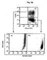
- the loading of tumor cell bodies labeled with 7-AAD by FITC-CD1a + DC was measured by flow cytometry as the percentage of DC doubly positive for FITC-CD1a+ and 7-AAD ( Fig. 1B ).
- Forward scatter/side scatter analysis revealed the increased granularity of the double positive population, further confirming the absorption of tumor cell bodies by DC ( Fig. 1B ).
- up to 45% of DC were able to load cell bodies of cell lines A2058, A375.S2, DU 145, PC3, LnCAP, and Jurkat T lymphoma.
- phagocytosis was observed in an autologous setting where dead or dying EBV transformed B-lymphoblastoid cell line or non-transformed B cells were used.
- the extent to which DC capture dead or dying tumor cells was independent of whether cell death was induced by Fas ligation or by ⁇ -irradiation.
- DNA staining and a standard Giemsa staining indicated that only a fraction, up to 45%, of the immature DC capture tumor cell bodies containing at least one nuclear fragment. This partial capture did not seem to be due to a lack of substrate as many labeled dead or dying tumor cells remained unengulfed by DC.
- results from fluorescence activated cell sorting and confocal microscopy demonstrated that immature DC have the ability to phagocytose dead or dying cell bodies derived from different tumor cell lines.
- Monocyte-derived DC which were generated by culturing monocytes of a prostate carcinoma patient with GM-CSF and IL-4 were shown to capture bodies derived from PrCa cell lines. Giemsa staining of MDDC indicated that the MDDC had phagocytosed bodies derived from prostate cancer cell line DU145. As shown in Fig. 2A , up to 20% of patient's MDDC phagocytosed DU145 cell bodies, similarly to DC generated from a healthy volunteer ( Fig. 2B ).
- Loaded DC induce proliferation of autologous CD4+ T cells and CD8+ T cells
- DC were loaded with tumor cell bodies as above and sorted, based on CD1a-FITC staining, to be used as stimulators for autologous T cells.
- Sources for T cells were unseparated PBMC, purified (>90%) CD4 + T cells and purified (>90%) CD8 + T cells.
- CD4 + T cells and CD8 + T cells were obtained from Ficoll-separated PBMC of healthy volunteers (with no history of blood transfusion) and depleted of other cells using purified CD3 (UCHT1), CD4 (13B8.2), CD8 (B9.11), CD14 (RMO52), CD16 (3G8), CD19 (J4.119), CD56 (NKH-1), anti-HLA-DR (B8.12.2), and anti-glycophorin A (D2.10) monoclonal antibodies (mAbs) (all from Beckman-Coulter, Miami, FL) ( Nouri-Shirazi, et al. 2000.
- mAbs monoclonal antibodies
- CD1a-labeled DC were cocultured with tumor cell bodies (with no DNA labeling), sorted based on CD1a expression (purity >98%) and plated in round bottom 96-well plates at graded doses from 0 to 5000 DC/well.
- Autologous PBMC (1x10 5 /well/200 gl), purified CD4 + T cells (5x10 4 /well200 ⁇ l) or purified CD8 + T (5x10 4 /well/200 ⁇ l) cells were added to the wells containing CD1a-labeled DC.
- the proliferation assay was carried out in culture medium supplemented with heat-inactivated 10% human AB serum (Gemini Bio-products, Woodland, CA).
- Soluble CD40L 200 ng/ml, Immunex, Seattle, WA was added at the culture initiation to induce DC maturation, and IL-2 (5 U/ml, Genzyme Co., Cambridge, MA) was added to support the proliferation of purified CD8 + T cells. After 4 days of incubation, tritiated thymidine (Wallac, Inc., Gaithersburg, MD) was added at the activity of 1 ⁇ Ci/well. The plates were harvested 16 hours later and the incorporated radioactivity was measured by liquid scintigraphy according to manufacturer's instructions.
- the same assay to detect T cell expansion was also performed in the presence of mAbs directed against HLA-DR and HLA-A,B,C to determine the antigenic peptides on MHC class I and II molecules presented by DC that are essential for the initiation of T cell proliferation.
- DC were incubated with mAbs against HLA-DR (clone L243 from Becton Dickinson) and HLA-A,B,C (W 6/32, from DAKO, Botany, N.S.W.) at a final concentration of 5 ⁇ g/ml in CM for 30 minutes before the addition of T cells.
- the mAbs were present throughout the culture period. Irrelevant mAbs were used as isotype controls.
- DC have the ability to phagocytose dead or dying cell bodies derived from different tumor cell lines and were able to induce proliferation of autologous T cells, it was then determined that DC loaded with tumor cell bodies permits the expansion of CTL possessing cytotoxic activity against the immunizing tumor cell line.
- DC loaded with dead or dying tumor cells were sorted as described above and used as stimulatory cells, while autologous PBMC or purified CD8+ T cells (as prepared above in this example) were used as responders.
- Cultures were prepared in 24-well plates (Costar, Corning, Inc., Acton, MA) by plating 0.5-1x10 5 cells/ml immature loaded DC with 2x10 6 cells/ml responder cells in a final volume of 2 ml. Culture medium was supplemented with 10% human AB serum (Gemini Bio-products, Woodland, CA) and IL-7 (10 U/ml from Immunex, Seattle, WA) in the first seven day cycle.
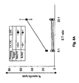
- the expanded T cells sensitized by DU145 loaded DC were able to lyse DU145 cells but not the K562 target, thereby demonstrating CTL, rather than NK activity.
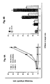
- CTL with equivalent cytotoxic activity were also expanded against the priming tumor cell line when total PBMC, rather than purified CD8+ T cells, were used as responder cells ( Fig.4A ).
- Comparable CTL priming was also obtained when DC were loaded with PC3 prostate carcinoma cell bodies ( Fig.4B ).
- the expanded CTL were able to lyse both the PC3 cells used for sensitization and cells from the other prostate carcinoma cell line DU 145, substantiating recognition by the CTL of shared antigens.
- T2 cells a standard HLA-A201 negative cell line obtained from the American Type Culture Collection, were loaded with PSA specific antigens, using synthetic PSA peptides PSA1 (NH2-FLTPKKLQCV-OH; amino acids 141-150) and PSA2 (NH2-KLQCVDLHV-OH; amino acids 146-154) (Biosynthesis, Lewisville, Texas). These PSA specific peptides were previously identified as HLA-A2 binding peptides ( Correale, et al. 1997. "In vitro generation of human cytotoxic T lymphocytes specific for peptides derived from prostate-specific antigen," J Natl Cancer Inst 89:293 ).
- HLA-A2 binding affinity of the PSA peptides was evaluated by up-regulation of A2 surface expression on T2 cells after peptide loading.
- About 1x10 6 T2 cells were incubated in serum-free complete medium with 50 ⁇ g peptide/ml at 37°C in 5% CO 2 overnight. The cells were then washed two times with PBS and subsequently stained with FITC conjugated HLA-A2-specific mAb (10% v/v), HLA-A2, 28 from One Lamda, Inc, Canoga Park, CA). The mean fluorescence intensity of HLA-A2 staining with or without peptide loading was analyzed by flow cytometry.
- DC were loaded with prostate carcinoma cell line LnCAP because these cells express PSA and present PSA peptides in the context of HLA-A2 for T cell-mediated lysis ( Correale, et al. 1998. "Generation of human cytolytic T lymphocyte lines directed against prostate-specific antigen (PSA) employing a PSA oligoepitope peptide," J Immunol 161:3186 ). Immunofluorescence staining confirmed the cytoplasmic expression of PSA in LnCAP cells but not in PC3 cells.
- PSA prostate-specific antigen
- immature DC generated from either male or female HLA-A2 + donors were cocultured with LnCAP cell bodies, then sorted and used as stimulators of autologous CD8 + T cells, obtained by methods presented earlier in this example. After three repeated cycles of stimulation, the responder cells were harvested and cytotoxic activity and specificity were determined using LnCAP cells, K562 cells and T2 cells loaded with PSA peptides as targets.
- DC generated from a HLA-A2+ male donor were loaded with LnCAP cell bodies and used as stimulators of autologous CD8+ T cells.
- the CTL generated from a male donor were also able to kill T2 cells loaded with PSA peptides but not unloaded T2 cells nor NK-sensitive K562 cells.
- the generated CTL were not able to kill T2 cells loaded with control influenza matrix peptide (MP), again showing the specificity of the response (data not shown).
- MP control influenza matrix peptide
- the Colo829 and SkMe128 melanoma cell lines, K562, LnCAP prostate carcinoma cell line, 1806 breast cancer cell line (established by Drs. A. Gazdar and J. Minna at UTSW Medical Center at Dallas) and T2 cells were purchased from the American Type Culture Collection (ATCC; Manassas, VA). The Me275 and Me290 cell lines were established at the Ludwig Cancer Institute in Lausanne. All cell lines were maintained in CM.
- ME275 cell death was induced by treatment with 10 ⁇ g/ml of betulinic acid for 48 hours.
- Colo829 cells were killed by the same method, or by gamma irradiation (150 Gy and then cultured 48 hours in serum-free medium).
- Cell death was assessed by morphology, externalization of phosphatidylserine using FITC-labeled annexin V (CALTAG, Burlingame, CA), and staining with DNA specific dyes: 7AAD and trypan blue (Sigma Chemical Co., St. Louis, MO).
- Colo829 cells could be killed by ⁇ -irradiation/serum starvation (150 Gy, >30% of dead cells).
- Me275 cells proved resistant to ⁇ -irradiation as well as receptor-mediated death via either Fas-ligation, TNF or TRAIL (not shown).
- Me275 cells undergo swift death when treated with Betulinic acid (BA) ( Pisha, et al. 1995.
- BA Betulinic acid
- melanoma cells were treated for 48 hours with 10 ⁇ g/ml BA (>50% of dead cells, a combination of apoptosis and necrosis). Both Colo829 and Me275 grow as adherent cells and detach when dying, thus the nonadherent fraction composed mostly of killed cells for DC loading was used.
- Immature DC were generated from Ficoll-separated PBMC of HLA-A201 + healthy volunteers or patients with stage IV melanoma.
- PBMC from nonmobilized or mobilized GM-CSF (Amgen, Thousand Oaks, CA) were suspended in CM and allowed to adhere onto plastic dishes (Falcon 6-well, Franklin Lakes, NJ) for 2 hours at 37°C.
- the non-adherent cells were removed, and the adherent cells were cultured in CM for 6 days.
- GM-CSF 100 ng/mL; Leukine, Immunex, Seattle, WA
- IL4 5 ng/mL; Schering-Plough
- the DC recovery at day 7 as determined by a standard immunofluorescence and FACS analysis was >90% of CD1a + cells.
- soluble CD40-ligand sCD40L; 200 ng/mL; Immunex, Seattle, WA
- Purified CD4 + and CD8 + T cells were depleted of other cells using purified CD4 (13B8.2), CD8 (89.11), CD14 (RM052), CD16 (3G8), CD19 (14,119), anti-CD45 RO (UCHL1), CD56 (NKH-1), anti-HLA-DR (B8.12.2), and anti-glycophorin A (D2.10) monoclonal antibodies (mAbs) (all from IMMUNOTECH, Marseille, France) and goat anti-mouse IgG Dynabeads (Dynal, Lake Success, NY) ( Nouri-Shirazi, et al. 2000.
- CD8+CD45RA+CD27+ T cells the CD8+ T cells were stained with anti CD45RA-PE and anti CD27-FITC (Becton-Dickinson Immunocytometry Systems, San Jose, CA) and FACS sorted. Purified CD4 + and CD8 + T cells were then used in the T-cell proliferation assay given below.
- FACS analysis was performed on a FACSCalibur and sorting on a FACS Vantage (Becton Dickinson, Mountain View, CA).
- Antibodies used to phenotype or sort the cells were Anti-CD1a-FITC labelled (Biosource), Anti-CD14-APC, Anti-CD80-PE, Anti CD83-PE, Anti CD86-PE, Anti-HLA DR-PerCP, Anti CD45RA-PE and Anti-CD27-FITC.
- CD1a-FITC labeled DC were cocultured for 1 hour at 37°C with tumor bodies, and then sorted based on CD1a expression (purity > 95%) and plated in U bottom 96-well plates at graded doses from 0 to 5000 DC/well.
- Purified CD4 + T cells or purified CD8 + T cells (10 5 /well/200 ⁇ l) were added to plates.
- the proliferation assay was carried out in culture medium supplemented with heat-inactivated 10% human AB serum (Gemini Bio-products, Woodland, CA).
- Soluble CD-40L (200ng/ml; Immunex, Seattle, WA) was added to induce DC maturation, and IL2 (10 U/ml; Genzyme Co., Cambridge MA) was added to support the proliferation of purified CD8 + T cells. After 5 days, tritiated thymidine (NEN, Boston, MA) was added at the activity of 1 Ci/well. The plates were harvested 16 hr later (Wallac, Inc., Gaithersburg, MD) and incorporated radioactivity was measured by liquid scintigraphy according to manufacturer's instructions.
- CTL cytotoxic T lymphocytes
- DC loaded with tumor derived cell bodies and sorted were used as stimulatory cells while autologous purified CD8 + T cells, CD8 + CD45RA + CD45RO - or CD8 + CDR45A + CD27 + T cells were used as responders.
- Cultures were prepared in 24-well plates (Costar) by plating loaded DC at 1x10 5 cells with 1x10 6 T cells in a final volume of 1ml. Culture medium was supplemented with 10%AB serum, sCD40L (200ng/ml; Immunex, Seattle, WA); , IL7 (10 UI/ml in the 1 st week) and IL2 (10 UI/ml in the 2 nd and 3 rd week). T cells were restimulated with fresh preparations of unloaded DC or DC loaded with killed tumor cells weekly for two other weeks. Six days after the last stimulation, cells were harvested and their cytotoxic activity as well as the capacity for IFN- ⁇ release were tested.
- ELISPOT enzyme-linked immunospot
- the capacity of immature monocyte derived DC to capture killed melanoma cells was evaluated.
- Immature DC were mixed with killed melanoma cells at 1:5 ratio and incubated for 1 hour at 37°C to allow phagocytosis. Thereafter, DC were sorted by flow cytometry based on forward scatter/side scatter properties reflecting size and granularity according to the procedures described herein.
- CD1a-FITC labeled immature DC were loaded with killed melanoma cells, sorted by flow cytometry based on CD1a expression (purity > 90%) and cocultured with purified (> 90%) autologous T cells in the presence of CD40L, which induces maturation of both loaded and unloaded DC as determined by CD83 expression (data not shown).
- the T cells proliferated when cultured with DC loaded with killed Me275 cells, but not when cultured with killed melanoma cells only ( Fig. 7 ).
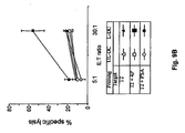
- HLA-A201 + DC Immature HLA-A201 + DC were loaded with killed Me275, sorted and used to stimulate purified autologous CD8 + T cells over a 3-week culture. Soluble CD40L was added to induce DC maturation, as well as IL-7 (10 U/ml 1 st week) and IL-2 (10 U/ml in the 2 nd and 3 rd week) to help T cell proliferation. The T cells were harvested after three stimulation cycles and their cytotoxic activity was determined using Me275, unrelated HLA-A201 + tumor cells, LnCAP, 1806, and NK-sensitive K562 cells. As shown in Fig.
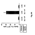
- T cell lines generated with killed melanoma cells loaded DC contain T cells specific for melanoma associated antigens: 1) an overnight IFN- ⁇ ELISPOT with the HLA-A201 + DC pulsed with melanoma peptides: Melan A/MART-1 27-35 , tyrosinase 368-376 and MAGE-3 271-279 as well as mutated gp100 g209-2M ; and 2) a cytotoxic chromium release assay using T2 cells pulsed with the four peptides as target cells. As shown in Fig.
- T cell lines generated with unloaded DC, and peptide-pulsed DC alone (without the T cells) yielded 3 ⁇ 1 and 4 ⁇ 1.5 spots, respectively. As shown in Fig.
- the ability to induce melanoma-specific CTL was not restricted to Me275 cells as the T cells elicited by DC loaded with the HLA-A201 - Colo829 also contained cells that could kill the T2 cells loaded with each of the four melanoma peptides (Table I).
- T cells elicited by killed melanoma cells loaded DCs kill melanoma-peptide pulsed T2 cells a No peptide Tyr Gp100 MAGE-3 MART-1 Exp.1/Me275 18 ⁇ 5 b 68 ⁇ 5 48 ⁇ 4 nd nd Exp.
- CD45RA + CD45RO - naive CD8 + T cells were cultured with DC loaded with killed Me275 cells.
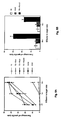
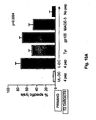
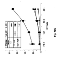
- the elicited T cells were able to kill (1) T2 cells pulsed with the four melanoma peptides (up to 70% specific lysis, Fig. 10A ), (2) T2 cells loaded with a single peptide: tyrosinase or gp100 or MAGE-3 ( Fig. 10A ), and ( 3 ) the Me275 cell line used for immunization (data not shown).
- CD8 + CD45RA + CD45RO - T cells contains a small fraction of "effector memory” cells that might expand in our culture conditions. Therefore, based on CD27 expression, the pool of CD8 + CD45RA + CD45RO - T cells was subdivided to distinguish truly inexperienced T cells (CD45RA + CD27 + ) from uncommitted effector T cells (CD45RA + CD27 - ) ( Hamaan, D., et al. "Phenotypic and finctional separation of memory and effector human CD8+ T cells" 1997. J. Exp. Med. 186:1407-1418 ).
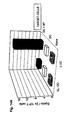
- these naive T cells primed by DC loaded with HLA-A201 - Colo829 cells were able to kill HLA-A201 + melanoma targets including Me275, Me290 and SkMe128 cells, thus demonstrating cross-priming ( Fig. 11 ).
- DC obtained from patients with stage IV melanoma capture dead or dying melanoma cells and induce proliferation of autologous T cells
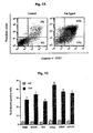
- Fig. 13A and 13B demonstrates immature monocyte-derived DC from a patient with stage IV melanoma cocultured with dead or dying cells from a melanoma cell line induce proliferation of both autologous CD4+ T cells ( Fig. 13A ) and CD8+ T cells ( Fig. 13B ).
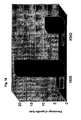
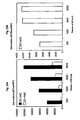
- CD8 + T cells cultured for 3 weeks with DC loaded with killed Me275 cells contain melanoma-specific T cells as determined by IFN- ⁇ ELISPOT using peptide-pulsed DCs (15 spots/2x10 5 T cells, patient # 6, Fig. 14A ).
- DC loaded with killed breast cancer cells can induce proliferation of autologous CD4 T cells.
- DC were first loaded with killed breast cancer cells, either sorted or enriched by differential centrifugation, and then cocultured at graded doses with 1x10 5 autologous CD4 T cells (purity > 85%).
- Monocyte-derived DC and T cells were prepared as described in the Examples 1 and 2. Capture of killed breast cancer cells by immature DC was monitored by flow cytometry and Giemsa staining as described in the Examples 1 and 2. After 5 days of co-culture with T cells, tritiated thymidine (NEN, Boston, MA) was added to each well at the activity of 1 Ci /well.
- the plates were harvested 16 hours later (Wallac, Inc., Gaithersburg, MD) and incorporated radioactivity was measured as described above. As shown in Fig. 15 , the loaded DC induced T cells from healthy volunteers to proliferate, indicating that DC can process antigens from killed breast cancer cells and present them to autologous CD4 + T cells.
- T cell lines having cytotoxic activity against breast cancer can be generated in vitro.
- Immature DC were mixed with killed allogeneic breast cancer 1806 cells (killed by a combination of CHX and Fas ligation as described in the Example 1), cultured for 48 hours at 1:1 ratio in the presence of cytokines inducing DC maturation TNF-a (10ng/mL), IL-6 (10 ng/mL) and IL-1 (10 ng/mL) (all from Genzyme). Thereafter, the DC were used to stimulate purified autologous CD8 T cells using the procedures described in the Examples 1 and 2. The T cells were harvested using a chromium release assay after three stimulation cycles, and their cytotoxic activity was determined using a panel of tumor cell lines.
- DC loaded with breast cancer 1806 bodies induced differentiation of CTL able to kill 1806 breast cancer cells with 30% of specific lysis but not the NK-sensitive K562 cell line nor other tumor cells, LnCaP and MCF-7.
- breast cancer specific CTL can be elicited by DC loaded with killed breast cancer cells using peripheral blood lymphocytes without adding cytokines to induce DC maturation during loading with killed breast cancer cells.
- the immature DCs are mixed with killed breast cancer cells (all procedures as above) and cultured for 48 hours in CM. Thereafter the loaded DCs are used as stimulators of autologous peripheral blood lymphocytes. All procedures are the same as above ( Fig. 18 ).
- Example 4 DC loaded with allogeneic cell lines can be used to eliminate unwanted immune responses
- T cells were loaded with COLOB29 bodies, sorted as described above and used as stimulators for autologous CD8 + T cell expansion plated in al:5 ratio.
- T cells were primed in the first cycle using the same procedure as described in Example 1 by COLO829 loaded DC, harvested and subsequently cocultured, in a second cycle, with DC loaded with the EBV transformed cell line COLO829-BL in the presence of exogenous Fas-ligand anti-Fas antibody as described in Example 1 (Immunotech, Marseille, France).
- the concept behind this approach was that the use of EBV cell bodies and Fas ligand allows elimination of allo-responsive T cells.
- the surviving CD8 + T cells were harvested and restimulated in two more seven day cycles by DC loaded with COLO829 melanoma cell bodies to expand the melanoma-antigen specific T cells.
- the resulting cytotoxic activity was determined in the standard 51 Cr release assay using melanoma peptides loaded in T2 cells and the NK-sensitive K562 cells as targets (same methods as described above).
- the elicited CTL line was able to specifically lyse T2 cells loaded with gp100, tyrosinase and to a lesser extent with MART-1 peptides.
- tumor antigen specific CTL can be enriched by elimination of allo-specific T cells.
- Example 5 Induction of antigen-specific tolerance with antigen-presenting cells loaded with killed allogeneic cells
- Dendritic cells can also be loaded with cells that express an antigen to which the T cells could be tolerized. This is demonstrated in Fig. 21 and 22 where loaded DCs are treated with compounds that allow antigen presentation but inhibit the expression of co-stimulatory molecules. In this way, the T cells can recognize the antigen but will become tolerant, as demonstrated in Fig 22 the T cells are unable to proliferate when restimulated with the same antigen.
- CD45RA+CD4+ T cells are obtained from Ficoll-separated PBMC of healthy volunteers, depleted of other cells using CD8 (B9.11), CD45RO (UCHL1), CD14 (RMO52) CD16 (3G8), CD19 (J4.119), CD56 (NKH-1), anti-HLA-DR (B8.12.2), anti-glycophorin A (D2.10) monoclonal antibodies (mAbs) (Beckman-Coulter, Miami, FL) and Goat anti-mouse IgG Dynabeads (Dynal, Lake Success, NY). The purity of the enriched populations was > 90%.
- Immature monocyte-derived DC are generated from adherent fraction of peripheral blood PBMC as described in the Example 1. Allogeneic cell lines (Colo 829, Colo-EBV and Hom2 EBV) are prepared as described in the Example 1. Antigen-loaded DCs are prepared as described above and used to stimulate na ⁇ ve CD4 T cells as described above.
- CD4+CD45RA+ T cells (10 6 /well) are cocultured in 24-well plates with sorted autologous immature DC (1X10 5 /well) loaded with killed allogeneic cell line (colo 829) in the presence or absence of an inhibitor of NFkB translocation NAC (25mM, SIGMA). After 5 days, the T cells from both cocultures (-NAC and +NAC) are harvested, washed and rested for an additional 2 days. T cells from primary cultures are rechallenged in a second proliferation assay with the stimulators indicated in the Fig. 22 (colo EBV, Hom2 EBV). Proliferation is determined by uptake of (1 ⁇ Ci/well for the last 16hr) tritiated thymidine.
- Example 6 Treatment of patient with antigen-presenting cells loaded with allogeneic tumor cells
- Antitumor responses in a patient can be obtained by administering antigen-presenting cells, including but not limited to DC, loaded with killed allogeneic tumor cells.
- antigen-presenting cells including but not limited to DC
- the multiple and/or shared tumor antigens presented on the loaded antigen-presenting cells can elicit immune responses in the patient, leading to tumor rejection.
- Example 7 Treatment of patient with antigen-specific T cells expanded ex vivo by antigen-presentingcells loaded with allogeneic killed tumor cells
- Antitumor responses in a patient can be obtained by administering tumor antigen-specific T cells elicited ex vivo by DC loaded with killed allogeneic tumor cells.
- the shared tumor antigens presented on the tumor cells can be recognized by T cells specific for multiple and/or shared tumor antigens, leading to tumor rejection.
- Example 8 Elimination of transplantation complications using antigen-presenting cells loaded with donor cells
- Yet another aspect of the present invention can be useful in bone marrow and organ transplantation.
- the present invention permits the elimination of undesired T cells that react either against the transplant or against the host, which eliminates or minimizes the risk of graft rejection or of graft versus host disease.
- Antigen-presenting cells of the donor or of the host are loaded with dead or dying EBV-transformed B cells from the host or from the donor, respectively.
- Immunosuppressive drugs such as azathioprine, cyclosporine or methotrexate are administered to the patient, permitting the killing of the activated T cells.
- Upon subsequent transplantation there will be no graft rejection or graft versus host disease, respectively, because the reactive T cells have been eliminated.
- Yet another aspect of the present invention can be useful in the elimination of the residual disease in post-transplant patients with hematological malignancies, including but not limited to leukemias and lymphomas.
- the donor antigen-presenting cells are loaded with dead or dying leukemic cells of the donor or allogeneic cell lines. Such loaded antigen-presenting cells are then administered to patients and the shared antigens presented on the loaded antigen-presenting cells elicits immune responses leading to the rejection of the tumor and elimination of the residual disease.
Landscapes
- Health & Medical Sciences (AREA)
- Life Sciences & Earth Sciences (AREA)
- General Health & Medical Sciences (AREA)
- Immunology (AREA)
- Engineering & Computer Science (AREA)
- Animal Behavior & Ethology (AREA)
- Public Health (AREA)
- Veterinary Medicine (AREA)
- Chemical & Material Sciences (AREA)
- Epidemiology (AREA)
- Biomedical Technology (AREA)
- Bioinformatics & Cheminformatics (AREA)
- Organic Chemistry (AREA)
- Wood Science & Technology (AREA)
- Genetics & Genomics (AREA)
- Microbiology (AREA)
- Biotechnology (AREA)
- Zoology (AREA)
- Medicinal Chemistry (AREA)
- Pharmacology & Pharmacy (AREA)
- Cell Biology (AREA)
- Hematology (AREA)
- Mycology (AREA)
- Biochemistry (AREA)
- General Engineering & Computer Science (AREA)
- Nuclear Medicine, Radiotherapy & Molecular Imaging (AREA)
- General Chemical & Material Sciences (AREA)
- Chemical Kinetics & Catalysis (AREA)
- Transplantation (AREA)
- Rheumatology (AREA)
- Medicines Containing Material From Animals Or Micro-Organisms (AREA)
- Micro-Organisms Or Cultivation Processes Thereof (AREA)
- Medicines Containing Antibodies Or Antigens For Use As Internal Diagnostic Agents (AREA)
Abstract
Description
- This invention relates to engineering immunogenic cells.
- Dendritic cells (hereinafter referred to as "DC") are the only antigen-presenting cells, or APC, able to induce primary immune responses (Cella, et al. 1997. "Origin, maturation and antigen presenting function of dendritic cells," Curr Opin Immunol 9:10; Banchereau, J., and R.M. Steinman. 1998. "Dendritic cells and the control of immunity," Nature 392:245). Circulating DC precursors circulate to tissues where they reside as immature, antigen-capturing cells with high endocytic and phagocytic activity. Following antigen uptake, DC migrate to the secondary lymphoid organs where they mature and become antigen-presenting cells able to select and activate naïve antigen-specific CD4+ T cells. This permits diversification of the response and activation of antigen-specific effectors such as antigen-specific cytotoxic T lymphocytes (hereinafter referred to as "CTL") and B cells as well as non-specific effectors such as NK cells, macrophages and eosinophils (Sogn, J.A. 1998. "Tumor immunology: the glass is half full," Immunity 9:757).
- DC have been used to elicit an immune response against tumors. The induction of tumor immunity can be viewed as a three-step process that includes: 1) presentation of tumor associated antigens; 2) selection and activation of tumor associated antigens - specific T cells as well as non-specific effectors; 3) localizing tumor associated antigens - specific T cells to the tumor site; and 4) recognition of restriction elements leading to the elimination of tumor cells.
U.S. Patent No. 5,637,483 issued to Dranoff et al. discloses a method by which modified tumor cells expressing cytokines such as GM-CSF and IL-2 are irradiated or rendered proliferation incompetent and then administered to a patient to act as a stimulator or suppressor of a patient's systemic immune system. Experiments in mice have demonstrated the development of both protective and therapeutic anti-tumor responses induced by DC loaded with tumor associated antigens (Bell, et al. 1999. "Dendritic cells," Adv Immunol 72:255). Furthermore, pilot clinical trials in humans show the feasibility of DC administration and the ability of peptide-loaded DC to induce peptide-specific T cell responses in patients with lymphoma, malignant melanoma and prostate carcinoma (Mukherji, et al. 1995. "Induction of antigen-specific cytolytic T cells in situ in human melanoma by immunization with synthetic peptide-pulsed autologous antigen presenting cells," Proc Natl Acad Sci USA 92:8078; Hsu, et al. 1996. "Vaccination of patients with B-cell lymphoma using autologous antigen-pulsed dendritic cells," Nat Med 2:52; Gilboa, E., et al. 1998. "Immunotherapy of cancer with dendritic-cell-based vaccines," Cancer Immunol Immunother 46:82; Nestle, et al. 1998. "Vaccination of melanoma patients with peptide- or tumor lysate-pulsed dendritic cells," Nat Med 4:328; Tjoa, et al. 1998. "Evaluation of phase I/II clinical trials in prostate cancer with dendritic cells and PSMA peptides," Prostate 36:39). - In order for DC to affect the immune system, the cells must possess processed peptide fragments to interact with T-cells, or be "loaded." One of the critical challenges for the use of DC as immunotherapy vectors is the identification of an efficient antigen loading strategy. Several systems have been employed to deliver tumor associated antigens to DC including: 1) defined peptides of known sequences (Mayordomo, et al. 1995. "Bone marrow-derived dendritic cells pulsed with synthetic tumor peptides elicit protective and therapeutic antitumor immunity," Nat Med 1:1297; Celluzzi, et al. 1996. "Peptide-pulsed dendritic cells induce antigen-specific CTL-mediated protective tumor immunity," J Exp Med 183:283); 2) undefined acid-eluted peptides from autologous tumor (Zitvogel, et al. 1996. "Therapy of murine tumors with tumor peptide-pulsed dendritic cells: dependence on T cells, B7 costimulation, and T helper cell 1-associated cytokines," J Exp Med 183:87); 3) whole tumor lysates (Fields, et al. 1998. "Murine dendritic cells pulsed with whole tumor lysates mediate potent antitumor immune responses in vitro and in vivo," Proc Natl Acad Sci USA 95:9482); 4) retroviral and adenoviral vectors (Song, et al. 1997. "Dendritic cells genetically modified with an adenovirus vector encoding the cDNA for a model antigen induce protective and therapeutic antitumor immunity," J Exp Med 186:1247; Specht, et al. 1997. "Dendritic cells retrovirally transduced with a model antigen gene are therapeutically effective against established pulmonary metastases," J Exp Med 186:1213); 5) tumor cell derived RNA (Boczkowski, et al. 1996. "Dendritic cells pulsed with RNA are potent antigen-presenting cells in vitro and in vivo," J Exp Med 184:465); and 6) fusion of DC with tumor cells (Gong, et al. 1998. "Reversal of tolerance to human MUCI antigen in MUCI transgenic mice immunized with fusions of dendritic and carcinoma cells, Proc Natl Acad Sci USA 95:6279).
- Although all methods for delivering tumor associated antigens described above induce T cell responses and cause considerable anti-tumor effects, each has potential drawbacks (Sogn, J.A. 1998. "Tumor immunology: the glass is half full," Immunity 9:757). Foremost, peptide-based strategies are limited by 1) the knowledge of the MHC haplotype of each patient; 2) the knowledge of the corresponding MHC class I binding motifs of the tumor associated antigens; 3) the variability of the MHC class I binding affinity of the synthetic peptides; and 4) the knowledge of whether or which various defined peptides represent tumor associated antigens in vivo.
- Unlike a peptide-based approach which requires information about MHC haplotype and peptide sequence, unfractionated tumor tissue can provide both MHC class I and MHC class II epitopes without identifying tumor associated antigens. Recent studies demonstrated the ability of DC to capture dead or dying cells and elicit MHC class I-restricted secondary CTL responses (Albert, et al. 1998. "Dendritic cells acquire antigen from apoptotic cells and induce class I- restricted CTLs," Nature 392:86; Albert, et al. 1998. "Tumor-specific killer cells in paraneoplastic cerebellar degeneration," Nat Med 4:1321). These studies indicated that 1) DC loaded with dead or dying influenza virus-infected cells can activate lymphocytes to mount virus-specific CTL responses; and 2) DC that capture dead or dying tumor cells can present antigen to tumor associated antigens peptide-specific T cells that were derived from patients with the paraneoplastic cerebellar degeneration. Because of the potential of engineered DC to contribute positively in the treatment of human disease, a simple method is desired for their production.
-
U.S. Patent No. 5,851,756 issued to Steinman et al. discloses methods by which proliferating cultures of DC precursors and mature DC can be produced in vitro. Steinman et al. disclose the production of T cell dependent antigens using mature DC cultures, wherein the antigens are comprised of DC modified antigens or antigen-activated DC. Steinman et al. further disclose that the DC modified antigens or antigen-activated DC can be used as immunogens for vaccines or treatment of infectious diseases. - Major problems in tumor immunotherapy as experienced in the methods presented above are the limited number of well-defined tumor associated antigens and the lack of evidence that the known tumor associated antigens actually represent rejection antigens in vivo. Furthermore, the use of MHC class I binding peptides is associated with the HLA restriction and the limitation of induced immune responses to CD8+ T cells. In this context, the use of unfractionated antigenic material, in the form of dead or dying allogeneic tumor cells, that provides both MHC class I and class II epitopes leading to a diversified immune response involving many clones of CD4+ T cells and CTL represents an attractive alternative.
- International Application No.
WO 99/42564 of Albert et al. - We have now developed a method for engineering immunogenic DC. We have perfected a process using immature human antigen-presenting cells, including DC, derived from healthy volunteers and from patients that can capture dead or dying allogeneic cells, and subsequently present their antigens to autologous T cells, thus inducing proliferation of autologous CD4+ T cells and CD8+ t cells. Furthermore, by using the processes described herein, it has been found that DC loaded with allogeneic tumor cells can activate CD8+ T cells, generate CTL specific for antigens expressed by the dead or dying cells, and also recognize antigens that are shared between different tumor cell lines. Yet another finding is that by using the process described herein, DC loaded with dead or dying allogeneic tumor cell lines can prime naive T cells and induce their differentiation to antigen-specific CTL.
- In one aspect, the invention is directed to inducing a cytotoxic tumor-specific immune response in a tumor-bearing human patient using dendritic cells which are prepared in vitro by coincubating dendritic cells from said patient with allogeneic apoptotic human tumor cells, to form loaded dendritic cells, wherein said allogeneic tumor cells possess at least one antigen shared between the allogeneic tumor cells and said patient's tumor cells, and wherein said cytotoxic immune response is directed against said shared tumor antigen to. Thereby, the patient's cells upon contact with the loaded dendritic cells mature to form cytotoxic cells expressing cytotoxic activity against tumor cells thus causing tumor rejection. The resulting cytotoxic cells can be either CD4 positive, or CD8 positive or both. The apoptotic cells can be obtained from allogeneic cell lines. The tumor specific immune response can be directed against multiple shared tumor antigens.
- In another aspect, the invention is directed to inducing a tumor-specific immune response in a tumor-bearing patient using cytotoxic T cells which are prepared in vitro by coincubating human dendritic cells with allogeneic apoptotic human tumor cells to form loaded dendritic cells; and coculturing said loaded dendritic cells with human naive T cells to form cytotoxic T cells, wherein said allogeneic tumor cells possess at least one antigen shared between the allogeneic tumor cells and said patient's tumor cells, and wherein said cytotoxic immune response is directed against said shared tumor antigen. The resulting cytotoxic cells can be natural killer cells or natural killer T cells; they can be either CD4 positive, or CD8 positive or both. The apoptotic cells can be obtained from allogeneic cell lines. The tumor specific immune response can be directed against multiple shared tumor antigens.
- In yet another aspect, the invention is directed to an in vitro assay to determine the degree of cytotoxic activity of cytotoxic cells against tumor cells from a patient comprising (a) coincubating human dendritic cells with apoptotic human allogeneic tumor cells possessing antigens to which the immune response is desired to form loaded dendritic cells; (b) coculturing said loaded dendritic cells with cytotoxic precursor cells to form cytotoxic cells; (c) isolating said cytotoxic cells; (d) incubating said cytotoxic cells with said patient's tumor cells; and (e) monitoring the death of said patient's tumor cells, wherein the degree of tumor cell death is proportional to the cytotoxic activity of said cytotoxic cells.
-
-
Fig. 1A-C depicts that immature DC capture dead or dying tumor cells. InFig. 1A , DU145 prostate carcinoma cells were induced to apoptosis by anti-Fas (1µg/ml, CH-11, 16hr), the generated dead or dying cells were stained with FITC-annexin V and PI. The percentage of dead or dying cells was measured by flow cytometry. InFig. 1B , DU145 prostate carcinoma cells were induced to apoptosis by anti-Fas (1µg/ml, CH-11, 16hr); the generated dead or dying cells were labeled with 7-AAD and incubated with CD1a-FITC labeled immature DC for 1hr at 37°C. The capture of dead or dying cells by DC was quantitated by flow cytometry as the percentage of CD1a+ DC that become positive for 7AAD. Forward scatter/side scatter analysis of CD1a+7-ADD+ DC (R1) and CD1a+7-ADD- DC (R2) reveals the increased granularity of the double positive population confirming the engulfment of cell bodies by DC. InFig. 1C , the immature DC were incubated with dead or dying tumor cells, generated from the indicated tumor cell lines, at either 4°C or 37°C, and the percentage of double positive cells was measured by flow cytometry assay. -
Fig. 2A-2B depict that monocyte-derived DC (MDDC) from patients with prostate carcinoma efficiently capture PrCa bodies. 7AAD labeled PrCa cell bodies were coincubated with CD1a-labeled monocyte-derived DC, from patient with prostate carcinoma (Fig. 2A ) or from healthy volunteers (Fig. 2B ), for 1h at 37°C. Uptake was evaluated by flow cytometry. -
Fig. 3A-3D depict that immature DC loaded with tumor cell bodies induce proliferation of purified CD4+ and CD8+ T cells. CD1a-FITC labeled immature DC were cocultured with dead or dying tumor cells (Ap) for 1hr at 37°C and sorted based on CD1a expression. The sorted cells were cocultured for 5 days with autologous purified CD4+ T cells (Fig. 3A ) or CD8+ T cells (5x10⁴ cells/well) (Fig. 3B ). After 4 days of incubation, tritiated thymidine was added, and the thymidine incorporation was measured 16hr later. The experiments assessing the proliferation of CD8+ T cells were carried out in the presence of CD40L (200ng/ml) to induce DC maturation and IL-2 (5U/ml) to enhance T cell proliferation. CD1a-FITC labeled immature DC were cocultured with dead or dying tumor cells (Ap) for 1hr at 37°C and sorted based on CD1a expression. The sorted cells were cocultured for 5 days with autologous purified CD4+ T cells (Fig. 3C ) or CD8+ T cells (5x10⁴ cells/well) (Fig. 3D ) in the presence of mAbs against MHC class I or II molecules before onset of cultures and subsequently during the entire cultures period. After 4 days of incubation, tritiated thymidine was added and the thymidine incorporation was measured 16hr later. -
Fig. 4A and 4B depict that DC loaded with dead or dying prostate carcinoma cell lines prime CTL. CD1a-FITC labeled immature DC were loaded with prostate carcinoma DU145 (Fig. 4A ) or PC3 (Fig. 4B ) derived cell bodies for 1hr at 37°C, sorted based on CD1a expression and used as stimulator cells. The cultures were set in a 24-well plate by plating stimulator cells with either autologous purified CD8+ T cells or total PBMC. Soluble CD40 ligand (Immunex Corp) was added to induce DC maturation. After 7 days of culture, the T cells were harvested, washed and replated with freshly prepared stimulator cells for 7 additional days. Cytotoxic activity of expanded CTLs was assessed in a standard 51Cr release assay using sensitizing cells, non-sensitizing cells and K562 as targets. Percent cytotoxicity was measured as a function of spontaneous and total release. Results are representative of three experiments, and each value represents the mean from triplicate wells. -
Fig. 5 depicts HLA-A2 binding of PSA peptides, PSA1 and PSA2. T2 cells were incubated with PSA peptides overnight, then the cells were washed and stained with FITC conjugated HLA-A2-specific mAb (HLA-A2, 28). Up-regulation of HLA-A2 molecules on T2 cells after peptide binding was demonstrated as shifted histograms. The filled histograms showed the isotype control staining. -
Fig. 6A and 6B depict DC loaded with dead or dying prostate carcinoma cell line LnCAP induce PSA-specific CTL. CD1a-FITC labeled immature DC, generated from female (Fig. 6A ) and male (Fig. 6B ) HLA-A2+ donors, were loaded with prostate carcinoma (LnCAP) derived cell bodies for 1hr at 37°C, sorted based on CD1a expression and used as stimulator cells. The cultures were set in a 24-well plate by plating stimulator cells with autologous purified CD8+ T cells. Soluble CD40 ligand (Immunex Corp) was added to induce DC maturation. After 7 days of culture, the T cells were harvested, washed and replated with freshly prepared stimulator cells for 7 additional days. Cytotoxic activity of expanded CD8+ T cells was assessed in a standard 51Cr release assay using sensitizing LnCAP cells, T2 cells pulsed with PSA peptides as well as NK-sensitive K562 as targets. Percent cytotoxicity was measured as a function of spontaneous and total release. Results are representative of three experiments, and each value represents the mean from triplicate wells. -
Fig. 7A and 7B depict DC loaded with killed melanoma cells induce T cell proliferation. Unloaded DC (UL-DC) and loaded DC (L-DC) are sorted and cultured with purified autologous CD4+ (Fig. 7A ) and CD8+ T cells (Fig. 7B ) (with CD40L and IL2). Thymidine incorporation atday 5. Representative of two experiments. -
Fig. 8A and8B depict the induction of CTL by DC loaded with killed melanoma cells. Purified CD8+ T cells are cultured for 3 weeks with unloaded DC (UL-DC) or DC loaded with Me275. T cells are tested in 4 hour chromium release assay using as targets immunizing Me275 cells and K562 cells as a control for NK activity (a, mean ± SE, n=4) (Fig. 8A ), as well as unrelated HLA-A201+ tumor cell lines: prostate cancer (LnCAP) and breast cancer (1806) (Fig. 8B ). No CTL are elicited when T cells are cultured with killed melanoma cells without the DC (not shown). -
Fig. 9A and9B depict the induction of melanoma specific CTL by killed melanoma cells loaded DC. Purified CD8+ T cells are cultured for 3 weeks with unloaded or Me275 loaded DC.Fig. 9A depicts IFN ELISPOT using as targets DC, either unpulsed or pulsed with the four melanoma peptides (4P). Representative experiment from 3 performed using DC and T cells from different donors.Fig. 9B depicts CTL activity in 4 hour chromium release assay using unpulsed and melanoma-peptide pulsed (4P; mean values of 3 experiments) and control peptide PSA pulsed T2 cells (one experiment). -
Fig. 10A-10C depict DC loaded with killed melanoma cells prime naive T cells. Naive CD8+CD45RA+CD45RO- T cells are cultured for 3 weeks with unloaded DC (UL-DC) or with DC loaded with killed melanoma cells (L-DC). T cells are tested in a 4 hour chromium release assay. CTL elicited by DC loaded with Me275 bodies kill the T2 cells pulsed with different melanoma peptides (paired two-tailed t-test on log transformed data comparing the killing of unpulsed and peptide-pulsed T2 cells) (Fig. 10A ). Naive CD27+CD45RA+CD8+T cells differentiate into CTL able to kill the cell line used for immunization (Fig. 10B ) and T2 cells pulsed with melanoma peptides but not PSA peptide (Fig. 10C ) (representative of two experiments). -
Fig. 11 depicts cross-priming using DC loaded with killed melanoma cells. Naïve CD27+CD45RA+CD8+T cells are primed by autologous HLA-A201+ DC loaded with killed HLA-A201- Colo829 cells. The elicited T cells are able to kill HLA-A201+ melanoma targets but neither HLA-A201+ breast cancer cells nor autologous monocytes. -
Fig. 12A and 12B depict that CD34+HPC-derived DC subsets in stage IV melanoma capture dead or dying melanoma cells and induce proliferation of autologous T cells. CD34+HPC were cultured for 9 days with GM-CSF, TNF and FLT3L. Total DC (Fig. 12A ) and their isolated subsets (Fig. 12B ) were then loaded with dead or dying melanoma cell line COLO829. After overnight culture in the presence of DC maturation factor CD40L, DC were subsequently cultured at graded doses (horizontal axis) with autologous T cells for 5 days. T cell proliferation was determined by thymidine uptake (vertical axis, x103). -
Fig. 13A and 13B depict that immature DC from melanoma patient capture dead or dying melanoma cells and induce proliferation of both CD4+ and CD8+ T cells. Immature DC from patient with stage IV melanoma were cocultured with melanoma bodies for 1hr, sorted and cocultured for 5 days with autologous purified CD4+ T cells (+CD40L) (Fig. 13A ) or CD8+ T cells (5x10⁴ cells/well) (Fig. 13B ) and tritiated thymidine was measured at Day 5 (vertical axis x 103). The proliferation of CD8+ T cells was carried out in the presence of CD40L (200 ng/ml) and IL-2 (5 U/ml). -
Fig. 14A-14C depict the induction of melanoma specific CTL by killed melanoma cells loaded DC using DC and T cells from patient with advanced malignant melanoma. Purified CD8+ T cells are cultured for 3 weeks with unloaded or Me275 loaded DC.Fig. 14A depicts IFN ELISPOT using as targets DC, either unpulsed or pulsed with the four melanoma peptides (4P). Results from one patient.Fig. 14B depicts CTL activity in 4 hour chromium release assay. T cells are able to kill Me275 used for immunization, and the killing is increased when Me275 cells are pulsed with the four melanoma peptides. Similar results were obtained in two patients.Fig. 14C depicts priming of naïve T cells. CD8+CD45RA+CD45RO- T cells cultured 3 weeks with DC loaded with killed Me275 cells, kill T2 cells pulsed with 4 melanoma peptides. -
Fig. 15 depicts that DC loaded with killed breast cancer cells induce proliferation of autologous CD4+ T cells. Loaded DC are prepared and cultured at graded does with 105 autologous CD4+ T cells (purity >85%). T cell proliferation is determined after 5 days by thymidine incorporation (cpm x 103). The control curve represents cpm from wells where killed BrCa cells were plated together with the T cells. -
Fig. 16 depicts loaded DC can sensitize T cells which in turn can kill specific tumor cells. Immature DC are mixed with killed allogeneicbreast cancer cells 1806. The DC are incubated with killed tumor cells for 48 hours in the presence of monocyte conditioned medium to induce DC maturation. Harvested DC are then used as stimulators of autologous CD8 T cells in 3-weeks cultures (3 stimulations). Harvested T cells are tested in a chromium release assay (percentage specific lysis, vertical axis). -
Fig. 17 depicts mature loaded DC can sensitize T cells to kill tumor cells. Immature DC are mixed with killed allogeneicbreast cancer cells 1806. The DC are incubated with killed tumor cells for 48 hours in the presence of monocyte conditioned medium to induce DC maturation. Harvested DC are then used as stimulators of autologous peripheral blood lymphocytes in 3-weeks cultures (3 stimulations). Harvested T cells are tested in a chromium release assay (percentage specific lysis, vertical axis). -
Fig. 18 depicts loaded DC can sensitize T cells to kill tumor cells without the need for exogenous DC maturation factors. Immature DC are mixed with killed allogeneicbreast cancer cells 1806. The DC are incubated with killed tumor cells for 48 hours. Harvested DC are then used as stimulators of autologous peripheral blood lymphocytes in 3-weeks cultures (3 stimulations). Harvested T cells are tested in a chromium release assay (percentage specific lysis, vertical axis). -
Fig. 19 depicts loaded DC can sensitize T cells which in turn can kill specific tumor cells. Immature DC are mixed with killed allogeneic breast cancer cells MCF. The DC are incubated with killed tumor cells for 48 hours in the presence of monocyte conditioned medium to induce DC maturation. Harvested DC are then used as stimulators of autologous CD8 T cells in 3-weeks cultures (3 stimulations). Harvested T cells are tested in a chromium release assay (percentage specific lysis, vertical axis). MCF-7 cells are incubated with IFN-γ for 7 days prior to use as CTL targets. -
Fig. 20 depicts that tumor antigen specific CTL can be enriched by elimination of allo-specific T cells. The graph depicts the percentage of specific lysis (ordinate axis) of peptide loaded T2 cells and K562 cells at indicated effector to target ratios. Effector cells were generated from adult CD8+ T cells, after elimination of allo-specific T cells by two rounds of stimulation (7 days each) with autologous DC loaded with COLO829 derived bodies in the presence of IL-7 (100/ml, first cycle) and IL-2 (10U/ml, 3rd and 4th cycle). CD40 ligand was added to induce DC maturation. T2 cells were loaded with peptide for 2 hours. -
Fig. 21 depicts DC loaded with killed EBV cell lines induce proliferation of naive CD4 T cells. Immature monocyte-derived DC are loaded with killed EBV cells, sorted based on CD1a expression and cultured for 5 days with autologous naive CD4+CD45RA+ T cells. Proliferation is measured by thymidine incorporation (cpm, vertical axis). -
Fig. 22 depicts DC loaded with killed EBV cell lines and treated with the NFkB inhibitor induce tolerance of naive CD4+ T cells. In the first stimulation cycle purified CD4+CD45RA+ T cells (106/well) are cocultured in 24-well plates with sorted autologous immature DC (1X10⁵/well) loaded with killed allogeneic cell line (colo 829) in the presence or absence of an inhibitor of NFkB translocation NAC (25mM, SIGMA). After 5 days, the T cells from both cocultures (-NAC and +NAC) are harvested, washed and rested for an additional 2 days. T cells from primary cultures are rechallenged in a second proliferation assay with the DC loaded with killed EBV cells that are either autologous (Colo-EBV) or allogeneic (Hom2-EBV) to the Colo829 cell line used in the first stimulation. Proliferation is determined by uptake of (1µCi/well for the last 16hr) thymidine (vertical axis). The T cells are tolerized to antigens from Colo or Colo-EBV cells (Left panel) but can proliferate in responses to another antigen (right panel). - Novel antigen-presenting cells, including but not limited to dendritic cells (DC), that are loaded with antigens from dead or dying allogeneic cell lines, and the methods for making such antigen-presenting cells are described. These loaded antigen-presenting cells are useful to induce both prophylactic immune responses and therapeutic immune responses in humans and animals. In particular, such loaded antigen-presenting cells are useful in the management of cancer and infectious diseases. Alternatively, the loaded antigen-presenting cells can be used to eliminate undesired immune responses such as autoimmune responses and graft versus host disease or host versus graft reaction, i.e., graft rejection in organ and bone marrow transplantation.
- Antigen-presenting cells useful in the present invention dendritic cells at various differentiation stages (precursors, immature dendritic cells and mature dendritic cells), dendritic cells derived from blood precursors including dendritic cells derived from CD34-hematopoiedc progenitor cells, subsets of dendritic cells such as Langerhans cells, interstitial DCs and lymphoid DCs. Preferably the antigen-presenting cells are of human origin. Unless specified otherwise, it is to understood that the terms "dendritic cell" or "DC" as used herein refer to all antigen-presenting cells useful in the present invention. In one embodiment of the present invention, the dendritic cells and responding T cells are derived from healthy volunteers. In another embodiment, the dendritic cells and T cells are derived from patients with cancer or other form of tumor disease. In yet another embodiment, dendritic cells are used for either autologous or allogeneic application.
- In the present invention, the novel dendritic cells are obtained by loading dendritic cells with apoptotic allogeneic cells. Apoptotic allogeneic tumor cells useful in the present invention include but are not limited to tumor cell lines and isolated allogeneic tumor cells. These dendritic cells loaded with apoptotic allogeneic cells are capable of eliciting cytotoxic cells able to kill tumor cells as well as targets loaded with tumor associated antigen derived peptides. The cytotoxic cells include but are not limited to CD8 T cells, CD4 T cells, natural killer cells, and natural killer T cells. It is to be understood hereinafter that unless stated otherwise, reference to cytotoxic T cells means one or more of the cytotoxic cells. Antigen-loaded dendritic cells prepared as described here can also prime naïve T cells to differentiate into effector cells able to recognize multiple and/or shared tumor antigens that are expressed either on the tumor cells that are used to load the dendritic cells and/or on other tumor cells. This cross-priming against multiple antigens shared between different cells, for instance tumor cells is important to elicit broad immune responses. The generation of CTL is associated with MHC class I restricted CD8+ T cell proliferation. The loaded DC can also induce MHC class II restricted CD4+ T cell proliferation This cross-presentation of captured antigens is of particular importance because CD4+ T cells can exert helper function for induction and maintenance of anti-tumor CD8+ T cells, and have a direct effector function against MHC class II positive tumors as well as indirect effector function by activation of other cells, e.g. macrophages.
- In the present invention, the CTL elicited by DC loaded with cell bodies from one specific allogeneic tumor source can be used to provide a killing effect on other tumors. For example, CTL elicited by DC loaded with cell bodies derived from a specific allogeneic prostate carcinoma cell line such as PC3 kill other tumor cells lines such as prostate carcinoma DU145 and LnCAP. Similar results have been obtained with melanoma cell lines, breast cancer cell lines, and CTL elicited by DC loaded with LnCAP against PSA peptide-pulsed T2 cells. Thus, an antitumor immune response can be obtained by activation of T cells specific for shared tumor antigens. Moreover, as demonstrated herein, the CTL activity against PSA peptides, using a third prostate cancer cell line, formally proves that 1) the induced CTL activity is not solely due to the existence of shared alloantigens and 2) the presence of alloantigens does not prevent activation of T cells specific for shared tumor antigens.
- In the present invention, allogeneic tumor cells can be used for targeting of tumor-associated antigen (TAA) to DC to permit generation of TAA-specific CTL. First, allogeneic cell bodies can be used to load DC to induce anti-tumor responses in patients. These well-characterized antigen sources can permit a more rigorous clinical assessment than the ill-defined autologous tumor preparations that cannot permit standardization and are often limited in their quantity. These shared tumor antigens can be used to generate immune responses that will lead to tumor rejection. An important consequence of the present invention is that the shared tumor antigens do not need to be overexpressed as evidenced with transfected cells or transgenic animals. Finally, this strategy offers the possibility to identify novel shared tumor antigens unavailable from the analysis of T cell clones reacting with autologous tumors.
- During the immune responses, alloantigen-specific responses can be eliminated using agents killing activated lymphocytes, (e.g.: anti-Fas antibody) without preventing the development of responses against shared antigens, providing for a method of inactivating autoreactive clones in autoimmune diseases, and allogeneic/xenogeneic specific cell clones in the context of allogeneic/xenogeneic transplantation.
- The T cells that are exposed to dendritic cells loaded with killed cells can be tolerized against the antigen(s) expressed on the killed cells. That can be achieved by manipulation of loaded dendritic cells that allows presentation of the antigen but inhibits the expression of co-stimulatory molecules.
- We determined that in vitro generated immature monocyte-derived DC (MDDC) were capable of capturing dead or dying tumor cells.
- Immature monocyte-derived DC were generated from the adherent fraction of peripheral blood monocytes (PBMC) (Bender, et al. 1996. "Improved methods for the generation of dendritic cells from nonproliferating progenitors in human blood," J Immunol Methods 196:121). PBMC from a healthy donor were suspended in complete media (CM) consisting of
RPMI 1640, 1% L- Glutamine, 1% penicillin/Streptomycin, 50 pM 2-mercaptoethanol, 1% sodium pyruvate, 1% essential amino acids and heat-inactivated 10% fetal calf serum (FCS) (all from GIBCO BRL, Grand Island, N.Y). The cells were allowed to adhere to plastic dishes (Falcon 6-well plates from Bectin-Dickinson, Franklin Lakes, NJ). After incubating for 2 hours at 37°C, the nonadherent cells were removed, and the adherent cells were cultured in CM with GM-CSF (100 ng/ml GM-CSF Leukine, Immunex, Seattle, WA) and IL-4 (5 ng/ml R & D System, Minneapolis, MN). Cultures were fed every 2 days. Cells were routinely used atDay 6, and the DC recovery utilizing this method as determined by immunofluorescence and flow cytometry was >90% of CD1a+ CD14- cells, phenotype characteristic of immature monocyte derived DC. - Tumor cell lines Jurkat T cell lymphoma (clone E6-1), prostate carcinomas DU145, PC3, LnCAP, and malignant melanomas A375.S2 and A2058 were purchased from American Type Culture Collection; Manassas, VA and maintained in CM. The tumor cells were induced to undergo apoptosis. The Jurkat T cell lymphoma and the melanoma cell lines A2058 and A375.S2 were induced to apoptose by treating cells (1x10⁶/ml) with anti-Fas mAb (1 µg/ml, clone CH-11, from Beckman-Coulter) for 16 hours in CM. This process resulted in approximately >80% of dead or dying cells as determined by annexin V binding and PI staining using a standard methodology well known in the art (
Fig. 1A ). Before prostate carcinoma PC3 cells were treated with the anti-Fas mAb, they were sensitized with cycloheximide (25 µg/ml, Sigma Chemical Co., St. Louis, MO) for 2 hours. The death of the prostate carcinoma LnCAP cells was induced by treatment with TNF-α (40 ng/ml; CellPro, Inc., Bothell, WA) for 24 hours followed by γ irradiation (80 Gy). - Prior to culturing the dying tumor cells and DC together, all cells were specifically labeled to detect uptake by the DC of the dying tumor cells. Dying tumor cells were harvested, washed with phosphate buffered saline (PBS) and labeled with the DNA specific fluorescent dye 7-aminoactinomycin (7-AAD) (Sigma Chemical Co.) at 20 µg/ml per 106 cells for 30 minutes at 4°C. The bodies were subsequently washed and resuspended in CM at 1x10⁶ cells/ml. The immature DC were harvested, washed with staining medium (PBS with 5mM EDTA and 2% fetal bovine serum (FBS)) and labeled with CD1a-FITC (DAKO, Carpenteria, CA) for 45 minutes at 4°C. After staining, DC were washed PBS with 2% FCS (Gibco) and resuspended in CM at a concentration of 2x10⁵cells/ml. The DC and dead or dying tumor cells were cocultured at a 1:5 ratio for 1 hour at 4°C or 37°C in CM. After coculture, cells were harvested, washed in PBS, and treated with 0.05% trypsin/0.02% EDTA for 5 minutes to disrupt cell-cell binding.
- The loading of tumor cell bodies labeled with 7-AAD by FITC-CD1a+ DC was measured by flow cytometry as the percentage of DC doubly positive for FITC-CD1a+ and 7-AAD (
Fig. 1B ). Forward scatter/side scatter analysis revealed the increased granularity of the double positive population, further confirming the absorption of tumor cell bodies by DC (Fig. 1B ). As shown inFig.1C , up to 45% of DC were able to load cell bodies of cell lines A2058, A375.S2, DU 145, PC3, LnCAP, and Jurkat T lymphoma. - In data not shown, phagocytosis was observed in an autologous setting where dead or dying EBV transformed B-lymphoblastoid cell line or non-transformed B cells were used. The extent to which DC capture dead or dying tumor cells was independent of whether cell death was induced by Fas ligation or by γ-irradiation. DNA staining and a standard Giemsa staining indicated that only a fraction, up to 45%, of the immature DC capture tumor cell bodies containing at least one nuclear fragment. This partial capture did not seem to be due to a lack of substrate as many labeled dead or dying tumor cells remained unengulfed by DC.
- Results from fluorescence activated cell sorting and confocal microscopy demonstrated that immature DC have the ability to phagocytose dead or dying cell bodies derived from different tumor cell lines.
- Monocyte-derived DC (MDDC) which were generated by culturing monocytes of a prostate carcinoma patient with GM-CSF and IL-4 were shown to capture bodies derived from PrCa cell lines. Giemsa staining of MDDC indicated that the MDDC had phagocytosed bodies derived from prostate cancer cell line DU145. As shown in
Fig. 2A , up to 20% of patient's MDDC phagocytosed DU145 cell bodies, similarly to DC generated from a healthy volunteer (Fig. 2B ). - DC loaded with dead or dying allogeneic tumor cells induced proliferation of autologous T cells.
- DC were loaded with tumor cell bodies as above and sorted, based on CD1a-FITC staining, to be used as stimulators for autologous T cells. Sources for T cells were unseparated PBMC, purified (>90%) CD4+ T cells and purified (>90%) CD8+ T cells. CD4+ T cells and CD8+ T cells were obtained from Ficoll-separated PBMC of healthy volunteers (with no history of blood transfusion) and depleted of other cells using purified CD3 (UCHT1), CD4 (13B8.2), CD8 (B9.11), CD14 (RMO52), CD16 (3G8), CD19 (J4.119), CD56 (NKH-1), anti-HLA-DR (B8.12.2), and anti-glycophorin A (D2.10) monoclonal antibodies (mAbs) (all from Beckman-Coulter, Miami, FL) (Nouri-Shirazi, et al. 2000. "Dendritic cells capture killed tumor cells and present their antigens to elicit tumor-specific immune responses," J Immunol 165:3797-3803) and goat anti-mouse IgG Dynabeads (Dynal, Lake Success, NY). The purity of the enriched populations was > 85%.
- CD1a-labeled DC were cocultured with tumor cell bodies (with no DNA labeling), sorted based on CD1a expression (purity >98%) and plated in round bottom 96-well plates at graded doses from 0 to 5000 DC/well. Autologous PBMC (1x10⁵/well/200 gl), purified CD4+ T cells (5x10⁴/well200 µl) or purified CD8+ T (5x10⁴/well/200 µl) cells were added to the wells containing CD1a-labeled DC. The proliferation assay was carried out in culture medium supplemented with heat-inactivated 10% human AB serum (Gemini Bio-products, Woodland, CA). Soluble CD40L (200 ng/ml, Immunex, Seattle, WA) was added at the culture initiation to induce DC maturation, and IL-2 (5 U/ml, Genzyme Co., Cambridge, MA) was added to support the proliferation of purified CD8+ T cells. After 4 days of incubation, tritiated thymidine (Wallac, Inc., Gaithersburg, MD) was added at the activity of 1 µCi/well. The plates were harvested 16 hours later and the incorporated radioactivity was measured by liquid scintigraphy according to manufacturer's instructions.
- The same assay to detect T cell expansion was also performed in the presence of mAbs directed against HLA-DR and HLA-A,B,C to determine the antigenic peptides on MHC class I and II molecules presented by DC that are essential for the initiation of T cell proliferation. DC were incubated with mAbs against HLA-DR (clone L243 from Becton Dickinson) and HLA-A,B,C (
W 6/32, from DAKO, Botany, N.S.W.) at a final concentration of 5 µg/ml in CM for 30 minutes before the addition of T cells. The mAbs were present throughout the culture period. Irrelevant mAbs were used as isotype controls. - As shown in
Fig. 3A , DC loaded with Jurkat lymphoma bodies induced proliferation of autologous CD4+ T cells independently of the presence of exogenous CD40L (which induces DC maturation). As shown inFig. 3C , CD4+ T cell proliferation was strongly inhibited by the presence of mAbs against class II MHC molecules. Furthermore, DC loaded with DU145 prostate carcinoma cell bodies induced proliferation of autologous CD8+ T cells (Fig. 3B ). In data not shown, the induction of DC maturation by the addition of exogenous CD40L and IL-2 was necessary to induce maximal proliferation of purified CD8+ T cells. The inhibition of CD8+ T cell proliferation was also seen when the mAb against class I MHC molecules was added to the cultures (Fig. 3D ). Although macrophages were more potent in capturing EBV-transformed cell bodies than immature DC, they failed to elicit CD8+ T cell responses (data not shown). - Once it was demonstrated that DC have the ability to phagocytose dead or dying cell bodies derived from different tumor cell lines and were able to induce proliferation of autologous T cells, it was then determined that DC loaded with tumor cell bodies permits the expansion of CTL possessing cytotoxic activity against the immunizing tumor cell line.
- DC loaded with dead or dying tumor cells (as prepared above in this example) were sorted as described above and used as stimulatory cells, while autologous PBMC or purified CD8+ T cells (as prepared above in this example) were used as responders. Cultures were prepared in 24-well plates (Costar, Corning, Inc., Acton, MA) by plating 0.5-1x10⁵ cells/ml immature loaded DC with 2x10⁶ cells/ml responder cells in a final volume of 2 ml. Culture medium was supplemented with 10% human AB serum (Gemini Bio-products, Woodland, CA) and IL-7 (10 U/ml from Immunex, Seattle, WA) in the first seven day cycle. After seven days, cells were harvested and washed in culture medium, and stimulation with tumor loaded DC was repeated for two additional seven days cycles in the presence of IL-2 (10 U/ml, Genzyme Co. Cambridge, MA). For each stimulation cycle, a fresh batch of antigen-loaded DC was prepared. The cells were then harvested, and their cytotoxic activity was tested in a standard chromium release assay (Sitkovsky, M.V., Cytotoxic Cells: Recognition, Effector Function, Generation and Methods, Henkart, P.A., eds., Birkhauser, Boston, 1993). Cytotoxicity of the T cells sensitized by the DC loaded with antigens from the DU145 prostate carcinoma cell line was compared to the NK-sensitive K562 cell line.
- As shown in
Fig. 4A , the expanded T cells sensitized by DU145 loaded DC were able to lyse DU145 cells but not the K562 target, thereby demonstrating CTL, rather than NK activity. CTL with equivalent cytotoxic activity were also expanded against the priming tumor cell line when total PBMC, rather than purified CD8+ T cells, were used as responder cells (Fig.4A ). Comparable CTL priming was also obtained when DC were loaded with PC3 prostate carcinoma cell bodies (Fig.4B ). Significantly, the expanded CTL were able to lyse both the PC3 cells used for sensitization and cells from the other prostate carcinoma cell line DU 145, substantiating recognition by the CTL of shared antigens. These results showed that these two prostate carcinoma cell lines share antigens able to elicit CTL generation, and demonstrated that DC loaded with tumor bodies allow expansion of CTL with cytotoxic activity against the tumor cell line used for DC loading as well as against shared tumor antigens. - The loading of DC with tumor cell bodies permitted the induction of c (TAA) specific CTL.
- T2 cells, a standard HLA-A201 negative cell line obtained from the American Type Culture Collection, were loaded with PSA specific antigens, using synthetic PSA peptides PSA1 (NH2-FLTPKKLQCV-OH; amino acids 141-150) and PSA2 (NH2-KLQCVDLHV-OH; amino acids 146-154) (Biosynthesis, Lewisville, Texas). These PSA specific peptides were previously identified as HLA-A2 binding peptides (Correale, et al. 1997. "In vitro generation of human cytotoxic T lymphocytes specific for peptides derived from prostate-specific antigen," J Natl Cancer Inst 89:293). HLA-A2 binding affinity of the PSA peptides was evaluated by up-regulation of A2 surface expression on T2 cells after peptide loading. About 1x10⁶ T2 cells were incubated in serum-free complete medium with 50 µg peptide/ml at 37°C in 5% CO2 overnight. The cells were then washed two times with PBS and subsequently stained with FITC conjugated HLA-A2-specific mAb (10% v/v), HLA-A2, 28 from One Lamda, Inc, Canoga Park, CA). The mean fluorescence intensity of HLA-A2 staining with or without peptide loading was analyzed by flow cytometry.
- Using methods described previously in this example, DC were loaded with prostate carcinoma cell line LnCAP because these cells express PSA and present PSA peptides in the context of HLA-A2 for T cell-mediated lysis (Correale, et al. 1998. "Generation of human cytolytic T lymphocyte lines directed against prostate-specific antigen (PSA) employing a PSA oligoepitope peptide," J Immunol 161:3186). Immunofluorescence staining confirmed the cytoplasmic expression of PSA in LnCAP cells but not in PC3 cells. To assess whether LNCaP bodies loaded in DC were able to elicit HLA-A2-restricted PSA-specific CTL in vitro, immature DC generated from either male or female HLA-A2+ donors were cocultured with LnCAP cell bodies, then sorted and used as stimulators of autologous CD8+ T cells, obtained by methods presented earlier in this example. After three repeated cycles of stimulation, the responder cells were harvested and cytotoxic activity and specificity were determined using LnCAP cells, K562 cells and T2 cells loaded with PSA peptides as targets.
- The binding of PSA peptides to T2 cells was confirmed by flow cytometry (
Fig. 5 ). As shown inFig. 6A , the elicited CTL from a female donor lysed the LnCAP cells but not the NK-sensitive K562 target cells. Importantly, the generated CTL were able to efficiently and specifically kill PSA peptide loaded T2 cells but not unloaded T2 cells. Up to 50% (a five fold increase) of specific lysis of LnCAP cells and PSA-loaded T2 cells was achieved at an effector:target ratio of 50:1, compared to unloaded T2 cells and K562 cells (Fig. 6A ). DC generated from a HLA-A2+ male donor were loaded with LnCAP cell bodies and used as stimulators of autologous CD8+ T cells. As shown inFig. 6B , the CTL generated from a male donor were also able to kill T2 cells loaded with PSA peptides but not unloaded T2 cells nor NK-sensitive K562 cells. In addition, the generated CTL were not able to kill T2 cells loaded with control influenza matrix peptide (MP), again showing the specificity of the response (data not shown). - DC loaded with killed allogeneic melanoma cells primed naïve T cells to differentiate into CTLs that were specific for a broad spectrum of shared melanoma antigens and were able to kill melanoma cell lines.
- The Colo829 and SkMe128 melanoma cell lines, K562, LnCAP prostate carcinoma cell line, 1806 breast cancer cell line (established by Drs. A. Gazdar and J. Minna at UTSW Medical Center at Dallas) and T2 cells were purchased from the American Type Culture Collection (ATCC; Manassas, VA). The Me275 and Me290 cell lines were established at the Ludwig Cancer Institute in Lausanne. All cell lines were maintained in CM.
- ME275 cell death was induced by treatment with 10 µg/ml of betulinic acid for 48 hours. Colo829 cells were killed by the same method, or by gamma irradiation (150 Gy and then cultured 48 hours in serum-free medium). Cell death was assessed by morphology, externalization of phosphatidylserine using FITC-labeled annexin V (CALTAG, Burlingame, CA), and staining with DNA specific dyes: 7AAD and trypan blue (Sigma Chemical Co., St. Louis, MO).
- To determine whether killed allogeneic melanoma cells delivered to DC will permit selection and expansion of T cells specific for shared melanoma antigens two melanoma cell lines were used: Me275 and Colo829. The choice was based on 1) the expression of HLA-A201 by Me275 cells but not by Colo829 cells, and 2) the expression of "known" melanoma TAA, both cell lines expressing Melan A/MART-1, gp100, tyrosinase and MAGE-3 at the RNA and protein level (de Vries, T.J., et al. 1997. "Heterogeneous expression of immunotherapy candidate proteins gp100, MART-1, and tyrosine in human melanoma cell lines and in human melanocytic lesions." Cancer Res. 57:3223).
- To generate killed tumor cells, several death-inducing factors were tested including DNA damage and receptor-mediated death, cell death being monitored using Annexin V/PI staining. Colo829 cells could be killed by γ-irradiation/serum starvation (150 Gy, >30% of dead cells). Me275 cells proved resistant to γ-irradiation as well as receptor-mediated death via either Fas-ligation, TNF or TRAIL (not shown). However, Me275 cells undergo swift death when treated with Betulinic acid (BA) (Pisha, et al. 1995. "Discovery of betulinic acid as a selective inhibitor of human melanoma that functions by induction of apoptosis," Nat Med 1:1046-1051; Fulda, et al. 1999. "Betulinic acid: a new cytotoxic agent against malignant brain-tumor cells," Int J Cancer 82:435-441; and Selzer, et al. 2000. "Effects of betulinic acid alone and in combination with irradiation in human melanoma cells," J Invest Dermatol 114:935-940). BA is particularly active against melanoma (as well as malignant brain tumor cells) and induces mitochondria-dependent death through activation of caspase-8 and caspase-3. Based on initial kinetic and dose response experiments (not shown) melanoma cells were treated for 48 hours with 10µg/ml BA (>50% of dead cells, a combination of apoptosis and necrosis). Both Colo829 and Me275 grow as adherent cells and detach when dying, thus the nonadherent fraction composed mostly of killed cells for DC loading was used.
- Immature DC were generated from Ficoll-separated PBMC of HLA-A201+ healthy volunteers or patients with stage IV melanoma. PBMC from nonmobilized or mobilized GM-CSF (Amgen, Thousand Oaks, CA) were suspended in CM and allowed to adhere onto plastic dishes (Falcon 6-well, Franklin Lakes, NJ) for 2 hours at 37°C. The non-adherent cells were removed, and the adherent cells were cultured in CM for 6 days. GM-CSF (100 ng/mL; Leukine, Immunex, Seattle, WA) and IL4 (5 ng/mL; Schering-Plough) were added to the culture every two days. The DC recovery at
day 7 as determined by a standard immunofluorescence and FACS analysis was >90% of CD1a+ cells. To induce DC maturation, soluble CD40-ligand (sCD40L; 200 ng/mL; Immunex, Seattle, WA) was added to the culture fromday 5 today 7. - Purified CD4+ and CD8+ T cells (autologous to the DC) were depleted of other cells using purified CD4 (13B8.2), CD8 (89.11), CD14 (RM052), CD16 (3G8), CD19 (14,119), anti-CD45 RO (UCHL1), CD56 (NKH-1), anti-HLA-DR (B8.12.2), and anti-glycophorin A (D2.10) monoclonal antibodies (mAbs) (all from IMMUNOTECH, Marseille, France) and goat anti-mouse IgG Dynabeads (Dynal, Lake Success, NY) (Nouri-Shirazi, et al. 2000. "Dendritic cells capture killed tumor cells and present their antigens to elicit tumor-specific immune responses," J Immunol 165:3797-3803). For CD8+CD45RA+CD27+ T cells, the CD8+ T cells were stained with anti CD45RA-PE and anti CD27-FITC (Becton-Dickinson Immunocytometry Systems, San Jose, CA) and FACS sorted. Purified CD4+ and CD8+ T cells were then used in the T-cell proliferation assay given below.
- FACS analysis was performed on a FACSCalibur and sorting on a FACS Vantage (Becton Dickinson, Mountain View, CA). Antibodies used to phenotype or sort the cells were Anti-CD1a-FITC labelled (Biosource), Anti-CD14-APC, Anti-CD80-PE, Anti CD83-PE, Anti CD86-PE, Anti-HLA DR-PerCP, Anti CD45RA-PE and Anti-CD27-FITC.
- CD1a-FITC labeled DC were cocultured for 1 hour at 37°C with tumor bodies, and then sorted based on CD1a expression (purity > 95%) and plated in U bottom 96-well plates at graded doses from 0 to 5000 DC/well. Purified CD4+ T cells or purified CD8+ T cells (105/well/200µl) were added to plates. The proliferation assay was carried out in culture medium supplemented with heat-inactivated 10% human AB serum (Gemini Bio-products, Woodland, CA). Soluble CD-40L (200ng/ml; Immunex, Seattle, WA) was added to induce DC maturation, and IL2 (10 U/ml; Genzyme Co., Cambridge MA) was added to support the proliferation of purified CD8+ T cells. After 5 days, tritiated thymidine (NEN, Boston, MA) was added at the activity of 1 Ci/well. The plates were harvested 16 hr later (Wallac, Inc., Gaithersburg, MD) and incorporated radioactivity was measured by liquid scintigraphy according to manufacturer's instructions.
- DC loaded with tumor derived cell bodies and sorted were used as stimulatory cells while autologous purified CD8+ T cells, CD8+CD45RA+CD45RO- or CD8+CDR45A+CD27+ T cells were used as responders. Cultures were prepared in 24-well plates (Costar) by plating loaded DC at 1x10⁵ cells with 1x10⁶ T cells in a final volume of 1ml. Culture medium was supplemented with 10%AB serum, sCD40L (200ng/ml; Immunex, Seattle, WA); , IL7 (10 UI/ml in the 1st week) and IL2 (10 UI/ml in the 2nd and 3rd week). T cells were restimulated with fresh preparations of unloaded DC or DC loaded with killed tumor cells weekly for two other weeks. Six days after the last stimulation, cells were harvested and their cytotoxic activity as well as the capacity for IFN-γ release were tested.
- Cytotoxicity was measured in a standard 4 hours 51Cr release assay. Briefly, T2 cells were pulsed overnight with 10 µg/ml of the various peptides. Then, the different targets were labeled with 51Cr (NEN, Boston, MA) and washed three times with PBS. Cytotoxic T-lymphocytes (CTL) were co-cultured at 37°C during 4 hours with 1x10³ 51Cr -labeled target cells in 200 µl of CM supplemented with 10%AB serum in 96-well culture plates. After 4 hours, 50 µl of supernatant was collected and the percentage of killed cells was calculated using the formula: percent release = 100 x (cpm experiment - cpm spontaneous release) / (cpm maximum release - cpm spontaneous release).
- To quantitate antigen-specific IFN-γ-releasing effector T cells, an enzyme-linked immunospot (ELISPOT) assay was used as recommended by manufacturer. CD8+ T cells (1 to 2x10⁵/well) were added in triplicate to nitrocellulose-bottomed 96-well plates (MAHA S4510; Millipore Corp.) pre-coated with the primary anti-IFN-γ mAb (1-D1K; Mabtech, Sweden) in 50µl cRPMI per well. For the detection of specific reactive T cells, autologous mature monocyte-derived dendritic cells pulsed with MHC class I-restricted-peptides were added at 104/well (final volume 100µl/well). After 20 hours, wells were washed six times, incubated with biotinylated second mAb to IFN-γ (7 B6-1; Mabtech) for 2 hours, washed and stained with Vectastain Elite kit (Vectors Labs.).
- The capacity of immature monocyte derived DC to capture killed melanoma cells was evaluated.
- Immature DC were mixed with killed melanoma cells at 1:5 ratio and incubated for 1 hour at 37°C to allow phagocytosis. Thereafter, DC were sorted by flow cytometry based on forward scatter/side scatter properties reflecting size and granularity according to the procedures described herein.
- It was then determined that presentation of loaded antigens by DC could induce proliferation of autologous T cells. As described above in this example, CD1a-FITC labeled immature DC were loaded with killed melanoma cells, sorted by flow cytometry based on CD1a expression (purity > 90%) and cocultured with purified (> 90%) autologous T cells in the presence of CD40L, which induces maturation of both loaded and unloaded DC as determined by CD83 expression (data not shown). The T cells proliferated when cultured with DC loaded with killed Me275 cells, but not when cultured with killed melanoma cells only (
Fig. 7 ). These results demonstrated that DC captured killed allogeneic melanoma cells and through presentation of their antigens, induced proliferation of autologous T cells. - It was determined that DC loaded with killed melanoma cells elicit T cells with cytotoxic activity against the melanoma cells used for immunization.
- Immature HLA-A201+ DC were loaded with killed Me275, sorted and used to stimulate purified autologous CD8+T cells over a 3-week culture. Soluble CD40L was added to induce DC maturation, as well as IL-7 (10 U/
ml 1st week) and IL-2 (10 U/ml in the 2nd and 3rd week) to help T cell proliferation. The T cells were harvested after three stimulation cycles and their cytotoxic activity was determined using Me275, unrelated HLA-A201+ tumor cells, LnCAP, 1806, and NK-sensitive K562 cells. As shown inFig. 8 , loaded DC induced differentiation of CTL able to kill the Me275 melanoma cells, 50±10% specific lysis (mean ± SE, n=4; 6±0.5% specific lysis using control T cells cultured with unloaded DC) but neither the unrelated HLA-A201+tumor cells, LnCAP, 1806, nor NK-sensitive K562 cell line. These results demonstrated that DC loaded with killed allogeneic melanoma cells can trigger CD8+T cells to differentiate into CTL able to kill the melanoma cells used for immunization. - Two assays described above in this example were developed to establish whether the T cell lines generated with killed melanoma cells loaded DC contain T cells specific for melanoma associated antigens: 1) an overnight IFN-γ ELISPOT with the HLA-A201+ DC pulsed with melanoma peptides: Melan A/MART-1 27-35, tyrosinase 368-376 and MAGE-3 271-279 as well as mutated gp100 g209-2M; and 2) a cytotoxic chromium release assay using T2 cells pulsed with the four peptides as target cells. As shown in
Fig. 9A , 3-week CTL lines elicited using DC loaded with killed Me275 cells contain cells recognizing a combination of the four melanoma peptides, 27±2.9 melanoma specific spots/2 x105 T cells (mean ± SE, n=3, p=0.03). T cell lines generated with unloaded DC, and peptide-pulsed DC alone (without the T cells) yielded 3±1 and 4±1.5 spots, respectively. As shown inFig. 9B , these T cell lines were able to kill T2 cells pulsed with a combination of the four peptides (56±13% of specific lysis; mean ± SE, n=3), but not T2 cells loaded with a PSA-derived peptide, thus indicating specificity for melanoma TAA. The ability to induce melanoma-specific CTL was not restricted to Me275 cells as the T cells elicited by DC loaded with the HLA-A201- Colo829 also contained cells that could kill the T2 cells loaded with each of the four melanoma peptides (Table I). Thus, different melanoma cell lines and different methods to kill melanoma cells can be used to load DCs to elicit CTL lines specific for melanoma associated antigens.Table I: T cells elicited by killed melanoma cells loaded DCs kill melanoma-peptide pulsed T2 cellsa No peptide Tyr Gp100 MAGE-3 MART-1 Exp.1/ Me275 18 ± 5b 68 ± 5 48 ± 4 nd nd Exp. 2/Me275 22 ± 2.5 60± 7 40± 9.5 nd nd Exp.3/Colo829 19 ± 4.5 65 ± 9.5* 34 ± 3.5* 33 ± 3* 36 ± 7.5* 64 ± 4 41 ± 7 Mean SD 20 ± 2 p=0.006 p=0.03 a Percentage of specific lysis, mean of triplicates ± SD. Paired two-tailed t-test on log transformed data comparing the killing of unpulsed and peptide-pulsed T2 cells.
b 23% of lysis using PSA peptide-loaded T2 cells.
c p=0.02; nd - not done. - It was then determined that DC loaded with killed melanoma cells prime naive CD8+ T cells to differentiate into melanoma specific CTL.
- First, CD45RA+CD45RO- naive CD8+T cells (>80% pure) were cultured with DC loaded with killed Me275 cells. In two independent experiments, the elicited T cells were able to kill (1) T2 cells pulsed with the four melanoma peptides (up to 70% specific lysis,
Fig. 10A ), (2) T2 cells loaded with a single peptide: tyrosinase or gp100 or MAGE-3 (Fig. 10A ), and (3 ) the Me275 cell line used for immunization (data not shown). - Though mostly comprised of truly naïve T cells, the pool of CD8+CD45RA+CD45RO- T cells contains a small fraction of "effector memory" cells that might expand in our culture conditions. Therefore, based on CD27 expression, the pool of CD8+CD45RA+CD45RO- T cells was subdivided to distinguish truly inexperienced T cells (CD45RA+CD27+) from uncommitted effector T cells (CD45RA+CD27-) (Hamaan, D., et al. "Phenotypic and finctional separation of memory and effector human CD8+ T cells" 1997. J. Exp. Med. 186:1407-1418). When cultured with killed melanoma cells loaded DC, these CD8+CD45RA+CD27+ T cells yielded melanoma-specific CTL able to kill both the melanoma cells used for immunization (up to 75% of specific lysis,
Fig. 10B ) and T2 cells pulsed with melanoma peptides (75% of melanoma-peptide specific killing) (Fig. 10C ). The induction of melanoma-specific T cells was further confirmed by IFN-γ ELISPOT with melanoma-peptides pulsed autologous DC (23 peptide-specific spots/105 T cells versus 5 spots/105 T cells background, data not shown). Most importantly, these naive T cells primed by DC loaded with HLA-A201- Colo829 cells were able to kill HLA-A201+ melanoma targets including Me275, Me290 and SkMe128 cells, thus demonstrating cross-priming (Fig. 11 ). - Taken together, the results demonstrated for the first time that DC loaded with killed allogeneic melanoma cells can prime naïve CD8+ T cells to differentiate into CTL specific for shared melanoma associated antigens and were able to kill tumor cells HLA matched to the dendritic cells.
- In another phase of the study, the capacity of DC derived from blood CD34+HPC in stage IV melanoma patients to capture dead or dying melanoma cells and to present their antigens to autologous T cells was measured. To this end, CD34DC at Day 9 of culture were sorted based on CD1a and CD 14 expression. The total CD34DC and their isolated subsets were cultured with allogeneic melanoma cell line COLO829 cells induced to die by γ-irradiation for 4 hours to allow antigen capture and subsequently overnight with CD40L to allow DC activation/maturation. Thereafter, DC were used as stimulators in culture with autologous T cells. As shown in
Fig. 12A and 12B , only mature CD34DC loaded with dead or dying melanoma cells induced proliferation of autologous T cells. Furthermore, while both subsets were capable of antigen capture and presentation in this system, CD1a DC seem more efficient in inducing proliferation of autologous T cells. These data indicate that CD34+HPC-derived dendritic cells, which consists of Langerhans cells and interstitial DC, can capture and present allogeneic tumor cell bodies.Fig. 13A and 13B demonstrates immature monocyte-derived DC from a patient with stage IV melanoma cocultured with dead or dying cells from a melanoma cell line induce proliferation of both autologous CD4+ T cells (Fig. 13A ) and CD8+ T cells (Fig. 13B ). - Although it would be desirable to vaccinate melanoma patients with DC loaded with killed melanoma cells to elicit immune responses, these patients could suffer from immune dysfunction that may be reflected in the inability of their blood T cells to mount melanoma-specific immune responses (Lee, P.P., et al. "Characterization of circulating T cells specific for tumor-associated antigens in melanoma patients" 1999. Nat. Med. 5:677-685). It was found that the DC loaded with killed allogeneic melanoma cell lines can elicit melanoma-specific CTL using blood T cells and DCs from patients with advanced malignant melanoma.
- To determine whether CD8+ T cells from patients with metastatic melanoma could be differentiated into effective melanoma specific CTL, purified CD8+T cells cultured for 3 weeks with DC loaded with killed Me275 cells contain melanoma-specific T cells as determined by IFN-γ ELISPOT using peptide-pulsed DCs (15 spots/2x105 T cells,
patient # 6,Fig. 14A ). As shown inFig. 14B , purified CD8+T cells cultured with killed Me275 loaded DC generated CTL lines able to kill Me275 cells, either unpulsed (50% specific lysis) or pulsed with the four melanoma peptides (88% specific lysis; patient #1). In two patients, CD45RA+CD45RO-CD8+T cells cultured for three weeks with killed melanoma cells loaded DC, were found to differentiate into melanoma-specific CTL as shown by their capacity to kill T2 cells pulsed with a combination of four melanoma peptides (Fig. 14C and data not shown). - It has now been found that DC loaded with killed breast cancer cells can induce proliferation of autologous CD4 T cells. DC were first loaded with killed breast cancer cells, either sorted or enriched by differential centrifugation, and then cocultured at graded doses with 1x10⁵ autologous CD4 T cells (purity > 85%). Monocyte-derived DC and T cells were prepared as described in the Examples 1 and 2. Capture of killed breast cancer cells by immature DC was monitored by flow cytometry and Giemsa staining as described in the Examples 1 and 2. After 5 days of co-culture with T cells, tritiated thymidine (NEN, Boston, MA) was added to each well at the activity of 1 Ci /well. The plates were harvested 16 hours later (Wallac, Inc., Gaithersburg, MD) and incorporated radioactivity was measured as described above. As shown in
Fig. 15 , the loaded DC induced T cells from healthy volunteers to proliferate, indicating that DC can process antigens from killed breast cancer cells and present them to autologous CD4+ T cells. - It was also determined that different tumor cells may require unique conditions for loading DCs with killed tumor cells. The method for DC loading that yielded sufficient antigen presentation when killed melanoma cell lines or prostate cancer cell lines were used, was not sufficient when killed breast cancer cells were used. Thus, a new method was established where DCs are co-cultured with killed breast cancer cells, at the 1:1 ration, for 48 hours.
- It was also determined that T cell lines having cytotoxic activity against breast cancer can be generated in vitro. Immature DC were mixed with killed
allogeneic breast cancer 1806 cells (killed by a combination of CHX and Fas ligation as described in the Example 1), cultured for 48 hours at 1:1 ratio in the presence of cytokines inducing DC maturation TNF-a (10ng/mL), IL-6 (10 ng/mL) and IL-1 (10 ng/mL) (all from Genzyme). Thereafter, the DC were used to stimulate purified autologous CD8 T cells using the procedures described in the Examples 1 and 2. The T cells were harvested using a chromium release assay after three stimulation cycles, and their cytotoxic activity was determined using a panel of tumor cell lines. As shown inFig. 16 , DC loaded withbreast cancer 1806 bodies induced differentiation of CTL able to kill 1806 breast cancer cells with 30% of specific lysis but not the NK-sensitive K562 cell line nor other tumor cells, LnCaP and MCF-7. - It was also determined that DC loaded with killed breast cancer cells can elicit cytotoxic T cells when cultured with the peripheral blood lymphocytes, without the need for T cell purification and without adding cytokines to help T cell proliferation (such as IL-2 and IL-7). All procedures are the same as above except that peripheral blood lymphocytes are used instead of purified CD8 T cells (
Fig. 17 ). - It was also determined that breast cancer specific CTL can be elicited by DC loaded with killed breast cancer cells using peripheral blood lymphocytes without adding cytokines to induce DC maturation during loading with killed breast cancer cells. Thus, the immature DCs are mixed with killed breast cancer cells (all procedures as above) and cultured for 48 hours in CM. Thereafter the loaded DCs are used as stimulators of autologous peripheral blood lymphocytes. All procedures are the same as above (
Fig. 18 ). - It was also determined that treatment of tumor cells with IFN-γ increased both the immunogenicity of killed tumor cells (not shown) as well as their susceptibility to CTL mediated lysis (
Fig. 19 ). All procedures are the same as above except that that breast cancer cells are treated with IFN-γ for 1 week before being used as targets in the cytotoxicity assay. By this method, breast cancer-specific CTLs can be elicited using different breast cancer lines (data not shown), indicating that DC loaded with dead or dying breast cancer cells can be used in immunizing people against breast cancer. - Because allogeneic tumor cell lines were used as a source of tumor antigens, the response to alloantigens could represent a major component of the induced CTL responses. Therefore, an in vitro system was found to allow for the elimination of allo-specific T cells and potential enrichment of tumor antigen specific CTL. The melanoma cell line COLO829 was the source of both alloantigens and tumor associated antigens, while the EBV transformed cell line (COLOB29-BL) derived from the same donor was the source of alloantigen.
- DC were loaded with COLOB29 bodies, sorted as described above and used as stimulators for autologous CD8+T cell expansion plated in al:5 ratio. T cells were primed in the first cycle using the same procedure as described in Example 1 by COLO829 loaded DC, harvested and subsequently cocultured, in a second cycle, with DC loaded with the EBV transformed cell line COLO829-BL in the presence of exogenous Fas-ligand anti-Fas antibody as described in Example 1 (Immunotech, Marseille, France). The concept behind this approach was that the use of EBV cell bodies and Fas ligand allows elimination of allo-responsive T cells. After these two seven day cycles, the surviving CD8+T cells were harvested and restimulated in two more seven day cycles by DC loaded with COLO829 melanoma cell bodies to expand the melanoma-antigen specific T cells. The resulting cytotoxic activity was determined in the standard 51Cr release assay using melanoma peptides loaded in T2 cells and the NK-sensitive K562 cells as targets (same methods as described above). As shown in
Fig. 20 , the elicited CTL line was able to specifically lyse T2 cells loaded with gp100, tyrosinase and to a lesser extent with MART-1 peptides. Thus, tumor antigen specific CTL can be enriched by elimination of allo-specific T cells. These results indicated cross-presentation of the tumor antigens and feasibility of the elimination of allo-specific CTL and provided the rationale and tools for immunization of prostate carcinoma patients with DC loaded with bodies of allogeneic prostate carcinoma cell lines, and identification of novel, shared, prostate tumor associated antigens. - Dendritic cells can also be loaded with cells that express an antigen to which the T cells could be tolerized. This is demonstrated in
Fig. 21 and22 where loaded DCs are treated with compounds that allow antigen presentation but inhibit the expression of co-stimulatory molecules. In this way, the T cells can recognize the antigen but will become tolerant, as demonstrated inFig 22 the T cells are unable to proliferate when restimulated with the same antigen. - Purified CD45RA+CD4+ T cells are obtained from Ficoll-separated PBMC of healthy volunteers, depleted of other cells using CD8 (B9.11), CD45RO (UCHL1), CD14 (RMO52) CD16 (3G8), CD19 (J4.119), CD56 (NKH-1), anti-HLA-DR (B8.12.2), anti-glycophorin A (D2.10) monoclonal antibodies (mAbs) (Beckman-Coulter, Miami, FL) and Goat anti-mouse IgG Dynabeads (Dynal, Lake Success, NY). The purity of the enriched populations was > 90%.
- Immature monocyte-derived DC are generated from adherent fraction of peripheral blood PBMC as described in the Example 1. Allogeneic cell lines (Colo 829, Colo-EBV and Hom2 EBV) are prepared as described in the Example 1. Antigen-loaded DCs are prepared as described above and used to stimulate naïve CD4 T cells as described above.
- In the tolerance assay purified CD4+CD45RA+ T cells (106/well) are cocultured in 24-well plates with sorted autologous immature DC (1X10⁵/well) loaded with killed allogeneic cell line (colo 829) in the presence or absence of an inhibitor of NFkB translocation NAC (25mM, SIGMA). After 5 days, the T cells from both cocultures (-NAC and +NAC) are harvested, washed and rested for an additional 2 days. T cells from primary cultures are rechallenged in a second proliferation assay with the stimulators indicated in the
Fig. 22 (colo EBV, Hom2 EBV). Proliferation is determined by uptake of (1µCi/well for the last 16hr) tritiated thymidine. - Antitumor responses in a patient can be obtained by administering antigen-presenting cells, including but not limited to DC, loaded with killed allogeneic tumor cells. The multiple and/or shared tumor antigens presented on the loaded antigen-presenting cells can elicit immune responses in the patient, leading to tumor rejection.
- Antitumor responses in a patient can be obtained by administering tumor antigen-specific T cells elicited ex vivo by DC loaded with killed allogeneic tumor cells. The shared tumor antigens presented on the tumor cells can be recognized by T cells specific for multiple and/or shared tumor antigens, leading to tumor rejection.
- Yet another aspect of the present invention can be useful in bone marrow and organ transplantation. In particular, the present invention permits the elimination of undesired T cells that react either against the transplant or against the host, which eliminates or minimizes the risk of graft rejection or of graft versus host disease.
- Antigen-presenting cells of the donor or of the host are loaded with dead or dying EBV-transformed B cells from the host or from the donor, respectively. Immunosuppressive drugs such as azathioprine, cyclosporine or methotrexate are administered to the patient, permitting the killing of the activated T cells. Upon subsequent transplantation, there will be no graft rejection or graft versus host disease, respectively, because the reactive T cells have been eliminated.
- Yet another aspect of the present invention can be useful in the elimination of the residual disease in post-transplant patients with hematological malignancies, including but not limited to leukemias and lymphomas.
- The donor antigen-presenting cells are loaded with dead or dying leukemic cells of the donor or allogeneic cell lines. Such loaded antigen-presenting cells are then administered to patients and the shared antigens presented on the loaded antigen-presenting cells elicits immune responses leading to the rejection of the tumor and elimination of the residual disease.
Claims (10)
- Use of dendritic cells for preparing a pharmaceutical composition for inducing a cytotoxic tumor-specific immune response in a tumor-bearing human patient, wherein said dendritic cells are prepared in vitro by(a) coincubating dendritic cells from said patient with allogeneic apoptotic human tumor cells, to form loaded dendritic cells,wherein said allogeneic tumor cells possess at least one antigen shared between the allogeneic tumor cells and said patient's tumor cells, and
wherein said cytotoxic immune response is directed against said shared tumor antigen to cause tumor rejection. - The use of Claim 1, wherein said apoptotic cells are obtained from allogeneic cell lines.
- The use of Claim 1 or 2, wherein said cytotoxic immune response is directed against multiple shared tumor antigens.
- Use of cytotoxic T cells for preparing a pharmaceutical composition for inducing a tumor-specific immune response in a tumor-bearing patient, wherein said cytotoxic T cells are prepared in vitro by:(a) coincubating human dendritic cells with allogeneic apoptotic human tumor cells to form loaded dendritic cells; and(b) coculturing said loaded dendritic cells with human naive T cells to form cytotoxic T cells,wherein said allogeneic tumor cells possess at least one antigen shared between the allogeneic tumor cells and said patient's tumor cells, and
wherein said cytotoxic immune response is directed against said shared tumor antigen. - The use of Claim 4, wherein said allogeneic apoptotic cells are obtained from allogeneic cell lines.
- The use of Claim 4 or 5, wherein said cytotoxic immune response is directed against multiple shared tumor antigens.
- The use of Claims 4 to 6, wherein cytotoxic cells are natural killer cells or natural killer T cells.
- The use of Claims 4 to 6, wherein cytotoxic cells are CD4 positive.
- The use of Claims 4 to 6, wherein cytotoxic cells are CD8 positive.
- An in vitro assay to determine the degree of cytotoxic activity of cytotoxic cells against tumor cells from a patient comprising:(a) coincubating human dendritic cells with allogeneic apoptotic human tumor cells possessing antigens to which the immune response is desired to form loaded dendritic cells;(b) coculturing said loaded dendritic cells with cytotoxic precursor cells to form cytotoxic cells;(c) isolating said cytotoxic cells;(d) incubating said cytotoxic cells with said patient's tumor cells; and(e) monitoring the death of said patient's tumor cells, wherein the degree of tumor cell death is proportional to the cytotoxic activity of said cytotoxic cells.
Priority Applications (3)
| Application Number | Priority Date | Filing Date | Title |
|---|---|---|---|
| EP09014698A EP2151245A1 (en) | 1999-10-15 | 2000-10-16 | Use of allogeneic cell lines to load antigen-presenting cells to elicit or eliminate immune responses |
| EP10013110A EP2302038A1 (en) | 1999-10-15 | 2000-10-16 | Use of allogeneic cell lines to load antigen-presenting cells to elicit or eliminate immune responses |
| CY20101100319T CY1110235T1 (en) | 1999-10-15 | 2010-04-07 | USE OF ALLOGENETIC CELL LINES FOR LOADING CELLS WITH ANTIGEN FOR PRESENTATION OR DESTRUCTION OF IMMUNE RESPONSES |
Applications Claiming Priority (3)
| Application Number | Priority Date | Filing Date | Title |
|---|---|---|---|
| US15990399P | 1999-10-15 | 1999-10-15 | |
| US159903P | 1999-10-15 | ||
| PCT/US2000/028670 WO2001029192A2 (en) | 1999-10-15 | 2000-10-16 | Use of allogeneic cell lines to load antigen-presenting cells to elicit or eliminate immune responses |
Related Child Applications (2)
| Application Number | Title | Priority Date | Filing Date |
|---|---|---|---|
| EP09014698A Division EP2151245A1 (en) | 1999-10-15 | 2000-10-16 | Use of allogeneic cell lines to load antigen-presenting cells to elicit or eliminate immune responses |
| EP09014698A Division-Into EP2151245A1 (en) | 1999-10-15 | 2000-10-16 | Use of allogeneic cell lines to load antigen-presenting cells to elicit or eliminate immune responses |
Publications (2)
| Publication Number | Publication Date |
|---|---|
| EP1224264A2 EP1224264A2 (en) | 2002-07-24 |
| EP1224264B1 true EP1224264B1 (en) | 2010-01-27 |
Family
ID=22574604
Family Applications (3)
| Application Number | Title | Priority Date | Filing Date |
|---|---|---|---|
| EP09014698A Withdrawn EP2151245A1 (en) | 1999-10-15 | 2000-10-16 | Use of allogeneic cell lines to load antigen-presenting cells to elicit or eliminate immune responses |
| EP00970961A Expired - Lifetime EP1224264B1 (en) | 1999-10-15 | 2000-10-16 | Use of allogeneic cell lines to load antigen-presenting cells to elicit immune responses |
| EP10013110A Withdrawn EP2302038A1 (en) | 1999-10-15 | 2000-10-16 | Use of allogeneic cell lines to load antigen-presenting cells to elicit or eliminate immune responses |
Family Applications Before (1)
| Application Number | Title | Priority Date | Filing Date |
|---|---|---|---|
| EP09014698A Withdrawn EP2151245A1 (en) | 1999-10-15 | 2000-10-16 | Use of allogeneic cell lines to load antigen-presenting cells to elicit or eliminate immune responses |
Family Applications After (1)
| Application Number | Title | Priority Date | Filing Date |
|---|---|---|---|
| EP10013110A Withdrawn EP2302038A1 (en) | 1999-10-15 | 2000-10-16 | Use of allogeneic cell lines to load antigen-presenting cells to elicit or eliminate immune responses |
Country Status (10)
| Country | Link |
|---|---|
| US (2) | US7988963B1 (en) |
| EP (3) | EP2151245A1 (en) |
| AT (1) | ATE456649T1 (en) |
| AU (1) | AU8026800A (en) |
| CY (1) | CY1110235T1 (en) |
| DE (1) | DE60043781D1 (en) |
| DK (1) | DK1224264T3 (en) |
| ES (1) | ES2340358T3 (en) |
| PT (1) | PT1224264E (en) |
| WO (1) | WO2001029192A2 (en) |
Families Citing this family (12)
| Publication number | Priority date | Publication date | Assignee | Title |
|---|---|---|---|---|
| US7541184B2 (en) | 2000-02-24 | 2009-06-02 | Invitrogen Corporation | Activation and expansion of cells |
| US7572631B2 (en) | 2000-02-24 | 2009-08-11 | Invitrogen Corporation | Activation and expansion of T cells |
| US20050084967A1 (en) | 2002-06-28 | 2005-04-21 | Xcyte Therapies, Inc. | Compositions and methods for eliminating undesired subpopulations of T cells in patients with immunological defects related to autoimmunity and organ or hematopoietic stem cell transplantation |
| US20040022813A1 (en) * | 2002-08-05 | 2004-02-05 | Jean-Claude Bystryn | Shed antigen vaccine with dendritic cells adjuvant |
| AR060424A1 (en) * | 2007-04-11 | 2008-06-18 | Consejo Nac Invest Cient Tec | CELLULAR LINES, COMPOSITIONS FOR THE TREATMENT OF MELANOMS THAT UNDERSTAND PROCEDURES TO PREPARE COMPOSITIONS AND TREATMENT METHODS |
| WO2010068680A1 (en) | 2008-12-10 | 2010-06-17 | Mount Sinai School Of Medicine Of New York University | Thelper cell type 17 lineage-specific adjuvants, compositions and methods |
| CN105288602A (en) | 2011-10-20 | 2016-02-03 | 新干细胞肿瘤学有限责任公司 | Antigen presenting cancer vaccine |
| WO2016145578A1 (en) | 2015-03-13 | 2016-09-22 | Syz Cell Therapy Co. | Methods of cancer treatment using activated t cells |
| US11472856B2 (en) | 2016-06-13 | 2022-10-18 | Torque Therapeutics, Inc. | Methods and compositions for promoting immune cell function |
| CN109563481B (en) | 2016-07-13 | 2024-01-05 | 俄亥俄州创新基金会 | Platform and method for optimizing host antigen presentation and host anti-tumor and anti-pathogen immunity |
| KR20210032924A (en) | 2017-09-05 | 2021-03-25 | 토크 테라퓨틱스, 인코포레이티드 | Therapeutic protein composition and its preparation and use method |
| WO2019183924A1 (en) | 2018-03-30 | 2019-10-03 | Syz Cell Therapy Co. | Improved multiple antigen specific cell therapy methods |
Family Cites Families (9)
| Publication number | Priority date | Publication date | Assignee | Title |
|---|---|---|---|---|
| US5637483A (en) | 1991-10-04 | 1997-06-10 | Whitehead Institute For Biomedical Research | Irradiated tumor cell vaccine engineered to express GM-CSF |
| DE69333433T2 (en) | 1992-04-01 | 2004-12-02 | The Rockefeller University | METHOD FOR THE VITRO CULTIVATION OF DENDRITIC PROCUREMENT CELLS AND THE USE THEREOF FOR IMMUNOGENOUS PRODUCTION |
| US6077519A (en) * | 1993-01-29 | 2000-06-20 | University Of Pittsburgh | Methods for isolation and use of T cell epitopes eluted from viable cells in vaccines for treating cancer patients |
| WO1995032734A1 (en) * | 1994-05-26 | 1995-12-07 | Innogenetics N.V. | New methods and compounds for the selective modulation of antigen-specific t-cell responsiveness |
| US6300090B1 (en) * | 1994-07-29 | 2001-10-09 | The Rockefeller University | Methods of use of viral vectors to deliver antigen to dendritic cells |
| WO1997029183A2 (en) * | 1996-02-08 | 1997-08-14 | The Government Of The United States Of America, Represented By The Secretary, Department Of Health And Human Services | Methods and compositions for transforming dendritic cells and activating t cells |
| EP0983074B1 (en) * | 1997-05-23 | 2003-07-30 | Hadasit Medical Research Services And Development Ltd. | Non-myeloablative tolerogenic treatment |
| CA2321093A1 (en) | 1998-02-20 | 1999-08-26 | The Rockefeller University | Apoptotic cell-mediated antigen presentation to dendritic cells |
| AU2002953094A0 (en) * | 2002-12-04 | 2002-12-19 | The University Of Queensland | Immunomodulating compositions, processes for their production and uses therefor |
-
2000
- 2000-10-16 AU AU80268/00A patent/AU8026800A/en not_active Abandoned
- 2000-10-16 US US10/110,553 patent/US7988963B1/en not_active Expired - Fee Related
- 2000-10-16 EP EP09014698A patent/EP2151245A1/en not_active Withdrawn
- 2000-10-16 DE DE60043781T patent/DE60043781D1/en not_active Expired - Lifetime
- 2000-10-16 DK DK00970961.9T patent/DK1224264T3/en active
- 2000-10-16 PT PT00970961T patent/PT1224264E/en unknown
- 2000-10-16 EP EP00970961A patent/EP1224264B1/en not_active Expired - Lifetime
- 2000-10-16 WO PCT/US2000/028670 patent/WO2001029192A2/en not_active Ceased
- 2000-10-16 ES ES00970961T patent/ES2340358T3/en not_active Expired - Lifetime
- 2000-10-16 AT AT00970961T patent/ATE456649T1/en active
- 2000-10-16 EP EP10013110A patent/EP2302038A1/en not_active Withdrawn
-
2010
- 2010-04-07 CY CY20101100319T patent/CY1110235T1/en unknown
-
2011
- 2011-06-27 US US13/169,456 patent/US20110268767A1/en not_active Abandoned
Non-Patent Citations (1)
| Title |
|---|
| PALUCKA K. ET AL, J INVEST DERMATOL, vol. 114, 2000, pages 236 * |
Also Published As
| Publication number | Publication date |
|---|---|
| EP2151245A1 (en) | 2010-02-10 |
| DK1224264T3 (en) | 2010-05-10 |
| EP1224264A2 (en) | 2002-07-24 |
| ES2340358T3 (en) | 2010-06-02 |
| US7988963B1 (en) | 2011-08-02 |
| DE60043781D1 (en) | 2010-03-18 |
| WO2001029192A2 (en) | 2001-04-26 |
| US20110268767A1 (en) | 2011-11-03 |
| ATE456649T1 (en) | 2010-02-15 |
| CY1110235T1 (en) | 2015-01-14 |
| EP2302038A1 (en) | 2011-03-30 |
| AU8026800A (en) | 2001-04-30 |
| WO2001029192A3 (en) | 2002-01-17 |
| PT1224264E (en) | 2010-03-15 |
Similar Documents
| Publication | Publication Date | Title |
|---|---|---|
| US20110268767A1 (en) | Use of allogeneic cell lines to load antigen-presenting cells to elicit or eliminate immune responses | |
| Schnurr et al. | Tumor cell lysate-pulsed human dendritic cells induce a T-cell response against pancreatic carcinoma cells: an in vitro model for the assessment of tumor vaccines | |
| US20220016164A1 (en) | Pharmaceutical composition for use in the treatment of pancreatic cancer | |
| Galea-Lauri et al. | Eliciting cytotoxic T lymphocytes against acute myeloid leukemia-derived antigens: evaluation of dendritic cell–leukemia cell hybrids and other antigen-loading strategies for dendritic cell-based vaccination | |
| Berard et al. | Cross-priming of naive CD8 T cells against melanoma antigens using dendritic cells loaded with killed allogeneic melanoma cells | |
| US7015205B1 (en) | Melanoma vaccine and methods of making and using same | |
| JP4662776B2 (en) | A rapid one-step method for generating antigen-loaded dendritic cell vaccines from precursors | |
| Jeras et al. | In vitro preparation and functional assessment of human monocyte-derived dendritic cells—potential antigen-specific modulators of in vivo immune responses | |
| JP2002504322A (en) | Apoptotic cell-mediated antigen presentation to dendritic cells | |
| CA2367590A1 (en) | In vitro induction of antigen-specific t-cells using dendritic cell-tumor cell or dendritic cell-viral cell derived immunogens | |
| Yoshida et al. | The generation of anti-tumoral cells using dendritic cells from the peripheral blood of patients with malignant brain tumors | |
| Cho et al. | In vitro induction of carcinoembryonic antigen (CEA)-specific cytotoxic T lymphocytes by dendritic cells transduced with recombinant adenoviruses | |
| Quah et al. | The application of dendritic cell-derived exosomes in tumour immunotherapy | |
| Vonderheide et al. | A translational bridge to cancer immunotherapy: exploiting costimulation and target antigens for active and passive T cell immunotherapy | |
| US20210353674A1 (en) | Pharmaceutical composition for use in the treatment of pancreatic cancer | |
| Kass et al. | In vitro induction of tumor-specific HLA class I-restricted CD8+ cytotoxic T lymphocytes from patients with locally advanced breast cancer by tumor antigen-pulsed autologous dendritic cells | |
| Banchereau | Dendritic cells: therapeutic potentials | |
| Santin et al. | Restoration of tumor specific human leukocyte antigens class I-restricted cytotoxicity by dendritic cell stimulation of tumor infiltrating lymphocytes in patients with advanced ovarian cancer | |
| Kass et al. | Restoration of tumor-specific HLA class I restricted cytotoxicity in tumor infiltrating lymphocytes of advanced breast cancer patients by in vitro stimulation with tumor antigen-pulsed autologous dendritic cells | |
| Lundqvist et al. | Allogeneic tumor-dendritic cell fusion vaccines for generation of broad prostate cancer T-cell responses | |
| Sloan et al. | Human autologous dendritic cell-glioma fusions: feasibility and capacity to stimulate T cells with proliferative and cytolytic activity | |
| Wang et al. | Effect of dendritic cell vaccine against a tongue squamous cell cancer cell line (Tca8113) in vivo and in vitro | |
| EP1227838B1 (en) | Melanoma vaccine and methods of making and using same | |
| HK1140971A (en) | Use of allogeneic cell lines to load antigen-presenting cells to elicit or eliminate immune responses | |
| Asada et al. | Combination vaccine of dendritic cells (DCs) and T cells effectively suppressed preestablished malignant melanoma in mice |
Legal Events
| Date | Code | Title | Description |
|---|---|---|---|
| PUAI | Public reference made under article 153(3) epc to a published international application that has entered the european phase |
Free format text: ORIGINAL CODE: 0009012 |
|
| 17P | Request for examination filed |
Effective date: 20020508 |
|
| AK | Designated contracting states |
Kind code of ref document: A2 Designated state(s): AT BE CH CY DE DK ES FI FR GB GR IE IT LI LU MC NL PT SE |
|
| AX | Request for extension of the european patent |
Free format text: AL;LT;LV;MK;RO;SI |
|
| 17Q | First examination report despatched |
Effective date: 20061025 |
|
| GRAP | Despatch of communication of intention to grant a patent |
Free format text: ORIGINAL CODE: EPIDOSNIGR1 |
|
| RTI1 | Title (correction) |
Free format text: USE OF ALLOGENEIC CELL LINES TO LOAD ANTIGEN-PRESENTING CELLS TO ELICIT IMMUNE RESPONSES |
|
| GRAS | Grant fee paid |
Free format text: ORIGINAL CODE: EPIDOSNIGR3 |
|
| GRAA | (expected) grant |
Free format text: ORIGINAL CODE: 0009210 |
|
| AK | Designated contracting states |
Kind code of ref document: B1 Designated state(s): AT BE CH CY DE DK ES FI FR GB GR IE IT LI LU MC NL PT SE |
|
| REG | Reference to a national code |
Ref country code: GB Ref legal event code: FG4D |
|
| REG | Reference to a national code |
Ref country code: CH Ref legal event code: EP |
|
| REG | Reference to a national code |
Ref country code: IE Ref legal event code: FG4D |
|
| REG | Reference to a national code |
Ref country code: PT Ref legal event code: SC4A Free format text: AVAILABILITY OF NATIONAL TRANSLATION Effective date: 20100309 Ref country code: CH Ref legal event code: NV Representative=s name: E. BLUM & CO. AG PATENT- UND MARKENANWAELTE VSP |
|
| REF | Corresponds to: |
Ref document number: 60043781 Country of ref document: DE Date of ref document: 20100318 Kind code of ref document: P |
|
| REG | Reference to a national code |
Ref country code: NL Ref legal event code: T3 |
|
| REG | Reference to a national code |
Ref country code: DK Ref legal event code: T3 |
|
| REG | Reference to a national code |
Ref country code: SE Ref legal event code: TRGR |
|
| REG | Reference to a national code |
Ref country code: GR Ref legal event code: EP Ref document number: 20100400890 Country of ref document: GR |
|
| REG | Reference to a national code |
Ref country code: ES Ref legal event code: FG2A Ref document number: 2340358 Country of ref document: ES Kind code of ref document: T3 |
|
| PLBE | No opposition filed within time limit |
Free format text: ORIGINAL CODE: 0009261 |
|
| STAA | Information on the status of an ep patent application or granted ep patent |
Free format text: STATUS: NO OPPOSITION FILED WITHIN TIME LIMIT |
|
| 26N | No opposition filed |
Effective date: 20101028 |
|
| PGFP | Annual fee paid to national office [announced via postgrant information from national office to epo] |
Ref country code: AT Payment date: 20101014 Year of fee payment: 11 |
|
| PGFP | Annual fee paid to national office [announced via postgrant information from national office to epo] |
Ref country code: DE Payment date: 20101013 Year of fee payment: 11 |
|
| PGFP | Annual fee paid to national office [announced via postgrant information from national office to epo] |
Ref country code: GR Payment date: 20101014 Year of fee payment: 11 Ref country code: GB Payment date: 20101013 Year of fee payment: 11 Ref country code: IT Payment date: 20101020 Year of fee payment: 11 |
|
| PGFP | Annual fee paid to national office [announced via postgrant information from national office to epo] |
Ref country code: FR Payment date: 20111103 Year of fee payment: 12 Ref country code: FI Payment date: 20111011 Year of fee payment: 12 Ref country code: MC Payment date: 20111011 Year of fee payment: 12 Ref country code: DK Payment date: 20111011 Year of fee payment: 12 Ref country code: ES Payment date: 20111115 Year of fee payment: 12 Ref country code: LU Payment date: 20111013 Year of fee payment: 12 Ref country code: IE Payment date: 20111011 Year of fee payment: 12 Ref country code: CH Payment date: 20111012 Year of fee payment: 12 Ref country code: BE Payment date: 20111012 Year of fee payment: 12 Ref country code: SE Payment date: 20111011 Year of fee payment: 12 Ref country code: NL Payment date: 20111021 Year of fee payment: 12 Ref country code: PT Payment date: 20111031 Year of fee payment: 12 |
|
| PGFP | Annual fee paid to national office [announced via postgrant information from national office to epo] |
Ref country code: CY Payment date: 20111014 Year of fee payment: 12 |
|
| REG | Reference to a national code |
Ref country code: PT Ref legal event code: MM4A Free format text: LAPSE DUE TO NON-PAYMENT OF FEES Effective date: 20130416 |
|
| BERE | Be: lapsed |
Owner name: BAYLOR RESEARCH INSTITUTE Effective date: 20121031 |
|
| REG | Reference to a national code |
Ref country code: NL Ref legal event code: V1 Effective date: 20130501 |
|
| PG25 | Lapsed in a contracting state [announced via postgrant information from national office to epo] |
Ref country code: MC Free format text: LAPSE BECAUSE OF NON-PAYMENT OF DUE FEES Effective date: 20121031 |
|
| REG | Reference to a national code |
Ref country code: CH Ref legal event code: PL |
|
| REG | Reference to a national code |
Ref country code: DK Ref legal event code: EBP |
|
| REG | Reference to a national code |
Ref country code: AT Ref legal event code: MM01 Ref document number: 456649 Country of ref document: AT Kind code of ref document: T Effective date: 20121016 |
|
| REG | Reference to a national code |
Ref country code: GR Ref legal event code: ML Ref document number: 20100400890 Country of ref document: GR Effective date: 20130508 |
|
| GBPC | Gb: european patent ceased through non-payment of renewal fee |
Effective date: 20121016 |
|
| REG | Reference to a national code |
Ref country code: IE Ref legal event code: MM4A |
|
| REG | Reference to a national code |
Ref country code: FR Ref legal event code: ST Effective date: 20130628 |
|
| PG25 | Lapsed in a contracting state [announced via postgrant information from national office to epo] |
Ref country code: AT Free format text: LAPSE BECAUSE OF NON-PAYMENT OF DUE FEES Effective date: 20121016 Ref country code: BE Free format text: LAPSE BECAUSE OF NON-PAYMENT OF DUE FEES Effective date: 20121031 Ref country code: SE Free format text: LAPSE BECAUSE OF NON-PAYMENT OF DUE FEES Effective date: 20121017 Ref country code: LI Free format text: LAPSE BECAUSE OF NON-PAYMENT OF DUE FEES Effective date: 20121031 Ref country code: IE Free format text: LAPSE BECAUSE OF NON-PAYMENT OF DUE FEES Effective date: 20121016 Ref country code: CH Free format text: LAPSE BECAUSE OF NON-PAYMENT OF DUE FEES Effective date: 20121031 Ref country code: DE Free format text: LAPSE BECAUSE OF NON-PAYMENT OF DUE FEES Effective date: 20130501 Ref country code: GB Free format text: LAPSE BECAUSE OF NON-PAYMENT OF DUE FEES Effective date: 20121016 Ref country code: CY Free format text: LAPSE BECAUSE OF NON-PAYMENT OF DUE FEES Effective date: 20121016 |
|
| REG | Reference to a national code |
Ref country code: DE Ref legal event code: R119 Ref document number: 60043781 Country of ref document: DE Effective date: 20130501 |
|
| PG25 | Lapsed in a contracting state [announced via postgrant information from national office to epo] |
Ref country code: IT Free format text: LAPSE BECAUSE OF NON-PAYMENT OF DUE FEES Effective date: 20121016 Ref country code: FI Free format text: LAPSE BECAUSE OF NON-PAYMENT OF DUE FEES Effective date: 20121016 Ref country code: GR Free format text: LAPSE BECAUSE OF NON-PAYMENT OF DUE FEES Effective date: 20130508 Ref country code: NL Free format text: LAPSE BECAUSE OF NON-PAYMENT OF DUE FEES Effective date: 20130501 Ref country code: PT Free format text: LAPSE BECAUSE OF NON-PAYMENT OF DUE FEES Effective date: 20130416 Ref country code: FR Free format text: LAPSE BECAUSE OF NON-PAYMENT OF DUE FEES Effective date: 20121031 |
|
| PG25 | Lapsed in a contracting state [announced via postgrant information from national office to epo] |
Ref country code: DK Free format text: LAPSE BECAUSE OF NON-PAYMENT OF DUE FEES Effective date: 20121031 |
|
| REG | Reference to a national code |
Ref country code: ES Ref legal event code: FD2A Effective date: 20140509 |
|
| PG25 | Lapsed in a contracting state [announced via postgrant information from national office to epo] |
Ref country code: LU Free format text: LAPSE BECAUSE OF NON-PAYMENT OF DUE FEES Effective date: 20121016 Ref country code: ES Free format text: LAPSE BECAUSE OF NON-PAYMENT OF DUE FEES Effective date: 20121017 |