FIELD OF THE INVENTION
The present invention relates to materials and methods for detection of HIV nucleic acids, in
particular to probes and primers for detection of HIV in hybridization and amplification assays.
BACKGROUND OF THE INVENTION
The genome of the Human Immunodeficiency Virus (HIV) is highly heterogeneous and
exhibits a mutation rate on the order of 10-4 per base per generation. Combined with the rapid rate
of viral propagation in infected individuals, this presents particular challenges for diagnostic nucleic
acid amplification techniques which typically amplify a single conserved sequence within a target
organism. Although a great deal of research activity has been directed to detecting HIV-1 in
hybridization and amplification assays, such molecular assays have so far been limited by their
inability to detect all genotypes with equal efficiency. Although a signal amplification system for the
detection of HIV-1 type O has been reported, none of the currently available diagnostic nucleic acid
amplification methods are able to detect this genotype of the virus.
SUMMARY OF THE INVENTION
The present invention provides primers and probes derived from the HIV-1 po/gene which
facilitate detection and/or quantification of all presently known genotypes of HIV-1 (A-I and O). A
single amplification primer pair according to the invention efficiently amplifies all known genotypes of
HIV-1, which may then be detected in a single detection step using the detector probes and primers
of the invention.
DESCRIPTION OF THE DRAWINGS
Fig. 1 shows the titration of internal control fluorescent signal (FAM) with increasing levels of
native HIV target (ROX).
DETAILED DESCRIPTION OF THE INVENTION
The primers, hybridization probes and detector probes of the present invention are
complementary to regions of the HIV-1 polymerase (pol) gene. Initially, design of the disclosed
primers and probes was based on conserved regions in an alignment of fifty-eight HIV-1 pol
sequences. Additional data was subsequently added to the alignment to provide a database with a
total of 115 pol sequences. One goal was to develop probes and primers which, in spite of the
heterogeneity of the pol sequence, provided amplification, detection and/or quantitation of all
presently known HIV-1 genotypes with essentially equal efficiency in amplification reactions. In some
cases this was accomplished by overlapping the hybridization site of the 5' ends of certain of the
detector probes with the hybridization site of the 3' end an amplification primer. This approach took
advantage of sequence conservation in the SDA primer region and avoided much of the sequence
heterogeneity evident in the intervening region between the two SDA primers. This technique also
allowed use of a smaller target sequence, thereby improving amplification efficiency.
As used herein, an amplification primer is an oligonucleotide for amplification of a target
sequence by extension of the oligonucleotide after hybridization to the target sequence or by ligation
of multiple oligonucleotides which are adjacent when hybridized to the target sequence. At least a
portion of the amplification primer hybridizes to the target. This portion is referred to as the target
binding sequence and it determines the target-specificity of the primer. In addition to the target
binding sequence, certain amplification methods require specialized non-target binding sequences in
the amplification primer. These specialized sequences are necessary for the amplification reaction to
proceed and typically serve to append the specialized sequence to the target. For example, the
amplification primers used in SDA include a restriction endonuclease recognition site 5' to the target
binding sequence (US Patent Nos. 5,270,184 and 5,455,166). NASBA, 3SR and transcription based
amplification primers require an RNA polymerase promoter linked to the target binding sequence of
the primer. Linking such specialized sequences to a target binding sequence for use in a selected
amplification reaction is routine in the art. In contrast, amplification methods such as PCR, which do
not require specialized sequences at the ends of the target, generally employ amplification primers
consisting of only target binding sequence.
As used herein, the terms "primer" and "probe" refer to the function of the oligonucleotide. A
primer is typically extended by polymerase or ligation following hybridization to the target but a probe
typically is not. A hybridized oligonucleotide may function as a probe if it is used to capture or detect
a target sequence, and the same oligonucleotide may function as a primer when it is employed as a
target binding sequence in an amplification primer. It will therefore be appreciated that any of the
target binding sequences disclosed herein for amplification, detection or quantitation of HIV-1 may be
used either as hybridization probes or as target binding sequences in primers for detection or
amplification, optionally linked to a specialized sequence required by the selected amplification
reaction or to facilitate detection.
Based on the alignment of multiple HIV-1
pol gene sequences, the following amplification
primers were designed for testing in SDA reactions. Target binding sequences are underlined. The
remaining 5' portion of the sequence comprises the restriction endonuclease recognition site (RERS)
that is required for the SDA reaction to proceed and a generic non-target-specific tail sequence. It
will be readily apparent that the target binding sequences may be used alone to amplify the target in
reactions which do not require specialized sequences or structures (e.g., PCR) and that different
specialized sequences required by amplification reactions other than SDA may be substituted for the
RERS-containing sequence shown below (e.g., an RNA polymerase promoter). "R" and "L" in the
primer name indicates "right" and "left" primers, respectively, when the oligonucleotides are used in
amplification reactions:
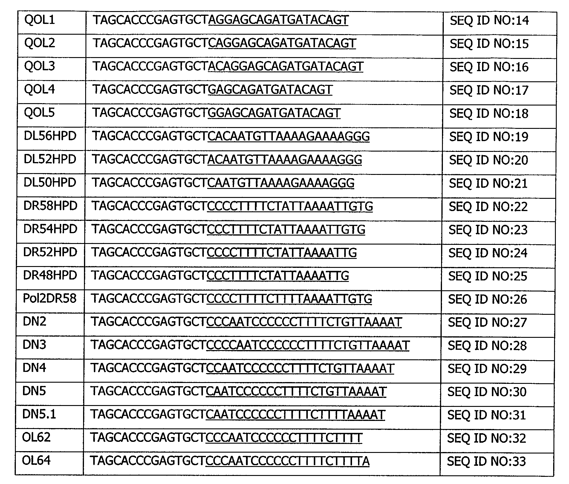
In addition, the following detector primers were designed for real-time detection of
amplification products produced using the amplification primers. The structure and use of such
detector primers is described, for example, in US Patent No. 5,547,861 and US Patent No. 5,928,869.
The target binding sequences are underlined. The remaining portion of the sequence forms a hairpin
structure which is typically labeled to facilitate detection of amplification products as is known in the
art. It will be readily apparent that the target sequence may be used alone for direct detection by
hybridization (typically linked to a detectable label) and that other directly and indirectly detectable
sequences and labels may be substituted for the hairpin as is known in the art. See, for example US
Patent No. 5,935,791; US Patent No. 5,846,726; US Patent No. 5,691,145; US Patent No. 5,550,025
and US Patent No. 5,593,867.
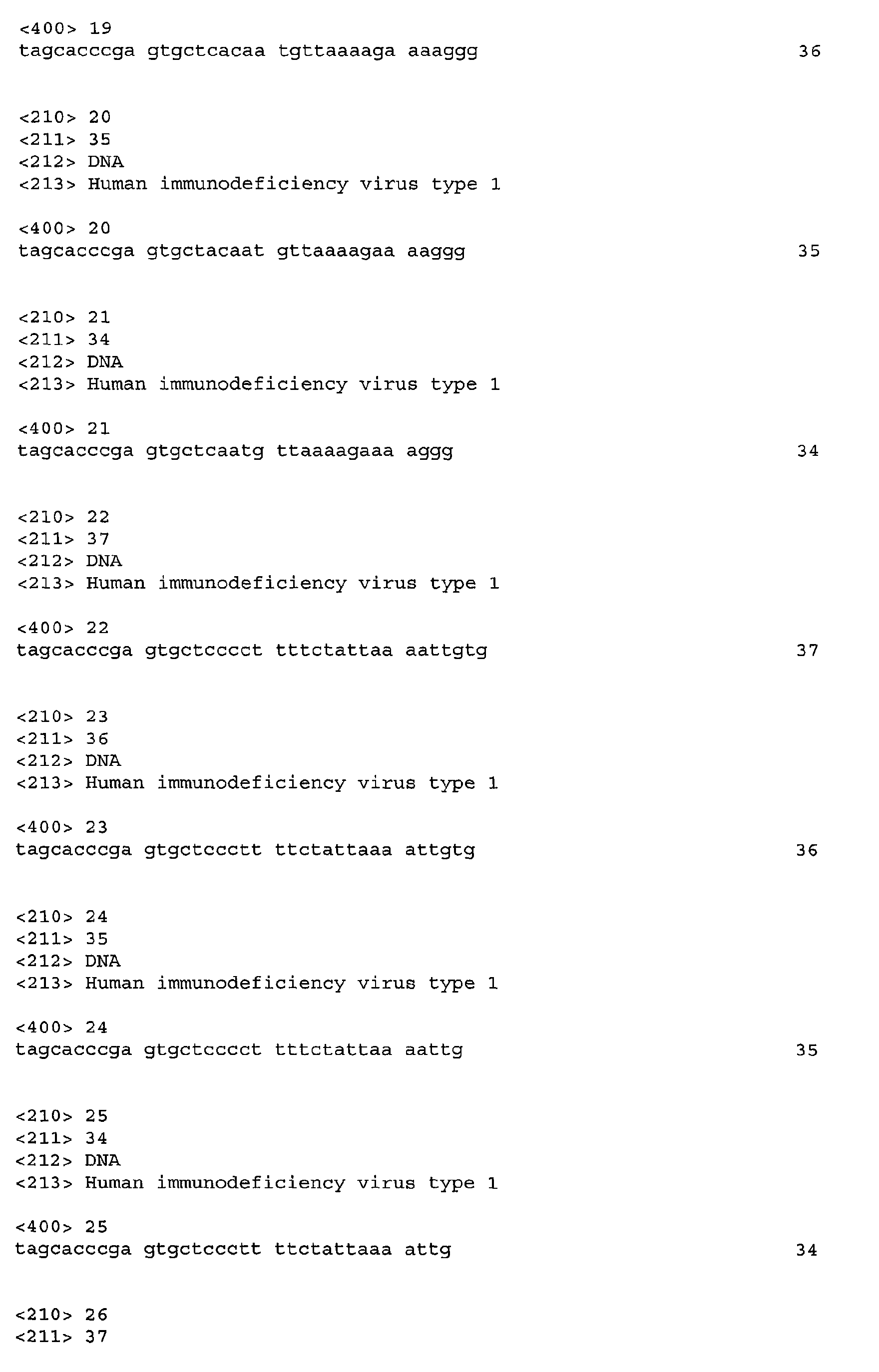
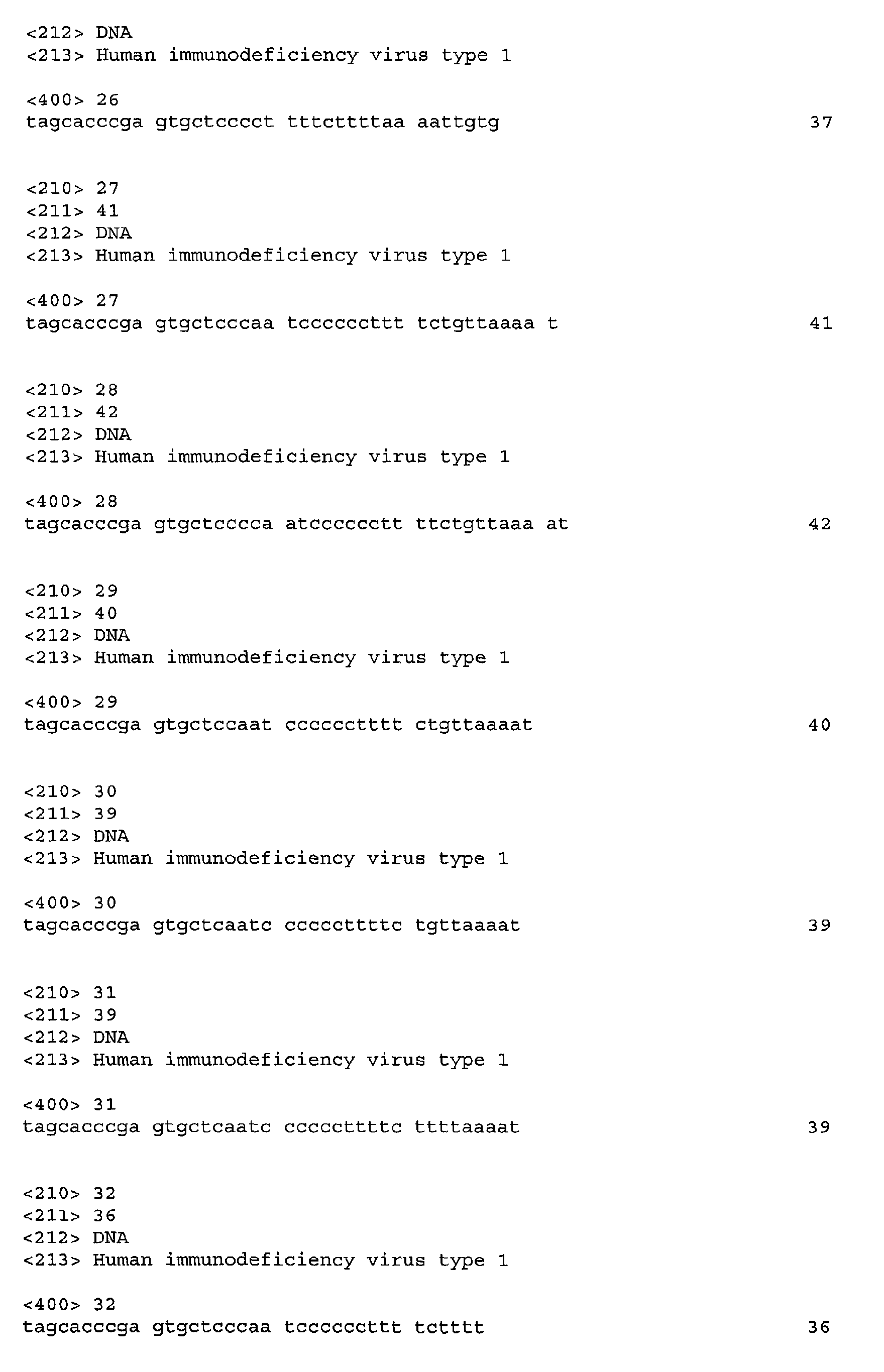
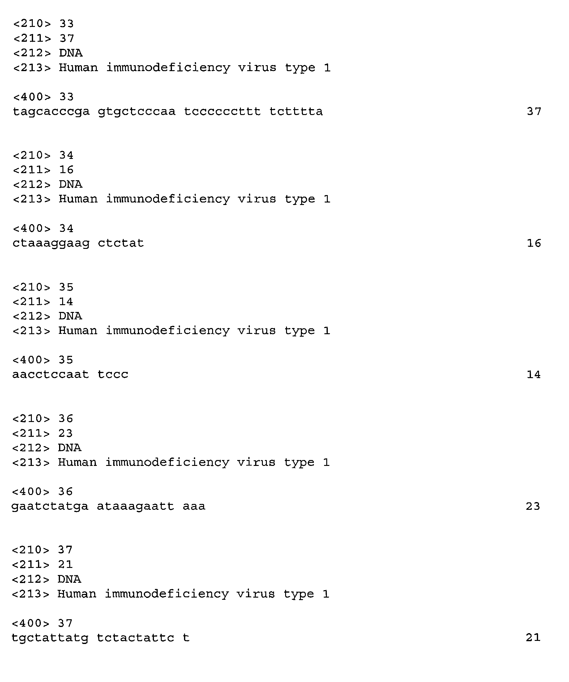
SEQ ID NOs:11-13 and 19-26 are conventional non-overlapping detector primers which
contain a hairpin as described in US Patent No. 5,928,869. SEQ ID NOs:14-18 and 27-33 also contain
the hairpin but the 5' end of the target binding sequences overlap with the 3' end of the target
binding sequences of the upstream amplification primers.
Bumper primers used in SDA (BR and BL) were also designed. The entire sequence of these
oligonucleotides consists of target binding sequence:
The sequences set forth above were selected to minimize the effects of heterogeneity in the
targeted region of the pol gene. Mismatches were confined to the middle or the 5' end of the
sequences to permit efficient 3' extension upon hybridization to the target sequence. Only three of
the 117 HIV-1 strains analyzed exhibit more than one mismatch with amplification primers SEQ ID
NO:5 and SEQ ID NO:9 or with detector SEQ ID NO:26. Detector SEQ ID NO:27-30 contain a
deliberate mismatch eight bases from the 3' end to minimize target-specific variations in priming
efficiency caused by heterogeneity in the target region.
Because the target binding sequence confers target specificity on the primer or probe, it
should be understood that the target binding sequences exemplified above for use as particular
components of a specific amplification reaction may also be used in a variety of other ways for
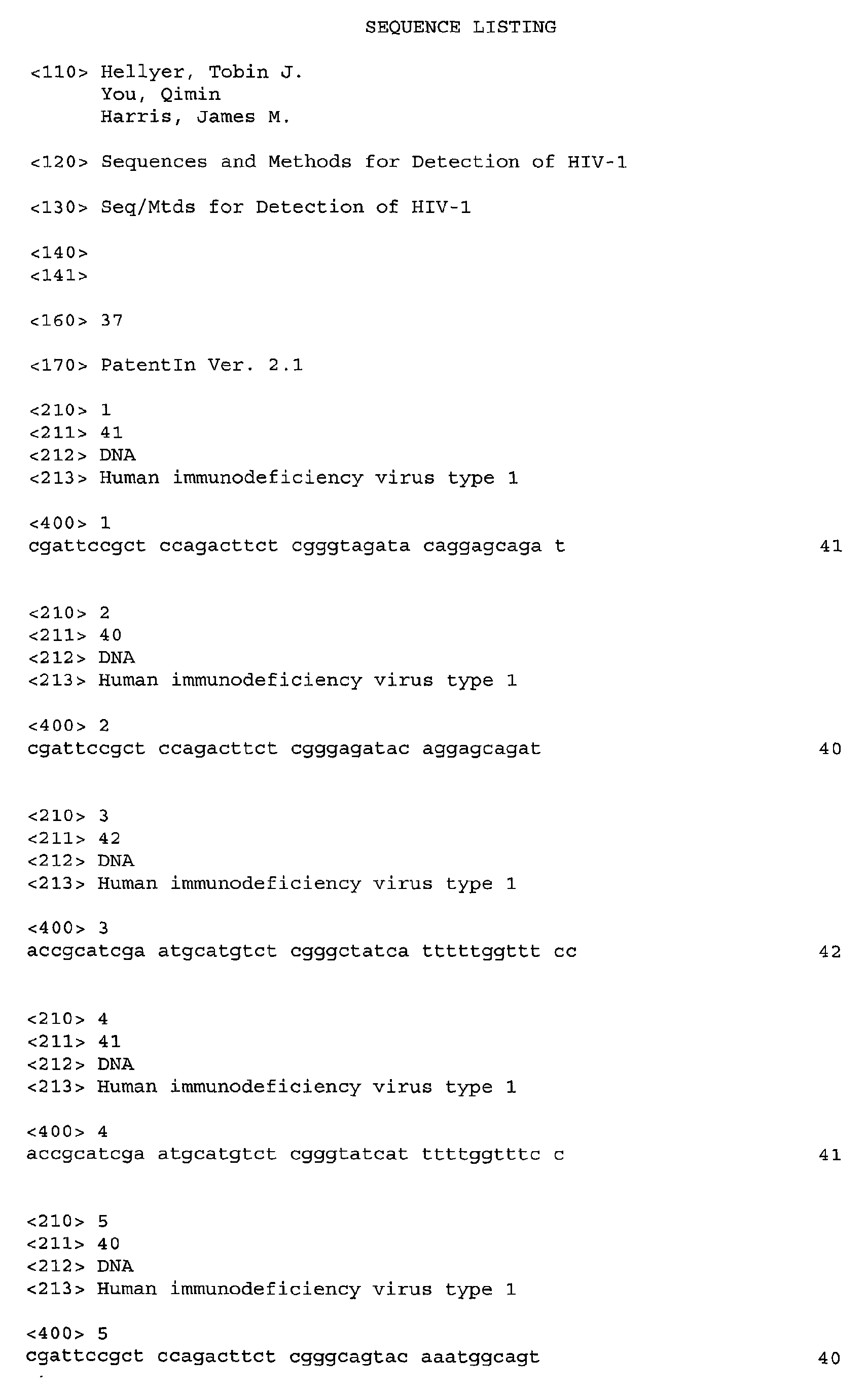
detection of HIV. For example, the target binding sequences of SEQ ID NOs:1-37 may alternatively
be used as hybridization probes for direct detection of HIV-1, either without prior amplification or as a
post-amplification assay. Such hybridization methods are well known in the art and typically employ
a detectable label associated with or linked to the target binding sequence to facilitate detection of
hybridization. Further, essentially all of the target binding sequences set forth above may be used as
amplification primers in amplification reactions which do not require additional specialized sequences
(such as PCR) or appended to the appropriate specialized sequences for use in 3SR, NASBA,
transcription-based or any other primer extension amplification reactions. For detection of
amplification products, amplification primers comprising the target binding sequences disclosed herein
may be labeled as is known in the art, or labeled detector primers comprising the disclosed target
binding sequences may be used in conjunction with the amplification primers as described in US
Patent No. 5,547,861 and US Patent No. 5,928,869 for real-time homogeneous detection of
amplification. Such detector primers typically comprise a directly or indirectly detectable sequence
which does not initially hybridize to the target but which facilitates detection of the detector primer
once it has hybridized to the target and been extended. For example, such detectable sequences
may be sequences which form a secondary structure, sequences which contain a restriction site, or
linear sequences which are detected by hybridization of their complements to a labeled
oligonucleotide (sometimes referred to as a reporter probe) as is known in the art. Alternatively, the
amplification products may be detected post-amplification by hybridization of a probe selected from
any of the target binding sequences disclosed herein which fall between a selected set of
amplification primers.
It is to be understood that an oligonucleotide according to the invention which consists of a
target binding sequence and, optionally, either a sequence required for a selected amplification
reaction or a sequence required for a selected detection reaction may also include certain other
sequences which serve as spacers, linkers, sequences for labeling or binding of an enzyme, etc. Such
additional sequences are typically known to be necessary to obtain optimum function of the
oligonucleotide in the selected reaction and are intended to be included by the term "consisting of."
EXAMPLE 1
SDA reactions were performed to determine the analytical sensitivity of the assay for the
detection of HIV DNA. Amplification was carried in the presence of 0, 5, 10, 50, 100 or 250 copies of
HIV target DNA. The target sequence was a fragment of the HIV-1 genome corresponding to
nucleotides 4659-4910 of strain B-WEAU (GenBank accession number U21135) that was cloned into
the plasmid vector pBlueScript SK+ (Stratagene). SDA was performed at 52°C using 500nM primers
(SEQ ID NO:5 and SEQ ID NO:9), 50nM bumpers (SEQ ID NO:36 and SEQ ID NO:37), and 200nM
detector probe, SEQ ID NO:26, in buffer containing: 100mM bicine; 30mM potassium hydroxide;
68mM KiPO4, pH7.6; 10.5% glycerol; 6.5% DMSO; 0.7mM dCsTP; 0.1mM dA-, dG- and dTTP; 70ng/µl
human placental DNA; 100ng/µl bovine serum albumin; 4mM magnesium acetate; 6U Bst polymerase
and 32U BsoBI restriction enzyme. The detector primer was labeled at the 5' terminus with a
fluorescein donor molecule and internally with a dabcyl quencher moiety. The two dyes on the
detector were separated by a BsoBI restriction endonuclease recognition sequence and held in close
juxtaposition by a hairpin structure within the tail sequence as described in US Patent Nos. 5,919,630;
5,928,869 and 5,958,700. Donor fluorescence was monitored during the course of amplification.
In the presence of target, donor fluorescence increased during the course of the reaction as
the hairpin holding the donor and quencher in close proximity unfolded and the restriction site was
cleaved. In contrast, in the absence of target, fluorescence remained consistently low throughout the
reaction. Results were expressed in terms of area under the curve or "MOTA." A larger MOTA score
indicates generation of more fluorescence and, generally, the presence of more input target. The
results are shown in Table 1.
| TARGETS PER REACTION | MEAN MOTA SCORE (n=28) |
| 0 | 606 |
| 5 | 14883 |
| 10 | 20565 |
| 50 | 47643 |
| 100 | 56740 |
| 150 | 61676 |
The limit of detection (LOD), defined as the input target level at which 95% of reactions
would yield a positive result, was determined to be between 9 and 11 copies of target DNA depending
on the MOTA score selected as a cut-off for determining a positive result. These results demonstrate
that the amplification and detector primers are capable of sensitive and reproducible detection of HIV
DNA.
EXAMPLE 2
A similar experiment to that described in Example 1 was performed using the SEQ ID NO:27
detector primer, the target hybridization region of which overlaps the SEQ ID NO:9 amplification
primer. This probe was designed to take advantage of sequence conservation in the amplification
primer binding region while maintaining the specificity afforded by detection of the internal region of
the SDA amplicon that lies between the two amplification primers. Buffer conditions were the same as
described above with the following modifications: 75mM KiPO4, 14% DMSO, 5% glycerol and 27U
BsoBI. The limit of detection using the overlapping probe was 61 to 86 copies of HIV target DNA
depending on the MOTA score selected for determining a positive result. These results demonstrate
sensitive and specific detection of HIV target DNA using an alternative probe design that offers
additional flexibility in the development of SDA-based systems.
EXAMPLE 3
The SDA assay for HIV DNA was converted to a two-step reverse transcriptase (RT)-SDA
format in which RNA was first copied to cDNA using an RT enzyme and then amplified in a
conventional SDA reaction. One important difference between the SDA conditions described in
Examples 1 and 2 and the RT-SDA described here is the absence of bumper primers. The need for
bumpers apparently was precluded by the choice of an RT enzyme which possesses RNase H activity
and which is therefore capable of degrading the RNA template following reverse transcription, thereby
liberating a single stranded DNA target sequence in to solution. An LOD study was performed for HIV
RNA using in vitro transcripts of a genotype B strain generated from the pBlueScript plasmid clone
described in Example 1.
Reverse transcription was carried out in microtiter wells using 15U avian myeloblastosis virus
(AMV)-RT in buffer containing: 69.3mM bicine; 12.3mM KOH; 20.8mM KiPO4; 3% glycerol; 4.5%
DMSO; 6mM magnesium acetate; 2.75mM dCsTP; 0.25mM dA-, dG- and dTTP; 100ng/µl BSA;
1250nM SEQ ID NO:5 and 700nM SEQ ID NO:9. In brief, the reverse transcription reaction mixture
without enzyme was incubated for 10 min at 68°C to denature the target RNA. Eighty microliters of
denatured target was then added to a microwell containing 20µl of an RT enzyme mixture that was
pre-equilibrated at 50°C. The complete RT reaction was incubated for 10 min to facilitate synthesis of
cDNA and degradation of the RNA template by the RNase H activity of the RT enzyme. To initiate
amplification, 40µl of the reverse transcription reaction was transferred to a second microtiter well at
52°C containing 60µl of an amplification mixture comprising Bst polymerase, BsoBI restriction
enzyme, and fluorescent detector primer together with certain buffer components. The final SDA
conditions were as follows: 72mM bicine; 24mM potassium hydroxide; 54.4mM KiPO4; 4% glycerol;
11% DMSO; 0.1mM dA-, dG- and dTTP; 1.1mM dCsTP; 700ng/µl hpDNA; 100ng/µl BSA; 500nM SEQ
ID NO:5; 300nM SEQ ID NO:9; 600nM SEQ ID NO:26; 12U BsoBI and 7U Bst polymerase. The wells
were sealed and incubated at 52°C. Donor fluorescence was monitored throughout the course of the
reaction.
The results of this experiment are shown in Table 2.
| RNA TARGETS PER REACTION | MEAN MOTA SCORE (n=17) | % POSITIVE |
| 0 | 139 | 0 |
| 10 | 3577 | 64.7 |
| 25 | 7810 | 88.0 |
| 50 | 13410 | 100.0 |
| 75 | 23641 | 100.0 |
| 100 | 35719 | 100.0 |
| 500 | 73870 | 100.0 |
MOTA scores ≥1000 were considered positive . All reactions containing ≥50 copies of HIV
target RNA were positive as were 88% containing 25 copies and 65% of those containing 10 copies.
These results demonstrate the sensitivity of the disclosed amplification and detector primers for the
detection of HIV-1 RNA.
EXAMPLE 4
RT-SDA was performed on purified RNA from representative isolates of nine different clades
of HIV-1 (Boston Biomedica, Inc.). In order to quantify the parental stocks of viral RNA, all except the
type O nucleic acid were tested with each of two commercially available quantitative HIV tests: the
Roche Amplicor HIV-1 Monitor V1.5 and the Chiron Quantiplex HIV-1 RNA 3.0 Assay. Since neither of
these tests is able to amplify RNA of HIV-1 genotype O, a third non-amplified system, the Digene
Hybrid Capture Assay, was used to quantify the type O nucleic acid. Viral RNAs for genotypes A-H
were diluted according to the results of the Roche assay to give the equivalent of 200 copies per RT-SDA
reaction. Type O RNA was diluted to the same level based on the results of the Digene test. RT-SDA
was performed according to the method described in Example 3. For eight of the nine clades, all
sixteen replicates were positive at 200 copies per RT-SDA reaction. For genotype F, 4/16 (25%)
replicates were positive. The reason for the apparently lower sensitivity with type F is unclear but
might be attributed to inaccurate quantification of the parental RNA stock of this genotype by the
Roche assay. Alternatively, the discrepancy might be due to the presence in the type F sequence of a
single base mismatch with the right amplification primer which is used to initiate first strand cDNA
synthesis (SEQ ID NO:9). Importantly, all reactions conducted with 2000 input copies of type F RNA
were positive.
These data demonstrate that the disclosed primers and probes are capable of detecting
multiple genotypes of HIV-1, including type O, with a high degree of sensitivity. Detection of type O
RNA is particularly important in view of the inability to detect this clade with the majority of
commercially available diagnostic nucleic acid assays.
EXAMPLE 5
In vitro transcripts generated from the plasmid clone described in Example 1 were quantified
by competitive RT-SDA. In brief, RT-SDA was performed as described above with the exception that
two detector primers were included in the reaction mixture, both at a concentration of 200nM. The
first primer, SEQ ID NO:26, was specific for HIV-1 and was labeled at the 5' end with dabcyl quencher
moiety and internally with rhodamine (Rox). The second probe was specific for an internal control
sequence and was labeled at its 5' end with fluorescein and internally with a dabcyl quencher. The
internal control comprised an RNA molecule that was generated by in vitro transcription of a mutated
clone of the HIV-1 pol gene. The internal control possessed the same primer binding regions as the
native HIV target but differed by a series of point mutations introduced to coincide with the 3' end of
the detector hybridization region. These mutations permitted discrimination of the native target and
internal control by preventing hybridization and extension of the mismatched probes during
amplification. The internal control was designed such that it amplified with similar efficiency to the
native target and would compete during the course of the reaction for one or more rate-limiting
reagents. The same amount of internal control was seeded into each test sample and into a series of
calibrator reactions containing known amounts of native target. Details of the theoretical aspects of
the quantitative SDA algorithm may be found elsewhere (J. G. Nadeau, et al. 1999. Anal Biochem 276, 177-187; C. M. Nycz, et al. 1998. Anal Biochem 259, 226-234). In brief, the fluorescence
produced by amplification of both the native target and internal control was monitored at discreet
intervals throughout the reaction. Data were processed through a series of normalization and
smoothing functions to produce a fluorescent signal for both sequences. From these values, the
natural log of the ratio of native target and internal control signals was calculated. The values
obtained from the calibrator wells were plotted as a regression against the natural log of the input
number of target molecules of RNA to produce calibration curves corresponding to each time point.
An automated algorithm was then used to determine the time at which it was statistically optimal to
perform quantification.
Fig. 1 shows the MOTA scores for the internal control with various copy numbers of input
target sequence. MOTA scores for the internal control decreased with increasing levels of native
target, demonstrating the competition between the two targets that is necessary for accurate
quantification. Table 3 shows the results of quantitative competitive RT-SDA with between 50 and
100000 copies of native RNA target and 5000 copies of internal control per reaction. At all but one
target level, accuracy and precision were better than ± 25%, thus demonstrating the ability to
quantify HIV-1 RNA using the disclosed amplification and detector primer sequences.
| TARGET RNA COPIES/REACTION | | PERCENT |
| REVERSE TRANSCR. | SDA | MEAN (n=8) | ACCURACY | PRECISION | |
| 0 | 0 | 16 | | 62.78 |
| 125 | 50 | 47 | -5.17 | 25.47 |
| 1250 | 500 | 537 | 7.45 | 24.19 |
| 12500 | 5000 | 8765 | 75.29 | 9.07 |
| 125000 | 50000 | 60009 | 20.02 | 5.48 |
| 250000 | 100000 | 76351 | -23.65 | 7.26 |
Sequence alignment data and initial testing of the additional target binding sequences
disclosed herein indicates that similar results would be obtained using these sequences in probes and
primers for amplification and/or detection of all genotypes of HIV-1.