EP1220949B1 - Chromosome-wide analysis of protein-dna interactions - Google Patents
Chromosome-wide analysis of protein-dna interactions Download PDFInfo
- Publication number
- EP1220949B1 EP1220949B1 EP00959909A EP00959909A EP1220949B1 EP 1220949 B1 EP1220949 B1 EP 1220949B1 EP 00959909 A EP00959909 A EP 00959909A EP 00959909 A EP00959909 A EP 00959909A EP 1220949 B1 EP1220949 B1 EP 1220949B1
- Authority
- EP
- European Patent Office
- Prior art keywords
- dna
- protein
- cell
- interest
- region
- Prior art date
- Legal status (The legal status is an assumption and is not a legal conclusion. Google has not performed a legal analysis and makes no representation as to the accuracy of the status listed.)
- Expired - Lifetime
Links
- 238000004458 analytical method Methods 0.000 title description 21
- 230000003993 interaction Effects 0.000 title description 7
- 108020004414 DNA Proteins 0.000 claims abstract description 173
- 108090000623 proteins and genes Proteins 0.000 claims abstract description 130
- 102000004169 proteins and genes Human genes 0.000 claims abstract description 79
- 238000000034 method Methods 0.000 claims abstract description 56
- 230000000295 complement effect Effects 0.000 claims abstract description 34
- 102000052510 DNA-Binding Proteins Human genes 0.000 claims abstract description 25
- 238000009396 hybridization Methods 0.000 claims abstract description 22
- 101710096438 DNA-binding protein Proteins 0.000 claims abstract description 19
- 239000012634 fragment Substances 0.000 claims description 69
- 210000004027 cell Anatomy 0.000 claims description 64
- 238000003752 polymerase chain reaction Methods 0.000 claims description 19
- WSFSSNUMVMOOMR-UHFFFAOYSA-N Formaldehyde Chemical compound O=C WSFSSNUMVMOOMR-UHFFFAOYSA-N 0.000 claims description 12
- 239000000203 mixture Substances 0.000 claims description 11
- 108091023040 Transcription factor Proteins 0.000 claims description 10
- 102000040945 Transcription factor Human genes 0.000 claims description 10
- 238000000018 DNA microarray Methods 0.000 claims description 9
- 238000004132 cross linking Methods 0.000 claims description 3
- 230000001404 mediated effect Effects 0.000 claims description 3
- 210000003527 eukaryotic cell Anatomy 0.000 claims description 2
- 238000000527 sonication Methods 0.000 claims description 2
- 108700020796 Oncogene Proteins 0.000 claims 1
- 238000000338 in vitro Methods 0.000 claims 1
- 238000002372 labelling Methods 0.000 claims 1
- 102100039556 Galectin-4 Human genes 0.000 description 42
- 108010001515 Galectin 4 Proteins 0.000 description 41
- 230000027455 binding Effects 0.000 description 29
- 230000014509 gene expression Effects 0.000 description 22
- 210000000349 chromosome Anatomy 0.000 description 20
- 229930182830 galactose Natural products 0.000 description 19
- 240000004808 Saccharomyces cerevisiae Species 0.000 description 18
- 230000001105 regulatory effect Effects 0.000 description 16
- 238000002487 chromatin immunoprecipitation Methods 0.000 description 11
- 230000006870 function Effects 0.000 description 11
- 239000012190 activator Substances 0.000 description 10
- 108700026244 Open Reading Frames Proteins 0.000 description 9
- 239000003016 pheromone Substances 0.000 description 9
- 238000002474 experimental method Methods 0.000 description 8
- 101150085096 fur4 gene Proteins 0.000 description 8
- 101100189545 Saccharomyces cerevisiae (strain ATCC 204508 / S288c) PCL10 gene Proteins 0.000 description 7
- 230000013011 mating Effects 0.000 description 7
- 239000000523 sample Substances 0.000 description 7
- 238000013518 transcription Methods 0.000 description 7
- 230000035897 transcription Effects 0.000 description 7
- 108700020911 DNA-Binding Proteins Proteins 0.000 description 6
- 230000004568 DNA-binding Effects 0.000 description 6
- IXKSXJFAGXLQOQ-XISFHERQSA-N WHWLQLKPGQPMY Chemical compound C([C@@H](C(=O)N[C@@H](CC=1C2=CC=CC=C2NC=1)C(=O)N[C@@H](CC(C)C)C(=O)N[C@@H](CCC(N)=O)C(=O)N[C@@H](CC(C)C)C(=O)N1CCC[C@H]1C(=O)NCC(=O)N[C@@H](CCC(N)=O)C(=O)N[C@@H](CC(O)=O)C(=O)N1CCC[C@H]1C(=O)N[C@@H](CCSC)C(=O)N[C@@H](CC=1C=CC(O)=CC=1)C(O)=O)NC(=O)[C@@H](N)CC=1C2=CC=CC=C2NC=1)C1=CNC=N1 IXKSXJFAGXLQOQ-XISFHERQSA-N 0.000 description 6
- 238000006243 chemical reaction Methods 0.000 description 6
- 230000002759 chromosomal effect Effects 0.000 description 6
- 230000006698 induction Effects 0.000 description 6
- WQZGKKKJIJFFOK-GASJEMHNSA-N Glucose Natural products OC[C@H]1OC(O)[C@H](O)[C@@H](O)[C@@H]1O WQZGKKKJIJFFOK-GASJEMHNSA-N 0.000 description 5
- 101000744394 Homo sapiens Oxidized purine nucleoside triphosphate hydrolase Proteins 0.000 description 5
- 102100039792 Oxidized purine nucleoside triphosphate hydrolase Human genes 0.000 description 5
- 238000003491 array Methods 0.000 description 5
- 239000008103 glucose Substances 0.000 description 5
- 238000001114 immunoprecipitation Methods 0.000 description 5
- 108091006106 transcriptional activators Proteins 0.000 description 5
- XLYOFNOQVPJJNP-UHFFFAOYSA-N water Substances O XLYOFNOQVPJJNP-UHFFFAOYSA-N 0.000 description 5
- 101100043662 Emericella nidulans (strain FGSC A4 / ATCC 38163 / CBS 112.46 / NRRL 194 / M139) steA gene Proteins 0.000 description 4
- 108700039691 Genetic Promoter Regions Proteins 0.000 description 4
- TWRXJAOTZQYOKJ-UHFFFAOYSA-L Magnesium chloride Chemical compound [Mg+2].[Cl-].[Cl-] TWRXJAOTZQYOKJ-UHFFFAOYSA-L 0.000 description 4
- 101150032846 STE12 gene Proteins 0.000 description 4
- 101100390137 Schizosaccharomyces pombe (strain 972 / ATCC 24843) fab1 gene Proteins 0.000 description 4
- ISAKRJDGNUQOIC-UHFFFAOYSA-N Uracil Chemical compound O=C1C=CNC(=O)N1 ISAKRJDGNUQOIC-UHFFFAOYSA-N 0.000 description 4
- 230000000694 effects Effects 0.000 description 4
- 238000002493 microarray Methods 0.000 description 4
- 230000008569 process Effects 0.000 description 4
- 238000011160 research Methods 0.000 description 4
- OKTJSMMVPCPJKN-UHFFFAOYSA-N Carbon Chemical compound [C] OKTJSMMVPCPJKN-UHFFFAOYSA-N 0.000 description 3
- 230000004543 DNA replication Effects 0.000 description 3
- 101150094690 GAL1 gene Proteins 0.000 description 3
- 101150038242 GAL10 gene Proteins 0.000 description 3
- 102100028501 Galanin peptides Human genes 0.000 description 3
- 102100024637 Galectin-10 Human genes 0.000 description 3
- 101710154606 Hemagglutinin Proteins 0.000 description 3
- 101100121078 Homo sapiens GAL gene Proteins 0.000 description 3
- 101710093908 Outer capsid protein VP4 Proteins 0.000 description 3
- 101710135467 Outer capsid protein sigma-1 Proteins 0.000 description 3
- 101710176177 Protein A56 Proteins 0.000 description 3
- HEMHJVSKTPXQMS-UHFFFAOYSA-M Sodium hydroxide Chemical compound [OH-].[Na+] HEMHJVSKTPXQMS-UHFFFAOYSA-M 0.000 description 3
- 230000004913 activation Effects 0.000 description 3
- 230000033228 biological regulation Effects 0.000 description 3
- 229910052799 carbon Inorganic materials 0.000 description 3
- 239000012154 double-distilled water Substances 0.000 description 3
- 239000000185 hemagglutinin Substances 0.000 description 3
- 238000007854 ligation-mediated PCR Methods 0.000 description 3
- 230000037353 metabolic pathway Effects 0.000 description 3
- 238000012544 monitoring process Methods 0.000 description 3
- 102000027450 oncoproteins Human genes 0.000 description 3
- 108091008819 oncoproteins Proteins 0.000 description 3
- 230000010076 replication Effects 0.000 description 3
- 230000004044 response Effects 0.000 description 3
- 108050006400 Cyclin Proteins 0.000 description 2
- 108090000266 Cyclin-dependent kinases Proteins 0.000 description 2
- 102000003903 Cyclin-dependent kinases Human genes 0.000 description 2
- 108010014303 DNA-directed DNA polymerase Proteins 0.000 description 2
- 102000016928 DNA-directed DNA polymerase Human genes 0.000 description 2
- LFQSCWFLJHTTHZ-UHFFFAOYSA-N Ethanol Chemical compound CCO LFQSCWFLJHTTHZ-UHFFFAOYSA-N 0.000 description 2
- 101150090134 FLO11 gene Proteins 0.000 description 2
- 101150037782 GAL2 gene Proteins 0.000 description 2
- 102100021735 Galectin-2 Human genes 0.000 description 2
- 102100039555 Galectin-7 Human genes 0.000 description 2
- 101000967087 Homo sapiens Metal-response element-binding transcription factor 2 Proteins 0.000 description 2
- 101001061518 Homo sapiens RNA-binding protein FUS Proteins 0.000 description 2
- 101000659267 Homo sapiens Tumor suppressor candidate 2 Proteins 0.000 description 2
- KFZMGEQAYNKOFK-UHFFFAOYSA-N Isopropanol Chemical compound CC(C)O KFZMGEQAYNKOFK-UHFFFAOYSA-N 0.000 description 2
- 102100040632 Metal-response element-binding transcription factor 2 Human genes 0.000 description 2
- 108010002747 Pfu DNA polymerase Proteins 0.000 description 2
- 102100036691 Proliferating cell nuclear antigen Human genes 0.000 description 2
- 102100028469 RNA-binding protein FUS Human genes 0.000 description 2
- 241000235070 Saccharomyces Species 0.000 description 2
- 101100012902 Saccharomyces cerevisiae (strain ATCC 204508 / S288c) FIG2 gene Proteins 0.000 description 2
- FAPWRFPIFSIZLT-UHFFFAOYSA-M Sodium chloride Chemical compound [Na+].[Cl-] FAPWRFPIFSIZLT-UHFFFAOYSA-M 0.000 description 2
- 230000003321 amplification Effects 0.000 description 2
- 230000003915 cell function Effects 0.000 description 2
- 230000004663 cell proliferation Effects 0.000 description 2
- 108091092356 cellular DNA Proteins 0.000 description 2
- 230000001413 cellular effect Effects 0.000 description 2
- 230000008859 change Effects 0.000 description 2
- 108010045512 cohesins Proteins 0.000 description 2
- 230000001419 dependent effect Effects 0.000 description 2
- 238000013461 design Methods 0.000 description 2
- 238000001514 detection method Methods 0.000 description 2
- 238000010195 expression analysis Methods 0.000 description 2
- 239000011521 glass Substances 0.000 description 2
- 230000002621 immunoprecipitating effect Effects 0.000 description 2
- 238000001727 in vivo Methods 0.000 description 2
- 229910001629 magnesium chloride Inorganic materials 0.000 description 2
- 230000007246 mechanism Effects 0.000 description 2
- 239000012528 membrane Substances 0.000 description 2
- 230000004060 metabolic process Effects 0.000 description 2
- 238000003199 nucleic acid amplification method Methods 0.000 description 2
- 102000039446 nucleic acids Human genes 0.000 description 2
- 108020004707 nucleic acids Proteins 0.000 description 2
- 150000007523 nucleic acids Chemical class 0.000 description 2
- 238000007639 printing Methods 0.000 description 2
- 239000011541 reaction mixture Substances 0.000 description 2
- 238000010008 shearing Methods 0.000 description 2
- 229940035893 uracil Drugs 0.000 description 2
- 210000005253 yeast cell Anatomy 0.000 description 2
- FQVLRGLGWNWPSS-BXBUPLCLSA-N (4r,7s,10s,13s,16r)-16-acetamido-13-(1h-imidazol-5-ylmethyl)-10-methyl-6,9,12,15-tetraoxo-7-propan-2-yl-1,2-dithia-5,8,11,14-tetrazacycloheptadecane-4-carboxamide Chemical compound N1C(=O)[C@@H](NC(C)=O)CSSC[C@@H](C(N)=O)NC(=O)[C@H](C(C)C)NC(=O)[C@H](C)NC(=O)[C@@H]1CC1=CN=CN1 FQVLRGLGWNWPSS-BXBUPLCLSA-N 0.000 description 1
- DOSMHBDKKKMIEF-UHFFFAOYSA-N 2-[3-(diethylamino)-6-diethylazaniumylidenexanthen-9-yl]-5-[3-[3-[4-(1-methylindol-3-yl)-2,5-dioxopyrrol-3-yl]indol-1-yl]propylsulfamoyl]benzenesulfonate Chemical compound C1=CC(=[N+](CC)CC)C=C2OC3=CC(N(CC)CC)=CC=C3C(C=3C(=CC(=CC=3)S(=O)(=O)NCCCN3C4=CC=CC=C4C(C=4C(NC(=O)C=4C=4C5=CC=CC=C5N(C)C=4)=O)=C3)S([O-])(=O)=O)=C21 DOSMHBDKKKMIEF-UHFFFAOYSA-N 0.000 description 1
- 102100026433 39S ribosomal protein L14, mitochondrial Human genes 0.000 description 1
- PDWZXKSZLRVSEH-UHFFFAOYSA-N 6-chloro-4-n-cyclopropyl-4-n-(thiophen-2-ylmethyl)pyrimidine-2,4-diamine Chemical compound NC1=NC(Cl)=CC(N(CC=2SC=CC=2)C2CC2)=N1 PDWZXKSZLRVSEH-UHFFFAOYSA-N 0.000 description 1
- 101150025253 AFR1 gene Proteins 0.000 description 1
- 101150031810 AGP1 gene Proteins 0.000 description 1
- 102100039601 ARF GTPase-activating protein GIT1 Human genes 0.000 description 1
- 102100034035 Alcohol dehydrogenase 1A Human genes 0.000 description 1
- 102100022463 Alpha-1-acid glycoprotein 1 Human genes 0.000 description 1
- 102100039160 Amiloride-sensitive amine oxidase [copper-containing] Human genes 0.000 description 1
- 102100033972 Amyloid protein-binding protein 2 Human genes 0.000 description 1
- 101100165241 Arabidopsis thaliana BCP1 gene Proteins 0.000 description 1
- 101100168642 Arabidopsis thaliana CRN gene Proteins 0.000 description 1
- 101100277337 Arabidopsis thaliana DDM1 gene Proteins 0.000 description 1
- 101100388296 Arabidopsis thaliana DTX51 gene Proteins 0.000 description 1
- 101100223316 Arabidopsis thaliana GAD2 gene Proteins 0.000 description 1
- 101100184272 Arabidopsis thaliana NIFS1 gene Proteins 0.000 description 1
- 101100408455 Arabidopsis thaliana PLC7 gene Proteins 0.000 description 1
- 101100257981 Arabidopsis thaliana S-ACP-DES3 gene Proteins 0.000 description 1
- 101100532684 Arabidopsis thaliana SCC3 gene Proteins 0.000 description 1
- 101100045632 Arabidopsis thaliana TCX2 gene Proteins 0.000 description 1
- 101100482999 Arabidopsis thaliana TSM1 gene Proteins 0.000 description 1
- 238000012935 Averaging Methods 0.000 description 1
- 101150083885 BIK1 gene Proteins 0.000 description 1
- 101150103205 BUD3 gene Proteins 0.000 description 1
- 102100030981 Beta-alanine-activating enzyme Human genes 0.000 description 1
- 102100031024 CCR4-NOT transcription complex subunit 1 Human genes 0.000 description 1
- 102100026049 CDP-diacylglycerol-glycerol-3-phosphate 3-phosphatidyltransferase, mitochondrial Human genes 0.000 description 1
- 101150059868 CTR86 gene Proteins 0.000 description 1
- 101100327917 Caenorhabditis elegans chup-1 gene Proteins 0.000 description 1
- 108091006146 Channels Proteins 0.000 description 1
- 108010077544 Chromatin Proteins 0.000 description 1
- 108091026890 Coding region Proteins 0.000 description 1
- 102100035590 Cohesin subunit SA-1 Human genes 0.000 description 1
- 102100037700 DNA mismatch repair protein Msh3 Human genes 0.000 description 1
- 102100031137 DNA-directed RNA polymerase II subunit RPB7 Human genes 0.000 description 1
- 101000785279 Dictyostelium discoideum Calcium-transporting ATPase PAT1 Proteins 0.000 description 1
- 101000609814 Dictyostelium discoideum Protein disulfide-isomerase 1 Proteins 0.000 description 1
- 101100166522 Dictyostelium discoideum cycB gene Proteins 0.000 description 1
- 101710167313 Drebrin-like protein Proteins 0.000 description 1
- 241000255581 Drosophila <fruit fly, genus> Species 0.000 description 1
- 101100237168 Drosophila melanogaster Mettl14 gene Proteins 0.000 description 1
- 101100080608 Drosophila melanogaster comt gene Proteins 0.000 description 1
- KCXVZYZYPLLWCC-UHFFFAOYSA-N EDTA Chemical compound OC(=O)CN(CC(O)=O)CCN(CC(O)=O)CC(O)=O KCXVZYZYPLLWCC-UHFFFAOYSA-N 0.000 description 1
- 101150031726 ERS1 gene Proteins 0.000 description 1
- 241000588724 Escherichia coli Species 0.000 description 1
- 102100026064 Exosome complex component RRP43 Human genes 0.000 description 1
- 101150032501 FUS3 gene Proteins 0.000 description 1
- 102000018711 Facilitative Glucose Transport Proteins Human genes 0.000 description 1
- 102100022366 Fatty acyl-CoA reductase 1 Human genes 0.000 description 1
- 102100026121 Flap endonuclease 1 Human genes 0.000 description 1
- 108090000652 Flap endonucleases Proteins 0.000 description 1
- 230000010190 G1 phase Effects 0.000 description 1
- 101150082479 GAL gene Proteins 0.000 description 1
- 101150103804 GAL3 gene Proteins 0.000 description 1
- 101150103317 GAL80 gene Proteins 0.000 description 1
- 101150031227 GDH2 gene Proteins 0.000 description 1
- 101150108435 GLK1 gene Proteins 0.000 description 1
- 101710193897 Galactose transporter Proteins 0.000 description 1
- 101710103223 Galactose-proton symporter Proteins 0.000 description 1
- 102100039558 Galectin-3 Human genes 0.000 description 1
- 101000887163 Gallus gallus Gallinacin-4 Proteins 0.000 description 1
- 101000887166 Gallus gallus Gallinacin-7 Proteins 0.000 description 1
- 101000892220 Geobacillus thermodenitrificans (strain NG80-2) Long-chain-alcohol dehydrogenase 1 Proteins 0.000 description 1
- 108091052347 Glucose transporter family Proteins 0.000 description 1
- 229920002527 Glycogen Polymers 0.000 description 1
- 102100028541 Guanylate-binding protein 2 Human genes 0.000 description 1
- 101150009006 HIS3 gene Proteins 0.000 description 1
- 101150069554 HIS4 gene Proteins 0.000 description 1
- 101150069289 HMLALPHA2 gene Proteins 0.000 description 1
- 241000282412 Homo Species 0.000 description 1
- 101000670360 Homo sapiens 39S ribosomal protein L32, mitochondrial Proteins 0.000 description 1
- 101000888659 Homo sapiens ARF GTPase-activating protein GIT1 Proteins 0.000 description 1
- 101000780443 Homo sapiens Alcohol dehydrogenase 1A Proteins 0.000 description 1
- 101000779309 Homo sapiens Amyloid protein-binding protein 2 Proteins 0.000 description 1
- 101000773364 Homo sapiens Beta-alanine-activating enzyme Proteins 0.000 description 1
- 101000919672 Homo sapiens CCR4-NOT transcription complex subunit 1 Proteins 0.000 description 1
- 101000692362 Homo sapiens CDP-diacylglycerol-glycerol-3-phosphate 3-phosphatidyltransferase, mitochondrial Proteins 0.000 description 1
- 101001027762 Homo sapiens DNA mismatch repair protein Msh3 Proteins 0.000 description 1
- 101000729332 Homo sapiens DNA-directed RNA polymerase II subunit RPB7 Proteins 0.000 description 1
- 101001130401 Homo sapiens E3 ubiquitin-protein ligase RAD18 Proteins 0.000 description 1
- 101001055989 Homo sapiens Exosome complex component RRP43 Proteins 0.000 description 1
- 101000824458 Homo sapiens Fatty acyl-CoA reductase 1 Proteins 0.000 description 1
- 101000608765 Homo sapiens Galectin-4 Proteins 0.000 description 1
- 101000608772 Homo sapiens Galectin-7 Proteins 0.000 description 1
- 101001058858 Homo sapiens Guanylate-binding protein 2 Proteins 0.000 description 1
- 101000998969 Homo sapiens Inositol-3-phosphate synthase 1 Proteins 0.000 description 1
- 101000971351 Homo sapiens KRR1 small subunit processome component homolog Proteins 0.000 description 1
- 101001121408 Homo sapiens L-amino-acid oxidase Proteins 0.000 description 1
- 101100021579 Homo sapiens L1RE1 gene Proteins 0.000 description 1
- 101001018064 Homo sapiens Lysosomal-trafficking regulator Proteins 0.000 description 1
- 101001131592 Homo sapiens Periodic tryptophan protein 2 homolog Proteins 0.000 description 1
- 101001099381 Homo sapiens Peroxisomal biogenesis factor 19 Proteins 0.000 description 1
- 101001072903 Homo sapiens Phosphoglucomutase-2 Proteins 0.000 description 1
- 101000579123 Homo sapiens Phosphoglycerate kinase 1 Proteins 0.000 description 1
- 101000994626 Homo sapiens Potassium voltage-gated channel subfamily A member 1 Proteins 0.000 description 1
- 101000842368 Homo sapiens Protein HIRA Proteins 0.000 description 1
- 101000582366 Homo sapiens Protein RER1 Proteins 0.000 description 1
- 101001114059 Homo sapiens Protein-arginine deiminase type-1 Proteins 0.000 description 1
- 101000713296 Homo sapiens Proton-coupled amino acid transporter 1 Proteins 0.000 description 1
- 101001074528 Homo sapiens Regulating synaptic membrane exocytosis protein 1 Proteins 0.000 description 1
- 101100043640 Homo sapiens STAG1 gene Proteins 0.000 description 1
- 101000632319 Homo sapiens Septin-7 Proteins 0.000 description 1
- 101000667110 Homo sapiens Vacuolar protein sorting-associated protein 13B Proteins 0.000 description 1
- 101000976610 Homo sapiens Zinc finger protein 410 Proteins 0.000 description 1
- 101000976645 Homo sapiens Zinc finger protein ZIC 3 Proteins 0.000 description 1
- 102100036881 Inositol-3-phosphate synthase 1 Human genes 0.000 description 1
- 108091029795 Intergenic region Proteins 0.000 description 1
- 101150055855 KAR5 gene Proteins 0.000 description 1
- 102100021559 KRR1 small subunit processome component homolog Human genes 0.000 description 1
- 102100026388 L-amino-acid oxidase Human genes 0.000 description 1
- 102100031764 LINE-1 retrotransposable element ORF1 protein Human genes 0.000 description 1
- 102100033472 Lysosomal-trafficking regulator Human genes 0.000 description 1
- 101150041583 MATALPHA1 gene Proteins 0.000 description 1
- 101150103998 MATALPHA2 gene Proteins 0.000 description 1
- 102100025169 Max-binding protein MNT Human genes 0.000 description 1
- 102000005455 Monosaccharide Transport Proteins Human genes 0.000 description 1
- 108010006769 Monosaccharide Transport Proteins Proteins 0.000 description 1
- 102100027110 N-alpha-acetyltransferase 38, NatC auxiliary subunit Human genes 0.000 description 1
- 108091028043 Nucleic acid sequence Proteins 0.000 description 1
- 101150110809 ORM1 gene Proteins 0.000 description 1
- 108010067244 Origin Recognition Complex Proteins 0.000 description 1
- 102100040591 Origin recognition complex subunit 1 Human genes 0.000 description 1
- KJWZYMMLVHIVSU-IYCNHOCDSA-N PGK1 Chemical compound CCCCC[C@H](O)\C=C\[C@@H]1[C@@H](CCCCCCC(O)=O)C(=O)CC1=O KJWZYMMLVHIVSU-IYCNHOCDSA-N 0.000 description 1
- 101150012394 PHO5 gene Proteins 0.000 description 1
- 101150054516 PRD1 gene Proteins 0.000 description 1
- 102100034421 Periodic tryptophan protein 2 homolog Human genes 0.000 description 1
- 102100038883 Peroxisomal biogenesis factor 19 Human genes 0.000 description 1
- 102100036629 Phosphoglucomutase-2 Human genes 0.000 description 1
- 102100028251 Phosphoglycerate kinase 1 Human genes 0.000 description 1
- 108091000080 Phosphotransferase Proteins 0.000 description 1
- 239000004743 Polypropylene Substances 0.000 description 1
- 102100034368 Potassium voltage-gated channel subfamily A member 1 Human genes 0.000 description 1
- 102100030473 Protein HIRA Human genes 0.000 description 1
- 102100030594 Protein RER1 Human genes 0.000 description 1
- 102100023222 Protein-arginine deiminase type-1 Human genes 0.000 description 1
- 102000001170 RAD18 Human genes 0.000 description 1
- 230000006819 RNA synthesis Effects 0.000 description 1
- 101150026113 RVS161 gene Proteins 0.000 description 1
- MUPFEKGTMRGPLJ-RMMQSMQOSA-N Raffinose Natural products O(C[C@H]1[C@@H](O)[C@H](O)[C@@H](O)[C@@H](O[C@@]2(CO)[C@H](O)[C@@H](O)[C@@H](CO)O2)O1)[C@@H]1[C@H](O)[C@@H](O)[C@@H](O)[C@@H](CO)O1 MUPFEKGTMRGPLJ-RMMQSMQOSA-N 0.000 description 1
- 102100036240 Regulating synaptic membrane exocytosis protein 1 Human genes 0.000 description 1
- 101100394989 Rhodopseudomonas palustris (strain ATCC BAA-98 / CGA009) hisI gene Proteins 0.000 description 1
- 101150077669 SAT4 gene Proteins 0.000 description 1
- 101150074303 SED4 gene Proteins 0.000 description 1
- 101150014136 SUC2 gene Proteins 0.000 description 1
- 101100433696 Saccharomyces cerevisiae (strain ATCC 204508 / S288c) AAD3 gene Proteins 0.000 description 1
- 101100215626 Saccharomyces cerevisiae (strain ATCC 204508 / S288c) ADP1 gene Proteins 0.000 description 1
- 101100162196 Saccharomyces cerevisiae (strain ATCC 204508 / S288c) AFI1 gene Proteins 0.000 description 1
- 101100001141 Saccharomyces cerevisiae (strain ATCC 204508 / S288c) AGA1 gene Proteins 0.000 description 1
- 101100004606 Saccharomyces cerevisiae (strain ATCC 204508 / S288c) BPH1 gene Proteins 0.000 description 1
- 101100272842 Saccharomyces cerevisiae (strain ATCC 204508 / S288c) BUD5 gene Proteins 0.000 description 1
- 101100191165 Saccharomyces cerevisiae (strain ATCC 204508 / S288c) CAB2 gene Proteins 0.000 description 1
- 101100059542 Saccharomyces cerevisiae (strain ATCC 204508 / S288c) CDC50 gene Proteins 0.000 description 1
- 101100043657 Saccharomyces cerevisiae (strain ATCC 204508 / S288c) CHA1 gene Proteins 0.000 description 1
- 101100495992 Saccharomyces cerevisiae (strain ATCC 204508 / S288c) CIK1 gene Proteins 0.000 description 1
- 101100439777 Saccharomyces cerevisiae (strain ATCC 204508 / S288c) CIT2 gene Proteins 0.000 description 1
- 101100439280 Saccharomyces cerevisiae (strain ATCC 204508 / S288c) CLB1 gene Proteins 0.000 description 1
- 101100062195 Saccharomyces cerevisiae (strain ATCC 204508 / S288c) CPR4 gene Proteins 0.000 description 1
- 101100275981 Saccharomyces cerevisiae (strain ATCC 204508 / S288c) CSS1 gene Proteins 0.000 description 1
- 101100497588 Saccharomyces cerevisiae (strain ATCC 204508 / S288c) CWH36 gene Proteins 0.000 description 1
- 101100334595 Saccharomyces cerevisiae (strain ATCC 204508 / S288c) FEN2 gene Proteins 0.000 description 1
- 101100281642 Saccharomyces cerevisiae (strain ATCC 204508 / S288c) FRM2 gene Proteins 0.000 description 1
- 101100121588 Saccharomyces cerevisiae (strain ATCC 204508 / S288c) GCY1 gene Proteins 0.000 description 1
- 101100282742 Saccharomyces cerevisiae (strain ATCC 204508 / S288c) GIC2 gene Proteins 0.000 description 1
- 101100070234 Saccharomyces cerevisiae (strain ATCC 204508 / S288c) HCM1 gene Proteins 0.000 description 1
- 101100507293 Saccharomyces cerevisiae (strain ATCC 204508 / S288c) HMLALPHA1 gene Proteins 0.000 description 1
- 101100507379 Saccharomyces cerevisiae (strain ATCC 204508 / S288c) HPF1 gene Proteins 0.000 description 1
- 101100451713 Saccharomyces cerevisiae (strain ATCC 204508 / S288c) HTL1 gene Proteins 0.000 description 1
- 101100396758 Saccharomyces cerevisiae (strain ATCC 204508 / S288c) ILV6 gene Proteins 0.000 description 1
- 101100532687 Saccharomyces cerevisiae (strain ATCC 204508 / S288c) IRR1 gene Proteins 0.000 description 1
- 101100452818 Saccharomyces cerevisiae (strain ATCC 204508 / S288c) ISC1 gene Proteins 0.000 description 1
- 101100509656 Saccharomyces cerevisiae (strain ATCC 204508 / S288c) KAR4 gene Proteins 0.000 description 1
- 101100453927 Saccharomyces cerevisiae (strain ATCC 204508 / S288c) KIN82 gene Proteins 0.000 description 1
- 101100075458 Saccharomyces cerevisiae (strain ATCC 204508 / S288c) LRE1 gene Proteins 0.000 description 1
- 101100459386 Saccharomyces cerevisiae (strain ATCC 204508 / S288c) MAK31 gene Proteins 0.000 description 1
- 101100459905 Saccharomyces cerevisiae (strain ATCC 204508 / S288c) NCP1 gene Proteins 0.000 description 1
- 101100296465 Saccharomyces cerevisiae (strain ATCC 204508 / S288c) PAU3 gene Proteins 0.000 description 1
- 101100407739 Saccharomyces cerevisiae (strain ATCC 204508 / S288c) PET18 gene Proteins 0.000 description 1
- 101100190357 Saccharomyces cerevisiae (strain ATCC 204508 / S288c) PHO87 gene Proteins 0.000 description 1
- 101100298553 Saccharomyces cerevisiae (strain ATCC 204508 / S288c) PRM1 gene Proteins 0.000 description 1
- 101100084229 Saccharomyces cerevisiae (strain ATCC 204508 / S288c) PRM2 gene Proteins 0.000 description 1
- 101100084234 Saccharomyces cerevisiae (strain ATCC 204508 / S288c) PRM3 gene Proteins 0.000 description 1
- 101100363772 Saccharomyces cerevisiae (strain ATCC 204508 / S288c) RPS8A gene Proteins 0.000 description 1
- 101100092377 Saccharomyces cerevisiae (strain ATCC 204508 / S288c) RRT16 gene Proteins 0.000 description 1
- 101100094103 Saccharomyces cerevisiae (strain ATCC 204508 / S288c) RSC6 gene Proteins 0.000 description 1
- 101100310949 Saccharomyces cerevisiae (strain ATCC 204508 / S288c) SRD1 gene Proteins 0.000 description 1
- 101100366623 Saccharomyces cerevisiae (strain ATCC 204508 / S288c) SRO9 gene Proteins 0.000 description 1
- 101100451681 Saccharomyces cerevisiae (strain ATCC 204508 / S288c) SSA4 gene Proteins 0.000 description 1
- 101100366701 Saccharomyces cerevisiae (strain ATCC 204508 / S288c) SSK22 gene Proteins 0.000 description 1
- 101100204213 Saccharomyces cerevisiae (strain ATCC 204508 / S288c) STE3 gene Proteins 0.000 description 1
- 101100204215 Saccharomyces cerevisiae (strain ATCC 204508 / S288c) STE50 gene Proteins 0.000 description 1
- 101100478693 Saccharomyces cerevisiae (strain ATCC 204508 / S288c) STP22 gene Proteins 0.000 description 1
- 101100205899 Saccharomyces cerevisiae (strain ATCC 204508 / S288c) TAF2 gene Proteins 0.000 description 1
- 101100102183 Saccharomyces cerevisiae (strain ATCC 204508 / S288c) USV1 gene Proteins 0.000 description 1
- 101100209558 Saccharomyces cerevisiae (strain ATCC 204508 / S288c) VMA9 gene Proteins 0.000 description 1
- 101100320203 Saccharomyces cerevisiae (strain ATCC 204508 / S288c) YCP4 gene Proteins 0.000 description 1
- 101100320202 Saccharomyces cerevisiae (strain ATCC 204508 / S288c) YCR001W gene Proteins 0.000 description 1
- 101100266867 Saccharomyces cerevisiae (strain ATCC 204508 / S288c) YCR061W gene Proteins 0.000 description 1
- 101100376726 Saccharomyces cerevisiae (strain ATCC 204508 / S288c) YNR064C gene Proteins 0.000 description 1
- 101100376764 Saccharomyces cerevisiae (strain ATCC 204508 / S288c) YOR343C gene Proteins 0.000 description 1
- 101100204733 Schizosaccharomyces pombe (strain 972 / ATCC 24843) gus1 gene Proteins 0.000 description 1
- 101100450131 Schizosaccharomyces pombe (strain 972 / ATCC 24843) hal4 gene Proteins 0.000 description 1
- 101100307193 Schizosaccharomyces pombe (strain 972 / ATCC 24843) rps1401 gene Proteins 0.000 description 1
- 101100233913 Schizosaccharomyces pombe (strain 972 / ATCC 24843) tht1 gene Proteins 0.000 description 1
- 102100027981 Septin-7 Human genes 0.000 description 1
- 101150023508 TEC1 gene Proteins 0.000 description 1
- 101150049692 THR4 gene Proteins 0.000 description 1
- 101100229372 Trypanosoma brucei brucei GK gene Proteins 0.000 description 1
- MUPFEKGTMRGPLJ-UHFFFAOYSA-N UNPD196149 Natural products OC1C(O)C(CO)OC1(CO)OC1C(O)C(O)C(O)C(COC2C(C(O)C(O)C(CO)O2)O)O1 MUPFEKGTMRGPLJ-UHFFFAOYSA-N 0.000 description 1
- 101150037250 Zhx2 gene Proteins 0.000 description 1
- 102100023547 Zinc finger protein 410 Human genes 0.000 description 1
- 102100025093 Zinc fingers and homeoboxes protein 2 Human genes 0.000 description 1
- 230000009471 action Effects 0.000 description 1
- 239000011543 agarose gel Substances 0.000 description 1
- WQZGKKKJIJFFOK-PHYPRBDBSA-N alpha-D-galactose Chemical compound OC[C@H]1O[C@H](O)[C@H](O)[C@@H](O)[C@H]1O WQZGKKKJIJFFOK-PHYPRBDBSA-N 0.000 description 1
- 239000000427 antigen Substances 0.000 description 1
- 108091007433 antigens Proteins 0.000 description 1
- 102000036639 antigens Human genes 0.000 description 1
- 238000013459 approach Methods 0.000 description 1
- WQZGKKKJIJFFOK-VFUOTHLCSA-N beta-D-glucose Chemical compound OC[C@H]1O[C@@H](O)[C@H](O)[C@@H](O)[C@@H]1O WQZGKKKJIJFFOK-VFUOTHLCSA-N 0.000 description 1
- 230000015572 biosynthetic process Effects 0.000 description 1
- KGBXLFKZBHKPEV-UHFFFAOYSA-N boric acid Chemical compound OB(O)O KGBXLFKZBHKPEV-UHFFFAOYSA-N 0.000 description 1
- 239000004327 boric acid Substances 0.000 description 1
- 230000022131 cell cycle Effects 0.000 description 1
- 230000025084 cell cycle arrest Effects 0.000 description 1
- 230000033077 cellular process Effects 0.000 description 1
- 210000002230 centromere Anatomy 0.000 description 1
- 238000012512 characterization method Methods 0.000 description 1
- 210000003483 chromatin Anatomy 0.000 description 1
- KRKNYBCHXYNGOX-UHFFFAOYSA-N citric acid Chemical compound OC(=O)CC(O)(C(O)=O)CC(O)=O KRKNYBCHXYNGOX-UHFFFAOYSA-N 0.000 description 1
- 239000002299 complementary DNA Substances 0.000 description 1
- 230000001351 cycling effect Effects 0.000 description 1
- 238000007405 data analysis Methods 0.000 description 1
- 210000001840 diploid cell Anatomy 0.000 description 1
- 238000007598 dipping method Methods 0.000 description 1
- 238000002224 dissection Methods 0.000 description 1
- 239000000975 dye Substances 0.000 description 1
- 238000010828 elution Methods 0.000 description 1
- 230000029578 entry into host Effects 0.000 description 1
- 230000028564 filamentous growth Effects 0.000 description 1
- 239000007850 fluorescent dye Substances 0.000 description 1
- 102000034356 gene-regulatory proteins Human genes 0.000 description 1
- 108091006104 gene-regulatory proteins Proteins 0.000 description 1
- 229940096919 glycogen Drugs 0.000 description 1
- 230000004121 glycogenesis Effects 0.000 description 1
- XLYOFNOQVPJJNP-ZSJDYOACSA-N heavy water Substances [2H]O[2H] XLYOFNOQVPJJNP-ZSJDYOACSA-N 0.000 description 1
- 150000002402 hexoses Chemical class 0.000 description 1
- 101150072994 hsp30 gene Proteins 0.000 description 1
- 101150013335 img1 gene Proteins 0.000 description 1
- 101150071665 img2 gene Proteins 0.000 description 1
- 238000010348 incorporation Methods 0.000 description 1
- 230000005764 inhibitory process Effects 0.000 description 1
- 230000003834 intracellular effect Effects 0.000 description 1
- 239000007788 liquid Substances 0.000 description 1
- 238000012423 maintenance Methods 0.000 description 1
- WSFSSNUMVMOOMR-NJFSPNSNSA-N methanone Chemical compound O=[14CH2] WSFSSNUMVMOOMR-NJFSPNSNSA-N 0.000 description 1
- 238000012775 microarray technology Methods 0.000 description 1
- 230000014925 multi-organism signaling Effects 0.000 description 1
- 101150081317 naa38 gene Proteins 0.000 description 1
- 239000013642 negative control Substances 0.000 description 1
- 239000002773 nucleotide Substances 0.000 description 1
- 125000003729 nucleotide group Chemical group 0.000 description 1
- 230000037361 pathway Effects 0.000 description 1
- 101150068749 pbn1 gene Proteins 0.000 description 1
- 230000010422 pheromone response pathway Effects 0.000 description 1
- 102000020233 phosphotransferase Human genes 0.000 description 1
- 230000004962 physiological condition Effects 0.000 description 1
- 101150112912 pol4 gene Proteins 0.000 description 1
- -1 polypropylene Polymers 0.000 description 1
- 229920001155 polypropylene Polymers 0.000 description 1
- 230000029279 positive regulation of transcription, DNA-dependent Effects 0.000 description 1
- 238000002360 preparation method Methods 0.000 description 1
- 238000012545 processing Methods 0.000 description 1
- 238000000746 purification Methods 0.000 description 1
- MUPFEKGTMRGPLJ-ZQSKZDJDSA-N raffinose Chemical compound O[C@H]1[C@H](O)[C@@H](CO)O[C@@]1(CO)O[C@@H]1[C@H](O)[C@@H](O)[C@H](O)[C@@H](CO[C@@H]2[C@@H]([C@@H](O)[C@@H](O)[C@@H](CO)O2)O)O1 MUPFEKGTMRGPLJ-ZQSKZDJDSA-N 0.000 description 1
- 230000014493 regulation of gene expression Effects 0.000 description 1
- 238000007634 remodeling Methods 0.000 description 1
- 101150056525 rps14a gene Proteins 0.000 description 1
- 238000000926 separation method Methods 0.000 description 1
- 239000011780 sodium chloride Substances 0.000 description 1
- 101150103732 sol2 gene Proteins 0.000 description 1
- 101150032207 srb8 gene Proteins 0.000 description 1
- 239000000758 substrate Substances 0.000 description 1
- 108091035539 telomere Proteins 0.000 description 1
- 102000055501 telomere Human genes 0.000 description 1
- 210000003411 telomere Anatomy 0.000 description 1
- QDYJUZRDCDGOMP-PMACEKPBSA-N tert-butyl 4-[(2s)-2-[4-[1-[(2s)-1-[4-[(2-methylpropan-2-yl)oxycarbonyl]piperazin-1-yl]-1-oxopropan-2-yl]triazol-4-yl]triazol-1-yl]propanoyl]piperazine-1-carboxylate Chemical compound O=C([C@H](C)N1N=NC(=C1)C=1N=NN(C=1)[C@@H](C)C(=O)N1CCN(CC1)C(=O)OC(C)(C)C)N1CCN(C(=O)OC(C)(C)C)CC1 QDYJUZRDCDGOMP-PMACEKPBSA-N 0.000 description 1
- 108091006107 transcriptional repressors Proteins 0.000 description 1
- 238000011144 upstream manufacturing Methods 0.000 description 1
Images




Classifications
-
- C—CHEMISTRY; METALLURGY
- C12—BIOCHEMISTRY; BEER; SPIRITS; WINE; VINEGAR; MICROBIOLOGY; ENZYMOLOGY; MUTATION OR GENETIC ENGINEERING
- C12Q—MEASURING OR TESTING PROCESSES INVOLVING ENZYMES, NUCLEIC ACIDS OR MICROORGANISMS; COMPOSITIONS OR TEST PAPERS THEREFOR; PROCESSES OF PREPARING SUCH COMPOSITIONS; CONDITION-RESPONSIVE CONTROL IN MICROBIOLOGICAL OR ENZYMOLOGICAL PROCESSES
- C12Q1/00—Measuring or testing processes involving enzymes, nucleic acids or microorganisms; Compositions therefor; Processes of preparing such compositions
- C12Q1/68—Measuring or testing processes involving enzymes, nucleic acids or microorganisms; Compositions therefor; Processes of preparing such compositions involving nucleic acids
- C12Q1/6809—Methods for determination or identification of nucleic acids involving differential detection
-
- C—CHEMISTRY; METALLURGY
- C12—BIOCHEMISTRY; BEER; SPIRITS; WINE; VINEGAR; MICROBIOLOGY; ENZYMOLOGY; MUTATION OR GENETIC ENGINEERING
- C12Q—MEASURING OR TESTING PROCESSES INVOLVING ENZYMES, NUCLEIC ACIDS OR MICROORGANISMS; COMPOSITIONS OR TEST PAPERS THEREFOR; PROCESSES OF PREPARING SUCH COMPOSITIONS; CONDITION-RESPONSIVE CONTROL IN MICROBIOLOGICAL OR ENZYMOLOGICAL PROCESSES
- C12Q1/00—Measuring or testing processes involving enzymes, nucleic acids or microorganisms; Compositions therefor; Processes of preparing such compositions
- C12Q1/68—Measuring or testing processes involving enzymes, nucleic acids or microorganisms; Compositions therefor; Processes of preparing such compositions involving nucleic acids
- C12Q1/6804—Nucleic acid analysis using immunogens
-
- C—CHEMISTRY; METALLURGY
- C12—BIOCHEMISTRY; BEER; SPIRITS; WINE; VINEGAR; MICROBIOLOGY; ENZYMOLOGY; MUTATION OR GENETIC ENGINEERING
- C12Q—MEASURING OR TESTING PROCESSES INVOLVING ENZYMES, NUCLEIC ACIDS OR MICROORGANISMS; COMPOSITIONS OR TEST PAPERS THEREFOR; PROCESSES OF PREPARING SUCH COMPOSITIONS; CONDITION-RESPONSIVE CONTROL IN MICROBIOLOGICAL OR ENZYMOLOGICAL PROCESSES
- C12Q1/00—Measuring or testing processes involving enzymes, nucleic acids or microorganisms; Compositions therefor; Processes of preparing such compositions
- C12Q1/68—Measuring or testing processes involving enzymes, nucleic acids or microorganisms; Compositions therefor; Processes of preparing such compositions involving nucleic acids
- C12Q1/6844—Nucleic acid amplification reactions
- C12Q1/6853—Nucleic acid amplification reactions using modified primers or templates
- C12Q1/6855—Ligating adaptors
-
- G—PHYSICS
- G01—MEASURING; TESTING
- G01N—INVESTIGATING OR ANALYSING MATERIALS BY DETERMINING THEIR CHEMICAL OR PHYSICAL PROPERTIES
- G01N33/00—Investigating or analysing materials by specific methods not covered by groups G01N1/00 - G01N31/00
- G01N33/48—Biological material, e.g. blood, urine; Haemocytometers
- G01N33/50—Chemical analysis of biological material, e.g. blood, urine; Testing involving biospecific ligand binding methods; Immunological testing
- G01N33/53—Immunoassay; Biospecific binding assay; Materials therefor
- G01N33/5308—Immunoassay; Biospecific binding assay; Materials therefor for analytes not provided for elsewhere, e.g. nucleic acids, uric acid, worms, mites
Definitions
- Transcriptional activators for example, bind to specific promoter sequences and recruit chromatin modifying complexes and the transcription apparatus to initiate RNA synthesis.
- the remodeling of gene expression that occurs as cells move through the cell cycle, or when cells sense changes in their environment, is effected in part by changes in the DNA-binding status of transcriptional activators.
- Distinct DNA-binding proteins are also associated with centromeres, telomeres, and origins of DNA replication, where they regulate chromosome replication and maintenance.
- Proteins which bind to a particular region of DNA can be detected using known methods. However, a need exists for a method which allows examination of the binding of proteins to DNA across the entire genome of an organism.
- Cell, Vol 98, 249-259 relates to cohesins which bind to preferential sites along yeast chromosome III, with differential regulation along arms versus the centric region.
- the document discloses a method to monitor the relative distribution of cohesins, or any other chromosomal protein of interest, along a whole yeast chromosome wherein existing protocols for chromatin immunoprecipitation were modified so that the immunoprecipitated DNA can be radiolabeled and used as a probe in a hybridisation experiment rather than as a template for PCR.
- the present invention relates to a method of identifying a region (one or more) of a genome of a cell to which a protein of interest binds.
- DNA binding protein of a cell is covalently crosslinked to genomic DNA of a cell.
- the genomic DNA to which the DNA binding protein is linked is identified and combined or contacted with DNA comprising a sequence complementary to genomic DNA of the cell (e.g ., all or a portion of a cell's genomic DNA such as one or more chromosome or chromosome region) under conditions in which hybridization between the identified genomic DNA and the sequence complementary to genomic DNA occurs.
- Region(s) of hybridization are region(s) of the genome of the cell to which the protein of interest binds.
- the methods of the present invention are preferably performed using living cells.
- proteins which bind DNA in a cell are crosslinked to the cellular DNA.
- the resulting mixture which includes DNA bound by protein and DNA which is not bound by protein is subject to shearing conditions.
- DNA fragments of the genome crosslinked to DNA binding protein are generated and the DNA fragment (one or more) to which the protein of interest is bound is removed from the mixture.
- the resulting DNA fragment is then separated from the protein of interest and amplified, using known methods.
- the DNA fragment is combined with DNA comprising a sequence complementary to genomic DNA of the cell, under conditions in which hybridization between the DNA fragment and a region of the sequence complementary to genomic DNA occurs; and the region of the sequence complementary to genomic DNA to which the DNA fragment hybridizes is identified.
- the identified region (one or more) is a region of the genome of the cell, such as a selected chromosome or chromosomes, to which the protein of interest binds.
- the present invention relates to a method of identifying a region of a genome (such as a region of a chromosome) of a cell to which a protein of interest binds, wherein the DNA binding protein of the cell is crosslinked to genomic DNA of the cell using formaldehyde.
- DNA fragments of the crosslinked genome are generated and the DNA fragment to which the protein of interest is bound is removed or separated from the mixture, such as through immunoprecipitation using an antibody that specifically binds the protein of interest. This results in separation of the DNA-protein complex.
- the DNA fragment in the complex is separated from the protein of interest, for example, by subjecting the complex to conditions which reverse the crosslinks.
- the separated DNA fragment is amplified using ligation-mediated polymerase chain reaction (LM-PCR), and then fluorescently labeled.
- the labeled DNA fragment is contacted with a DNA microarray comprising a sequence complementary to genomic DNA of the cell, under conditions in which hybridization between the DNA fragment and a region of the sequence complementary to genomic DNA occurs.
- the region of the sequence complementary to genomic DNA to which the DNA fragment hybridizes is identified by measuring fluorescence intensity, and the fluorescence intensity of the region of the sequence complementary to genomic DNA to which the DNA fragment hybridizes is compared to the fluorescence intensity of a control. Fluorescence intensity in a region of the sequence complementary to genomic DNA which is greater than the fluorescence intensity of the control in that region of the sequence complementary to genomic DNA marks the region of the genome in the cell to which the protein of interest binds.
- Also encompassed by the present invention is a method of determining a function of a protein of interest which binds to the genomic DNA of a cell.
- DNA binding protein of the cell is crosslinked to the genomic DNA of the cell.
- DNA fragments of the genome crosslinked to DNA binding protein are then generated, as described above, and the DNA fragment (one or more) to which the protein of interest is bound is removed from the mixture.
- the resulting DNA fragment is then separated from the protein of interest and amplified.
- the DNA fragment is combined with DNA comprising a sequence complementary to genomic DNA of the cell, under conditions in which hybridization between the DNA fragment and a region of the sequence complementary to genomic DNA occurs; and the region of the sequence complementary to genomic DNA to which the DNA fragment hybridizes is identified.
- This identified region is a region of the genome of the cell to which the protein of interest binds.
- the identified region is characterized and the characteristic of the identified region indicates the function of the protein of interest (e.g ., a regulatory protein such as a transcription factor; an oncoprotein).
- the present invention also relates to a method of determining whether a protein of interest which binds to genomic DNA of a cell functions as a transcription factor.
- DNA binding protein of the cell is crosslinked to the genomic DNA of the cell.
- DNA fragments of the crosslinked genome are generated and the DNA fragment to which the protein of interest is bound is removed from the mixture.
- the resulting DNA fragment is separated from the protein of interest and amplified.
- the DNA fragment is combined with DNA comprising a sequence complementary to genomic DNA of the cell, under conditions in which hybridization between the DNA fragment and a region of the sequence complementary to genomic DNA occurs.
- the region of the sequence complementary to genomic DNA to which the DNA fragments hybridizes is identified; wherein if the region of the genome is a regulatory region, then the protein of interest is a transcription factor.
- the methods described herein facilitate the dissection of the cells regulatory network of gene expression across the entire genome and aid in the identification of gene function.
- telomeres chromosomal replication and cellular proliferation would be facilitated by identification of the chromosomal locations at which these proteins function in vivo.
- Described herein is a genome-wide location profiling method for DNA-bound proteins, which has been used to monitor dynamic binding of gene-specific transcription factors and components of the general transcription apparatus in yeast cells.
- the genome-wide location method correctly identified known sites of action for the transcriptional activators Gal4 and Stel2 and revealed unexpected functions for these activators.
- the combination of expression and location profiles identified the global set of genes whose expression is under the direct control of specific activators and components of the transcription apparatus as cells responded to changes in their extracellular environment. Genome-wide location analysis provides a powerful tool for further dissecting gene regulatory networks, annotating gene functions and exploring how genomes are replicated.
- the present invention provides methods of examining the binding of proteins to DNA across the genome (e.g ., the entire genome or a portion thereof, such as one or more chromosomes or a chromosome regions) of an organism.
- the present invention relates to a method of identifying a region (one or more) of genomic DNA of a cell to which a protein of interest binds.
- proteins which bind DNA in a cell are crosslinked to the cellular DNA.
- the resulting mixture which includes DNA bound by protein and DNA which is not bound by protein is subject to shearing conditions.
- DNA fragments of the genome crosslinked to DNA binding protein are generated and the DNA fragment (one or more) to which the protein of interest is bound is removed from the mixture.
- the resulting DNA fragments are then separated from the protein of interest and amplified using known techniques.
- the DNA fragment is then combined with DNA comprising a sequence complementary to genomic DNA of the cell, under conditions in which hybridization between the DNA fragments and the sequence complementary to genomic DNA occurs; and the region of the sequence complementary to genomic DNA to which the DNA fragment hybridizes is identified.
- the identified region is a region of the genome of the cell to which the protein of interest binds.
- Also encompassed by the present invention is a method of determining a function of a protein of interest which binds to the genomic DNA of a cell.
- DNA binding protein of the cell is crosslinked to the genomic DNA of the cell.
- DNA fragments of the genome crosslinked to DNA binding protein are then generated, as described above, and the DNA fragment (one or more) to which the protein of interest is bound is removed.
- the resulting DNA fragment is then separated from the protein of interest and amplified.
- the DNA fragment is then combined with DNA comprising a sequence complementary to genomic DNA of the cell, under conditions in which hybridization between the DNA fragment and a region of the sequence complementary to genomic DNA occurs; and the region of the sequence complementary to genomic DNA to which the DNA fragment hybridizes is identified and is a region of the genome of the cell to which the protein of interest binds.
- the identified region is characterized ( e.g ., a regulatory region) and the characteristic of the identified region indicates a function of the protein of interest (e.g ., a transcription factor; an oncoprotein).
- the present invention also relates to a method of determining whether a protein of interest which binds to genomic DNA of a cell functions as a transcription factor.
- DNA fragments of the crosslinked genome are generated.
- the DNA fragment to which the protein of interest is bound are removed.
- the resulting DNA fragment is separated from the protein of interest and amplified.
- the DNA fragment is combined with DNA comprising a sequence complementary to genomic DNA of the cell, under conditions in which hybridization between the DNA fragments and sequence complementary to genomic DNA occurs.
- the region of the sequence complementary to genomic DNA to which the DNA fragments hybridizes is identified wherein if the region of the genome is a regulatory region, then the protein of interest is a transcription factor.
- the methods of the present invention can be used to examine and/or identify DNA binding of proteins across the entire genome of a eukaryotic organism. For example, DNA binding proteins across the entire genome of eukaryotic organisms such as yeast, Drosophila and humans can be analyzed. Alternatively, they can be used to examine and/or identify DNA binding of proteins to an entire chromosome or set of chromosomes of interest.
- a variety of proteins which bind to DNA can be analyzed.
- any protein involved in DNA replication such as a transcription factor, or an oncoprotein can be examined in the methods of the present invention.
- DNA binding protein of the cell there are a variety of methods which can be used to link DNA binding protein of the cell to the genome of the cell.
- UV light can be used.
- formaldehyde is used to crosslink DNA binding proteins to the genomic DNA of a cell.
- identification of DNA fragments bound to the protein of interest can be removed from the mixture comprising DNA fragment(s) bound to the protein of interest and DNA fragments which are not bound to the protein of interest, using a variety of methods.
- immunoprecipitation using an antibody e.g ., polyclonal, monoclonal
- antigen binding fragment thereof which binds (specifically) to the protein of interest can be used.
- the protein of interest can be labeled or tagged using, for example, an antibody epitope (e.g ., hemagglutinin (HA)).
- an antibody epitope e.g ., hemagglutinin (HA)
- DNA fragments in the methods described herein are amplified using, ligation-mediated polymerase chain reaction (e.g., see Current Protocols in Molecular Biology , Ausubel, F.M. et al., eds. 1991.
- ligation-mediated polymerase chain reaction e.g., see Current Protocols in Molecular Biology , Ausubel, F.M. et al., eds. 1991.
- the DNA comprising the complement sequence of the genome of the cell can be combined with the isolated DNA fragment to which the protein of interest binds using a variety of methods.
- the complement sequence can be immobilized on a glass slide (e.g ., Coming Microarray Technology (CMT TM ) GAPS TM ) or on a microchip.
- Conditions of hybridization used in the methods of the present invention include, for example, high stringency conditions and/or moderate stringency conditions. See e.g ., pages 2.10.1-2.10.16 (see particularly 2.10.8-11) and pages 6.3.1-6 in Current Protocols in Molecular Biology ).
- Factors such as probe length, base composition, percent mismatch between the hybridizing sequences, temperature and ionic strength influence the stability of hybridization.
- high or moderate stringency conditions can be determined empirically, and depend in part upon the characteristics of the known nucleic acids (DNA, RNA) and the other nucleic acids to be assessed for hybridization thereto.
- the methods of the present invention can further comprise comparing the results to a control.
- the methods of the present invention can be carried out using a control protein which is not a DNA binding protein.
- immunoprecipitation is performed using an antibody against an HA or MYC epitope tag. The results of immunoprecipitating the protein of interest containing the tag, and the protein of interest without the tag are compared. The untagged protein should not be immunoprecipitated, and thus, serves as a negative control.
- a particular embodiment of the present invention comprises the combined use of Chromatin Immunoprecipitation (ChIP) and Genome-wide expression monitoring microarrays.
- Chromatin immunoprecipitation allows the detection of proteins that are bound to a particular region of DNA. It involves four steps: (1) formaldehyde cross-linking proteins to DNA in living cells, (2) disrupting and then sonicating the cells to yield small fragments of cross-linked DNA, (3) immunoprecipitating the protein-DNA crosslinks using an antibody which specifically binds the protein of interest, and (4) reversing the crosslinks and amplifying the DNA region of interest using the Polymerase Chain Reaction (PCR).
- PCR Polymerase Chain Reaction
- the present method is not limited to amplifying individual DNA regions by performing PCR with specific primers. Rather the entire genome is amplified using a Ligation-mediated PCR (LMPCR) strategy.
- the amplified DNA was fluorescently labeled by including fluorescently-tagged nucleotides in the LM-PCR reaction.
- the labeled DNA was hybridized to a DNA microarray containing spots representing all or a subset (e.g., a chromosome or chromosomes) of the genome.
- the fluorescent intensity of each spot on the microarray relative to a non-immunoprecipitated control demonstrated whether the protein of interest bound to the DNA region located at that particular spot.
- the methods described herein allow the detection of protein-DNA interactions across the entire genome.
- DNA microarrays consisting of most of yeast chromosome III plus approximately 15 model genes whose expression have been well studied were constructed. These arrays were used in conjunction with the ChIP technique to study the DNA-binding properties of transcription factors and the transcription apparatus genome-wide. The methods described herein provide insights into the mechanism and regulation of gene expression in eukaryotic cells.
- the genome-wide location analysis method described herein allows proteinDNA interactions to be monitored across the entire yeast genome and is diagramed in Figure 1 .
- the method combines a modified Ch romatin I mmuno p recipitation (ChIP) procedure, which has been previously used to study in vivo protein-DNA interactions at one or a small number of specific DNA sites, with DNA microarray analysis. Briefly, cells are fixed with formaldehyde, harvested by sonication, and DNA fragments that are crosslinked to a protein of interest are enriched by immunoprecipitation with a specific antibody. After reversal of the crosslinking, the enriched DNA is amplified and labeled with a fluorescent dye using ligation-mediated PCR (LM-PCR).
- LM-PCR ligation-mediated PCR
- a sample of DNA that has not been enriched by immunoprecipitation is subjected to LM-PCR in the presence of a different fluorophore, and both IP-enriched and unenriched pools of labeled-DNA are hybridized to a single DNA microarray containing all yeast intergenic sequences.
- the IP-enriched/unenriched ratio of fluorescence intensity obtained from three independent experiments can be used with a weighted average analysis method to calculate the relative binding of the protein of interest to each sequence represented on the array (see Figure 2 ).
- a 1,2 are the intensities measured in the two channels for each spot
- ⁇ 1,2 are the uncertainties due to background subtraction
- fis a fractional multiplicative error such as would come from hybridization non-uniformities, fluctuations in the dye incorporation efficiency, scanner gain fluctuations, ets.
- X is approximately normal.
- the parameters ⁇ and f were chosen such that X has unit variance.
- Array contains all non-overlapping open reading frames (ORF) on Chromosome III (See the Table). When a sequence contains part or all of two potential reading frames, the larger sequence was chosen to represent the ORF. Any remaining sequence was included in intergenic fragments.
- SGD Saccharomyces Genomic Database
- Reactions were quantified by visualizing 1 ⁇ l of the purified DNA on an agarose gel compared to a known quantity of lambda DNA cut with HindIII (Promega).
- DNA was stored at -20 until shortly before printing. The DNA was then dried down by speed vac in the Coming microtiter plates to less than 5 ⁇ l.
- PCR reactions were resuspended to approximately 0.5 mg/ml in 3XSSC.
- SSC was made as a 20X stock (3M NaCl, 0.3M Na 3 citrate ⁇ 2H 2 O, pH'd to 7.0 with HCl) and diluted to the desired concentration with H 2 O.
- PCR products should be greater than 250 pb.
- Gal4 was selected because it is among the best characterized transcriptional activators, it is known to be responsible for induction of 10 genes necessary for galactose metabolism, and a consensus DNA binding sequence (the UAS G ) has been identified for Gal4 in the promoters of the GAL genes. Very little Gal4 is bound at the UAS G of the GAL1 and GAL10 promoters when cells are grown in glucose (the repressed state), whereas relatively high levels of Gal4 are bound in galactose (the activated state).
- the genome-wide location of epitope-tagged Gal4p in both glucose and galactose media was investigated in three independent experiments, as described in more detail below.
- the location analysis experiment identified seven genes previously reported to be regulated by Gal4 and three additional genes encoding activities that are physiologically relevant to cells that utilize galactose as the sole carbon source, but which were not previously known to be regulated by this activator ( Figures 6A ).
- Gal4 does not functionally activate all of these genes, however, since only a subset of the genes that share intergenic regions bound by Gal4 will be regulated by this activator ( Figure 6B ).
- Figure 6A To identify genes that are both bound by Gal4 and activated by galactose, genome-wide expression analysis was carried out.
- the upper panel of Figure 6A shows genes whose expression is induced in galactose, whereas the lower panel shows genes whose expression is galactose independent.
- Gal4 Seven genes previously reported to be regulated by Gal4 ( GAL1, GAL2, GAL3, GAL7, GAL10, GAL80 and GCY1 ) bound Gal4 and were activated in galactose. Three genes whose expression was not previously associated with the Gal4 activator, MTH, PCL10 and FUR4, were also found to be bound by Gal4 and activated in galactose. Substantially less Gal4 was associated with each of these promoters in cells grown in glucose, as expected. Gal4p was not bound to the promoters of GAL4 and PGM2, genes previously thought to be regulated by Gal4, although direct evidence for Gal4 binding to these promoters had not been demonstrated. Each of these results was confirmed by conventional ChIP analysis ( Figure 6C ), demonstrating that the microarray results accurately reflect results obtained by the conventional approach, which has until now been used to study binding sites individually.
- the ten genes that are both bound and regulated by Gal4 were selected and the AlignAce program was used to identify a consensus binding site for this activator ( Figure 6D ).
- This binding site sequence is similar to, but refines, the sequence previously determined for Gal4.
- the Gal4 binding sequence occurs at approximately 50 sites through the yeast genome where Gal4 binding is not detected, indicating that the simple presence of this sequence is not sufficient for Gal4 binding.
- MTH, PCL10 and FUR4 Three genes whose expression was not previously associated with the Gal4 activator, MTH, PCL10 and FUR4, were found to be bound by Gal4 and activated in galactose. It is likely that these three genes are genuine Gal4p targets because they share the following three features with the well established Gal4-dependent GAL genes.
- MTH, PCL10 and FUR4 are galactose-induced ( Figure 6A ).
- Galactose induction depends on Gal4 ( Figure 6C ).
- MTH, PCL10 and FUR4 promoters are bound by Gal4 when cells are grown in galactose but not in glucose ( Figure 6A ). The binding of Gal4p to the MTH, PCL10 and FUR4 promoters was verified by conventional CHIP analysis ( Figure 6C ).
- MTH1 and MTH, PCL10 and FUR4 as Gal4-regulated genes reveals how regulation of several different metabolic pathways are interconnected ( Figure 6F ).
- MTH1 encodes a transcriptional repressor of many genes involved in metabolic pathways that would be unnecessary when cells utilize galactose as a sole carbon source.
- HTX HTX genes involved in hexose transport.
- the results described herein indicate that the cell responds to galactose by modifying the concentration of its hexose transporters at the membrane in a Gal4-dependent fashion; Gal4 activates the galactose transporter gene GAL2 and, by activation of MTH1 repressor, causes reduced levels of glucose transporter expression.
- the Pc110 cyclin associates with Pho85p and appears to repress the formation of glycogen.
- PCL10 is Gal4-activated indicates that reduced glycogenesis occurs to maximize the energy obtained from galactose metabolism.
- FUR4 encodes a uracil pennease and its induction by Gal4 may reflect a need to increase intracellular pools of uracil to permit efficient UDP addition to galactose catalyzed by Gal7.
- Gal4 binds to at least some GAL gene promoters when cells are grown on carbon sources other than galactose, as long as glucose is absent. Genome-wide location analysis of Gal4 in cells grown on raffinose was repeated and it was found that the results were essentially identical to those obtained when cells were grown on galactose. These results indicate that Gal4 exhibits the same binding behavior at all its genomic binding sites and demonstrate that the genome-wide location method is highly reproducible.
- the genome-wide binding profile of the DNA-binding transcription activator St1l2 was also investigated.
- Stel2 is of interest because it has a defined cellular role - it is key to the response of haploid yeast to mating pheromones - but only a few genes regulated by Stel2 have been identified.
- Activation of the pheromone-response pathway causes cell cycle arrest and transcriptional activation of more than 100 genes.
- Expression analysis using ste12 mutant cells has shown that Ste12 is required for the pheromone induction of all of these genes. However, the mechanism by which Ste12 activates transcription of these genes in response to pheromone has not been elucidated.
- Ste12 binds to some promoters in the absence of pheromone signaling, however, its binding to most genes is enhanced by alpha factor. Interestingly, Ste12p is bound to its own promoter both before and after pheromone treatment. Together, the binding and expression data argue that the regulation of the STE12 gene involves a positive feedback loop. STE12 expression is increased immediately after pheromone treatment, indicating that the bound but inactive Ste12 activator is rapidly converted to an active form. Increased expression of STE12 gene would allow more Ste12p to be made and this would, in turn, activate its genes.
- Ste12 target gene encodes a G1 cyclin that forms complexes with the cyclin-dependent kinase (cdk) Pho85.
- the Pcl2-Pho85 and PC11-Pho85 complexes act in concert with Cln1-Cdc28 and Cln-2-Cdc28 cyclin dependent kinase complexes to promote G1cell cycle progression (Measday et al., 1994).
- the Pcl2-Pho85 kinase complex has a substrate specificity that is overlapping but different from that of the Cln1-Cdc28 and Cln2-Cdc28.
- haploid yeast cells are arrested at start of the late G1 phase, due to the inhibition of Cln1-Cdc28 and Cln2-Cdc28 activities by Farl, which is encoded by another Ste12 target gene.
- Activation of PCL2 by Ste12 after pheromone treatment indicates that increased Pho85 complex activities are likely necessary to compensate for the loss of Cdc28 activities.
- Ste12 target genes identified by analysis of genome locations of Ste12 and expression profiles during pheromone induction encode proteins involved in various steps of the mating response. Among them are 11 previously uncharacterized. The cellular roles for these genes, including YNL279W, YOR129C, YOR343C, YPL192C, YER019W, YIL083C, YIL037C, YIL169C, YNL105W, YOL155C and YNR064C, are therefore most likely related to mating.
- Ste12 has also been implicated in other cellular processes. Together with Tec1, Ste12 regulates the filmamentation of diploid cells and invasive growth in haploids. Two genes, TEC1 and FLO11, have been identified as Ste12 targets in filamentous growth pathway. Ste12 binding to these genes either in the presence or absence of alpha factor was not detected. It is likely that Ste12p's binding to these promoters is regulated by different physiological conditions.
Landscapes
- Chemical & Material Sciences (AREA)
- Life Sciences & Earth Sciences (AREA)
- Health & Medical Sciences (AREA)
- Organic Chemistry (AREA)
- Engineering & Computer Science (AREA)
- Immunology (AREA)
- Wood Science & Technology (AREA)
- Analytical Chemistry (AREA)
- Zoology (AREA)
- Proteomics, Peptides & Aminoacids (AREA)
- Molecular Biology (AREA)
- Microbiology (AREA)
- General Health & Medical Sciences (AREA)
- Biotechnology (AREA)
- Biochemistry (AREA)
- Physics & Mathematics (AREA)
- Bioinformatics & Cheminformatics (AREA)
- General Engineering & Computer Science (AREA)
- Biophysics (AREA)
- Genetics & Genomics (AREA)
- Biomedical Technology (AREA)
- Urology & Nephrology (AREA)
- Pathology (AREA)
- Hematology (AREA)
- Food Science & Technology (AREA)
- Tropical Medicine & Parasitology (AREA)
- General Physics & Mathematics (AREA)
- Medicinal Chemistry (AREA)
- Cell Biology (AREA)
- Chemical Kinetics & Catalysis (AREA)
- Measuring Or Testing Involving Enzymes Or Micro-Organisms (AREA)
- Investigating Or Analysing Biological Materials (AREA)
- Peptides Or Proteins (AREA)
Abstract
Description
- Many proteins involved in regulating genome expression, chromosomal replication and cellular proliferation function through their ability to bind specific sites in the genome. Transcriptional activators, for example, bind to specific promoter sequences and recruit chromatin modifying complexes and the transcription apparatus to initiate RNA synthesis. The remodeling of gene expression that occurs as cells move through the cell cycle, or when cells sense changes in their environment, is effected in part by changes in the DNA-binding status of transcriptional activators. Distinct DNA-binding proteins are also associated with centromeres, telomeres, and origins of DNA replication, where they regulate chromosome replication and maintenance. Although considerable knowledge of many fundamental aspects of gene expression and DNA replication has been obtained from studies of DNA-binding proteins, an understanding of these proteins and their functions is limited by our knowledge of their binding sites in the genome.
- Proteins which bind to a particular region of DNA can be detected using known methods. However, a need exists for a method which allows examination of the binding of proteins to DNA across the entire genome of an organism.
- Cell, Vol 98, 249-259 relates to cohesins which bind to preferential sites along yeast chromosome III, with differential regulation along arms versus the centric region. The document discloses a method to monitor the relative distribution of cohesins, or any other chromosomal protein of interest, along a whole yeast chromosome wherein existing protocols for chromatin immunoprecipitation were modified so that the immunoprecipitated DNA can be radiolabeled and used as a probe in a hybridisation experiment rather than as a template for PCR.
- The present invention relates to a method of identifying a region (one or more) of a genome of a cell to which a protein of interest binds. In the methods described herein, DNA binding protein of a cell is covalently crosslinked to genomic DNA of a cell. The genomic DNA to which the DNA binding protein is linked is identified and combined or contacted with DNA comprising a sequence complementary to genomic DNA of the cell (e.g., all or a portion of a cell's genomic DNA such as one or more chromosome or chromosome region) under conditions in which hybridization between the identified genomic DNA and the sequence complementary to genomic DNA occurs. Region(s) of hybridization are region(s) of the genome of the cell to which the protein of interest binds. The methods of the present invention are preferably performed using living cells.
- In the invention, proteins which bind DNA in a cell are crosslinked to the cellular DNA. The resulting mixture, which includes DNA bound by protein and DNA which is not bound by protein is subject to shearing conditions. As a result, DNA fragments of the genome crosslinked to DNA binding protein are generated and the DNA fragment (one or more) to which the protein of interest is bound is removed from the mixture. The resulting DNA fragment is then separated from the protein of interest and amplified, using known methods. The DNA fragment is combined with DNA comprising a sequence complementary to genomic DNA of the cell, under conditions in which hybridization between the DNA fragment and a region of the sequence complementary to genomic DNA occurs; and the region of the sequence complementary to genomic DNA to which the DNA fragment hybridizes is identified. The identified region (one or more) is a region of the genome of the cell, such as a selected chromosome or chromosomes, to which the protein of interest binds.
- The present invention relates to a method of identifying a region of a genome (such as a region of a chromosome) of a cell to which a protein of interest binds, wherein the DNA binding protein of the cell is crosslinked to genomic DNA of the cell using formaldehyde. DNA fragments of the crosslinked genome are generated and the DNA fragment to which the protein of interest is bound is removed or separated from the mixture, such as through immunoprecipitation using an antibody that specifically binds the protein of interest. This results in separation of the DNA-protein complex. The DNA fragment in the complex is separated from the protein of interest, for example, by subjecting the complex to conditions which reverse the crosslinks. The separated DNA fragment is amplified using ligation-mediated polymerase chain reaction (LM-PCR), and then fluorescently labeled. The labeled DNA fragment is contacted with a DNA microarray comprising a sequence complementary to genomic DNA of the cell, under conditions in which hybridization between the DNA fragment and a region of the sequence complementary to genomic DNA occurs. The region of the sequence complementary to genomic DNA to which the DNA fragment hybridizes is identified by measuring fluorescence intensity, and the fluorescence intensity of the region of the sequence complementary to genomic DNA to which the DNA fragment hybridizes is compared to the fluorescence intensity of a control. Fluorescence intensity in a region of the sequence complementary to genomic DNA which is greater than the fluorescence intensity of the control in that region of the sequence complementary to genomic DNA marks the region of the genome in the cell to which the protein of interest binds.
- Also encompassed by the present invention is a method of determining a function of a protein of interest which binds to the genomic DNA of a cell. In this method, DNA binding protein of the cell is crosslinked to the genomic DNA of the cell. DNA fragments of the genome crosslinked to DNA binding protein are then generated, as described above, and the DNA fragment (one or more) to which the protein of interest is bound is removed from the mixture. The resulting DNA fragment is then separated from the protein of interest and amplified. The DNA fragment is combined with DNA comprising a sequence complementary to genomic DNA of the cell, under conditions in which hybridization between the DNA fragment and a region of the sequence complementary to genomic DNA occurs; and the region of the sequence complementary to genomic DNA to which the DNA fragment hybridizes is identified. This identified region is a region of the genome of the cell to which the protein of interest binds. The identified region is characterized and the characteristic of the identified region indicates the function of the protein of interest (e.g., a regulatory protein such as a transcription factor; an oncoprotein).
- The present invention also relates to a method of determining whether a protein of interest which binds to genomic DNA of a cell functions as a transcription factor. In one embodiment, DNA binding protein of the cell is crosslinked to the genomic DNA of the cell. DNA fragments of the crosslinked genome are generated and the DNA fragment to which the protein of interest is bound is removed from the mixture. The resulting DNA fragment is separated from the protein of interest and amplified. The DNA fragment is combined with DNA comprising a sequence complementary to genomic DNA of the cell, under conditions in which hybridization between the DNA fragment and a region of the sequence complementary to genomic DNA occurs. The region of the sequence complementary to genomic DNA to which the DNA fragments hybridizes is identified; wherein if the region of the genome is a regulatory region, then the protein of interest is a transcription factor.
- The methods described herein facilitate the dissection of the cells regulatory network of gene expression across the entire genome and aid in the identification of gene function.
-
-
Figure 1 is an illustration of the Genome-wide Monitoring Protein-DNA interactions described herein. -
Figure 2 shows how the relative binding of the protein of interest to each sequence represented on an array was calculated using a weighted average analysis. -
Figure 3 is a graph of chromosomal position versus fold change of Genome-wide Monitoring Protein-DNA interactions. -
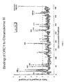
Figure 4 is a graph of chromosome position versus ratio of tagged to untagged for binding of ORC1 to yeast chromosome III. -
Figure 5A is an example of a scanned image. The unenriched and IP enriched DNA generates green fluorescence and red fluorescence respectively. The close-up image shows examples of spots for which the red intensity is over-represented, indicating binding of the targeted protein to these DNA sequences. -
Figure 5B show that small amounts of DNA can be quantitatively amplified and labeled with Cy3 and Cy5 fluorophores. Cy3- and Cy5-labeled DNA from 1 ng of yeast genomic DNA was prepared using the LM-PCR method described in the text. The resulting DNA samples were mixed and hybridized to a yeast intragenic DNA microarray. Low intensity spots have larger variations than high intensity spots, probably due to background noise. -
Figure 6A shows the set of 24 genes whose promoter regions are most likely to be bound by Gal4 by the analysis criteria described herein. -
Figure 6B is a schematic of the Gal4 binding intergenic regions. -
Figure 6C shows the results of conventional ChIP analysis. -
Figure 6D shows the results of the AlignAce program used to identify a consensus binding site for the Gal4 activator. -
Figure 6E is a bar graph showing relative expression of PLC10 and MTH1. -
Figure 6F is a schematic illustrating how the identification of MTH1 and MTH,PCL 10 and FUR4 as Gal4-regulated genes reveals how several different metabolic pathways are interconnected. -
Figure 7 lists the set of genes whose promoter regions are most likely to be bound by Stel12 by the analysis criteria described herein. - Understanding how DNA-binding proteins control global gene expression, chromosomal replication and cellular proliferation would be facilitated by identification of the chromosomal locations at which these proteins function in vivo. Described herein is a genome-wide location profiling method for DNA-bound proteins, which has been used to monitor dynamic binding of gene-specific transcription factors and components of the general transcription apparatus in yeast cells. The genome-wide location method correctly identified known sites of action for the transcriptional activators Gal4 and Stel2 and revealed unexpected functions for these activators. The combination of expression and location profiles identified the global set of genes whose expression is under the direct control of specific activators and components of the transcription apparatus as cells responded to changes in their extracellular environment. Genome-wide location analysis provides a powerful tool for further dissecting gene regulatory networks, annotating gene functions and exploring how genomes are replicated.
- Accordingly, the present invention which is defined in the appended claims, provides methods of examining the binding of proteins to DNA across the genome (e.g., the entire genome or a portion thereof, such as one or more chromosomes or a chromosome regions) of an organism. In particular, the present invention relates to a method of identifying a region (one or more) of genomic DNA of a cell to which a protein of interest binds. In the invention, proteins which bind DNA in a cell are crosslinked to the cellular DNA. The resulting mixture, which includes DNA bound by protein and DNA which is not bound by protein is subject to shearing conditions. As a result, DNA fragments of the genome crosslinked to DNA binding protein are generated and the DNA fragment (one or more) to which the protein of interest is bound is removed from the mixture. The resulting DNA fragments are then separated from the protein of interest and amplified using known techniques. The DNA fragment is then combined with DNA comprising a sequence complementary to genomic DNA of the cell, under conditions in which hybridization between the DNA fragments and the sequence complementary to genomic DNA occurs; and the region of the sequence complementary to genomic DNA to which the DNA fragment hybridizes is identified. The identified region is a region of the genome of the cell to which the protein of interest binds.
- Also encompassed by the present invention is a method of determining a function of a protein of interest which binds to the genomic DNA of a cell. In this method, DNA binding protein of the cell is crosslinked to the genomic DNA of the cell. DNA fragments of the genome crosslinked to DNA binding protein are then generated, as described above, and the DNA fragment (one or more) to which the protein of interest is bound is removed. The resulting DNA fragment is then separated from the protein of interest and amplified. The DNA fragment is then combined with DNA comprising a sequence complementary to genomic DNA of the cell, under conditions in which hybridization between the DNA fragment and a region of the sequence complementary to genomic DNA occurs; and the region of the sequence complementary to genomic DNA to which the DNA fragment hybridizes is identified and is a region of the genome of the cell to which the protein of interest binds. The identified region is characterized (e.g., a regulatory region) and the characteristic of the identified region indicates a function of the protein of interest (e.g., a transcription factor; an oncoprotein).
- The present invention also relates to a method of determining whether a protein of interest which binds to genomic DNA of a cell functions as a transcription factor. In the invention, DNA fragments of the crosslinked genome are generated. The DNA fragment to which the protein of interest is bound are removed. The resulting DNA fragment is separated from the protein of interest and amplified. The DNA fragment is combined with DNA comprising a sequence complementary to genomic DNA of the cell, under conditions in which hybridization between the DNA fragments and sequence complementary to genomic DNA occurs. The region of the sequence complementary to genomic DNA to which the DNA fragments hybridizes is identified wherein if the region of the genome is a regulatory region, then the protein of interest is a transcription factor.
- The methods of the present invention can be used to examine and/or identify DNA binding of proteins across the entire genome of a eukaryotic organism. For example, DNA binding proteins across the entire genome of eukaryotic organisms such as yeast, Drosophila and humans can be analyzed. Alternatively, they can be used to examine and/or identify DNA binding of proteins to an entire chromosome or set of chromosomes of interest.
- A variety of proteins which bind to DNA can be analyzed. For example, any protein involved in DNA replication such as a transcription factor, or an oncoprotein can be examined in the methods of the present invention.
- There are a variety of methods which can be used to link DNA binding protein of the cell to the genome of the cell. For example, UV light can be used. In a particular embodiment, formaldehyde is used to crosslink DNA binding proteins to the genomic DNA of a cell.
- In the methods of the present invention, identification of DNA fragments bound to the protein of interest can be removed from the mixture comprising DNA fragment(s) bound to the protein of interest and DNA fragments which are not bound to the protein of interest, using a variety of methods. For example, immunoprecipitation using an antibody (e.g., polyclonal, monoclonal) or antigen binding fragment thereof which binds (specifically) to the protein of interest, can be used. In addition, the protein of interest can be labeled or tagged using, for example, an antibody epitope (e.g., hemagglutinin (HA)).
- The DNA fragments in the methods described herein are amplified using, ligation-mediated polymerase chain reaction (e.g., see Current Protocols in Molecular Biology, Ausubel, F.M. et al., eds. 1991.
- The DNA comprising the complement sequence of the genome of the cell can be combined with the isolated DNA fragment to which the protein of interest binds using a variety of methods. For example, the complement sequence can be immobilized on a glass slide (e.g., Coming Microarray Technology (CMT™) GAPS™) or on a microchip. Conditions of hybridization used in the methods of the present invention include, for example, high stringency conditions and/or moderate stringency conditions. See e.g., pages 2.10.1-2.10.16 (see particularly 2.10.8-11) and pages 6.3.1-6 in Current Protocols in Molecular Biology). Factors such as probe length, base composition, percent mismatch between the hybridizing sequences, temperature and ionic strength influence the stability of hybridization. Thus, high or moderate stringency conditions can be determined empirically, and depend in part upon the characteristics of the known nucleic acids (DNA, RNA) and the other nucleic acids to be assessed for hybridization thereto.
- The methods of the present invention can further comprise comparing the results to a control. For example, in one embodiment, the methods of the present invention can be carried out using a control protein which is not a DNA binding protein. In one embodiment, immunoprecipitation is performed using an antibody against an HA or MYC epitope tag. The results of immunoprecipitating the protein of interest containing the tag, and the protein of interest without the tag are compared. The untagged protein should not be immunoprecipitated, and thus, serves as a negative control.
- As described in the exemplification, a particular embodiment of the present invention comprises the combined use of Chromatin Immunoprecipitation (ChIP) and Genome-wide expression monitoring microarrays. Chromatin immunoprecipitation allows the detection of proteins that are bound to a particular region of DNA. It involves four steps: (1) formaldehyde cross-linking proteins to DNA in living cells, (2) disrupting and then sonicating the cells to yield small fragments of cross-linked DNA, (3) immunoprecipitating the protein-DNA crosslinks using an antibody which specifically binds the protein of interest, and (4) reversing the crosslinks and amplifying the DNA region of interest using the Polymerase Chain Reaction (PCR). Analysis of the PCR product yield compared to a non-immunoprecipitated control determines whether the protein of interest binds to the DNA region tested. However, each region of DNA must be tested individually by PCR. Thus, the ChIP technique is limited to the small set of DNA regions that are chosen to be tested.
- In contrast, the present method is not limited to amplifying individual DNA regions by performing PCR with specific primers. Rather the entire genome is amplified using a Ligation-mediated PCR (LMPCR) strategy. The amplified DNA was fluorescently labeled by including fluorescently-tagged nucleotides in the LM-PCR reaction. Finally, the labeled DNA was hybridized to a DNA microarray containing spots representing all or a subset (e.g., a chromosome or chromosomes) of the genome. The fluorescent intensity of each spot on the microarray relative to a non-immunoprecipitated control demonstrated whether the protein of interest bound to the DNA region located at that particular spot. Hence, the methods described herein allow the detection of protein-DNA interactions across the entire genome.
- In particular, DNA microarrays consisting of most of yeast chromosome III plus approximately 15 model genes whose expression have been well studied were constructed. These arrays were used in conjunction with the ChIP technique to study the DNA-binding properties of transcription factors and the transcription apparatus genome-wide. The methods described herein provide insights into the mechanism and regulation of gene expression in eukaryotic cells.
- The genome-wide location analysis method described herein allows proteinDNA interactions to be monitored across the entire yeast genome and is diagramed in
Figure 1 . The method combines a modified Chromatin Immunoprecipitation (ChIP) procedure, which has been previously used to study in vivo protein-DNA interactions at one or a small number of specific DNA sites, with DNA microarray analysis. Briefly, cells are fixed with formaldehyde, harvested by sonication, and DNA fragments that are crosslinked to a protein of interest are enriched by immunoprecipitation with a specific antibody. After reversal of the crosslinking, the enriched DNA is amplified and labeled with a fluorescent dye using ligation-mediated PCR (LM-PCR). A sample of DNA that has not been enriched by immunoprecipitation is subjected to LM-PCR in the presence of a different fluorophore, and both IP-enriched and unenriched pools of labeled-DNA are hybridized to a single DNA microarray containing all yeast intergenic sequences. The IP-enriched/unenriched ratio of fluorescence intensity obtained from three independent experiments can be used with a weighted average analysis method to calculate the relative binding of the protein of interest to each sequence represented on the array (seeFigure 2 ). - Four features of the global location profiling method were found to be critical for consistent, high-quality results. First, DNA microarrays with consistent spot quality and even signal background play an obvious role. An example of an image generated by the technique described herein is shown in
figure 5A . Second, the LM-PCR method described herein was developed to permit reproducible amplification of very small amounts of DNA; signals for greater than 99.9% of genes were essentially identical within the error range when independent samples of 1 ng of genomic DNA were amplified with the LM-PCR method (Figure 5B ). Third, each experiment was carried out in triplicate, allowing an assessment of the reproducibility of the binding data. And fourth, a single-array error model described by Hughs et al, (2000) was adopted to handle noise associated with low intensity spots and to average repeated experiments with appropriate weights - The quantitative amplification of small amount of DNA generates some uncertainty for the low intensity spots. In order to track that uncertainty and to be able to average repeated experiments with appropriate related weights, we adopted an single-array error model that was first described by Hughs et al, (2000). According to this error model, the significance of a measured ratio at a spot is defined by a statistic X, which takes the form

where a1,2 are the intensities measured in the two channels for each spot, σ1,2 are the uncertainties due to background subtraction, and fis a fractional multiplicative error such as would come from hybridization non-uniformities, fluctuations in the dye incorporation efficiency, scanner gain fluctuations, ets. X is approximately normal. The parameters σ and f were chosen such that X has unit variance. The significance of a change of magnitude |x| is then calculated as
- Array contains all non-overlapping open reading frames (ORF) on Chromosome III (See the Table). When a sequence contains part or all of two potential reading frames, the larger sequence was chosen to represent the ORF. Any remaining sequence was included in intergenic fragments.
- All intergenic regions larger than 100bp are represented by fragments averaging 500bp. Where regions are greater than 700bp, they are broken into multiple fragments of 300 to 600bps. PCR primers for each region were chosen using the Saccharomyces Genomic Database (SGD) "Design Primers" program from Stanford University. The total number of intergenic fragments equals 241 for Chromosome III.
- The location and size of open reading frames were determined from the Saccharomyces Genomic Database (SGD) functional chromosomal map.
- An additional 17 model genes (see the Table) were selected based on their high frequency of citation in transcription literature. Each gene was amplified as well as 1-2kb upstream and 500bp downstream of the coding region.
-
- PCR generation of unmodified yeast ORF DNA
- 100 µl reaction generally yields approximately 5-6µg DNA
-
- 10.0 µl 10X PCR buffer (Perkin Elmer, AmpliTaq)
- 8.0 µl 25mM MgCl2 (Perkin Elver, AmpliTaq)
- 10.0 µl 10X dNTPs (2mM each; Pharmacia 100mM stocks)
- 1.0-2.0 µl ORF DNA (Research Genetics, approximately 10 ng)
- 2.5µl each universal primer (Research Genetics, 20 µM solution)
- 1.6 µl diluted Pfu DNA polymerase (diluted 1:100 in water, Strategene, 0.02U)
- 1.0 µl AmpliTaq DNA polymerase (5U, Perkin Elmer)
- 63.4 µl ddH2O
- PCR Generation of Yeast Intergenic regions
- 100 µl reaction generally yields approximately 5-6 ug DNA
-
- 10.0 µl 10X PCR buffer (Perkin Elmer, AmpliTaq)
- 8.0 µl 25mM MgCl2 (Perkin Elmer, AmpliTaq)
- 10.0 µl 10X dNTPs (2mM each, Pharmacia 100mM stocks)
- 1.0µl Yeast Genomic DNA (Research Genetics, approximately 100 ng)
- 5.0 µl each primer (Research Genetics, 20 µM solution)
- 1.6 µl diluted Pfu DNA polymerase (diluted 1:100 in water, Strategene 0.02U)
- 1.0µl AmpliTaq DNA polymerase (5U, Perkin Elmer)
- 58.4 µl ddH2O
- Cycling for ORF and intergenic DNA
- 95°
C 3 min - 30 cycles of:
- 94°
C 30 sec - 60°
C 30 sec - 72°
C 2 min - Reactions were cleaned by Qiagen QIAquick 96 PCR purification kits according to the manufacturers' protocol with the following exception. DNA was eluted with 120 µl ofT.E. 8.0 (10mmTris, 1mm EDTA, pH8.0). T.E. 8.0 was applied to the Qiagen membrane and allowed to sit 5 minutes before elution. The DNA was collected into a Corning polypropylene 96 well plate.
- Reactions were quantified by visualizing 1µl of the purified DNA on an agarose gel compared to a known quantity of lambda DNA cut with HindIII (Promega).
- DNA was stored at -20 until shortly before printing. The DNA was then dried down by speed vac in the Coming microtiter plates to less than 5µl.
- PCR reactions were resuspended to approximately 0.5 mg/ml in 3XSSC. SSC was made as a 20X stock (3M NaCl, 0.3M Na3citrate·2H2O, pH'd to 7.0 with HCl) and diluted to the desired concentration with H2O.
- 10-15 µl of the DNA was placed in a Coming 96 or 384 well plate and GAPS coated slides were printed using the Cartessian Robot. PCR products should be greater than 250 pb.
-
- 1. Rehydrated arrays by holding slides over a dish of hot ddH2O (∼10 sec).
- 2. Snap-dried each array (DNA side up) on a 100°C hot plate for ∼ 3 seconds.
- 3. UV X-linked DNA to the glass by using a Stratalinker set for 60 mJoules.
- 4. Dissolved 5g of succinic anhybride (Aldrich) in 315mL of n-methyl-pyrrilidinone.
- 5. To this, added 35mL of 0.2M NaBorate pH 8.0, and stirred until dissolved (Boric Acid pH'd with NaOH).
- 6. Soaked arrays in this solution for 15 minutes with shaking.
- 7. Transferred arrays to 95°C water bath for 2 minutes.
- 8. Quickly transferred arrays to 95% EtOH for 1 minute.
- 9. Air dried slides array side up at a slight angle (close to vertical).
-
- 1. Incubated slide in 3.5XSSC, 0.1%SDS, 10mg/ml BSA (Sigma) in a Coplin jar for 20 minutes at 50°C (Place Coplin jar in water bath).
- 2. Washed slide by dipping in water and then isopropanol.
- 3. Air dried array side up at slight angle (close to vertical).
Probe preparation - 1. The probe volume should be 20-30 µl for a small coverslip (25 mm2) and 40-60 µl for a large cover slip (24 x 60 mm).
- 2. Brought probe (cDNA or PCR based) up to final hyb volume in 3XSSC, 0.1 % SDS with 10 µg E. coli tRNA (Boehringer-Mannheim).
- 3. Boiled in heat block for 3-5 minutes.
- 4. Snaped cool on ice. And spun.
-
- 1. Pipetted probe onto slide. Dropped cover slip onto liquid avoiding bubbles.
- 2. Assembled over 50°C waterbath in hybridization chamber. Clamped shut.
- 3. Submerged in 50°C waterbath overnight.
-
- 1. Dissambled hybridization right side up.
- 2. Removed coverslip with fingers or tweezers.
- 3. Placed in 0.1 X SSC, 0.1% SDS at room temperature for 5-10 minutes.
- 4. Transfered slides to 0.1 X SSC for 2.5 minutes and again for 2.5 minutes.
- 5. Blew dry and scan slide.
- The data generated from scanning was analyzed using the ImaGene software.
The Table Yeast ORF Model Genes YCL001 w RER1 YOL086c ADH1 YCL001w-a YBR115c LYS2 YCL002c YBR039c PHO5 YCL004w PGS1 YIR019c FLO11 YCL005w YDL215c GDH2 YCL006c YER103w SSA4 YCL007c CWH36 YHR053c CUP1 YCL008c STP22 YKL178c STE3 YCL009c ILV6 YIL163c SUC2 YCL010c YOR202w HIS3 YCL011c GBP2 YJR048w CYC1 YCL012w YJR153c INO1 YCL014w BUD3 YBR020w GAL1 YCL016c YBR019c GAL10 YCL017c NSF1 YDL227c HO YCL018w LEU2 YPL256c CLN2 YCL019w YGR108w CLB1 YCL020w YCL024w YCL025c AGP1 YCL026ca FRM2 YCL027w FUS1 YCL028w YCL029w BIK1 YCL030c HIS4 YCL031c RPB7 YCL032w STE50 YCL033c YCL034w YCL035c YCL036w YCL037c SRO9 YCL038c YCL039w YCL040w GLK1 YCL041c YCL042w YCL043c PDI1 YCL044c YCL045c YCL046w YCL047c YCL048w YCL049c YCL050c APA1 Yeast ORF Model Genes YCL051w LRE1 YCL052c PBN1 YCL054w YCL055w KAR4 YCL056w YCL057w PRD1 YCL058c YCL059c KRR1 YCL061c YCL063w YCL064c CHA1 YCL065w YCL066w HMLALPHA1 YCL067c HMLALPHA2 YCL068c YCL069w YCL073c YCL074w YCLO75w YCL076w YCR001W YYCR002c CDC10 YCR003w MRPL32 YCR004c YCP4 YCR005c CIT2 YCR006c YCR007c YCR008w SAT4 YCR009c RVS161 YCR010c YCR011c ADP1 YCR012w PGK1 YCR014c POL4 YCR015c YCR016w YCR017c YCR018c SRD1 YCR018ca YCR019w YCR020c PET18 YCR020CA MAK31 YCR020wb HTL1 YCR021c HSP30 YCR022c YCR023c YCR024c YCR024CA PMP1 YCR025c YCR026c YCR027c YCR028c FEN2 YCR028CA RIM1 YCR030c YCR031c RPS14A YCR032w BPH1 YCR033w YCR034w FEN1 YCR035c RRP43 YCR036w RBK1 YCR037c PHO87 YCR038c BUD5 YCR039c MATALPHA2 YCR040w MATALPHA1 YCR041w YCR042c TSM1 YCR043c YCR044c YCR045c YCR046c IMG1 YCR047c YCR048w ARE 1 YCR051w YCR052w RSC6 YCR053w THR4 YCR054c CTR86 YCR057c PWP2 YCR059c YCR060w YCR061W YCR063w YCR064c YCR065w HCM1 YCR066w RAD18 YCR067c SED4 YCR068w YCR069w SCC3 YCR071c IMG2 YCR072c YCR073c SSK22 YCR073wa SOL2 YCR075c ERS1 YCR076c YCR077c PAT1 YCR079w YCR081w SRB8 YCR082w YCR083w YCR084c TUP1 YCR085w YCR086w YCR087w YCR088w ABP1 YCR089w FIG2 YCR090c YCR091w KIN82 YCR092c MSH3 YCR093w CDC39 YCR094w CDC50 YCR095c YCR096c A2 YCR097w A1 YCR098c GIT1 YCR099c YCR100c YCR101c YCR102c YCR102wa YCR103 YCR104w PAU3 YCR105w YCR106w YCR107w AAD3 - To investigate the accuracy of the genome-wide location analysis method, the analysis was used to identify sites bound by the transcriptional activator Gal4 in the yeast genome. Gal4 was selected because it is among the best characterized transcriptional activators, it is known to be responsible for induction of 10 genes necessary for galactose metabolism, and a consensus DNA binding sequence (the UASG) has been identified for Gal4 in the promoters of the GAL genes. Very little Gal4 is bound at the UASG of the GAL1 and GAL10 promoters when cells are grown in glucose (the repressed state), whereas relatively high levels of Gal4 are bound in galactose (the activated state).
- The genome-wide location of epitope-tagged Gal4p in both glucose and galactose media was investigated in three independent experiments, as described in more detail below. The location analysis experiment identified seven genes previously reported to be regulated by Gal4 and three additional genes encoding activities that are physiologically relevant to cells that utilize galactose as the sole carbon source, but which were not previously known to be regulated by this activator (
Figures 6A ). - The set of 24 genes whose promoter regions are most likely to be bound by Gal4 by the analysis criteria (p-value < 0.00001) described herein, is listed in
Figure 6A . Gal4 does not functionally activate all of these genes, however, since only a subset of the genes that share intergenic regions bound by Gal4 will be regulated by this activator (Figure 6B ). To identify genes that are both bound by Gal4 and activated by galactose, genome-wide expression analysis was carried out. The upper panel ofFigure 6A shows genes whose expression is induced in galactose, whereas the lower panel shows genes whose expression is galactose independent. Seven genes previously reported to be regulated by Gal4 (GAL1, GAL2, GAL3, GAL7, GAL10, GAL80 and GCY1) bound Gal4 and were activated in galactose. Three genes whose expression was not previously associated with the Gal4 activator, MTH, PCL10 and FUR4, were also found to be bound by Gal4 and activated in galactose. Substantially less Gal4 was associated with each of these promoters in cells grown in glucose, as expected. Gal4p was not bound to the promoters of GAL4 and PGM2, genes previously thought to be regulated by Gal4, although direct evidence for Gal4 binding to these promoters had not been demonstrated. Each of these results was confirmed by conventional ChIP analysis (Figure 6C ), demonstrating that the microarray results accurately reflect results obtained by the conventional approach, which has until now been used to study binding sites individually. - The ten genes that are both bound and regulated by Gal4 were selected and the AlignAce program was used to identify a consensus binding site for this activator (
Figure 6D ). This binding site sequence is similar to, but refines, the sequence previously determined for Gal4. The Gal4 binding sequence occurs at approximately 50 sites through the yeast genome where Gal4 binding is not detected, indicating that the simple presence of this sequence is not sufficient for Gal4 binding. - Three genes whose expression was not previously associated with the Gal4 activator, MTH, PCL10 and FUR4, were found to be bound by Gal4 and activated in galactose. It is likely that these three genes are genuine Gal4p targets because they share the following three features with the well established Gal4-dependent GAL genes. MTH, PCL10 and FUR4 are galactose-induced (
Figure 6A ). Galactose induction depends on Gal4 (Figure 6C ). MTH, PCL10 and FUR4 promoters are bound by Gal4 when cells are grown in galactose but not in glucose (Figure 6A ). The binding of Gal4p to the MTH, PCL10 and FUR4 promoters was verified by conventional CHIP analysis (Figure 6C ). - The identification of MTH1 and MTH, PCL10 and FUR4 as Gal4-regulated genes reveals how regulation of several different metabolic pathways are interconnected (
Figure 6F ). MTH1 encodes a transcriptional repressor of many genes involved in metabolic pathways that would be unnecessary when cells utilize galactose as a sole carbon source. Among the most interesting of its targets are a subset of the HTX genes involved in hexose transport. The results described herein indicate that the cell responds to galactose by modifying the concentration of its hexose transporters at the membrane in a Gal4-dependent fashion; Gal4 activates the galactose transporter gene GAL2 and, by activation of MTH1 repressor, causes reduced levels of glucose transporter expression. The Pc110 cyclin associates with Pho85p and appears to repress the formation of glycogen. The observation that PCL10 is Gal4-activated indicates that reduced glycogenesis occurs to maximize the energy obtained from galactose metabolism. FUR4 encodes a uracil pennease and its induction by Gal4 may reflect a need to increase intracellular pools of uracil to permit efficient UDP addition to galactose catalyzed by Gal7. - Previous studies have shown that Gal4 binds to at least some GAL gene promoters when cells are grown on carbon sources other than galactose, as long as glucose is absent. Genome-wide location analysis of Gal4 in cells grown on raffinose was repeated and it was found that the results were essentially identical to those obtained when cells were grown on galactose. These results indicate that Gal4 exhibits the same binding behavior at all its genomic binding sites and demonstrate that the genome-wide location method is highly reproducible.
- The genome-wide binding profile of the DNA-binding transcription activator St1l2 was also investigated. Stel2 is of interest because it has a defined cellular role - it is key to the response of haploid yeast to mating pheromones - but only a few genes regulated by Stel2 have been identified. Activation of the pheromone-response pathway causes cell cycle arrest and transcriptional activation of more than 100 genes. Expression analysis using ste12 mutant cells has shown that Ste12 is required for the pheromone induction of all of these genes. However, the mechanism by which Ste12 activates transcription of these genes in response to pheromone has not been elucidated.
- The genome-wide location of epitope-tagged Ste12p before and after pheromone treatment was investigated in three independent experiments. The set of genes whose promoter regions are most likely to be bound by
Ste 12 by the analysis criteria (p-value < 0.005) described herein is listed inFigure 7 ; the upper panel shows genes whose expression is induced by alpha factor, whereas the lower panel shows genes whose expression is not significantly induced by alpha factor. Of the 36 genes that are induced by alpha factor and are bound by Ste12, 12 are known to participate in various steps of the mating process (FIG2 , AFR1, GIC2, STE12, CHS1, KAR5, FUS1, AGA1, FUS3, CIK1, FAR1,FIG1 ). - Ste12 binds to some promoters in the absence of pheromone signaling, however, its binding to most genes is enhanced by alpha factor. Interestingly, Ste12p is bound to its own promoter both before and after pheromone treatment. Together, the binding and expression data argue that the regulation of the STE12 gene involves a positive feedback loop. STE12 expression is increased immediately after pheromone treatment, indicating that the bound but inactive Ste12 activator is rapidly converted to an active form. Increased expression of STE12 gene would allow more Ste12p to be made and this would, in turn, activate its genes.
- Twenty-four genes whose expression were not previously associated with Ste12 and the mating process were found to be bound by Ste12 and activated by alpha factor. Considering that their pheromone induction is eliminated in ste12 mutant cells, it is likely that these 24 genes are also genuine Ste12 targets. The identities of these genes indicate interesting details about various steps of the mating process. For example, one Ste12 target gene, PCL2, encodes a G1 cyclin that forms complexes with the cyclin-dependent kinase (cdk) Pho85. The Pcl2-Pho85 and PC11-Pho85 complexes act in concert with Cln1-Cdc28 and Cln-2-Cdc28 cyclin dependent kinase complexes to promote G1cell cycle progression (Measday et al., 1994). The Pcl2-Pho85 kinase complex has a substrate specificity that is overlapping but different from that of the Cln1-Cdc28 and Cln2-Cdc28. During the mating process, haploid yeast cells are arrested at start of the late G1 phase, due to the inhibition of Cln1-Cdc28 and Cln2-Cdc28 activities by Farl, which is encoded by another Ste12 target gene. Activation of PCL2 by Ste12 after pheromone treatment indicates that increased Pho85 complex activities are likely necessary to compensate for the loss of Cdc28 activities.
- Most Ste12 target genes identified by analysis of genome locations of Ste12 and expression profiles during pheromone induction encode proteins involved in various steps of the mating response. Among them are 11 previously uncharacterized. The cellular roles for these genes, including YNL279W, YOR129C, YOR343C, YPL192C, YER019W, YIL083C, YIL037C, YIL169C, YNL105W, YOL155C and YNR064C, are therefore most likely related to mating.
- Ste12 has also been implicated in other cellular processes. Together with Tec1, Ste12 regulates the filmamentation of diploid cells and invasive growth in haploids. Two genes, TEC1 and FLO11, have been identified as Ste12 targets in filamentous growth pathway. Ste12 binding to these genes either in the presence or absence of alpha factor was not detected. It is likely that Ste12p's binding to these promoters is regulated by different physiological conditions.
Claims (6)
- An in vitro genome-wide location profiling method of identifying a region of a genome of a living cell to which a protein of interest binds, comprising the steps of:a) cross-linking DNA binding protein in the living cells to genomic DNA of the living cell, thereby producing DNA binding protein cross-linked to genomic DNA;b) generating DNA fragments of the genomic DNA cross-linked to DNA binding protein in a), thereby producing a mixture comprising DNA fragments to which DNA binding protein is bound;c) removing the fragment (one or more) to which the protein of interest is bound from the mixture produced in b);d) separating the DNA fragment (one or more) obtained in c) from the protein of interest;e) amplifying the DNA fragment (one or more) of d) using a ligation mediated polymerase chain reaction strategy; characterised in that the method further comprises the steps of:f) fluorescently labelling the DNA fragment (one or more) of (e) with a first fluorophore during or after step (e);g) generating a control by amplifying a sample of DNA that has not been enriched in accordance with step c) using LM-PCR in the presence of a second fluorophore;h) combining the labelled DNA fragment (one or more) of (f) with a DNA micro-array comprising a sequence complementary to genomic DNA of the cell under conditions in which hybridisation between the DNA fragment (one or more) and a region complementary to the genomic DNA occurs;i) combining the labelled DNA fragment of (g) with the same DNA micro-array used in step (h) under conditions in which hybridisation between the DNA fragments and a region complementary to the genomic DNA occurs;j) identifying the region of the sequence complementary to genomic DNA to which the DNA fragment (one or more) hybridise by measuring the fluorescence intensity of the first chromophore;k) comparing the fluorescence intensity measured in step (j) with the fluorescence intensity of the control, whereby the fluorescence intensity in a region of the genome which is greater than the fluorescence intensity of the control in the region indicates the region of the genome in the cell in which the protein of interest binds.
- The method of Claim 1 wherein the cell is a eukaryotic cell.
- The method of Claim 1 or 2 wherein the protein of interest is selected from the group consisting of: a transcription factor and an oncogene.
- The method of any preceding claim wherein the DNA binding protein of the cell is crosslinked to the genome of the cell using formaldehyde.
- The method of any preceding claim, wherein the DNA fragments of (c) to which is bound the protein of interest is obtained using an antibody which binds to the protein of interest.
- The method of any preceding claim wherein the DNA fragments of b) are generated by sonication.
Priority Applications (1)
| Application Number | Priority Date | Filing Date | Title |
|---|---|---|---|
| DE20023863U DE20023863U1 (en) | 1999-09-01 | 2000-09-01 | Microarray obtainable by total chromosome analysis of protein-DNA interaction |
Applications Claiming Priority (3)
| Application Number | Priority Date | Filing Date | Title |
|---|---|---|---|
| US15197299P | 1999-09-01 | 1999-09-01 | |
| US151972P | 1999-09-01 | ||
| PCT/US2000/024358 WO2001016378A2 (en) | 1999-09-01 | 2000-09-01 | Chromosome-wide analysis of protein-dna interactions |
Publications (2)
| Publication Number | Publication Date |
|---|---|
| EP1220949A2 EP1220949A2 (en) | 2002-07-10 |
| EP1220949B1 true EP1220949B1 (en) | 2011-03-30 |
Family
ID=22541045
Family Applications (1)
| Application Number | Title | Priority Date | Filing Date |
|---|---|---|---|
| EP00959909A Expired - Lifetime EP1220949B1 (en) | 1999-09-01 | 2000-09-01 | Chromosome-wide analysis of protein-dna interactions |
Country Status (7)
| Country | Link |
|---|---|
| US (1) | US6410243B1 (en) |
| EP (1) | EP1220949B1 (en) |
| JP (1) | JP2003508066A (en) |
| AT (1) | ATE503843T1 (en) |
| CA (1) | CA2383928A1 (en) |
| DE (2) | DE60045796D1 (en) |
| WO (1) | WO2001016378A2 (en) |
Families Citing this family (51)
| Publication number | Priority date | Publication date | Assignee | Title |
|---|---|---|---|---|
| US7470507B2 (en) | 1999-09-01 | 2008-12-30 | Whitehead Institute For Biomedical Research | Genome-wide location and function of DNA binding proteins |
| US20030087231A1 (en) * | 2000-05-19 | 2003-05-08 | Albertson Donna G. | Methods and compositions for preparation of a polynucleotide array |
| EP1362122A2 (en) * | 2000-09-12 | 2003-11-19 | TransGenetics Incorporated | Microarrayed organization of transcription factor target genes |
| DE10056899A1 (en) | 2000-11-16 | 2002-05-29 | Aventis Pharma Gmbh | Promoter for the functional characterization of G-protein coupled receptors in the yeast Saccharomyces cerevisiae |
| EP1381692A2 (en) * | 2000-12-21 | 2004-01-21 | Whitehead Institute For Biomedical Research | Genome-wide location and function of dna binding proteins |
| US20040058356A1 (en) * | 2001-03-01 | 2004-03-25 | Warren Mary E. | Methods for global profiling gene regulatory element activity |
| WO2004024084A2 (en) * | 2002-09-12 | 2004-03-25 | Massachusetts Institute Of Technology | Rb pahtway and chromatin remodeling genes that antagonize let-60 ras signaling |
| US20060099713A1 (en) * | 2002-10-01 | 2006-05-11 | Buck Institute | Targeted-assisted iterative screening (tais):a novel screening format for large molecular repertoires |
| US20040161779A1 (en) * | 2002-11-12 | 2004-08-19 | Affymetrix, Inc. | Methods, compositions and computer software products for interrogating sequence variations in functional genomic regions |
| US20040115794A1 (en) * | 2002-12-12 | 2004-06-17 | Affymetrix, Inc. | Methods for detecting transcriptional factor binding sites |
| US7750908B2 (en) * | 2003-04-04 | 2010-07-06 | Agilent Technologies, Inc. | Focus plus context viewing and manipulation of large collections of graphs |
| US7601492B2 (en) | 2003-07-03 | 2009-10-13 | The Regents Of The University Of California | Genome mapping of functional DNA elements and cellular proteins |
| US8222005B2 (en) * | 2003-09-17 | 2012-07-17 | Agency For Science, Technology And Research | Method for gene identification signature (GIS) analysis |
| JP2007515954A (en) * | 2003-11-26 | 2007-06-21 | ホワイトヘッド・インスティテュート・フォー・バイオメディカル・リサーチ | Transcriptional regulator and method thereof |
| US20070031883A1 (en) * | 2004-03-04 | 2007-02-08 | Kincaid Robert H | Analyzing CGH data to identify aberrations |
| US8321138B2 (en) * | 2005-07-29 | 2012-11-27 | Agilent Technologies, Inc. | Method of characterizing quality of hybridized CGH arrays |
| WO2005088306A2 (en) * | 2004-03-04 | 2005-09-22 | Whitehead Institute For Biomedical Research | Biologically-active dna-binding sites and related methods |
| US20050282193A1 (en) * | 2004-04-23 | 2005-12-22 | Bulyk Martha L | Space efficient polymer sets |
| US20060035251A1 (en) * | 2004-06-30 | 2006-02-16 | Whitehead Institute For Biomedical Research | Novel methods for high-throughput genome-wide location analysis |
| JP2006078313A (en) * | 2004-09-09 | 2006-03-23 | Hitachi Software Eng Co Ltd | Protein binding position identifying method |
| US20070099227A1 (en) * | 2004-10-12 | 2007-05-03 | Curry Bo U | Significance analysis using data smoothing with shaped response functions |
| EP1904648A4 (en) * | 2005-07-06 | 2010-03-10 | Uppsala Uni Projekt Ab | Method for localization of nucleic acid associated molecules and modifications |
| US20090221428A1 (en) * | 2005-09-07 | 2009-09-03 | Young Richard A | Methods of Genome-Wide Location Analysis in Stem Cells |
| WO2007041621A2 (en) * | 2005-10-03 | 2007-04-12 | Xingsheng Sean Ling | Hybridization assisted nanopore sequencing |
| EP1954835A4 (en) * | 2005-12-02 | 2009-07-22 | Whitehead Biomedical Inst | METHODS OF MATCHING SIGNAL TRANSDUCTION PATHWAYS TO GENE EXPRESSION PROGRAMS |
| US7297477B2 (en) * | 2006-02-13 | 2007-11-20 | Agilent Technologies, Inc. | Methods and compositions for detecting viral nucleic acid in a cell |
| US8071296B2 (en) | 2006-03-13 | 2011-12-06 | Agency For Science, Technology And Research | Nucleic acid interaction analysis |
| US20070238104A1 (en) * | 2006-04-07 | 2007-10-11 | Agilent Technologies, Inc. | Competitive oligonucleotides |
| US8058055B2 (en) | 2006-04-07 | 2011-11-15 | Agilent Technologies, Inc. | High resolution chromosomal mapping |
| US20080124707A1 (en) * | 2006-06-09 | 2008-05-29 | Agency For Science, Technology And Research | Nucleic acid concatenation |
| US7437249B2 (en) * | 2006-06-30 | 2008-10-14 | Agilent Technologies, Inc. | Methods and systems for detrending signal intensity data from chemical arrays |
| US8077951B2 (en) | 2006-10-12 | 2011-12-13 | Agilent Technologies, Inc. | Method and system for dynamic, automated detection of outlying feature and feature background regions during processing of data scanned from a chemical array |
| US7881876B2 (en) * | 2006-10-12 | 2011-02-01 | Agilent Technologies, Inc. | Methods and systems for removing offset bias in chemical array data |
| US20080090236A1 (en) * | 2006-10-13 | 2008-04-17 | Yakhini Zohar H | Methods and systems for identifying tumor progression in comparative genomic hybridization data |
| US20080102453A1 (en) * | 2006-10-31 | 2008-05-01 | Jayati Ghosh | Methods and systems and analysis of CGH data |
| WO2008088839A2 (en) * | 2007-01-16 | 2008-07-24 | Cytocure, Inc. | Methods of isolating and purifying nucleic acid-binding biomolecules and compositions including same |
| WO2009046094A1 (en) | 2007-10-01 | 2009-04-09 | Nabsys, Inc. | Biopolymer sequencing by hybridization of probes to form ternary complexes and variable range alignment |
| US8263367B2 (en) * | 2008-01-25 | 2012-09-11 | Agency For Science, Technology And Research | Nucleic acid interaction analysis |
| US9650668B2 (en) | 2008-09-03 | 2017-05-16 | Nabsys 2.0 Llc | Use of longitudinally displaced nanoscale electrodes for voltage sensing of biomolecules and other analytes in fluidic channels |
| US8882980B2 (en) * | 2008-09-03 | 2014-11-11 | Nabsys, Inc. | Use of longitudinally displaced nanoscale electrodes for voltage sensing of biomolecules and other analytes in fluidic channels |
| US8262879B2 (en) | 2008-09-03 | 2012-09-11 | Nabsys, Inc. | Devices and methods for determining the length of biopolymers and distances between probes bound thereto |
| US8583380B2 (en) | 2008-09-05 | 2013-11-12 | Aueon, Inc. | Methods for stratifying and annotating cancer drug treatment options |
| RU2565550C2 (en) | 2010-09-24 | 2015-10-20 | Те Борд Оф Трастиз Оф Те Лилэнд Стэнфорд Джуниор Юниверсити | Direct capture, amplification and sequencing of target dna using immobilised primers |
| US8715933B2 (en) | 2010-09-27 | 2014-05-06 | Nabsys, Inc. | Assay methods using nicking endonucleases |
| US20120115132A1 (en) | 2010-11-05 | 2012-05-10 | Chromatin, Inc. | Identification of centromere sequences using centromere associated proteins and uses thereof |
| EP2640849B1 (en) | 2010-11-16 | 2016-04-06 | Nabsys 2.0 LLC | Methods for sequencing a biomolecule by detecting relative positions of hybridized probes |
| WO2012109574A2 (en) | 2011-02-11 | 2012-08-16 | Nabsys, Inc. | Assay methods using dna binding proteins |
| WO2013188789A2 (en) | 2012-06-14 | 2013-12-19 | Whitehead Institute For Biomedical Research | Genome-wide method of assessing interactions between chemical entities and their target molecules |
| WO2014052433A2 (en) | 2012-09-26 | 2014-04-03 | Nabsys, Inc. | Method and system for analysis of protein and other modifications on dna and rna |
| US9914966B1 (en) | 2012-12-20 | 2018-03-13 | Nabsys 2.0 Llc | Apparatus and methods for analysis of biomolecules using high frequency alternating current excitation |
| WO2014113557A1 (en) | 2013-01-18 | 2014-07-24 | Nabsys, Inc. | Enhanced probe binding |
Family Cites Families (4)
| Publication number | Priority date | Publication date | Assignee | Title |
|---|---|---|---|---|
| US4882269A (en) * | 1985-12-13 | 1989-11-21 | Princeton University | Amplified hybridization assay |
| US6046165A (en) * | 1997-06-23 | 2000-04-04 | Ophidian Pharmaceuticals, Inc. | Compositions and methods for identifying and testing TGF-β pathway agonists and antagonists |
| US6066452A (en) * | 1997-08-06 | 2000-05-23 | Yale University | Multiplex selection technique for identifying protein-binding sites and DNA-binding proteins |
| US6109776A (en) * | 1998-04-21 | 2000-08-29 | Gene Logic, Inc. | Method and system for computationally identifying clusters within a set of sequences |
-
2000
- 2000-09-01 WO PCT/US2000/024358 patent/WO2001016378A2/en not_active Ceased
- 2000-09-01 JP JP2001520923A patent/JP2003508066A/en active Pending
- 2000-09-01 EP EP00959909A patent/EP1220949B1/en not_active Expired - Lifetime
- 2000-09-01 DE DE60045796T patent/DE60045796D1/en not_active Expired - Lifetime
- 2000-09-01 AT AT00959909T patent/ATE503843T1/en not_active IP Right Cessation
- 2000-09-01 CA CA002383928A patent/CA2383928A1/en not_active Abandoned
- 2000-09-01 US US09/654,409 patent/US6410243B1/en not_active Expired - Lifetime
- 2000-09-01 DE DE2000959909 patent/DE00959909T1/en active Pending
Non-Patent Citations (1)
| Title |
|---|
| FRANCIS BARANY: "The Ligase Chain Reaction in a PCR World", PCR METHODS AND APPLICATIONS, vol. 1, 1991, pages 5 - 16, XP009024434 * |
Also Published As
| Publication number | Publication date |
|---|---|
| CA2383928A1 (en) | 2001-03-08 |
| US6410243B1 (en) | 2002-06-25 |
| JP2003508066A (en) | 2003-03-04 |
| EP1220949A2 (en) | 2002-07-10 |
| WO2001016378A2 (en) | 2001-03-08 |
| DE60045796D1 (en) | 2011-05-12 |
| ATE503843T1 (en) | 2011-04-15 |
| WO2001016378A9 (en) | 2002-11-28 |
| WO2001016378A3 (en) | 2002-05-02 |
| DE00959909T1 (en) | 2005-11-10 |
Similar Documents
| Publication | Publication Date | Title |
|---|---|---|
| US6410243B1 (en) | Chromosome-wide analysis of protein-DNA interactions | |
| US6821724B1 (en) | Methods of genetic analysis using nucleic acid arrays | |
| US7468249B2 (en) | Detection of chromosomal disorders | |
| US20230235384A1 (en) | Compositions and methods for in situ single cell analysis using enzymatic nucleic acid extension | |
| Reijans et al. | Quantitative comparison of cDNA-AFLP, microarrays, and GeneChip expression data in Saccharomyces cerevisiae | |
| US7575869B2 (en) | Genome wide location and function of DNA binding proteins | |
| JP2020188774A (en) | Systems and methods for clonal replication and amplification of nucleic acid molecules for genomic and therapeutic applications | |
| EA004271B1 (en) | Methods of nucleic acid amplification and sequencing | |
| JPH04502553A (en) | In-situ hybrid formation analysis method | |
| JP2003530116A (en) | Identification of biological (micro) organisms by detection of homologous nucleotide sequences on arrays | |
| AU2008204338A1 (en) | Circular chromosome conformation capture (4c) | |
| AU2385500A (en) | Statistical combining of cell expression profiles | |
| JP2007525998A (en) | Detection of STRP such as fragile X syndrome | |
| EP3103884B1 (en) | Nucleic acid detection or quantification method using mask oligonucleotide, and device for same | |
| Bilitewski | DNA microarrays: an introduction to the technology | |
| Ray et al. | Negative subtraction hybridization: an efficient method to isolate large numbers of condition-specific cDNAs | |
| US8476011B1 (en) | Construction and use of catalogued nucleic acid libraries that contain advantageously adjusted representations of defined components | |
| JP2001509017A (en) | East transcriptome characterization | |
| WO2003048727A2 (en) | Identification of rearrangements in nucleic acid molecules | |
| EP1381692A2 (en) | Genome-wide location and function of dna binding proteins | |
| JP2001512961A (en) | Microsatellite sequences for canine genotyping. | |
| DE20023863U1 (en) | Microarray obtainable by total chromosome analysis of protein-DNA interaction | |
| US7556921B2 (en) | Methods for mapping signal transduction pathways to gene expression programs | |
| AU2002246845A1 (en) | Genome-wide location and function of DNA binding proteins | |
| JP2003009862A (en) | METHOD FOR LABELING cDNA |
Legal Events
| Date | Code | Title | Description |
|---|---|---|---|
| PUAI | Public reference made under article 153(3) epc to a published international application that has entered the european phase |
Free format text: ORIGINAL CODE: 0009012 |
|
| 17P | Request for examination filed |
Effective date: 20020321 |
|
| AK | Designated contracting states |
Kind code of ref document: A2 Designated state(s): AT BE CH CY DE DK ES FI FR GB GR IE IT LI LU MC NL PT SE |
|
| RIN1 | Information on inventor provided before grant (corrected) |
Inventor name: WYRICK, JOHN Inventor name: ROBERT, FRANCOIS Inventor name: YOUNG, RICHARD, A. |
|
| 17Q | First examination report despatched |
Effective date: 20030724 |
|
| DET | De: translation of patent claims | ||
| APBN | Date of receipt of notice of appeal recorded |
Free format text: ORIGINAL CODE: EPIDOSNNOA2E |
|
| APAF | Appeal reference modified |
Free format text: ORIGINAL CODE: EPIDOSCREFNE |
|
| APBR | Date of receipt of statement of grounds of appeal recorded |
Free format text: ORIGINAL CODE: EPIDOSNNOA3E |
|
| APBT | Appeal procedure closed |
Free format text: ORIGINAL CODE: EPIDOSNNOA9E |
|
| GRAP | Despatch of communication of intention to grant a patent |
Free format text: ORIGINAL CODE: EPIDOSNIGR1 |
|
| GRAS | Grant fee paid |
Free format text: ORIGINAL CODE: EPIDOSNIGR3 |
|
| GRAA | (expected) grant |
Free format text: ORIGINAL CODE: 0009210 |
|
| AK | Designated contracting states |
Kind code of ref document: B1 Designated state(s): AT BE CH CY DE DK ES FI FR GB GR IE IT LI LU MC NL PT SE |
|
| REG | Reference to a national code |
Ref country code: GB Ref legal event code: FG4D |
|
| REG | Reference to a national code |
Ref country code: CH Ref legal event code: EP |
|
| REG | Reference to a national code |
Ref country code: IE Ref legal event code: FG4D |
|
| REF | Corresponds to: |
Ref document number: 60045796 Country of ref document: DE Date of ref document: 20110512 Kind code of ref document: P |
|
| REG | Reference to a national code |
Ref country code: DE Ref legal event code: R096 Ref document number: 60045796 Country of ref document: DE Effective date: 20110512 |
|
| REG | Reference to a national code |
Ref country code: NL Ref legal event code: VDEP Effective date: 20110330 |
|
| PG25 | Lapsed in a contracting state [announced via postgrant information from national office to epo] |
Ref country code: GR Free format text: LAPSE BECAUSE OF FAILURE TO SUBMIT A TRANSLATION OF THE DESCRIPTION OR TO PAY THE FEE WITHIN THE PRESCRIBED TIME-LIMIT Effective date: 20110701 Ref country code: SE Free format text: LAPSE BECAUSE OF FAILURE TO SUBMIT A TRANSLATION OF THE DESCRIPTION OR TO PAY THE FEE WITHIN THE PRESCRIBED TIME-LIMIT Effective date: 20110330 |
|
| PG25 | Lapsed in a contracting state [announced via postgrant information from national office to epo] |
Ref country code: FI Free format text: LAPSE BECAUSE OF FAILURE TO SUBMIT A TRANSLATION OF THE DESCRIPTION OR TO PAY THE FEE WITHIN THE PRESCRIBED TIME-LIMIT Effective date: 20110330 Ref country code: AT Free format text: LAPSE BECAUSE OF FAILURE TO SUBMIT A TRANSLATION OF THE DESCRIPTION OR TO PAY THE FEE WITHIN THE PRESCRIBED TIME-LIMIT Effective date: 20110330 Ref country code: CY Free format text: LAPSE BECAUSE OF FAILURE TO SUBMIT A TRANSLATION OF THE DESCRIPTION OR TO PAY THE FEE WITHIN THE PRESCRIBED TIME-LIMIT Effective date: 20110330 |
|
| PG25 | Lapsed in a contracting state [announced via postgrant information from national office to epo] |
Ref country code: BE Free format text: LAPSE BECAUSE OF FAILURE TO SUBMIT A TRANSLATION OF THE DESCRIPTION OR TO PAY THE FEE WITHIN THE PRESCRIBED TIME-LIMIT Effective date: 20110330 |
|
| PG25 | Lapsed in a contracting state [announced via postgrant information from national office to epo] |
Ref country code: PT Free format text: LAPSE BECAUSE OF FAILURE TO SUBMIT A TRANSLATION OF THE DESCRIPTION OR TO PAY THE FEE WITHIN THE PRESCRIBED TIME-LIMIT Effective date: 20110801 |
|
| PG25 | Lapsed in a contracting state [announced via postgrant information from national office to epo] |
Ref country code: ES Free format text: LAPSE BECAUSE OF FAILURE TO SUBMIT A TRANSLATION OF THE DESCRIPTION OR TO PAY THE FEE WITHIN THE PRESCRIBED TIME-LIMIT Effective date: 20110711 |
|
| PG25 | Lapsed in a contracting state [announced via postgrant information from national office to epo] |
Ref country code: NL Free format text: LAPSE BECAUSE OF FAILURE TO SUBMIT A TRANSLATION OF THE DESCRIPTION OR TO PAY THE FEE WITHIN THE PRESCRIBED TIME-LIMIT Effective date: 20110330 |
|
| PLBE | No opposition filed within time limit |
Free format text: ORIGINAL CODE: 0009261 |
|
| STAA | Information on the status of an ep patent application or granted ep patent |
Free format text: STATUS: NO OPPOSITION FILED WITHIN TIME LIMIT |
|
| PG25 | Lapsed in a contracting state [announced via postgrant information from national office to epo] |
Ref country code: DK Free format text: LAPSE BECAUSE OF FAILURE TO SUBMIT A TRANSLATION OF THE DESCRIPTION OR TO PAY THE FEE WITHIN THE PRESCRIBED TIME-LIMIT Effective date: 20110330 |
|
| 26N | No opposition filed |
Effective date: 20120102 |
|
| REG | Reference to a national code |
Ref country code: DE Ref legal event code: R097 Ref document number: 60045796 Country of ref document: DE Effective date: 20120102 |
|
| PG25 | Lapsed in a contracting state [announced via postgrant information from national office to epo] |
Ref country code: MC Free format text: LAPSE BECAUSE OF NON-PAYMENT OF DUE FEES Effective date: 20110930 |
|
| REG | Reference to a national code |
Ref country code: CH Ref legal event code: PL |
|
| PG25 | Lapsed in a contracting state [announced via postgrant information from national office to epo] |
Ref country code: IT Free format text: LAPSE BECAUSE OF FAILURE TO SUBMIT A TRANSLATION OF THE DESCRIPTION OR TO PAY THE FEE WITHIN THE PRESCRIBED TIME-LIMIT Effective date: 20110330 |
|
| REG | Reference to a national code |
Ref country code: IE Ref legal event code: MM4A |
|
| PG25 | Lapsed in a contracting state [announced via postgrant information from national office to epo] |
Ref country code: LI Free format text: LAPSE BECAUSE OF NON-PAYMENT OF DUE FEES Effective date: 20110930 Ref country code: CH Free format text: LAPSE BECAUSE OF NON-PAYMENT OF DUE FEES Effective date: 20110930 Ref country code: IE Free format text: LAPSE BECAUSE OF NON-PAYMENT OF DUE FEES Effective date: 20110901 |
|
| PG25 | Lapsed in a contracting state [announced via postgrant information from national office to epo] |
Ref country code: LU Free format text: LAPSE BECAUSE OF NON-PAYMENT OF DUE FEES Effective date: 20110901 |
|
| REG | Reference to a national code |
Ref country code: FR Ref legal event code: PLFP Year of fee payment: 17 |
|
| REG | Reference to a national code |
Ref country code: FR Ref legal event code: PLFP Year of fee payment: 18 |
|
| REG | Reference to a national code |
Ref country code: FR Ref legal event code: PLFP Year of fee payment: 19 |
|
| PGFP | Annual fee paid to national office [announced via postgrant information from national office to epo] |
Ref country code: FR Payment date: 20190730 Year of fee payment: 20 Ref country code: DE Payment date: 20190820 Year of fee payment: 20 |
|
| PGFP | Annual fee paid to national office [announced via postgrant information from national office to epo] |
Ref country code: GB Payment date: 20190830 Year of fee payment: 20 |
|
| REG | Reference to a national code |
Ref country code: DE Ref legal event code: R071 Ref document number: 60045796 Country of ref document: DE |
|
| REG | Reference to a national code |
Ref country code: GB Ref legal event code: PE20 Expiry date: 20200831 |
|
| PG25 | Lapsed in a contracting state [announced via postgrant information from national office to epo] |
Ref country code: GB Free format text: LAPSE BECAUSE OF EXPIRATION OF PROTECTION Effective date: 20200831 |