FIELD OF THE INVENTION
This invention relates to the fields of genetics, genomics, and immunology.
More specifically, this invention relates to methods for the detection of an increased
risk for developing an inflammatory disorder.
BACKGROUND OF THE INVENTION
Inflammation has recently been described as a complex protective
physiologic response elicited by various stimuli such as infectious agent, localized
tissue injury, or other trauma (Gallin et al., Inflammation: Basic Principles and
Clinical Correlates, Raven Press, New York (1988)). The inflammatory response
involves numerous mediators and various immune cells. The migration of white
blood cells from the blood stream through the endothelial cell wall to sites of injury,
infection, or immunological reaction is recognized as a characteristic feature and a
critical step of the inflammatory response (Issekutz et al., Immunol. 88:569-576
(1996)). The accumulation of leukocytes into inflamed tissues occurs as a
consequence of endothelial cell activation and leukocyte migration to the
inflammatory foci.
Numerous human inflammatory disorders and diseases result from
abnormal inflammatory responses including, without limitation, coronary artery
disease, osteoporosis, nephropathy in diabetes mellitus, alopecia areata, Graves'
disease, systemic lupus erythematosus, lichen sclerosis, ulcerative colitis,
periodontal disease, juvenile chronic arthritis, chronic iridocyclitis, psoriases, insulin
dependent diabetes, diabetic complications, diabetic retinopathy, atherosclerosis,
Crohn's disease, rheumatoid arthritis, osteoarthritis, congestive heart failure,
neurodegenerative diseases, and any other disease with an inflammatory
component.
Interleukin-1 (IL-1) is a cytokine comprising both IL-1α and IL-1β that is able
to initiation a wide variety of proinflamatory activities (Bevilacqua et al., Science
243:1160-1164 (1989); Guan et al., J. Bio. Chem. 273:28670-28676 (1998);
Flannery et al., Matrix Bio. 18:225-236 (1999)). The gene cluster encoding IL-1,
which in humans lies on chromosome 2q, has been reported to be the site of
polymorphisms (see, for example, EI-Omar et al., Nature 404:398-402 (2000)).
Piciot et al. reported that a polymorphism in the IL-1β gene allegedly correlates with
increased IL-1β secretion in vitro (Piciot et al., Eur. J. Clin. Invest. 22:396-402
(1992)). Several studies have analyzed whether or not these polymorphisms in the
IL-1 gene are associated with inflammatory disorders.
In certain studies, IL-1 gene polymorphisms have been described as being
associated with inflammatory disease. For example, PCT publication WO98/54359
(PCT Application No. PCT/GB98/01481), and counterpart U.S. Patent No.
6,268,142, describe a method involving the detection of at least one allele in a IL-1
haplotype that allegedly allows detection of a propensity to develop an inflammatory
disorder. The alleles described in WO98/54359 and U.S. Patent No. 6,268,142
include one IL-1β allele having a polymorphism at position +3953, and another IL-1β
allele having a polymorphism at position -511. Zheng et al. describe the
occurrence of a polymorphism in exon 5 of IL-1β, and conclude that this
polymorphism does not confer susceptibility to the development of multiple
myeloma (Zheng et al., Br. J. Haematology 109:39-45 (2000)). Takamatsu et al.
describe an IL-1β allele marked by polymorphisms at -511 and +3953 which is
allegedly associated with the development of alcoholic cirrhosis in Japanese
patients (Takamatsu et al., Am. J. Gastroenterol. 95:1305-1311 (2000)). Huang et
al. describe the presence of a IL-1β gene polymorphism in exon 5 and allegedly
demonstrate that myasthenia gravis patients with a polymorphism in the IL-1
receptor antagonist (IL-1 Ra) gene have a statistically higher percentage of this IL-1β
exon 5 polymorphism (Huang et al., Journal of Neuroimmunol. 81:76-81 (1998)).
EI-Omar et al. describe an IL-1β haplotype marked by polymorphisms at -511 and
-31 which is allegedly associated with increased risk of gastric cancer (El Omar et
al., Nature 404:398-402 (2000)).
However, Santtila et al. teach that these IL-1β gene polymorphisms at
positions -511 and +3953 are not major regulators of the in vitro IL-1β production
(Santtila et al., Scand. J. Immunol. 47:195-198 (1998)). Other studies also state
that there is no correlation between IL-1 gene polymorphisms and inflammatory
disease. For example, Hacker et al. studied patients with ulcerative colitis or
Crohn's disease and concluded that there was no difference in the patient with
these inflammatory bowel diseases as compared to healthy controls with regard to
the allele and genotype frequencies of IL-1β polymorphism (Hacker et al., Eur. J. of
Clin. Invest. 28:214-219 (1998)). Cookson et al. studied patients with type I
autoimmune hepatitis (AIH) and concluded that there were no significant
differences in the distributions of the IL-1β alleles, genotypes, or haplotypes in AIH
patients and healthy controls (Cookson et al., Hepatology 30:851-856 (1999)).
Given the discrepancies in the previous studies of the association of
polymorphisms in the human IL-1 gene cluster with inflammatory disorders, there
remains a need for a reliable method for detecting an increased risk for developing
an inflammatory disorder in patients. Such an early detection method could aid in
decreasing or even preventing the damaging effects of an abnormal or excessive
inflammatory response on the body.
All of the documents cited herein, including the foregoing, are incorporated by
reference herein in their entireties.
SUMMARY OF THE INVENTION
This invention is based on the observation that when at least one copy of a
haplotype of the human IL-1β gene comprising a cytosine nucleotide at position -31
and a cytosine nucleotide at position +3953 is present in the genome of a mammalian
patient (e.g., a human being), that patient's cells secrete higher amounts of IL-1β in
response to suboptimal stimulus. Given the role of IL-1 (of which IL-1β is a
component) as a primary mediator in the pathogenesis of inflammatory diseases and
disorders, excessive secretion of IL-1β protein by a patient's cells in response to
suboptimal stimulus is associated with an increased risk of developing an
inflammatory disorder by the patient. Accordingly, this invention provides a reliable
method for detecting an increased risk for developing an inflammatory disorder in a
patient. Such an increased risk can be detected by detecting the presence of at least
one copy of an IL-1β gene haplotype defined by the presence of cytosine nucleotides
at positions -31 and +3953 in that patient.
Accordingly, in a first aspect, the invention provides a method for detecting
an increased risk for developing an inflammatory disorder in a mammal, comprising
detecting the presence of at least one copy of an IL-1β gene haplotype, the
haplotype comprising cytosine nucleotides at positions -31 and +3953 in the
mammal. The presence of at least one copy of the IL-1β gene haplotype
comprising cytosine nucleotides at positions -31 and +3953 indicates that the
mammal has an increased risk for developing an inflammatory disorder. Preferably,
the method detects an increased risk for developing an inflammatory disorder in a
human being. Preferably, the mammal has two copies of the IL-1β gene haplotype
comprising cytosine nucleotides at positions -31 and +3953 in the mammal.
In various embodiments of the first aspect of the invention, the IL-1β gene
haplotype further comprises a thymidine nucleotide at position -511. In certain
embodiments, the inflammatory disorder is coronary artery disease, osteoporosis,
nephropathy in diabetes mellitus, alopecia areata, Graves' disease, systemic lupus
erythematosus, lichen sclerosis, ulcerative colitis, periodontal disease, juvenile
chronic arthritis, chronic iridocyclitis, psoriases, insulin dependent diabetes, diabetic
complications, diabetic retinopathy, atherosclerosis, Crohn's disease, rheumatoid
arthritis, osteoarthritis, congestive heart failure, or a neurodegenerative disease.
In a second aspect, the invention provides a method for detecting a
mammalian patient who requires an increased dosage of an agent that reduces the
effect of IL-1β. This method comprises detecting the presence of at least one copy of
an IL-1β gene haplotype comprising cytosine nucleotides at positions -31 and +3953
in the patient. The presence of at least one copy of the IL-1β gene haplotype
comprising cytosine nucleotides at positions -31 and +3953 indicates that the patient
requires a dosage of the agent to reduce the effect of IL-1β in the patient that is
higher than the dosage of the agent required to reduce the effect of IL-1β in a second
mammal who does not have at least one copy of the IL-1β haplotype comprising
cytosine nucleotides at positions -31 and +3953.
In preferred embodiments of the second aspect, the mammalian patient
having at least one copy of the IL-1β gene haplotype comprising cytosine
nucleotides at positions -31 and +3953 has two copies of the IL-1β gene haplotype.
Preferably, the mammalian patient having at least one copy of the IL-1β gene
haplotype is a human being. Preferably, the mammalian patient and the second
mammal are of the same species.
In various embodiments of the second aspect of the invention, the IL-1β
gene haplotype further comprises a thymidine nucleotide at position -511. In certain
embodiments, the inflammatory disorder is coronary artery disease, osteoporosis,
nephropathy in diabetes mellitus, alopecia areata, Graves' disease, systemic lupus
erythematosus, lichen sclerosis, ulcerative colitis, periodontal disease, juvenile
chronic arthritis, chronic iridocyclitis, psoriases, insulin dependent diabetes, diabetic
complications, diabetic retinopathy, atherosclerosis, Crohn's disease, rheumatoid
arthritis, osteoarthritis, congestive heart failure, or a neurodegenerative disease.
In a third aspect, the invention provides kits for detecting an increased risk
of developing an inflammatory disorder in a mammal. In a preferred embodiment,
the kits comprise a first set of PCR primers for amplifying a first DNA sequence
comprising the nucleotide at position -31 of an IL-1β gene; a second set of PCR
primers for amplifying a second DNA sequence comprising the nucleotide at
position +3953 of the IL-1β gene; and instructions for determining the presence of
at least one copy of a IL-1β gene haplotype comprising cytosine nucleotides at
positions -31 and +3953 in the mammal based on the size of the PCR products.
Preferably, the mammal is a human being.
In certain embodiments of the third aspect of the invention, the kit further
comprises a third set of primers for amplifying a third DNA sequence comprising
the nucleotide at position -511 of the IL-1β gene and instructions for determining
the presence of at least one IL-1β gene haplotype comprising cytosine nucleotides
at positions -31 and +3953 and a thymidine nucleotide at position -511 in the
mammal based on the size of the PCR products.
In certain embodiments, the inflammatory disorder is is coronary artery
disease, osteoporosis, nephropathy in diabetes mellitus, alopecia areata, Graves'
disease, systemic lupus erythematosus, lichen sclerosis, ulcerative colitis,
periodontal disease, juvenile chronic arthritis, chronic iridocyclitis, psoriases, insulin
dependent diabetes, diabetic complications, diabetic retinopathy, atherosclerosis,
Crohn's disease, rheumatoid arthritis, osteoarthritis, congestive heart failure, or a
neurodegenerative disease.
BRIEF DESCRIPTION OF THE DRAWING
The foregoing and other objects of the present invention, the various
features thereof, as well as the invention itself may be more fully understood from
the following description, when read together with the accompanying drawing in
which:
Figure 1 is a schematic representation of a scatter plot from individuals with
the indicated IL-1β gene haplotype (on the X axis) showing the average combined
IL-1β protein secretion (ng/ml) following lipopolysaccharide (LPS) stimulation only
from three independent assays performed three different time points (baseline, day
0, and day 16), as quantitated using an enzyme-linked immunosorbent assay
(ELISA). The closed squares with the standard deviation bar indicates the average
of all the individuals in the indicated haplotype. Individuals having two IL-1β
haplotypes comprising a T at -511, a C at -31, and a C at +3953 show a
significantly greater IL-1β secretion in response to stimulation with LPS alone as
compared to those individuals with no "TCC" haplotype or only one "TCC"
haplotype.
DETAILED DESCRIPTION OF THE INVENTION
Those skilled in the art will fully understand the terms used herein to describe
the present invention; nonetheless, the following terms used herein, unless otherwise
provided herein, are as described below.
By "agent that reduces the effect of IL-1β" is meant any chemical that can
target the IL-1β pathway and reduce the inflammation caused by IL-1β. Agents of
this definition include, without limitation, interleukin-1 receptor antagonist,
neutralizing antibodies specific for IL-1β or the IL-1 receptor, Anakinra (produced
by Amgen, Inc., Thousand Oaks, CA), and any other small molecules that bind to
either the IL-1 receptor or to IL-1β and prevent IL-1β from binding to and activating
the IL-1 receptor. The determination of the dosage of such an agent for a mammal
who does not have at least one copy of an IL-1β gene haplotype comprising
cytosine nucleotides at positions -31 and +3953 is within the skill of the ordinary
health care professional (e.g., physician or pharmacist) and will depend upon the
particular agent being used.
By "allele" is meant one of at least two alternative forms of a genetic locus,
where a single allele for each genetic locus is inherited separately from each parent.
If an individual has the same haplotype from each parent for the same gene, that
individual is "homozygous" for that gene. If an individual has different haplotypes from
each parent for the same gene, that individual is heterozygous for that gene.
By "genotype" is meant the sum total of the elements associated with a
single genetic locus. Thus, genotyping a patient allows the determination of
whether the patient has a copy (or two copies) of an IL-1β gene haplotype of the
invention.
By "haplotype" is meant the particular combination of closely linked alleles or
markers found in the same region of a chromosome (e.g., found in the same gene).
For example, an IL-1β gene haplotype of the invention is one that has the following
markers: a cytosine nucleotide ("C") at position -31 and a cytosine nucleotide at
position +3953.
By "increased risk for developing an inflammatory disorder" is meant an
individual determined to have an increased risk for developing an inflammatory
disorder according to the methods of the invention. Such an individual has a greater
predisposition for developing the disorder than if the individual were determined to not
have an increased risk for developing an inflammatory disorder according to the
methods of the invention. Preferably, the individual determined to have an increased
risk for developing an inflammatory disorder according to the methods of the
invention has a greater predisposition for developing the disorder as compared to an
individual subjected to similar environmental conditions, but who is determined not to
have an increased risk of an inflammatory disorder. The increased risk is preferably
at least about 10% higher in an individual determined according to the methods of the
invention to have an increased risk for developing an inflammatory disorder as
compared to an individual determined not to have an increased risk for developing an
inflammatory disorder. More preferably, the increased risk is at least about 25%
higher; still more preferably, the increased risk is at least about 50% higher; even
more preferably, the increased risk is at least about 75% higher; even more
preferably, the increased risk is at least about 100% higher (i.e., about 2 fold higher);
even more preferably, the increased risk is at least about 200% higher (i.e., about 4
fold higher); and most preferably, the increased risk for developing an inflammatory
disorder in an individual determined to have an increased risk for developing an
inflammatory disorder is at least about 400% higher (i.e., 8 fold higher) as compared
to an individual determined not to have a appreciable increased risk for developing an
inflammatory disorder. Preferably, the compared individuals are matched for species,
gender, age, and/or exposure to similar environmental conditions. Methods for
determining whether or not an individual has developed an immune disorder are
known to the ordinarily skilled health care professional, such as a nurse or a
physician. Such methods include, without limitation, detection of an increase in white
blood cell count, detection of redness, detection of swelling (as measured, for
example, by calipers), and/or detection of fever.
By "inflammatory disorder" is meant any disease or disorder characterized
by an abnormal and/or excessive inflammatory response. Inflammatory disorders
include, without limitation, coronary artery disease, rheumatoid arthritis,
osteoporosis, nephropathy in diabetes mellitus, alopecia areata, Graves' disease,
systemic lupus erythramatosus, lichen sclerosis, ulcerative colitis, periodontal
disease, juvenile chronic arthritis, chronic iridocyclitis, psoriases, insulin dependent
diabetes, diabetic complications, diabetic retinopathy, atherosclerosis, Crohn's
disease, osteoarthritis, congestive heart failure, neurodegenerative diseases, and
any other diseases with an inflammatory component.
By "linkage disequilibrium" is meant the state in which the frequencies of the
different haplotypes present do not conform to a random distribution as predicted by
the individual frequencies of the various alleles. That is, linkage equilibrium means
that two markers (e.g., a C at position -31 in the IL-1β gene and a C at position
+3953 in the IL-1β gene) are inherited dependently of each other, and not
independently.
By "marker" is meant a difference a nucleotide (e.g., C instead of T) at a
specific position (e.g., at position -31) in the DNA sequence of a gene (e.g., IL-1β
gene) among individuals in a population. Thus, one allele of the human IL-1β gene
has a cytosine nucleotide at position -31 and another allele of the human IL-1β gene
has a thymidine nucleotide at position -31. By convention of usage in the genomics
art, the presence of a cytosine nucleotide at a position in a gene is called allele 1 of
that gene and the presence of a thymidine nucleotide at a position in a gene is called
allele 2 of that gene (EI-Omar et al., Nature 404: 398-402 (2000)).
The published patent and scientific literature referred to herein establishes
knowledge that is available to those with skill in the art. The U.S. patents, allowed
applications, published foreign patent applications, and references, including
GenBank database sequences, that are cited herein are hereby incorporated by
reference to the same extent as if each was specifically and individually indicated to
be incorporated by reference. Any inconsistency between these publications and the
present disclosure shall be resolved in favor of the present disclosure.
The present invention provides reliable methods for detecting a predisposition
to develop an inflammatory disease in a mammalian patient, particularly a human
being. The methods of the invention are useful as analytical tools and as therapeutic
tools. The invention also provides methods which may be manipulated and fine-tuned
by those skilled in the art based on the present description to fit the condition(s) to be
detected. Standard reference works setting forth the general principles of the genetic
and molecular biology technology described herein include Ott and Hoh, "Statistical
Approaches to Genetic Mapping," Am. J. Hum. Genet. 67:289-294 (2000); Zubay G.,
Genetics The Benjamin/Cummings Publishing Co., Inc., Menlo Park, CA (1987);
Ausubel et al., Current Protocols in Molecular Biology, John Wiley & Sons, New York,
NY (1999); Sambrook et al., Molecular Cloning: A Laboratory Manual, 2d Ed., Cold
Spring Harbor Laboratory Press, Plainview, NY (1989); Kaufman et al. (Eds.),
Handbook of Molecular and Cellular Methods in Biology and Medicine, CRC Press,
Boca Raton, LA (1995); and McPherson, Ed., Directed Mutagenesis: A Practical
Approach, IRL Press, Oxford (1991). Standard reference works setting for the
general principles of immunology and inflammation include Gallin et al., Inflammation:
Basic Principles and Clinical Correlates, Raven Press, New York (1988); Kuby, J.,
Immunology, 3rd ed., W.H. Freeman, New York (1997); Coligan et al. (Eds.), Current
Protocols in in Immunology, John Wiley & Sons, New York (1991); and Hurley, J.V.,
Acute Inflammation, 2nd ed., Churchill Livingstone, New York (1983).
The present invention is based on the observation that the presence of a
specific haplotype of the IL-1β gene in the genome of a patient is a reliable indicator
for that patient having an increased risk for developing an inflammatory disorder. The
inventors have discovered at least two distinct alleles in the IL-1β gene which are in
linkage disequilibrium.
To understand linkage disequilibrium, it is useful to understand linkage
equilibrium, which is the natural state of alleles assuming random mating. In the
simplest example, with random mating, alleles at any genetic locus will acquire
random association into genotypes of the following specific frequencies: Sperm
contains allele A or a for gene X; Egg contains allele A or a for the same gene X;
and, accordingly, Offspring will have alleles AA 25% of time, alleles Aa 50% of the
time, and alleles aa 25% of the time.
In a more complicated example, for two genes (
e.g., gene A at
locus 1 with
alleles A1 and A2 and frequencies of occurrence denoted p1 and p2 and gene B at
locus 2 with alleles B1 and B2 and frequencies denoted as q1 and q2), the frequency
at which both are inherited together on the same chromosome is as follows:
(Note that since there are only two alleles of each of genes A and B, p1 + p2 =1 and
q1 + q2 = 1.) Thus, the frequency of observing these two genes inherited together as
a haplotype is the product of their allele frequency.
Using the IL-1β gene markers as an example, the allele frequencies for the -
511 (C or T) marker are, for the purposes of this example, 37% T and 63% C. The
allele frequencies for the -31 (T or C), for the purposes of this example, is 40%C and
60% T. Therefore, the expected frequency of observing the T at -511 and the C at -
31 would be (37%) X (40%) = 14.8%. If the observed frequency of inheriting the T at
-511 and the C at -31 together reaches a statistically significant value higher or lower
than the expected 14.8%, then these two markers are said to be in linkage
disequilibrium (where statistical significance is as described, e.g., in Joseph
Terwilliger and Jurg Ott, Handbook of Human Genetic Linkage, The Johns Hopkins
University Press (1994)).
Therefore, the formula for calculating disequilibrium is: DAB = PAB - (PA X PB),
where "DAB" stands for linkage disequilibrium of allele A (e.g., nucleotide T) at locus 1
(e.g., -511 on the IL-1 gene) and allele B (e.g., nucleotide C) at locus 2 (e.g., -31on
the IL-1β gene); where "PAB" stands for the frequency of A at locus 1 with B at locus
2; where "PA" stands for the frequency of allele A at locus 1; and where "PB" stands
for the frequency of allele B at locus 2. Where two markers (e.g., alleles A at locus 1
and allele B at locus 2) are not in linkage disequilibrium, the value for DAB should be 0.
The linkage disequilibrium information can be used to estimate haplotypes
through the use of a statistical algorithm called the Expectation Maximization (EM)
algorithm, which is used by the statistical software package called Estimating
Haplotype-frequencies (EH) (ftp://linkage.rockefeller.edu/software/eh) designed by
Jurg Ott (explained in Joseph Terwilliger and Jurg Ott, Handbook of Human Genetic
Linkage, The Johns Hopkins University Press (1994)). Haplotype frequencies for
pairs of alleles are estimated, for example, using the EH software program. One
estimate of linkage disequilibrium, D' = DAB /Dmax (where Dmax= min (p1q1, p2q2,
where p and q are allele frequencies for the two respective markers in question), and
χ2 values are calculated for pairs of the most common alleles at each locus using the
LINKDOS software program (distributed with GENEPOP, ftp://ftp.cefe.cnrs-mop.fr/pub/PC/MSDOS/GENEPOP/).
Odds ratios with Cornfield 95% confidence
intervals and logistic regression models controlling for the effects of possible
confounders can be computed using STATA version 5.0 software (STATA Press). D'
was calculated according to formulas which can be found in "Genetic Data Analysis
II: Methods for Discrete Population Genetic Data", Bruce Weir, Sinauer Associates,
Inc., Sunderland, MA 1996.
The two (or three) markers identified herein as being in linkage disequilibrium
define the specific allele of the IL-1β gene, the presence of which is a reliable
indicator for increased risk of developing an inflammatory disorder.
The inventors have found that two markers, namely cytosine at position -31
and cytosine at position +3953, are in linkage disequilibrium in the human IL-1β gene,
where the +1 position indicates the start site of transcription. Position -31 is in the
regulatory portion of the IL-1β gene upstream (i.e., 5') of the start site of transcription,
whereas position +3953 is in exon 5 of the IL-1β gene. Note that, depending upon
the method used for numbering the start site of transcription, position +3953 is
sometimes numbered as +3954 (see Kornman et al., J. Periodontal Res. 34:353-357
(1999)). Regardless, this site is recognized by the Taql restriction endonuclease,
which specifically recognizes the sequence 5'...TCGA...3'. When the cytosine
nucleotide appears at position +3953 in the IL-1β gene, the Taq I recognition site is
present.
Moreover, as described in detail in Example I below, when at least one copy,
or preferably two copies, of a haplotype of IL-1β comprising a cytosine nucleotide at
position -31 and a cytosine nucleotide at position +3953 is present in the genome of a
patient, that patient's cells secrete higher amounts of IL-1β in response to suboptimal
stimulus (as compared to the amount of IL-1β secreted by cells of a patient whose
genome does not have at least one copy of a IL-1β haplotype comprising C
nucleotides at positions -31 and +3953). Since interleukin-1 (IL-1) is a primary
mediator in the pathogenesis of inflammatory diseases and disorders, and since IL-1β
is a component of IL-1, excessive secretion of IL-1β protein by a patient's cells in
response to suboptimal stimulus is associated with an increased risk of developing an
inflammatory disorder by the patient.
Accordingly, the invention provides a method for detecting an increased risk
for developing an inflammatory disorder in a mammalian patient. This method
comprises detecting the presence of at least one copy of an IL-1β gene haplotype
comprising cytosine nucleotides at positions -31 and +3953 in the patient, where the
presence of at least one copy of the IL-1β gene haplotype comprising cytosine
nucleotides at positions -31 and +3953 indicates that the patient has an increased
risk for developing an inflammatory disorder. Preferably, the patient is a human
being. Preferably, the patient who has at least one copy of an IL-1β gene haplotype
comprising cytosine nucleotides at positions -31 and +3953 has two copies of an IL-1β
gene haplotype comprising cytosine nucleotides at positions -31 and +3953.
While the patient of the invention is preferably a human being, the methods
and kits of the invention may also be employed with other mammals such as, for
example, companion animals (e.g., cats, dogs, horses, elephants), livestock (e.g.,
cattle, sheep, goats, pigs), and laboratory animals (e.g., mice, rats, non-human
primates).
This invention further relates to a third polymorphism that occurs at position
-511 in the IL-1β gene haplotype of the invention. Thus, when cytosine nucleotides
are found to occur at positions -31 and +3953 in a patient's IL-1β gene, the patient
has an even more pronounced increased risk for developing an inflammatory
disorder when the nucleotide found at position -511 in that IL-1β allele is a
thymidine. Accordingly, the IL-1β gene haplotype may further comprises a
thymidine nucleotide at position -511.
Moreover, it is well known that inflammatory cytokines, such as IL-1β, are
involved with diseases (including, without limitation, rheumatoid arthritis and
psoriasis), and animal models have shown that by reducing the effects of IL-1β,
through recombinant forms of its natural antagonist IL-1RA, the disease effects of
IL-1β can be reduced. Currently, there are no therapies on the market that
specifically inhibit IL-1; however, Anakinra, produced by Amgen, Inc. (Thousand
Oaks, CA), a recombinant form of the natural receptor antagonist IL-1 RA, has been
reported to produce improvements in the signs and symptoms of rheumatoid
arthritis and a reduction in the progression of joint destruction in phase II trials.
Thus, therapies specifically blocking the effects of IL-1β could be influenced
by the concentration of extracellular IL-1β that differs between individuals being
treated. Based upon the present description, once knowledge of a subject's IL-1β
haplotype is obtained, such knowledge can be used to determine appropriate
dosing of future therapies targeting the IL-1β pathway. In one example, subjects
homozygous for the TCC haplotype may require larger doses or prolonged therapy
to obtain equivalent pharmacological inhibition and/or clinical response observed in
subjects carrying only one or zero copies of this haplotype.
Accordingly, the invention also provides methods for detecting a
mammalian patient who requires an increased dosage of an agent that reduces the
effect of IL-1β comprising detecting the presence of at least one copy of an IL-1β
gene haplotype comprising cytosine nucleotides at positions -31 and +3953 in the
patient. The presence of at least one copy of the IL-1β gene haplotype comprising
cytosine nucleotides at positions -31 and +3953 indicates that the patient requires a
dosage of the agent to reduce the effect of IL-1β in the patient that is higher than
the dosage of the agent required to reduce the effect of IL-1β in a second mammal
who does not have at least one copy of the IL-1β haplotype comprising cytosine
nucleotides at positions -31 and +3953.
Preferably, the patient having at least one copy of the IL-1β gene haplotype
comprising cytosine nucleotides at positions -31 and +3953 has two copies of the
IL-1β gene haplotype. Preferably, the patient having at least one copy of the IL-1β
gene haplotype is a human being. Preferably, the mammalian patient and the
second mammal are of the same species, age, and/or are exposure to similar
environmental conditions.
In certain embodiments, the IL-1β gene haplotype comprises cytosine
nucleotides at positions -31 and +3953 and a thymidine nucleotide at position -511.
The inflammatory disorder of the methods of the invention may be coronary
artery disease, osteoporosis, nephropathy in diabetes mellitus, alopecia areata,
Graves' disease, systemic lupus erythematosus, lichen sclerosis, ulcerative colitis,
periodontal disease, juvenile chronic arthritis, chronic iridocyclitis, psoriases, insulin
dependent diabetes, diabetic complications, diabetic retinopathy, atherosclerosis,
Crohn's disease, rheumatoid arthritis, osteoarthritis, congestive heart failure, or a
neurodegenerative disease.
This invention is also directed to kits for detecting increased risk for
developing an inflammatory disorder in a mammalian patient, such as a human
being. These kits allow for the rapid detection of the presence of an IL-1β gene
haplotype of the invention (e.g., comprising a C at position -31 and a C at position
+3953). Accordingly, the invention also provides kits for detecting an increased risk
of developing an inflammatory disorder in a patient comprising a means for
detecting the presence of at least one IL-1β gene haplotype comprising cytosine
nucleotides at positions -31 and +3953 in the patient. Preferably, the IL-1β gene
haplotype comprises cytosine nucleotides at positions -31 and +3953 and a
thymidine nucleotide at position -511.
Any means for detecting the presence of cytosine nucleotides at positions -
31 and +3953 can be used in the kits of the invention. For example, the means may
be two sets of polymerase chain reaction (PCR) primers (one set for the -31
position and another for the +3953 position) and reagents for performing PCR to
determine the genotype of the patient being tested.
In one non-limiting example, the kit may have PCR means for detecting a C
at position -31 and a C at position +3953 in an IL-1β gene, where the presence or
absence of these nucleotides would result in a PCR product of different lengths.
Thus, if the patient has the IL-1β haplotype of the invention (i.e., C nucleotides at
positions -31 and +3953) and so has an increased risk for developing an
inflammatory disease, the PCR amplification, using the means of the kit, would
result in two PCR product, one 800 basepairs (bp) in length and the other of 400 bp
in length. The presence of an IL-1β haplotype not having a C nucleotide at position
-31 and +3953 results in two PCR products, one 1.2 kB in length and another 2.0
kB in length. Resolution of the PCR products by agarose gel electrophoresis
identifies a patient having at least one copy of the IL-1β haplotype of the invention
(four distinct bands at 400 bp, 800 bp, 1.2 kB, and 2.0 kB), a patient having two
copies of the IL-1β haplotype of the invention (two distinct bands at 400 bp and 800
bp), and a patient having no copies of the IL-1β haplotype of the invention (and
thus at no increased risk for developing an inflammatory disorder) (two distinct
bands at 1.2 kB and 2.0 kB). The kit can include instructions describing and
explaining the determination of the presence or absence of an IL-1β haplotype of
the invention based on occurrence of these distinct bands.
In another non-limiting example, the kit contains PCR primers and
amplification instructions as described below in Example I. The kit may additionally
include the following restriction enzymes and their digestion buffers, Ava I, Nla III, and
Taq I, and instructions for digesting the PCR product from the C-511T sample with
Ava I, the C+3953T sample with Taq I, and the T-31C sample with Nla III. The kit
may further include directions for resolving the Taq I digested C+3953T samples and
the Ava I digested C-511T samples on a 2.5% agarose gel, and the Nla III digested
T-31C samples on a 3% agarose gel. The kits may further include instructions for
visually examining the resolved bands from the digests where the expected allele
fragment are as follows: C-511T sample: the C allele if two bands at 191 bp and 113
bp; the T allele if a single band at 304 bp; T-31C sample: the T allele one band at 117
bp; the C allele if two bands at 100 bp and 17 bp; and C+3953T sample: the C allele
if two bands at 135 bp and 114 bp.
Another method for detecting the presence of an IL-1β haplotype of the
invention in a patient is restriction fragment length polymorphism or RFLP
(described, e.g., in Ausubel et al., Current Protocols in Molecular Biology, John
Wiley & Sons, New York, NY (1999)). Thus, the kits of the invention include any
suitable restriction endonucleases (including, for example, the Taq I restriction
endonuclease) and an instruction booklet showing the restriction fragment lengths
observed in a patient with no copies of the IL-1β haplotype of the invention, a
patient with one copy of the IL-1β haplotype of the invention, and a patient with two
copies of the IL-1β haplotype of the invention.
Another non-limiting example of a method to detect the presence of an IL-1β
haplotype of the invention is by polymerase chain reaction single-strand conformation
polymorphism (PCR-SSCP). In one non-limiting embodiment of such a method, the
method for determining genotypes of subjects is as follows: cells are obtained from
the subjects, the cells' DNA is isolated according to standard methods, and the cells'
genotypes are determined using the polymerase chain reaction single-strand
conformation polymorphism (PCR-SSCP) and/or the 5' nuclease PCR assays. For
PCR-SSCP, 50 ng DNA can be amplified in a GeneAmp PCR System 9700 (PE
Applied Biosystems, Mississauga, Ontario, Canada), using primer pairs that will
expand the portion of the IL-1β gene containing the polymorphism of the haplotype.
PCR amplification can be performed in a volume of 20 µl, containing 10 mM Tris-HCI
pH 8.3, 50 mM KCI, 1.5 mM MgCl2, 200 µM each of dATP, dTTP and dGTP, 100µM
dCTP, 1 µCi of |a-32P|dCTP (3,000 Ci mol-1), 80 ng of each primer, and 1 unit of Taq
polymerase. The thermocycling conditions are, for example, 94°C for 10 min.; then 5
cycles of 94°C for 30 sec, 65°C for 30 sec and 72°C for 30 sec; then 30 cycles of
94°C for 30 sec, 60°C for 30 sec and 72°C for 30 sec; then 5 cycles of 94°C for 30
sec, 55°C for 30 sec and 72°C for 30 sec. SSCP analysis of the radiolabelled
amplification products can then be performed according to standard methods (see,
e.g., Law et al., Analytical Biochemistry 236: 373-375 (1996)).
Yet another way to detect the presence of an IL-1β haplotype of the invention
is by the 5' nuclease PCR assay (TaqMan, PE Applied Biosystems). Primers and
probes can be designed using, for example, the Primer Express software (PE Applied
Biosystems). Probes for the T or C allele can be 5'-labelled with either FAM (6-carboxyfluoresceine)
or V1C fluorogenic dyes, and 3'-labeled with TAMRA (6-carboxytetramethylrhodamine)
quencher. PCR amplification can be performed in a
volume of 25 µl containing 50 ng genomic DNA, I X TaqMan Universal Master Mix
(PE Applied Biosystems), 200 nM of each probe, and 900 nM of primers.
Amplification conditions can be, for example, 50°C for 2 min, 95°C for 10 min, then
40 cycles of 95°C for 15 sec and 62°C for 1 min, as recommended by the
manufacturer. Thermal cycling of optical plates can be performed in the ABI PRISM
7700 Sequence Detection System (PE Applied Biosystems).
Additional methods for detecting the presence of an IL-1β haplotype of the
invention include, without limitation, sequence analysis, molecular haplotyping,
fluorescence polarization, mass spectrometry, fluorescence resonance energy
transfer or FRET, any methodology that reads the product of a primer-extension
reaction, and hybridization-based technologies. Such methods are well known to
those of skill in the art and are described, e.g., in Ausubel et al., Current Protocols
in Molecular Biology, John Wiley & Sons, New York, NY (1999) and Sambrook et
al., Molecular Cloning: A Laboratory Manual, 2d Ed., Cold Spring Harbor
Laboratory Press, Plainview, NY (1989).
In another non-limiting example, the presence of an IL-1β haplotype of the
invention is shown to be causative for an increased IL-1β protein secretion in
response to suboptimal stimulus and, accordingly, an increased risk in a patient for
developing an inflammatory disorder. In this non-limiting example, the blood cells
from patients described in Example I are immortalized (e.g., by infection with Epstein-Barr
Virus (EBV)). Once stable cell lines are established, the levels of IL-1β protein
secreted by the cells following no stimulus, stimulus with LPS only, or stimulus with
LPS followed by ATP are determined.
Artificial chromosomes may be generated, where the chromosomes
comprises the entire IL-1β gene (human IL-1β gene sequence is available, for
example, from GenBank at Accession Nos. U26540, M15330, M15534; M15534;
M15840; M15840; AF043335). Particularly, at least two artificial chromosomes are
generated, one which has an IL-1β gene comprising a C at position -31 and a C at
position +3953; and another which has an IL-1β gene comprising a T at position -511,
a C at position -31, and a C at position +3953.
The artificial chromosome according to this example may be designed to exist
episomally once transfected into a cell, or may be designed to integrate into the
genome of the cell to which it is transfected using homologous recombination.
Because the transfection of an episomal chromosomal into a cell would introduce a
third copy of the IL-1β gene, the homologous recombination method is preferably
employed. However, since the presence of the IL-1β haplotype of the invention (i.e.,
comprising either C at position -31 and C at position +3953 or T at position -511, C at
position -31, and C at position +3953) does not affect the production of the IL-1β
protein, but rather, affects the secretion of the intracellularly stored protein, it is likely
that an effect on IL-1β protein secretion will be observed even with the episomal
artificial chromosome.
For generation of an episomal artificial chromosome, for example, if the cells
are immortalized by infection with EBV, then an artificial chromosome may be
generated on a circular vector, where the vector comprises the IL-1β gene, a
selectable marker (e.g., sequence encoding resistant to hygromycin), and the OriP
origin of replication for Epstein Barr Virus. Such EBV vectors containing the OriP
sequence and sequences encoding hygromycin resistance (i.e., the hisD sequence)
are commercially available from, for example, Invitrogen, Carlsbad, CA). The
immortalized cell lines are transfected (e.g., by electroporation or calcium phosphate
(CaPO4) transfection) with the artificial chromosome, and selected for an ability to
grow in the presence of hygromycin (or any other drug to which the marker has made
the transfected cell resistant including, without limitation, for example, neomycin
(G418), puromycin, and tetracycline).
The artificial chromosome designed to be integrated into the genome of a cell
may be designed, for example, by flanking the IL-1β gene sequence with two
sequences that will undergo homologous recombination with other sequences in the
IL-1 gene cluster. Preferably, the artificial chromosome will be designed to include
two selectable markers such that integration of the chromosome at the correct locus
(i.e., such that the chromosome replaces the cell's original IL-1β gene) can be
selected for. The selected cells can then either be retained as heterozygotes (i.e.,
having one copy of the artificial chromosome and one copy of the original IL-1β gene)
or be rendered homozygous (i.e., two copies of the artificial chromosome) using
standard techniques. Homologous recombination is well known and is described, for
example, in Ausubel et al., Current Protocols in Molecular Biology, John Wiley &
Sons, New York, NY 1999 (particularly supplement 45, sections 9.15.1 through
9.17.3).
Once cell lines comprising the artificial chromosomes are established, their IL-1β
protein secretion is measured following no stimulus, stimulation with LPS only, or
stimulation with LPS followed by ATP. These measurements are compared to the
levels of IL-1β protein secreted by the immortalized cells not transfected with the
artificial chromosome.
The presence of an IL-1β haplotype of the invention in a cell is causative for
that cell's increased secretion of IL-1β protein in response to suboptimal stimulus
(i.e., stimulus with LPS only). Thus, a cell whose genome had no copies of the IL-1β
haplotype defined by T(-511), C(-31), and C(+3953), when transfected with the
artificial chromosome containing an IL-1β gene with either C at position -31 and C at
position +3953 or T at position -511, C at position -31, and C at position +3953, has
increased IL-1β secretion in response to LPS only as compared to the original cell
which lacked an IL-1β haplotype of the invention. Furthermore, if the cell comprises
two copies of an IL-1β gene with either C at position -31 and C at position +3953 or T
at position -511, C at position -31, and C at position +3953, that cell has an even
more pronounced increase in IL-1β secretion in response to LPS only as compared
to the original cell which lacks a "TCC" IL-1β haplotype of the invention.
The following examples are intended to further illustrate certain preferred
embodiments of the invention and are not limiting in nature. Those skilled in the art
will recognize, or be able to ascertain, using no more than routine experimentation,
numerous equivalents to the specific substances and procedures described herein.
Such equivalents are considered to be within the scope of this invention, and are
covered by the following claims.
EXAMPLE I
Presence of an IL-1β Haplotype Correlated With Increased IL-1β Protein
Secretion in Response to Suboptimal Stimulus
The genotypes were determined for thirty-one healthy Caucasian human
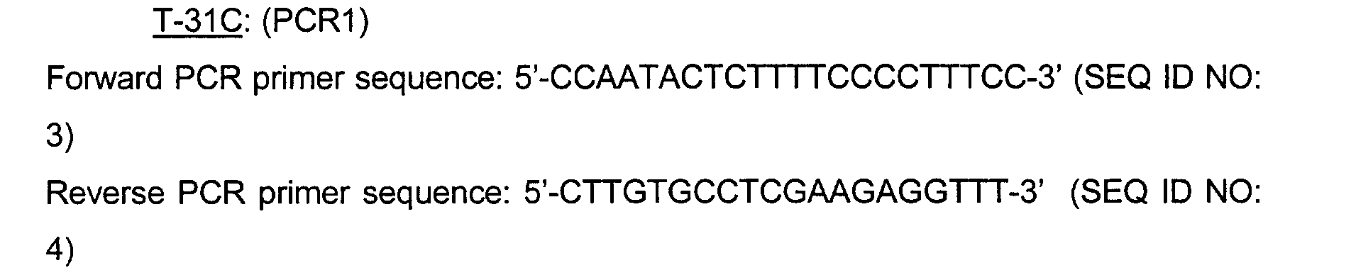
subjects of Northern European descent. To do this, the following primers flanking the
indicated IL-1β gene polymorphisms were generated:
PCR amplifications were performed using a PTC-225 tetrad model by MJ
Research, Inc. (Waltham, MA). Note that there were two reactions for the T-31C
polymorphism. The first reaction amplified a region that contained the polymorphism
at position -31. The second reaction used a reverse primer that introduces a G in
place of the A three nucleotides downstream of the T-31 C polymorphism. These two
reactions introduced a NlaIII restriction site that can be used to score the genotype,
rather than the naturally occurring Alul restriction site which is a very common
restriction endonuclease and thus is difficult to use to interpret the genotypes.
To perform the PCR amplifications, 10 ng genomic DNA was amplified in a
50µl reaction containing 10 mM Tris-CI, pH 8.7, 50 mM KCI, 1.5 mM MgCl2, 200 µM
each of the dNTPs, 0.5 µM each of the PCR primers and 2.5 Units HotStarTaq DNA
polymerase (Qiagen, Santa Clarita, CA). Samples were cycled at 96°C for 15 min.
followed by 30 cycles of 96°C for 30 sec., 58°C for 45 sec., and 72°C for 1 min. A
second PCR reaction is necessary for the T-31C marker since the reverse primer in
the reaction introduces an Nla III site which has proven to be more reliable in the
RFLP assays than the native Alu I site. PCR products from the initial PCR reaction
are diluted 1:800 and 1 µl PCR product used as template in the second PCR reaction
using the same conditions described above.
For genotyping, the PCR products were digested with the appropriate
restriction enzyme and buffer provided by the manufacturer (New England Biolabs,
Beverly, MA). 80-100ng PCR product was digested with restriction enzyme in a 50 µl
volume. C-511T samples were digested with Ava I and the T-31C samples digested
with Nla III at 37°C for 90 minutes. The C3953T samples were digested with Taq
α I at
65°C for 2 hours. The Ava I and Taq I digests were resolved on 2.5% agarose gels in
1X TBE buffer (0.1M Tris, 0.09M Boric acid, 0.001M EDTA). The Nla III digests were
resolved by gel electrophoresis on a 3% agarose gel in 1X TBE buffer. Genotypes
were scored by visual examination of the size of the PCR products present.
| Expected allele fragments: |
| C-511T | C allele: 191 bp and 113 bp | T allele: 304 bp |
| T-31C | T allele: 117 bp | C allele: 100 bp and 17 bp |
| C3953T | C allele: 135 bp and 114 bp | T allele: 249 bp |
Table I shows a typical EH output from the EH software package designed by
Jurg Ott (explained in Joseph Terwilliger and Jurg Ott,
Handbook of Human Genetic
Linkage, The Johns Hopkins University Press (1994)).
| IL-1β Statistical Estimation of Haplotype |
| | | | Haplotype Frequency |
| | | | Independent | w/Association |
| Allele at -511 | Allele at -31 | Allele at +3953 |
| C | C | C | 0.202315 | 0.016667 |
| C | C | T | 0.040463 | 0.000000 |
| C | T | C | 0.325463 | 0.450001 |
| C | T | T | 0.065093 | 0.166666 |
| T | C | C | 0.117130 | 0.366665 |
| T | C | T | 0.023426 | 0.000001 |
| T | T | C | 0.188426 | 0.000000 |
| T | T | T | 0.037685 | 0.000000 |
wherein: X
2 4=63.5 (where there were 4 degrees of freedom used in the X
2 test
statistics), P<10
-12, N=31.
Note that under the column "Independent" in Table I, the program is
calculating the expected haplotype frequencies by taking the product of all three allele
(i.e., marker) frequencies.
Allele frequencies for the 3 markers rounded up to the nearest percentage are
listed in Table II.
| Allele at -511 | Allele at -31 | Allele at +3953 |
| C= 0.59 | C=0.38 | C=0.84 |
| T= 0.41 | T= 0.62 | T=0.16 |
The column marked "w/Association" is the observed frequencies of the three
alleles being inherited together and is calculated via the EM algorithm. The difference
between the expected (i.e., "Independent" and observed (i.e., "w/Association")
frequencies in Table I demonstrates that these markers (i.e., T at -511, C at -31 and
C at 3953) are in linkage disequilibrium such that they are not inherited
independently. Pairwise calculations of linkage disequilibrium (D') show that allele T
at -511 and allele C at -31 have a D' value of .984 (p=0). Allele C at -31 and allele C
at 3953 have a D' value of .632 (p=.00607). Those skilled in the art will appreciate
that these values confirm that these alleles are not independently inherited.
Note that the EH program tests for the inheritance of these markers under the
null hypothesis of independence. Accordingly, the X2 value tests how significant the
observed is different than the expected.
Following genotype determination, an ex vivo blood assay was used to
measure the levels of secreted IL-1β protein levels. This assay consisted of a two-step
procedure in which a blood sample was first stimulated with LPS to promote IL-1β
synthesis, followed by treatment with ATP to promote postranslational processing.
The IL-1β protein released extracellularly was measured by ELISA.
To do this assay, blood was collected from patients in heparin-containing
vaccutainer tubes (commercially available from, e.g., Becton Dickinson, Franklin
Lakes, NJ). These samples could be stored on ice for up to four hours with no
adverse effect on assay performance. Seven-five microliters (µl) of blood was placed
into an individual well of a 96-well place and diluted with 75 µl of RPMI 1640
containing 20 mM Hepes, pH 7.3 (commercially available from, for example, Gibco-BRL).
The diluted blood samples were then incubated for two hours in the presence
or absence of lipopolysaccharide (LPS; 100-200 ng/ml; E. coli serotype 055:B5)
(commercially available from Sigma Chemical Co., St. Louis, MO) at 37°C in a 5%
CO2 environment. After incubation, 6 mM ATP (final concentration) was introduced
as a secretion stimulation. Thus, a solution of 100 mM ATP in 20 mM Hepes, pH 7.0
was made, and an appropriate volume of this solution was added to each well to
obtain a final concentration of 6 mM ATP.
The supematants of the blood cells was measured by ELISA. To do this, LPS
and ATP were added at each timepoint, with IL-1β protein measurements taken
before addition of LPS, after addition of LPS, and after addition of LPS/ATP. At day
16, the patients were "washed out" to ensure that the they did not have any active
study drug in their systems. From these "washed out' patients, cells were collected
as above and IL-1β protein measured following stimulation (e.g., with LPS alone and
LPS/ATP). The IL-1β assay was performed in its entirety at each timepoint to give us
three independent measures to control for assay variability. The ELISA for IL-1β
protein was purchased from R&D Systems (Minneapolis, MN), and used according to
manufacturer's specifications. Absolute IL-1β protein levels were calculated based on
comparison of assay performance in the presence of known quantities of
recombinant IL-1β protein (included with the ELISA kit) (see also Journal of
Immunology 165: 4615-4623 (2000)).
Substantially all of the cells from all subjects secreted maximal amounts of IL-1β
protein following stimulation with both LPS and ATP. Differences in the level of IL-1β
protein secretion were observed among cells from individuals with different IL-1β
haplotypes at all time points examined (i.e., at baseline, at day 0, and at day 16).
Note that the cells were in LPS for about two hours prior to each reading.
Accordingly, the measured levels of IL-1β protein secretion in response to stimulation
with LPS only at three different timepoints (baseline, day 0, and day 16) were
averaged and correlated with respect to the IL-1β haplotypes. As shown in Figure 1,
this analysis revealed a significant association between the inheritance of at least one
copy, and more so with two copies of a IL-1β gene haplotype defined by T(-511), C(-31),
and C(+3953) and increased levels of IL-1β protein that were secreted in
response to suboptimal stimulation with LPS alone (p=0.036).
EXAMPLE II
Presence of an IL-1β Haplotype Correlated With
An Increased Occurrence of Psoriasis
In another experiment, the genotypes of Caucasian human subjects with
psoriasis (and healthy control subjects) was determined as substantially described
above in Example I. The results demonstrate that the patients with psoriasis have at
least one copy of an IL-1β gene haplotype comprising a cysteine nucleotide at
position -31 and a cysteine nucleotide at position +3953 as compared to control
subjects (i.e., with no psoriasis). Thus, those skilled in the art will, once again,
appreciate the usefulness of the methods of the present invention, e.g., in detecting
an increased risk in developing an inflammatory disease, condition or disorder.
EQUIVALENTS
As will be apparent to those skilled in the art to which the invention pertains,
the present invention may be embodied in forms other than those specifically
disclosed above without departing from the spirit or essential characteristics of the
invention. The particular embodiments of the invention described above, are,
therefore, to be considered as illustrative and not restrictive. The scope of the
invention is as set forth in the appended claims rather than being limited to the
examples contained in the foregoing description.