EP1213349A1 - HUMAN 1p36 HOMOZYGOSITY DELETION DOMAIN - Google Patents
HUMAN 1p36 HOMOZYGOSITY DELETION DOMAIN Download PDFInfo
- Publication number
- EP1213349A1 EP1213349A1 EP00956853A EP00956853A EP1213349A1 EP 1213349 A1 EP1213349 A1 EP 1213349A1 EP 00956853 A EP00956853 A EP 00956853A EP 00956853 A EP00956853 A EP 00956853A EP 1213349 A1 EP1213349 A1 EP 1213349A1
- Authority
- EP
- European Patent Office
- Prior art keywords
- lane
- region
- deletion
- human
- tumor
- Prior art date
- Legal status (The legal status is an assumption and is not a legal conclusion. Google has not performed a legal analysis and makes no representation as to the accuracy of the status listed.)
- Withdrawn
Links
- 238000012217 deletion Methods 0.000 title claims abstract description 69
- 230000037430 deletion Effects 0.000 title claims abstract description 69
- 210000004027 cell Anatomy 0.000 claims abstract description 38
- 206010029260 Neuroblastoma Diseases 0.000 claims abstract description 34
- 210000000349 chromosome Anatomy 0.000 claims abstract description 17
- 108700025716 Tumor Suppressor Genes Proteins 0.000 abstract description 13
- 102000044209 Tumor Suppressor Genes Human genes 0.000 abstract description 13
- 206010028980 Neoplasm Diseases 0.000 description 35
- 239000002585 base Substances 0.000 description 35
- 108020004414 DNA Proteins 0.000 description 27
- 238000003752 polymerase chain reaction Methods 0.000 description 25
- 108090000623 proteins and genes Proteins 0.000 description 25
- 238000000034 method Methods 0.000 description 20
- 239000013641 positive control Substances 0.000 description 18
- 239000000203 mixture Substances 0.000 description 13
- 230000007170 pathology Effects 0.000 description 12
- XLYOFNOQVPJJNP-UHFFFAOYSA-N water Chemical compound O XLYOFNOQVPJJNP-UHFFFAOYSA-N 0.000 description 12
- 201000011510 cancer Diseases 0.000 description 11
- 239000012153 distilled water Substances 0.000 description 11
- 238000004458 analytical method Methods 0.000 description 10
- 238000004393 prognosis Methods 0.000 description 10
- 238000012216 screening Methods 0.000 description 10
- 238000003745 diagnosis Methods 0.000 description 9
- 239000000047 product Substances 0.000 description 9
- 239000002246 antineoplastic agent Substances 0.000 description 8
- 230000014509 gene expression Effects 0.000 description 8
- 239000003153 chemical reaction reagent Substances 0.000 description 7
- 239000003550 marker Substances 0.000 description 7
- 239000013642 negative control Substances 0.000 description 7
- 239000000243 solution Substances 0.000 description 7
- LFQSCWFLJHTTHZ-UHFFFAOYSA-N Ethanol Chemical compound CCO LFQSCWFLJHTTHZ-UHFFFAOYSA-N 0.000 description 6
- 102000004169 proteins and genes Human genes 0.000 description 6
- 230000003321 amplification Effects 0.000 description 5
- 238000001962 electrophoresis Methods 0.000 description 5
- 238000003199 nucleic acid amplification method Methods 0.000 description 5
- 239000006228 supernatant Substances 0.000 description 5
- 239000000439 tumor marker Substances 0.000 description 5
- 239000000872 buffer Substances 0.000 description 4
- 238000006243 chemical reaction Methods 0.000 description 4
- 210000003917 human chromosome Anatomy 0.000 description 4
- 208000005623 Carcinogenesis Diseases 0.000 description 3
- 206010009944 Colon cancer Diseases 0.000 description 3
- 201000000582 Retinoblastoma Diseases 0.000 description 3
- 230000005856 abnormality Effects 0.000 description 3
- 230000036952 cancer formation Effects 0.000 description 3
- 231100000504 carcinogenesis Toxicity 0.000 description 3
- 230000032823 cell division Effects 0.000 description 3
- 208000029742 colonic neoplasm Diseases 0.000 description 3
- 238000011161 development Methods 0.000 description 3
- 238000010586 diagram Methods 0.000 description 3
- 230000002779 inactivation Effects 0.000 description 3
- 210000004698 lymphocyte Anatomy 0.000 description 3
- 210000002826 placenta Anatomy 0.000 description 3
- 238000011160 research Methods 0.000 description 3
- 239000000523 sample Substances 0.000 description 3
- 238000012163 sequencing technique Methods 0.000 description 3
- 210000004881 tumor cell Anatomy 0.000 description 3
- QKNYBSVHEMOAJP-UHFFFAOYSA-N 2-amino-2-(hydroxymethyl)propane-1,3-diol;hydron;chloride Chemical compound Cl.OCC(N)(CO)CO QKNYBSVHEMOAJP-UHFFFAOYSA-N 0.000 description 2
- HRPVXLWXLXDGHG-UHFFFAOYSA-N Acrylamide Chemical compound NC(=O)C=C HRPVXLWXLXDGHG-UHFFFAOYSA-N 0.000 description 2
- HEDRZPFGACZZDS-UHFFFAOYSA-N Chloroform Chemical compound ClC(Cl)Cl HEDRZPFGACZZDS-UHFFFAOYSA-N 0.000 description 2
- KCXVZYZYPLLWCC-UHFFFAOYSA-N EDTA Chemical compound OC(=O)CN(CC(O)=O)CCN(CC(O)=O)CC(O)=O KCXVZYZYPLLWCC-UHFFFAOYSA-N 0.000 description 2
- 108090000790 Enzymes Proteins 0.000 description 2
- 102000004190 Enzymes Human genes 0.000 description 2
- 108010025020 Nerve Growth Factor Proteins 0.000 description 2
- 102000015336 Nerve Growth Factor Human genes 0.000 description 2
- 108700020796 Oncogene Proteins 0.000 description 2
- 238000009004 PCR Kit Methods 0.000 description 2
- FAPWRFPIFSIZLT-UHFFFAOYSA-M Sodium chloride Chemical compound [Na+].[Cl-] FAPWRFPIFSIZLT-UHFFFAOYSA-M 0.000 description 2
- IQFYYKKMVGJFEH-XLPZGREQSA-N Thymidine Chemical compound O=C1NC(=O)C(C)=CN1[C@@H]1O[C@H](CO)[C@@H](O)C1 IQFYYKKMVGJFEH-XLPZGREQSA-N 0.000 description 2
- 239000007984 Tris EDTA buffer Substances 0.000 description 2
- 208000008383 Wilms tumor Diseases 0.000 description 2
- 102100022748 Wilms tumor protein Human genes 0.000 description 2
- 238000010521 absorption reaction Methods 0.000 description 2
- 239000011543 agarose gel Substances 0.000 description 2
- 238000000246 agarose gel electrophoresis Methods 0.000 description 2
- 238000000137 annealing Methods 0.000 description 2
- 238000005119 centrifugation Methods 0.000 description 2
- 150000001875 compounds Chemical class 0.000 description 2
- 238000012258 culturing Methods 0.000 description 2
- 238000013461 design Methods 0.000 description 2
- 230000002068 genetic effect Effects 0.000 description 2
- 210000004408 hybridoma Anatomy 0.000 description 2
- 238000011534 incubation Methods 0.000 description 2
- 238000011835 investigation Methods 0.000 description 2
- PHTQWCKDNZKARW-UHFFFAOYSA-N isoamylol Chemical compound CC(C)CCO PHTQWCKDNZKARW-UHFFFAOYSA-N 0.000 description 2
- 238000002156 mixing Methods 0.000 description 2
- 229940053128 nerve growth factor Drugs 0.000 description 2
- 108020004707 nucleic acids Proteins 0.000 description 2
- 102000039446 nucleic acids Human genes 0.000 description 2
- 150000007523 nucleic acids Chemical class 0.000 description 2
- 238000010837 poor prognosis Methods 0.000 description 2
- 238000007894 restriction fragment length polymorphism technique Methods 0.000 description 2
- HUDPLKWXRLNSPC-UHFFFAOYSA-N 4-aminophthalhydrazide Chemical compound O=C1NNC(=O)C=2C1=CC(N)=CC=2 HUDPLKWXRLNSPC-UHFFFAOYSA-N 0.000 description 1
- 229920000936 Agarose Polymers 0.000 description 1
- 101100222854 Bacillus subtilis (strain 168) czcO gene Proteins 0.000 description 1
- DWRXFEITVBNRMK-UHFFFAOYSA-N Beta-D-1-Arabinofuranosylthymine Natural products O=C1NC(=O)C(C)=CN1C1C(O)C(O)C(CO)O1 DWRXFEITVBNRMK-UHFFFAOYSA-N 0.000 description 1
- 208000003174 Brain Neoplasms Diseases 0.000 description 1
- 206010006187 Breast cancer Diseases 0.000 description 1
- 208000026310 Breast neoplasm Diseases 0.000 description 1
- 208000031639 Chromosome Deletion Diseases 0.000 description 1
- 108010067770 Endopeptidase K Proteins 0.000 description 1
- 101000621309 Homo sapiens Wilms tumor protein Proteins 0.000 description 1
- 208000026350 Inborn Genetic disease Diseases 0.000 description 1
- 208000036626 Mental retardation Diseases 0.000 description 1
- 101100537961 Methanosarcina mazei (strain ATCC BAA-159 / DSM 3647 / Goe1 / Go1 / JCM 11833 / OCM 88) trkA2 gene Proteins 0.000 description 1
- 108700026495 N-Myc Proto-Oncogene Proteins 0.000 description 1
- 238000000636 Northern blotting Methods 0.000 description 1
- 108091028043 Nucleic acid sequence Proteins 0.000 description 1
- 102000003992 Peroxidases Human genes 0.000 description 1
- 102000004160 Phosphoric Monoester Hydrolases Human genes 0.000 description 1
- 108090000608 Phosphoric Monoester Hydrolases Proteins 0.000 description 1
- 101150020201 RB gene Proteins 0.000 description 1
- 238000002105 Southern blotting Methods 0.000 description 1
- YZCKVEUIGOORGS-NJFSPNSNSA-N Tritium Chemical compound [3H] YZCKVEUIGOORGS-NJFSPNSNSA-N 0.000 description 1
- 108090000631 Trypsin Proteins 0.000 description 1
- 102000004142 Trypsin Human genes 0.000 description 1
- 201000007960 WAGR syndrome Diseases 0.000 description 1
- 108700020467 WT1 Proteins 0.000 description 1
- 239000002253 acid Substances 0.000 description 1
- 150000007513 acids Chemical class 0.000 description 1
- DZBUGLKDJFMEHC-UHFFFAOYSA-N acridine Chemical class C1=CC=CC2=CC3=CC=CC=C3N=C21 DZBUGLKDJFMEHC-UHFFFAOYSA-N 0.000 description 1
- 230000001919 adrenal effect Effects 0.000 description 1
- 238000001042 affinity chromatography Methods 0.000 description 1
- 239000003513 alkali Substances 0.000 description 1
- 208000008303 aniridia Diseases 0.000 description 1
- 210000004507 artificial chromosome Anatomy 0.000 description 1
- IQFYYKKMVGJFEH-UHFFFAOYSA-N beta-L-thymidine Natural products O=C1NC(=O)C(C)=CN1C1OC(CO)C(O)C1 IQFYYKKMVGJFEH-UHFFFAOYSA-N 0.000 description 1
- 210000004369 blood Anatomy 0.000 description 1
- 239000008280 blood Substances 0.000 description 1
- 238000004113 cell culture Methods 0.000 description 1
- 239000007795 chemical reaction product Substances 0.000 description 1
- 238000010367 cloning Methods 0.000 description 1
- 230000009089 cytolysis Effects 0.000 description 1
- 238000000605 extraction Methods 0.000 description 1
- 230000001605 fetal effect Effects 0.000 description 1
- GNBHRKFJIUUOQI-UHFFFAOYSA-N fluorescein Chemical compound O1C(=O)C2=CC=CC=C2C21C1=CC=C(O)C=C1OC1=CC(O)=CC=C21 GNBHRKFJIUUOQI-UHFFFAOYSA-N 0.000 description 1
- 239000000499 gel Substances 0.000 description 1
- 238000001502 gel electrophoresis Methods 0.000 description 1
- 208000016361 genetic disease Diseases 0.000 description 1
- 210000005260 human cell Anatomy 0.000 description 1
- 238000009396 hybridization Methods 0.000 description 1
- 238000007901 in situ hybridization Methods 0.000 description 1
- 230000003834 intracellular effect Effects 0.000 description 1
- 208000022013 kidney Wilms tumor Diseases 0.000 description 1
- 230000003902 lesion Effects 0.000 description 1
- 201000007270 liver cancer Diseases 0.000 description 1
- 208000014018 liver neoplasm Diseases 0.000 description 1
- 238000010369 molecular cloning Methods 0.000 description 1
- 201000008026 nephroblastoma Diseases 0.000 description 1
- 238000010606 normalization Methods 0.000 description 1
- 201000008106 ocular cancer Diseases 0.000 description 1
- 210000000056 organ Anatomy 0.000 description 1
- 108040007629 peroxidase activity proteins Proteins 0.000 description 1
- 150000002989 phenols Chemical class 0.000 description 1
- 238000000746 purification Methods 0.000 description 1
- 210000003765 sex chromosome Anatomy 0.000 description 1
- 239000011780 sodium chloride Substances 0.000 description 1
- 230000003068 static effect Effects 0.000 description 1
- 239000000126 substance Substances 0.000 description 1
- 238000006467 substitution reaction Methods 0.000 description 1
- 238000001356 surgical procedure Methods 0.000 description 1
- 239000000725 suspension Substances 0.000 description 1
- 210000003411 telomere Anatomy 0.000 description 1
- 229940104230 thymidine Drugs 0.000 description 1
- 210000001519 tissue Anatomy 0.000 description 1
- 229910052722 tritium Inorganic materials 0.000 description 1
- 101150111535 trk gene Proteins 0.000 description 1
- 101150025395 trkA gene Proteins 0.000 description 1
- 101150113435 trkA1 gene Proteins 0.000 description 1
- 239000012588 trypsin Substances 0.000 description 1
- 238000001262 western blot Methods 0.000 description 1
Images




Classifications
-
- C—CHEMISTRY; METALLURGY
- C07—ORGANIC CHEMISTRY
- C07K—PEPTIDES
- C07K14/00—Peptides having more than 20 amino acids; Gastrins; Somatostatins; Melanotropins; Derivatives thereof
- C07K14/435—Peptides having more than 20 amino acids; Gastrins; Somatostatins; Melanotropins; Derivatives thereof from animals; from humans
- C07K14/46—Peptides having more than 20 amino acids; Gastrins; Somatostatins; Melanotropins; Derivatives thereof from animals; from humans from vertebrates
- C07K14/47—Peptides having more than 20 amino acids; Gastrins; Somatostatins; Melanotropins; Derivatives thereof from animals; from humans from vertebrates from mammals
- C07K14/4701—Peptides having more than 20 amino acids; Gastrins; Somatostatins; Melanotropins; Derivatives thereof from animals; from humans from vertebrates from mammals not used
- C07K14/4702—Regulators; Modulating activity
- C07K14/4703—Inhibitors; Suppressors
-
- C—CHEMISTRY; METALLURGY
- C07—ORGANIC CHEMISTRY
- C07K—PEPTIDES
- C07K14/00—Peptides having more than 20 amino acids; Gastrins; Somatostatins; Melanotropins; Derivatives thereof
- C07K14/82—Translation products from oncogenes
Definitions
- This invention relates to a homozygous deletion region (hereunder also referred to simply as “deletion region”) present in common at position 36 on the short arm of chromosome 1 (hereunder also referred to as "1p36”) of human neuroblastomas.
- deletion region a homozygous deletion region present in common at position 36 on the short arm of chromosome 1 (hereunder also referred to as "1p36”) of human neuroblastomas.
- Cancer-associated genes linked to oncogenesis are known to include tumor suppressor genes which promote oncogenesis when inactivated, and for example, the tumor suppressor genes RB and WT-1 have already been discovered and identified.
- the RB gene was so named because it was discovered as the gene responsible for retinoblastoma, an ocular cancer of childhood. In most cases of retinoblastoma, region 14 of the long arm of human chromosome 13 (13q14) is shortened (Yunis, JJ. et al., Am. J. Dis. Child. 132, 161-163(1978)), and it has long been conjectured that inactivation of a gene on this portion results in predisposition to cancer. However, cloning of the gene was difficult, and RB was first identified as a human tumor suppressor gene in 1986 (Friend, SH. et al., Nature, 323, 643-646(1986)).
- Wilms' tumor which is also called nephroblastoma
- nephroblastoma is a fetal malignant tumor originating in the nephric blastema tissue in infants.
- Patients often also exhibit abnormalities of the genitourinary organs, but epidemiological data tend to suggest the "two-hit theory" as with retinoblastoma, leading to conjecture on the role of a recessive oncogene.
- WAGR syndrome which also exhibit aniridia (A) and mental retardation (R) in addition to the tumor (W) and genitourinary (G) abnormalities
- a microdeletion is often found in region 13 on the short arm of chromosome 11 (11p13).
- WT1 has been isolated as the gene located in this common deletion region (Call, KM. et al., Cell 60, 509-520(1990)).
- the base sequence data for this region also allowed development of a tumor gene diagnosis method and an anticancer agent, as well as development of a method of analyzing the tumor pathology mechanism and a reagent to be used for analysis of the tumor pathology mechanism, and this invention was thereupon further completed.
- the invention relates to a homozygous deletion region present in common at position 36 on the short arm of chromosome 1 of human neuroblastomas.
- this region is a region characterized by being defined by at least from between a primer set at the SP6 end of the PAC clone dJ1028013 and a primer set at the SP6 end of the PAC clone dJ371E1 to between a primer set D1S2736 and a primer set at the T7 end of the PAC clone dJ142A6.
- the region is also a region characterized by being further defined by at least the primer set SGC30343 and D1S2736.
- the region is yet also a region characterized by being deleted in the human neuroblastoma cell lines NB-1 and MASS-NB-SCH-1.
- the invention further includes a tumor marker, a tumor diagnostic reagent, a tumor prognostic reagent and an anticancer agent which are based on the base sequence data obtained from this region.
- the invention still further includes a tumor pathology mechanism analysis method and a tumor pathology mechanism analysis reagent which are based on the base sequence data obtained from this region.
- the present inventors focused on the fact that the human 1p36 deletion has been found in at least 14 different human tumors (colon cancer, breast cancer, brain tumor, etc.), that several tumor suppressor genes are predicted to be present in this region and that the deletion has a high correlation with poor prognosis of neuroblastoma (Schwab M. et al., Genes Chromosomes Cancer 16, 211-229(1996), Fong C.T. et al., Cancer Res. 52, 1780-1785(1992)), and also focused on the fact that normalization of cancer cells has been achieved by introduction of normal 1p36 in colon cancer (Tanaka K.
- LOH Loss of Heterozygosity
- LOH refers to a pattern whereby a small genetic abnormality (deletion or substitution of a base or part of the gene) exists in one copy, while the chromosome on which the other copy resides undergoes the entire deletion of a large region including the gene. Consequently, in patients experiencing onset of cancer due to inactivation of a tumor suppressor gene, it is thought that there are two copies of the chromosome on which the tumor suppressor gene resides in normal cells, and one copy in tumor cells.
- the present inventors analyzed neuroblastoma cell lines and discovered an approximately 700 kb homozygous deletion region on the neuroblastoma chromosome 1p36. These 700 kb constituted part of the deletion region (approximately 8-10 Mb) elucidated by analysis of loss of heterozygosity.
- the base sequence disclosed by this invention may be used to investigate the presence or absence of the 1p36 homozygous deletion region in cell specimens to allow diagnosis and prognosis of tumors.
- the base sequence disclosed by this invention may be used to investigate the presence or absence of the 1p36 homozygous deletion region in cell specimens to allow diagnosis and prognosis of tumors.
- these probes may be labeled with radioisotopes, enzymes (for example, alkali phosphatase or peroxidase), fluorescent compounds (for example, fluorescein) or chemiluminescent compounds (for example, acridinium esters or isoluminol).
- hybridization methods there may be used, for example, Southern blotting, Northern blotting, dot blotting, Fluorescence in situ Hybridization (FISH) or the like.
- PCR Polymerase Chain Reaction
- Identification of loss of heterozygosity may be accomplished using, for example, Restriction Fragment Length Polymorphism (RFLP).
- RFLP Restriction Fragment Length Polymorphism
- Cells having the 1p36 homozygous deletion or loss of heterozygosity can be found using base sequences disclosed by the invention, thus allowing screening of anticancer agents. Because the cells are cancerous, their cell division occurs faster than that of normal cells. By adding candidate anticancer agents to the cell culture solution and observing the rate of cell division, it is possible to screen for anticancer agents against tumors having the 1p36 homozygous deletion or loss of heterozygosity. The cell division rate can be measured based on, for example, the amount of uptake of radioisotope (for example, tritium)-labeled thymidine.
- radioisotope for example, tritium
- Cells having the 1p36 homozygous deletion or loss of heterozygosity can be found using base sequences disclosed by the invention, thus allowing analysis of the tumor pathology mechanism.
- the proteins may be extracted from these and normal cells, and the protein expressed only by the normal cells identified by, for example, Western blotting. This protein may then be used for identification of other proteins that specifically interact therewith by, for example, affinity chromatography. By repeating such methods it is possible to identify proteins connected with oncogenesis and thus analyze the tumor pathology mechanism through analysis of their intracellular function.
- SK-N-BE ATCC Number: CRL-2271
- SK-N-DZ ATCC Number: CRL-2149
- RT-BM1 NLF donated by Garrett M.
- Brön LA-N-5 (Riken Cell Bank (The Institute of Physical and Chemical Research): RC0485)
- IMR32 donated by Garrett M.
- Brillion NMB donated by Garrett M.
- Brillion OAN donated by Garrett M.
- Brillion NGP donated by Garrett M.
- Brillion NB1 donated by Dr. Takafumi Matsumura of Kyoto Prefectural University, Dept.
- KP-N-NB donated by Dr. Takafumi Matsumura of Kyoto Prefectural University, Dept. of Pediatrics NB69: (Riken Cell Bank: RC0480)
- NBTu-1 donated by Dr. Kinji Yokomori of Japanese Red Cross Medical Center, Dept. of Pediatrics SK-N-MC (ATCC Number: HTB-10) SK-N-SH (ATCC Number: HTB-11).
- TEN buffer 50 mM Tris-HCl (pH 8.0), 1 mM EDTA, 100 mM NaCl
- SDS SDS
- EDTA Proteinase K
- the genomic DNA extracted from human neuroblastoma cell lines was purified under the following conditions using a QIAamp Blood Kit (QIAGEN).
- the extracted genomic DNA was quantified by ultraviolet absorption at 260 nm, and to 5-10 ⁇ g of the DNA there was added an appropriate amount of TE buffer to 225 ⁇ l. After further adding 210 ⁇ l of ethanol, the mixture was stirred.
- the stirred solution was transferred to a QIAamp column (kit accessory, QIAGEN), and centrifugation was performed at 8,000 rpm for one minute to adsorb the genomic DNA onto the column.
- a 500 ⁇ l portion of Buffer AW (kit accessory, QIAGEN) was added to the column, and centrifugation was performed at 8,000 rpm for one minute.
- Buffer AW kit accessory, QIAGEN
- the column was transferred to a 1.5 ml fresh tube, 200 ⁇ l of Buffer AE prewarmed to 70°C was added, and the column was allowed to stand at room temperature for one minute and then centrifuged at 8,000 rpm for one minute to purify the DNA.
- the purified genomic DNA was quantified based on the ultraviolet absorption at 260 nm.
- the genomic DNA of the 27 human neuroblastoma cell lines extracted and purified in Examples 1 and 2 was used for identification of the 1p36 deletion by the following site-specific PCR.
- the PCR primers used were products of Sawady Technology Co., Ltd., unless otherwise specified.
- a 10 ⁇ l mixture was prepared by combining 1 ⁇ l of genomic DNA solution (2 ng/ ⁇ l), 1 ⁇ l of 10xPfu buffer, 1 ⁇ l of dNTPs (2 mM), 0.2 ⁇ l of D1S548F (20 pmole/ ⁇ l), 0.2 ⁇ l of D1S548R (20 pmole/ ⁇ l), 0.2 ⁇ l of D1S244F (20 pmole/ ⁇ l), 0.2 ⁇ l of D1S244R (20 pmole/ ⁇ l), 6.12 ⁇ l of sterile distilled water and 0.08 ⁇ l of Pfu.
- the D1S548 and D1S244 are both known primer sets capable of specifically amplifying the 1p36.23-31 region, and their sequences are shown in Table 1.
- the 1.3 Mb chromosome telomere end was amplified by D1S548 and D1S244.
- F and R indicate the directions of the primers.
- a band near 169 bases amplified by D1S548 as the positive control primer set was observed in all of the cell lines.
- a band near 285-296 bases amplified by D1S244 was observed only in lines NB-1 and MASS-NB-SCH-1.
- PCR was conducted with each of 5 other primer sets, SGC-30343, D1S2021, D1S1768, PGD and D1S2736, which are known to specifically amplify the 1p36.23-31 region of the two cell lines NB-1 and ESS-NB-SCH-1.
- As a positive control there were used two different types of human Nerve Growth Factor ⁇ (hNGF ⁇ ) located at position 13 of the short arm of human chromosome 1 (1p13).
- hNGF ⁇ 60s is a primer with an annealing temperature near 60°C
- hMGF ⁇ 57s is a primer with an annealing temperature near 57°C
- the bands are amplified near 253 bp and 229 bp, respectively.
- the sequences for these primers are shown in Table 2.
- the human neuroblastoma lines MB-1 and MASS-NB-SCH-1 have deletions at least in the region from SGC-30343 to D1S2736 of 1p36, on both chromosomes.
- the primer set D1S244 which was used to confirm the 1p36.23-31 region deletion in Example 3 was used for PCR with PAC clones (P1-derived artificial chromosomes, product of Oligo Service, Tsukuba Laboratory) having the human genome at random, using an LA PCR kit (Takara Shuzo), and PAC clones containing the deletion region sequence were selected.
- PAC clones P1-derived artificial chromosomes, product of Oligo Service, Tsukuba Laboratory
- a 20 ⁇ l mixture was prepared by combining 5 ⁇ l of PAC DNA pool solution (Z1-Z4: Oligo Service, Tsukuba Laboratory), 2 ⁇ l of 10xLA PCR buffer, 2.5 ⁇ l of dNTPs (2 mM), 0.4 ⁇ l of D1S244F (100 pmole/ ⁇ l), 0.4 ⁇ l of D1S244R (100 pmole/ ⁇ l), 9.6 ⁇ l of sterile distilled water and 0.1 ⁇ l of LA Taq (5 u/ ⁇ l).
- the PCR product was subjected to electrophoresis, and then only the PAC DNA pools for which an amplification band near 285-296 bases was observed were subjected to a second PCR by the same procedure to narrow down the clones.
- Fig. 3 shows the results for only dJ142A6, dJ332G17 and dJ369D13.
- the clones obtained in this manner were taken as the 1st screening.
- the 18 clones obtained by the 1st screening are shown in Table 3.
- the total of 18 clones obtained by the 1st screening are believed to retain the genome sequence of the deleted region of the two strains NB-1 and MASS-NB-SCH-1, and since some sequences were detected by multiple primer sets, 3 general contigs were formed (respectively designated as Contig 1, Contig 2 and Contig 3).
- Contig 1, Contig 2 and Contig 3 The contigs for the 18 PAC clones obtained by the 1st screening are shown in Fig. 4.
- RPN2434 (Kit: Amersham Co.) was used according to the procedure described below to determine the base sequences at both ends of a total of 9 PAC clones, namely the 2 clones dJ99E23 and dJ371E1 from Contig 1, the 4 clones dJ326J10, dJ739D18, dJ756A17 and dJ373D24 from Contig 2 and the 3 clones dJ369D13, dJ142A6 and dJ345P21 from Contig 3.
- Table 4 shows the selected clones and the sequences of their respective primer sets.
- the newly designed primer sets were used for reselection of PAC clones from PAC DNA pools by PCR, and the process of designing of walking primers based on both end sequences of the selected clones and determination of contigs based on walking was repeated. These were taken as the 2nd and 3rd screenings. Table 5 shows the selected clones and the sequences of their respective primers sets.
- Example 4 Based on Example 4 it was judged that NB-1 and MASS-NB-SCH-1 have deletions on both chromosomes in at least the regions defined from SGC-30343 to D1S2736 of 1p36, but the following procedure was carried out next to identify both ends of the deletion regions.
- New primers were synthesized near the deletion region based on the contig data for the PAC clones obtained in Example 5 and the NB-1 and MASS-NB-SCH-1 strains and positive controls were subjected to PCR under the conditions described below, after which the presence or absence of specific band amplification was investigated.
- An LA PCR kit (Takara Shuzo) was used for PCR in the following manner.
- a 10 ⁇ l mixture was prepared by combining 1 ⁇ l of genomic DNA solution (100 ng/ ⁇ l), 1 ⁇ l of 10xLA PCR buffer, 0.6 ⁇ l of dNTPs (2.5 mM, kit accessories), 0.1 ⁇ l of Primer F (100 mM), 0.1 ⁇ l of Primer R(100 mM), 7.15 ⁇ l of sterile distilled water and 0.05 ⁇ l of LA Taq (5 u/ ⁇ l, kit accessory). After denaturing the mixture at 95°C for 2 minutes, it was provided for PCR with 30 repeated cycles of 95°C for 30 seconds and 58°C for one minute.
- the PCR reaction product was subjected to electrophoresis using 2.5% agarose gel. The results are shown in Fig. 6.
- results gave a range for the genome deletion region of 1p36 as defined by from between the primer set at the SP6 end of the PAC clone dJ1028O13 and the primer set at the SP6 end of the PAC clone dJ371E1 to between the primer set D1S2736 and the primer set at the T7 end of the PAC clone dJ142A6, as shown in Fig. 7.
- the minimal PAC. clone group including the entire deletion region is shown in Fig. 8. Sequencing of these PAC clones yields the base sequence contained in the deletion region of NB-1 and MASS-NB-SCH-1.
- Table 6 shows the sequences of the primer sets used to determine the 1p36 genome deletion region range as elucidated by this invention.
- a PAC clone group was selected which included essentially all of the deletion region, in the same manner as Example 6. These PAC clones were dJ371E1, dJ756A17, dJ266I16 and dJ345P21, the overall structural map of which is shown in Fig. 9. Thus, by sequencing each of these PAC clones and aligning their sequences, it is possible to determine approximately the entire length base sequence of the deletion region.
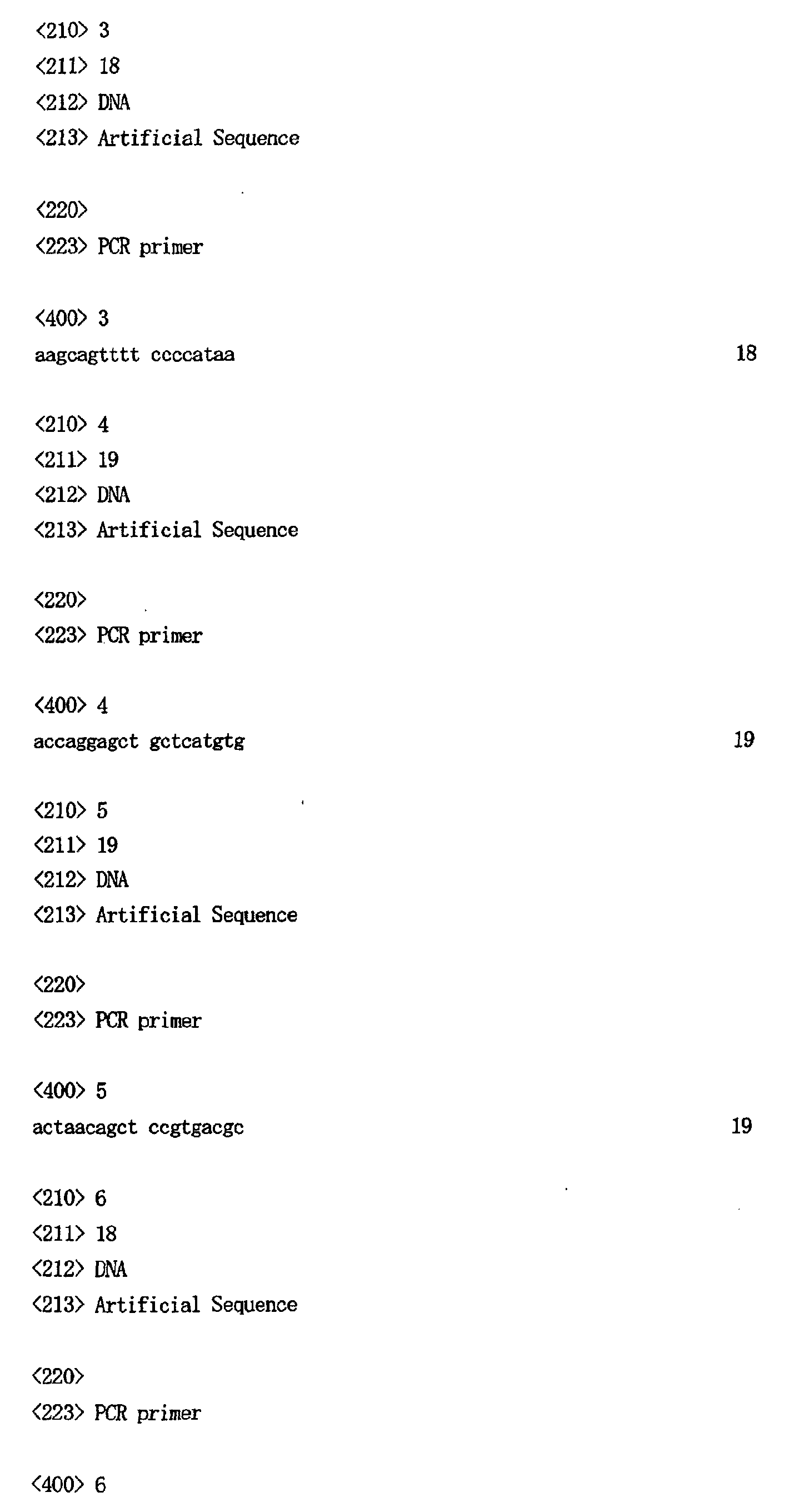
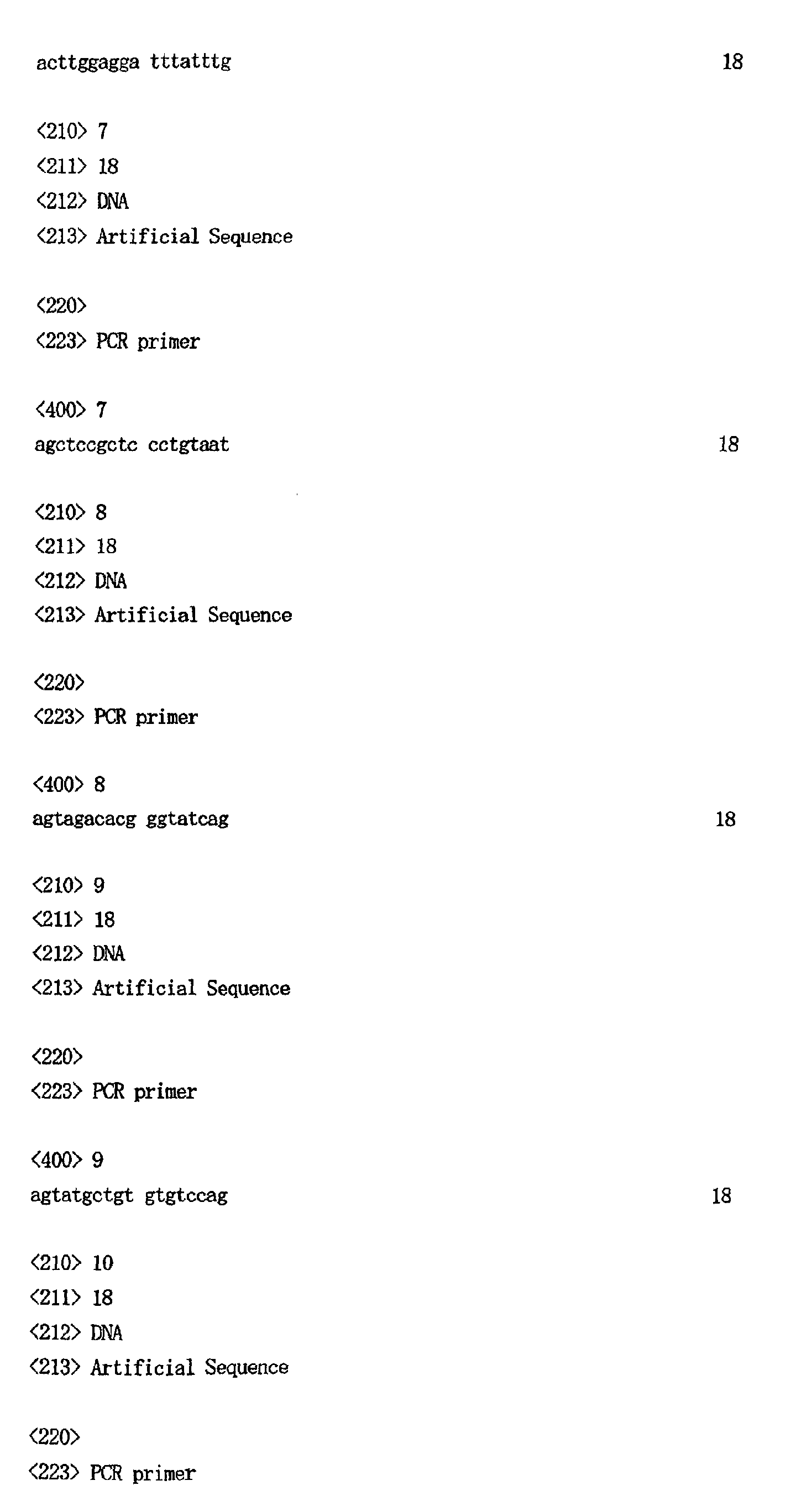
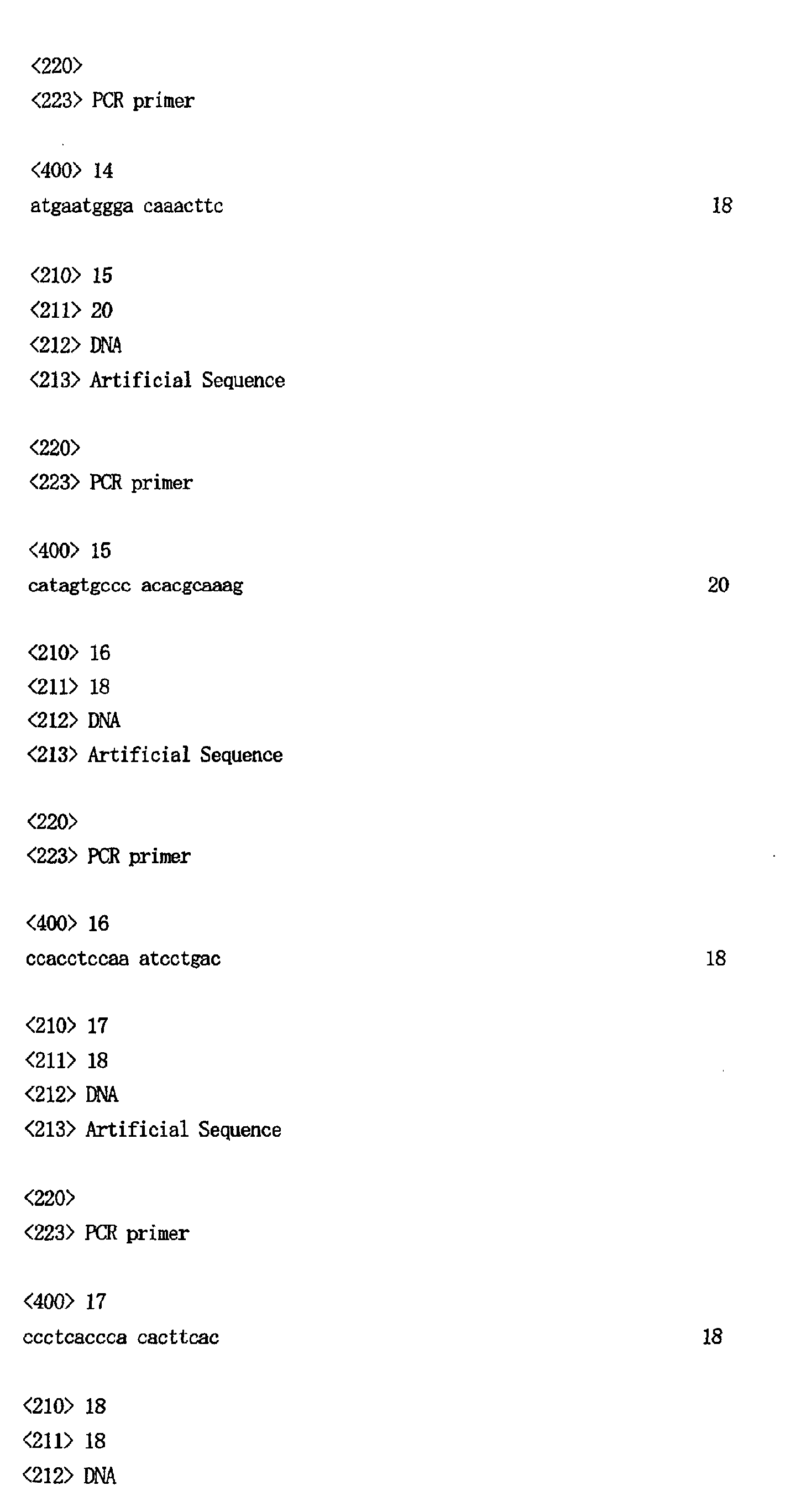
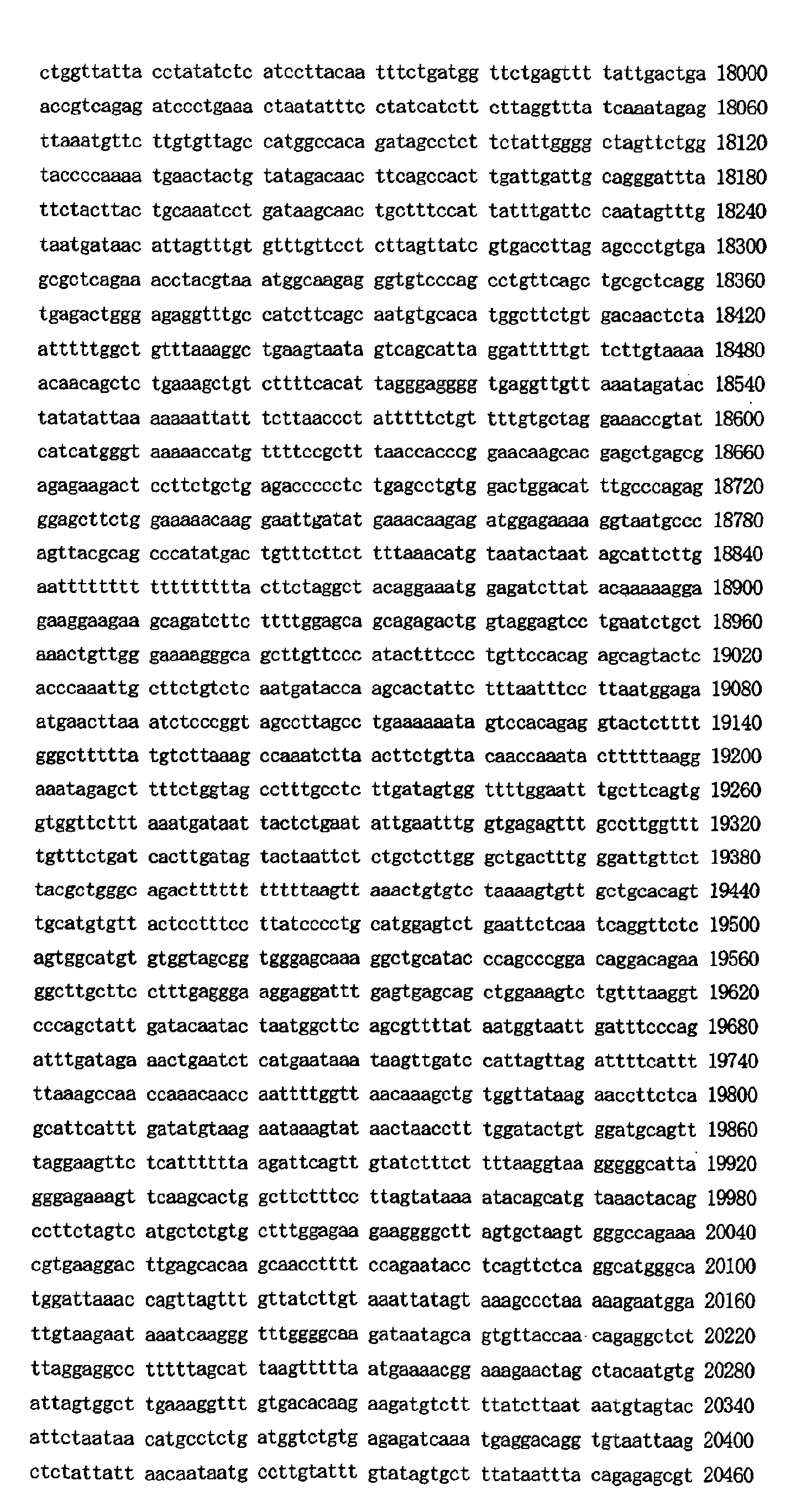
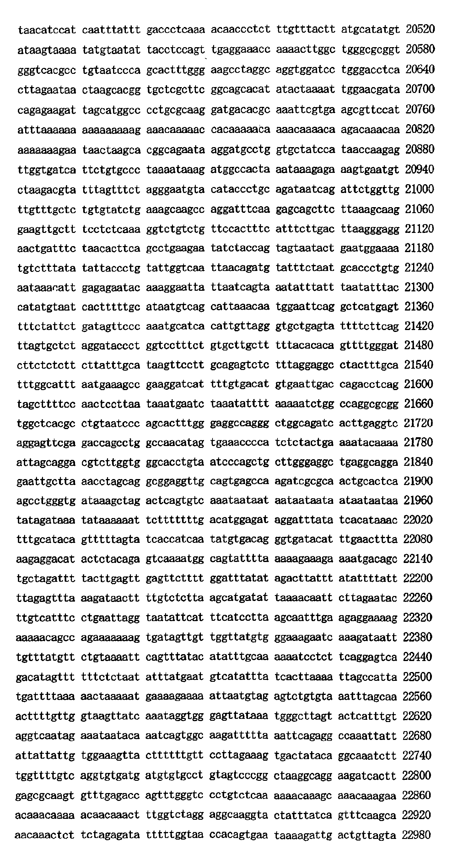
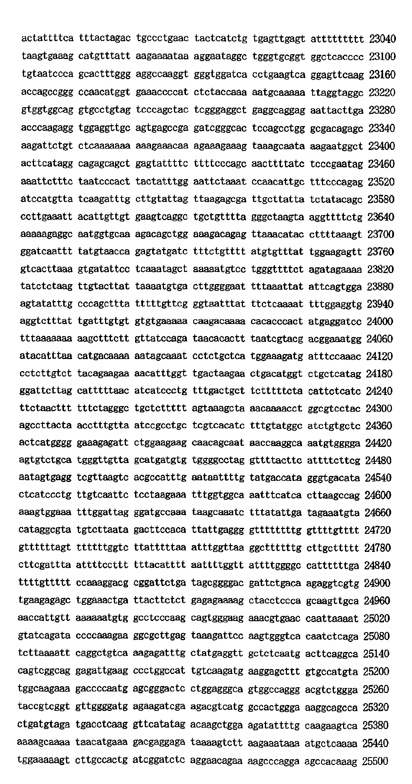
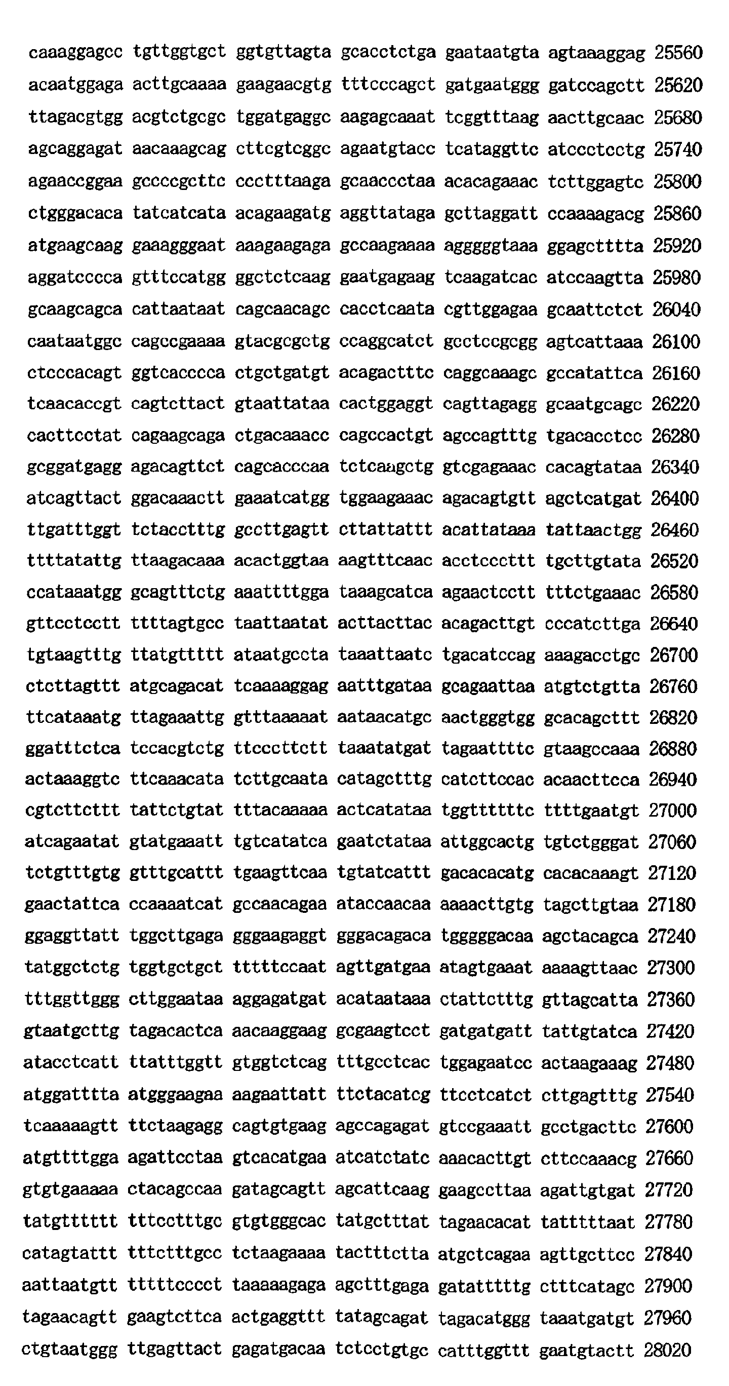
- the base sequences of each of the clones, including the deletion region were determined. Specifically, the base sequences of 5 contiguous partial sequences including the deletion region were found for PAC clone dJ371E1. Their partial sequences were designated as dJ371E1-1 (SEQ. ID. No.60), dJ371E1-2 (SEQ. ID. No.61), dJ371E1-3 (SEQ. ID. No.62), dJ371E1-4 (SEQ. ID. No.63) and dJ371E1-5 (SEQ. ID. No.64). The respective base sequences are listed in the Sequence Listing.
- the base sequences of 7 contiguous partial sequences including the deletion region were found for PAC clone dJ756A17. Their partial sequences were designated as dJ756A17-1 (SEQ. ID. No.65), dJ756A17-2 (SEQ. ID. No.66), dJ756A17-3 (SEQ. ID. No.67), dJ756A17-4 (SEQ. ID, No.68), dJ756A17-5 (SEQ. ID. No.69), dJ756A17-6 (SEQ. ID. No.70) and dJ756A17-7 (SEQ. ID. No.71). The respective base sequences are listed in the Sequence Listing.
- dJ266I16-1 SEQ. ID. No.72
- dJ266I16-2 SEQ. ID. No.73
- dJ266I16-3 SEQ. ID. No.74
- dJ266I16-4 SEQ. ID. No.75
- dJ266I16-5 SEQ. ID. No.76
- dJ266I16-6 SEQ. ID. No.77
- dJ266I16-7 SEQ. ID. No.78
- dJ266I16-8 SEQ. ID.
- Prognosis is generally also poor for patients with high age of onset (Nakagawara A. et al., Med. Pediatr. Oncol. 31, 113-115(1998)), patients with primary lesion in the adrenal body (Nakagawara A. et al., Surgery 104, 34-40(1988)), and patients with high expression of the oncogene N-myc (Brön GM. et al., Science 224, 1121-1124(1984)), and a very stable correlation was found with the presence or absence of LOH for 1p36.
- a homozygous deletion region present in common at position 36 on the short arm of chromosome 1 of human neuroblastoma. Based on the base sequence data of the deletion region it is possible to design a method for diagnosis, prognosis of tumors, as well as a tumor marker which can be used therefor, while the base sequence data of the deletion region can also be used to develop a method of tumor gene diagnosis and an anticancer agent, and to develop a method of analyzing the tumor pathology mechanism and a reagent to be used for analysis of the tumor pathology mechanism.
Landscapes
- Chemical & Material Sciences (AREA)
- Health & Medical Sciences (AREA)
- Organic Chemistry (AREA)
- Life Sciences & Earth Sciences (AREA)
- General Health & Medical Sciences (AREA)
- Gastroenterology & Hepatology (AREA)
- Biochemistry (AREA)
- Biophysics (AREA)
- Genetics & Genomics (AREA)
- Medicinal Chemistry (AREA)
- Molecular Biology (AREA)
- Proteomics, Peptides & Aminoacids (AREA)
- Zoology (AREA)
- Toxicology (AREA)
- Oncology (AREA)
- Measuring Or Testing Involving Enzymes Or Micro-Organisms (AREA)
Abstract
Description
- This invention relates to a homozygous deletion region (hereunder also referred to simply as "deletion region") present in common at position 36 on the short arm of chromosome 1 (hereunder also referred to as "1p36") of human neuroblastomas.
- Cancer-associated genes linked to oncogenesis are known to include tumor suppressor genes which promote oncogenesis when inactivated, and for example, the tumor suppressor genes RB and WT-1 have already been discovered and identified.
- The RB gene was so named because it was discovered as the gene responsible for retinoblastoma, an ocular cancer of childhood. In most cases of retinoblastoma,
region 14 of the long arm of human chromosome 13 (13q14) is shortened (Yunis, JJ. et al., Am. J. Dis. Child. 132, 161-163(1978)), and it has long been conjectured that inactivation of a gene on this portion results in predisposition to cancer. However, cloning of the gene was difficult, and RB was first identified as a human tumor suppressor gene in 1986 (Friend, SH. et al., Nature, 323, 643-646(1986)). - Wilms' tumor, which is also called nephroblastoma, is a fetal malignant tumor originating in the nephric blastema tissue in infants. Patients often also exhibit abnormalities of the genitourinary organs, but epidemiological data tend to suggest the "two-hit theory" as with retinoblastoma, leading to conjecture on the role of a recessive oncogene. In the case of patients with WAGR syndrome which also exhibit aniridia (A) and mental retardation (R) in addition to the tumor (W) and genitourinary (G) abnormalities, a microdeletion is often found in
region 13 on the short arm of chromosome 11 (11p13). WT1 has been isolated as the gene located in this common deletion region (Call, KM. et al., Cell 60, 509-520(1990)). - Numerous tumor suppressor genes have since been discovered based on the data for chromosome deletion regions, but as of the current time no tumor suppressor gene for neuroblastoma has been identified from the 1p36 deletion region.
- It is an object of this invention to provide data relating to a novel homozygous deletion region at position 36 on the short arm of
chromosome 1 which is common to human neuroblastomas. - It is another object to provide a method for diagnosis and prognosis of tumors based on the base sequence data for this region, as well as a tumor marker which can be used therefor.
- It is yet another object to develop a method of gene diagnosis and an anticancer agent, and to develop a method of analyzing the tumor pathology mechanism and a reagent to be used for analysis of the tumor pathology mechanism, based on this base sequence data.
- As a result of much diligent research toward finding a tumor suppressor gene for neuroblastoma, the present inventors have discovered a novel homozygous deletion region at position 36 on the short arm of
chromosome 1 of NB-1, a human neuroblastoma cell line. - This finding made it possible to design a method for tumor diagnosis and prognosis and a tumor marker which can be used therefor, and this invention was thus completed. The base sequence data for this region also allowed development of a tumor gene diagnosis method and an anticancer agent, as well as development of a method of analyzing the tumor pathology mechanism and a reagent to be used for analysis of the tumor pathology mechanism, and this invention was thereupon further completed.
- More specifically, the invention relates to a homozygous deletion region present in common at position 36 on the short arm of
chromosome 1 of human neuroblastomas. - Even more specifically, this region is a region characterized by being defined by at least from between a primer set at the SP6 end of the PAC clone dJ1028013 and a primer set at the SP6 end of the PAC clone dJ371E1 to between a primer set D1S2736 and a primer set at the T7 end of the PAC clone dJ142A6.
- The region is also a region characterized by being further defined by at least the primer set SGC30343 and D1S2736.
- The region is yet also a region characterized by being deleted in the human neuroblastoma cell lines NB-1 and MASS-NB-SCH-1.
- The invention further includes a tumor marker, a tumor diagnostic reagent, a tumor prognostic reagent and an anticancer agent which are based on the base sequence data obtained from this region.
- The invention still further includes a tumor pathology mechanism analysis method and a tumor pathology mechanism analysis reagent which are based on the base sequence data obtained from this region.
-
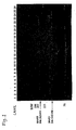
- Fig. 1 is a photograph of 4% agarose gel electrophoresis of a PCR product for human neuroblastoma genomic DNA using the D1S548 and D1S244 primer sets. (Lane 1: molecular weight marker (1330, 828, 750, 696, 536, 517, 501, 396, 351, 221, 75, 65, 35, 22 bp from top), Lane 2: SK-N-BE, Lane 3: SK-N-DZ, Lane 4: RT-BM1, Lane 5: NLF, Lane 6: LA-N-5, Lane 7: IMR32, Lane 8: NMB, Lane 9: OAN, Lane 10: NGP, Lane 11: NB1, Lane 12: CHP134, Lane 13: KCN, Lane 14: NBL-S, Lane 15: CHP901, Lane 16: SY5Y, Lane 17: GANB, Lane 18: SK-N-AS, Lane 19: KAN, Lane 20: LHN, Lane 21: SAN, Lane 22: TGW, Lane 23: positive control (human lymphocytes), Lane 24: MASS-NB-SCH-1, Lane 25: negative control (sterile distilled water), Lane 26: molecular weight marker (same as Lane 1)).
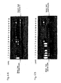
- Fig. 2 is a photograph of 2% agarose gel electrophoresis of PCR products for NB-1 and MASS-NB-SCH-1 genomic DNA using the D1S2021, PGD, SGC-30343, D1S1768 and D1S2736 primer sets. All of the PCR products include primers that specifically amplify the region of human Nerve Growth Factor β (hNGFβ) as positive controls. (Lane 1: empty lane, Lane 2: molecular weight marker (1030, 734, 341, 258, 192, 105, 78, 75, 46, 36, 18, 17, 12, 11, 8 bp from top), Lane 3: positive control (SYSY), Lane 4: NB-1, Lane 5: MASS-NB-SCH-1, Lane 6: negative control (sterile distilled water), Lane 7: positive control (SY5Y), Lane 8: NB-1, Lane 9: MASS-NB-SCH-1, Lane 10: negative control (sterile distilled water), Lane 11: positive control (SYSY), Lane 12: NB-1, Lane 13: MASS-NB-SCH-1, Lane 14: negative control (sterile distilled water), Lane 15: positive control (SYSY), Lane 16: NB-1, Lane 17: MASS-NB-SCH-1, Lane 18: negative control (sterile distilled water), Lane 19: positive control (SY5Y), Lane 20: NB-1, Lane 21: MASS-NB-SCH-1, Lane 22: negative control (sterile distilled water), Lane 23: molecular weight marker (same as Lane 2)).
- Fig. 3 is a photograph of 5% acrylamide electrophoresis of a PCR product for a PAC clone DNA using the D1S244 primer set. (Lane 1: molecular weight marker (1202 bp, 517 bp, 396 bp, 201 bp), Lanes 2-4: dJ142A6, Lane 5-7: dJ332G17, Lane 8-10: dJ369D13, Lane 11: positive control (human placenta DNA), Lane 12: negative control (sterile distilled water)).
- Fig. 4 is a contig map for the PAC18 clone included in 1p36 found in strains NB-1 and MASS-NB-SCH-1 by the 1st screening.
- Fig. 5 is a contig map for the PAC34 clone included in 1p36 found in strains NB-1 and MASS-NB-SCH-1 in the final screening.
- Fig. 6 is a pair of photographs of electrophoresis of PCR products for NB-1 and MASS-NB-SCH-1 genomic DNA using primers designed based on PAC clone data. The gel used was 2.5 agarose. (Lane 1: marker (1202 bp, 517 bp, 396 bp, 201 bp), Lane 2: positive control (human placenta DNA), Lane 3: normal cells derived from neuroblastoma patient without 1p36 deletion, Lane 4: tumor cells derived from neuroblastoma patient without 1p36 deletion, Lane 5: normal cells derived from MASS-NB-SCH-1 patient, Lane 6: MASS-NB-SCH-1, Lane 7: NB1, Lane 8: positive control (mouse hybridoma with human chromosome), Lane 9: positive control (human lymphocytes), Lane 10: positive control (human placenta DNA), Lane 11: normal cells derived from neuroblastoma patient without 1p36 deletion, Lane 12: tumor cells. derived from neuroblastoma patient without 1p36 deletion, Lane 13: normal cells derived from MASS-NB-SCH-1 patient, Lane 14: MASS-NB-SCH-1, Lane 15: NB1, Lane 16: positive control (mouse hybridoma with human chromosome), Lane 17: positive control (human lymphocytes), Lane 18: empty lane). In A, dJ1028013 was used as primer for Lanes 1-8; dJ142A6-T7 was used as primer for Lanes 9-17. In B, dJ371E1-SP6 was used as primer for Lanes 1-8; dJ587c9-SP6 was used as primer for Lanes 9-17.
- Fig. 7 is a diagram showing the genome deletion region area for 1p36 elucidated by this invention.
- Fig. 8 is a diagram showing the positions for a minimal PAC clone group included in the genome deletion region area for 1p36 elucidated by this invention.
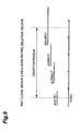
- Fig. 9 is a diagram showing the positions for a minimal PAC clone group included in the genome deletion region area for 1p36 elucidated by this invention, which differs somewhat from the combination shown in Fig. 8.
-
- The present inventors focused on the fact that the human 1p36 deletion has been found in at least 14 different human tumors (colon cancer, breast cancer, brain tumor, etc.), that several tumor suppressor genes are predicted to be present in this region and that the deletion has a high correlation with poor prognosis of neuroblastoma (Schwab M. et al., Genes Chromosomes Cancer 16, 211-229(1996), Fong C.T. et al., Cancer Res. 52, 1780-1785(1992)), and also focused on the fact that normalization of cancer cells has been achieved by introduction of normal 1p36 in colon cancer (Tanaka K. et al., Oncogene, 8, 2253-2258(1993)) and that the 1p36 deletion is found from the initial stages of liver cancer and colon cancer (Kuroki T. et al., Genes Chromosomes Cancer 13, 163-167(1995)), which suggested a high possibility that the 1p36 deletion plays a role in the early stages of canceration of normal cells.
- Identification of the tumor suppressor gene is possible by analysis of Loss of Heterozygosity (hereunder also abbreviated as LOH). The chromosomes of human cells, except for the sex chromosomes, exist as pairs, and two copies of each gene are present, one from the mother and one from the father. According to the "two-hit theory" advocated by Kundson A.G. et al. (Kundson A.G., Pediatr. Res. 513- (1976)), inactivation of a gene is only found to occur when both of each copy from the mother and father are inactivated, and LOH is observed most often by this mechanism. LOH refers to a pattern whereby a small genetic abnormality (deletion or substitution of a base or part of the gene) exists in one copy, while the chromosome on which the other copy resides undergoes the entire deletion of a large region including the gene. Consequently, in patients experiencing onset of cancer due to inactivation of a tumor suppressor gene, it is thought that there are two copies of the chromosome on which the tumor suppressor gene resides in normal cells, and one copy in tumor cells.
- However, the commonly observed range for LOH is very large in terms of the molecular genetic distance, and the numbers of genes and numbers of bases contained therein are exceedingly huge. The current actual deletion region of 1p36 is limited to the approximately 8-10 Mb region of 1p36.2-1p36.3 according to LOH analysis, but this region must be further narrowed down.
- In order to identify the tumor suppressor gene, the present inventors analyzed neuroblastoma cell lines and discovered an approximately 700 kb homozygous deletion region on the neuroblastoma chromosome 1p36. These 700 kb constituted part of the deletion region (approximately 8-10 Mb) elucidated by analysis of loss of heterozygosity.
- The base sequence disclosed by this invention may be used to investigate the presence or absence of the 1p36 homozygous deletion region in cell specimens to allow diagnosis and prognosis of tumors. For investigation of the presence or absence of the deletion region there may be applied all of the methods using any desired sequence of the 1p36 homozygous deletion region and base sequences (probes) that can hybridize thereto. When necessary, these probes may be labeled with radioisotopes, enzymes (for example, alkali phosphatase or peroxidase), fluorescent compounds (for example, fluorescein) or chemiluminescent compounds (for example, acridinium esters or isoluminol). That is, when nucleic acids that hybridize to the probe sequences are not present in the cell specimen, a high possibility exists for tumor onset, while in specimens from known cancer onset cases the prognosis can be predicted to be poor. As hybridization methods there may be used, for example, Southern blotting, Northern blotting, dot blotting, Fluorescence in situ Hybridization (FISH) or the like.
- For investigation of the presence or absence of the deletion region there may also be used a Polymerase Chain Reaction (PCR) with any desired sequences from the 1p36 homozygous deletion region as the primers.
- Identification of loss of heterozygosity may be accomplished using, for example, Restriction Fragment Length Polymorphism (RFLP).
- These methods are carried out according to protocols known by skilled professionals. Reference may be made to, for example, Molecular Cloning, A Laboratory Manual (T. Maniatis: Cold Spring Harbor Laboratory Press) and Idenshibyo Nyumon [Introduction to Genetic Diseases] (F. Takahisa: Nankodo Publishing).
- Cells having the 1p36 homozygous deletion or loss of heterozygosity can be found using base sequences disclosed by the invention, thus allowing screening of anticancer agents. Because the cells are cancerous, their cell division occurs faster than that of normal cells. By adding candidate anticancer agents to the cell culture solution and observing the rate of cell division, it is possible to screen for anticancer agents against tumors having the 1p36 homozygous deletion or loss of heterozygosity. The cell division rate can be measured based on, for example, the amount of uptake of radioisotope (for example, tritium)-labeled thymidine.
- Cells having the 1p36 homozygous deletion or loss of heterozygosity can be found using base sequences disclosed by the invention, thus allowing analysis of the tumor pathology mechanism. The proteins may be extracted from these and normal cells, and the protein expressed only by the normal cells identified by, for example, Western blotting. This protein may then be used for identification of other proteins that specifically interact therewith by, for example, affinity chromatography. By repeating such methods it is possible to identify proteins connected with oncogenesis and thus analyze the tumor pathology mechanism through analysis of their intracellular function.
- The 1p36 homozygous deletion region common to neuroblastoma which was discovered by this invention will now be described in detail by way of examples.
- Each of the following human neuroblastoma-derived cell lines was cultured on a 10 cm culturing plate to general confluency. The names and sources of the cell lines used are summarized below.
SK-N-BE (ATCC Number: CRL-2271)
SK-N-DZ (ATCC Number: CRL-2149)
RT-BM1
NLF: donated by Garrett M. Brodeur
LA-N-5: (Riken Cell Bank (The Institute of Physical and Chemical Research): RC0485)
IMR32: donated by Garrett M. Brodeur
NMB: donated by Garrett M. Brodeur
OAN: donated by Garrett M. Brodeur
NGP: donated by Garrett M. Brodeur
NB1: donated by Dr. Takafumi Matsumura of Kyoto Prefectural University, Dept. of Pediatrics (Considered one of the oldest neuroblastoma cell lines in Japan, established at the Niigata University Brain Research Laboratory)
CHP134: donated by Garrett M. Brodeur
KCN: donated by Garrett M. Brodeur
NBL-S: donated by Garrett M. Brodeur
CHP901: cell line established from patient surgically extracted specimen
SYSY: donated by Garrett M. Brodeur
GANB: cell line established from patient surgically extracted specimen
SK-N-AS (ATCC Number: CRL-2137)
KAN: donated by Garrett M. Brodeur
LHN: donated by Garrett M. Brodeur
SAN: donated by Garrett M. Brodeur
TGW: donated by Garrett M. Brodeur
MASS-NB-SCH-1: donated by Dr. Hisayuki Hiraiwa of Shizuoka Children's Hospital, Clinical Pathology Section. Cancer 79, 2036-2044 (1997)
KP-N-NB: donated by Dr. Takafumi Matsumura of Kyoto Prefectural University, Dept. of Pediatrics
NB69: (Riken Cell Bank: RC0480)
NBTu-1: donated by Dr. Kinji Yokomori of Japanese Red Cross Medical Center, Dept. of Pediatrics
SK-N-MC (ATCC Number: HTB-10)
SK-N-SH (ATCC Number: HTB-11). - Two of the obtained confluent culturing plates were used to obtain cells by trypsin treatment. The obtained cells were suspended in 5 ml of TEN buffer (TEN: 50 mM Tris-HCl (pH 8.0), 1 mM EDTA, 100 mM NaCl) and homogenized. To the homogenized suspension there were added 750 µl of SDS (10%) and 125 µl of Proteinase K (MERCK Co., 20 mg/ml), and the mixture was gently mixed by inversion and incubated overnight at 50°C for lysis of the cells.
- The next day, 5.1 ml of saturated phenol was added, and then the mixture was mixed for one hour and centrifuged at 10,000 rpm for 10 minutes, after which the supernatant was collected. This was further centrifuged at 3,000 rpm for 10 minutes and the supernatant was collected.
- After adding 5.1 ml of chloroform:isoamyl alcohol = 24:1 to the collected supernatant and mixing for one hour, the mixture was centrifuged at 3000 rpm for 10 minutes and the supernatant was collected.
- Next, 10.2 ml of ethanol which had been precooled at -20°C was added to and mixed with the collected supernatant, the resulting filamentous DNA was collected with a Pasteur pipette, the excess ethanol was removed and the DNA was dried. An appropriate amount of TE buffer (Tris-HCl (pH 8.0), 1 mM EDTA) was added to the dried DNA, and mixing was effected at room temperature for 1-2 days to dissolve the DNA.
- The genomic DNA extracted from human neuroblastoma cell lines was purified under the following conditions using a QIAamp Blood Kit (QIAGEN).
- The extracted genomic DNA was quantified by ultraviolet absorption at 260 nm, and to 5-10 µg of the DNA there was added an appropriate amount of TE buffer to 225 µl. After further adding 210 µl of ethanol, the mixture was stirred.
- The stirred solution was transferred to a QIAamp column (kit accessory, QIAGEN), and centrifugation was performed at 8,000 rpm for one minute to adsorb the genomic DNA onto the column. A 500 µl portion of Buffer AW (kit accessory, QIAGEN) was added to the column, and centrifugation was performed at 8,000 rpm for one minute. After then further adding Buffer AW and centrifuging at 15,000 rpm for 3 minutes, the column was transferred to a 1.5 ml fresh tube, 200 µl of Buffer AE prewarmed to 70°C was added, and the column was allowed to stand at room temperature for one minute and then centrifuged at 8,000 rpm for one minute to purify the DNA.
- The purified genomic DNA was quantified based on the ultraviolet absorption at 260 nm.
- The genomic DNA of the 27 human neuroblastoma cell lines extracted and purified in Examples 1 and 2 was used for identification of the 1p36 deletion by the following site-specific PCR. The PCR primers used were products of Sawady Technology Co., Ltd., unless otherwise specified.
- A 10 µl mixture was prepared by combining 1 µl of genomic DNA solution (2 ng/µl), 1 µl of 10xPfu buffer, 1 µl of dNTPs (2 mM), 0.2 µl of D1S548F (20 pmole/µl), 0.2 µl of D1S548R (20 pmole/µl), 0.2 µl of D1S244F (20 pmole/µl), 0.2 µl of D1S244R (20 pmole/µl), 6.12 µl of sterile distilled water and 0.08 µl of Pfu.
- The D1S548 and D1S244 are both known primer sets capable of specifically amplifying the 1p36.23-31 region, and their sequences are shown in Table 1.

- The 1.3 Mb chromosome telomere end was amplified by D1S548 and D1S244. F and R indicate the directions of the primers.
- When absolutely no deletion is present in the 1p36.23-31 region, an amplified band is observed near 167 bases for D1S548 and near 285-296 bases for D1S244. When a deletion is present in the 1p36.23-31 region specified by this invention, a band is observed near 167 bases for D1S548, but no amplified band is observed near 285-296 bases for D1S244.
- After denaturing the mixture at 96°C for 3 minutes, it was provided for PCR with 30 repeated cycles of 94°C for 30 seconds, 56°C for 45 seconds and 72°C for one minute, followed by a final incubation at 72°C for 10 minutes. The obtained PCR product was subjected to electrophoresis using 4% agarose gel. The results are shown in Fig. 1.
- A band near 169 bases amplified by D1S548 as the positive control primer set was observed in all of the cell lines. In contrast, a band near 285-296 bases amplified by D1S244 was observed only in lines NB-1 and MASS-NB-SCH-1.
- A PCR was conducted with each of 5 other primer sets, SGC-30343, D1S2021, D1S1768, PGD and D1S2736, which are known to specifically amplify the 1p36.23-31 region of the two cell lines NB-1 and ESS-NB-SCH-1. As a positive control, there were used two different types of human Nerve Growth Factor β (hNGF β) located at
position 13 of the short arm of human chromosome 1 (1p13). hNGFβ 60s is a primer with an annealing temperature near 60°C while hMGF β 57s is a primer with an annealing temperature near 57°C, and the bands are amplified near 253 bp and 229 bp, respectively. The sequences for these primers are shown in Table 2.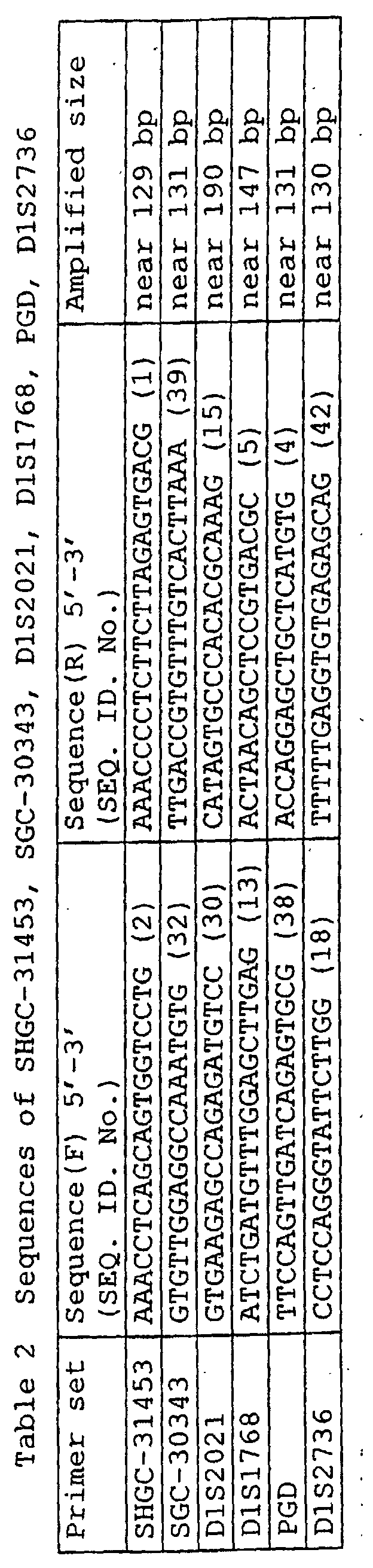
- As seen by the results (Fig. 2), there was detected a band for amplification with the hNGFβ positive controls, but no band was observed for amplification in the 1p36.23-31 region.
- Based on these results it was determined that the human neuroblastoma lines MB-1 and MASS-NB-SCH-1 have deletions at least in the region from SGC-30343 to D1S2736 of 1p36, on both chromosomes.
- The primer set D1S244 which was used to confirm the 1p36.23-31 region deletion in Example 3 was used for PCR with PAC clones (P1-derived artificial chromosomes, product of Oligo Service, Tsukuba Laboratory) having the human genome at random, using an LA PCR kit (Takara Shuzo), and PAC clones containing the deletion region sequence were selected. A 20 µl mixture was prepared by combining 5 µl of PAC DNA pool solution (Z1-Z4: Oligo Service, Tsukuba Laboratory), 2 µl of 10xLA PCR buffer, 2.5 µl of dNTPs (2 mM), 0.4 µl of D1S244F (100 pmole/µl), 0.4 µl of D1S244R (100 pmole/µl), 9.6 µl of sterile distilled water and 0.1 µl of LA Taq (5 u/µl).
- After denaturing the mixture at 94°C for 3 minutes, it was provided for PCR reaction with 35 repeated cycles of 94°C for 30 seconds, 56°C for 30 seconds and 72°C for one minute, followed by a final static incubation at 72°C for 5 minutes.
- The PCR product was subjected to electrophoresis, and then only the PAC DNA pools for which an amplification band near 285-296 bases was observed were subjected to a second PCR by the same procedure to narrow down the clones.
- The results are shown in Fig. 3. An amplified band near 285-296 bases was observed for the 4 PAC clones dJ142A6, dJ332G17, dJ345P21 and dJ369D13. Fig. 3 shows the results for only dJ142A6, dJ332G17 and dJ369D13.
- As a result of screening in the same manner with the other primer sets used to confirm the 1p36.23-31 region deletion, SHGC-31453, SGC-30343, D1S2021, D1S1768, PGD and D1S2736, there were isolated 14 clones for which amplification was observed with the five primer sets excluding SHGC-31453.
- The clones obtained in this manner were taken as the 1st screening. The 18 clones obtained by the 1st screening are shown in Table 3.

- The total of 18 clones obtained by the 1st screening are believed to retain the genome sequence of the deleted region of the two strains NB-1 and MASS-NB-SCH-1, and since some sequences were detected by multiple primer sets, 3 general contigs were formed (respectively designated as
Contig 1,Contig 2 and Contig 3). The contigs for the 18 PAC clones obtained by the 1st screening are shown in Fig. 4. - RPN2434 (Kit: Amersham Co.) was used according to the procedure described below to determine the base sequences at both ends of a total of 9 PAC clones, namely the 2 clones dJ99E23 and dJ371E1 from
Contig 1, the 4 clones dJ326J10, dJ739D18, dJ756A17 and dJ373D24 fromContig 2 and the 3 clones dJ369D13, dJ142A6 and dJ345P21 fromContig 3. Sterile distilled water was added to a mixture of 2 µg of PAC DNA, 6 pmol of SP6 or T7 primer and 2 µl of each reagent for A, C, G and T (kit accessories), to prepare a total of 6 µl of the mixture. After denaturing the mixture at 95°C for 2 minutes, it was provided for PCR reaction with 30 repeated cycles of 95°C for 30 seconds, 50°C for 30 seconds and 72°C for one minute. After adding 5 µl of reaction suspending solution (kit accessory), the reaction solution was denatured at 95°C for 2 minutes, cooled on ice, subjected to 6% acrylamide gel electrophoresis and sequenced. Ten new walking primer sets were designed from the obtained base sequence data. - Table 4 shows the selected clones and the sequences of their respective primer sets.

- The newly designed primer sets were used for reselection of PAC clones from PAC DNA pools by PCR, and the process of designing of walking primers based on both end sequences of the selected clones and determination of contigs based on walking was repeated. These were taken as the 2nd and 3rd screenings. Table 5 shows the selected clones and the sequences of their respective primers sets.

- The sequences of the walking primers used for the 2nd and 3rd screenings are shown in Table 4. Finally, one contig of 34 PAC clones was obtained. The results are shown in Fig. 5.
- Based on Example 4 it was judged that NB-1 and MASS-NB-SCH-1 have deletions on both chromosomes in at least the regions defined from SGC-30343 to D1S2736 of 1p36, but the following procedure was carried out next to identify both ends of the deletion regions.
- New primers were synthesized near the deletion region based on the contig data for the PAC clones obtained in Example 5 and the NB-1 and MASS-NB-SCH-1 strains and positive controls were subjected to PCR under the conditions described below, after which the presence or absence of specific band amplification was investigated. An LA PCR kit (Takara Shuzo) was used for PCR in the following manner.
- A 10 µl mixture was prepared by combining 1 µl of genomic DNA solution (100 ng/µl), 1 µl of 10xLA PCR buffer, 0.6 µl of dNTPs (2.5 mM, kit accessories), 0.1 µl of Primer F (100 mM), 0.1 µl of Primer R(100 mM), 7.15 µl of sterile distilled water and 0.05 µl of LA Taq (5 u/µl, kit accessory). After denaturing the mixture at 95°C for 2 minutes, it was provided for PCR with 30 repeated cycles of 95°C for 30 seconds and 58°C for one minute.
- The PCR reaction product was subjected to electrophoresis using 2.5% agarose gel. The results are shown in Fig. 6.
- The results gave a range for the genome deletion region of 1p36 as defined by from between the primer set at the SP6 end of the PAC clone dJ1028O13 and the primer set at the SP6 end of the PAC clone dJ371E1 to between the primer set D1S2736 and the primer set at the T7 end of the PAC clone dJ142A6, as shown in Fig. 7.
- The minimal PAC. clone group including the entire deletion region is shown in Fig. 8. Sequencing of these PAC clones yields the base sequence contained in the deletion region of NB-1 and MASS-NB-SCH-1.
- Table 6 shows the sequences of the primer sets used to determine the 1p36 genome deletion region range as elucidated by this invention.

- In order to obtain the base sequence data for the deletion region, a search was made of an internet gene map database (a database containing recorded human genome data for over 30,000 mapped genes, ESTs etc. according to chromosome: http://www.ncbi.nlm.nim.nih.gov/genemap99/) with the keywords "primer D1S244", etc. which were used in the aforementioned examples, resulting in numerous hits for genes and ESTs. Upon conducting a DNA sequence homology search through the internet [NCBI BLAST (National Center of Biotechnology Information USA, http://www.ncbi.nlm.nih.gov/BLAST)] for portions of the genome sequence, some ESTs were found. The data for these sequences are shown in Table 7, and those with recorded base sequences are listed in the Sequence Listing.

- A PAC clone group was selected which included essentially all of the deletion region, in the same manner as Example 6. These PAC clones were dJ371E1, dJ756A17, dJ266I16 and dJ345P21, the overall structural map of which is shown in Fig. 9. Thus, by sequencing each of these PAC clones and aligning their sequences, it is possible to determine approximately the entire length base sequence of the deletion region.
- The sequences of each of the clones were determined by the nested deletion (ND) method. See Hattori, M., Tanpakushitsu, Kakusan, Kouso [Proteins, Nucleic acids, Enzymes] 42, 17, 296-303(1997) or Hattori, M. et al., Nucl. Acids Res. 25, 1802-1808(1997).
- In this manner, the base sequences of each of the clones, including the deletion region, were determined. Specifically, the base sequences of 5 contiguous partial sequences including the deletion region were found for PAC clone dJ371E1. Their partial sequences were designated as dJ371E1-1 (SEQ. ID. No.60), dJ371E1-2 (SEQ. ID. No.61), dJ371E1-3 (SEQ. ID. No.62), dJ371E1-4 (SEQ. ID. No.63) and dJ371E1-5 (SEQ. ID. No.64). The respective base sequences are listed in the Sequence Listing. The base sequences of 7 contiguous partial sequences including the deletion region were found for PAC clone dJ756A17. Their partial sequences were designated as dJ756A17-1 (SEQ. ID. No.65), dJ756A17-2 (SEQ. ID. No.66), dJ756A17-3 (SEQ. ID. No.67), dJ756A17-4 (SEQ. ID, No.68), dJ756A17-5 (SEQ. ID. No.69), dJ756A17-6 (SEQ. ID. No.70) and dJ756A17-7 (SEQ. ID. No.71). The respective base sequences are listed in the Sequence Listing. The base sequences of 9 contiguous partial sequences including the deletion region were found for PAC clone dJ266I16. Their partial sequences were designated as dJ266I16-1 (SEQ. ID. No.72), dJ266I16-2 (SEQ. ID. No.73), dJ266I16-3 (SEQ. ID. No.74), dJ266I16-4 (SEQ. ID. No.75), dJ266I16-5 (SEQ. ID. No.76), dJ266I16-6 (SEQ. ID. No.77), dJ266I16-7 (SEQ. ID. No.78), dJ266I16-8 (SEQ. ID. No.79) and dJ266I16-9 (SEQ. ID. No.80). The respective base sequences are listed in the Sequence Listing. The base sequences of 8 contiguous partial sequences including the deletion region were found for PAC clone dJ345p-21. Their partial sequences were designated as dJ345p-21-1 (SEQ. ID. No.81), dJ345p-21-2 (SEQ. ID. No.82), dJ345p-21-3 (SEQ. ID. No.83), dJ345p-21-4 (SEQ. ID. No.84), dJ345p-21-5 (SEQ. ID. No.85), dJ345p-21-6 (SEQ. ID. No.86), dJ345p-21-7 (SEQ. ID. No.87) and dJ345p-21-8 (SEQ. ID. No.88). The respective base sequences are listed in the Sequence Listing.
- Cells of patients with pediatric neuroblastoma were used to examine the correlation between the presence or absence of LOH for 1p36 demonstrated here and various factors which are associated with tumor prognosis. The results are shown in Table 8.

- Generally speaking, patients with high expression of the oncogene trkA are considered to have poor prognosis. See Nakagawara, A. et al., Cancer Res. 52, 1364-1368(1992), Nakagawara, A. et al., N. Engl. J. Med. 328, 847-854(1993).
- In fact, upon examining LOH for 1p36 across 157 pediatric neuroblastoma patients based on comparison of the degree of trk gene expression, among 42 patients who had LOH on 1p36, 24 patients had high trk expression while 18 had low trk expression. Conversely, among the 115 patients with no LOH on 1p36, 36 patients had high trk expression while 79 had low trk expression. A good correlation was therefore obtained between LOH on 1p36 and trk expression.
- Prognosis is generally also poor for patients with high age of onset (Nakagawara A. et al., Med. Pediatr. Oncol. 31, 113-115(1998)), patients with primary lesion in the adrenal body (Nakagawara A. et al., Surgery 104, 34-40(1988)), and patients with high expression of the oncogene N-myc (Brodeur GM. et al., Science 224, 1121-1124(1984)), and a very stable correlation was found with the presence or absence of LOH for 1p36.
- This confirms that the presence or absence of LOH for 1p36 can serve as an excellent marker for prognosis and identification.
- According to this invention, there has been identified a homozygous deletion region present in common at position 36 on the short arm of
chromosome 1 of human neuroblastoma. Based on the base sequence data of the deletion region it is possible to design a method for diagnosis, prognosis of tumors, as well as a tumor marker which can be used therefor, while the base sequence data of the deletion region can also be used to develop a method of tumor gene diagnosis and an anticancer agent, and to develop a method of analyzing the tumor pathology mechanism and a reagent to be used for analysis of the tumor pathology mechanism.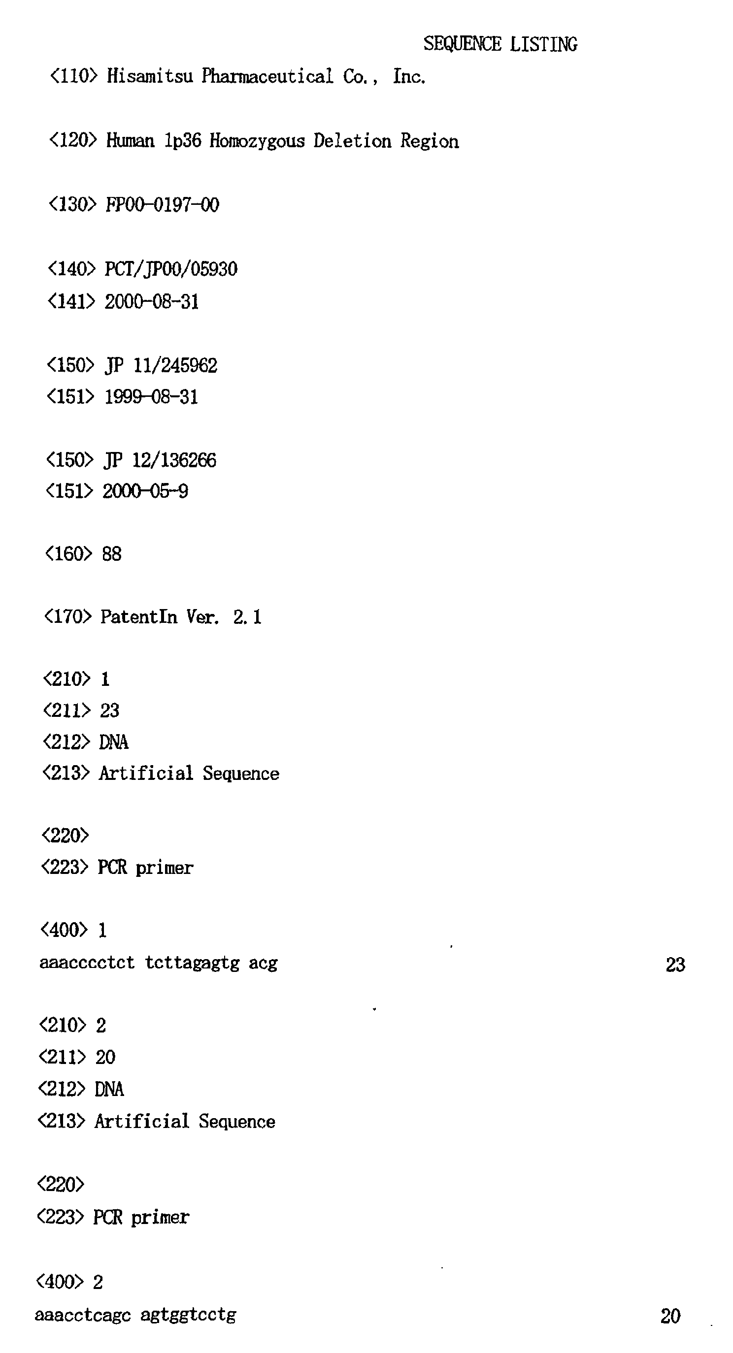









































































Claims (5)
- A homozygous deletion region present in common at position 36 on the short arm of chromosome 1 of human neuroblastoma (1p36).
- The region according to claim 1, characterized by being defined by at least from between a primer set at the SP6 end of the PAC clone dJ1028013 and a primer set at the SP6 end of the PAC clone dJ371E1 to between the primer set D1S2736 and a primer set at the T7 end of the PAC clone dJ142A6.
- The region according to claim 1 or 2, characterized by being further defined by at least the primer set SGC30343 and D1S2736.
- The region according to claim 1 or 2, characterized by being deleted in the human neuroblastoma cell lines NB-1 and MASS-NB-SCH-1.
- The region according to claim 3, characterized by being deleted in the human neuroblastoma cell lines NB-1 and MASS-NB-SCH-1.
Applications Claiming Priority (5)
| Application Number | Priority Date | Filing Date | Title |
|---|---|---|---|
| JP24596299 | 1999-08-31 | ||
| JP24596299 | 1999-08-31 | ||
| JP2000136266 | 2000-05-09 | ||
| JP2000136266 | 2000-05-09 | ||
| PCT/JP2000/005930 WO2001016311A1 (en) | 1999-08-31 | 2000-08-31 | HUMAN 1p36 HOMOZYGOSITY DELETION DOMAIN |
Publications (2)
| Publication Number | Publication Date |
|---|---|
| EP1213349A1 true EP1213349A1 (en) | 2002-06-12 |
| EP1213349A4 EP1213349A4 (en) | 2003-01-02 |
Family
ID=26537499
Family Applications (1)
| Application Number | Title | Priority Date | Filing Date |
|---|---|---|---|
| EP00956853A Withdrawn EP1213349A4 (en) | 1999-08-31 | 2000-08-31 | DOMAIN OF DELETION 1p36 OF HUMAN HOMOZYGOTISM |
Country Status (3)
| Country | Link |
|---|---|
| EP (1) | EP1213349A4 (en) |
| AU (1) | AU6867300A (en) |
| WO (1) | WO2001016311A1 (en) |
Cited By (1)
| Publication number | Priority date | Publication date | Assignee | Title |
|---|---|---|---|---|
| EP1173461A4 (en) * | 1999-03-26 | 2003-10-29 | Human Genome Sciences Inc | 50 HUMAN SECRETED PROTEINS |
-
2000
- 2000-08-31 WO PCT/JP2000/005930 patent/WO2001016311A1/en not_active Ceased
- 2000-08-31 EP EP00956853A patent/EP1213349A4/en not_active Withdrawn
- 2000-08-31 AU AU68673/00A patent/AU6867300A/en not_active Abandoned
Non-Patent Citations (7)
| Title |
|---|
| HIRAIWA HISAYUKI ET AL: "A neuroblastoma cell line derived from a case detected through a mass screening system in Japan: A case report including the biological and phenotypic characteristics of cell line." CANCER, vol. 79, no. 10, 1997, pages 2036-2044, XP002218785 ISSN: 0008-543X * |
| IOANNOU P A ET AL: "A NEW BACTERIOPHAGE P1-DERIVED VECTOR FOR THE PROPAGATION OF LARGE HUMAN DNA FRAGMENTS" NATURE GENETICS, NEW YORK, NY, US, vol. 6, 1994, pages 84-89, XP000770742 ISSN: 1061-4036 * |
| SCHWAB MANFRED ET AL: "Genomic instability in Ip and human malignancies." GENES CHROMOSOMES & CANCER, vol. 16, no. 4, 1996, pages 211-229, XP001118678 ISSN: 1045-2257 * |
| See also references of WO0116311A1 * |
| SPIEKER NICOLE ET AL: "An integrated 5-Mb physical, genetic, and radiation hybrid map of a 1p36.1 region implicated in neuroblastoma pathogenesis." GENES CHROMOSOMES & CANCER, vol. 27, no. 2, February 2000 (2000-02), pages 143-152, XP002218801 ISSN: 1045-2257 * |
| WHITE P S ET AL: "Molecular analysis of the region of distal 1p commonly deleted in Neuroblastoma" EUROPEAN JOURNAL OF CANCER, PERGAMON PRESS, OXFORD, GB, vol. 33, no. 12, October 1997 (1997-10), pages 1957-1961, XP004284624 ISSN: 0959-8049 * |
| WHITE P S ET AL: "Physical mapping of the CA6, ENO1, and SLC2A5 (GLUT5) genes and reassignment of SLC2A5 to 1p36.2." CYTOGENETICS AND CELL GENETICS, vol. 81, no. 1, 1998, pages 60-64, XP002218787 ISSN: 0301-0171 * |
Cited By (1)
| Publication number | Priority date | Publication date | Assignee | Title |
|---|---|---|---|---|
| EP1173461A4 (en) * | 1999-03-26 | 2003-10-29 | Human Genome Sciences Inc | 50 HUMAN SECRETED PROTEINS |
Also Published As
| Publication number | Publication date |
|---|---|
| AU6867300A (en) | 2001-03-26 |
| WO2001016311A1 (en) | 2001-03-08 |
| EP1213349A4 (en) | 2003-01-02 |
Similar Documents
| Publication | Publication Date | Title |
|---|---|---|
| Kalachikov et al. | Cloning and gene mapping of the chromosome 13q14 region deleted in chronic lymphocytic leukemia | |
| EP0820526B1 (en) | Coding sequences of the human brca1 gene | |
| US6191268B1 (en) | Compositions and methods relating to DNA mismatch repair genes | |
| Lange et al. | Detection by enzymatic amplification of bcr-abl mRNA in peripheral blood and bone marrow cells of patients with chronic myelogenous leukemia | |
| Sunahara et al. | Mutational analysis of p51A/TAp63γ, a p53 homolog, in non-small cell lung cancer and breast cancer | |
| JP2008099698A (en) | Multiple tumor abnormal growth genes | |
| JP2002505888A (en) | Neoplasm detection by saliva analysis | |
| CN112391466B (en) | Methylation biomarkers or combinations and applications thereof for detecting breast cancer | |
| JP4201057B2 (en) | Diagnostic method and gene therapy using a reagent derived from human metastasis suppressor gene KAII | |
| Solinhac et al. | Integrative mapping analysis of chicken microchromosome 16 organization | |
| JP2003529331A (en) | Modification of long QT syndrome genes KVLQT1 and SCN5A and method for detecting the same | |
| Schmidt et al. | RT-PCR and FISH analysis of acute myeloid leukemia with t (8; 16)(p11; p13) and chimeric MOZ and CBP transcripts: breakpoint cluster region and clinical implications | |
| EP0943017B1 (en) | Diagnostic assay for breast cancer susceptibility | |
| Yang et al. | Genomic structure and mutational analysis of the human KIF1B gene which is homozygously deleted in neuroblastoma at chromosome 1p36. 2 | |
| Keohavong et al. | Detection of K-ras and p53 mutations in sputum samples of lung cancer patients using laser capture microdissection microscope and mutation analysis | |
| JPH07143884A (en) | Tumor suppressor gene merlin and its use | |
| Hughes et al. | The use of whole genome amplification to study chromosomal changes in prostate cancer: insights into genome-wide signature of preneoplasia associated with cancer progression | |
| KR101206029B1 (en) | Multiple SNP for diagnosing colorectal cancer, microarray and kit comprising the same, and method for diagnosing colorectal cancer using the same | |
| JP2800850B2 (en) | Methods for detecting neoplasia | |
| Magovcevic et al. | Regional localization of the human G protein αi2 (GNAI2) gene: Assignment to 3p21 and a related sequence (GNAI2L) to 12p12–p13 | |
| CN109234282B (en) | BRCA1 gene g.43063754T>G mutant and its primers | |
| Paulsen et al. | Regional localisation of X chromosome short arm probes | |
| JPWO2001016311A1 (en) | Human 1p36 homozygous deletion region | |
| Dawson et al. | Array comparative genomic hybridization and cytogenetic analysis in pediatric acute leukemias | |
| ES2319533T3 (en) | DIAGNOSIS OF THE RISK OF CHEST CANCER. |
Legal Events
| Date | Code | Title | Description |
|---|---|---|---|
| PUAI | Public reference made under article 153(3) epc to a published international application that has entered the european phase |
Free format text: ORIGINAL CODE: 0009012 |
|
| 17P | Request for examination filed |
Effective date: 20020328 |
|
| AK | Designated contracting states |
Kind code of ref document: A1 Designated state(s): AT BE CH CY DE DK ES FI FR GB GR IE IT LI LU MC NL PT SE |
|
| A4 | Supplementary search report drawn up and despatched |
Effective date: 20021113 |
|
| AK | Designated contracting states |
Kind code of ref document: A4 Designated state(s): AT BE CH CY DE DK ES FI FR GB GR IE IT LI LU MC NL PT SE |
|
| STAA | Information on the status of an ep patent application or granted ep patent |
Free format text: STATUS: THE APPLICATION IS DEEMED TO BE WITHDRAWN |
|
| 18D | Application deemed to be withdrawn |
Effective date: 20060627 |