EP1212356B1 - Protein structures and protein fibres - Google Patents
Protein structures and protein fibres Download PDFInfo
- Publication number
- EP1212356B1 EP1212356B1 EP00964388A EP00964388A EP1212356B1 EP 1212356 B1 EP1212356 B1 EP 1212356B1 EP 00964388 A EP00964388 A EP 00964388A EP 00964388 A EP00964388 A EP 00964388A EP 1212356 B1 EP1212356 B1 EP 1212356B1
- Authority
- EP
- European Patent Office
- Prior art keywords
- saf
- protein
- peptide
- protein structure
- monomer units
- Prior art date
- Legal status (The legal status is an assumption and is not a legal conclusion. Google has not performed a legal analysis and makes no representation as to the accuracy of the status listed.)
- Expired - Lifetime
Links
- 108090000623 proteins and genes Proteins 0.000 title claims abstract description 78
- 102000004169 proteins and genes Human genes 0.000 title claims abstract description 78
- 108090000765 processed proteins & peptides Proteins 0.000 claims abstract description 128
- 239000000835 fiber Substances 0.000 claims abstract description 36
- 238000000034 method Methods 0.000 claims abstract description 13
- 239000000178 monomer Substances 0.000 claims description 50
- 239000000203 mixture Substances 0.000 claims description 47
- 235000018102 proteins Nutrition 0.000 claims description 28
- 150000001413 amino acids Chemical group 0.000 claims description 13
- 230000000295 complement effect Effects 0.000 claims description 13
- 239000000833 heterodimer Substances 0.000 claims description 12
- 125000000539 amino acid group Chemical group 0.000 claims description 8
- 239000011159 matrix material Substances 0.000 claims description 8
- AGPKZVBTJJNPAG-WHFBIAKZSA-N L-isoleucine Chemical compound CC[C@H](C)[C@H](N)C(O)=O AGPKZVBTJJNPAG-WHFBIAKZSA-N 0.000 claims description 5
- 125000000613 asparagine group Chemical class N[C@@H](CC(N)=O)C(=O)* 0.000 claims description 4
- 229960000310 isoleucine Drugs 0.000 claims description 4
- AGPKZVBTJJNPAG-UHFFFAOYSA-N isoleucine Natural products CCC(C)C(N)C(O)=O AGPKZVBTJJNPAG-UHFFFAOYSA-N 0.000 claims description 4
- ROHFNLRQFUQHCH-YFKPBYRVSA-N L-leucine Chemical compound CC(C)C[C@H](N)C(O)=O ROHFNLRQFUQHCH-YFKPBYRVSA-N 0.000 claims description 3
- ROHFNLRQFUQHCH-UHFFFAOYSA-N Leucine Natural products CC(C)CC(N)C(O)=O ROHFNLRQFUQHCH-UHFFFAOYSA-N 0.000 claims description 3
- 235000009582 asparagine Nutrition 0.000 claims description 3
- 238000004132 cross linking Methods 0.000 claims description 3
- 125000001165 hydrophobic group Chemical group 0.000 claims description 3
- WHUUTDBJXJRKMK-UHFFFAOYSA-N Glutamic acid Natural products OC(=O)C(N)CCC(O)=O WHUUTDBJXJRKMK-UHFFFAOYSA-N 0.000 claims description 2
- WHUUTDBJXJRKMK-VKHMYHEASA-N L-glutamic acid Chemical compound OC(=O)[C@@H](N)CCC(O)=O WHUUTDBJXJRKMK-VKHMYHEASA-N 0.000 claims description 2
- 235000013922 glutamic acid Nutrition 0.000 claims description 2
- 239000004220 glutamic acid Substances 0.000 claims description 2
- 125000003588 lysine group Chemical group [H]N([H])C([H])([H])C([H])([H])C([H])([H])C([H])([H])C([H])(N([H])[H])C(*)=O 0.000 claims description 2
- KZSNJWFQEVHDMF-BYPYZUCNSA-N L-valine Chemical compound CC(C)[C@H](N)C(O)=O KZSNJWFQEVHDMF-BYPYZUCNSA-N 0.000 claims 1
- KZSNJWFQEVHDMF-UHFFFAOYSA-N Valine Natural products CC(C)C(N)C(O)=O KZSNJWFQEVHDMF-UHFFFAOYSA-N 0.000 claims 1
- 239000004474 valine Substances 0.000 claims 1
- 102000004196 processed proteins & peptides Human genes 0.000 abstract description 47
- 230000015572 biosynthetic process Effects 0.000 abstract description 16
- 229920001184 polypeptide Polymers 0.000 abstract description 7
- 238000002983 circular dichroism Methods 0.000 description 17
- 230000003993 interaction Effects 0.000 description 17
- 238000002267 linear dichroism spectroscopy Methods 0.000 description 16
- 238000013461 design Methods 0.000 description 15
- 239000000243 solution Substances 0.000 description 12
- 238000000429 assembly Methods 0.000 description 11
- 230000000712 assembly Effects 0.000 description 11
- 238000001228 spectrum Methods 0.000 description 11
- 235000001014 amino acid Nutrition 0.000 description 10
- 229940024606 amino acid Drugs 0.000 description 10
- 239000000539 dimer Substances 0.000 description 9
- 230000002209 hydrophobic effect Effects 0.000 description 9
- 238000002474 experimental method Methods 0.000 description 8
- 150000003839 salts Chemical class 0.000 description 8
- 239000002071 nanotube Substances 0.000 description 7
- 238000004627 transmission electron microscopy Methods 0.000 description 7
- 238000001142 circular dichroism spectrum Methods 0.000 description 6
- 239000000463 material Substances 0.000 description 6
- 238000012856 packing Methods 0.000 description 6
- 239000011698 potassium fluoride Substances 0.000 description 6
- 239000000523 sample Substances 0.000 description 6
- 239000000356 contaminant Substances 0.000 description 5
- 230000000694 effects Effects 0.000 description 5
- 230000035800 maturation Effects 0.000 description 5
- 230000003595 spectral effect Effects 0.000 description 5
- DVLFYONBTKHTER-UHFFFAOYSA-N 3-(N-morpholino)propanesulfonic acid Chemical compound OS(=O)(=O)CCCN1CCOCC1 DVLFYONBTKHTER-UHFFFAOYSA-N 0.000 description 4
- 239000008280 blood Substances 0.000 description 4
- 210000004369 blood Anatomy 0.000 description 4
- 238000005119 centrifugation Methods 0.000 description 4
- 238000010276 construction Methods 0.000 description 4
- 230000035557 fibrillogenesis Effects 0.000 description 4
- NROKBHXJSPEDAR-UHFFFAOYSA-M potassium fluoride Chemical compound [F-].[K+] NROKBHXJSPEDAR-UHFFFAOYSA-M 0.000 description 4
- 238000002360 preparation method Methods 0.000 description 4
- 108020004414 DNA Proteins 0.000 description 3
- 239000007993 MOPS buffer Substances 0.000 description 3
- 238000002835 absorbance Methods 0.000 description 3
- 239000011230 binding agent Substances 0.000 description 3
- 238000012937 correction Methods 0.000 description 3
- 239000013078 crystal Substances 0.000 description 3
- 125000000151 cysteine group Chemical group N[C@@H](CS)C(=O)* 0.000 description 3
- 238000001459 lithography Methods 0.000 description 3
- 238000002156 mixing Methods 0.000 description 3
- 239000011295 pitch Substances 0.000 description 3
- 229920000642 polymer Polymers 0.000 description 3
- 238000004626 scanning electron microscopy Methods 0.000 description 3
- 238000001338 self-assembly Methods 0.000 description 3
- 239000000126 substance Substances 0.000 description 3
- XLYOFNOQVPJJNP-UHFFFAOYSA-N water Substances O XLYOFNOQVPJJNP-UHFFFAOYSA-N 0.000 description 3
- DCXYFEDJOCDNAF-UHFFFAOYSA-N Asparagine Chemical group OC(=O)C(N)CC(N)=O DCXYFEDJOCDNAF-UHFFFAOYSA-N 0.000 description 2
- OKTJSMMVPCPJKN-UHFFFAOYSA-N Carbon Chemical compound [C] OKTJSMMVPCPJKN-UHFFFAOYSA-N 0.000 description 2
- 102000005937 Tropomyosin Human genes 0.000 description 2
- 108010030743 Tropomyosin Proteins 0.000 description 2
- 241000700605 Viruses Species 0.000 description 2
- 238000004458 analytical method Methods 0.000 description 2
- 238000003491 array Methods 0.000 description 2
- 125000003118 aryl group Chemical group 0.000 description 2
- 229960001230 asparagine Drugs 0.000 description 2
- 238000004630 atomic force microscopy Methods 0.000 description 2
- 239000013060 biological fluid Substances 0.000 description 2
- 210000004027 cell Anatomy 0.000 description 2
- 239000004568 cement Substances 0.000 description 2
- 230000001268 conjugating effect Effects 0.000 description 2
- 238000001212 derivatisation Methods 0.000 description 2
- 238000011161 development Methods 0.000 description 2
- 230000018109 developmental process Effects 0.000 description 2
- 229920000359 diblock copolymer Polymers 0.000 description 2
- 238000001493 electron microscopy Methods 0.000 description 2
- 230000009881 electrostatic interaction Effects 0.000 description 2
- 238000011065 in-situ storage Methods 0.000 description 2
- 238000011534 incubation Methods 0.000 description 2
- 125000001909 leucine group Chemical group [H]N(*)C(C(*)=O)C([H])([H])C(C([H])([H])[H])C([H])([H])[H] 0.000 description 2
- 229920002521 macromolecule Polymers 0.000 description 2
- 238000004519 manufacturing process Methods 0.000 description 2
- 238000005259 measurement Methods 0.000 description 2
- 238000012900 molecular simulation Methods 0.000 description 2
- 235000003270 potassium fluoride Nutrition 0.000 description 2
- 238000000746 purification Methods 0.000 description 2
- 238000000926 separation method Methods 0.000 description 2
- 150000003384 small molecules Chemical class 0.000 description 2
- 238000010186 staining Methods 0.000 description 2
- 239000013638 trimer Substances 0.000 description 2
- 125000003088 (fluoren-9-ylmethoxy)carbonyl group Chemical group 0.000 description 1
- PUMZXCBVHLCWQG-UHFFFAOYSA-N 1-(4-Hydroxyphenyl)-2-aminoethanol hydrochloride Chemical compound [Cl-].[NH3+]CC(O)C1=CC=C(O)C=C1 PUMZXCBVHLCWQG-UHFFFAOYSA-N 0.000 description 1
- 125000004042 4-aminobutyl group Chemical group [H]C([*])([H])C([H])([H])C([H])([H])C([H])([H])N([H])[H] 0.000 description 1
- 229920001817 Agar Polymers 0.000 description 1
- 239000004475 Arginine Substances 0.000 description 1
- 238000012935 Averaging Methods 0.000 description 1
- 0 C(C1)C2CC1*C2 Chemical compound C(C1)C2CC1*C2 0.000 description 1
- 108090000565 Capsid Proteins Proteins 0.000 description 1
- 241001012508 Carpiodes cyprinus Species 0.000 description 1
- RYGMFSIKBFXOCR-UHFFFAOYSA-N Copper Chemical compound [Cu] RYGMFSIKBFXOCR-UHFFFAOYSA-N 0.000 description 1
- 241000701533 Escherichia virus T4 Species 0.000 description 1
- 102000011782 Keratins Human genes 0.000 description 1
- 108010076876 Keratins Proteins 0.000 description 1
- AHLPHDHHMVZTML-BYPYZUCNSA-N L-Ornithine Chemical compound NCCC[C@H](N)C(O)=O AHLPHDHHMVZTML-BYPYZUCNSA-N 0.000 description 1
- ODKSFYDXXFIFQN-BYPYZUCNSA-P L-argininium(2+) Chemical compound NC(=[NH2+])NCCC[C@H]([NH3+])C(O)=O ODKSFYDXXFIFQN-BYPYZUCNSA-P 0.000 description 1
- DCXYFEDJOCDNAF-REOHCLBHSA-N L-asparagine Chemical compound OC(=O)[C@@H](N)CC(N)=O DCXYFEDJOCDNAF-REOHCLBHSA-N 0.000 description 1
- CKLJMWTZIZZHCS-REOHCLBHSA-N L-aspartic acid Chemical compound OC(=O)[C@@H](N)CC(O)=O CKLJMWTZIZZHCS-REOHCLBHSA-N 0.000 description 1
- AHLPHDHHMVZTML-UHFFFAOYSA-N Orn-delta-NH2 Natural products NCCCC(N)C(O)=O AHLPHDHHMVZTML-UHFFFAOYSA-N 0.000 description 1
- UTJLXEIPEHZYQJ-UHFFFAOYSA-N Ornithine Natural products OC(=O)C(C)CCCN UTJLXEIPEHZYQJ-UHFFFAOYSA-N 0.000 description 1
- 108010038006 Peptide Nanotubes Proteins 0.000 description 1
- 102000002067 Protein Subunits Human genes 0.000 description 1
- 108010001267 Protein Subunits Proteins 0.000 description 1
- 241000669298 Pseudaulacaspis pentagona Species 0.000 description 1
- 101100163901 Rattus norvegicus Asic2 gene Proteins 0.000 description 1
- 238000003917 TEM image Methods 0.000 description 1
- 101710166729 Tail fiber protein Proteins 0.000 description 1
- 108091023040 Transcription factor Proteins 0.000 description 1
- 102000040945 Transcription factor Human genes 0.000 description 1
- COQLPRJCUIATTQ-UHFFFAOYSA-N Uranyl acetate Chemical compound O.O.O=[U]=O.CC(O)=O.CC(O)=O COQLPRJCUIATTQ-UHFFFAOYSA-N 0.000 description 1
- 238000002441 X-ray diffraction Methods 0.000 description 1
- TUCNEACPLKLKNU-UHFFFAOYSA-N acetyl Chemical compound C[C]=O TUCNEACPLKLKNU-UHFFFAOYSA-N 0.000 description 1
- 239000008272 agar Substances 0.000 description 1
- 239000007864 aqueous solution Substances 0.000 description 1
- ODKSFYDXXFIFQN-UHFFFAOYSA-N arginine Natural products OC(=O)C(N)CCCNC(N)=N ODKSFYDXXFIFQN-UHFFFAOYSA-N 0.000 description 1
- 235000003704 aspartic acid Nutrition 0.000 description 1
- OQFSQFPPLPISGP-UHFFFAOYSA-N beta-carboxyaspartic acid Natural products OC(=O)C(N)C(C(O)=O)C(O)=O OQFSQFPPLPISGP-UHFFFAOYSA-N 0.000 description 1
- 230000008033 biological extinction Effects 0.000 description 1
- 229920001222 biopolymer Polymers 0.000 description 1
- 230000002051 biphasic effect Effects 0.000 description 1
- 210000004899 c-terminal region Anatomy 0.000 description 1
- 229910052799 carbon Inorganic materials 0.000 description 1
- 239000002041 carbon nanotube Substances 0.000 description 1
- 229910021393 carbon nanotube Inorganic materials 0.000 description 1
- 238000006555 catalytic reaction Methods 0.000 description 1
- 230000010261 cell growth Effects 0.000 description 1
- 238000012512 characterization method Methods 0.000 description 1
- 238000005352 clarification Methods 0.000 description 1
- 238000004891 communication Methods 0.000 description 1
- 238000004624 confocal microscopy Methods 0.000 description 1
- 239000000470 constituent Substances 0.000 description 1
- 229910052802 copper Inorganic materials 0.000 description 1
- 239000010949 copper Substances 0.000 description 1
- 230000008878 coupling Effects 0.000 description 1
- 238000010168 coupling process Methods 0.000 description 1
- 238000005859 coupling reaction Methods 0.000 description 1
- 235000018417 cysteine Nutrition 0.000 description 1
- XUJNEKJLAYXESH-UHFFFAOYSA-N cysteine Natural products SCC(N)C(O)=O XUJNEKJLAYXESH-UHFFFAOYSA-N 0.000 description 1
- 238000013480 data collection Methods 0.000 description 1
- 238000006073 displacement reaction Methods 0.000 description 1
- 238000009826 distribution Methods 0.000 description 1
- 238000001035 drying Methods 0.000 description 1
- 238000005516 engineering process Methods 0.000 description 1
- 238000011067 equilibration Methods 0.000 description 1
- 102000034240 fibrous proteins Human genes 0.000 description 1
- 108091005899 fibrous proteins Proteins 0.000 description 1
- GNBHRKFJIUUOQI-UHFFFAOYSA-N fluorescein Chemical compound O1C(=O)C2=CC=CC=C2C21C1=CC=C(O)C=C1OC1=CC(O)=CC=C21 GNBHRKFJIUUOQI-UHFFFAOYSA-N 0.000 description 1
- 238000002866 fluorescence resonance energy transfer Methods 0.000 description 1
- 125000000524 functional group Chemical group 0.000 description 1
- 229920001002 functional polymer Polymers 0.000 description 1
- WHUUTDBJXJRKMK-VKHMYHEASA-L glutamate group Chemical group N[C@@H](CCC(=O)[O-])C(=O)[O-] WHUUTDBJXJRKMK-VKHMYHEASA-L 0.000 description 1
- PCHJSUWPFVWCPO-UHFFFAOYSA-N gold Chemical compound [Au] PCHJSUWPFVWCPO-UHFFFAOYSA-N 0.000 description 1
- 229910052737 gold Inorganic materials 0.000 description 1
- 239000010931 gold Substances 0.000 description 1
- 230000005661 hydrophobic surface Effects 0.000 description 1
- 238000003384 imaging method Methods 0.000 description 1
- 238000004949 mass spectrometry Methods 0.000 description 1
- 238000001840 matrix-assisted laser desorption--ionisation time-of-flight mass spectrometry Methods 0.000 description 1
- 230000007246 mechanism Effects 0.000 description 1
- 238000000302 molecular modelling Methods 0.000 description 1
- 238000005232 molecular self-assembly Methods 0.000 description 1
- 239000002808 molecular sieve Substances 0.000 description 1
- 239000002086 nanomaterial Substances 0.000 description 1
- 238000010899 nucleation Methods 0.000 description 1
- 108020004707 nucleic acids Proteins 0.000 description 1
- 102000039446 nucleic acids Human genes 0.000 description 1
- 150000007523 nucleic acids Chemical class 0.000 description 1
- 229960001576 octopamine Drugs 0.000 description 1
- 238000000238 one-dimensional nuclear magnetic resonance spectroscopy Methods 0.000 description 1
- 229960003104 ornithine Drugs 0.000 description 1
- 239000002049 peptide nanotube Substances 0.000 description 1
- 230000000737 periodic effect Effects 0.000 description 1
- 238000009428 plumbing Methods 0.000 description 1
- 239000002244 precipitate Substances 0.000 description 1
- 230000008569 process Effects 0.000 description 1
- 239000000047 product Substances 0.000 description 1
- 230000012846 protein folding Effects 0.000 description 1
- 230000004850 protein–protein interaction Effects 0.000 description 1
- 230000007115 recruitment Effects 0.000 description 1
- 238000011160 research Methods 0.000 description 1
- 238000004007 reversed phase HPLC Methods 0.000 description 1
- PYWVYCXTNDRMGF-UHFFFAOYSA-N rhodamine B Chemical compound [Cl-].C=12C=CC(=[N+](CC)CC)C=C2OC2=CC(N(CC)CC)=CC=C2C=1C1=CC=CC=C1C(O)=O PYWVYCXTNDRMGF-UHFFFAOYSA-N 0.000 description 1
- 238000004062 sedimentation Methods 0.000 description 1
- 238000012772 sequence design Methods 0.000 description 1
- 238000007873 sieving Methods 0.000 description 1
- URGAHOPLAPQHLN-UHFFFAOYSA-N sodium aluminosilicate Chemical compound [Na+].[Al+3].[O-][Si]([O-])=O.[O-][Si]([O-])=O URGAHOPLAPQHLN-UHFFFAOYSA-N 0.000 description 1
- 239000007787 solid Substances 0.000 description 1
- 238000010532 solid phase synthesis reaction Methods 0.000 description 1
- 241000894007 species Species 0.000 description 1
- 238000004611 spectroscopical analysis Methods 0.000 description 1
- 238000009987 spinning Methods 0.000 description 1
- 238000006467 substitution reaction Methods 0.000 description 1
- 239000006228 supernatant Substances 0.000 description 1
- 238000003786 synthesis reaction Methods 0.000 description 1
- 229920001059 synthetic polymer Polymers 0.000 description 1
- 238000012360 testing method Methods 0.000 description 1
- 125000003396 thiol group Chemical class [H]S* 0.000 description 1
- 230000036962 time dependent Effects 0.000 description 1
- 230000017423 tissue regeneration Effects 0.000 description 1
- 238000012546 transfer Methods 0.000 description 1
- 125000002987 valine group Chemical group [H]N([H])C([H])(C(*)=O)C([H])(C([H])([H])[H])C([H])([H])[H] 0.000 description 1
- 238000012800 visualization Methods 0.000 description 1
- 238000002424 x-ray crystallography Methods 0.000 description 1
Images




Classifications
-
- G—PHYSICS
- G01—MEASURING; TESTING
- G01Q—SCANNING-PROBE TECHNIQUES OR APPARATUS; APPLICATIONS OF SCANNING-PROBE TECHNIQUES, e.g. SCANNING PROBE MICROSCOPY [SPM]
- G01Q60/00—Particular types of SPM [Scanning Probe Microscopy] or microscopes; Essential components thereof
- G01Q60/24—AFM [Atomic Force Microscopy] or apparatus therefor, e.g. AFM probes
- G01Q60/38—Probes, their manufacture, or their related instrumentation, e.g. holders
-
- B—PERFORMING OPERATIONS; TRANSPORTING
- B82—NANOTECHNOLOGY
- B82Y—SPECIFIC USES OR APPLICATIONS OF NANOSTRUCTURES; MEASUREMENT OR ANALYSIS OF NANOSTRUCTURES; MANUFACTURE OR TREATMENT OF NANOSTRUCTURES
- B82Y15/00—Nanotechnology for interacting, sensing or actuating, e.g. quantum dots as markers in protein assays or molecular motors
-
- B—PERFORMING OPERATIONS; TRANSPORTING
- B82—NANOTECHNOLOGY
- B82Y—SPECIFIC USES OR APPLICATIONS OF NANOSTRUCTURES; MEASUREMENT OR ANALYSIS OF NANOSTRUCTURES; MANUFACTURE OR TREATMENT OF NANOSTRUCTURES
- B82Y35/00—Methods or apparatus for measurement or analysis of nanostructures
-
- C—CHEMISTRY; METALLURGY
- C07—ORGANIC CHEMISTRY
- C07K—PEPTIDES
- C07K1/00—General methods for the preparation of peptides, i.e. processes for the organic chemical preparation of peptides or proteins of any length
- C07K1/107—General methods for the preparation of peptides, i.e. processes for the organic chemical preparation of peptides or proteins of any length by chemical modification of precursor peptides
- C07K1/113—General methods for the preparation of peptides, i.e. processes for the organic chemical preparation of peptides or proteins of any length by chemical modification of precursor peptides without change of the primary structure
- C07K1/1136—General methods for the preparation of peptides, i.e. processes for the organic chemical preparation of peptides or proteins of any length by chemical modification of precursor peptides without change of the primary structure by reversible modification of the secondary, tertiary or quarternary structure, e.g. using denaturating or stabilising agents
-
- C—CHEMISTRY; METALLURGY
- C07—ORGANIC CHEMISTRY
- C07K—PEPTIDES
- C07K14/00—Peptides having more than 20 amino acids; Gastrins; Somatostatins; Melanotropins; Derivatives thereof
- C07K14/001—Peptides having more than 20 amino acids; Gastrins; Somatostatins; Melanotropins; Derivatives thereof by chemical synthesis
-
- G—PHYSICS
- G01—MEASURING; TESTING
- G01Q—SCANNING-PROBE TECHNIQUES OR APPARATUS; APPLICATIONS OF SCANNING-PROBE TECHNIQUES, e.g. SCANNING PROBE MICROSCOPY [SPM]
- G01Q70/00—General aspects of SPM probes, their manufacture or their related instrumentation, insofar as they are not specially adapted to a single SPM technique covered by group G01Q60/00
- G01Q70/08—Probe characteristics
- G01Q70/10—Shape or taper
- G01Q70/12—Nanotube tips
-
- B—PERFORMING OPERATIONS; TRANSPORTING
- B82—NANOTECHNOLOGY
- B82Y—SPECIFIC USES OR APPLICATIONS OF NANOSTRUCTURES; MEASUREMENT OR ANALYSIS OF NANOSTRUCTURES; MANUFACTURE OR TREATMENT OF NANOSTRUCTURES
- B82Y10/00—Nanotechnology for information processing, storage or transmission, e.g. quantum computing or single electron logic
-
- G—PHYSICS
- G11—INFORMATION STORAGE
- G11B—INFORMATION STORAGE BASED ON RELATIVE MOVEMENT BETWEEN RECORD CARRIER AND TRANSDUCER
- G11B9/00—Recording or reproducing using a method not covered by one of the main groups G11B3/00 - G11B7/00; Record carriers therefor
- G11B9/12—Recording or reproducing using a method not covered by one of the main groups G11B3/00 - G11B7/00; Record carriers therefor using near-field interactions; Record carriers therefor
- G11B9/14—Recording or reproducing using a method not covered by one of the main groups G11B3/00 - G11B7/00; Record carriers therefor using near-field interactions; Record carriers therefor using microscopic probe means, i.e. recording or reproducing by means directly associated with the tip of a microscopic electrical probe as used in Scanning Tunneling Microscopy [STM] or Atomic Force Microscopy [AFM] for inducing physical or electrical perturbations in a recording medium; Record carriers or media specially adapted for such transducing of information
- G11B9/1409—Heads
Definitions
- This invention relates to protein structures, to methods of producing those protein structures, and to protein fibres and other materials and assemblies produced using those protein structures.
- coiled coils are relatively straightforward: they are ⁇ -helical bundles with between 2 and 5 strands that can be arranged in parallel, antiparallel or mixed topologies.
- the basic sequence features that guide the formation of coiled coils from peptides are reasonably well understood (P. B. Harbury et al (1993) Science 262, 1401; D. N. Woolfson and T. Alber (1995) Protein Sci. 4, 1596).
- most coiled-coil sequences are dominated by a 7-residue repeat of hydrophobic (H) and polar (P) residues, (HPPHPPP) n , known as the "heptad repeat".
- both the number and the direction of chains within a coiled-coil bundle is determined predominantly by residues that form or flank the hydrophobic core namely, residues at the first, fourth, fifth and seventh positions of the heptad repeat.
- coiled coils which form dimers i.e. two-stranded assemblies
- trimers i.e.
- hetero-oligomers that is coiled coils made from strands with different amino-acid sequences
- sequences with multiple, offset heptad repeats which help explain oligomer-state specification in coiled coils. For example, sequences with two heptad repeats offset by two residues; i.e a / f-b / g'-c / a'-d / b'-e / c'-f / d'-g / e' set up two hydrophobic seams on opposite sides of the helix formed. Such helices may combine to bury these hydrophobic surfaces in two different ways and form two distinct structures: open " ⁇ -sheets" and closed " ⁇ -cylinders".
- the building block of the design was a staggered heterodimer with overhanging or "sticky” ends. This contrasts with and distinguishes it from the natural and designer coiled-coil assemblies that have been characterized to date, in which the polypeptide strands align in-register, i.e they have blunt or "flush” ends. Complementary core interactions and flanking ion-pairs were incorporated into the overhangs to facilitate longitudinal association of the heterodimers (Figs. 1&2). This principle of using "sticky ends” is well developed in molecular biology for assembling DNA (S. J. Palmer et al (1998) Nucleic Acids Res. 26, 2560), and has been used to design intricate DNA crystals (E.
- US-A-5,712, 366 discloses self-assembling protein material but does not provide details of how to make a staggered parallel heterodimer.
- WO 96/11947 discloses protein nanostructures based on bacteriophage T4 tail fiber proteins but does not disclose a staggered parallel heterodimer coiled coil structure.
- a protein structure comprising a plurality of first peptide monomer units arranged in a first strand and a plurality of second peptide monomer units arranged in a second strand, the strands preferably forming a coiled-coil structure, and in which a first peptide monomer unit in the first strand extends beyond a corresponding second peptide monomer unit in the second strand in the direction of the strands.
- the protein structures of the invention have numerous advantages. For example, relatively long protein fibres can be formed with little material - 1 ⁇ l of a 100 ⁇ M solution of the peptide monomers may provide enough material to form 10 m of fibre 50 mm thick.
- At least one charged amino acid residue of the first peptide monomer unit may be arranged to attract an oppositely-charged amino acid residue of the second peptide monomer unit.
- the charged amino acid residue is in an end portion of the first peptide monomer unit, which extends beyond the corresponding second peptide monomer unit in the second strand.
- At least one strand may consist solely of first or second peptide monomer units respectively i.e homogenous strands. Heterologous strands are also contemplated.
- the peptide monomer units may comprise a repeating structural unit.
- the repeating structural unit comprises a heptad repeat motif, having the pattern:
- the repeat may include isoleucine or asparagine at position a and leucine at position d.
- Other repeats e.g hendecads - abcdefghijk
- amino acid compositions may also be used (see WO99/11774).
- the heptad repeat comprises oppositely-charged residues at positions e and g respectively.
- the oppositely-charged residues may be, for example, glutamic acid and lysine residues or arginine and aspartic acid.
- the use of synthetic amino acids, such as ornithine is also envisaged.
- a protein structure in accordance with the invention may be also specified by pairs of asparagine residues in the "a" positions provided by corresponding first and second peptide monomer units.
- the first and second peptide monomer units have the following sequences:
- a method of producing protein structures comprising providing a mixture of first and second peptide monomer units which associate to form a protein structure according to the invention.
- the structure can be derivatised and/or stabilized by cross-linking.
- Derivatization of the peptide monomer units before or after assembly into the protein structures of the invention may be performed.
- fluorescent moieties fluorophores
- the addition of fluorescent moieties may assist visualization of the protein structure.
- Substitution with functional groups at the "f" position in the heptad repeat is especially preferred as that position is on the outside of the helix (see Fig. 1C and 1E).
- Other derivatives may include attaching binders to the peptide monomer units for example so that units which can bind other entities can be produced.
- the first and second peptide monomers and the strands may have the characteristics described above.
- the invention also provides protein fibres produced by an association of protein structures according to the invention.
- the protein structures may also be arranged to form tubular structures.
- the structures may be arranged to form nanotubes.
- kits for making protein structures comprising first and second peptide monomer units which associate to form a protein structure or protein fibres according to the invention.
- the protein structures of the invention may be assembled in two and three dimensional arrays.
- two dimensional mats can be formed which can function, for example as filters.
- Three dimensional grids or matrices can also be formed again, for example, for use as sieves or filters or for organising other associated or conjugated molecules in three dimensions.
- a matrix is assembled in situ.
- a matrix can be formed in a solution to entrap contaminants in the solution and then the matrix, together with contaminants, can be removed from the solution for example by centrifugation.
- the stability of the protein structures at higher temperatures may be improved by making the peptide monomers longer, such that the overlap between corresponding first and second monomer unit residues is increased. Increases in monomer length have previously been shown to stabilize coiled coil structures.
- protein structures may be stabilised and derivatised by using them to template the polymerisation of synthetic polymers.
- a coiled-coil is a peptide/protein sequence usually with a contiguous pattern of hydrophobic residues spaced 3 and 4 residues apart, which assembles (folds) to form a multi-meric bundle of helices. Coiled-coils including sequences with multiple offset repeats are also contemplated.
- a dimer is a two stranded structure.
- a heterodimer is a dimeric structure formed by two different stands.
- a staggered heterodimer is a structure in which the two strands assemble to leave overlapping ends that are not interacting within the heterodimer.
- Blunt-end assembly is association where the two strands combine to give flushed i.e non-overlapping ends.
- a protofibril is a protein structure assembled longitudinally from staggered heterodimers interacting through their overhanging ends.
- a fibre is a structure formed by lateral association of two or more protofibrils.
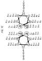
- Fig. 1 shows (A) A mechanism for self assembly: complementary charges in "companion” peptides direct the formation of staggered, parallel heterodimers; the resulting “sticky” ends are also complementary and promote longitudinal association into extended structures.
- Fig 1(B) shows the designed amino acid sequences: each peptide comprised canonical heptad repeats ( abcdefg ) with Ile at a and Leu at d to guide the formation of coiled-coil dimers; oppositely-charged residues were incorporated at e and g to favour the staggered dimer with sticky ends; asparagine residues (which preferentially pairs with each other at a sites (Gonzalez L et al (1996) Nature Structural Biology 3, 13 : 1011-1018) were included to cement the prescribed register further and to favour the parallel structures.
- Fig. 1(C) is a helical-wheel representation, summarizing the designed sequences in context. The view is from the N-terminus with heptad sites labeled a-g and assumes 3.5 residues per helical turn to emphasise the heptad repeat.
- the peptides were synthesized on an Applied Biosystems 432A Peptide Synthesizer using solid-phase methods and Fmoc chemistry. Peptide samples were purified using reversed-phase HPLC and their identities confirmed by MALDI-TOF mass spectrometry.
- This design should combine with SAF-p2D to form a blunt-ended structure, which should not form fibres.
- a model of the three-dimensional structure of the designed protein fibre resulting from the assembly of SAF-p1 and SAF-p2 was made from the minimised structure of a model coiled-coil 35-mer, (LAALAAA) 5 , which was generated using Crick's Equation and had an ideally packed interface (G. Offer and R. Sessions, J. Mol. Biol. 249, 967 (1995)). Copies of the 35-mer were superimposed with an overlap of one heptad repeat to extend the structural template, and the backbone was rejoined after removal of overlapping segments.
- LAALAAA model coiled-coil 35-mer
- Residues in the two-stranded template were replaced with the sequences of the SAF peptides, staggered relative to each other by two heptad repeats according to the alignment in Fig. 1B.
- the structure was soaked in a 5 ⁇ layer of water and energy minimised until the average absolute derivative of coordinates with respect to energy fell below 0.01 kcal ⁇ -1 .
- the structure was built and visualized using Insight II 97.0 (Molecular Simulations Inc.), and was energy-minimized using Discover 2.9.8 (Molecular Simulations Inc.) with the consistent valence forcefield.
- peptides SAF-p1 and SAF-p2 (each coloured dark grey-to-light grey from the N-terminus) interact through core residues including asparagine pairs (coloured mid-grey) to form the two strands of a staggered, parallel, coiled-coil fibre.
- Fig. 2(B) negatively charged glutamate side chains (coloured light grey) and positively charged lysine side chains (coloured black) form complementary charge interactions between the SAF peptides.
- Fig. 3 provides spectroscopic evidence for the formation of helical structures by the SAF peptides.
- Fig. 3(A) shows circular dichroism (CD) spectra at 10 ⁇ M for: SAF-p1 (----), SAF-p2 (- -), the average of these spectra (- - -), and the experimental SAF peptide mixture (o).
- Fig 3(B) shows CD spectra at 100 ⁇ M - the key is the same as for Fig 3(A), but with the additional spectrum (•) being for the SAF peptide mixture after "maturation" for 1 h.
- CD data were wholly consistent with the desired ⁇ -helical SAF design and, moreover, indicated the formation of large assemblies.
- SAF-p3 (the permutation of SAF-p1 (identical to SAF-p1c)) was designed to form a blunt-ended heterodimer with SAF-p1 that should not assemble further into fibres.
- Linear dichroism (LD) spectroscopy was also used to test if elongated structures were being formed as designed. Long polymers such as DNA molecules can be oriented by shear flow. This effect can be monitored by LD spectroscopy provided that chromophores also become aligned by the flow (M. Bloemendal (1994) Chem. Soc. Rev. 23, 265; A. Rodger and B.
- Peptide samples were prepared for LD as for CD.
- LD data were collected on samples spinning in a couette flow cell by integrating the signal for 2 s every nm in the range 210-320 nm, using a JASCO J-715 spectropolarimeter.
- absorbance was converted to molar extinction coefficient (1 mol-res -1 cm -1 ) by normalizing for the concentration of peptide bonds.
- a linear correction for a sloping baseline was made to the data from the 100 ⁇ M SAF peptide mixture.
- Fig. 3D shows linear dichroism (LD) spectra for: 20 ⁇ M tropomyosin (- - -), the SAF peptide mixture at 10 ⁇ M (----), and the SAF peptide mixture at 100 ⁇ M in the absence (•) and presence (o) of 0.5 M KF.
- peptide samples were incubated for 1 h at 5 °C in filtered 10 mM MOPS, pH 7.
- a drop of peptide solution was applied to a carbon-coated copper specimen grid (Agar Scientific Ltd, Stansted, UK), and dried with filter paper before negative staining with 0.5% aqueous uranyl acetate and then dried at 5 °C.
- a "fresh" SAF peptide mixture was prepared by mixing preincubated solutions of the individual peptides at 200 ⁇ M directly on the specimen grid, before drying and negative staining as described.
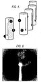
- the X-ray fibre diffraction pattern collected from SAF peptide fibres showed the following features (Fig. 6): (1) a short meridional (that is, parallel to the long fibre axis) reflection at 5.11 ⁇ 0.03 ⁇ ; (2) the harmonic of this 5.11 reflection at 10.19 ⁇ 0.05 ⁇ ; and (3) a stronger, more diffuse reflection centered at 8.8 ⁇ 0.15 ⁇ on the equator. These features are consistent with ⁇ -helical coiled-coils aligned with the fibre axis. The 5.1 ⁇ meridional reflection corresponds to the pitch of the helices within the coiled-coils.
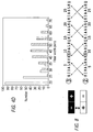
- TEM images of a 100 ⁇ M SAF mixture also demonstrated that fibres were not formed in the presence of 0.5 M KF (Fig. 4E).
- Fig. 4E shows the results of TEM of a matured SAF peptide mixture at 100 ⁇ M incubated in the presence of 0.5 M KF.
- the inventors did not knowingly design any features into the SAF peptides to foster further association of the two-stranded coiled coils.
- the observation of thick fibres in SAF peptide preparations therefore, raised the question: what interactions guided and stabilized these higher-order assemblies?
- the inventors therefore propose that features inherent in repeating structures of the type that they designed will naturally promote such fibre assembly (fibrillogenesis).
- any sequence feature presented on its surface by either, or both of the constituent peptides will be repeated at regular intervals along the protofibril.
- the repeat length will be equal to the length of the peptides (for SAF-p1 and SAF-p2 this was 28 residues, or about 4.2 nm).
- the motif will spiral around the protofibril tracking the superhelix of the coiled coil, which has a pitch of about 15 nm for a contiguous, heptad-based, dimeric structure.
- protofibril-protofibril interactions may be promoted if another sequence motif complementary to the first is present in the potential partner.
- Sequence offsets of 2 residues are potentially more interesting than the 1- and 3-residue offsets. This is because of the possibility of placing hydrophobic (H) residues at a, c, d, and f, with c and f effectively making up the a' and d' positions of the second, offset heptad. This is represented below, where P signifies polar (non-core) residues.
- TolC has two ⁇ -barrel-like domains (Koronakis, V. et al (2000) Nature 405, 914-919). Both have 12 helices contributed by 3 monomers. In the lower barrel each helix pairs with another from the same protomer to form separate supercoiled, antiparallel coiled-coils; SOCKET analysis revealed extensive antiparallel knobs into holes (KIH) interactions within these pairs, but not between them. In contrast, the helices of the upper barrel appear to pack more uniformly, albeit with a slant, to describe an ⁇ -cylinder. The SOCKET output for this part of the structure revealed many fewer KIH interactions than found in the lower barrel.
- the TolC barrel represents a variation of the cylinders formed by protein structures of the invention.
- a second consideration for ⁇ -cylinder construction is the consequences of helix and coiled-coil supercoiling.
- the upper barrel of TolC has 12 helices. Based on a structure of parallel helices with canonical supercoiling, i.e an angular separation of 154° between the two seams in each helix, we calculated that the cylinder should close at 14 helices. However, variations in helix number are expected. One reason for this is that helices cannot supercoil in two direction simultaneously, and some distortion is required to maintain packing at both interfaces.
- the protein structures of the invention may have various applications such as in:
- a 36-residue peptide with a 7-11-7-11 repeat offset by 2 residues should form a spiral of helices the ends of which meet to close the tube.
- Such nanotubes maybe particularly useful in the production of nanoscale piping and plumbing.
- the interior of the tube may be derivatised to control the flow of different small (2-40 ⁇ ) molecules.
- the self-assembling peptides of the invention are relatively small and synthetically accessible.
- non-standard derivatisable side chains may be incorporated in them.
- the monomer units can be made with a single cysteine residue at an exterior f position.
- These can be used to couple small molecules and other peptides using thiol-based chemistry.
- a wide variety of thiol-reactive probes are available.
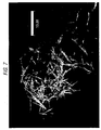
- the peptides can be tagged with fluorophores. For instance, with one peptide labelled with Fluorescein and the other with Rhodamine fibres visualised by confocal microscopy appear green and red, respectively (Fig. 7).
- T-shaped conjugated peptides can be made. These are covalent heterodimers made by mixing and coupling together variants of two SAF peptides: one with a terminal cysteine and the other having a central cysteine residue.
- the desired products can be purified from the mix of disulphide-linked peptide by PHLC.
- Doping the conjugated ("T") peptides into fresh SAF mixtures should propagate fibre assembly in three dimensions as both the "bar” and the "stem” of the "T” could become incorporated in, or initiate, fibres.
- the resulting networks can be visualised and characterised by EM.
- Peptide synthetic diblock copolymer hybrids may be produced. Suitable methods for preparing water soluble diblock copolymers using atom transfer radical polymerisation are described in X. S. Wang et al Chemical Communications 1817 (1999) and X. S Wang et al Macromolecules 33, 257 (2000).
- the protein fibres of the invention may be used to template and control this polymerisation either to produce hybrid fibres or if the peptide template is subsequently disassembled and marked away, to provide routes to water soluble "fishnet" nanotubes.
- Other possibilities include: conjugating polymers onto preassembled peptide fibres; conjugating the polymers and peptides prior to fibre assembly; and effecting polymerisation on the pre-assembled fibres.
- the protein fibres of the invention may be arranged to form two and three dimensional grids and matrices respectively.
- An affinity matrix could be assembled (for example in situ in blood) to remove blood contaminants such as viruses.
- a binder for the target contaminant e.g a peptide or protein with natural or engineered affinities for a viral coat protein
- the matrix can then be removed from blood along with any bound contaminants by light centrifugation. For example, it is estimated that a 100 nm length of fibre would have a mass of ⁇ 12 MDa which would readily be removed.
- affinity matrices have a number of advantages over larger naturally occurring proteins. In the assembled matrices any binders are aligned to give high effective avidities for the targeted molecules.
Landscapes
- Chemical & Material Sciences (AREA)
- Health & Medical Sciences (AREA)
- General Health & Medical Sciences (AREA)
- Organic Chemistry (AREA)
- Nanotechnology (AREA)
- Engineering & Computer Science (AREA)
- Molecular Biology (AREA)
- Life Sciences & Earth Sciences (AREA)
- Chemical Kinetics & Catalysis (AREA)
- Physics & Mathematics (AREA)
- Genetics & Genomics (AREA)
- Medicinal Chemistry (AREA)
- Radiology & Medical Imaging (AREA)
- Proteomics, Peptides & Aminoacids (AREA)
- General Chemical & Material Sciences (AREA)
- Biochemistry (AREA)
- Biophysics (AREA)
- General Physics & Mathematics (AREA)
- Crystallography & Structural Chemistry (AREA)
- Nuclear Medicine, Radiotherapy & Molecular Imaging (AREA)
- Analytical Chemistry (AREA)
- Gastroenterology & Hepatology (AREA)
- Peptides Or Proteins (AREA)
- Nonwoven Fabrics (AREA)
- Preparation Of Compounds By Using Micro-Organisms (AREA)
Abstract
Description
- This invention relates to protein structures, to methods of producing those protein structures, and to protein fibres and other materials and assemblies produced using those protein structures.
- The process of molecular self-assembly is central to all biological systems and is assuming increasing importance and application in biotechnology (L. Q. Gu, et al (1999) Nature 398, 686) and nanotechnology (K. E. Drexler, (1999) TIBTECH 17, 5). The characterization of natural biomolecular assemblies motivates and directs the development of model self-assembling systems and, in turn, these advance our understanding of biology. For proteins at least, the coiled coil is arguably the simplest self-assembling system. Coiled coils are protein-folding motifs that direct and cement a wide variety of protein-protein interactions (A. Lupas, (1996) Trends Biochem. Sci 21, 375). In structural terms, coiled coils are relatively straightforward: they are α-helical bundles with between 2 and 5 strands that can be arranged in parallel, antiparallel or mixed topologies. The basic sequence features that guide the formation of coiled coils from peptides are reasonably well understood (P. B. Harbury et al (1993) Science 262, 1401; D. N. Woolfson and T. Alber (1995) Protein Sci. 4, 1596). For instance, most coiled-coil sequences are dominated by a 7-residue repeat of hydrophobic (H) and polar (P) residues, (HPPHPPP) n , known as the "heptad repeat". When configured into an α-helix this pattern gives an amphipathic structure, the hydrophobic face of which directs oligomer-assembly. Furthermore, both the number and the direction of chains within a coiled-coil bundle is determined predominantly by residues that form or flank the hydrophobic core namely, residues at the first, fourth, fifth and seventh positions of the heptad repeat. For instance, coiled coils which form dimers (i.e. two-stranded assemblies) usually have isoleucine or valine residues at the first position and a leucine residue at the fourth position. By contrast, coiled coils that form trimers (i.e. three-stranded assemblies) often have the same residues (i.e both isoleucine or both leucine) at both "H" positions. Finally, hetero-oligomers (that is coiled coils made from strands with different amino-acid sequences) may be directed by complementary charged interactions that flank the hydrophobic core. For these reasons, there have been a number of successful de novo protein designs based on the coiled coil These include some ambitious structures that extend the natural repertoire of coiled-coil motifs (S. Nautiyal et al (1995) Biochemistry 34, 11645; A. Lombardi et al (1996)
Biopolymers 40, 495; D. H. Lee et al (1996) Nature 382, 525; P. B. Harbury et al (1998) Science 282, 1462; J. P. Schneider et al (1998) Folding Des. 3, R29). - In addition to commonly accepted structures with a single, contiguous heptad repeat, the inventors have identified sequences with multiple, offset heptad repeats which help explain oligomer-state specification in coiled coils. For example, sequences with two heptad repeats offset by two residues; i.e a/f-b/g'-c/a'-d/b'-e/c'-f/d'-g/e' set up two hydrophobic seams on opposite sides of the helix formed. Such helices may combine to bury these hydrophobic surfaces in two different ways and form two distinct structures: open "α-sheets" and closed "α-cylinders".
- Other relevant aspects of coiled-coil structure are described in WO99/11774, the disclosure of which is incorporated herein by way of reference.
- This understanding of coiled coils, and the resulting protein designs, centres on short structures as exemplified by the leucine-zipper motifs (E. K. O'Shea et al (1989) Science 243, 538; E. K. O'Shea et al (1991) Science 254, 539), which are found in a variety of transcription factors. In contrast, most natural coiled coils extend over hundreds of amino acids (A. Lupas (1996) supra; J. Sodek et al (1972) Proc. Natl. Acad. Sci. U.S.A 69, 3800) and many assemble further to form thicker, multi-stranded filaments (H. Herrmann and U. Aebi (1998) Curr. Opin. Struct. Biol. 8, 177).
- With the goal of making elongated structures to improve our understanding of coiled coils, and to develop protein-design studies, we initially designed two 28-residue peptides - dubbed Self-Assembling Fibre peptides, SAF-p1 and SAF-p2 - to fold and form extended fibres when mixed. Focusing on the buried, hydrophobic-core positions of the structure, rules were incorporated to direct parallel dimer formation and to guard against alternative oligomers and topologies (P. B. Harbury et al (1993) supra; D. N. Woolfson and T. Alber (1995) supra; L. J. Gonzalez et al (1996) Nature Struct. Biol. 3, 1011). The building block of the design was a staggered heterodimer with overhanging or "sticky" ends. This contrasts with and distinguishes it from the natural and designer coiled-coil assemblies that have been characterized to date, in which the polypeptide strands align in-register, i.e they have blunt or "flush" ends. Complementary core interactions and flanking ion-pairs were incorporated into the overhangs to facilitate longitudinal association of the heterodimers (Figs. 1&2). This principle of using "sticky ends" is well developed in molecular biology for assembling DNA (S. J. Palmer et al (1998) Nucleic Acids Res. 26, 2560), and has been used to design intricate DNA crystals (E. Winfree et al (1998) Nature 394, 539). However, to OUI knowledge, our application of sticky end-directed molecular assembly to peptides is new; although we do note that head-to-tail packing of helices has been observed in recently solved crystal structures for two designer peptides (N. L. Ogihara et al (1997) Protein Sci. 6, 80; G. G. Prive et al (1999) Protein Sci. 8, 1400). These were helical peptides that crystallised with their helical ends in contact so as to form pseudo-continnous helices in the solid state. In other words they formed "blunt-ended" arrangements.
- US-A-5,712, 366 discloses self-assembling protein material but does not provide details of how to make a staggered parallel heterodimer. WO 96/11947 discloses protein nanostructures based on bacteriophage T4 tail fiber proteins but does not disclose a staggered parallel heterodimer coiled coil structure.
- Pandya et al., Biochemistry, 29, 8728-34, 2000 (published after the priority date of the present application) does not disclose a method of making nanotubes and does not disclose a matrix comprising the protein structures of the present invention.
- According to one aspect of the invention there is provided a protein structure comprising a plurality of first peptide monomer units arranged in a first strand and a plurality of second peptide monomer units arranged in a second strand, the strands preferably forming a coiled-coil structure, and in which a first peptide monomer unit in the first strand extends beyond a corresponding second peptide monomer unit in the second strand in the direction of the strands. The protein structures of the invention have numerous advantages. For example, relatively long protein fibres can be formed with little material - 1 µl of a 100 µM solution of the peptide monomers may provide enough material to form 10 m of
fibre 50 mm thick. - At least one charged amino acid residue of the first peptide monomer unit may be arranged to attract an oppositely-charged amino acid residue of the second peptide monomer unit. Preferably, the charged amino acid residue is in an end portion of the first peptide monomer unit, which extends beyond the corresponding second peptide monomer unit in the second strand. At least one strand may consist solely of first or second peptide monomer units respectively i.e homogenous strands. Heterologous strands are also contemplated. The peptide monomer units may comprise a repeating structural unit. Preferably, the repeating structural unit comprises a heptad repeat motif, having the pattern:

- Preferably, the repeat may include isoleucine or asparagine at position a and leucine at position d. Other repeats (e.g hendecads - abcdefghijk) and amino acid compositions may also be used (see WO99/11774).
- Preferably, the heptad repeat comprises oppositely-charged residues at positions e and g respectively. The oppositely-charged residues may be, for example, glutamic acid and lysine residues or arginine and aspartic acid. The use of synthetic amino acids, such as ornithine is also envisaged.
- A protein structure in accordance with the invention may be also specified by pairs of asparagine residues in the "a" positions provided by corresponding first and second peptide monomer units.
- In a preferred protein structure, the first and second peptide monomer units have the following sequences:
- a) KIAALKQKIASLKQEIDALEYENDALEQ (SAF-p1C) and
- b) KIRALKAKNAHLKQEIAALEQEIAALEQ (SAF-p2D) respectively; or
- c) KIAALKQKIAALKQEIDALEYENDALEQ (SAF-p1A) and
- d) KIRALKWKNAHLKQEIAALEQEIAALEQ (SAF-p2C) respectively; or
- e) KIAALKQKIASLKQEIDALEYENDALEQ (SAF-p1C) and
- f) KIRALKWKNAHLKQEIAALEQEIAALEQ (SAF-p2C) respectively.
- It will be appreciated that these are examples only of 4-heptad structures and that other lengths are possible and envisaged for use in the invention.
- According to another aspect of the invention, there is provided a method of producing protein structures, the method comprising providing a mixture of first and second peptide monomer units which associate to form a protein structure according to the invention. The structure can be derivatised and/or stabilized by cross-linking.
- Derivatization of the peptide monomer units before or after assembly into the protein structures of the invention may be performed. For example, fluorescent moieties (fluorophores) may be attached to the coiled coil as described in WO99/11774. The addition of fluorescent moieties may assist visualization of the protein structure. Substitution with functional groups at the "f" position in the heptad repeat is especially preferred as that position is on the outside of the helix (see Fig. 1C and 1E). Other derivatives may include attaching binders to the peptide monomer units for example so that units which can bind other entities can be produced.
- The first and second peptide monomers and the strands may have the characteristics described above.
- The invention also provides protein fibres produced by an association of protein structures according to the invention.
- The protein structures may also be arranged to form tubular structures. In particular, the structures may be arranged to form nanotubes.
- According to another aspect of the invention, there is provided a kit for making protein structures, the kit comprising first and second peptide monomer units which associate to form a protein structure or protein fibres according to the invention.
- The protein structures of the invention may be assembled in two and three dimensional arrays. For example, two dimensional mats can be formed which can function, for example as filters. Three dimensional grids or matrices can also be formed again, for example, for use as sieves or filters or for organising other associated or conjugated molecules in three dimensions.
- In a preferred embodiment, a matrix is assembled in situ. For example, a matrix can be formed in a solution to entrap contaminants in the solution and then the matrix, together with contaminants, can be removed from the solution for example by centrifugation.
- The stability of the protein structures at higher temperatures may be improved by making the peptide monomers longer, such that the overlap between corresponding first and second monomer unit residues is increased. Increases in monomer length have previously been shown to stabilize coiled coil structures.
- Additionally, the protein structures may be stabilised and derivatised by using them to template the polymerisation of synthetic polymers.
- The terms used in the specification are to be given the ordinary meaning attributed to them by the skilled addressee. The following is given by way of clarification:
- This term embraces both naturally-occuring amino acids and synthetic amino acids as well as naturally-occuring amino acids which have been modified in some way to alter certain properties such as charge. In all cases references to naturally-occurring amino acids may be considered to include synthetic amino acids which may be substituted therefor.
- A coiled-coil is a peptide/protein sequence usually with a contiguous pattern of hydrophobic residues spaced 3 and 4 residues apart, which assembles (folds) to form a multi-meric bundle of helices. Coiled-coils including sequences with multiple offset repeats are also contemplated.
- A dimer is a two stranded structure.
- A heterodimer is a dimeric structure formed by two different stands.
- A staggered heterodimer is a structure in which the two strands assemble to leave overlapping ends that are not interacting within the heterodimer.
- Blunt-end assembly is association where the two strands combine to give flushed i.e non-overlapping ends.
- A protofibril is a protein structure assembled longitudinally from staggered heterodimers interacting through their overhanging ends.
- A fibre is a structure formed by lateral association of two or more protofibrils.
- Protein structures and methods of producing protein structures in accordance with the invention will now be described, by way of example only, with reference to the accompanying Figures 1 to 8 in which:
- Fig. 1 illustrates the design and the sequences of self-assembling fibre (SAF) peptide monomers of the invention.
- Fig. 2 illustrates computer modelling of the designed self-assembling fibre of the invention.
- Fig. 3 illustrates the results of circular dichroism (CD) and linear dichroism (LD) experiments on protein structures of the invention.
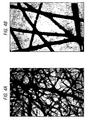
- Fig. 4 illustrates the assembly of synthetic protein fibres visualized directly by transmission electron microscopy and an analysis of fibre width In all panels, the white scale bars represent 100 nm. Fig. 4D is a histogram showing the distribution of fibre widths determined using TEM for fresh (white bars) and matured (black bars) mixtures of SAF peptides at 100 µM (a width value of "x" on the histogram includes all measurements made from "(x-5) to x").
- Fig. 5 is a cartoon showing the possible anti-typic association of parallel helical peptides leading to a homo-oligomeric peptide nanotube.
- Fig. 6 is an x-ray diffraction pattern of an aligned protein fibre of the invention.
- Fig. 7 is an electromicrograph showing fibres which have been derivatised through the inclusion of flurophores; and
- Fig. 8 shows amino acid sequences designed to form blunt-ended heterodimers.
- Various peptide monomer units were designed as described above. The monomers and capping peptides (designed to complement the sticky ends of the monomers so as to produce flush, or blunt ends and, so, arrest longitudinal fibre assembly) are set out in Table 1:
TABLE 1 PEPTIDE SEQUENCE DESIGN CD DATA LD DATA EM DATA 
@ 10 µM @ 100 µM @ 10 µM @ 100 µM @ 10 µM @ 100 µM CAP-p1A *YGPGE IAALEQE NAALEQ prototype unfolded SAF-p1A K IAALKQK IAALKQE IDALEYE NDALEQ prototype; slowly precipitates unfolded ~45% α-helix no fibres SAF-p1B *K IAALKQK IAALKQE IDALEYE NDALEQ* chemical capping of the ends (↑ stability) ~ 60 % α-helix ~ 70% α-helix SAF-p1C K IAALKQK TASLKQE IDALEYE NDALEQ no capping (↓ stability); mutate A11u → S (↑ solubility & ↓ helix stability) unfolded ~ 20% α-helix no signal no fibres CAP-p2A K TAALKQK DAALKQG GW* prototype unfolded SAF-p2A K ISALKWK NASLKQE IAALEQE IAALEQ prototype; low solubility unfolded SAF-p2B *K IRALKWK NAHLKQE IAALEQE IAALEQ* mutate S3 → R & S11 → H(↑ solubility & ↑ helix stability) ~ 60% α-helix ~ 95% α-helix SAF-p2C K IRALKWK NAHLKQE IAALEQE IAALEQ no capping (↓ stability) unfolded ~ 20% α-helix thin fibres SAF-p2D K IRALKAK NAHLKQE IAALEQE IAALEQ mutate W7 → A (investigate role of Trp in fibrillogenesis) ~ 15 % α-helix ~ 45% α-helix no signal no fibres SAF-p2E K IRALKCK NAHLKQE IAALEQE IAALEQ mutate A7 → C (for derivatization & cross-linking) *=Chemical capping = CH3CO at the N terminus and NH2 at the C terminus - Fig. 1 shows (A) A mechanism for self assembly: complementary charges in "companion" peptides direct the formation of staggered, parallel heterodimers; the resulting "sticky" ends are also complementary and promote longitudinal association into extended structures. Fig 1(B) shows the designed amino acid sequences: each peptide comprised canonical heptad repeats ( abcdefg ) with Ile at a and Leu at d to guide the formation of coiled-coil dimers; oppositely-charged residues were incorporated at e and g to favour the staggered dimer with sticky ends; asparagine residues (which preferentially pairs with each other at a sites (Gonzalez L et al (1996)
Nature Structural Biology 3, 13: 1011-1018) were included to cement the prescribed register further and to favour the parallel structures. Fig. 1(C) is a helical-wheel representation, summarizing the designed sequences in context. The view is from the N-terminus with heptad sites labeled a-g and assumes 3.5 residues per helical turn to emphasise the heptad repeat. - The peptides were synthesized on an Applied Biosystems 432A Peptide Synthesizer using solid-phase methods and Fmoc chemistry. Peptide samples were purified using reversed-phase HPLC and their identities confirmed by MALDI-TOF mass spectrometry.
- Various combinations of peptide monomers and capping peptides were tested as set out in Table 2:
TABLE 2 MISC. PEPTIDE MIXTURE EQUILIBRATION CD DATA LD DATA EM DATA @ 10 µM @ 100µM @ 10 µM @ 100m µM @ 10 µM @ 100µM SAF-p1A, SAF-p2A ~ 20% α-helix ~ 40 % α-helix Tm ≈ 30 °C SAF-p1A, SAF-p2A, CAP-p1A, CAP-p2A ~ 20 % α-helix SAF-p1A, SAF-p2B rapid ~ 65% α-helix Tm ≈ 36/46 °C (expect 38 °C if no interaction) SAF-p1B, SAF-p2B rapid ~ 70 % α-helix Tm ≈ 25 °C (expect 19 °C if no interaction) ~ 80% α-Helix Tm ≈ 35 °C (expect 36°C if no interaction) no fibres SAF-p1A, SAF-p2C slow @ 100µM; clouding occurs ~ 20 % α-helix ~ 80 % α-helix biphasic thermal melt no fibres thick fibres (- 45 nm wide) no signal from fibres in 1D-NMR SAF-p1C, SAF-p2C slow @ 100 µM; clouding occurs ~ 25 % α-helix ~ 65 % α-helix (~ 25% in 0.5 M salt); unusual spectral shape; no aromatic signal no signal strong signal from backbone & aromatics no fibres thick fibres (~ 45 nm wide); no fibres in 0.5 M salt SAF-p1C, SAF-p2D slow @ 100 µM; clouding occurs ~ 25 % α-helix ~ 85 % α-helix (~ 20% in 0.5 M salt); unusual spectral shape no signal strong signal from backbone & aromatics; no signal in 0.5 M salt no fibres thick fibres (- 45 nm wide); no fibres in 0.5 M salt - In addition and as a control, the SAF-p1c sequence was permuted (N- and C-terminal halves were swapped) to produce peptide SAF-p3:
- This design should combine with SAF-p2D to form a blunt-ended structure, which should not form fibres.
- A model of the three-dimensional structure of the designed protein fibre resulting from the assembly of SAF-p1 and SAF-p2 was made from the minimised structure of a model coiled-coil 35-mer, (LAALAAA)5, which was generated using Crick's Equation and had an ideally packed interface (G. Offer and R. Sessions, J. Mol. Biol. 249, 967 (1995)). Copies of the 35-mer were superimposed with an overlap of one heptad repeat to extend the structural template, and the backbone was rejoined after removal of overlapping segments. Residues in the two-stranded template were replaced with the sequences of the SAF peptides, staggered relative to each other by two heptad repeats according to the alignment in Fig. 1B. The structure was soaked in a 5 Å layer of water and energy minimised until the average absolute derivative of coordinates with respect to energy fell below 0.01 kcal Å-1. The structure was built and visualized using Insight II 97.0 (Molecular Simulations Inc.), and was energy-minimized using Discover 2.9.8 (Molecular Simulations Inc.) with the consistent valence forcefield. In Fig 2(A) peptides SAF-p1 and SAF-p2 (each coloured dark grey-to-light grey from the N-terminus) interact through core residues including asparagine pairs (coloured mid-grey) to form the two strands of a staggered, parallel, coiled-coil fibre. In Fig. 2(B), negatively charged glutamate side chains (coloured light grey) and positively charged lysine side chains (coloured black) form complementary charge interactions between the SAF peptides.
- Peptide samples were incubated at 5°C in 10 mM MOPS (3-(N-Morpholino)propanesulfonic acid), pH 7. Sample concentrations were determined from their UV absorbance at 280 nm (SAF-pl) and 214 nm (SAF-p2). After baseline correction, ellipticities in mdeg were converted to molar ellipticities (deg cm2 dmol-res-1) by normalizing for the concentration of peptide bonds. Data were recorded in a cell of 1 mm path length by integrating the signal for 5s (and 1s for the fresh 100 µM peptide mixture) every nm in the range 205-260 nm. CD measurements were made using a JASCO J-715 spectropolarimeter fitted with a Peltier temperature controller.
- The CD data shown in Fig. 3 provides spectroscopic evidence for the formation of helical structures by the SAF peptides. Fig. 3(A) shows circular dichroism (CD) spectra at 10 µM for: SAF-p1 (----), SAF-p2 (- -), the average of these spectra (- - -), and the experimental SAF peptide mixture (o). Fig 3(B) shows CD spectra at 100 µM - the key is the same as for Fig 3(A), but with the additional spectrum (•) being for the SAF peptide mixture after "maturation" for 1 h.
- Consistent with our design, neither SAF-p1 nor SAF-p2 was highly structured in aqueous solution at
pH 7 and 5 °C (Fig. 3). However, when mixed in equal proportions the circular dichroism (CD) spectrum changed and, moreover, was markedly different from the theoretical spectrum generated by averaging the spectra for the isolated peptides. In particular, the spectrum for the mixture had intense minima at 208 and 222 nm consistent with the formation of α-helical structure, but these features were not as pronounced in the spectra of the individual peptides. This was clear evidence that the two peptides interacted to form an α-helical structure as designed. Furthermore, and as expected for a multimerization event, the magnitude of these spectral changes depended on peptide concentration; a SAF mixture with 10 µM of each peptide, did show a weak signal indicative of some α-helical structure, however, a 100 µM mixture gave a much stronger signal (Figs. 3A&B). - The shape and intensity of spectra from 100 µM mixtures of the SAF peptides also changed with time (Fig. 3B). Spectra recorded immediately after mixing a "fresh" sample displayed some α-helical structure. After incubation of the mixture for 1 hour at 5 °C ("maturation"), however, the signal at 222 nm was more intense, and indicated approximately 75 % α-helix, consistent with substantial coiled-coil formation.
- Maturation of 100 µM SAF peptide mixtures was also accompanied by slight clouding of the samples. Scattering effects from such samples can lead to attenuation and distortion of CD spectra (D. Mao and B. A. Wallace, (1984) Biochemistry 23, 2667). However, we could disregard this possibility because altering the distance between the sample and the detector in the CD instrument did not affect the shape or the intensity of the spectrum. Furthermore, we established that the majority of the CD signal from the mixtures derived from the suspended material: a supernatant without the suspended material, which was recovered by centrifugation of a matured 100 µM SAF mixture, gave only a weak CD signal similar to the 10 µM mixture.
- Thus, the CD data were wholly consistent with the desired α-helical SAF design and, moreover, indicated the formation of large assemblies.
- As a control, SAF-p3 (the permutation of SAF-p1 (identical to SAF-p1c)) was designed to form a blunt-ended heterodimer with SAF-p1 that should not assemble further into fibres. 100 µM mixtures of SAF-p2 (identical to SAF-p2D) and SAF-p3 were analysed by sedimentation equilibrium in the analytical ultracentrifuge. The resulting data were best fitted assuming a single ideal species in solution, and the molecular weight was allowed to vary during the fit. An Mr of 6422 (with 95% confidence limits of 5924 and 6911) was obtained, which is very close to the expected heterodimer value of 6303 calculated from mass spectrometry of the individual peptides. CD spectra for 100 µM fibre-producing mixtures (SAF-p1 with SAF-p2), and for blunt dimer-producing mixtures (SAF-p2 with SAF-p3), were recorded. For the blunt dimer-producing mixtures, the shape and intensity of the CD spectrum were fully consistent with coiled-coil formation as designed. In contrast to the fibre-producing mixtures, the blunt dimer-producing mixtures showed no signs of maturation; that is, negligible spectral changes and no clouding of solutions occurred upon incubation. Interestingly, the intensity of the minimum near 222 nm, which is an accepted indicator of α-helical structure and degree of α-helical folding, was similar for both mixtures. This strongly supports the formation of α-helical structure as designed in the fibre-producing mixtures despite the spectral shifts observed upon maturation.
- Linear dichroism (LD) spectroscopy was also used to test if elongated structures were being formed as designed. Long polymers such as DNA molecules can be oriented by shear flow. This effect can be monitored by LD spectroscopy provided that chromophores also become aligned by the flow (M. Bloemendal (1994) Chem. Soc. Rev. 23, 265; A. Rodger and B.
- Norden (1997) Oxford Chemistry Masters (Oxford University Press, Oxford), vol. 1).
- Peptide samples were prepared for LD as for CD. LD data were collected on samples spinning in a couette flow cell by integrating the signal for 2 s every nm in the range 210-320 nm, using a JASCO J-715 spectropolarimeter. After baseline correction, absorbance was converted to molar extinction coefficient (1 mol-res-1 cm-1) by normalizing for the concentration of peptide bonds. A linear correction for a sloping baseline was made to the data from the 100 µM SAF peptide mixture.
- The results are depicted in Fig. 3D, which shows linear dichroism (LD) spectra for: 20 µM tropomyosin (- - -), the SAF peptide mixture at 10 µM (----), and the SAF peptide mixture at 100 µM in the absence (•) and presence (o) of 0.5 M KF.
- For instance, we found that tropomyosin, which forms a dimeric coiled coil approximately 42 nm in length, could be aligned to give a LD signal (Fig. 3D). In contrast and consistent with our design and the CD data, experiments with a 10 µM SAF mixture, (Fig. 3D), and for the individual peptides at 100 µM (data not shown), LD signals were not detected. However, a matured 100 µM SAF peptide mixture gave a strong absorbance from the peptide backbone (210-240 nm) and some signal in the aromatic region (260-290 nm) during flow orientation (Fig. 3D). As only long structures are aligned by this technique, the data demonstrated that long fibres at least 500 nm in length were present in solutions of the matured 100 µM SAF peptide mixtures.
- To confirm fibre assembly, we used electron microscopy to visualize structures in the peptide preparations directly. For TEM experiments, peptide samples were incubated for 1 h at 5 °C in filtered 10 mM MOPS, pH 7. A drop of peptide solution was applied to a carbon-coated copper specimen grid (Agar Scientific Ltd, Stansted, UK), and dried with filter paper before negative staining with 0.5% aqueous uranyl acetate and then dried at 5 °C. A "fresh" SAF peptide mixture was prepared by mixing preincubated solutions of the individual peptides at 200 µM directly on the specimen grid, before drying and negative staining as described. Grids were examined in a Hitachi 7100 TEM at 100 kV and digital images were acquired with a (800 x 1200 pixel) charge-coupled device camera (Digital Pixel Co. Ltd., Brighton, UK) and analyzed (Kinetic Imaging Ltd., Liverpool, UK).
- For scanning electron microscopy (SEM) experiments, negatively-stained specimen grids were sputter-coated with gold and examined in a Leo Stereoscan 420 SEM at 20 kV and with a probe current of 10 pA.
- No structures were visible up to 100 000 times magnification by transmission electron microscopy (TEM) for either the 10 µM SAF mixture, or for the individual peptides at 100 µM concentration (data not shown). However, TEM of a 100 µM SAF mixture at 50 000 times magnification revealed time-dependent formation of long fibrous structures, consistent with the CD and LD data. Fresh mixtures showed large numbers of extended fibres of various widths. The majority of these had a diameter of about 20 nm (Figs. 4A (a fresh mixture at 100 µM) & Fig 4D); finer fibres were present, but their widths could not be measured reliably. Images recorded for the matured mixtures showed fewer fibres, but these were more distinct and thicker than those observed in the fresh mixture (Fig. 4B&D).
- Scanning electron microscopy (SEM) of a matured mixture showed no evidence for fibre branching. Rather, the fibres were simply intertwined as if layered on top of each other (Fig. 4C). It was not possible to follow the full length of fibres due to intertwining, but they were at least several hundred microns in length. Although the density of fibres varied across the surface of the EM grid, for the matured samples at least, their diameters were quite uniform with a mean width of 43.3 (SD = 9.3) nm (Fig. 4D). As the original design was for a longitudinally extended, but otherwise two-stranded coiled coil the average diameter that we might have expected was about 2 nm. Therefore, the EM data suggested that the designed two-stranded coiled-coil fibres associate laterally into higher order assemblies.
- Mixtures of SAF peptides at 500 µM in 10 mM MOPS, pH 7, were incubated on ice for at least 1h, before centrifugation at 6500g for 5 min. Droplets of fibre-containing solutions, taken from the bottom of the centrifuged tubes, were suspended between the ends of two wax-filled capillaries and allowed to dry slowly overnight at 4°C, yielding clumps of partially aligned fibres. X-ray fibre diffraction images were collected using a Rigaku CuKα rotating anode source (wavelength 1.5418 Å) and a R-AXIS IV detector. Samples were maintained at 5°C during data collection with cool air from a cryostream (Oxford Cryo-systems). The X-ray fibre diffraction pattern collected from SAF peptide fibres showed the following features (Fig. 6): (1) a short meridional (that is, parallel to the long fibre axis) reflection at 5.11± 0.03 Å; (2) the harmonic of this 5.11 reflection at 10.19 ± 0.05 Å; and (3) a stronger, more diffuse reflection centered at 8.8 ± 0.15 Å on the equator. These features are consistent with α-helical coiled-coils aligned with the fibre axis. The 5.1 Å meridional reflection corresponds to the pitch of the helices within the coiled-coils. The other expected reflection on the meridian-that is, that at 1.5 Å and corresponding to the rise per residue-lies out of the resolution of the current data sets, whereas the equatorial reflection reveals the mean distance between α-helical axes. This value at 8.8 Å is less than the observed value for keratin but falls within reported ranges for dimeric coiled-coil peptides.
- Molecular modeling of the SAF sequences into an extended two-stranded coiled coil also highlighted potential complementary charge interactions on the surface of the protofibrils, Figs 1&2. In accordance with this, experimentally it was found that moderate concentrations of salt inhibited protofibril and thick fibre assembly. First, CD spectra recorded for both the individual peptides-and a 100 µM mixture of SAF peptide samples with 0.5 M potassium fluoride showed reduced helical CD signals and there was no evidence of "maturing" in the mixed samples (Fig. 3C). Second, the LD signal described previously for the matured 100 µM SAF peptide mixture was also lost when the experiment was repeated in the presence of salt (Fig. 3D). Finally, TEM images of a 100 µM SAF mixture also demonstrated that fibres were not formed in the presence of 0.5 M KF (Fig. 4E). Fig. 4E shows the results of TEM of a matured SAF peptide mixture at 100 µM incubated in the presence of 0.5 M KF.
The inventors did not knowingly design any features into the SAF peptides to foster further association of the two-stranded coiled coils. The observation of thick fibres in SAF peptide preparations, therefore, raised the question: what interactions guided and stabilized these higher-order assemblies? The inventors therefore propose that features inherent in repeating structures of the type that they designed will naturally promote such fibre assembly (fibrillogenesis). - Consider a protofibril as depicted in Fig. 1B and 2A. Any sequence feature presented on its surface by either, or both of the constituent peptides will be repeated at regular intervals along the protofibril. The repeat length will be equal to the length of the peptides (for SAF-p1 and SAF-p2 this was 28 residues, or about 4.2 nm). Furthermore, the motif will spiral around the protofibril tracking the superhelix of the coiled coil, which has a pitch of about 15 nm for a contiguous, heptad-based, dimeric structure. In this scenario, protofibril-protofibril interactions may be promoted if another sequence motif complementary to the first is present in the potential partner. This is because the pitches of the complementary motifs on each protofibril will match precisely. Thus, once initiated, lateral association of protofibrils - that is, fibrillogenesis - will be cemented by many regularly spaced interactions as in a crystal. As a result, the complementary interactions need only be weak as the stability of the protofibril-protofibril interaction rests on an avidity effect rather than a small number of strong interactions. Provided that the components of the assembly can make more than one type of complementary surface very extensive molecular assemblies may result. The inventors used electrostatic interactions both to direct heterodimer formation, and to promote elongation of the protofibrils (Figs. 1 and 2). These features would also create periodic and alternating patches of charge in the protofibrils provided they are regular as envisaged (Fig. 1B and 2B). These charged patches could guide and stabilize the higher order assemblies. Indeed, similar features have been noted in several natural fibrous proteins and have been implicated in the assembly of multi-protein filaments (J. J. Meng et al (1994) Biol. Chem. 269, 18679; A. D. McLachlan and M. Stewart (1976) Mol. Biol. 103, 271), and small synthetic peptide systems (S. G. Zhang et al (1993) Proc. Natl. Sc. U.S.A 90, 3334). The experiments with salt (KF) described above suggest that salt-bridges (electrostatic interaction) may be at least in part the cause of fibrillogenesis.
- a. For two superimposed heptads there are three possible sequence offsets of 1, 2 and 3 residue(s), which are equivalent to 6, 5 and 4-residue offsets, respectively. For a regular 3.6-residue-per-tum α-helix, these set up two hydrophobic faces with angular offsets of 100°, 160° (360-200) and 60° (360-300), respectively, around the outside of the helix. This is best seen on a helical wheel. Accounting for helical supercoiling - i.e assuming 3.5 residues per turn and using the accepted helical-wheel representation for the coiled-coil these angular offsets are altered to 103°, 154° and 51°, respectively. However, both sets of angles are over-simplifications when considering helix-helix interactions in actual coiled-coil systems because side-chain size, geometry and packing also affect the helix interfaces (Harbury, P. B. et al (1993) Science 262, 1401-1407; Harbury, P. B. et al (1994) Nature 371, 80-83; Malashkevich, V. N. et al (1996) Science 274, 761-765). Nonetheless, we found that many natural coiled-coil assemblies, at least, were consistent with the approximate angular offsets: Trimers could be considered as having overlapping heptads separated by 3 residues (angular offset = 51/60°). Whereas, tetrameric and pentameric coiled-coils were often variations on a theme with two hepad repeats offset by 1 residue (100/103°).
- Sequence offsets of 2 residues are potentially more interesting than the 1- and 3-residue offsets. This is because of the possibility of placing hydrophobic (H) residues at a, c, d, and f, with c and f effectively making up the a' and d' positions of the second, offset heptad. This is represented below, where P signifies polar (non-core) residues.
a b c d e f g a b c d e f g repeat 1 H P P H P P P H P P H P P P binary pattern 1P P H P P H P P P H P P H P binary pattern 2f'g'a'b'c'd'e'f'g'a'b'c'd'e' repeat 2a b c d e f g a b c d e f g assigned register H P H H P H P H P H H P H P overall binary pattern - Such sequence patterns would results in two hydrophobic seams with a wide angular separation (154/160°), which would place them roughly on opposite sides of the helix. Furthermore, it offers two possibilities for parallel helix-helix packing arrangements: syn, where two like faces - i.e a / d with a / d , or c/f with c/f - from neighbouring helices combine to produce an openα-sheet, Fig. 6a; anti, where a/d faces pair with c/f. In the anti-arrangement the structure can close to form α α-cylinder. For antiparallel pairs of helices syn-typic association should lead to cylinders, whereas sheets should be formed from anti-typic antiparallel interfaces.
- TolC has two α-barrel-like domains (Koronakis, V. et al (2000) Nature 405, 914-919). Both have 12 helices contributed by 3 monomers. In the lower barrel each helix pairs with another from the same protomer to form separate supercoiled, antiparallel coiled-coils; SOCKET analysis revealed extensive antiparallel knobs into holes (KIH) interactions within these pairs, but not between them. In contrast, the helices of the upper barrel appear to pack more uniformly, albeit with a slant, to describe an α-cylinder. The SOCKET output for this part of the structure revealed many fewer KIH interactions than found in the lower barrel. Furthermore, KIH interactions were not contiguous around the cylinder and, in particular, they were more extensive between helices in the same monomer, but less regular between the helices abutting the monomers. In our view, the TolC barrel represents a variation of the cylinders formed by protein structures of the invention.
- Nevertheless, the inventors were able to assign heptad registers for the helices of the upper barrel unambiguously. This revealed knobs at relative a, c, d, and f positions and syn-typic association of two seams adjacent helices; i.e fully consistent with the theory outlined above.
- We believe that it will be possible to constuct α-sheets and α-cylinders using helices in parallel. The use of parallel helices does have one interesting consequence for the construction of α-cylinders, however: as the pairing in these structures will be anti-typic, a residues on one helix partner c residues of a neighbouring helix at the same level in the structure. Similarly, d and f residues pair at the intervening levels. The result will be that successive helices will be translated up the helix and cylinder axes by two residues, which is equivalent to ≈3Å. Thus, attempts to construct α-cylinders from parallel helices will give spirals of helices which may or may not close. This is, however, potentially extremely interesting as it opens up possibilities for making peptide-based nanotubes as described above.
- A second consideration for α-cylinder construction is the consequences of helix and coiled-coil supercoiling. The upper barrel of TolC has 12 helices. Based on a structure of parallel helices with canonical supercoiling, i.e an angular separation of 154° between the two seams in each helix, we calculated that the cylinder should close at 14 helices. However, variations in helix number are expected. One reason for this is that helices cannot supercoil in two direction simultaneously, and some distortion is required to maintain packing at both interfaces. We found structural precedents for this in the Protein Data Book PDB where tight knobs-into-holes packing was maintained (Walshaw & Woolfson, unpublished); indeed, the central helices of the 3-helix α-sheets are straight, Fig. 7b. (n.b . The slanting of the helices in the upper barrel of TolC may offer a compromise between straight and supercoiled helices). Assuming the packing of completely straight helices, the angular offset becomes 160° and 18 helices would close a cylinder. However, given that, as in 3-, 4- and 5-stranded coiled coils, side chains mediate the helix-helix contact angles other oligonmerisation states might be possible (Harbury, P. B et al (1993) Science 262, 1401-1407; Harbury, P. B. et al (1994) 371, 80-83; Malashkevich, V. N. et al (1996) Science 274, 761-765): we calculate that small adjustments in the angular offset between 144° to 162° varies the helix number from 10 to 20.
- As mentioned above, the protein structures of the invention may have various applications such as in:
- a. This can be achieved for example by combining the aforementioned 7- and 11-residue repeats with offsets in the sequence. The effect would be eliminate the overall hydrophobic displacement. In other words, alternating heptad and hendecad repeats give an 18-residue repeat to match the α-helical repeat; in the α-helix, 18 residues span 5 helical turns exactly. It may therefore be possible to create a completely closed peptide nantotube (Fig. 5 shows part of a nanotube) In the parallel, straight helix case there would be 18 helices per turn of the "cylinder", and the rise per turn is 36 residues. Thus, a 36-residue peptide with a 7-11-7-11 repeat offset by 2 residues should form a spiral of helices the ends of which meet to close the tube. Such nanotubes maybe particularly useful in the production of nanoscale piping and plumbing. The interior of the tube may be derivatised to control the flow of different small (2-40Å) molecules.
- The self-assembling peptides of the invention are relatively small and synthetically accessible. Thus, non-standard derivatisable side chains may be incorporated in them. For example, the monomer units can be made with a single cysteine residue at an exterior f position. These can be used to couple small molecules and other peptides using thiol-based chemistry. A wide variety of thiol-reactive probes are available. In particular, the peptides can be tagged with fluorophores. For instance, with one peptide labelled with Fluorescein and the other with Rhodamine fibres visualised by confocal microscopy appear green and red, respectively (Fig. 7). There is a possibility for FRET between the probes, which may pack closely in the fibres, and this may confuse interpretation. To avoid this the tagged peptides can be doped into fresh, assembling SAF mixtures. Having available fluorescently labelled peptides and fibres offers another route to tracking fibre/network assembly and orientation.
- To generate branched self-assembling fibres "T-shaped" conjugated peptides can be made. These are covalent heterodimers made by mixing and coupling together variants of two SAF peptides: one with a terminal cysteine and the other having a central cysteine residue. The desired products can be purified from the mix of disulphide-linked peptide by PHLC. Doping the conjugated ("T") peptides into fresh SAF mixtures should propagate fibre assembly in three dimensions as both the "bar" and the "stem" of the "T" could become incorporated in, or initiate, fibres. The resulting networks can be visualised and characterised by EM.
- Peptide synthetic diblock copolymer hybrids may be produced. Suitable methods for preparing water soluble diblock copolymers using atom transfer radical polymerisation are described in X. S. Wang et al Chemical Communications 1817 (1999) and X. S Wang et al Macromolecules 33, 257 (2000).
- The protein fibres of the invention may be used to template and control this polymerisation either to produce hybrid fibres or if the peptide template is subsequently disassembled and marked away, to provide routes to water soluble "fishnet" nanotubes. Other possibilities include: conjugating polymers onto preassembled peptide fibres; conjugating the polymers and peptides prior to fibre assembly; and effecting polymerisation on the pre-assembled fibres.
- The protein fibres of the invention may be arranged to form two and three dimensional grids and matrices respectively. One application for such matices is in the purification of biological fluids such as blood. An affinity matrix could be assembled (for example in situ in blood) to remove blood contaminants such as viruses. In the case of virus removal, a binder for the target contaminant (e.g a peptide or protein with natural or engineered affinities for a viral coat protein) can be fused to a peptide monomer units in the protein structure of the invention. The matrix can then be removed from blood along with any bound contaminants by light centrifugation. For example, it is estimated that a 100 nm length of fibre would have a mass of ≥ 12 MDa which would readily be removed. Such affinity matrices have a number of advantages over larger naturally occurring proteins. In the assembled matrices any binders are aligned to give high effective avidities for the targeted molecules.
- Other applications for protein structures in accordance with the invention include:
- i. preparation of organised networks for seeding the crystalisation of biomolecules for X-ray crystallography;
- ii. using ordered fibres to promote cell growth for tissue engineering;
- iii. the construction of nanoscale molecular sieves
- iv. the preparation of nanoscale molecular grids/scaffolds that could be used as supports for a variety of functional small or macromolecules.
- v. functionalised grids and networks could be used in, for example, catalysis, affinity-sieving/purification of biological fluids and other research solutions, the recruitment of endogenous molecules and co-factors to promote tissue repair and tissue engineering in general.
- vi. to create novel lab-on-chip technologies, peptide self-assembly could be combined with lithography as follows.
Lithography and related techniques can be used to pattern a variety of surfaces with channels, which can be made of a suitable size (e.g 20-100 nm wide and deep) to accommodate peptide fibres. These can then be used to direct the assembly of the fibres from solutions mixed directly on the surfaces. Furthermore, using well-established chemistry, the inventors envisage funtionalising the peptide fibres with a variety of small molecules and other proteins. This proposed combination of peptide design, self-assembly and lithography should allow the development of ordered arrays of functional polymers on specific surfaces. - vii. Assembled fibres could also be used as fine (therefore, high resolution) tips in AFM (atomic force microscopy) the current limit is about 10-25 nm using carbon nanotubes.
-
- <110> University of Sussex
- <120> Peptides
- <130> P100087PCT
- <140> PCT/GB00/03576
<141> 2000-09-18 - <150> 9922013.9
<151> 1999-09-17 - <160> 14
- <170> SeqWin99, version 1.02
- <210> 1
<211> 28
<212> PRT
<213> Artificial Sequence - <220>
<223> artificial sequence - <400> 1

- <210> 2
<211> 28
<212> PRT
<213> Artificial Sequence - <220>
<223> artificial sequence - <400> 2

- <210> 3
<211> 28
<212> PRT
<213> Artificial Sequence - <220>
<223> artificial sequence - <400> 3

- <210> 4
<211> 28
<212> PRT
<213> Artificial Sequence - <220>
<223> artificial sequence - <400> 4

- <210> 5
<211> 28
<212> PRT
<213> Artificial Sequence - <220>
<223> artificial sequence - <400> 5

- <210> 6
<211> 28
<212> PRT
<213> Artificial Sequence - <220>
<223> artificial sequence - <400> 6

- <210> 7
<211> 18
<212> PRT
<213> Artificial Sequence - <220>
<223> artificial sequence - <400> 7

- <210> 8
<211> 17
<212> PRT
<213> Artificial Sequence - <220>
<223> artificial sequence - <400> 8

- <210> 9
<211> 28
<212> PRT
<213> Artificial Sequence - <220>
<223> artificial sequence - <400> 9

- <210> 10
<211> 28
<212> PRT
<213> Artificial Sequence - <220>
<223> artificial sequence - <400> 10

- <210> 11
<211> 28
<212> PRT
<213> Artificial Sequence - <220>
<223> artificial sequence - <400> 11

- <210> 12
<211> 35
<212> PRT
<213> Artificial Sequence - <220>
<223> artificial sequence - <400> 12


- <210> 13
<211> 42
<212> PRT
<213> Artificial Sequence - <220>
<223> artificial sequence - <400> 13

- <210> 14
<211> 42
<212> PRT
<213> Artificial Sequence - <220>
<223> artificial sequence - <400> 14

Claims (16)
- A protein structure comprising a plurality of first peptide monomer units arranged in a first strand and a plurality of second peptide monomer units arranged in a second strand wherein the first and second monomer units comprise a plurality of heptad repeat motifs (abcdefg) and wherein there is a pair of asparagines at complementary "a" positions in complementary heptad repeats within corresponding first and second monomer units and charged amino acid residues at positions "e" and "g" in each first peptide monomer unit and oppositely-charged amino acid residues at positions "e" and "g" in each corresponding second peptide monomer unit and at least one charged amino acid residue is in an end portion of each first peptide monomer unit which extends beyond the corresponding second peptide monomer unit in the second strand, to ensure that the first strand and the second form a staggered parallel heterodimer coiled coil structure, and wherein hydrophobic residues are positioned at the other "a" positions and all the "d" positions in the first and second monomer units.
- A protein structure according to claim 1 in which at least one strand consists solely of first or second peptide monomer units respectively.
- A protein structure according to claim 1 wherein the hydrophobic residues at the other "a" positions are selected from isoleucine or valine.
- A protein structure according to any one of the preceding claims having a leucine at one or more of the "d" positions of the first and second monomer units.
- A protein structure according to claim 1 in which the oppositely-charged residues are glutamic acid and lysine residues, or synthetic derivatives of these amino acid residues.
- A protein structure according to any preceding claim in which the first and second peptide monomer units have the sequence:a) KIAALKQKIASLKQEIDALEYENDALEQ (SAF-p1C) andb) KIRALKAKNAHLKQEIAALEQEIAALEQ (SAF-p2D) respectively; orc) KIAALKQKIAALKQEIDALEYENDALEQ (SAF-p1A) andd) KIRALKWKNAHLKQEIAALEQEIAALEQ (SAF-p2C) respectively; ore) KIAALKQKIASLKQEIDALEYENDALEQ (SAF-p 1 C) andf) KIRALKWKNAHLKQEIAALEQEIAALEQ (SAF-p2C) respectively.
- A peptide monomer unit for use in preparing a protein structure the peptide monomer unit having an amino acid sequence selected from:a) KIAALKQKIASLKQEIDALEYENDALEQ (SAF-p1C);b) KIRALKAKNAHLKQEIAALEQEIAALEQ (SAF-p2D);c) KIAALKQKIAALKQEIDALEYENDALEQ (SAF-p1A); andd) KIRALKWKNAHLKQEIAALEQEIAALEQ (SAF-p2C).
- A protein structure according to any one of claims 1 to 6 or a peptide monomer unit according to claim 8 wherein at least one amino acid residue is derivatised.
- A method of producing protein structures, the method comprising providing a mixture of first and second monomer units which associate to form a protein structure according to any one of claims 1 to 5, wherein the first and second monomer units comprise the heptad repeat motif (abcdefg).
- A method according to claim 9 in which the protein structure is derivatised.
- A method according to claim 9 or 10 in which the protein structure is stabilised by cross-linking.
- A protein fibre produced by an association of protein structures according to any one of claims 1 to 6.
- A kit for making a protein structure, the kit comprising first and second peptide monomer units which associate to form a protein structure according to any one of claims 1 to 6 or a protein fibre according to claim 12, wherein the first and second monomer units comprise the heptad repeat motif (abcdefg).
- A two dimensional grid comprising a protein structure according to any one of claims 1 to 6 or a protein fibre according to claim 12.
- A three dimensional matrix comprising a protein structure according to any one of claims 1 to 6 or a protein fibre according to claim 12.
- A matrix according to claim 15 for use as a sieve or filter or for organising other associated or conjugated molecules in three dimensions.
Applications Claiming Priority (3)
| Application Number | Priority Date | Filing Date | Title |
|---|---|---|---|
| GB9922013 | 1999-09-17 | ||
| GBGB9922013.9A GB9922013D0 (en) | 1999-09-17 | 1999-09-17 | Peptides |
| PCT/GB2000/003576 WO2001021646A1 (en) | 1999-09-17 | 2000-09-18 | Protein structures and protein fibres |
Publications (2)
| Publication Number | Publication Date |
|---|---|
| EP1212356A1 EP1212356A1 (en) | 2002-06-12 |
| EP1212356B1 true EP1212356B1 (en) | 2007-03-07 |
Family
ID=10861113
Family Applications (1)
| Application Number | Title | Priority Date | Filing Date |
|---|---|---|---|
| EP00964388A Expired - Lifetime EP1212356B1 (en) | 1999-09-17 | 2000-09-18 | Protein structures and protein fibres |
Country Status (10)
| Country | Link |
|---|---|
| US (2) | US7045537B1 (en) |
| EP (1) | EP1212356B1 (en) |
| JP (1) | JP4703079B2 (en) |
| AT (1) | ATE356141T1 (en) |
| AU (1) | AU783156B2 (en) |
| CA (1) | CA2384849C (en) |
| DE (1) | DE60033828T2 (en) |
| DK (1) | DK1212356T3 (en) |
| GB (1) | GB9922013D0 (en) |
| WO (1) | WO2001021646A1 (en) |
Families Citing this family (26)
| Publication number | Priority date | Publication date | Assignee | Title |
|---|---|---|---|---|
| GB0219980D0 (en) * | 2002-08-28 | 2002-10-09 | Univ Sussex | Peptides |
| US7781396B2 (en) * | 2002-01-31 | 2010-08-24 | Tel Aviv University Future Technology Development L.P. | Peptides directed for diagnosis and treatment of amyloid-associated disease |
| US20040052928A1 (en) * | 2002-09-06 | 2004-03-18 | Ehud Gazit | Peptides and methods using same for diagnosing and treating amyloid-associated diseases |
| GB0220805D0 (en) * | 2002-09-06 | 2002-10-16 | Univ Sussex | Peptide monomers and protein structures |
| US7491699B2 (en) | 2002-12-09 | 2009-02-17 | Ramot At Tel Aviv University Ltd. | Peptide nanostructures and methods of generating and using the same |
| EP1583713B1 (en) * | 2003-01-07 | 2009-03-25 | Ramot at Tel Aviv University Ltd. | Peptide nanostructures encapsulating a foreign material and method of manufacturing same |
| GB0315993D0 (en) * | 2003-07-08 | 2003-08-13 | Sussex Ip Ltd | Peptide-based linker |
| WO2005027901A1 (en) * | 2003-09-25 | 2005-03-31 | Tel Aviv University Future Technology Development L.P. | Compositions and methods using same for treating amyloid-associated diseases |
| WO2005031362A2 (en) * | 2003-10-02 | 2005-04-07 | Ramot At Tel Aviv University Ltd. | Novel antibacterial agents and methods of identifying and utilizing same |
| US20090156471A1 (en) * | 2004-07-15 | 2009-06-18 | Ramot At Tel Aviv University Ltd. | Use of anti-amyloid agents for treating and typing pathogen infections |
| WO2006013552A2 (en) | 2004-08-02 | 2006-02-09 | Ramot At Tel Aviv University Ltd. | Articles of peptide nanostructures and method of forming the same |
| US7732479B2 (en) | 2004-08-19 | 2010-06-08 | Tel Aviv University Future Technology Development L.P. | Compositions for treating amyloid associated diseases |
| US7786086B2 (en) * | 2004-09-08 | 2010-08-31 | Ramot At Tel-Aviv University Ltd. | Peptide nanostructures containing end-capping modified peptides and methods of generating and using the same |
| GB0425033D0 (en) * | 2004-11-12 | 2004-12-15 | Univ Sussex | Peptides |
| EP1973928A2 (en) * | 2005-10-11 | 2008-10-01 | Ramot at Tel-Aviv University Ltd. | Self-assembled fmoc-ff hydrogels |
| US7879212B2 (en) * | 2005-11-03 | 2011-02-01 | Ramot At Tel-Aviv University Ltd. | Peptide nanostructure-coated electrodes |
| US20080177521A1 (en) * | 2007-01-24 | 2008-07-24 | The University Of Kentucky | Modeling of mPGES-1 three-dimensional structures: applications in drug design and discovery |
| US8993714B2 (en) * | 2007-10-26 | 2015-03-31 | Imiplex Llc | Streptavidin macromolecular adaptor and complexes thereof |
| US9102526B2 (en) | 2008-08-12 | 2015-08-11 | Imiplex Llc | Node polypeptides for nanostructure assembly |
| WO2010132363A1 (en) | 2009-05-11 | 2010-11-18 | Imiplex Llc | Method of protein nanostructure fabrication |
| EP2488546B1 (en) * | 2009-10-15 | 2014-12-31 | Kemijski Institut | Self-assembled structures composed of single polypeptide comprising at least three coiled-coil forming elements |
| US20110189284A1 (en) * | 2009-12-21 | 2011-08-04 | University Of Bristol | Peptide Fibres |
| SI23902A (en) * | 2011-10-07 | 2013-04-30 | Kemijski inštitut | Self-assembling polypeptide polyhedrons |
| GB201306634D0 (en) | 2013-04-11 | 2013-05-29 | Univ Bristol | Cage-like particles |
| GB201714478D0 (en) * | 2017-09-08 | 2017-10-25 | Univ Bristol | Sensor |
| CN111995798B (en) * | 2018-06-18 | 2022-04-15 | 杭州沸创生命科技股份有限公司 | A kind of composite material comprising molecular sieve and fiber, its preparation method and use |
Family Cites Families (2)
| Publication number | Priority date | Publication date | Assignee | Title |
|---|---|---|---|---|
| US5712366A (en) * | 1993-05-25 | 1998-01-27 | The United States Of America As Represented By The Secretary Of The Army | Fabrication of nanoscale materials using self-assembling proteins |
| US5877279A (en) * | 1994-10-13 | 1999-03-02 | Nanoframes, Llc | Materials for the production of nanometer structures and use thereof |
-
1999
- 1999-09-17 GB GBGB9922013.9A patent/GB9922013D0/en not_active Ceased
-
2000
- 2000-09-18 AT AT00964388T patent/ATE356141T1/en not_active IP Right Cessation
- 2000-09-18 US US10/088,417 patent/US7045537B1/en not_active Expired - Fee Related
- 2000-09-18 DK DK00964388T patent/DK1212356T3/en active
- 2000-09-18 EP EP00964388A patent/EP1212356B1/en not_active Expired - Lifetime
- 2000-09-18 DE DE60033828T patent/DE60033828T2/en not_active Expired - Lifetime
- 2000-09-18 WO PCT/GB2000/003576 patent/WO2001021646A1/en not_active Ceased
- 2000-09-18 CA CA2384849A patent/CA2384849C/en not_active Expired - Fee Related
- 2000-09-18 JP JP2001525220A patent/JP4703079B2/en not_active Expired - Fee Related
- 2000-09-18 AU AU75329/00A patent/AU783156B2/en not_active Ceased
-
2005
- 2005-03-15 US US11/079,139 patent/US7402599B2/en not_active Expired - Fee Related
Also Published As
| Publication number | Publication date |
|---|---|
| EP1212356A1 (en) | 2002-06-12 |
| JP2003511353A (en) | 2003-03-25 |
| DE60033828D1 (en) | 2007-04-19 |
| ATE356141T1 (en) | 2007-03-15 |
| DK1212356T3 (en) | 2007-07-02 |
| US7402599B2 (en) | 2008-07-22 |
| GB9922013D0 (en) | 1999-11-17 |
| CA2384849A1 (en) | 2001-03-29 |
| WO2001021646A1 (en) | 2001-03-29 |
| US7045537B1 (en) | 2006-05-16 |
| DE60033828T2 (en) | 2007-11-15 |
| US20060009620A1 (en) | 2006-01-12 |
| JP4703079B2 (en) | 2011-06-15 |
| AU783156B2 (en) | 2005-09-29 |
| AU7532900A (en) | 2001-04-24 |
| CA2384849C (en) | 2012-01-24 |
Similar Documents
| Publication | Publication Date | Title |
|---|---|---|
| AU783156B2 (en) | Protein structures and protein fibres | |
| Strelkov et al. | Molecular architecture of intermediate filaments | |
| Wu et al. | Polymers with controlled assembly and rigidity made with click-functional peptide bundles | |
| Tanrikulu et al. | Peptide tessellation yields micrometre-scale collagen triple helices | |
| Meier et al. | Vimentin coil 1A—a molecular switch involved in the initiation of filament elongation | |
| Ulijn et al. | Designing peptide based nanomaterials | |
| Steinmetz et al. | A distinct 14 residue site triggers coiled‐coil formation in cortexillin I | |
| O'leary et al. | Multi-hierarchical self-assembly of a collagen mimetic peptide from triple helix to nanofibre and hydrogel | |
| US20240083957A1 (en) | Self-assembly of protein-based biomaterials with multiple morphologies | |
| EP2984102B1 (en) | Self-assembling peptide cages from coiled-coil peptide modules | |
| Chen et al. | Basement membrane macromolecules: insights from atomic force microscopy | |
| Tian et al. | Transition from disordered aggregates to ordered lattices: kinetic control of the assembly of a computationally designed peptide | |
| US12545707B2 (en) | Peptidic macromolecular assemblies | |
| JP2006117602A (en) | Conductive peptide nanofiber and method for producing the same | |
| Willems et al. | Nature of amorphous hydrophilic block affects self-assembly of an artificial viral coat polypeptide | |
| Ryadnov et al. | The leucine zipper as a building block for self-assembled protein fibers | |
| Fraser et al. | The three-dimensional structure of IF | |
| JP4544997B2 (en) | Fiber shaping peptide capable of interacting with self-assembling peptides | |
| McCahill | Coiled Coil Peptides as Monomers for Targeted Self-Assembly and Crosslinking of Nanostructured Materials | |
| Langenstein | Template Induced Assembly and Sequential Growth: New Mechanisms for the Growth of Bundlemer Nanorods and Brushes | |
| MacDonald | Molecular basis of laminin isoform assembly | |
| Nguyen | Self-Assemblies of Electrostatically Self-Complementary Peptides Derived From Different Structural Motifs | |
| Haider | Computationally designed coiled coil peptides for the solution assembly of nanostructured materials | |
| Merg | Development of Programmable Peptide Constructs for Controlling the Assembly Architecture and Properties of Gold Nanoparticle Superstructures and Soft Materials | |
| WO2005005464A2 (en) | Peptide-based linker |
Legal Events
| Date | Code | Title | Description |
|---|---|---|---|
| PUAI | Public reference made under article 153(3) epc to a published international application that has entered the european phase |
Free format text: ORIGINAL CODE: 0009012 |
|
| 17P | Request for examination filed |
Effective date: 20020315 |
|
| AK | Designated contracting states |
Kind code of ref document: A1 Designated state(s): AT BE CH CY DE DK ES FI FR GB GR IE IT LI LU MC NL PT SE |
|
| AX | Request for extension of the european patent |
Free format text: AL;LT;LV;MK;RO;SI |
|
| 17Q | First examination report despatched |
Effective date: 20030605 |
|
| RIN1 | Information on inventor provided before grant (corrected) |
Inventor name: WOOLFSON, DEREK N.,UNIVERSITY OF SUSSEX Inventor name: WALSHAW, JOHN,UNIVERSITY OF SUSSEX Inventor name: COLYER, JOHN Inventor name: PANDYA, MAYA J.,UNIVERSITY OF SUSSEX |
|
| GRAP | Despatch of communication of intention to grant a patent |
Free format text: ORIGINAL CODE: EPIDOSNIGR1 |
|
| RIC1 | Information provided on ipc code assigned before grant |
Ipc: C07K 1/113 20060101ALI20060418BHEP Ipc: C07K 14/00 20060101AFI20060418BHEP |
|
| GRAS | Grant fee paid |
Free format text: ORIGINAL CODE: EPIDOSNIGR3 |
|
| GRAA | (expected) grant |
Free format text: ORIGINAL CODE: 0009210 |
|
| AK | Designated contracting states |
Kind code of ref document: B1 Designated state(s): AT BE CH CY DE DK ES FI FR GB GR IE IT LI LU MC NL PT SE |
|
| PG25 | Lapsed in a contracting state [announced via postgrant information from national office to epo] |
Ref country code: FI Free format text: LAPSE BECAUSE OF FAILURE TO SUBMIT A TRANSLATION OF THE DESCRIPTION OR TO PAY THE FEE WITHIN THE PRESCRIBED TIME-LIMIT Effective date: 20070307 Ref country code: BE Free format text: LAPSE BECAUSE OF FAILURE TO SUBMIT A TRANSLATION OF THE DESCRIPTION OR TO PAY THE FEE WITHIN THE PRESCRIBED TIME-LIMIT Effective date: 20070307 Ref country code: NL Free format text: LAPSE BECAUSE OF FAILURE TO SUBMIT A TRANSLATION OF THE DESCRIPTION OR TO PAY THE FEE WITHIN THE PRESCRIBED TIME-LIMIT Effective date: 20070307 Ref country code: AT Free format text: LAPSE BECAUSE OF FAILURE TO SUBMIT A TRANSLATION OF THE DESCRIPTION OR TO PAY THE FEE WITHIN THE PRESCRIBED TIME-LIMIT Effective date: 20070307 |
|
| REG | Reference to a national code |
Ref country code: GB Ref legal event code: FG4D |
|
| REG | Reference to a national code |
Ref country code: CH Ref legal event code: EP |
|
| REF | Corresponds to: |
Ref document number: 60033828 Country of ref document: DE Date of ref document: 20070419 Kind code of ref document: P |
|
| REG | Reference to a national code |
Ref country code: IE Ref legal event code: FG4D |
|
| PG25 | Lapsed in a contracting state [announced via postgrant information from national office to epo] |
Ref country code: SE Free format text: LAPSE BECAUSE OF FAILURE TO SUBMIT A TRANSLATION OF THE DESCRIPTION OR TO PAY THE FEE WITHIN THE PRESCRIBED TIME-LIMIT Effective date: 20070607 |
|
| PG25 | Lapsed in a contracting state [announced via postgrant information from national office to epo] |
Ref country code: ES Free format text: LAPSE BECAUSE OF FAILURE TO SUBMIT A TRANSLATION OF THE DESCRIPTION OR TO PAY THE FEE WITHIN THE PRESCRIBED TIME-LIMIT Effective date: 20070618 |
|
| REG | Reference to a national code |
Ref country code: DK Ref legal event code: T3 |
|
| REG | Reference to a national code |
Ref country code: CH Ref legal event code: NV Representative=s name: AMMANN PATENTANWAELTE AG BERN |
|
| PG25 | Lapsed in a contracting state [announced via postgrant information from national office to epo] |
Ref country code: PT Free format text: LAPSE BECAUSE OF FAILURE TO SUBMIT A TRANSLATION OF THE DESCRIPTION OR TO PAY THE FEE WITHIN THE PRESCRIBED TIME-LIMIT Effective date: 20070807 |
|
| NLV1 | Nl: lapsed or annulled due to failure to fulfill the requirements of art. 29p and 29m of the patents act | ||
| EN | Fr: translation not filed | ||
| ET | Fr: translation filed | ||
| REG | Reference to a national code |
Ref country code: FR Ref legal event code: EERR Free format text: CORRECTION DE BOPI 07/43 - BREVETS EUROPEENS DONT LA TRADUCTION N A PAS ETE REMISE A L INPI. IL Y A LIEU DE SUPPRIMER : LA MENTION DE LA NON REMISE. LA REMISE DE LA TRADUCTION EST PUBLIEE DANS LE PRESENT BOPI. |
|
| PLBE | No opposition filed within time limit |
Free format text: ORIGINAL CODE: 0009261 |
|
| STAA | Information on the status of an ep patent application or granted ep patent |
Free format text: STATUS: NO OPPOSITION FILED WITHIN TIME LIMIT |
|
| 26N | No opposition filed |
Effective date: 20071210 |
|
| PG25 | Lapsed in a contracting state [announced via postgrant information from national office to epo] |
Ref country code: MC Free format text: LAPSE BECAUSE OF NON-PAYMENT OF DUE FEES Effective date: 20070930 Ref country code: GR Free format text: LAPSE BECAUSE OF FAILURE TO SUBMIT A TRANSLATION OF THE DESCRIPTION OR TO PAY THE FEE WITHIN THE PRESCRIBED TIME-LIMIT Effective date: 20070608 Ref country code: IT Free format text: LAPSE BECAUSE OF FAILURE TO SUBMIT A TRANSLATION OF THE DESCRIPTION OR TO PAY THE FEE WITHIN THE PRESCRIBED TIME-LIMIT Effective date: 20070307 |
|
| PG25 | Lapsed in a contracting state [announced via postgrant information from national office to epo] |
Ref country code: IE Free format text: LAPSE BECAUSE OF NON-PAYMENT OF DUE FEES Effective date: 20070918 |
|
| PG25 | Lapsed in a contracting state [announced via postgrant information from national office to epo] |
Ref country code: CY Free format text: LAPSE BECAUSE OF FAILURE TO SUBMIT A TRANSLATION OF THE DESCRIPTION OR TO PAY THE FEE WITHIN THE PRESCRIBED TIME-LIMIT Effective date: 20070307 |
|
| PG25 | Lapsed in a contracting state [announced via postgrant information from national office to epo] |
Ref country code: LU Free format text: LAPSE BECAUSE OF NON-PAYMENT OF DUE FEES Effective date: 20070918 |
|
| PGFP | Annual fee paid to national office [announced via postgrant information from national office to epo] |
Ref country code: CH Payment date: 20100923 Year of fee payment: 11 |
|
| PGFP | Annual fee paid to national office [announced via postgrant information from national office to epo] |
Ref country code: FR Payment date: 20101005 Year of fee payment: 11 |
|
| PGFP | Annual fee paid to national office [announced via postgrant information from national office to epo] |
Ref country code: GB Payment date: 20100910 Year of fee payment: 11 |
|
| PGFP | Annual fee paid to national office [announced via postgrant information from national office to epo] |
Ref country code: DK Payment date: 20100913 Year of fee payment: 11 |
|
| PGFP | Annual fee paid to national office [announced via postgrant information from national office to epo] |
Ref country code: DE Payment date: 20100922 Year of fee payment: 11 |
|
| REG | Reference to a national code |
Ref country code: CH Ref legal event code: PL Ref country code: DK Ref legal event code: EBP |
|
| GBPC | Gb: european patent ceased through non-payment of renewal fee |
Effective date: 20110918 |
|
| REG | Reference to a national code |
Ref country code: FR Ref legal event code: ST Effective date: 20120531 |
|
| REG | Reference to a national code |
Ref country code: DE Ref legal event code: R119 Ref document number: 60033828 Country of ref document: DE Effective date: 20120403 |
|
| PG25 | Lapsed in a contracting state [announced via postgrant information from national office to epo] |
Ref country code: CH Free format text: LAPSE BECAUSE OF NON-PAYMENT OF DUE FEES Effective date: 20110930 Ref country code: LI Free format text: LAPSE BECAUSE OF NON-PAYMENT OF DUE FEES Effective date: 20110930 Ref country code: DE Free format text: LAPSE BECAUSE OF NON-PAYMENT OF DUE FEES Effective date: 20120403 |
|
| PG25 | Lapsed in a contracting state [announced via postgrant information from national office to epo] |
Ref country code: FR Free format text: LAPSE BECAUSE OF NON-PAYMENT OF DUE FEES Effective date: 20110930 Ref country code: GB Free format text: LAPSE BECAUSE OF NON-PAYMENT OF DUE FEES Effective date: 20110918 |
|
| PG25 | Lapsed in a contracting state [announced via postgrant information from national office to epo] |
Ref country code: DK Free format text: LAPSE BECAUSE OF NON-PAYMENT OF DUE FEES Effective date: 20110930 |