EP1212106B1 - Phospholipid-coated implants - Google Patents
Phospholipid-coated implants Download PDFInfo
- Publication number
- EP1212106B1 EP1212106B1 EP00954791A EP00954791A EP1212106B1 EP 1212106 B1 EP1212106 B1 EP 1212106B1 EP 00954791 A EP00954791 A EP 00954791A EP 00954791 A EP00954791 A EP 00954791A EP 1212106 B1 EP1212106 B1 EP 1212106B1
- Authority
- EP
- European Patent Office
- Prior art keywords
- phospholipid
- implant
- prosthesis
- use according
- coating
- Prior art date
- Legal status (The legal status is an assumption and is not a legal conclusion. Google has not performed a legal analysis and makes no representation as to the accuracy of the status listed.)
- Expired - Lifetime
Links
Images





Classifications
-
- A—HUMAN NECESSITIES
- A61—MEDICAL OR VETERINARY SCIENCE; HYGIENE
- A61L—METHODS OR APPARATUS FOR STERILISING MATERIALS OR OBJECTS IN GENERAL; DISINFECTION, STERILISATION OR DEODORISATION OF AIR; CHEMICAL ASPECTS OF BANDAGES, DRESSINGS, ABSORBENT PADS OR SURGICAL ARTICLES; MATERIALS FOR BANDAGES, DRESSINGS, ABSORBENT PADS OR SURGICAL ARTICLES
- A61L27/00—Materials for grafts or prostheses or for coating grafts or prostheses
- A61L27/28—Materials for coating prostheses
- A61L27/34—Macromolecular materials
-
- A—HUMAN NECESSITIES
- A61—MEDICAL OR VETERINARY SCIENCE; HYGIENE
- A61P—SPECIFIC THERAPEUTIC ACTIVITY OF CHEMICAL COMPOUNDS OR MEDICINAL PREPARATIONS
- A61P19/00—Drugs for skeletal disorders
- A61P19/08—Drugs for skeletal disorders for bone diseases, e.g. rachitism, Paget's disease
Definitions
- the invention relates to prostheses or implants having a surface modification which improves the process of osteointegration.
- the invention also relates to methods for making such prostheses or implants and prosthetic or implant kits.
- orthopaedic prostheses which achieve permanent fixation to surrounding bone.
- metal devices whether cemented or non-cemented, show only poor osteointegration with a finite lifetime before loosening; porous ceramic coatings provide improvements but do not offer complete resolution 1 .
- orthopaedic implants particularly in development of uncemented devices, which aim to improve osteointegration between implant and bone.
- Cemented prostheses suffer from problems associated with thermal and chemical bone necrosis, cement shrinkage and stiffness mismatch, weak-link zones at ⁇ bone:cement:imptant ⁇ interfaces and cement particles causing inflammation and bone erosion 2,3,4 .
- Uncemented prostheses were developed to overcome the problems associated with cement, however the clinical outcomes of these systems have been below expectation; the main problems being associated with failure of the bone:implant interface and osteolysis (in common with cemented systems) 5 .
- the outcome of surgery to install the implant is heavily dependent upon how the implant interacts with the host both in acute and chronic phases of healing.
- the inflammatory response is directly related to the surgical intervention.
- the implant characteristics and its proximity to the bone come into play, fundamentally influencing the degree of osteointegration and therefore the longevity of the implant.
- the implant surface is thought to play an important role in osteointegration. Therefore the positioning of a biomaterial with a physical ultra-structure capable of forming a matrix or a scaffold for osteogenic cell attachment between the ⁇ bone:implant ⁇ interface may be advantageous in promoting osteointegration.
- Critical factors influencing the success of such biomaterials include biocompatibility, cellular adhesion, physical ultra-structure, and degradation,(related to residence time of the system).
- Metals e.g. titanium
- ceramics e.g. hydroxyapatite, bioglasses
- polymers e.g. polyethylene oxide
- osteoconductive since they appear to offer acceptable support for cell attachment, growth and vascularisation. Beyond osteoconductivity, however, the principal properties demanded of these materials are mechanical strength and osteointegration.
- osteointegration defined as a "direct structural and functional connection between ordered living bone and the surface of a load-carrying implant", is the major characteristic which ensures good long-term prosthesis functionality.
- European Patent Number EP 0806212 refers to a technology to precipitate calcium phosphate onto the surface of an implantable device and lists the co-precipitation of biologically active substances onto the surface during the manufacture of the coating. No mention is given for the co-precipitation of calcium phosphate and phospholipids, no information is supplied as to their function and no contribution is claimed for an increase in the rate of precipitation.
- Japanese patent number JP 3294221 refers to the coating of ceramics with phospholipids and drug molecules. The phospholipids form liposomes containing the drug and appear to be entrapped within the holes in the ceramics.
- EP0479582 refers to the use of antibiotic-containing liposomes combined with hydroxyapatite and collagen and placed into the area of resorbed jawbone to generate new bone tissue. No other claims are made with regards to any properties attributed to the presence of the phospholipids other than as carriers for the antibiotic.
- US Patent number US5755788 describes the binding of liposomes to the surface of prostheses and implants which are designed to resist thrombosis development in the body.
- the invention is based on the discovery that a coating of a phospholipid-type material on a surface can induce or enhance the precipitation on to that surface of calcium phosphate from a simulated body fluid.
- the invention provides in one aspect a prosthesis or implant having a surface carrying a coating comprising at least one phospholipid at a concentration to improve osteointegration.
- Other aspects of the invention are defined in the claims. "Osteointegration" is defined above.
- the invention is mainly concerned with orthopaedic and load-bearing prostheses, for it is with these that osteointegration is of particular importance.
- the prosthesis or implant is generally a metal (e.g. titanium), a ceramic (e.g. hydroxyapatite or bioglas), or an organic polymer (e.g. polyethylene). These are in general osteoconductive rather than bioactive materials.
- the surface which carries a phospholipid coating may be the whole surface of the prosthesis or implant, but is more usually a part of the surface, particularly the part that is likely to be subjected to mechanical stress in vivo.
- the surface may be smooth, porous or made rough as well known in the art to provide an improved mechanical key for osteointegration.
- the prosthesis or implant ideally needs to be sterile, and one method by which this may conveniently be achieved by sterilising the device and then applying the phospholipid coating under sterile conditions.
- Phospholipid is a term of art which defined a group of phosphate-containing lipids including the major structural lipids of most cellular membranes e.g. phosphatidyl phospholipids and sphingomyelins.
- Preferred phospholipids are those carrying a negative charge, including phosphatidylserine and phosphatidylinositol. These negatively charged phospholipids are believed to preferentially bind calcium in vivo, and phosphatidylserine is known to bind phosphate also. Such phospholipids are not themselves apt to form liposomes on mixing with water.
- the phospholipid may be provided either as a solution in an organic solvent, or alternatively as a liposomal or other suspension in an aqueous fluid.
- the coating may comprise a phospholipid suspension which may be lyophilised or otherwise dried on the surface.
- the formulation may contain components such as phosphatidlycholine and cholesterol adapted to promote liposomal formation on mixing with an aqueous fluid but this has been demonstrated to be not essential for the binding of calcium. Generally, however, phosphatidylcholine will not comprise an effective amount of the phospholipid, except in instances where liposome formation is desired.
- the phospholipid formulation may also contain biologically active materials, as known in the art, including antibiotics and antithrombotic pharmaceuticals, It is possible to include an ionophore, but not necessary, as the examples below demonstrate improved osteointegration even in the absence of added ionophore.
- the phospholipid coating is present at a concentration to improve, that is to say, to enhance, osteointegration.
- concentrations are in the range of 0.1 to 100 ⁇ mol/cm 2 particularly 1 - 10 ⁇ mol/cm 2 .
- the invention also relates to a method of making a prosthesis or implant having improved osteointegration, which method comprises providing a prosthesis or implant and coating a surface thereof with a phospholipid.
- the invention also describes a method of surgery that comprises introducing a prosthesis or implant into a patient, the improvement which consists in coating a surface of the prosthesis or implant with a phospholipid at a concentration to improve osteointegration.
- Such methods comprise the step of treating the phospholipid coated prosthesis or implant with a simulated body fluid prior to use.
- simulated body fluid is taken to mean a fluid which has free calcium and phosphate ions which are generally provided in the form of calcium and phosphate salts.
- the invention also relates to a prosthetic or implant kit as described in claims 21 to 25.
- EXAMPLE 1 Precipitation of calcium phosphate by phospholipid-containing vesicles in an aqueous environment
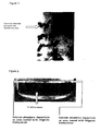
- Figure 1 shows a TEM micrograph of a sample from the phosphatidylcholine:phosphatidylserine:cholesterol incubation and clearly demonstrates the presence of crystalline structures in intimate contact with the multi-lamellar vesicles (large multi-layered liposomes).
- Implant materials can be metal, ceramic or polymeric implant coupons.
- the first coating methodology involves the dissolving of the excipients in HPLC-grade chloroform.
- the formulations were applied either as 5 ⁇ L aliquots onto defined areas of the implant surface or as a greater volume to cover the entire implant surface.
- the chloroform quickly evaporates at room temperature, leaving the excipients in the form of a thin film.
- the second coating methodology involves the production of a phospholipid suspension in an aqueous media.
- the phospholipid was weighed out and dissolved in 100 ⁇ L HPLC grade chloroform at a concentration of 222 ⁇ mol/mL.
- a thin film of phospholipid was produced on the wall of a glass round bottomed flask as described in Example 1.
- 1mL of deionised water was then added stepwise to the flask and the flask shaken, resulting in a phospholipid suspension being produced.
- 40 ⁇ L of the phospholipid suspension was then gently layered onto a defined area of the material surface.
- the suspension was then dried onto the surface of the implant material by means such as air drying, freeze drying or rotary evaporation. This results in a thin film of phospholipid being deposited onto the surface of the material.
- spot sizes were produced on the Ti-Pore300 implant coupons. In the case of 444 ⁇ mol/mL solution, the spot concentration was estimated at 5 ⁇ mol/cm 2 .
- Total Amount Excipients ⁇ m/mL
- Spot Size Produced for phosphatidylserine formulation mm
- Spot Size Produced for phosphatidylinositol formulation mm 444 7 3 222 9 6.5 111 10 6 56 11 7
- EXAMPLE 3 Induction of calcium phosphate precipitation on the surface of a metal surgical implant using a phospholipid formulation containing phosphatidylserine.
- FIG. 1 is of the surface of the Ti-Pore300 implant coupon whilst Figure 3 is a side on view of the 444 ⁇ m/mL coated spot.
- the implant coupons were rinsed 3 times in 10mL of distilled water and placed in a drying oven at 60°C for 18 hours.
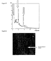
- FIG. 4 is an EDAX analysis undertaken on the uncoated Ti surface and demonstrates no calcium or phosphorus present. This indicates that the Ti surface itself does not act as a nucleating site for the precipitation of any significant amounts of calcium phosphate from the Simulated Body Fluid used.
- Figure 5 is an EDAX analysis of the area coated with excipients at 444 ⁇ mol/mL. Significant peaks for calcium and phosphorous were observed. The calcium peak can only be due to the presence of the deposited calcium whilst the phosphorus peak may be partially due to the presence of the phospholipids. EDAX spot analysis was also undertaken on the area coated with excipients at 222 ⁇ mol/mL and calcium and phosphorus peaks were again detected, demonstrating that a range of excipient concentrations are capable of inducing calcium precipitation onto the implant surface.
- FIG. 6 is a Scanning Electron Microscopy image of the surface previously coated with excipients at a concentration of 444mmol/mL whilst Figure 7 is of the uncoated Ti-Pore300 surface. The difference in morphology is particularly striking.
- EDAX analysis was undertaken ( Figure 8) and demonstrates that calcium and phosphorus peaks were still present. It should be noted that there is also a peak for carbon present in the EDAX analysis ( Figure 8) which suggests that some of the phospholipid may still be bound to the surface in association with the calcium deposit.
- EXAMPLE 4 Induction of calcium phosphate precipitation on the surface of a metal surgical implant using a phospholipid formulation containing phosphatidylinositol.
- Figure 9 is of the Ti-Pore300 implant coupon coated with the formulation containing phosphatidylinositol with the area corresponding to the 444 and 222 ⁇ m/mL spots to the forefront.
- the implant coupon was rinsed and dried as in Example 3.
- the surface of the implant coupon was then viewed by Scanning Electron Microscopy and subjected to EDAX analysis.
- Figure 10 is an EDAX analysis of the area coated with excipients at 444 ⁇ mol/mL and demonstrates that the phosphatidylinositol formulation is also capable of binding calcium to the surface of the implant coupon.
- EXAMPLE 5 Induction of calcium phosphate precipitation on the surface of a metal surgical implant using a phospholipid formulation of phosphatidylserine alone.
- Phosphatidylserine was weighed out and dissolved in HPLC grade chloroform to give a final concentration of 222 ⁇ mol/mL.
- the experimental protocol used was as described in Example 3.
- the implant coupons were rinsed and dried as in Example 3.
- the surface of the implant coupon was then viewed by Scanning Electron Microscopy and subjected to EDAX analysis.
- EDAX analysis of the area coated with excipients at 222 ⁇ mol/mL demonstrated that phosphatidylserine alone is also capable of binding calcium to the surface of the implant coupon.
- EXAMPLE 6 Induction of calcium phosphate precipitation on the surface of a surgical implant using a formulation containing phosphatidylcholine.
- FIG. 11 is an EDAX analysis of the area coated with excipients and demonstrates that the phosphatidylcholine formulation is also capable of binding calcium to the surface of the implant coupon after 7 days.
- EXAMPLE 7 Induction of calcium phosphate precipitation on the surface of a metal surgical implant using a different Simulated Body Fluid.
- Example 5 The experimental procedure described in Example 5 was repeated using a different Simulated Body Fluid 12 (152mM NaCl; 5mM KCl; 1mM K 2 HPO 4 ; 1.5mM MgCl 2 ; 27mM NaHCO 3 ; 0.5mM Na 2 SO 4 ; 2.6mM CaCl 2 and 50mM Tris buffer at pH 7.4).
- This Simulated Body Fluid contains ions that are known to compete with calcium and phosphate in the precipitation of calcium phosphate in vivo.
- EXAMPLE 8 Induction of calcium phosphate precipitation on different metal surgical implant surfaces using a phospholipid formulation containing phosphatidylserine.
- Ti-Pore600 implant coupons were coated with the phosphatidylserine formulation described in Example 3 at 444 ⁇ mol/mL and incubated in 10mL Simulated Body Fluid at 37°C. Photographs of the incubation chamber were taken at day 0,1,2,3 and 7 and Scanning Electron Microscopy/EDAX analysis undertaken after 9 days incubation, again without coating the implant coupon with palladium.
- FIG. 12 is an EDAX analysis of the area coated with excipients and demonstrates that the formulation is capable of binding calcium to the surface of different implant coupons.
- EXAMPLE 9 Induction of calcium phosphate precipitation on different metal implant surfaces using a formulation containing phosphatidylserine only.
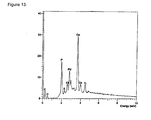
- Figure 13 is an EDAX analysis of the area coated with excipients and demonstrates that the formulation is capable of binding calcium to the surface of different implant coupons.
- EXAMPLE 10 Induction of calcium phosphate precipitation on ceramic surgical implant surfaces using a formulation containing phosphatidylserine.
- Ti-Pore300-HA implant coupons were coated with the phosphatidylserine formulation at 444 ⁇ mol/mL as described in Example 3 and incubated in 10mL Simulated Body Fluid at 37°C. Photographs were taken at day 0,1,2,3 and 7.
- Figure 14 is the surface of the Ti-Pore300-HA implant coupon after incubation for 2 hours in Simulated Body Fluid demonstrating that a similar matrix can be formed on a ceramic surface.
- EXAMPLE 11 Induction of calcium phosphate precipitation on a polymeric implant surface using a formulation containing phosphatidylserine only.
- Polymeric films of poly(methyl methacrylate) (PMMA) were cast using techniques well known to those skilled in the art. The smooth polymeric films were then coated with a phosphatidylserine suspension in an aqueous medium as described in Example 2 and incubations set up and analysed as described in Example 8.
- PMMA poly(methyl methacrylate)
- EXAMPLE 12 Induction of calcium phosphate precipitation on a metal surgical implant surface coated with a formulation containing phosphatidylserine, conditioned by incubation in serum.
- a Ti-Pore300 implant coupon was coated with the phosphatidylserine formulation as described in Example 3.
- the implant coupon was then incubated for 1 hour in commercially available human serum at 37°C, washed twice with 20mL and then incubated in Simulated Body Fluid as described in Example 4. Scanning Electron Microscopy/EDAX analysis was then undertaken after 9 days incubation, again without coating the implant coupon with palladium.
Landscapes
- Health & Medical Sciences (AREA)
- Medicinal Chemistry (AREA)
- Veterinary Medicine (AREA)
- Public Health (AREA)
- General Health & Medical Sciences (AREA)
- Animal Behavior & Ethology (AREA)
- Chemical & Material Sciences (AREA)
- Life Sciences & Earth Sciences (AREA)
- Dermatology (AREA)
- Epidemiology (AREA)
- Transplantation (AREA)
- Oral & Maxillofacial Surgery (AREA)
- Physical Education & Sports Medicine (AREA)
- Organic Chemistry (AREA)
- Nuclear Medicine, Radiotherapy & Molecular Imaging (AREA)
- Engineering & Computer Science (AREA)
- Rheumatology (AREA)
- Pharmacology & Pharmacy (AREA)
- Orthopedic Medicine & Surgery (AREA)
- General Chemical & Material Sciences (AREA)
- Chemical Kinetics & Catalysis (AREA)
- Bioinformatics & Cheminformatics (AREA)
- Materials For Medical Uses (AREA)
- Medicinal Preparation (AREA)
- Pharmaceuticals Containing Other Organic And Inorganic Compounds (AREA)
- Medicines That Contain Protein Lipid Enzymes And Other Medicines (AREA)
- Prostheses (AREA)
Abstract
Description
There is considerable scope for improvement of orthopaedic implants, particularly in development of uncemented devices, which aim to improve osteointegration between implant and bone.
The invention also describes a method of surgery that comprises introducing a prosthesis or implant into a patient, the improvement which consists in coating a surface of the prosthesis or implant with a phospholipid at a concentration to improve osteointegration. Typically such methods comprise the step of treating the phospholipid coated prosthesis or implant with a simulated body fluid prior to use. In this specification the term "simulated body fluid" is taken to mean a fluid which has free calcium and phosphate ions which are generally provided in the form of calcium and phosphate salts.
and
| Total Amount Excipients (µm/mL) | Spot Size Produced for phosphatidylserine formulation (mm) | Spot Size Produced for phosphatidylinositol formulation (mm) |
| 444 | 7 | 3 |
| 222 | 9 | 6.5 |
| 111 | 10 | 6 |
| 56 | 11 | 7 |
Claims (22)
- The use of a phospholipid or a mixture of phospholipids for coating a surface of a prosthesis or an implant, to improve osteointegration of the prosthesis or implant by inducing or enhancing calcium phosphate deposition on to the surface.
- The use according to claim 1, wherein the phospholipid or one of the phospholipids is a negatively charged phospholipid.
- The use according to claim 2, wherein the negatively charged phospholipid is phosphatidylserine or phosphatidylinositol or a mixture thereof.
- The use according to any one of claims 1 to 3, wherein the phospholipid concentration in the coating is 0.1 - 100µmol/cm2.
- The use according to claim 4, wherein the phospholipid concentration in the coating is 1.0 - 10µmol/cm2.
- The use according to any one of claims 1 to 5, wherein the surface of the prosthesis or implant is smooth, rough or porous.
- The use according to any one of claims 1 to 6, wherein the surface of the prosthesis or implant is metal, ceramic or polymeric.
- The use according to any one of claims 1 to 7, in which the phospholipid coating primarily coats only the part of the surface of the prosthesis or implant that is likely to be subject to mechanical stress in vivo.
- The use according to any one of claims 1 to 8, wherein the phospholipid coating is substantially free of effective amounts of phosphatidylcholine.
- The use according to any one of claims 1 to 9, wherein the surface of the prosthesis or implant has a coating of calcium phosphate.
- The use according to any one of claims 1 to 10, wherein the phospholipid coating additionally comprises a biologically active material, such as an antibiotic or an antithrombotic pharmaceutical.
- The use according to any one of claims 1 to 11, wherein the phospholipid coating is produced by lyophilizing or otherwise drying a suspension or solution of phospholipid onto the surface of the prosthesis or implant.
- A prosthesis or Implant kit comprising:(1) a prosthesis or implant; and(2) an osteointegration enhancement composition comprising at least one phospholipid at a concentration to improve osteointegration by inducing or enhancing calcium phosphate deposition when coated on a surface of the prosthesis or implant.
- A kit as claimed in claim 13 further comprising a simulated body fluid or means for making a simulated body fluid.
- A prosthesis or implant kit comprising:a prosthesis or implant having a surface carrying an osteointegration enhancement coating comprising at least one phospholipid at a concentration to improve osteointegration by inducing or enhancing calcium phosphate deposition on to the surface; anda simulated body fluid or means for making a simulated body fluid.
- The use of a phospholipid or mixture of phospholipids in the manufacture of a material for enhancing osteointegration of a prosthesis or implant into an animal body, including human, by inducing or enhancing calcium phosphate deposition on to a surface of the material.
- The use according to claim 16, wherein the phospholipid or one of the phospholipids is a negatively charged phospholipid.
- The use according to claim 17, wherein the negatively charged phospholipid is phosphatidylserine or phosphatidylinositol or a mixture thereof.
- The use according to any one of claims 16 to 18, wherein the phospholipid or mixture of phospholipids is applied to a surface of the prosthesis or implant to provide a coating thereon having a phospholipid concentration in the range of from 0.1 to 100µmol/cm2.
- The use according to claim 19, wherein the phospholipid concentration in the costing is 1.0 - 10µmol/cm2.
- The use according to any one of claims 16 to 20, wherein the phospholipid or mixture of phospholipids is substantially free of effective amounts of phosphatidylcholine.
- The use according to any one of claims 16 to 21 wherein the phospholipid or mixture of phospholipids is present in a composition additionally comprising a biologically active material, such as an antibiotic or an antithrombotic pharmaceutical.
Applications Claiming Priority (3)
| Application Number | Priority Date | Filing Date | Title |
|---|---|---|---|
| GBGB9920547.8A GB9920547D0 (en) | 1999-08-31 | 1999-08-31 | Coated implant |
| GB9920547 | 1999-08-31 | ||
| PCT/GB2000/003290 WO2001015752A1 (en) | 1999-08-31 | 2000-08-29 | Phospholipid-coated implants |
Publications (2)
| Publication Number | Publication Date |
|---|---|
| EP1212106A1 EP1212106A1 (en) | 2002-06-12 |
| EP1212106B1 true EP1212106B1 (en) | 2005-10-12 |
Family
ID=10860080
Family Applications (1)
| Application Number | Title | Priority Date | Filing Date |
|---|---|---|---|
| EP00954791A Expired - Lifetime EP1212106B1 (en) | 1999-08-31 | 2000-08-29 | Phospholipid-coated implants |
Country Status (9)
| Country | Link |
|---|---|
| US (1) | US6863693B2 (en) |
| EP (1) | EP1212106B1 (en) |
| JP (1) | JP2003508129A (en) |
| AT (1) | ATE306289T1 (en) |
| AU (1) | AU6714600A (en) |
| CA (1) | CA2419618A1 (en) |
| DE (1) | DE60023157D1 (en) |
| GB (1) | GB9920547D0 (en) |
| WO (1) | WO2001015752A1 (en) |
Families Citing this family (16)
| Publication number | Priority date | Publication date | Assignee | Title |
|---|---|---|---|---|
| DE10113108B4 (en) * | 2001-03-15 | 2007-07-26 | Dot Gmbh | Active substance-containing calcium phosphate materials |
| WO2003030957A1 (en) * | 2001-10-11 | 2003-04-17 | Straumann Holding Ag | Osteophilic implants |
| AU2002359715A1 (en) * | 2001-12-19 | 2003-07-09 | The University Of Texas Health Science Center At San Antonio | Implant coatings |
| US8318235B2 (en) | 2003-01-22 | 2012-11-27 | Cordis Corporation | Method for applying drug coating to a medical device in surgeon room |
| WO2005027991A1 (en) * | 2003-09-19 | 2005-03-31 | Cambridge University Technical Services Ltd | Orthopaedic devices |
| DE102006029247A1 (en) * | 2006-06-26 | 2007-12-27 | Biotronik Vi Patent Ag | Implant with a coating comprises one or more components such as cholesterol or cholesterol ester |
| GB0624423D0 (en) * | 2006-12-06 | 2007-01-17 | Univ Brighton | Biomaterials with Functionalised Surfaces |
| US20080181928A1 (en) * | 2006-12-22 | 2008-07-31 | Miv Therapeutics, Inc. | Coatings for implantable medical devices for liposome delivery |
| EP2187838A4 (en) * | 2007-08-24 | 2012-03-07 | Univ Brown | METHOD FOR MANUFACTURING NANOSTRUCTURES ON A SURFACE OF A MEDICAL IMPLANT |
| CA2711847C (en) * | 2008-01-09 | 2018-05-08 | Innovative Health Technologies, Llc | Implant pellets and methods for performing bone augmentation and preservation |
| US20130288951A1 (en) * | 2012-04-27 | 2013-10-31 | Biomet Manufacturing Corp. | Compositions and methods for coating implant surfaces to inhibit surgical infections |
| AU2013251346B2 (en) * | 2012-04-27 | 2016-12-22 | Biomet Manufacturing, Llc | Compositions and methods for coating implant surfaces to inhibit surgical infections |
| US9775853B2 (en) | 2013-03-15 | 2017-10-03 | Biomet Manufacturing, Llc. | Hemostatic compositions and methods |
| WO2018064556A1 (en) * | 2016-09-29 | 2018-04-05 | Gesea Biosciences Inc. | Bioerodible implant for long-term drug delivery and associated methods of manufacture and use |
| EP3530266A1 (en) * | 2018-02-27 | 2019-08-28 | LipoCoat B.V. | A lipid-based coating composition, and an object having a lipid-based coating |
| JP7618206B2 (en) | 2020-12-15 | 2025-01-21 | 国立研究開発法人産業技術総合研究所 | Implant material and method for manufacturing implant material |
Family Cites Families (18)
| Publication number | Priority date | Publication date | Assignee | Title |
|---|---|---|---|---|
| US4749585A (en) * | 1986-04-11 | 1988-06-07 | University Of Medicine And Dentistry Of New Jersey | Antibiotic bonded prosthesis and process for producing same |
| US5755788A (en) * | 1987-02-19 | 1998-05-26 | Rutgers, The State University | Prosthesis and implants having liposomes bound thereto and methods of preparation |
| WO1991000745A1 (en) * | 1989-07-07 | 1991-01-24 | Macnaught Pty Limited | A method for producing a biocompatible surface |
| JP2767152B2 (en) | 1990-04-10 | 1998-06-18 | 日本特殊陶業株式会社 | Sustained drug-containing ceramic body |
| EP0479582A1 (en) | 1990-10-03 | 1992-04-08 | Alexander Karl Friedrich Wilhelm Overweg | Restorative periodontal technique |
| US5342621A (en) * | 1992-09-15 | 1994-08-30 | Advanced Cardiovascular Systems, Inc. | Antithrombogenic surface |
| US6099562A (en) * | 1996-06-13 | 2000-08-08 | Schneider (Usa) Inc. | Drug coating with topcoat |
| US5607475A (en) * | 1995-08-22 | 1997-03-04 | Medtronic, Inc. | Biocompatible medical article and method |
| US5722984A (en) * | 1996-01-16 | 1998-03-03 | Iso Stent, Inc. | Antithrombogenic radioactive coating for an intravascular stent |
| CA2199890C (en) * | 1996-03-26 | 2002-02-05 | Corvita Corporation | Stents and stent-grafts having enhanced hoop strength and methods of making the same |
| EP0806212B1 (en) | 1996-05-10 | 2003-04-02 | IsoTis N.V. | Device for incorporation and release of biologically active agents |
| US6290718B1 (en) * | 1998-02-02 | 2001-09-18 | Regeneration Technologies, Inc. | Luminal graft, stent or conduit made of cortical bone |
| US6254634B1 (en) * | 1998-06-10 | 2001-07-03 | Surmodics, Inc. | Coating compositions |
| US6187024B1 (en) * | 1998-11-10 | 2001-02-13 | Target Therapeutics, Inc. | Bioactive coating for vaso-occlusive devices |
| US6428576B1 (en) * | 1999-04-16 | 2002-08-06 | Endospine, Ltd. | System for repairing inter-vertebral discs |
| US6663607B2 (en) * | 1999-07-12 | 2003-12-16 | Scimed Life Systems, Inc. | Bioactive aneurysm closure device assembly and kit |
| US6416549B1 (en) * | 1999-07-19 | 2002-07-09 | Sulzer Carbomedics Inc. | Antithrombogenic annuloplasty ring having a biodegradable insert |
| US6273901B1 (en) * | 1999-08-10 | 2001-08-14 | Scimed Life Systems, Inc. | Thrombosis filter having a surface treatment |
-
1999
- 1999-08-31 GB GBGB9920547.8A patent/GB9920547D0/en not_active Ceased
-
2000
- 2000-08-29 WO PCT/GB2000/003290 patent/WO2001015752A1/en not_active Ceased
- 2000-08-29 EP EP00954791A patent/EP1212106B1/en not_active Expired - Lifetime
- 2000-08-29 JP JP2001520163A patent/JP2003508129A/en active Pending
- 2000-08-29 AT AT00954791T patent/ATE306289T1/en not_active IP Right Cessation
- 2000-08-29 AU AU67146/00A patent/AU6714600A/en not_active Abandoned
- 2000-08-29 DE DE60023157T patent/DE60023157D1/en not_active Expired - Lifetime
- 2000-08-29 CA CA002419618A patent/CA2419618A1/en not_active Abandoned
-
2002
- 2002-02-28 US US10/084,677 patent/US6863693B2/en not_active Expired - Fee Related
Also Published As
| Publication number | Publication date |
|---|---|
| US20020165617A1 (en) | 2002-11-07 |
| EP1212106A1 (en) | 2002-06-12 |
| ATE306289T1 (en) | 2005-10-15 |
| AU6714600A (en) | 2001-03-26 |
| WO2001015752A1 (en) | 2001-03-08 |
| GB9920547D0 (en) | 1999-11-03 |
| US6863693B2 (en) | 2005-03-08 |
| CA2419618A1 (en) | 2001-03-08 |
| JP2003508129A (en) | 2003-03-04 |
| DE60023157D1 (en) | 2005-11-17 |
Similar Documents
| Publication | Publication Date | Title |
|---|---|---|
| US6863693B2 (en) | Phospholipid-coated implants | |
| Barrère et al. | Osteogenecity of octacalcium phosphate coatings applied on porous metal implants | |
| de Jonge et al. | Organic–inorganic surface modifications for titanium implant surfaces | |
| Duan et al. | Surface modifications of bone implants through wet chemistry | |
| Yaylaoǧlu et al. | Development of a calcium phosphate–gelatin composite as a bone substitute and its use in drug release | |
| Lickorish et al. | A three-phase, fully resorbable, polyester/calcium phosphate scaffold for bone tissue engineering: Evolution of scaffold design | |
| EP1385449B1 (en) | Biologically-functionalised, metabolically-inductive implant surfaces | |
| EP1492579B1 (en) | Medical prosthetic devices having improved biocompatibility | |
| Lee et al. | Effects of porous beta-tricalcium phosphate-based ceramics used as an E. coli-derived rhBMP-2 carrier for bone regeneration | |
| Belcarz et al. | Covalent coating of hydroxyapatite by keratin stabilizes gentamicin release | |
| Desante et al. | Surface multifunctionalization of inert ceramic implants by calcium phosphate biomimetic coating doped with nanoparticles encapsulating antibiotics | |
| Ter Brugge et al. | Effect of calcium phosphate coating composition and crystallinity on the response of osteogenic cells in vitro | |
| McLeod et al. | Adsorption of bisphosphonate onto hydroxyapatite using a novel co‐precipitation technique for bone growth enhancement | |
| SE1251042A1 (en) | Method of producing an alginate coated titanium dioxide caffold | |
| de Faria et al. | Different compact hybrid Langmuir–Blodgett‐film coatings modify biomineralization and the ability of osteoblasts to grow | |
| Navarro et al. | Biomimetic mineralization of ceramics and glasses | |
| US20040131754A1 (en) | Antibiotic calcium phosphate coating | |
| Daculsi et al. | Outcome and perspectives in bioactive coatings: What's new, what's coming | |
| Katsanevakis et al. | Biomineralization of polymer scaffolds | |
| WO2022242414A1 (en) | Anti-cancer bionic bone material, and pharmaceutical composition comprising bionic bone material and preparation method for pharmaceutical composition | |
| CN115137875A (en) | High-efficiency two-phase calcium phosphate coating method | |
| KR101070345B1 (en) | Organic/Inorganic hybrid coating layer containg body vitality factor, Biomedical implants and Method thereof | |
| Layrolle | 1.16 Calcium phosphate coatings | |
| Mur | Accelerating mineralization of biomimetic surfaces | |
| CN115382010B (en) | Bionic bone material and preparation method thereof |
Legal Events
| Date | Code | Title | Description |
|---|---|---|---|
| PUAI | Public reference made under article 153(3) epc to a published international application that has entered the european phase |
Free format text: ORIGINAL CODE: 0009012 |
|
| 17P | Request for examination filed |
Effective date: 20020328 |
|
| AK | Designated contracting states |
Kind code of ref document: A1 Designated state(s): AT BE CH CY DE DK ES FI FR GB GR IE IT LI LU MC NL PT SE |
|
| AX | Request for extension of the european patent |
Free format text: AL;LT;LV;MK;RO;SI |
|
| 17Q | First examination report despatched |
Effective date: 20030923 |
|
| GRAP | Despatch of communication of intention to grant a patent |
Free format text: ORIGINAL CODE: EPIDOSNIGR1 |
|
| GRAS | Grant fee paid |
Free format text: ORIGINAL CODE: EPIDOSNIGR3 |
|
| GRAA | (expected) grant |
Free format text: ORIGINAL CODE: 0009210 |
|
| RAP3 | Party data changed (applicant data changed or rights of an application transferred) |
Owner name: UNIVERSITY OF BRIGHTON Owner name: DESTINY PHARMA LIMITED |
|
| AK | Designated contracting states |
Kind code of ref document: B1 Designated state(s): AT BE CH CY DE DK ES FI FR GB GR IE IT LI LU MC NL PT SE |
|
| PG25 | Lapsed in a contracting state [announced via postgrant information from national office to epo] |
Ref country code: IT Free format text: LAPSE BECAUSE OF FAILURE TO SUBMIT A TRANSLATION OF THE DESCRIPTION OR TO PAY THE FEE WITHIN THE PRE;WARNING: LAPSES OF ITALIAN PATENTS WITH EFFECTIVE DATE BEFORE 2007 MAY HAVE OCCURRED AT ANY TIME BEFORE 2007. THE CORRECT EFFECTIVE DATE MAY BE DIFFERENT FROM THE ONE RECORDED.SCRIBED TIME-LIMIT Effective date: 20051012 Ref country code: CH Free format text: LAPSE BECAUSE OF FAILURE TO SUBMIT A TRANSLATION OF THE DESCRIPTION OR TO PAY THE FEE WITHIN THE PRESCRIBED TIME-LIMIT Effective date: 20051012 Ref country code: LI Free format text: LAPSE BECAUSE OF FAILURE TO SUBMIT A TRANSLATION OF THE DESCRIPTION OR TO PAY THE FEE WITHIN THE PRESCRIBED TIME-LIMIT Effective date: 20051012 Ref country code: FI Free format text: LAPSE BECAUSE OF FAILURE TO SUBMIT A TRANSLATION OF THE DESCRIPTION OR TO PAY THE FEE WITHIN THE PRESCRIBED TIME-LIMIT Effective date: 20051012 Ref country code: NL Free format text: LAPSE BECAUSE OF FAILURE TO SUBMIT A TRANSLATION OF THE DESCRIPTION OR TO PAY THE FEE WITHIN THE PRESCRIBED TIME-LIMIT Effective date: 20051012 Ref country code: AT Free format text: LAPSE BECAUSE OF FAILURE TO SUBMIT A TRANSLATION OF THE DESCRIPTION OR TO PAY THE FEE WITHIN THE PRESCRIBED TIME-LIMIT Effective date: 20051012 Ref country code: BE Free format text: LAPSE BECAUSE OF FAILURE TO SUBMIT A TRANSLATION OF THE DESCRIPTION OR TO PAY THE FEE WITHIN THE PRESCRIBED TIME-LIMIT Effective date: 20051012 |
|
| REG | Reference to a national code |
Ref country code: GB Ref legal event code: FG4D |
|
| REG | Reference to a national code |
Ref country code: CH Ref legal event code: EP |
|
| REG | Reference to a national code |
Ref country code: IE Ref legal event code: FG4D |
|
| REF | Corresponds to: |
Ref document number: 60023157 Country of ref document: DE Date of ref document: 20051117 Kind code of ref document: P |
|
| PG25 | Lapsed in a contracting state [announced via postgrant information from national office to epo] |
Ref country code: SE Free format text: LAPSE BECAUSE OF FAILURE TO SUBMIT A TRANSLATION OF THE DESCRIPTION OR TO PAY THE FEE WITHIN THE PRESCRIBED TIME-LIMIT Effective date: 20060112 Ref country code: GR Free format text: LAPSE BECAUSE OF FAILURE TO SUBMIT A TRANSLATION OF THE DESCRIPTION OR TO PAY THE FEE WITHIN THE PRESCRIBED TIME-LIMIT Effective date: 20060112 Ref country code: DK Free format text: LAPSE BECAUSE OF FAILURE TO SUBMIT A TRANSLATION OF THE DESCRIPTION OR TO PAY THE FEE WITHIN THE PRESCRIBED TIME-LIMIT Effective date: 20060112 |
|
| PG25 | Lapsed in a contracting state [announced via postgrant information from national office to epo] |
Ref country code: DE Free format text: LAPSE BECAUSE OF FAILURE TO SUBMIT A TRANSLATION OF THE DESCRIPTION OR TO PAY THE FEE WITHIN THE PRESCRIBED TIME-LIMIT Effective date: 20060113 |
|
| PG25 | Lapsed in a contracting state [announced via postgrant information from national office to epo] |
Ref country code: ES Free format text: LAPSE BECAUSE OF FAILURE TO SUBMIT A TRANSLATION OF THE DESCRIPTION OR TO PAY THE FEE WITHIN THE PRESCRIBED TIME-LIMIT Effective date: 20060123 |
|
| PG25 | Lapsed in a contracting state [announced via postgrant information from national office to epo] |
Ref country code: PT Free format text: LAPSE BECAUSE OF FAILURE TO SUBMIT A TRANSLATION OF THE DESCRIPTION OR TO PAY THE FEE WITHIN THE PRESCRIBED TIME-LIMIT Effective date: 20060313 |
|
| NLV1 | Nl: lapsed or annulled due to failure to fulfill the requirements of art. 29p and 29m of the patents act | ||
| REG | Reference to a national code |
Ref country code: CH Ref legal event code: PL |
|
| PLBE | No opposition filed within time limit |
Free format text: ORIGINAL CODE: 0009261 |
|
| STAA | Information on the status of an ep patent application or granted ep patent |
Free format text: STATUS: NO OPPOSITION FILED WITHIN TIME LIMIT |
|
| PG25 | Lapsed in a contracting state [announced via postgrant information from national office to epo] |
Ref country code: IE Free format text: LAPSE BECAUSE OF NON-PAYMENT OF DUE FEES Effective date: 20060829 |
|
| PG25 | Lapsed in a contracting state [announced via postgrant information from national office to epo] |
Ref country code: MC Free format text: LAPSE BECAUSE OF NON-PAYMENT OF DUE FEES Effective date: 20060831 |
|
| 26N | No opposition filed |
Effective date: 20060713 |
|
| PG25 | Lapsed in a contracting state [announced via postgrant information from national office to epo] |
Ref country code: FR Free format text: LAPSE BECAUSE OF FAILURE TO SUBMIT A TRANSLATION OF THE DESCRIPTION OR TO PAY THE FEE WITHIN THE PRESCRIBED TIME-LIMIT Effective date: 20061020 |
|
| EN | Fr: translation not filed | ||
| REG | Reference to a national code |
Ref country code: IE Ref legal event code: MM4A |
|
| PGFP | Annual fee paid to national office [announced via postgrant information from national office to epo] |
Ref country code: GB Payment date: 20070829 Year of fee payment: 8 |
|
| PG25 | Lapsed in a contracting state [announced via postgrant information from national office to epo] |
Ref country code: LU Free format text: LAPSE BECAUSE OF NON-PAYMENT OF DUE FEES Effective date: 20060829 |
|
| PG25 | Lapsed in a contracting state [announced via postgrant information from national office to epo] |
Ref country code: FR Free format text: LAPSE BECAUSE OF FAILURE TO SUBMIT A TRANSLATION OF THE DESCRIPTION OR TO PAY THE FEE WITHIN THE PRESCRIBED TIME-LIMIT Effective date: 20051012 Ref country code: CY Free format text: LAPSE BECAUSE OF FAILURE TO SUBMIT A TRANSLATION OF THE DESCRIPTION OR TO PAY THE FEE WITHIN THE PRESCRIBED TIME-LIMIT Effective date: 20051012 |
|
| GBPC | Gb: european patent ceased through non-payment of renewal fee |
Effective date: 20080829 |
|
| PG25 | Lapsed in a contracting state [announced via postgrant information from national office to epo] |
Ref country code: GB Free format text: LAPSE BECAUSE OF NON-PAYMENT OF DUE FEES Effective date: 20080829 |