EP1202728B1 - Peroxovanadium compounds as protein tyrosine phosphatase (ptp) inhibitors: their inhibitory effects on angiogenesis, restenosis and the production of endothelins. - Google Patents
Peroxovanadium compounds as protein tyrosine phosphatase (ptp) inhibitors: their inhibitory effects on angiogenesis, restenosis and the production of endothelins. Download PDFInfo
- Publication number
- EP1202728B1 EP1202728B1 EP00952813A EP00952813A EP1202728B1 EP 1202728 B1 EP1202728 B1 EP 1202728B1 EP 00952813 A EP00952813 A EP 00952813A EP 00952813 A EP00952813 A EP 00952813A EP 1202728 B1 EP1202728 B1 EP 1202728B1
- Authority
- EP
- European Patent Office
- Prior art keywords
- angiogenesis
- use according
- compounds
- endothelins
- bpv
- Prior art date
- Legal status (The legal status is an assumption and is not a legal conclusion. Google has not performed a legal analysis and makes no representation as to the accuracy of the status listed.)
- Expired - Lifetime
Links
- 150000001875 compounds Chemical class 0.000 title claims abstract description 57
- 102000002045 Endothelin Human genes 0.000 title claims abstract description 38
- 108050009340 Endothelin Proteins 0.000 title claims abstract description 38
- 208000037803 restenosis Diseases 0.000 title claims abstract description 18
- 230000033115 angiogenesis Effects 0.000 title claims abstract description 17
- 238000004519 manufacturing process Methods 0.000 title abstract description 9
- 102000002727 Protein Tyrosine Phosphatase Human genes 0.000 title abstract description 6
- 108020000494 protein-tyrosine phosphatase Proteins 0.000 title abstract description 6
- 230000002401 inhibitory effect Effects 0.000 title abstract description 5
- 239000003112 inhibitor Substances 0.000 title description 11
- NOESYZHRGYRDHS-UHFFFAOYSA-N insulin Chemical compound N1C(=O)C(NC(=O)C(CCC(N)=O)NC(=O)C(CCC(O)=O)NC(=O)C(C(C)C)NC(=O)C(NC(=O)CN)C(C)CC)CSSCC(C(NC(CO)C(=O)NC(CC(C)C)C(=O)NC(CC=2C=CC(O)=CC=2)C(=O)NC(CCC(N)=O)C(=O)NC(CC(C)C)C(=O)NC(CCC(O)=O)C(=O)NC(CC(N)=O)C(=O)NC(CC=2C=CC(O)=CC=2)C(=O)NC(CSSCC(NC(=O)C(C(C)C)NC(=O)C(CC(C)C)NC(=O)C(CC=2C=CC(O)=CC=2)NC(=O)C(CC(C)C)NC(=O)C(C)NC(=O)C(CCC(O)=O)NC(=O)C(C(C)C)NC(=O)C(CC(C)C)NC(=O)C(CC=2NC=NC=2)NC(=O)C(CO)NC(=O)CNC2=O)C(=O)NCC(=O)NC(CCC(O)=O)C(=O)NC(CCCNC(N)=N)C(=O)NCC(=O)NC(CC=3C=CC=CC=3)C(=O)NC(CC=3C=CC=CC=3)C(=O)NC(CC=3C=CC(O)=CC=3)C(=O)NC(C(C)O)C(=O)N3C(CCC3)C(=O)NC(CCCCN)C(=O)NC(C)C(O)=O)C(=O)NC(CC(N)=O)C(O)=O)=O)NC(=O)C(C(C)CC)NC(=O)C(CO)NC(=O)C(C(C)O)NC(=O)C1CSSCC2NC(=O)C(CC(C)C)NC(=O)C(NC(=O)C(CCC(N)=O)NC(=O)C(CC(N)=O)NC(=O)C(NC(=O)C(N)CC=1C=CC=CC=1)C(C)C)CC1=CN=CN1 NOESYZHRGYRDHS-UHFFFAOYSA-N 0.000 claims abstract description 24
- 102000004877 Insulin Human genes 0.000 claims abstract description 12
- 108090001061 Insulin Proteins 0.000 claims abstract description 12
- 229940125396 insulin Drugs 0.000 claims abstract description 12
- QVGXLLKOCUKJST-UHFFFAOYSA-N atomic oxygen Chemical group [O] QVGXLLKOCUKJST-UHFFFAOYSA-N 0.000 claims description 16
- 239000001301 oxygen Substances 0.000 claims description 16
- 229910052760 oxygen Inorganic materials 0.000 claims description 16
- 150000002978 peroxides Chemical class 0.000 claims description 13
- 206010029113 Neovascularisation Diseases 0.000 claims description 9
- 238000002399 angioplasty Methods 0.000 claims description 9
- 239000003814 drug Substances 0.000 claims description 9
- 241001465754 Metazoa Species 0.000 claims description 8
- 206010028980 Neoplasm Diseases 0.000 claims description 8
- 230000005764 inhibitory process Effects 0.000 claims description 7
- 208000002249 Diabetes Complications Diseases 0.000 claims description 5
- 206010012655 Diabetic complications Diseases 0.000 claims description 5
- 125000002887 hydroxy group Chemical group [H]O* 0.000 claims description 5
- 208000017169 kidney disease Diseases 0.000 claims description 4
- 208000010412 Glaucoma Diseases 0.000 claims description 3
- 206010038933 Retinopathy of prematurity Diseases 0.000 claims description 3
- 208000002780 macular degeneration Diseases 0.000 claims description 3
- 201000003142 neovascular glaucoma Diseases 0.000 claims description 3
- 230000010046 negative regulation of endothelial cell proliferation Effects 0.000 claims description 2
- XAEFZNCEHLXOMS-UHFFFAOYSA-M potassium benzoate Chemical compound [K+].[O-]C(=O)C1=CC=CC=C1 XAEFZNCEHLXOMS-UHFFFAOYSA-M 0.000 claims 3
- 230000003389 potentiating effect Effects 0.000 abstract description 21
- 210000004027 cell Anatomy 0.000 abstract description 20
- 230000001772 anti-angiogenic effect Effects 0.000 abstract description 19
- 241000700159 Rattus Species 0.000 abstract description 12
- 230000035755 proliferation Effects 0.000 abstract description 12
- 108010082117 matrigel Proteins 0.000 abstract description 11
- 230000002792 vascular Effects 0.000 abstract description 11
- 238000001727 in vivo Methods 0.000 abstract description 9
- 230000005012 migration Effects 0.000 abstract description 9
- 238000013508 migration Methods 0.000 abstract description 9
- 238000003556 assay Methods 0.000 abstract description 7
- 230000004069 differentiation Effects 0.000 abstract description 4
- 238000000338 in vitro Methods 0.000 abstract description 4
- 210000003556 vascular endothelial cell Anatomy 0.000 abstract description 4
- 230000007423 decrease Effects 0.000 abstract description 3
- 239000002955 immunomodulating agent Substances 0.000 abstract description 3
- 229940121354 immunomodulator Drugs 0.000 abstract description 3
- 230000006698 induction Effects 0.000 abstract description 3
- 102000004196 processed proteins & peptides Human genes 0.000 abstract description 3
- 108090000765 processed proteins & peptides Proteins 0.000 abstract description 3
- 239000012528 membrane Substances 0.000 abstract description 2
- 241000287828 Gallus gallus Species 0.000 abstract 1
- 231100001231 less toxic Toxicity 0.000 abstract 1
- 230000002265 prevention Effects 0.000 abstract 1
- 230000028327 secretion Effects 0.000 abstract 1
- DGEZNRSVGBDHLK-UHFFFAOYSA-N [1,10]phenanthroline Chemical compound C1=CN=C2C3=NC=CC=C3C=CC2=C1 DGEZNRSVGBDHLK-UHFFFAOYSA-N 0.000 description 39
- YNPNZTXNASCQKK-UHFFFAOYSA-N Phenanthrene Natural products C1=CC=C2C3=CC=CC=C3C=CC2=C1 YNPNZTXNASCQKK-UHFFFAOYSA-N 0.000 description 35
- LSGOVYNHVSXFFJ-UHFFFAOYSA-N vanadate(3-) Chemical compound [O-][V]([O-])([O-])=O LSGOVYNHVSXFFJ-UHFFFAOYSA-N 0.000 description 30
- 210000004204 blood vessel Anatomy 0.000 description 26
- 210000002889 endothelial cell Anatomy 0.000 description 23
- 230000000694 effects Effects 0.000 description 18
- 230000002491 angiogenic effect Effects 0.000 description 13
- 241000699670 Mus sp. Species 0.000 description 12
- 230000012010 growth Effects 0.000 description 12
- ROFVEXUMMXZLPA-UHFFFAOYSA-N Bipyridyl Chemical group N1=CC=CC=C1C1=CC=CC=N1 ROFVEXUMMXZLPA-UHFFFAOYSA-N 0.000 description 10
- 230000015572 biosynthetic process Effects 0.000 description 10
- 238000000034 method Methods 0.000 description 10
- 210000001534 vitelline membrane Anatomy 0.000 description 10
- 230000000259 anti-tumor effect Effects 0.000 description 8
- 150000003682 vanadium compounds Chemical class 0.000 description 8
- OUYCCCASQSFEME-QMMMGPOBSA-N L-tyrosine Chemical compound OC(=O)[C@@H](N)CC1=CC=C(O)C=C1 OUYCCCASQSFEME-QMMMGPOBSA-N 0.000 description 7
- 230000017531 blood circulation Effects 0.000 description 7
- 239000003446 ligand Substances 0.000 description 7
- 230000008569 process Effects 0.000 description 7
- 235000018102 proteins Nutrition 0.000 description 7
- 102000004169 proteins and genes Human genes 0.000 description 7
- 108090000623 proteins and genes Proteins 0.000 description 7
- 238000011282 treatment Methods 0.000 description 7
- OUYCCCASQSFEME-UHFFFAOYSA-N tyrosine Natural products OC(=O)C(N)CC1=CC=C(O)C=C1 OUYCCCASQSFEME-UHFFFAOYSA-N 0.000 description 7
- 102000009123 Fibrin Human genes 0.000 description 6
- 108010073385 Fibrin Proteins 0.000 description 6
- BWGVNKXGVNDBDI-UHFFFAOYSA-N Fibrin monomer Chemical compound CNC(=O)CNC(=O)CN BWGVNKXGVNDBDI-UHFFFAOYSA-N 0.000 description 6
- 239000003795 chemical substances by application Substances 0.000 description 6
- 229950003499 fibrin Drugs 0.000 description 6
- 230000026731 phosphorylation Effects 0.000 description 6
- 238000006366 phosphorylation reaction Methods 0.000 description 6
- SIOXPEMLGUPBBT-UHFFFAOYSA-N picolinic acid Chemical compound OC(=O)C1=CC=CC=N1 SIOXPEMLGUPBBT-UHFFFAOYSA-N 0.000 description 6
- 210000001715 carotid artery Anatomy 0.000 description 5
- 230000004663 cell proliferation Effects 0.000 description 5
- 230000001413 cellular effect Effects 0.000 description 5
- 208000037265 diseases, disorders, signs and symptoms Diseases 0.000 description 5
- 210000002257 embryonic structure Anatomy 0.000 description 5
- ZUBDGKVDJUIMQQ-UBFCDGJISA-N endothelin-1 Chemical compound C([C@@H](C(=O)N[C@@H](CC(C)C)C(=O)N[C@@H](CC(O)=O)C(=O)N[C@@H]([C@@H](C)CC)C(=O)N[C@@H]([C@@H](C)CC)C(=O)N[C@@H](CC=1C2=CC=CC=C2NC=1)C(O)=O)NC(=O)[C@H]1NC(=O)[C@H](CC=2C=CC=CC=2)NC(=O)[C@@H](CC=2C=CC(O)=CC=2)NC(=O)[C@H](C(C)C)NC(=O)[C@H]2CSSC[C@@H](C(N[C@H](CO)C(=O)N[C@@H](CO)C(=O)N[C@H](CC(C)C)C(=O)N[C@@H](CCSC)C(=O)N[C@H](CC(O)=O)C(=O)N[C@@H](CCCCN)C(=O)N[C@@H](CCC(O)=O)C(=O)N2)=O)NC(=O)[C@@H](CO)NC(=O)[C@H](N)CSSC1)C1=CNC=N1 ZUBDGKVDJUIMQQ-UBFCDGJISA-N 0.000 description 5
- 239000001963 growth medium Substances 0.000 description 5
- 238000002513 implantation Methods 0.000 description 5
- 239000011159 matrix material Substances 0.000 description 5
- -1 phosphotyrosyl Chemical group 0.000 description 5
- 239000000243 solution Substances 0.000 description 5
- 210000001519 tissue Anatomy 0.000 description 5
- 229910052723 transition metal Inorganic materials 0.000 description 5
- 150000003624 transition metals Chemical group 0.000 description 5
- IJGRMHOSHXDMSA-UHFFFAOYSA-N Atomic nitrogen Chemical compound N#N IJGRMHOSHXDMSA-UHFFFAOYSA-N 0.000 description 4
- 102000003974 Fibroblast growth factor 2 Human genes 0.000 description 4
- 108090000379 Fibroblast growth factor 2 Proteins 0.000 description 4
- 241000222727 Leishmania donovani Species 0.000 description 4
- 102000007327 Protamines Human genes 0.000 description 4
- 108010007568 Protamines Proteins 0.000 description 4
- 102000004022 Protein-Tyrosine Kinases Human genes 0.000 description 4
- 108090000412 Protein-Tyrosine Kinases Proteins 0.000 description 4
- 108010073929 Vascular Endothelial Growth Factor A Proteins 0.000 description 4
- 102000005789 Vascular Endothelial Growth Factors Human genes 0.000 description 4
- 108010019530 Vascular Endothelial Growth Factors Proteins 0.000 description 4
- 208000032594 Vascular Remodeling Diseases 0.000 description 4
- 230000012292 cell migration Effects 0.000 description 4
- 230000003247 decreasing effect Effects 0.000 description 4
- 201000010099 disease Diseases 0.000 description 4
- 210000004969 inflammatory cell Anatomy 0.000 description 4
- 210000002540 macrophage Anatomy 0.000 description 4
- 230000008520 organization Effects 0.000 description 4
- 229940048914 protamine Drugs 0.000 description 4
- 230000009467 reduction Effects 0.000 description 4
- GPPXJZIENCGNKB-UHFFFAOYSA-N vanadium Chemical compound [V]#[V] GPPXJZIENCGNKB-UHFFFAOYSA-N 0.000 description 4
- 229910052720 vanadium Inorganic materials 0.000 description 4
- 108090000386 Fibroblast Growth Factor 1 Proteins 0.000 description 3
- 102000003971 Fibroblast Growth Factor 1 Human genes 0.000 description 3
- PEDCQBHIVMGVHV-UHFFFAOYSA-N Glycerine Chemical compound OCC(O)CO PEDCQBHIVMGVHV-UHFFFAOYSA-N 0.000 description 3
- 206010020772 Hypertension Diseases 0.000 description 3
- MUBZPKHOEPUJKR-UHFFFAOYSA-N Oxalic acid Chemical compound OC(=O)C(O)=O MUBZPKHOEPUJKR-UHFFFAOYSA-N 0.000 description 3
- 108010038512 Platelet-Derived Growth Factor Proteins 0.000 description 3
- 102000010780 Platelet-Derived Growth Factor Human genes 0.000 description 3
- ZLMJMSJWJFRBEC-UHFFFAOYSA-N Potassium Chemical compound [K] ZLMJMSJWJFRBEC-UHFFFAOYSA-N 0.000 description 3
- 102100033237 Pro-epidermal growth factor Human genes 0.000 description 3
- 102000004887 Transforming Growth Factor beta Human genes 0.000 description 3
- 108090001012 Transforming Growth Factor beta Proteins 0.000 description 3
- 102400001320 Transforming growth factor alpha Human genes 0.000 description 3
- 101800004564 Transforming growth factor alpha Proteins 0.000 description 3
- 108060008682 Tumor Necrosis Factor Proteins 0.000 description 3
- 102000000852 Tumor Necrosis Factor-alpha Human genes 0.000 description 3
- 239000002870 angiogenesis inducing agent Substances 0.000 description 3
- 230000001028 anti-proliverative effect Effects 0.000 description 3
- 230000033228 biological regulation Effects 0.000 description 3
- 210000003837 chick embryo Anatomy 0.000 description 3
- 229940079593 drug Drugs 0.000 description 3
- 238000002474 experimental method Methods 0.000 description 3
- 230000014509 gene expression Effects 0.000 description 3
- 230000002440 hepatic effect Effects 0.000 description 3
- 238000007912 intraperitoneal administration Methods 0.000 description 3
- 210000001161 mammalian embryo Anatomy 0.000 description 3
- 230000007246 mechanism Effects 0.000 description 3
- 229920000609 methyl cellulose Polymers 0.000 description 3
- 239000001923 methylcellulose Substances 0.000 description 3
- 229940081066 picolinic acid Drugs 0.000 description 3
- 229910052700 potassium Inorganic materials 0.000 description 3
- 239000011591 potassium Substances 0.000 description 3
- 230000001023 pro-angiogenic effect Effects 0.000 description 3
- 230000002062 proliferating effect Effects 0.000 description 3
- 102000005962 receptors Human genes 0.000 description 3
- 108020003175 receptors Proteins 0.000 description 3
- 230000004044 response Effects 0.000 description 3
- 239000000126 substance Substances 0.000 description 3
- ZRKFYGHZFMAOKI-QMGMOQQFSA-N tgfbeta Chemical compound C([C@H](NC(=O)[C@H](C(C)C)NC(=O)CNC(=O)[C@H](CCC(O)=O)NC(=O)[C@H](CCCNC(N)=N)NC(=O)[C@H](CC(N)=O)NC(=O)[C@H](CC(C)C)NC(=O)[C@H]([C@@H](C)O)NC(=O)[C@H](CCC(O)=O)NC(=O)[C@H]([C@@H](C)O)NC(=O)[C@H](CC(C)C)NC(=O)CNC(=O)[C@H](C)NC(=O)[C@H](CO)NC(=O)[C@H](CCC(N)=O)NC(=O)[C@@H](NC(=O)[C@H](C)NC(=O)[C@H](C)NC(=O)[C@@H](NC(=O)[C@H](CC(C)C)NC(=O)[C@@H](N)CCSC)C(C)C)[C@@H](C)CC)C(=O)N[C@@H]([C@@H](C)O)C(=O)N[C@@H](C(C)C)C(=O)N[C@@H](CC=1C=CC=CC=1)C(=O)N[C@@H](C)C(=O)N1[C@@H](CCC1)C(=O)N[C@@H]([C@@H](C)O)C(=O)N[C@@H](CC(N)=O)C(=O)N[C@@H](CCC(O)=O)C(=O)N[C@@H](C)C(=O)N[C@@H](CC=1C=CC=CC=1)C(=O)N[C@@H](CCCNC(N)=N)C(=O)N[C@@H](C)C(=O)N[C@@H](CC(C)C)C(=O)N1[C@@H](CCC1)C(=O)N1[C@@H](CCC1)C(=O)N[C@@H](CCCNC(N)=N)C(=O)N[C@@H](CCC(O)=O)C(=O)N[C@@H](CCCNC(N)=N)C(=O)N[C@@H](CO)C(=O)N[C@@H](CCCNC(N)=N)C(=O)N[C@@H](CC(C)C)C(=O)N[C@@H](CC(C)C)C(O)=O)C1=CC=C(O)C=C1 ZRKFYGHZFMAOKI-QMGMOQQFSA-N 0.000 description 3
- 125000005287 vanadyl group Chemical group 0.000 description 3
- 230000007998 vessel formation Effects 0.000 description 3
- 201000001320 Atherosclerosis Diseases 0.000 description 2
- 102000018233 Fibroblast Growth Factor Human genes 0.000 description 2
- 108050007372 Fibroblast Growth Factor Proteins 0.000 description 2
- WSFSSNUMVMOOMR-UHFFFAOYSA-N Formaldehyde Chemical compound O=C WSFSSNUMVMOOMR-UHFFFAOYSA-N 0.000 description 2
- 108010010803 Gelatin Proteins 0.000 description 2
- 239000012981 Hank's balanced salt solution Substances 0.000 description 2
- HTTJABKRGRZYRN-UHFFFAOYSA-N Heparin Chemical compound OC1C(NC(=O)C)C(O)OC(COS(O)(=O)=O)C1OC1C(OS(O)(=O)=O)C(O)C(OC2C(C(OS(O)(=O)=O)C(OC3C(C(O)C(O)C(O3)C(O)=O)OS(O)(=O)=O)C(CO)O2)NS(O)(=O)=O)C(C(O)=O)O1 HTTJABKRGRZYRN-UHFFFAOYSA-N 0.000 description 2
- MHAJPDPJQMAIIY-UHFFFAOYSA-N Hydrogen peroxide Chemical compound OO MHAJPDPJQMAIIY-UHFFFAOYSA-N 0.000 description 2
- 208000004554 Leishmaniasis Diseases 0.000 description 2
- 206010027476 Metastases Diseases 0.000 description 2
- 108091005804 Peptidases Proteins 0.000 description 2
- 102000035195 Peptidases Human genes 0.000 description 2
- KFSLWBXXFJQRDL-UHFFFAOYSA-N Peracetic acid Chemical compound CC(=O)OO KFSLWBXXFJQRDL-UHFFFAOYSA-N 0.000 description 2
- 102000004160 Phosphoric Monoester Hydrolases Human genes 0.000 description 2
- 108090000608 Phosphoric Monoester Hydrolases Proteins 0.000 description 2
- 239000004365 Protease Substances 0.000 description 2
- 206010047505 Visceral leishmaniasis Diseases 0.000 description 2
- 230000002159 abnormal effect Effects 0.000 description 2
- 239000002253 acid Substances 0.000 description 2
- 230000004913 activation Effects 0.000 description 2
- 239000002671 adjuvant Substances 0.000 description 2
- 239000004037 angiogenesis inhibitor Substances 0.000 description 2
- 210000002376 aorta thoracic Anatomy 0.000 description 2
- 210000004369 blood Anatomy 0.000 description 2
- 239000008280 blood Substances 0.000 description 2
- 210000001772 blood platelet Anatomy 0.000 description 2
- 230000015624 blood vessel development Effects 0.000 description 2
- 210000004556 brain Anatomy 0.000 description 2
- 230000011712 cell development Effects 0.000 description 2
- 230000001276 controlling effect Effects 0.000 description 2
- 210000004087 cornea Anatomy 0.000 description 2
- 230000001419 dependent effect Effects 0.000 description 2
- 238000011161 development Methods 0.000 description 2
- 230000018109 developmental process Effects 0.000 description 2
- 230000029087 digestion Effects 0.000 description 2
- 230000003292 diminished effect Effects 0.000 description 2
- 231100000673 dose–response relationship Toxicity 0.000 description 2
- 210000003038 endothelium Anatomy 0.000 description 2
- 210000002744 extracellular matrix Anatomy 0.000 description 2
- 210000002950 fibroblast Anatomy 0.000 description 2
- 229940126864 fibroblast growth factor Drugs 0.000 description 2
- 239000000499 gel Substances 0.000 description 2
- 239000008273 gelatin Substances 0.000 description 2
- 229920000159 gelatin Polymers 0.000 description 2
- 235000019322 gelatine Nutrition 0.000 description 2
- 235000011852 gelatine desserts Nutrition 0.000 description 2
- 239000003102 growth factor Substances 0.000 description 2
- 230000000004 hemodynamic effect Effects 0.000 description 2
- 229960002897 heparin Drugs 0.000 description 2
- 229920000669 heparin Polymers 0.000 description 2
- 208000015181 infectious disease Diseases 0.000 description 2
- 238000002347 injection Methods 0.000 description 2
- 239000007924 injection Substances 0.000 description 2
- 208000014674 injury Diseases 0.000 description 2
- 230000003902 lesion Effects 0.000 description 2
- 210000000265 leukocyte Anatomy 0.000 description 2
- 230000009401 metastasis Effects 0.000 description 2
- 210000004088 microvessel Anatomy 0.000 description 2
- MEFBJEMVZONFCJ-UHFFFAOYSA-N molybdate Chemical compound [O-][Mo]([O-])(=O)=O MEFBJEMVZONFCJ-UHFFFAOYSA-N 0.000 description 2
- 210000000440 neutrophil Anatomy 0.000 description 2
- 229910052757 nitrogen Inorganic materials 0.000 description 2
- 125000004433 nitrogen atom Chemical group N* 0.000 description 2
- 235000015097 nutrients Nutrition 0.000 description 2
- PHEDXBVPIONUQT-RGYGYFBISA-N phorbol 13-acetate 12-myristate Chemical compound C([C@]1(O)C(=O)C(C)=C[C@H]1[C@@]1(O)[C@H](C)[C@H]2OC(=O)CCCCCCCCCCCCC)C(CO)=C[C@H]1[C@H]1[C@]2(OC(C)=O)C1(C)C PHEDXBVPIONUQT-RGYGYFBISA-N 0.000 description 2
- 230000036470 plasma concentration Effects 0.000 description 2
- 230000009822 protein phosphorylation Effects 0.000 description 2
- 238000011552 rat model Methods 0.000 description 2
- 210000003491 skin Anatomy 0.000 description 2
- 210000000329 smooth muscle myocyte Anatomy 0.000 description 2
- 230000009885 systemic effect Effects 0.000 description 2
- 238000012360 testing method Methods 0.000 description 2
- IHIXIJGXTJIKRB-UHFFFAOYSA-N trisodium vanadate Chemical compound [Na+].[Na+].[Na+].[O-][V]([O-])([O-])=O IHIXIJGXTJIKRB-UHFFFAOYSA-N 0.000 description 2
- 210000004881 tumor cell Anatomy 0.000 description 2
- 230000005748 tumor development Effects 0.000 description 2
- 230000004614 tumor growth Effects 0.000 description 2
- PBYZMCDFOULPGH-UHFFFAOYSA-N tungstate Chemical compound [O-][W]([O-])(=O)=O PBYZMCDFOULPGH-UHFFFAOYSA-N 0.000 description 2
- 238000002255 vaccination Methods 0.000 description 2
- 230000003639 vasoconstrictive effect Effects 0.000 description 2
- 239000005526 vasoconstrictor agent Substances 0.000 description 2
- 238000004804 winding Methods 0.000 description 2
- VEEGZPWAAPPXRB-BJMVGYQFSA-N (3e)-3-(1h-imidazol-5-ylmethylidene)-1h-indol-2-one Chemical compound O=C1NC2=CC=CC=C2\C1=C/C1=CN=CN1 VEEGZPWAAPPXRB-BJMVGYQFSA-N 0.000 description 1
- NMWKYTGJWUAZPZ-WWHBDHEGSA-N (4S)-4-[[(4R,7S,10S,16S,19S,25S,28S,31R)-31-[[(2S)-2-[[(1R,6R,9S,12S,18S,21S,24S,27S,30S,33S,36S,39S,42R,47R,53S,56S,59S,62S,65S,68S,71S,76S,79S,85S)-47-[[(2S)-2-[[(2S)-4-amino-2-[[(2S)-2-[[(2S)-2-[[(2S)-2-[[(2S)-2-[[(2S)-2-amino-3-methylbutanoyl]amino]-3-methylbutanoyl]amino]-3-hydroxypropanoyl]amino]-3-(1H-imidazol-4-yl)propanoyl]amino]-3-phenylpropanoyl]amino]-4-oxobutanoyl]amino]-3-carboxypropanoyl]amino]-18-(4-aminobutyl)-27,68-bis(3-amino-3-oxopropyl)-36,71,76-tribenzyl-39-(3-carbamimidamidopropyl)-24-(2-carboxyethyl)-21,56-bis(carboxymethyl)-65,85-bis[(1R)-1-hydroxyethyl]-59-(hydroxymethyl)-62,79-bis(1H-imidazol-4-ylmethyl)-9-methyl-33-(2-methylpropyl)-8,11,17,20,23,26,29,32,35,38,41,48,54,57,60,63,66,69,72,74,77,80,83,86-tetracosaoxo-30-propan-2-yl-3,4,44,45-tetrathia-7,10,16,19,22,25,28,31,34,37,40,49,55,58,61,64,67,70,73,75,78,81,84,87-tetracosazatetracyclo[40.31.14.012,16.049,53]heptaoctacontane-6-carbonyl]amino]-3-methylbutanoyl]amino]-7-(3-carbamimidamidopropyl)-25-(hydroxymethyl)-19-[(4-hydroxyphenyl)methyl]-28-(1H-imidazol-4-ylmethyl)-10-methyl-6,9,12,15,18,21,24,27,30-nonaoxo-16-propan-2-yl-1,2-dithia-5,8,11,14,17,20,23,26,29-nonazacyclodotriacontane-4-carbonyl]amino]-5-[[(2S)-1-[[(2S)-1-[[(2S)-3-carboxy-1-[[(2S)-1-[[(2S)-1-[[(1S)-1-carboxyethyl]amino]-4-methyl-1-oxopentan-2-yl]amino]-4-methyl-1-oxopentan-2-yl]amino]-1-oxopropan-2-yl]amino]-1-oxopropan-2-yl]amino]-3-(1H-imidazol-4-yl)-1-oxopropan-2-yl]amino]-5-oxopentanoic acid Chemical compound CC(C)C[C@H](NC(=O)[C@H](CC(C)C)NC(=O)[C@H](CC(O)=O)NC(=O)[C@H](C)NC(=O)[C@H](Cc1c[nH]cn1)NC(=O)[C@H](CCC(O)=O)NC(=O)[C@@H]1CSSC[C@H](NC(=O)[C@@H](NC(=O)[C@@H]2CSSC[C@@H]3NC(=O)[C@H](Cc4ccccc4)NC(=O)[C@H](CCC(N)=O)NC(=O)[C@@H](NC(=O)[C@H](Cc4c[nH]cn4)NC(=O)[C@H](CO)NC(=O)[C@H](CC(O)=O)NC(=O)[C@@H]4CCCN4C(=O)[C@H](CSSC[C@H](NC(=O)[C@@H](NC(=O)CNC(=O)[C@H](Cc4c[nH]cn4)NC(=O)[C@H](Cc4ccccc4)NC3=O)[C@@H](C)O)C(=O)N[C@@H](CCCNC(N)=N)C(=O)N[C@@H](Cc3ccccc3)C(=O)N[C@@H](CC(C)C)C(=O)N[C@@H](C(C)C)C(=O)N[C@@H](CCC(N)=O)C(=O)N[C@@H](CCC(O)=O)C(=O)N[C@@H](CC(O)=O)C(=O)N[C@@H](CCCCN)C(=O)N3CCC[C@H]3C(=O)N[C@@H](C)C(=O)N2)NC(=O)[C@H](CC(O)=O)NC(=O)[C@H](CC(N)=O)NC(=O)[C@H](Cc2ccccc2)NC(=O)[C@H](Cc2c[nH]cn2)NC(=O)[C@H](CO)NC(=O)[C@@H](NC(=O)[C@@H](N)C(C)C)C(C)C)[C@@H](C)O)C(C)C)C(=O)N[C@@H](Cc2c[nH]cn2)C(=O)N[C@@H](CO)C(=O)NCC(=O)N[C@@H](Cc2ccc(O)cc2)C(=O)N[C@@H](C(C)C)C(=O)NCC(=O)N[C@@H](C)C(=O)N[C@@H](CCCNC(N)=N)C(=O)N1)C(=O)N[C@@H](C)C(O)=O NMWKYTGJWUAZPZ-WWHBDHEGSA-N 0.000 description 1
- FSJSYDFBTIVUFD-SUKNRPLKSA-N (z)-4-hydroxypent-3-en-2-one;oxovanadium Chemical compound [V]=O.C\C(O)=C\C(C)=O.C\C(O)=C\C(C)=O FSJSYDFBTIVUFD-SUKNRPLKSA-N 0.000 description 1
- HVAUUPRFYPCOCA-AREMUKBSSA-N 2-O-acetyl-1-O-hexadecyl-sn-glycero-3-phosphocholine Chemical compound CCCCCCCCCCCCCCCCOC[C@@H](OC(C)=O)COP([O-])(=O)OCC[N+](C)(C)C HVAUUPRFYPCOCA-AREMUKBSSA-N 0.000 description 1
- FRIBMENBGGCKPD-UHFFFAOYSA-N 3-(2,3-dimethoxyphenyl)prop-2-enal Chemical compound COC1=CC=CC(C=CC=O)=C1OC FRIBMENBGGCKPD-UHFFFAOYSA-N 0.000 description 1
- JIVLDFFWTQYGSR-UHFFFAOYSA-N 4,7-dimethyl-[1,10]phenanthroline Chemical compound C1=CC2=C(C)C=CN=C2C2=C1C(C)=CC=N2 JIVLDFFWTQYGSR-UHFFFAOYSA-N 0.000 description 1
- UPXRTVAIJMUAQR-UHFFFAOYSA-N 4-(9h-fluoren-9-ylmethoxycarbonylamino)-1-[(2-methylpropan-2-yl)oxycarbonyl]pyrrolidine-2-carboxylic acid Chemical compound C1C(C(O)=O)N(C(=O)OC(C)(C)C)CC1NC(=O)OCC1C2=CC=CC=C2C2=CC=CC=C21 UPXRTVAIJMUAQR-UHFFFAOYSA-N 0.000 description 1
- TYJOQICPGZGYDT-UHFFFAOYSA-N 4-methylsulfonylbenzenesulfonyl chloride Chemical compound CS(=O)(=O)C1=CC=C(S(Cl)(=O)=O)C=C1 TYJOQICPGZGYDT-UHFFFAOYSA-N 0.000 description 1
- 102100022987 Angiogenin Human genes 0.000 description 1
- 206010059245 Angiopathy Diseases 0.000 description 1
- 102400000345 Angiotensin-2 Human genes 0.000 description 1
- 101800000733 Angiotensin-2 Proteins 0.000 description 1
- 239000004342 Benzoyl peroxide Substances 0.000 description 1
- OMPJBNCRMGITSC-UHFFFAOYSA-N Benzoylperoxide Chemical compound C=1C=CC=CC=1C(=O)OOC(=O)C1=CC=CC=C1 OMPJBNCRMGITSC-UHFFFAOYSA-N 0.000 description 1
- BVKZGUZCCUSVTD-UHFFFAOYSA-M Bicarbonate Chemical compound OC([O-])=O BVKZGUZCCUSVTD-UHFFFAOYSA-M 0.000 description 1
- 206010007559 Cardiac failure congestive Diseases 0.000 description 1
- 206010007572 Cardiac hypertrophy Diseases 0.000 description 1
- 208000006029 Cardiomegaly Diseases 0.000 description 1
- 102000019034 Chemokines Human genes 0.000 description 1
- 108010012236 Chemokines Proteins 0.000 description 1
- 208000017667 Chronic Disease Diseases 0.000 description 1
- 108060005980 Collagenase Proteins 0.000 description 1
- 102000029816 Collagenase Human genes 0.000 description 1
- 208000001333 Colorectal Neoplasms Diseases 0.000 description 1
- 206010011668 Cutaneous leishmaniasis Diseases 0.000 description 1
- 102000004127 Cytokines Human genes 0.000 description 1
- 108090000695 Cytokines Proteins 0.000 description 1
- 108020004414 DNA Proteins 0.000 description 1
- FEWJPZIEWOKRBE-JCYAYHJZSA-N Dextrotartaric acid Chemical compound OC(=O)[C@H](O)[C@@H](O)C(O)=O FEWJPZIEWOKRBE-JCYAYHJZSA-N 0.000 description 1
- 101100457919 Drosophila melanogaster stg gene Proteins 0.000 description 1
- 102400000686 Endothelin-1 Human genes 0.000 description 1
- 101800004490 Endothelin-1 Proteins 0.000 description 1
- 102000004190 Enzymes Human genes 0.000 description 1
- 108090000790 Enzymes Proteins 0.000 description 1
- 101800003838 Epidermal growth factor Proteins 0.000 description 1
- 108010037362 Extracellular Matrix Proteins Proteins 0.000 description 1
- 102000010834 Extracellular Matrix Proteins Human genes 0.000 description 1
- 108010049003 Fibrinogen Proteins 0.000 description 1
- 102000008946 Fibrinogen Human genes 0.000 description 1
- WQZGKKKJIJFFOK-GASJEMHNSA-N Glucose Natural products OC[C@H]1OC(O)[C@H](O)[C@@H](O)[C@@H]1O WQZGKKKJIJFFOK-GASJEMHNSA-N 0.000 description 1
- 206010019280 Heart failures Diseases 0.000 description 1
- 208000009889 Herpes Simplex Diseases 0.000 description 1
- CZGUSIXMZVURDU-JZXHSEFVSA-N Ile(5)-angiotensin II Chemical group C([C@@H](C(=O)N[C@@H]([C@@H](C)CC)C(=O)N[C@@H](CC=1NC=NC=1)C(=O)N1[C@@H](CCC1)C(=O)N[C@@H](CC=1C=CC=CC=1)C([O-])=O)NC(=O)[C@@H](NC(=O)[C@H](CCCNC(N)=[NH2+])NC(=O)[C@@H]([NH3+])CC([O-])=O)C(C)C)C1=CC=C(O)C=C1 CZGUSIXMZVURDU-JZXHSEFVSA-N 0.000 description 1
- 206010061218 Inflammation Diseases 0.000 description 1
- 102000000589 Interleukin-1 Human genes 0.000 description 1
- 108010002352 Interleukin-1 Proteins 0.000 description 1
- 102000000588 Interleukin-2 Human genes 0.000 description 1
- 108010002350 Interleukin-2 Proteins 0.000 description 1
- ZDXPYRJPNDTMRX-VKHMYHEASA-N L-glutamine Chemical compound OC(=O)[C@@H](N)CCC(N)=O ZDXPYRJPNDTMRX-VKHMYHEASA-N 0.000 description 1
- 229930182816 L-glutamine Natural products 0.000 description 1
- 206010057168 Leishmania infections Diseases 0.000 description 1
- 108010052285 Membrane Proteins Proteins 0.000 description 1
- 101710151803 Mitochondrial intermediate peptidase 2 Proteins 0.000 description 1
- 208000034827 Neointima Diseases 0.000 description 1
- 102000008299 Nitric Oxide Synthase Human genes 0.000 description 1
- 108010021487 Nitric Oxide Synthase Proteins 0.000 description 1
- 102000011779 Nitric Oxide Synthase Type II Human genes 0.000 description 1
- 108010076864 Nitric Oxide Synthase Type II Proteins 0.000 description 1
- 108700020796 Oncogene Proteins 0.000 description 1
- 102000043276 Oncogene Human genes 0.000 description 1
- 229930012538 Paclitaxel Natural products 0.000 description 1
- 208000037273 Pathologic Processes Diseases 0.000 description 1
- 108010003541 Platelet Activating Factor Proteins 0.000 description 1
- 102100027384 Proto-oncogene tyrosine-protein kinase Src Human genes 0.000 description 1
- 101710122944 Proto-oncogene tyrosine-protein kinase Src Proteins 0.000 description 1
- 201000004681 Psoriasis Diseases 0.000 description 1
- 238000003639 Student–Newman–Keuls (SNK) method Methods 0.000 description 1
- 210000001744 T-lymphocyte Anatomy 0.000 description 1
- FEWJPZIEWOKRBE-UHFFFAOYSA-N Tartaric acid Natural products [H+].[H+].[O-]C(=O)C(O)C(O)C([O-])=O FEWJPZIEWOKRBE-UHFFFAOYSA-N 0.000 description 1
- 108090000190 Thrombin Proteins 0.000 description 1
- 208000024248 Vascular System injury Diseases 0.000 description 1
- 208000012339 Vascular injury Diseases 0.000 description 1
- 206010047163 Vasospasm Diseases 0.000 description 1
- 208000027418 Wounds and injury Diseases 0.000 description 1
- HCHKCACWOHOZIP-UHFFFAOYSA-N Zinc Chemical compound [Zn] HCHKCACWOHOZIP-UHFFFAOYSA-N 0.000 description 1
- 229920000392 Zymosan Polymers 0.000 description 1
- 230000001154 acute effect Effects 0.000 description 1
- 239000000853 adhesive Substances 0.000 description 1
- 230000001070 adhesive effect Effects 0.000 description 1
- 239000000556 agonist Substances 0.000 description 1
- 238000004458 analytical method Methods 0.000 description 1
- 230000003872 anastomosis Effects 0.000 description 1
- 229940121369 angiogenesis inhibitor Drugs 0.000 description 1
- 108010072788 angiogenin Proteins 0.000 description 1
- 230000000964 angiostatic effect Effects 0.000 description 1
- 229950006323 angiotensin ii Drugs 0.000 description 1
- 150000001450 anions Chemical class 0.000 description 1
- 230000003527 anti-angiogenesis Effects 0.000 description 1
- 230000000791 anti-collagenolytic effect Effects 0.000 description 1
- 230000003276 anti-hypertensive effect Effects 0.000 description 1
- 210000000709 aorta Anatomy 0.000 description 1
- 239000007864 aqueous solution Substances 0.000 description 1
- 230000003143 atherosclerotic effect Effects 0.000 description 1
- 230000003305 autocrine Effects 0.000 description 1
- 210000002469 basement membrane Anatomy 0.000 description 1
- 230000008901 benefit Effects 0.000 description 1
- 235000019400 benzoyl peroxide Nutrition 0.000 description 1
- 230000008827 biological function Effects 0.000 description 1
- 210000001043 capillary endothelial cell Anatomy 0.000 description 1
- 210000000845 cartilage Anatomy 0.000 description 1
- 230000003197 catalytic effect Effects 0.000 description 1
- 101150069072 cdc25 gene Proteins 0.000 description 1
- 230000021164 cell adhesion Effects 0.000 description 1
- 230000009134 cell regulation Effects 0.000 description 1
- 230000007969 cellular immunity Effects 0.000 description 1
- 238000012512 characterization method Methods 0.000 description 1
- 230000001684 chronic effect Effects 0.000 description 1
- 229960002424 collagenase Drugs 0.000 description 1
- 239000013068 control sample Substances 0.000 description 1
- 238000007887 coronary angioplasty Methods 0.000 description 1
- 210000004748 cultured cell Anatomy 0.000 description 1
- 125000004122 cyclic group Chemical group 0.000 description 1
- 125000000151 cysteine group Chemical group N[C@@H](CS)C(=O)* 0.000 description 1
- 230000006378 damage Effects 0.000 description 1
- 230000007850 degeneration Effects 0.000 description 1
- 230000002939 deleterious effect Effects 0.000 description 1
- RHMZKSWPMYAOAZ-UHFFFAOYSA-N diethyl peroxide Chemical compound CCOOCC RHMZKSWPMYAOAZ-UHFFFAOYSA-N 0.000 description 1
- 208000035475 disorder Diseases 0.000 description 1
- 238000004090 dissolution Methods 0.000 description 1
- 230000004064 dysfunction Effects 0.000 description 1
- 210000002969 egg yolk Anatomy 0.000 description 1
- 150000002066 eicosanoids Chemical class 0.000 description 1
- 230000008030 elimination Effects 0.000 description 1
- 238000003379 elimination reaction Methods 0.000 description 1
- 238000013156 embolectomy Methods 0.000 description 1
- 230000009762 endothelial cell differentiation Effects 0.000 description 1
- 230000010595 endothelial cell migration Effects 0.000 description 1
- 230000003511 endothelial effect Effects 0.000 description 1
- 239000002158 endotoxin Substances 0.000 description 1
- 229940088598 enzyme Drugs 0.000 description 1
- 229940116977 epidermal growth factor Drugs 0.000 description 1
- 238000011156 evaluation Methods 0.000 description 1
- 238000000605 extraction Methods 0.000 description 1
- 229940012952 fibrinogen Drugs 0.000 description 1
- 239000012634 fragment Substances 0.000 description 1
- 238000004108 freeze drying Methods 0.000 description 1
- 230000006870 function Effects 0.000 description 1
- 239000008103 glucose Substances 0.000 description 1
- 235000011187 glycerol Nutrition 0.000 description 1
- 210000000224 granular leucocyte Anatomy 0.000 description 1
- 210000003547 hepatic macrophage Anatomy 0.000 description 1
- 238000010191 image analysis Methods 0.000 description 1
- 230000002584 immunomodulator Effects 0.000 description 1
- 230000001771 impaired effect Effects 0.000 description 1
- 230000001939 inductive effect Effects 0.000 description 1
- 230000002757 inflammatory effect Effects 0.000 description 1
- 230000004054 inflammatory process Effects 0.000 description 1
- 230000028709 inflammatory response Effects 0.000 description 1
- 230000000977 initiatory effect Effects 0.000 description 1
- 230000030214 innervation Effects 0.000 description 1
- 238000003780 insertion Methods 0.000 description 1
- 230000037431 insertion Effects 0.000 description 1
- 230000003993 interaction Effects 0.000 description 1
- 208000028867 ischemia Diseases 0.000 description 1
- 229960004184 ketamine hydrochloride Drugs 0.000 description 1
- 230000002147 killing effect Effects 0.000 description 1
- 210000001865 kupffer cell Anatomy 0.000 description 1
- 230000021633 leukocyte mediated immunity Effects 0.000 description 1
- 229920006008 lipopolysaccharide Polymers 0.000 description 1
- 239000007788 liquid Substances 0.000 description 1
- 230000033001 locomotion Effects 0.000 description 1
- 230000007774 longterm Effects 0.000 description 1
- 238000012423 maintenance Methods 0.000 description 1
- 238000005259 measurement Methods 0.000 description 1
- 230000001404 mediated effect Effects 0.000 description 1
- 239000002609 medium Substances 0.000 description 1
- 210000004379 membrane Anatomy 0.000 description 1
- 230000002503 metabolic effect Effects 0.000 description 1
- 230000001394 metastastic effect Effects 0.000 description 1
- 206010061289 metastatic neoplasm Diseases 0.000 description 1
- 238000001000 micrograph Methods 0.000 description 1
- 230000001617 migratory effect Effects 0.000 description 1
- 239000003226 mitogen Substances 0.000 description 1
- 230000002297 mitogenic effect Effects 0.000 description 1
- 230000011278 mitosis Effects 0.000 description 1
- 239000000203 mixture Substances 0.000 description 1
- 150000002751 molybdenum Chemical class 0.000 description 1
- 238000013425 morphometry Methods 0.000 description 1
- 230000035772 mutation Effects 0.000 description 1
- 210000000107 myocyte Anatomy 0.000 description 1
- 230000008692 neointimal formation Effects 0.000 description 1
- 230000007935 neutral effect Effects 0.000 description 1
- 210000000056 organ Anatomy 0.000 description 1
- 201000008482 osteoarthritis Diseases 0.000 description 1
- 235000006408 oxalic acid Nutrition 0.000 description 1
- 239000007800 oxidant agent Substances 0.000 description 1
- 125000004430 oxygen atom Chemical group O* 0.000 description 1
- 229960001592 paclitaxel Drugs 0.000 description 1
- 239000012188 paraffin wax Substances 0.000 description 1
- 230000036961 partial effect Effects 0.000 description 1
- 230000009054 pathological process Effects 0.000 description 1
- 210000003668 pericyte Anatomy 0.000 description 1
- 239000008194 pharmaceutical composition Substances 0.000 description 1
- 238000002135 phase contrast microscopy Methods 0.000 description 1
- 239000002644 phorbol ester Substances 0.000 description 1
- 230000035790 physiological processes and functions Effects 0.000 description 1
- 238000006116 polymerization reaction Methods 0.000 description 1
- 210000003240 portal vein Anatomy 0.000 description 1
- 239000013641 positive control Substances 0.000 description 1
- 230000001737 promoting effect Effects 0.000 description 1
- 244000000040 protozoan parasite Species 0.000 description 1
- 230000002685 pulmonary effect Effects 0.000 description 1
- UMJSCPRVCHMLSP-UHFFFAOYSA-N pyridine Natural products COC1=CC=CN=C1 UMJSCPRVCHMLSP-UHFFFAOYSA-N 0.000 description 1
- JUJWROOIHBZHMG-UHFFFAOYSA-N pyridine Substances C1=CC=NC=C1 JUJWROOIHBZHMG-UHFFFAOYSA-N 0.000 description 1
- 238000011002 quantification Methods 0.000 description 1
- 238000004445 quantitative analysis Methods 0.000 description 1
- 108091008598 receptor tyrosine kinases Proteins 0.000 description 1
- 102000027426 receptor tyrosine kinases Human genes 0.000 description 1
- 230000007115 recruitment Effects 0.000 description 1
- 230000002829 reductive effect Effects 0.000 description 1
- 230000008929 regeneration Effects 0.000 description 1
- 238000011069 regeneration method Methods 0.000 description 1
- 230000001105 regulatory effect Effects 0.000 description 1
- 230000010076 replication Effects 0.000 description 1
- 230000001177 retroviral effect Effects 0.000 description 1
- 230000002441 reversible effect Effects 0.000 description 1
- 206010039073 rheumatoid arthritis Diseases 0.000 description 1
- 150000003839 salts Chemical class 0.000 description 1
- 239000000523 sample Substances 0.000 description 1
- 230000003248 secreting effect Effects 0.000 description 1
- 210000002966 serum Anatomy 0.000 description 1
- 230000011664 signaling Effects 0.000 description 1
- 239000002356 single layer Substances 0.000 description 1
- 210000002460 smooth muscle Anatomy 0.000 description 1
- 238000013222 sprague-dawley male rat Methods 0.000 description 1
- 238000007619 statistical method Methods 0.000 description 1
- 238000013179 statistical model Methods 0.000 description 1
- 230000004936 stimulating effect Effects 0.000 description 1
- 235000000346 sugar Nutrition 0.000 description 1
- 150000008163 sugars Chemical class 0.000 description 1
- 230000004083 survival effect Effects 0.000 description 1
- 238000003786 synthesis reaction Methods 0.000 description 1
- 235000002906 tartaric acid Nutrition 0.000 description 1
- 239000011975 tartaric acid Substances 0.000 description 1
- RCINICONZNJXQF-MZXODVADSA-N taxol Chemical compound O([C@@H]1[C@@]2(C[C@@H](C(C)=C(C2(C)C)[C@H](C([C@]2(C)[C@@H](O)C[C@H]3OC[C@]3([C@H]21)OC(C)=O)=O)OC(=O)C)OC(=O)[C@H](O)[C@@H](NC(=O)C=1C=CC=CC=1)C=1C=CC=CC=1)O)C(=O)C1=CC=CC=C1 RCINICONZNJXQF-MZXODVADSA-N 0.000 description 1
- CIHOLLKRGTVIJN-UHFFFAOYSA-N tert‐butyl hydroperoxide Chemical compound CC(C)(C)OO CIHOLLKRGTVIJN-UHFFFAOYSA-N 0.000 description 1
- 229940124597 therapeutic agent Drugs 0.000 description 1
- 230000001225 therapeutic effect Effects 0.000 description 1
- 229960004072 thrombin Drugs 0.000 description 1
- 208000037816 tissue injury Diseases 0.000 description 1
- 230000001988 toxicity Effects 0.000 description 1
- 231100000419 toxicity Toxicity 0.000 description 1
- 230000001131 transforming effect Effects 0.000 description 1
- 230000008733 trauma Effects 0.000 description 1
- 230000001173 tumoral effect Effects 0.000 description 1
- 230000002476 tumorcidal effect Effects 0.000 description 1
- WFKWXMTUELFFGS-UHFFFAOYSA-N tungsten Chemical compound [W] WFKWXMTUELFFGS-UHFFFAOYSA-N 0.000 description 1
- 229910052721 tungsten Inorganic materials 0.000 description 1
- 239000010937 tungsten Substances 0.000 description 1
- 230000007306 turnover Effects 0.000 description 1
- 239000005483 tyrosine kinase inhibitor Substances 0.000 description 1
- 125000001493 tyrosinyl group Chemical group [H]OC1=C([H])C([H])=C(C([H])=C1[H])C([H])([H])C([H])(N([H])[H])C(*)=O 0.000 description 1
- 210000003606 umbilical vein Anatomy 0.000 description 1
- VBEQCZHXXJYVRD-GACYYNSASA-N uroanthelone Chemical compound C([C@@H](C(=O)N[C@H](C(=O)N[C@@H](CS)C(=O)N[C@@H](CC(N)=O)C(=O)N[C@@H](CS)C(=O)N[C@H](C(=O)N[C@@H]([C@@H](C)CC)C(=O)NCC(=O)N[C@@H](CC=1C=CC(O)=CC=1)C(=O)N[C@@H](CO)C(=O)NCC(=O)N[C@@H](CC(O)=O)C(=O)N[C@@H](CCCNC(N)=N)C(=O)N[C@@H](CS)C(=O)N[C@@H](CCC(N)=O)C(=O)N[C@@H]([C@@H](C)O)C(=O)N[C@@H](CCCNC(N)=N)C(=O)N[C@@H](CC(O)=O)C(=O)N[C@@H](CC(C)C)C(=O)N[C@@H](CCCNC(N)=N)C(=O)N[C@@H](CC=1C2=CC=CC=C2NC=1)C(=O)N[C@@H](CC=1C2=CC=CC=C2NC=1)C(=O)N[C@@H](CCC(O)=O)C(=O)N[C@@H](CC(C)C)C(=O)N[C@@H](CCCNC(N)=N)C(O)=O)C(C)C)[C@@H](C)O)NC(=O)[C@H](CO)NC(=O)[C@H](CC(O)=O)NC(=O)[C@H](CC(C)C)NC(=O)[C@H](CO)NC(=O)[C@H](CCC(O)=O)NC(=O)[C@@H](NC(=O)[C@H](CC=1NC=NC=1)NC(=O)[C@H](CCSC)NC(=O)[C@H](CS)NC(=O)[C@@H](NC(=O)CNC(=O)CNC(=O)[C@H](CC(N)=O)NC(=O)[C@H](CC(C)C)NC(=O)[C@H](CS)NC(=O)[C@H](CC=1C=CC(O)=CC=1)NC(=O)CNC(=O)[C@H](CC(O)=O)NC(=O)[C@H](CC=1C=CC(O)=CC=1)NC(=O)[C@H](CO)NC(=O)[C@H](CO)NC(=O)[C@H]1N(CCC1)C(=O)[C@H](CS)NC(=O)CNC(=O)[C@H]1N(CCC1)C(=O)[C@H](CC=1C=CC(O)=CC=1)NC(=O)[C@H](CO)NC(=O)[C@@H](N)CC(N)=O)C(C)C)[C@@H](C)CC)C1=CC=C(O)C=C1 VBEQCZHXXJYVRD-GACYYNSASA-N 0.000 description 1
- UUUGYDOQQLOJQA-UHFFFAOYSA-L vanadyl sulfate Chemical compound [V+2]=O.[O-]S([O-])(=O)=O UUUGYDOQQLOJQA-UHFFFAOYSA-L 0.000 description 1
- 229940041260 vanadyl sulfate Drugs 0.000 description 1
- 229910000352 vanadyl sulfate Inorganic materials 0.000 description 1
- 230000006459 vascular development Effects 0.000 description 1
- 208000019553 vascular disease Diseases 0.000 description 1
- 230000004865 vascular response Effects 0.000 description 1
- 238000007631 vascular surgery Methods 0.000 description 1
- 210000003462 vein Anatomy 0.000 description 1
- 230000003612 virological effect Effects 0.000 description 1
- 108010047303 von Willebrand Factor Proteins 0.000 description 1
- 102100036537 von Willebrand factor Human genes 0.000 description 1
- 239000002699 waste material Substances 0.000 description 1
- 229960004175 xylazine hydrochloride Drugs 0.000 description 1
- 229910052725 zinc Inorganic materials 0.000 description 1
- 239000011701 zinc Substances 0.000 description 1
- 229910000166 zirconium phosphate Inorganic materials 0.000 description 1
Images


Classifications
-
- A—HUMAN NECESSITIES
- A61—MEDICAL OR VETERINARY SCIENCE; HYGIENE
- A61K—PREPARATIONS FOR MEDICAL, DENTAL OR TOILETRY PURPOSES
- A61K33/00—Medicinal preparations containing inorganic active ingredients
- A61K33/24—Heavy metals; Compounds thereof
-
- A—HUMAN NECESSITIES
- A61—MEDICAL OR VETERINARY SCIENCE; HYGIENE
- A61K—PREPARATIONS FOR MEDICAL, DENTAL OR TOILETRY PURPOSES
- A61K31/00—Medicinal preparations containing organic active ingredients
- A61K31/33—Heterocyclic compounds
- A61K31/395—Heterocyclic compounds having nitrogen as a ring hetero atom, e.g. guanethidine or rifamycins
- A61K31/435—Heterocyclic compounds having nitrogen as a ring hetero atom, e.g. guanethidine or rifamycins having six-membered rings with one nitrogen as the only ring hetero atom
- A61K31/44—Non condensed pyridines; Hydrogenated derivatives thereof
-
- A—HUMAN NECESSITIES
- A61—MEDICAL OR VETERINARY SCIENCE; HYGIENE
- A61K—PREPARATIONS FOR MEDICAL, DENTAL OR TOILETRY PURPOSES
- A61K31/00—Medicinal preparations containing organic active ingredients
- A61K31/28—Compounds containing heavy metals
-
- A—HUMAN NECESSITIES
- A61—MEDICAL OR VETERINARY SCIENCE; HYGIENE
- A61K—PREPARATIONS FOR MEDICAL, DENTAL OR TOILETRY PURPOSES
- A61K31/00—Medicinal preparations containing organic active ingredients
- A61K31/327—Peroxy compounds, e.g. hydroperoxides, peroxides, peroxyacids
-
- A—HUMAN NECESSITIES
- A61—MEDICAL OR VETERINARY SCIENCE; HYGIENE
- A61K—PREPARATIONS FOR MEDICAL, DENTAL OR TOILETRY PURPOSES
- A61K31/00—Medicinal preparations containing organic active ingredients
- A61K31/33—Heterocyclic compounds
- A61K31/395—Heterocyclic compounds having nitrogen as a ring hetero atom, e.g. guanethidine or rifamycins
- A61K31/435—Heterocyclic compounds having nitrogen as a ring hetero atom, e.g. guanethidine or rifamycins having six-membered rings with one nitrogen as the only ring hetero atom
- A61K31/44—Non condensed pyridines; Hydrogenated derivatives thereof
- A61K31/4427—Non condensed pyridines; Hydrogenated derivatives thereof containing further heterocyclic ring systems
- A61K31/444—Non condensed pyridines; Hydrogenated derivatives thereof containing further heterocyclic ring systems containing a six-membered ring with nitrogen as a ring heteroatom, e.g. amrinone
-
- A—HUMAN NECESSITIES
- A61—MEDICAL OR VETERINARY SCIENCE; HYGIENE
- A61K—PREPARATIONS FOR MEDICAL, DENTAL OR TOILETRY PURPOSES
- A61K31/00—Medicinal preparations containing organic active ingredients
- A61K31/33—Heterocyclic compounds
- A61K31/395—Heterocyclic compounds having nitrogen as a ring hetero atom, e.g. guanethidine or rifamycins
- A61K31/435—Heterocyclic compounds having nitrogen as a ring hetero atom, e.g. guanethidine or rifamycins having six-membered rings with one nitrogen as the only ring hetero atom
- A61K31/47—Quinolines; Isoquinolines
- A61K31/4738—Quinolines; Isoquinolines ortho- or peri-condensed with heterocyclic ring systems
- A61K31/4745—Quinolines; Isoquinolines ortho- or peri-condensed with heterocyclic ring systems condensed with ring systems having nitrogen as a ring hetero atom, e.g. phenantrolines
-
- A—HUMAN NECESSITIES
- A61—MEDICAL OR VETERINARY SCIENCE; HYGIENE
- A61K—PREPARATIONS FOR MEDICAL, DENTAL OR TOILETRY PURPOSES
- A61K31/00—Medicinal preparations containing organic active ingredients
- A61K31/33—Heterocyclic compounds
- A61K31/555—Heterocyclic compounds containing heavy metals, e.g. hemin, hematin, melarsoprol
-
- A—HUMAN NECESSITIES
- A61—MEDICAL OR VETERINARY SCIENCE; HYGIENE
- A61P—SPECIFIC THERAPEUTIC ACTIVITY OF CHEMICAL COMPOUNDS OR MEDICINAL PREPARATIONS
- A61P29/00—Non-central analgesic, antipyretic or antiinflammatory agents, e.g. antirheumatic agents; Non-steroidal antiinflammatory drugs [NSAID]
-
- A—HUMAN NECESSITIES
- A61—MEDICAL OR VETERINARY SCIENCE; HYGIENE
- A61P—SPECIFIC THERAPEUTIC ACTIVITY OF CHEMICAL COMPOUNDS OR MEDICINAL PREPARATIONS
- A61P43/00—Drugs for specific purposes, not provided for in groups A61P1/00-A61P41/00
-
- A—HUMAN NECESSITIES
- A61—MEDICAL OR VETERINARY SCIENCE; HYGIENE
- A61P—SPECIFIC THERAPEUTIC ACTIVITY OF CHEMICAL COMPOUNDS OR MEDICINAL PREPARATIONS
- A61P9/00—Drugs for disorders of the cardiovascular system
- A61P9/10—Drugs for disorders of the cardiovascular system for treating ischaemic or atherosclerotic diseases, e.g. antianginal drugs, coronary vasodilators, drugs for myocardial infarction, retinopathy, cerebrovascula insufficiency, renal arteriosclerosis
-
- Y—GENERAL TAGGING OF NEW TECHNOLOGICAL DEVELOPMENTS; GENERAL TAGGING OF CROSS-SECTIONAL TECHNOLOGIES SPANNING OVER SEVERAL SECTIONS OF THE IPC; TECHNICAL SUBJECTS COVERED BY FORMER USPC CROSS-REFERENCE ART COLLECTIONS [XRACs] AND DIGESTS
- Y02—TECHNOLOGIES OR APPLICATIONS FOR MITIGATION OR ADAPTATION AGAINST CLIMATE CHANGE
- Y02A—TECHNOLOGIES FOR ADAPTATION TO CLIMATE CHANGE
- Y02A50/00—TECHNOLOGIES FOR ADAPTATION TO CLIMATE CHANGE in human health protection, e.g. against extreme weather
- Y02A50/30—Against vector-borne diseases, e.g. mosquito-borne, fly-borne, tick-borne or waterborne diseases whose impact is exacerbated by climate change
Definitions
- This invention relates to the use of peroxovanadium compounds, such as potassium bisperoxo(1,10-phenanthroline)oxovanadate [bpV(phen)], potassium bisperoxo(pyridine-2-carboxylato)oxovanadate [bpV(pic)] and potassium bisperoxo(2,2'-bipyridyl) oxovanadate [bpV(bipy)], for preventing angiogenesis, restenosis and the production of endothelins, and as adjuvants for vaccination.
- peroxovanadium compounds such as potassium bisperoxo(1,10-phenanthroline)oxovanadate [bpV(phen)], potassium bisperoxo(pyridine-2-carboxylato)oxovanadate [bpV(pic)] and potassium bisperoxo(2,2'-bipyridyl) oxovanadate [bpV(bipy)]
- Synthetic peroxovanadium (pV) compounds are structurally versatile molecules which are potent inhibitors of phosphotyrosyl phosphatases (PTPs) (1). These compounds contain one oxo ligand, one or two peroxo groups coordinated to vanadium, and one ancillary ligand, and are stable in aqueous solution at physiological pH when shielded from light. The mechanisms underlying the PTPs' inhibitory potency and the specificity of these peroxanion compounds have been characterized. Compared to known inhibitors of PTPs such as orthovanadate, molybdate, tungstate and zinc, they have the ability to irreversibly oxidize an essential, conserved cysteine residue located in the catalytic domain (2).
- PTPs phosphotyrosyl phosphatases
- vascular network which provides cells with nutrients and oxygen and enables the elimination of metabolic waste.
- the vascular network is a stable system with a slow rate of cellular turn over (6) and yet, the endothelium can become one of the most rapidly proliferating of all cell types when stimulated (7). Indeed, the formation of new blood vessels can cause serious physiological complications.
- cornea and cartilage are avascular in healthy situations, several diseases involving these tissues are complicated by the massive arrival of blood vessels. Eye angiogenic diseases include neovascular glaucoma, retrolental fibroplasia, macular degeneration and neovascularization of corneal grafts.
- Joint angiogenic diseases include rheumatoid arthritis and arthrosis.
- Psoriasis a chronic condition of the skin, also exhibits hypervascularization at the surface of the skin.
- solid tumor growth is critically dependent upon the formation of new blood vessels to progress locally and spread all over the body (8,9).
- the maintenance of existing blood vessels also requires the regulation of cell replication and/or differentiation.
- acute and chronic pathological processes such as atherosclerosis, post-angioplastic restenosis and hypertension, involve the proliferation of different cellular components of mature blood vessels (endothelial cells, smooth muscle cells, myocytes and fibroblasts).
- EGF/TGF- ⁇ Extracellular Growth Factor/Transforming Growth Factor-alpha
- TGF- ⁇ Transforming Growth Factor-beta
- TNF- ⁇ Tumor Necrosis Factor-alpha
- angiogenin prostaglandine E 2
- monobutyrine monobutyrine
- Inflammatory cells produce some factors, such as aFGF (acidic Fibroblast Growth Factor), bFGF (basic Fibroblast Growth Factor), PDGF (Platelet-Derived Growth Factor), and VEGF (Vascular Endothelial Growth Factor), which are capable of stimulating the proliferation of endothelial cells in vitro and angiogenesis in vivo (19-28).
- aFGF acidic Fibroblast Growth Factor
- bFGF basic Fibroblast Growth Factor
- PDGF Plateratived Growth Factor
- VEGF Vascular Endothelial Growth Factor
- angiogenic process as currently understood, can be summarized as follows: a cell activated by a mutation, lack of oxygen, etc, releases angiogenic molecules (29-34) that attract inflammatory and endothelial cells and promote their proliferation. Following the binding of leukocytes to vascular endothelial cells, the latter reorganize the protein arrangements on their membranes to activate the angiogenic process (35, 36). During migration to the target tissue, inflammatory cells release substances that intensify the angiogenic effect (37, 38). Activated vascular endothelial cells respond to the angiogenic signal by secreting proteases which digest blood vessel walls to enable migration toward the target tissue (39-41).
- Angiogenesis is thus a complex process consisting of several critical cellular events (45-47), among which the following may be readily identified:
- pV compounds are more powerful anti-tumor molecules than oxo compounds, such as vanadate.
- the direct antiproliferative activity of pV compounds on transformed cells is known (4).
- pV compounds are efficacious anti-tumor compounds.
- vanadium compounds Although the anti-tumor activity of vanadium compounds is known, their anti-angiogenic activity has not heretofore been disclosed in relation to either oxo or peroxo derivatives thereof.
- Endothelins a family of three isopeptides, acting as important regulators of the physiological state of mature blood vessels. They are the most potent vasoconstrictors identified to date. In addition, they are known to stimulate the proliferation of endothelial cells, smooth muscles, myocytes and fibroblasts, as well as the synthesis of various growth factors, including VEGF (55, 56).
- ETs are also considered to be angiogenic factors involved in tumor development (57). Most tumor cells synthesize and secrete ETs (58-62). Patients affected by different cancers show elevated blood concentrations of ETs (63-65). A reduction in the expression of ET receptor type B (RET B ) decreases the growth of tumor cells incubated in the presence of ETs (65). As indicated above, ETs promote proliferation and migration of endothelial cells (66). Expression of ET ligands and of ET receptors (RET A and RET B ) was observed in the endothelial cells of a plurality of tumors (67, 68). ETs act in an autocrine fashion, promoting local angiogenesis (69). ETs are further involved in hemodynamic changes that go along with metastatic development.
- the ratio of arterial hepatic blood flow to portal vein blood flow is abnormally high in patients having hepatic metastasis from colorectal tumors (70). This high blood flow ratio is due to the presence of a humoral mediator as demonstrated in vivo (71).
- a tumoral vascular bed has no innervation and consequently does not respond to vasoconstrictive drugs.
- these drugs decrease normal hepatic blood flow and increase blood flow in tumors.
- ETs are potent vasoconstrictive and angiogenic factors involved in vascular remodeling and tumor development, they may be responsible for these altered hemodynamics.
- An ET inhibitor may therefore be a valuable tool for controlling intratumor blood flow and for influencing the growth and degeneration of tumors. Additionally, such an inhibitor could be useful in verifying whether the anti-tumor effect of vanadium compounds involves ETs.
- ETs are also known to stimulate the production of extracellular matrices by endothelial cells (72, 73). That is frequently observed upon vascular reconstruction or angioplasty (74, 75). This effect is particularly deleterious following vascular trauma due to restenosis. In 30-50% of patients, restenosis is characterized by the reexpansion of atherosclerotic lesions. The causes of this vascular disorder are due to a local vascular blockage caused by cell proliferation, cell migration and extracellular matrix production. Recently, ETs have been shown to be major players in vascular remodelling, particularly in such conditions as long term atherosclerosis, cardiac hypertrophy (congestive heart failure), hypertension (pulmonary and other), renal problems and certain systemic dysfunctions (76-80).
- ETs are also strongly involved in coronary and brain vasospasm leading to ischemia and a reduced rate of survival (81, 82). Thus, because of the potent vasoconstrictor and nitogenic activities of ETs, inhibitors of ET production would be expected to be generally useful as anti-hypertensive and anti-proliferative agents, and particularly useful prior to, during and after vascular surgery.
- the present invention provides pV compounds that are useful against angiogenesis, restenosis and endothelin production. These molecules comprise vanadium, and one peroxo groups. Molecules comprising peroxo groups are the more potent anti-angiogenic molecules. Preferably, the molecules also contain an ancillary ligand, which includes any molecule capable of binding the transition metal atom (usually, through bonds involving oxygen and nitrogen). Phenanthroline, picolinic acid, bipyridine, oxalic acid, 4,7-dimethyl-phenanthroline and peptides are examples of such ligands.
- ETS endothelins
- the molecules containing peroxo anions are more powerful than their oxo counterparts. Therefore, the former can be used at much lower concentrations to reduce the toxicity that results from overexposure to transition metals (4).
- Oxo transition metal complexes include oxo complexes such as vanadate, tungstate, molybdate, and vanadyl complexes, such as the following: methavanadate (VO 3 - ), orthovanadate (VO 4 3- ), salts thereof, vanadyl compounds (VO 2+ ) like vanadyl acetyl acetonate and vanadyl sulfate. Similar complexes exist for other transition metals.
- Other suitable tungsten and molybdenum complexes include hydroxo derivatives derived from glycerol, tartaric acid and sugars, for example.
- the peroxo transition metal complexes include any oxidizing agent capable of combining with the transition metal.
- the preferred peroxides comprise the following: t-butylhydroperoxide, benzoyl peroxide, m-chloroperoxibenzoic acid, cumene hydroperoxide, peracetic acid, hydroperoxiloneic acid, ethyl peroxide, pyridine peroxide and hydrogen peroxide.
- Y is oxygen
- Z and Z' are peroxide
- L and L' are the nitrogen atoms of 1,10-phenanthroline.
- Y is oxygen
- Z and Z' are peroxide
- L and L' are nitrogen or oxygen atoms of picolinic acid.
- Y is oxygen
- Z and Z' are peroxide
- L and L' are nitrogen atoms of 2,2'-bipyridine.
- the above pV compounds are potent anti-angiogenics, since they inhibit endothelial cell proliferation. They further inhibit neovascularization and the production of endothelins.
- pV compound bpV(bipy) administered in the rat model revealed a significant reduction in the degree of post-angioplastic vascular remodeling of the carotid artery.
- the pV compound bpV(phen) was found to be a potent immunomodulator based on its capacity to induce cytokine and chemokine gene expression, to enhance cellular recruitment in response to an agonist and consequently act as an adjuvant in the context of vaccination.
- HUVECs Human umbilical vein endothelial cells
- Endothelial cells were plated at a density of 2500 cells/cm 2 in a sterile plate coated with gelatin.
- Cell proliferation was evaluated with the amount of DNA present in the petri dishes. Each experiment was performed in triplicate. The culture medium was changed daily. After 96 hours in culture, cells were lysed with Na-Citrate-SDS solution and incubated with Hoescht 33358. Samples were read at 365 nm with a spectrofluorometer.
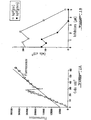
- the results show a dose-response inhibition of endothelial cell proliferation with the pV compounds ( Figure 1).
- the approximate IC 50 is 2 ⁇ M for bpV(phen) and 3.5 ⁇ M for bpV(pic).
- HUVECs cultured in a fibrin matrix can form 3-dimensional tubular-like structures in the presence of serum (106). This assay was performed in the presence of either bpV(phen), bpV(pic) or vanadate to assess the influence of each one of these compounds on HUVEC differentiation and organization (Table 1). HUVECs were seeded on the bottom of gelatin-coated wells at high density to provide a confluent monolayer at 48 hours.
- vanadate has an anti-angiogenic effect inasmuch as the stellate structures are affected (organization and terminal differentiation). Vanadate is at least 3 and 5 times less potent than bpV(pic) and bpV(phen), respectively.
- the anti-angiogenic effect observed with vanadate is in contradiction to the teachings of Montesano (48).
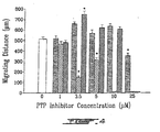
- Rat aorta rings were embedded in fibrin matrix as previously described (107). Thoracic aortas excised from adult rats were rinsed in Hank's balanced salt solution (HBSS), and cleansed of periadventitial. One-mm long aortic rings were sectioned and rinsed in culture medium. Each of the aortic rings were then embedded in fibrin gel matrix. Migration of microvessels in fibrin gel was quantified by measuring the distance from the external surface of rat aortic rings towards the migrating vessels. Measurements were performed at day 15 by digitizing morphometry with a NIH image analysis system (Fig. 3 and 4).
- test-system a. Definition of the test-system :
- chick embryo The normal development of a chick embryo involves the formation of an external vascular system which is located in the vitelline membrane and which carries nutrients from the vitellus (yolk of the egg) to the developing embryo. When placed onto the vitelline membrane, anti-angiogenic substances can inhibit blood vessel development in the vitelline membrane. To facilitate access to the vitelline membrane, chick embryos were transferred to a sterile culture box (Petri dish) and placed in a humidity- and temperature-controlled incubator. Embryos could then develop in this ex ovo condition for several days.
- a sterile culture box Petri dish
- Methylcellulose forms an inert matrix from which the tested compound can diffuse slowly.
- Methylcellulose discs containing the tested compound were placed on the external border of the vascular perimeter of the vitelline membrane where the angiogenic process was still active.
- the effects of the discs containing the tested compound on proximal vascular development were, in all cases, compared with those of discs containing control buffer.
- the discs were placed on the embryos' vitelline membrane on Day 0 or Day 1 of the ex ovo growth process; at this point, only beginnings of the main blood vessels are invading the vitellus.
- the embryos were then put in culture conditions until vascularization was assessed (approximately 24 h).
- Control buffer- and compound-containing discs were in all cases added simultaneously on the vitelline membrane of the same embryo. Both discs were arranged in a symmetrical fashion with respect to the cephalo-caudal axis of the embryo in order to minimize inter-individual variations when comparing the tested compounds with controls.
- EVTs Embryonic vascularization tests
- a score of 2 was attributed when at least one of the following conditions was met:
- a score of 1 was attributed when at least one of the following conditions was met:
- a score of 3 meant that blood vessel development was normal whereas a score of 1 indicated the greatest degree of angiostatic activity.
- This assay was performed as previously described (108). Briefly, Matrigel (liquid at 4°C) was mixed with 200 ng/ml ECGS and injected s.c. into four C57B1/6N female mice (1 ml/mice). After injection, the Matrigel polymerized to form a plug. After 7 days, the animals were sacrified, and the Matrigel plugs were removed and fixed in 10% neutral-buffered formalin solution (Sigma Chemical Co.) and embedded in paraffin. Histological sections were stained with Masson's trichrome and angiogenesis was scored by counting the number of cells that migrated in three microscopic fields for each s.c. Matrigel plug.
- pV compounds are potent inhibitors of the angiogenic process (see below), and this includes high doses of vanadate.
- Table III show the effect of bpV(phen) on the plasmatic levels of ETs.
- rats were injected intraperitoneally with bpV(phen) (0.5 mg/100g b.w.) 16 hours before the administration of either insulin or vehicle. Two minutes following insulin administration (1.5 ⁇ g/100g b.w, or vehicle), the plasma levels of ETs were determined. Insulin administration induced a strong increase of seric ET concentration (55). This increase was completely abolished by bpV(phen). In addition, bpV(phen) decreased the insulin-stimulated levels of plasmatic ETs below control levels.
- the data presented in Table IV show the effect of the pV compound bpV(bipy) on restenosis.
- Male Sprague-Dawley rats (325-350g) were anaesthetized with Rogarsetic, a mixture of ketamine hydrochloride and xylazine hydrochloride.
- the animals were divided into two groups of 14-31 rats that underwent balloon angioplasty of the left carotid artery, with or without treatment (109).
- the treated group received bpV (bipy) (10 mg / kg / i.p. b.i.d.) for 14 days, starting on the day of the angioplasty.
- the balloon was then inflated and retracted with a twisting motion to its insertion point. This procedure was repeated twice and the external carotid was ligated.
- compositions would comprise potent pV compounds capable of achieving an extracellular concentration of about 0.1 to 100 ⁇ M, preferably 2-40 ⁇ M. In rats, a dose of 20 ⁇ moles per kilogram was successful in reversing the endothelin increase after insulin administration.
- pV compounds which as previously stated are more potent and provoke less side effects than the vanadium "oxo" compounds, can be administered to achieve the desired dosage.
- bpV (bipy) on Restenosis NIP mm I mm 2 M I / M L mm 2 Control 0.064 0.138 0.090 1.546 0.090 bpV(bipy) 0.045 0.112 0.104 1.086 0.112 p ⁇ x 0.002 0.04 0.005 0.003 0.003
- I intima
- M media
- NIP neointimal proliferation
- L lumina.
Landscapes
- Health & Medical Sciences (AREA)
- Life Sciences & Earth Sciences (AREA)
- Medicinal Chemistry (AREA)
- Public Health (AREA)
- General Health & Medical Sciences (AREA)
- Veterinary Medicine (AREA)
- Chemical & Material Sciences (AREA)
- Pharmacology & Pharmacy (AREA)
- Animal Behavior & Ethology (AREA)
- Epidemiology (AREA)
- General Chemical & Material Sciences (AREA)
- Nuclear Medicine, Radiotherapy & Molecular Imaging (AREA)
- Organic Chemistry (AREA)
- Chemical Kinetics & Catalysis (AREA)
- Engineering & Computer Science (AREA)
- Bioinformatics & Cheminformatics (AREA)
- Inorganic Chemistry (AREA)
- Vascular Medicine (AREA)
- Heart & Thoracic Surgery (AREA)
- Cardiology (AREA)
- Pain & Pain Management (AREA)
- Rheumatology (AREA)
- Urology & Nephrology (AREA)
- Pharmaceuticals Containing Other Organic And Inorganic Compounds (AREA)
- Acyclic And Carbocyclic Compounds In Medicinal Compositions (AREA)
- Medicines That Contain Protein Lipid Enzymes And Other Medicines (AREA)
- Compositions Of Macromolecular Compounds (AREA)
- Heterocyclic Carbon Compounds Containing A Hetero Ring Having Oxygen Or Sulfur (AREA)
- Adhesives Or Adhesive Processes (AREA)
- Nitrogen Condensed Heterocyclic Rings (AREA)
- Pyridine Compounds (AREA)
Abstract
Description
- This invention relates to the use of peroxovanadium compounds, such as potassium bisperoxo(1,10-phenanthroline)oxovanadate [bpV(phen)], potassium bisperoxo(pyridine-2-carboxylato)oxovanadate [bpV(pic)] and potassium bisperoxo(2,2'-bipyridyl) oxovanadate [bpV(bipy)], for preventing angiogenesis, restenosis and the production of endothelins, and as adjuvants for vaccination.
- Synthetic peroxovanadium (pV) compounds are structurally versatile molecules which are potent inhibitors of phosphotyrosyl phosphatases (PTPs) (1). These compounds contain one oxo ligand, one or two peroxo groups coordinated to vanadium, and one ancillary ligand, and are stable in aqueous solution at physiological pH when shielded from light. The mechanisms underlying the PTPs' inhibitory potency and the specificity of these peroxanion compounds have been characterized. Compared to known inhibitors of PTPs such as orthovanadate, molybdate, tungstate and zinc, they have the ability to irreversibly oxidize an essential, conserved cysteine residue located in the catalytic domain (2). The possibility to manipulate the ancillary ligands may be important in regulating potency and specificity (1, 3). A reversible arrest of the proliferation of transformed cells by two pV compounds, bpV(phen) and bpV(pic), has been reported (4). The administration of pV compounds to mice has cured aggressive infections induced by a protozoan parasite (Leishmaniasis) (5).
- Almost all tissues and organs develop a vascular network which provides cells with nutrients and oxygen and enables the elimination of metabolic waste. Once formed, the vascular network is a stable system with a slow rate of cellular turn over (6) and yet, the endothelium can become one of the most rapidly proliferating of all cell types when stimulated (7). Indeed, the formation of new blood vessels can cause serious physiological complications. For example, while cornea and cartilage are avascular in healthy situations, several diseases involving these tissues are complicated by the massive arrival of blood vessels. Eye angiogenic diseases include neovascular glaucoma, retrolental fibroplasia, macular degeneration and neovascularization of corneal grafts. Joint angiogenic diseases include rheumatoid arthritis and arthrosis. Psoriasis, a chronic condition of the skin, also exhibits hypervascularization at the surface of the skin. Finally, solid tumor growth is critically dependent upon the formation of new blood vessels to progress locally and spread all over the body (8,9). The maintenance of existing blood vessels also requires the regulation of cell replication and/or differentiation. For example, acute and chronic pathological processes, such as atherosclerosis, post-angioplastic restenosis and hypertension, involve the proliferation of different cellular components of mature blood vessels (endothelial cells, smooth muscle cells, myocytes and fibroblasts).
- Several factors, including certain peptides and proteins, can induce a vascular response in vivo. They are endogenous substances, such as EGF/TGF-α (Epidermal Growth Factor/Transforming Growth Factor-alpha), TGF-β (Transforming Growth Factor-beta), TNF-α (Tumor Necrosis Factor-alpha), angiogenin, prostaglandine E2, and monobutyrine (10-15). However, these factors have almost no mitogenic effect on endothelial cells in culture (TGF-α, EGF, angiogenine, prostaglandine E2, monobutyrine) and, paradoxically, inhibit their growth (TGF-β, TNF-α) (10-17). Their angiogenic action is thus indirect, depending for the most part upon the inflammatory response (18). Inflammatory cells produce some factors, such as aFGF (acidic Fibroblast Growth Factor), bFGF (basic Fibroblast Growth Factor), PDGF (Platelet-Derived Growth Factor), and VEGF (Vascular Endothelial Growth Factor), which are capable of stimulating the proliferation of endothelial cells in vitro and angiogenesis in vivo (19-28).
- The angiogenic process, as currently understood, can be summarized as follows: a cell activated by a mutation, lack of oxygen, etc, releases angiogenic molecules (29-34) that attract inflammatory and endothelial cells and promote their proliferation. Following the binding of leukocytes to vascular endothelial cells, the latter reorganize the protein arrangements on their membranes to activate the angiogenic process (35, 36). During migration to the target tissue, inflammatory cells release substances that intensify the angiogenic effect (37, 38). Activated vascular endothelial cells respond to the angiogenic signal by secreting proteases which digest blood vessel walls to enable migration toward the target tissue (39-41). Several protein fragments produced by the digestion of the blood-vessel walls intensify the proliferative and migratory activity of the endothelial cells (42-44). Finally, the endothelial cells rearrange their adhesive membrane proteins to generate the formation of capillary tubes.
- Angiogenesis is thus a complex process consisting of several critical cellular events (45-47), among which the following may be readily identified:
- binding of leukocytes to endothelial cells and induction;
- migration of inflammatory cells to the target tissue;
- regression of the pericytes of the existing vascular system;
- dissolution of blood-vessel walls by proteases;
- endothelial cell migration;
- endothelial cell proliferation;
- endothelial cell differentiation and arrangement into a tubular shape;
- formation of the capillary network;
- anastomosis; and
- initiation of blood flow.
- Agents that are known to induce the proliferation of endothelial cells include sodium orthovanadate (48). The mechanism by which this agent induces an invasive phenotype in capillary endothelial cells is not clearly understood. However, the effects of vanadate on cultured cells are similar, in many respects, to those elicited by PMA (49), bFGF (50), and certain retroviral transforming proteins, which act by inducing tyrosine-specific protein phosphorylation (51-54). Consistent with these observations is the finding that both phorbol ester and various growth factors including bFGF, VEGF and PDGF stimulate the phosphorylation of cellular proteins on tyrosine residues whereas vanadate, perhaps by inhibiting tyrosine phosphatase(s), produces a marked increase in tyrosine phosphorylation.
- Other derivatives of vanadate (bpV(phen); bpV(pic)) have been synthesized and have proven to be potent PTP inhibitors (1). These products were expected to promote endothelial cell proliferation, like vanadate. On the contrary, these agents have been shown to inhibit the proliferation of several cell types (4). In some cases, the cells are arrested at G2/M. The simplest hypothesis to explain this restriction is that PTPs controlling cell mitosis are targets for bpV(phen) and pbV (pic) inhibitors. The cdc25 protein was proposed as a candidate target for pV compounds (4).
- International Patent Publication WO 95/19177 teaches the use of vanadate compounds for the treatment of proliferative disorders, metastasis and drug-resistant tumors. Although these vanadate compounds are stated to be anti-proliferative and anti-collagenolytic, no serious indication of any anti-angiogenic activity has been ascribed to them. This publication further shows that an anti-tumor effect is observed at dosages of vanadate higher than 5 mM. It is admitted that a concentration of vanadate compound of 1.3 mM or lower has no apparent anti-tumor effect.
- Montesano et al. (48) teach, on the contrary, that vanadate compounds cause endothelial cells to proliferate. Hence, their findings would indicate that these compounds are pro-angiogenic and not anti-angiogenic.
- US Patent 5,716,981 (Hunter et al.) mentions the use of vanadium compounds, namely oxovanadate, orthovanadate and vanadyl compounds, in anti-angiogenic applications. However, this reference provides no experimental evidence to substantiate the anti-angiogenic effects of these compounds. The compounds are stated to be possibly equivalent to Paclitaxel (an anti-angiogenic compound described in detail). Since the remaining body of the prior art suggests that vanadium compounds are pro-angiogenic, there is no enabling teaching in this patent to validate the use of these compounds as anti-angiogenics.
- pV compounds are more powerful anti-tumor molecules than oxo compounds, such as vanadate. The direct antiproliferative activity of pV compounds on transformed cells is known (4). At concentrations found to be ineffective for vanadate compounds in International Patent Publication WO 95/19177, pV compounds are efficacious anti-tumor compounds.
- Although the anti-tumor activity of vanadium compounds is known, their anti-angiogenic activity has not heretofore been disclosed in relation to either oxo or peroxo derivatives thereof.
- Endothelins (ETs), a family of three isopeptides, acting as important regulators of the physiological state of mature blood vessels. They are the most potent vasoconstrictors identified to date. In addition, they are known to stimulate the proliferation of endothelial cells, smooth muscles, myocytes and fibroblasts, as well as the synthesis of various growth factors, including VEGF (55, 56).
- ETs are also considered to be angiogenic factors involved in tumor development (57). Most tumor cells synthesize and secrete ETs (58-62). Patients affected by different cancers show elevated blood concentrations of ETs (63-65). A reduction in the expression of ET receptor type B (RETB) decreases the growth of tumor cells incubated in the presence of ETs (65). As indicated above, ETs promote proliferation and migration of endothelial cells (66). Expression of ET ligands and of ET receptors (RETA and RETB) was observed in the endothelial cells of a plurality of tumors (67, 68). ETs act in an autocrine fashion, promoting local angiogenesis (69). ETs are further involved in hemodynamic changes that go along with metastatic development. For example, the ratio of arterial hepatic blood flow to portal vein blood flow is abnormally high in patients having hepatic metastasis from colorectal tumors (70). This high blood flow ratio is due to the presence of a humoral mediator as demonstrated in vivo (71).
- A tumoral vascular bed has no innervation and consequently does not respond to vasoconstrictive drugs. However, these drugs decrease normal hepatic blood flow and increase blood flow in tumors. Since ETs are potent vasoconstrictive and angiogenic factors involved in vascular remodeling and tumor development, they may be responsible for these altered hemodynamics. An ET inhibitor may therefore be a valuable tool for controlling intratumor blood flow and for influencing the growth and degeneration of tumors. Additionally, such an inhibitor could be useful in verifying whether the anti-tumor effect of vanadium compounds involves ETs.
- ETs are also known to stimulate the production of extracellular matrices by endothelial cells (72, 73). That is frequently observed upon vascular reconstruction or angioplasty (74, 75). This effect is particularly deleterious following vascular trauma due to restenosis. In 30-50% of patients, restenosis is characterized by the reexpansion of atherosclerotic lesions. The causes of this vascular disorder are due to a local vascular blockage caused by cell proliferation, cell migration and extracellular matrix production. Recently, ETs have been shown to be major players in vascular remodelling, particularly in such conditions as long term atherosclerosis, cardiac hypertrophy (congestive heart failure), hypertension (pulmonary and other), renal problems and certain systemic dysfunctions (76-80). ETs are also strongly involved in coronary and brain vasospasm leading to ischemia and a reduced rate of survival (81, 82). Thus, because of the potent vasoconstrictor and nitogenic activities of ETs, inhibitors of ET production would be expected to be generally useful as anti-hypertensive and anti-proliferative agents, and particularly useful prior to, during and after vascular surgery.
- The present invention provides pV compounds that are useful against angiogenesis, restenosis and endothelin production. These molecules comprise vanadium, and one peroxo groups. Molecules comprising peroxo groups are the more potent anti-angiogenic molecules. Preferably, the molecules also contain an ancillary ligand, which includes any molecule capable of binding the transition metal atom (usually, through bonds involving oxygen and nitrogen). Phenanthroline, picolinic acid, bipyridine, oxalic acid, 4,7-dimethyl-phenanthroline and peptides are examples of such ligands.
- All these molecules can be used to inhibit the formation of new blood vessels and/or control systemic and local levels of endothelins (ETs) in the reparation of existing blood vessels.
- The molecules containing peroxo anions are more powerful than their oxo counterparts. Therefore, the former can be used at much lower concentrations to reduce the toxicity that results from overexposure to transition metals (4).
- Oxo transition metal complexes include oxo complexes such as vanadate, tungstate, molybdate, and vanadyl complexes, such as the following: methavanadate (VO3 -), orthovanadate (VO4 3-), salts thereof, vanadyl compounds (VO2+) like vanadyl acetyl acetonate and vanadyl sulfate. Similar complexes exist for other transition metals. Other suitable tungsten and molybdenum complexes include hydroxo derivatives derived from glycerol, tartaric acid and sugars, for example. The peroxo transition metal complexes include any oxidizing agent capable of combining with the transition metal. As such, the preferred peroxides comprise the following: t-butylhydroperoxide, benzoyl peroxide, m-chloroperoxibenzoic acid, cumene hydroperoxide, peracetic acid, hydroperoxiloneic acid, ethyl peroxide, pyridine peroxide and hydrogen peroxide.
- The general structure of the compounds of the present invention is the following:wherein:
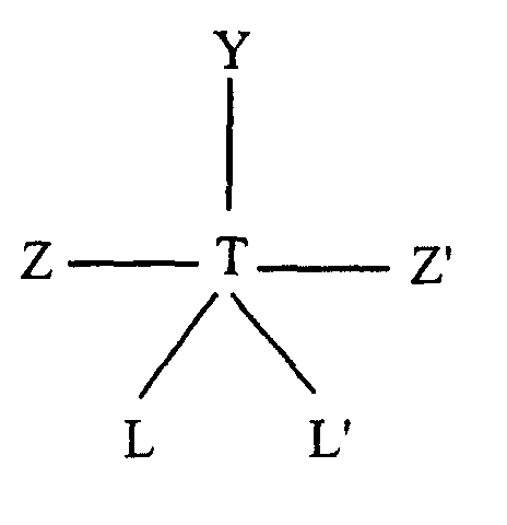
- T is vanadium;
- Y is oxygen or hydroxyl;
- Z and Z' are independently selected from oxygen and peroxide; and
- L and L' are any group which can donate an electron pair.
-
- In a preferred embodiment, Y is oxygen, Z and Z' are peroxide and L and L' are the nitrogen atoms of 1,10-phenanthroline.
- In another preferred embodiment, Y is oxygen, Z and Z' are peroxide and L and L' are nitrogen or oxygen atoms of picolinic acid.
- In yet another preferred embodiment, Y is oxygen, Z and Z' are peroxide and L and L' are nitrogen atoms of 2,2'-bipyridine.
- The above pV compounds are potent anti-angiogenics, since they inhibit endothelial cell proliferation. They further inhibit neovascularization and the production of endothelins.
- Moreover, administration of the pV compound bpV(bipy) in the rat model revealed a significant reduction in the degree of post-angioplastic vascular remodeling of the carotid artery. Additionally, the pV compound bpV(phen) was found to be a potent immunomodulator based on its capacity to induce cytokine and chemokine gene expression, to enhance cellular recruitment in response to an agonist and consequently act as an adjuvant in the context of vaccination.
- Other objects, advantages and features of the present invention will become apparent upon reading the following non-restrictive description of preferred embodiments thereof, with references to the accompanying drawings.
- This invention will now be described by referring to specific embodiments and the appended Figures, which purpose is to illustrate the invention rather than to limit its scope.
-
- Figure 1: illustrates the concentration-response effect of bpV(phen), bpV(pic) and vanadate on the growth rate of human vein umbilical endothelial cells (HUVECs).
- Figures 2: A and B show an anti-angiogenic response to bpV(phen), orthovanadate (Van) and protamine (Prot) using a vascularization test on the vitelline membrane of chick embryos. Each point represents a minimum of 15 embryos (15 to 28). In Figure 2A), neovascularization was assessed using N/+ scoring system; the proportion of embryos showing anti-angiogenesis increased with dosage, bpV(phen) being the most potent agent. In Figure 2B, neovascularization was assessed using the 1-2-3 scoring system; the angiogenic score decreased with dosage, bpV(phen) being the most potent agent.
- Figure 3: Micrographs of the cell migration from aortic rings embedded in fibrin (x12). Compared to the sample control (A), cell migration was impaired in the presence of doses of bpV(phen) (1, 3.5 µM; B, C) with a strong inhibition at 3.5 µM (C). In the presence of bpV(pic) (3.5, 10, 25 µM; D, E, F), inhibition was seen at 10-25µM. In the presence of orthovanadate, partial inhibition was reached at 25 µM (G) with no effect at 3.5 µM (H). The migration of well-defined microvessels in the presence of bpV(pic) is evident at 10 µM, while in the control sample very thin cell strands were frequently observed.
- Figure 4: Quantification of the cell migration from aortic rings in the presence of orthovanadate (plain bars); bpV(phen) (light gray bars); and bpV(pic) (dark gray bars). The control appears as an empty bar. Means and standard errors of the means are presented. The Student-Newman-Keuls method was used for statistical analysis (p value=0.001)
- Figure 5: bpV(phen) inhibits angiogenesis in vivo in the s.c. Matrigel assay. Graph showing the results of an experiment expressed as the number of cells that migrated in three microscopic fields for each s.c. Matrigel plug. Fields were equidistant from the edge of the plug. (1) Four mice that did not receive any treatment after the Matrigel plug implantation. (2) Four mice that received daily administration of 10 µg bpV(phen) during the 7 days post-implantation. (3) Four mice that received daily administration of 50 µg bpV(phen) during the 7 days post-implantation. (4) Four mice that received daily administration of 100 µg bpV(phen) during the 7 days post-implantation. (5) Four mice that received daily administration of PBS during the 7 days post-implantation.
-
- Human umbilical vein endothelial cells (HUVECs) were extracted with collagenase-controlled digestion, as previously described (105). Pure HUVECs were used before the fourth passage (trypsin-EDTA at each passage). The cells were analyzed for their capacity to incorporate di-acetyl LDL and to be labelled with factor VIII-related antigen.
- Endothelial cells were plated at a density of 2500 cells/cm2 in a sterile plate coated with gelatin. Cells were cultured in complete medium (M199: heparin (90 µg/ml) or L-glutamine (2mM), bicarbonate, FBS (20%) and ECGS (100 µg/ml)) for 24 hours to ensure cell adhesion. Then, cells were washed 3 times with PBS and culture medium was added according to experimental conditions. The last PBS wash was considered as time t=0.
- Cell proliferation was evaluated with the amount of DNA present in the petri dishes. Each experiment was performed in triplicate. The culture medium was changed daily. After 96 hours in culture, cells were lysed with Na-Citrate-SDS solution and incubated with Hoescht 33358. Samples were read at 365 nm with a spectrofluorometer.
- The results show a dose-response inhibition of endothelial cell proliferation with the pV compounds (Figure 1). The approximate IC50 is 2 µM for bpV(phen) and 3.5 µM for bpV(pic).
- Alternatively, HUVECs cultured in a fibrin matrix can form 3-dimensional tubular-like structures in the presence of serum (106). This assay was performed in the presence of either bpV(phen), bpV(pic) or vanadate to assess the influence of each one of these compounds on HUVEC differentiation and organization (Table 1). HUVECs were seeded on the bottom of gelatin-coated wells at high density to provide a confluent monolayer at 48 hours.
- Then, 50 000 HUVECs/ml were embedded in fibrinogen solution prior to polymerization. The fibrin matrix was covered with culture medium containing the molecule to be tested, while culture medium without any of these molecules served as controls. Cell behavior was observed periodically by phase contrast microscopy. After 21 days in culture in the presence of 1 µM bpV(phen), cord-like structures (or cords), tubelike structures (or tubes) and stellate structures (or Stell.struct.) were observed. At higher doses (2 and 3.5 µM), fragmented cord-like structures were apparent. In the presence of bpV(pic), cords and tubes were observed at 1, 2 and 3.5 µM. In the presence of orthovanadate, cords were still apparent at 10 µM, and at 25 µM dead cells were sparsely distributed. These results suggest that pV compounds interfere with endothelial cell organization and terminal differentiation. Furthermore, the nature of the ancillary ligand is important since bpV(phen) is more potent in inhibiting tube formation than bpV(pic).
- The results show that vanadate has an anti-angiogenic effect inasmuch as the stellate structures are affected (organization and terminal differentiation). Vanadate is at least 3 and 5 times less potent than bpV(pic) and bpV(phen), respectively. The anti-angiogenic effect observed with vanadate is in contradiction to the teachings of Montesano (48).
- Indeed, it was observed that at low doses, all the tested vanadium compounds seemed to be pro-angiogenic, while at higher doses (i.e., at almost double the dose), the same compounds were clearly anti-angiogenic. The type of radical L, L' greatly affects the potency of the vanadium compounds; for example, phenanthroline was found to be twice as potent as picolinic acid.
Effects of bpV(phen), bpV(pic) and Vanadate on HUVEC Differentiation and Organization Doses (µM) cords tubes Stell. struct. bpV (phen) 0 +++ +++ +++ 1 +++ +++ +++ 2 ++ + - 3.5 - - - bpV (pic) 0 +++ +++ +++ 1 +++ +++ +++ 2 +++ +++ +++ 3.5 ++ + - van 0 +++ +++ +++ 2.5 +++ +++ +++ 10 +++ +++ - 25 - Rat aorta rings were embedded in fibrin matrix as previously described (107). Thoracic aortas excised from adult rats were rinsed in Hank's balanced salt solution (HBSS), and cleansed of periadventitial. One-mm long aortic rings were sectioned and rinsed in culture medium. Each of the aortic rings were then embedded in fibrin gel matrix. Migration of microvessels in fibrin gel was quantified by measuring the distance from the external surface of rat aortic rings towards the migrating vessels. Measurements were performed at
day 15 by digitizing morphometry with a NIH image analysis system (Fig. 3 and 4). - The normal development of a chick embryo involves the formation of an external vascular system which is located in the vitelline membrane and which carries nutrients from the vitellus (yolk of the egg) to the developing embryo. When placed onto the vitelline membrane, anti-angiogenic substances can inhibit blood vessel development in the vitelline membrane. To facilitate access to the vitelline membrane, chick embryos were transferred to a sterile culture box (Petri dish) and placed in a humidity- and temperature-controlled incubator. Embryos could then develop in this ex ovo condition for several days.
- An aliquot of the tested compound was mixed with a methylcellulose solution and allowed to air-dry into thin discs. Methylcellulose forms an inert matrix from which the tested compound can diffuse slowly. Methylcellulose discs containing the tested compound were placed on the external border of the vascular perimeter of the vitelline membrane where the angiogenic process was still active.
- The effects of the discs containing the tested compound on proximal vascular development were, in all cases, compared with those of discs containing control buffer. The discs were placed on the embryos' vitelline membrane on
Day 0 orDay 1 of the ex ovo growth process; at this point, only beginnings of the main blood vessels are invading the vitellus. The embryos were then put in culture conditions until vascularization was assessed (approximately 24 h). Control buffer- and compound-containing discs were in all cases added simultaneously on the vitelline membrane of the same embryo. Both discs were arranged in a symmetrical fashion with respect to the cephalo-caudal axis of the embryo in order to minimize inter-individual variations when comparing the tested compounds with controls. - Embryonic vascularization tests (EVTs) were performed using different concentrations of protamine (5 to 20 µg) as a positive control or a tested compound (0.001 to 10 µg). After one day in culture, the level of vascularization in the area covered by the discs was graded by a pair of scientists in the usual blind fashion. To facilitate the location of the discs, black O-rings were placed around them just after their placement on the vitelline membrane. The evaluation scale for the EVTs was based on two different scoring systems.
- Blood vessel formation was assessed in a blind fashion. The areas of the vitelline membranes that lay beneath the methylcellulose discs were scored for the degree of vascularization, using two scoring systems (" N/+" or " 1-2-3"). The following selection criteria were used:
- A score of "N" for "Normal" was attributed when all the following conditions were met:
- Blood vessels in the evaluated area grew along their path with no abnormal deviation. Collateral branching density was normal and the growth path of the lateral branches was also normal.
- A score of " +" was attributed when at least one of the following conditions was met:
- Major blood vessels grew across the evaluated area but their paths were clearly affected (winding).
- Major blood vessels grew across the evaluated area but collateral branching density was clearly diminished.
- Major blood vessels penetrated the evaluated area but their growth path rapidly deviated.
- A kink was observed in the blood vessel.
- Major blood vessels penetrated the evaluated area but were stunted. No growth was observed beyond that point.
- A drastic deviation in the growth path occurred when major blood vessels reached the proximal ridge of the disc.
- A score of 3 was attributed when the following conditions were met:
- Blood vessels, present in the evaluated area, grew along their paths with no abnormal deviation. Collateral branching density was normal and the growth paths of the lateral branches were also normal.
- A score of 2 was attributed when at least one of the following conditions was met:
- Major blood vessels grew across the evaluated area but their paths were clearly affected (winding).
- Major blood vessels grew across the evaluated area but collateral branching density was clearly diminished.
- A score of 1 was attributed when at least one of the following conditions was met:
- Major blood vessels penetrated the evaluated area but their growth paths were rapidly deviated.
- A kink was observed in the blood vessels.
- Major blood vessels penetrated the evaluated area but were stunted. No growth was observed beyond that point.
- A drastic deviation in the growth paths occurred when major blood vessels reached the proximal ridge of the disc.
- A score of 3 meant that blood vessel development was normal whereas a score of 1 indicated the greatest degree of angiostatic activity.

- A dose-response inhibition was obtained with protamine, bpV(phen), and orthovanadate (Figure 2). The approximate IC50 is 0.1 µg for bpV(phen), 0.6 µg for orthovanadate, and 11 µg for protamine (regardless of the scoring system). A summary of the experimental data collected is shown in Table II.
- The results show that bpV(phen) is a potent inhibitor of the angiogenic process. Prior art on the subject (vanadate) taught the opposite, namely that vanadium compounds promote angiogenesis.
- This assay was performed as previously described (108). Briefly, Matrigel (liquid at 4°C) was mixed with 200 ng/ml ECGS and injected s.c. into four C57B1/6N female mice (1 ml/mice). After injection, the Matrigel polymerized to form a plug. After 7 days, the animals were sacrified, and the Matrigel plugs were removed and fixed in 10% neutral-buffered formalin solution (Sigma Chemical Co.) and embedded in paraffin. Histological sections were stained with Masson's trichrome and angiogenesis was scored by counting the number of cells that migrated in three microscopic fields for each s.c. Matrigel plug.
- Experiments were performed to determine whether bpV(phen) inhibits vessel formation in vivo. Endothelial cells invade and form vessels in Matrigel plugs containing angiogenic factors ECGS (109). Animals were divided into five groups (4 mice/group): two control groups that received no treatment or daily i.p. PBS injection, respectively, and three groups that received daily administration of various doses (10, 50 and 100 µg) of bpV(phen), from
day 1 to day 7 (intraperitoneal daily administration of 100 µl solution). The results show that bpV(phen) is a potent angiogenesis inhibitor in vivo (Fig. 5). Daily administration of 10 µg/mice or higher doses decreased by half the number of endothelial cells having invaded Matrigel plugs. - The angiogenic potential of pV compounds was tested using in vitro, ex ovo, and in vitro systems of analysis. The results show that in contrast to what might have been expected from the teaching of the prior art, pV compounds are potent inhibitors of the angiogenic process (see below), and this includes high doses of vanadate.
- The data presented in Table III show the effect of bpV(phen) on the plasmatic levels of ETs. In this study, rats were injected intraperitoneally with bpV(phen) (0.5 mg/100g b.w.) 16 hours before the administration of either insulin or vehicle. Two minutes following insulin administration (1.5 µg/100g b.w, or vehicle), the plasma levels of ETs were determined. Insulin administration induced a strong increase of seric ET concentration (55). This increase was completely abolished by bpV(phen). In addition, bpV(phen) decreased the insulin-stimulated levels of plasmatic ETs below control levels. These results suggest that bpV(phen) inhibits the insulin-induced release of ETs that subsequently can lead to diabetic complications like vasculopathy and nephropathy.
Effect of bpV (phen) on Endothelin Plasma Levels Plasma endothelins (pg/ml) Control 113.41 ±10.91 Insulin 253.10 Insulin, bpV(phen) 77.68 ±3.08 - ETs (ET-1, ET-2, ET-3) were measured by RIA (Amersham kit RPA 555) after lyophilization and extraction on C2 columns (500 mg). Results are mean ±SEM, p<0.0209, control (n=4) vs Insulin, pV (n=3).
- The data presented in Table IV show the effect of the pV compound bpV(bipy) on restenosis. Male Sprague-Dawley rats (325-350g) were anaesthetized with Rogarsetic, a mixture of ketamine hydrochloride and xylazine hydrochloride. The animals were divided into two groups of 14-31 rats that underwent balloon angioplasty of the left carotid artery, with or without treatment (109). The treated group received bpV (bipy) (10 mg / kg / i.p. b.i.d.) for 14 days, starting on the day of the angioplasty. Every animal in each group underwent balloon angioplasty of the left external carotid whereas the contra-lateral carotid served as a control for each animal. The left carotid artery balloon angioplasty was performed under aseptic conditions. The left external carotid was isolated and an arteriotomy was performed. A 2F Fogarty arterial embolectomy catheter was then inserted in the left common carotid and positioned near the aortic arch. The balloon was then inflated and retracted with a twisting motion to its insertion point. This procedure was repeated twice and the external carotid was ligated. There were no differences in the lumina (L), intima (I), media (M), the ratio of I/M and calculated value of (M - L / P) as a measure of neointimal proliferation (NIP) between normal, non-ballooned carotid artery of treated and non-treated rats, serving as controls (data not shown). Treatment caused a 32% decreased in NIP and the I/M ratio, a 21 % reduction in the thickness of the I and a 28% increase in the opening of the L. Thus, treatment with bpV (bipy) revealed a significant reduction in the degree of post-angioplastic vascular remodeling of the carotid artery in the rat model.
- The above results illustrate the antiangiogenic and anti-restenosis potential of vanadium compounds. The administration of pV compounds could be used to control the progression of several angiogeno-dependent conditions as well as post-angioplastic vascular remodeling. Since vanadates are anti-tumor compounds, the present pV compounds should function as anti-tumor therapeutic agents, and part of this activity should be due to an anti-angiogenic effect. Pharmaceutical compositions would comprise potent pV compounds capable of achieving an extracellular concentration of about 0.1 to 100 µM, preferably 2-40 µM. In rats, a dose of 20 µmoles per kilogram was successful in reversing the endothelin increase after insulin administration. Moreover, pV compounds, which as previously stated are more potent and provoke less side effects than the vanadium "oxo" compounds, can be administered to achieve the desired dosage. For example, see in vivo effects on blood glucose levels have been reported (1, 3).
Effect of bpV (bipy) on Restenosis NIP mm I mm2 M I / M L mm2 Control 0.064 0.138 0.090 1.546 0.090 bpV(bipy) 0.045 0.112 0.104 1.086 0.112 p < x 0.002 0.04 0.005 0.003 0.003 Abbreviations: I, intima; M, media; NIP, neointimal proliferation; L, lumina. -
- 1. Posner, B.I., Faure, R., Burgess, J.W., Bevan, A.P., Lachance, D., Zhan-Sun, G., Fantus, I.G., Ng, J.B., Hall, D.A., Lum, B.S. & Shaver, A. (1994). Peroxovanadium compounds: a new class of potent phosphotyrosine-phosphatase inhibitors which are insulin mimetics. J. Biol. Chem. 269: 4596-4604.
- 2. Shaver, A., Ng, J.B., Hall, D.A., & Posner, B.I. (1995). The chemistry of peroxovanadium compounds relevant to insulin mimesis. Mol. Cell. Biochem. 153: 5-15.
- 3. Bevan, A. P., Burgess, J. W., Yale, J. F., Drake, P. G., Lachance, D., Baquiran, G., Shaver, A., & Posner, B. I. (1995). In vivo insulin mimetic effects of pV compounds: role for tissue targeting in determining potency. Am J Physiol 268: E60-66..
- 4. Faure, R., Vincent, M., Dufour, M., Shaver, A. & Posner, B. (1995). Arrest at the G2/M transition of the cell-cycle by protein-tyrosine phosphatase inhibition: Studies on a neuronal and a glial cell line. J. Cell. Biochem. 58: 389-401.
- 5. Olivier, M, Romero-Gallo B. J., St-Laurent, R., Racette, N., Stevenson, M., Jacobs, P., Posner, B.I., Tremblay, M., & Faure, R. (1998). Modulation of INFg induced macrophage activation by phosphotyrosine phosphatase inhibition. J. Biol. Chem. 273: 13944-13949.
- 6. Reydi, M. A. & Schwartz, S. M. (1981) Endothelial regeneration: Time course of initial changes after small defined injury to rat aortic endothelium. Lab. Invest. 44: 301-308.
- 7. Schwartz, S. M. & Liaw, L. (1993) Growth control and morphogenesis in the development of pathology of arteries. J. Cardiovasc. Pharmacol. 21 (suppl): S31-S49.
- 8. Auerback, R. (1994). Angiogenesis inhibition: a review. Pharmac. Ther. 63: 265-311.
- 9. Folkman, J. (1995). Angiogenesis in cancer, vascular, rhumatoid and other diseases. Nature Med. 1: 27-31.
- 10. Schreiber, A.B., Winkler, M.E. & Derynck, R. (1986). Transforming growth factor-alpha: a more potent angiogenic factor than epidermal growth factor. Science 232: 1250-1254.
- 11. Roberts, A.B., Sporn, M.B., Assoian, R.K., Smith, J.M., Roche, N.J. & Wakefield, L.M. (1986). Transforming growth factor type beta: rapid induction of fibrosis and angiogenesis in vivo and stimulation of collagen formation in vitro. Proc. Natl. Acad. Sci. USA 84: 4167-4171.
- 12. Fràter-Schröder, M., Risau, W., Hallmann, R., Gautschi, R. & Bohlen, P. (1987). Tumor necrosis factor type-alpha, a potent inhibitor of endothelial cell growth in vitro, is angiogenic in vivo. Proc. Natl. Acad. Sci. USA 84: 5277-5282.
- 13. Fett, J.W., Strydom, D.J., Lobb, R.R., Alderman, R.M., Riordn, J.F. & Vallee, B.L. (1985). Isolation and characterization of angiogenin, an angiogenic protein from human carcinoma cells. Biochemistry 24: 5480-5488.
- 14. Ziche, M., Jones, J. & Gullino, P. (1982). Role of prostaglandin E1 and copper in angiogenesis. J. Natl. Cancer Inst. 69: 475-482.
- 15. Dobson, D.E., Kambe, A., Block, E., Dion, T., Lu, H. & Castellot, Jr. J.J., Speigelman, B.M. (1990). 1-Butyrylglycerol: a novel angiogenesis factor secreted by differentiating adipocytes. Cell 61: 223-230.
- 16. Baird, A. & Durkin, T. (1986). Inhibition of endothelial cell proliferation by type-beta transforming growth factor: interaction with acidic and basic fibroblast growth factors. Biochem. Biophys. Res. Commun. 138: 476-482.
- 17. Fràter-Schröder, M., Muller, G., Birchmeier, W. & Bohlen, P. (1986). Transforming growth factor-beta inhibits endothelial cell proliferation. Biochem. Biophys. Res. Commun. 137: 295-302.
- 18. Folkman, J., & Klagsbrun, M. (1987). Angiogenic factors. Science 235: 442-447.
- 19. Klagsbrun, M. & D'Amore, P.A. (1991). Regulators of angiogenesis. Annu. Rev. Physiol. 53: 217-232.
- 20. Gospodarowicz, D., Ferrara, N., Schweigerer, L. & Neufeld, G. (1987). Structural characterization and biological functions of fibroblast growth factor. Endocr. Rev. 8: 95-104.
- 21. Thomas, K., Rios Candelore, M., Gimenez-Gallego, G., Di Salvo, J., Bennet, C., Rodkey, J. & Fizpatrick, S. (1985). Pure brain-derived acidic fibroblast growth factor is a potent vascular endothelial cell mitogen with sequence homology to interleukin-1. Proc. Natl. Acad. Sci. USA 82: 6409-6416.
- 22. Miyazono, K., Okabe, T., Urabe, A., Takata, F. & Heldin, C.-H. (1987). Purification and properties of an endothelial cell mitogen from human platelets. J. Biol. Chem. 262: 4098-4104.
- 23. Ishikawa, F., Miyazono, K., Hellman, U., Drexler, H., Westemdt, C., Hagiwara, K., Usuki, K., Takaku, F., Risau, W. & Heldin, C.-H. (1989). Identification of angiogenic activity and the cloning and expression of platelet-derived endothelial cell growth factor. Nature 338: 557-561.
- 24. Leung, D.W., Cachianes, G., Kuang, W.-J., Goeddel, D.V. & Ferrara, N. (1989). Vascular endothelial growth factor is a secreted angiogenic mitogen. Science 246: 1306-1312.
- 25. Houck, K.A., Ferrara, N., Winer, J., Cachianes, G., Li, B. & Leung, D.W. (1991). The vascular endothelial growth factor family: Identification of a fourth molecular species and characterization of alternative splicing of RNA. Mol. Endocrinol. 5: 1806-1814.
- 26. Breier, G., Albrecht, U., Sterrer, S. & Risau, W. (1992). Expression of vascular endothelial growth factor during embryonic angiogenesis and endothelial cell differentiation. Development 114: 521-532.
- 27. Bussolino, F., Albini, A., Garnussi, G., Presta, M., Viglietto, G., Ziche, M. & Persico, G. (1996). Role of soluble mediators in angiogenesis. Eur. J. Cancer 32A: 2401-2412.
- 28. Colville-Nash, P.R. & Willoughby, D.A. (1997). Growth factors in angiogenesis: current interest and therapeutic potential. Mol. Med. Today. 3: 14-23.
- 29. Knighton, D.R., Hunt, T.K., Scheuenstuhl, H., Halliday, B.J., Werb, Z. & Banda, M.J. (1983). Oxygen tension regulates the expression of angiogenesis factor by macrophages. Science 221: 1283-1285.
- 30. Ladoux, A. & Frelin, C. (1993). Hypoxia is a strong inducer of vascular endothelial growth factor mRNA expression in the heart. Biochem. Biophys. Res. Commun. 195: 1005-1010.
- 31. Nomura, M., Yamagishi, S.I., Harada, S.I., Hayashi Y., Yarnashima, T., Yamashita, J. & Yamamoto H. (1995). Possible participation of autocrine and paracrine vascular endothelial growth factors in hypoxia-induced proliferation of endothelial cells and pericytes. J. Biol. Chem. 270: 28316-28324.
- 32. Walgenbach, K.J., Gratas, C., Shestak, K.C. & Becker, D. (1995). Ischaemia-induced expression of bFGF in normal skeletal muscle: a potential paracrine mechanism for mediating angiogenesis in ischaemic skeletal muscle. Nature Med. 1: 453-459.
- 33. Lew, A.P., Lew, N.S., Iliopoulos, O., Jiang, C., Kaeling Jr., W.G. & Goldberg, M.A. (1997). Regulation of vascular endothelial growth factor by hypoxia and its modulation by the von Hippel-Lindau tumor suppressor gene. Kidney Int. 51: 575-578.
- 34. Sandner, P., Wolf, K., Bergmaier, U., Gess, B. & Kurtz, A. (1997). Induction of VEGF and VEGF receptor gene expression by hypoxia: Divergent regulation in vivo and in vitro. Kidney Int. 51: 448-453.
- 35. Ferrara, N. (1995). Missing link in angiogenesis. Nature 376: 467.
- 36. Koch, A.E., Halloran, M.M., Haskell, C.J., Shaw, M.R. & Polverini, P.J. (1995). Angiogenesis mediated by soluble forms of E-selectin and vascular cell adhesion molecule-1. Nature 376: 517-519.
- 37. Polverini, J.P., Cotran, R.S., Gimbrone Jr, M.A. & Unanue, E.R. (1977). Activated macrophages induce vascular proliferation. Nature 269: 804-806.
- 38. Sunderkotter, C., Steinbrink, K., Goebeler, M., Bhardwaj, R. & Sorg, C. (1994). Macrophages and angiogenesis. J. Leukocyte Biol. 55: 410-422.
- 39. Polverini, P.J. (1996). How the extracellular matrix & macrophages contribute to angiogenesis-dependant diseases. Eur. J. Cancer 14: 2430-2437.
- 40. Gross, J.L., Moscatelli, D., Jaffe, E.A. & Rifkin, D.B. (1982). Plasminogen activator and collagenase production by cultured capillary endothelial cells. J. Cell Biol. 95: 974-981.
- 41. Moscatelli, D.A., Rifkin, D.B. & Jaffe, E.A. (1985). Production of latent collagenase by human umbilical vein endothelial cells in response to angiogenic preparations. Exp. Cell Res. 156: 379-390.
- 42. Ingber, D.E. (1991). Extracellular matrix and cell shape: potential control points for inhibition of angiogenesis. J. Cell. Biochem. 47: 236-241.
- 43. Ingber, D.E. (1992). Extracellular matrix as a solid-state regulator in angiogenesis: identification of new targets for anti-cancer therapy. Sem. Cancer Biol. 3: 57-63.
- 44. Bischoff, J. (1995). Approaches to studying cell adhesion molecules in angiogenesis. Trends Cell Biol. 5: 69-74.
- 45. Eisenstein, R. (1991). Angiogenesis in arteries: review. Pharmac. Ther. 49: 1-19.
- 46. Folkman, J. & Ingber, D. (1992). Inhibition of angiogenesis. Cancer Biol. 3: 89-96.
- 47. Bicknell, R. (1994). Vascular targeting and the inhibition of angiogenesis. Ann. Oncol. 5: S45-S50.
- 48. Montesano, R., Pepper, M.S., Belin, D., Vassalli, J.-D. & Orci, L. (1988). Induction of angiogenesis in vitro by vanadate, an inhibitor of phosphotyrosine phosphatases. J. Cell. Physiol. 134: 460-466.
- 49. Montesano, R. & Orci, L. (1985). Tumor-promoting phorbol esters induce angiogenesis in vitro. Cell 42: 469-477.
- 50. Montesano, R., Vassalli, J.D., Baird, A., Guillemin, R. & Orci, L. (1986). Basic fibroblast growth factor induces angiogenesis in vitro. Proc. Natl. Acad. Sci. USA. 83: 7297-7301.
- 51. Sefton, B.M. & Hunter, T. (1984). Tyrosine protein kinases. Adv. Cyclic Nucleotide Protein Phosphorylation Res. 18: 195-203.
- 52. Hunter, T. & Cooper, J.A. (1985). Protein-tyrosine kinases. Annu. Rev. Biochem. 54: 897-930.
- 53. Hunter, T. & Cooper, J.A. (1986). Viral oncogenes and tyrosine phosphorylation. In: The Enzymes, Vol. 17. P.D. Boyer and E.G. Krebs, eds. Academic Press, Orlando, Florida, pp. 191 -246.
- 54. Ramachandran, J. & Ullrich, A. (1987). Hormonal regulation of protein tyrosine kinase activity. Trends Pharmacol. Sci. 8: 28-31.
- 55. Battistini, B., Chailler, P., D'Orléans, J., Brière, N. & Sirois, P. (1993). Growth regulatory properties of endothelins. Peptides 14: 385-399.
- 56. Pedram, A., Razandi, M., Hu R-M & Levin, E.R. (1997). Vasoactive peptides modulate vascular endothelial cell growth factor production and endothelial cell proliferation and invasion. J. Biol. Chem. 272: 17097-17013.
- 57. Asham, E.H., Loizidou, M. & Taylor, I. (1998). Endothelin-1 and tumour development. Eur. J. Surg. Oncol. 24: 57-59.
- 58. Kusuhara, M., Yamaguchi, K., Nagasaki, K., Hayashi, C., Suzaki, A., Nori, S., Hanada, S., Nakamura, Y. and Abe, K. (1990). Production of endothelin in human cancer cell lines. Cancer Res. 50: 3257-3261.
- 59. Schichiri, M., Hirata, Y., Nakajiroa, T., & o, K., Imal, T., Yanagisawa, M., Mazaki, T. and Marumo, F. (1991). Endothelin-1 is an autocrine/paracrine growth factor for human cancer cell lines. J. Clin. Invest. 87: 1867- 1871.
- 60. Pagotto, U., Arzberger, T., Hopfer, U., Weidnl, A. & Stella, G. (1995). Cellular localization of endothelin receptor mRNA ETA and ETB in the brain tumour and normal human brain. J. Cardiovasc. Pharmacol. 26 (Suppl. 3): S104-106.
- 61. Stiles, J.D., Ostrow, P.T., Balos, L.L., Greenberg, S.J., Plunkett, R., Grand, W. & Heffner Jr., R.R. (1997). Correlation of endothelin-1 and transforming growth factor β1 with malignancy and vascularity in human gliomas. J. Neurobiol. 56: 435-439.
- 62. Ishibashi, M., Fujita, M., Nagai, K., Koko, M., Furue, H., Haku, E., Osamura, Y. & Yamaji, T. (1993). Production and secretion of endothelin by hepatocellular carcinoma. J. Clin. Endocrinol. Metab. 76: 378-383.
- 63. Giaid, A., Hamid, Q.A., Springall, D.R., Yanagisawa, M., Shinmi, O., Sawamura, T., Masaki, T., Kimura, S., Corrin, B. and Polak, J.M. (1990). Detection of endothelin immunoreactivity and mRNA in pulmonary tumours. J. Pathol. 162: 15-22.
- 64. Nelson, J., Chan, K., Hedican, S., Magnuson, S., Opgenorth, T., Bova, G. & Simons, J. (1996). Endothelin- 1 production decreased B receptor expression in advanced prostate cancer. Cancer Res. 56: 663-668.
- 65. Kikuchik, K., Nakagawa, H., Kadono, T., Etoh, T., Byers, H., Mihm, M. & Tamaki, K. (1996). Decreased ETB receptor expression in human metastatic melanoma cells. Biochem. Biophys. Res. Commun. 219: 734-739.
- 66. Ziche, M., Morbidelli, L., Donnini, S. & Ledda, F. (1995). ETB receptor promote proliferation and migration of endothelial cells. J. Cardiovasc. Pharmacol. 26 (Suppl. 3): S284-S286.
- 67. Inagaki, H., Bishop, A., Escrig, C., Wharton, J., Allen-Mersh, T. & Polak, J. (1991). Localization of endothelin like immunoreactivity and endothelin binding sites in human colon. Gastroenterology 101: 47-54.
- 68. Inagaki, H., Bishop, A., Eimoto, T. & Polak, J. (1992). Autoradiographic localization of endothelin-1 binding sites in human colonic cancer tissue. J. Pathol. 168: 263-267.
- 69. Eguchi, S., Hirata, Y., Imai, T. & Marumo, P. (1995). ET-1 as an autocrine growth factor for endothelial cells. J. Cardiovasc. Pharmacol. 26 (Suppl. 3): S279-S283.
- 70. Leveson, S., Wiggins, P., Giles, G., Parkin, A. & Robinson, P. (1985). Deranged blood flow patterns in the detection of liver metastases. Br. J. Surg. 72: 128-130.
- 71. Carter, R., Anderson, J., Cooke, T., Baxter, J. & Angerson, W. (1994). Splanchnic blood flow changes in the presence of hepatic tumours, evidence of humoral mediator. Br. J. Cancer 69: 1025-1026.
- 72. Nakamura, T., Ebihara, I., Fukui, M., Tomino, Y. & Koide, H. (1995). Effect of a specific endothelin receptor A antagonist on mRNA levels for extracellular matrix components and growth factors in diabetic glomeruli. Diabetes 44: 895-899.
- 73. Lee, K.M., Tsai, K.Y., Wang, N. & Ingber, D.E. (1998). Extracellular matrix and pulmonary hypertension: control of vascular smooth muscle cell contractility. Am. J. Physiol. 274: H76-82.
- 74. Hirshfeld, J.W. Jr, Schwartz, J.S., Jugo, R., MacDonald, R.G., Goldberg, S., Savage, M.P., Bass, T.A., Vetrovec, G., Cowley, M., Taussig, A.S., et al. (1991) Restenosis after coronary angioplasty: a multivariate statistical model to relate lesion and procedure variables to restenosis. The M-HEART Investigators. J. Am. Coll. Cardiol. 18: 647-656.
- 75. Maseri, A. (1996). An unresolved, persisting challenge for percutaneous transluminal coronary angioplasty: how to identify the lesions that will not restenose. Eur. Heart J. 17: 814-815.
- 76. Kirchengast, M. & Munter, K. (1998). Endothelin and restenosis. Cardiovasc. Res. 39: 550-555.
- 77. Kramer, B.K., Ackermann, M., Kohler, S.M. & Riegger, G.A. (1994). Role of endothelin in hypertension. Clin. Investig. 72: 88-93.
- 78. Levin, E. R. (1995). Endothelins: mechanisms of disease. New Eng. J. Med. 333: 356-363.
- 79. Rohmeiss, P., Birck, R., Braun, C., Kirchengast, M. & van der Woude, F.J. (1999). Targets for endothelin in the diseased kidney: clues for therapeutic intervention. Exp. Nephrol. 7: 1-10.
- 80. Cardell, L.O., Uddman, R. & Edvinsson, L. (1994). Endothelins: a role in cerebrovascular disease? Cephalalgia 14: 259-65.
- 81. Cosentino, F. & Katusic, Z. S. (1994). Does endothelin-1 play a role in the pathogenesis of cerebral vasospasm? Stroke 25: 904-908.
- 82. Battistini, B. & Dussault, P. (1998). The many aspects of endothelins in ischemia-reperfusion injury: emergence of a key mediator. J. Invest. Surg. 11: 297-313.
- 83. Mauel, J. (1990). Macrophage-parasite interactions in Leishmania infections. J. Leukoc. Biol. 47: 187-193.
- 84. Belosevic, M., Findbloom, D.S., Meltzer, M.S. & Nacy, C.A. (1990). IL-2. A cofactor for induction of activated macrophage resistance to infection. J. Immunol. 145: 831-839.
- 85. Olivier, M., Bertrand, S. & Tanner, C. E. (1989). Killing of Leishmania donovani by activated liver macrophages from resistant and susceptible strains mice. Int. J. Parasitol. 19: 377-383 .
- 86. Skov, C.B. & Twohy, W. (1974). Cellular immunity to Leishmania donovani. I. The effect of T cell depletion on resistance to L. donovani in mice. J. Immunol. 113: 2004-2011.
- 87. Murray, H.W., Masur, H. & Keithly, J.S. (1982). Cell-mediated immune response in experimental visceral leishmaniasis. I. Correlation between resistance to Leishmania donovani and lymphokine-generating capacity. J. Immunol. 129: 344-350.
- 88. Olivier, M. & Tanner, C. (1989). The effect of cyclosporin A in murine visceral leishmaniasis. Trop. Med. Parasitol. 40: 32-38.
- 89. Reiner, N.E. & Finke, J.H. (1983).
Interleukin 2 deficiency in murine leishmaniasis donovani and its relationship to depressed spleen cell responsees to phytohemagglutinin. J. Immunol. 131: 1487-1491. - 90. Murray, H.W., Stem, J.J., Welte, K., Rubin, B.Y., Carriero, S.M. & Nathan, C.F.
(1987). Experimental visceral leishmaniasis: production of
interleukin 2 and interferon-gamma, tissue immune reaction, and response to treatment withinterleukin 2 and interferon-gamma. J. Immunol. 138: 2290-2297. - 91. Olivier, M., Proulx, C. & Tanner, C.E. (1989). Importance of lymphokines in the control of the multiplication and dispersion of Leishmania donovani within liver macrophages of resistant and susceptible mice. J. Parasitol. 75: 720-727.
- 92. Green, S.P., Hamilton, J.A. & Phillips, W.A. (1992). Zymosan-triggered tyrosine phosphorylation in mouse bone-marrow-derived macrophages is enhanced by respiratory-burst priming agents. Biochem. J. 288: 427-432.
- 93. Greenberg, S., Chang. P. & Silverstein, S.C. (1993). Tyrosine phosphorylation of the gamma subunit of Fc gamma receptors, p72syk, and paxillin during Fc receptor-mediated phagocytosis in macrophages. J. Biol. Chem. 269: 3897-3902.
- 94. Dong, Z., Qi, X., Xie, K. & Fidler, I.J. (1993). Protein tyrosine kinase inhibitors decrease induction of nitric oxide synthase activity in lipopolysaccharide-responsive and lipopolysaccharide-nonresponsive murine macrohages. J. Immunol. 151: 2717-2724.
- 95. Dong, Z., O'Brian, C.A. & Fidler (1993). I.J., Activation of tumoricidal properties in macrophages by lipopolysaccharide requires protein-tyrosine kinase activity. J. Leukoc. Biol. 53: 53-60.
- 96. Glaser, K.B., Sung, A., Bauer, J. & Weichman, B.M. (1993). Regulation of eicosanoid biosynthesis in the macrophage. Involvement of protein tyrosine phosphorylation and modulation by selective protein tyrosine kinase inhibitors. Biochem. Pharmacol. 45: 711-721.
- 97. Golden, A., Nemeth, S.P. & Brugge, J.S. (1986). Blood platelets express high levels of the pp60c-src-specific tyrosine kinase activity. Proc. Natl. Acad. Sci. USA 83: 852-856.
- 98. Golden, A. & Brugge, J.S. (1989). Thrombin treatment induces rapid changes in tyrosine phosphorylation in platelets. Proc. Natl. Acad. Sci. USA 86: 901-905.
- 99. Walton, K.M. & Dixon, J.E. (1993). Protein tyrosine phosphatases. Annu. Rev. Biochem. 62: 101-120.
- 100. Fantl, W.J., Johnson, D.E. & Williams, L.T. (1993). Signalling by receptor tyrosine kinases. Annu. Rev. Biochem. 62: 453-481.
- 101. Chao, W., Liu, H., Hanahan, D.J. & Olson, M.S. (1992). Protein tyrosine phosphorylation and regulation of the receptor for platelet-activating factor in rat Kupffer cells. Effect of sodium vanadate. Biochem. J. 288: 777-784.
- 102. Secrist, J.P., Bums, L.A., Kamitz, L., Koretsky, G.A. & Abraham, R.T. (1993). Stimulatory effects of the protein tyrosine phosphatase inhibitor, pervanadate, on T-cell activation events. J. Biol. Chem. 268: 5886-5893.
- 103. Imbert, V., Peyron, J.F., Farahi Far, D., Mari, B., Auberger, P. & Rossi, B. (1994). Induction of tyrosine phosphorylation and T-cell activation by vanadate peroxide, an inhibitor of protein tyrosine phosphatase. Biochem. J. 297: 163-173.
- 104. Barbeau, B., Bernier, R., Dumais, N., Briand, G., Olivier, M., Faure, R.,. Posner, B.I. & Tremblay, M. (1997). Activation of HIV-1 LTR transcription and virus replication via NF-κB-dependent and -independent pathways by potent phosphotyrosine phosphatase inhibitors, the peroxovanadium compounds. J. Biol. Chem. 272: 12968-12977.
- 105. Sirois, E., Côté, M.F. & Doillon, C.J. (1993). Growth factors and biological supports for endothelial cell lining: in vitro study. Int. J. Artificial Organs 16: 609-619.
- 106. Janvier, R., Sourla, A., Koutsilieris, M. & Doillon, C. (1997). Stromal Fibroblasts are required for PC-3 human prostate cancer cells to produce capillary-like formation of endothelial cells in a three-dimensional co-culture system. Anticancer res. 17: 1551-1558.
- 107. Nicosia, R.F. & Ottineti, A. (1990) Growth of microvessels in serum-free-matrix culture of aorta. A quantitative assay of angiogenesis in vitro. Lab. Invest., 63:115-122.
- 108. Passaniti, A., Taylor, R.M., Pili, R., Guo, Y., Long, P.V., Haney, J.A., Pauly, R.R., Grant, D.S. & Martin, G.R. (1992). A simple quantitative method for assessing angiogenesis and antiangiogenic agents using reconstituted basement membrane, heparin, and fibroblast growth factor. Lab. Invest. 67 : 519-528.
- 109. Laporte, S., & Escher, E. (1992). Neointima formation after vascular injury is angiotensin II mediated. Biochem. Biophys. Res. Comm. 187:1510-1516.
- 110. Lima, G.M., Vallochi, A.L., Silva, U.R., Bevilacqua, E.M., Kiffer, M.M. & Abrahamsohn, I.A. (1998). The role of polymorphonuclear leukocytes in the resistance to cutaneous leishmaniasis. Immunol. Lett. 64: 145-151.
- 111.Hobbs, A.J., Higgs, A. & Moncada, S. (1999). Inhibition of nitric oxide synthase as a potential therapeutic target. Annu. Rev. Pharmacol. Toxicol. 39: 191-220.
- 112.Ajuevor, M.N., Virag, L., Flower, R.J., Perretti, M. & Szabo, C. (1998). Role of inducible nitric oxide synthase in the regulation of neutrophil migration in zymosan-induced inflammation. Immunology. 95: 625-630.
- 113.Yan, X.T., Tumpey, T.M., Kunkel, S.L., Oakes, J.E. & Lausch, R.N. (1998). Role of MIP-2 in neutrophil migration and tissue injury in the herpes simplex virus-1-infected cornea. Invest. Ophthalmol. Vis. Sci. 39: 1854-1862.
- 114. Duong, M., Ouellet, N., Simard, M., Bergeron, Y., Olivier, M. & Bergeron M.G.B. (1998). Kinetic study of host defense and inflammatory response to Aspergillus fumigatus in steroid-induced immunosuppressed mice. J. Infect. Dis. 178: 1472-1482.
- 115. Tam, F.W., Karkar, A.M., Smith, J., Yoshimura, T., Steinkasserer, Kurrle, R., Langner, K. & Rees, A.J. (1996) Differential expression of macrophage inflammatory protein-2 and monocyte chemoattractant protein-1 in experimental glomerulonephritis. Kidney Int. 49: 715-721.
- 116. Stenger, S., Thüring, H., Röllinghoff, M. & Bogdan, C. (1994). Tissue expression of inducible nitric oxide synthase is closely associated with resistance to Leishmania major. J. Exp. Med. 180: 783-793.
- 117. Stenger, S., Donhauser, N., Thüring, H., Röllinghoff, M. & Bogdan, C. (1996). Reactivation of latent leishmaniasis by inhibition of inducible nitric oxide synthase. J. Exp. Med. 183: 1501-1514.
- 118. Pani, G., Kozlowski, M., Cambier, J., Mills, G.B. & Siminovitch, K.A. (1995) Identification of the tyrosine phosphatase PTP1C as a B cell antigen receptor-associated protein involved in the regulation of B cell signaling. J. Exp. Med.. 181: 2077-2084.
- 119. Klingmüller, U., Lorenz, U., Cantley, L.C., Neel, B.G. & Lodish, H.F. (1995). Specific recruitment of SH-PTP1 to the erythropoietin receptor causes inactivation of JAK2 and termination of proliferative signals. Cell 80: 729-738.
-
Claims (17)
- Use of a compound of the following formula:wherein
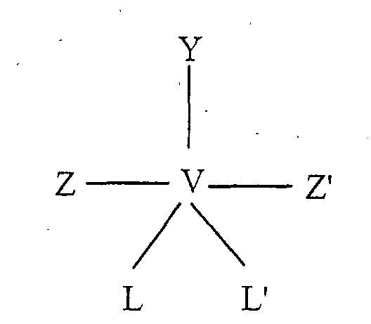 Y is oxygen or hydroxyl,Z and Z' are independently selected from oxygen and peroxide and at least one of them is peroxide, andL or L' are any group which can donate one electron pair, in the preparation of a medicament for treating any condition susceptible of being improved or prevented by inhibition of endothelial cell proliferation in an animal, preferably a human, said condition being selected from the group consisting ofangiogenesis, said angiogenesis condition being selected from to neovascular glaucoma, retrolental fibroplasia, macular degeneration, neovascularization of corneal grafts, or neovascularization of solid tumors,a diabetic complication consisting of vasculopathy or nephropathy, andrestenosis following angioplasty.
 - Use according to claim 1, wherein the angiogenesis condition is, selected from neovascular glaucoma, retrolental fibroplasia, macular degeneration or neovascularization of corneal grafts.
- Use according to claim 1, wherein the angiogenesis condition is neovascularization of solid tumors.
- Use according to claim 1, wherein the condition is the diabetic complication consisting of vasculopathy or nephropathy.
- Use according to claim 1, wherein the condition is the diabetic complication consisting of vasculopathy.
- Use according to claim 1, wherein the condition is restenosis following angioplasty.
- Use of a compound of the following formula:wherein
 in the preparation of a medicament for treating at least one diabetic complication selected from the group consisting of vasculopathy and nephropathy, which is susceptible of being improved or prevented by inhibition of an insulin-induced increase in endothelins in an animal, preferably a human.Y is oxygen or hydroxyl,Z and Z' are independently selected from oxygen and peroxide and at least one of them is peroxide, andL or L' are any group which can donate one electron pair,
 - Use of a compound of the following formula:wherein
 in the preparation of a medicament for preventing restenosis following angioplasty in an animal, preferably a human.Y is oxygen or hydroxyl,Z and Z' are independently selected from oxygen and peroxide and at least one of them is peroxide, andL or L' are any group which can donate one electron pair,
 - Use according to any one of claims 1 to 8 wherein Y is hydroxyl.
- Use according to any one of claims 1 to 8 wherein Y is oxygen.
- Use according to any one of claims 1 to 8 wherein Z is oxygen and Z' is peroxide.
- Use according to any one of claims 1 to 8 wherein Z and Z' are peroxide.
- Use according to any one of claims 1 to 8 wherein said compound has the following structure:and its potassium salt.
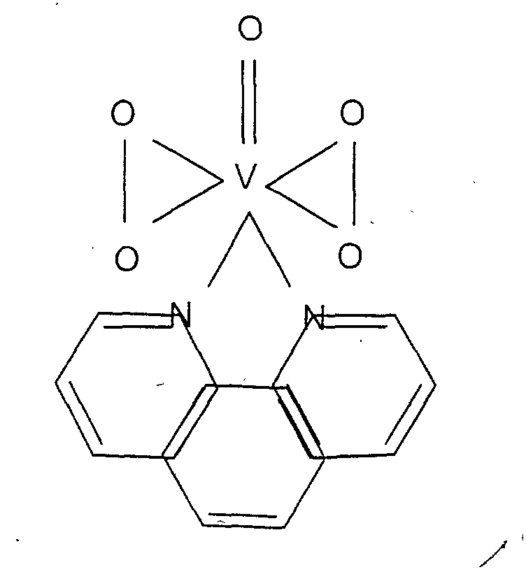
- Use according to any one of claims 1 to 8 wherein said compound has the following structure:and its potassium salt.

- Use according to any one of claims 1 to 8 wherein said compound has the following structure:and its potassium salt.

- Use according to any one of claims 1 to 15, wherein the medicament is capable of achieving an extracellular concentration of about 0.1 to 100 µM.
- Use according to claim 16, wherein the medicament is capable of achieving an extracellular concentration of obout 2 - 40 µM.
Applications Claiming Priority (3)
| Application Number | Priority Date | Filing Date | Title |
|---|---|---|---|
| CA002280249A CA2280249A1 (en) | 1999-08-12 | 1999-08-12 | Vanadium compounds as anti-angiogenics and as inhibitors of endothelin production |
| CA2280249 | 1999-08-12 | ||
| PCT/CA2000/000898 WO2001012180A2 (en) | 1999-08-12 | 2000-08-03 | Peroxovanadium compounds as protein tyrosine phosphatase (ptp) inhibitors: their inhibitory effects on angiogenesis, restenosis and the production of endothelins, and their stimulating effects on the immune responce |
Related Child Applications (1)
| Application Number | Title | Priority Date | Filing Date |
|---|---|---|---|
| EP05004368 Division | 2005-02-28 |
Publications (2)
| Publication Number | Publication Date |
|---|---|
| EP1202728A2 EP1202728A2 (en) | 2002-05-08 |
| EP1202728B1 true EP1202728B1 (en) | 2005-10-19 |
Family
ID=4163962
Family Applications (1)
| Application Number | Title | Priority Date | Filing Date |
|---|---|---|---|
| EP00952813A Expired - Lifetime EP1202728B1 (en) | 1999-08-12 | 2000-08-03 | Peroxovanadium compounds as protein tyrosine phosphatase (ptp) inhibitors: their inhibitory effects on angiogenesis, restenosis and the production of endothelins. |
Country Status (11)
| Country | Link |
|---|---|
| EP (1) | EP1202728B1 (en) |
| JP (1) | JP2003506487A (en) |
| AT (1) | ATE306918T1 (en) |
| AU (1) | AU781675B2 (en) |
| BR (1) | BR0013147A (en) |
| CA (1) | CA2280249A1 (en) |
| DE (1) | DE60023304D1 (en) |
| IL (1) | IL148081A0 (en) |
| MX (1) | MXPA02001481A (en) |
| NZ (1) | NZ517159A (en) |
| WO (1) | WO2001012180A2 (en) |
Families Citing this family (8)
| Publication number | Priority date | Publication date | Assignee | Title |
|---|---|---|---|---|
| EP1225891A2 (en) * | 1999-10-26 | 2002-07-31 | Virocell Inc. | Use of a bis-peroxovanadium compound for the treatment of immunosupressed patients |
| AU2001262819A1 (en) * | 2000-05-09 | 2001-11-20 | Johan Varland | Matching and positioning system for mobile units in communication networks |
| EP1343507A2 (en) * | 2000-08-02 | 2003-09-17 | Virocell Inc. | Method to treat infectious diseases and/or to enhance antimicrobial efficacy of drugs |
| AUPR598901A0 (en) * | 2001-06-28 | 2001-07-19 | Unisearch Limited | Inhibition of angiogenesis by targeting protein tyrosine phosphatases |
| EP1487501B1 (en) | 2001-10-09 | 2012-12-05 | The Johns Hopkins University | A phosphatase associated with metastasis |
| CA2386759A1 (en) * | 2002-05-17 | 2003-11-17 | Universite Laval | Therapeutic uses of peroxometallic compounds |
| CA2663259C (en) | 2006-09-12 | 2017-01-03 | Prophy Med Ab | Selective chemokine modulation |
| US20090023700A1 (en) * | 2007-07-20 | 2009-01-22 | University Of Louisville Research Foundation | Neuroprotective treatments |
Citations (1)
| Publication number | Priority date | Publication date | Assignee | Title |
|---|---|---|---|---|
| WO2000057860A2 (en) * | 1999-03-26 | 2000-10-05 | Centre For Translational Research In Cancer | Peroxovanadium compounds as antineoplastic agents for the treatment of cancer |
-
1999
- 1999-08-12 CA CA002280249A patent/CA2280249A1/en not_active Abandoned
-
2000
- 2000-08-03 AT AT00952813T patent/ATE306918T1/en not_active IP Right Cessation
- 2000-08-03 WO PCT/CA2000/000898 patent/WO2001012180A2/en not_active Ceased
- 2000-08-03 MX MXPA02001481A patent/MXPA02001481A/en unknown
- 2000-08-03 BR BR0013147-4A patent/BR0013147A/en not_active IP Right Cessation
- 2000-08-03 DE DE60023304T patent/DE60023304D1/en not_active Expired - Lifetime
- 2000-08-03 JP JP2001516526A patent/JP2003506487A/en active Pending
- 2000-08-03 EP EP00952813A patent/EP1202728B1/en not_active Expired - Lifetime
- 2000-08-03 NZ NZ517159A patent/NZ517159A/en unknown
- 2000-08-03 IL IL14808100A patent/IL148081A0/en unknown
- 2000-08-03 AU AU65509/00A patent/AU781675B2/en not_active Ceased
Patent Citations (1)
| Publication number | Priority date | Publication date | Assignee | Title |
|---|---|---|---|---|
| WO2000057860A2 (en) * | 1999-03-26 | 2000-10-05 | Centre For Translational Research In Cancer | Peroxovanadium compounds as antineoplastic agents for the treatment of cancer |
Also Published As
| Publication number | Publication date |
|---|---|
| AU6550900A (en) | 2001-03-13 |
| EP1202728A2 (en) | 2002-05-08 |
| IL148081A0 (en) | 2002-09-12 |
| ATE306918T1 (en) | 2005-11-15 |
| DE60023304D1 (en) | 2006-03-02 |
| BR0013147A (en) | 2002-07-23 |
| NZ517159A (en) | 2004-04-30 |
| AU781675B2 (en) | 2005-06-02 |
| MXPA02001481A (en) | 2003-07-21 |
| WO2001012180A3 (en) | 2001-08-16 |
| CA2280249A1 (en) | 2001-02-12 |
| JP2003506487A (en) | 2003-02-18 |
| WO2001012180A2 (en) | 2001-02-22 |
Similar Documents
| Publication | Publication Date | Title |
|---|---|---|
| Zhou et al. | Artesunate inhibits angiogenesis and downregulates vascular endothelial growth factor expression in chronic myeloid leukemia K562 cells | |
| Teicher et al. | Comparison of several antiangiogenic regimens alone and with cytotoxic therapies in the Lewis lung carcinoma | |
| Wilmanns et al. | Orthotopic and ectopic organ environments differentially influence the sensitivity of murine colon carcinoma cells to doxorubicin and 5‐fluorouracil | |
| US20220152070A1 (en) | Pharmaceutical composition and method for neoangiogenesis/revascularization useful in treating ischemic heart disease | |
| US10960017B2 (en) | Sorafenib or regorafenib troxacitabine phosphoramidate prodrug combination therapy for liver cancer | |
| EP3302478B1 (en) | Pac-1 combination therapy | |
| AU781675B2 (en) | Therapeutic uses of Peroxometallic compounds | |
| Boodhwani et al. | The future of therapeutic myocardial angiogenesis | |
| RU2407527C2 (en) | Methods, compositions and products for cancer treatment | |
| US20030055106A1 (en) | Therapeutic uses of peroxometallic compounds | |
| Kerr et al. | Conventional cytotoxic and novel therapeutic concepts in colorectal cancer | |
| CA2314915A1 (en) | Peroxovanadium compounds as protein tyrosine phosphate (ptp) inhibitors: their inhibitory effects on angiogenesis, restenosis and the production of endothelins, and their stimulating effects on the immune response | |
| CA2386759A1 (en) | Therapeutic uses of peroxometallic compounds | |
| US20220073642A1 (en) | Methods of using surufatinib in treating advanced pancreatic and extra-pancreatic neuroendocrine tumors | |
| Li et al. | The use of artemisinin compounds as angiogenesis inhibitors to treat cancer | |
| WO1998007415A2 (en) | Methods for prevention of cellular proliferation and restenosis | |
| US20210268010A1 (en) | Pharmaceutical composition and method for neoangiogenesis/revascularization useful in treating ischemic heart disease | |
| EP3615560B1 (en) | Fibroblast growth factor for use in treating patients unergoing ischemia-reperfusion therapy | |
| CN109789136B (en) | Pharmaceutical combination composition for preventing or treating colorectal cancer | |
| HK40040637B (en) | Combination therapy with sorafenib or regorafenib and a phosphoramidate prodrug of troxacitabine | |
| Theurl et al. | anuscript | |
| US20120095013A1 (en) | Use of kmup-3 for myocardial infarction | |
| HK1253828B (en) | Pac-1 combination therapy | |
| WO2012082337A2 (en) | Combination |
Legal Events
| Date | Code | Title | Description |
|---|---|---|---|
| PUAI | Public reference made under article 153(3) epc to a published international application that has entered the european phase |
Free format text: ORIGINAL CODE: 0009012 |
|
| 17P | Request for examination filed |
Effective date: 20020213 |
|
| AK | Designated contracting states |
Kind code of ref document: A2 Designated state(s): AT BE CH CY DE DK ES FI FR GB GR IE IT LI LU MC NL PT SE |
|
| AX | Request for extension of the european patent |
Free format text: AL;LT;LV;MK;RO;SI |
|
| 17Q | First examination report despatched |
Effective date: 20021010 |
|
| GRAP | Despatch of communication of intention to grant a patent |
Free format text: ORIGINAL CODE: EPIDOSNIGR1 |
|
| RTI1 | Title (correction) |
Free format text: PEROXOVANADIUM COMPOUNDS AS PROTEIN TYROSINE PHOSPHATASE (PTP) INHIBITORS: THEIR INHIBITORY EFFECTS ON ANGIOGENESIS, REST |
|
| GRAS | Grant fee paid |
Free format text: ORIGINAL CODE: EPIDOSNIGR3 |
|
| GRAA | (expected) grant |
Free format text: ORIGINAL CODE: 0009210 |
|
| AK | Designated contracting states |
Kind code of ref document: B1 Designated state(s): AT BE CH CY DE DK ES FI FR GB GR IE IT LI LU MC NL PT SE |
|
| PG25 | Lapsed in a contracting state [announced via postgrant information from national office to epo] |
Ref country code: IT Free format text: LAPSE BECAUSE OF FAILURE TO SUBMIT A TRANSLATION OF THE DESCRIPTION OR TO PAY THE FEE WITHIN THE PRESCRIBED TIME-LIMIT;WARNING: LAPSES OF ITALIAN PATENTS WITH EFFECTIVE DATE BEFORE 2007 MAY HAVE OCCURRED AT ANY TIME BEFORE 2007. THE CORRECT EFFECTIVE DATE MAY BE DIFFERENT FROM THE ONE RECORDED. Effective date: 20051019 Ref country code: LI Free format text: LAPSE BECAUSE OF FAILURE TO SUBMIT A TRANSLATION OF THE DESCRIPTION OR TO PAY THE FEE WITHIN THE PRESCRIBED TIME-LIMIT Effective date: 20051019 Ref country code: NL Free format text: LAPSE BECAUSE OF FAILURE TO SUBMIT A TRANSLATION OF THE DESCRIPTION OR TO PAY THE FEE WITHIN THE PRESCRIBED TIME-LIMIT Effective date: 20051019 Ref country code: CH Free format text: LAPSE BECAUSE OF FAILURE TO SUBMIT A TRANSLATION OF THE DESCRIPTION OR TO PAY THE FEE WITHIN THE PRESCRIBED TIME-LIMIT Effective date: 20051019 Ref country code: FI Free format text: LAPSE BECAUSE OF FAILURE TO SUBMIT A TRANSLATION OF THE DESCRIPTION OR TO PAY THE FEE WITHIN THE PRESCRIBED TIME-LIMIT Effective date: 20051019 Ref country code: BE Free format text: LAPSE BECAUSE OF FAILURE TO SUBMIT A TRANSLATION OF THE DESCRIPTION OR TO PAY THE FEE WITHIN THE PRESCRIBED TIME-LIMIT Effective date: 20051019 Ref country code: AT Free format text: LAPSE BECAUSE OF FAILURE TO SUBMIT A TRANSLATION OF THE DESCRIPTION OR TO PAY THE FEE WITHIN THE PRESCRIBED TIME-LIMIT Effective date: 20051019 |
|
| REG | Reference to a national code |
Ref country code: GB Ref legal event code: FG4D |
|
| REG | Reference to a national code |
Ref country code: CH Ref legal event code: EP |
|
| REG | Reference to a national code |
Ref country code: IE Ref legal event code: FG4D |
|
| PG25 | Lapsed in a contracting state [announced via postgrant information from national office to epo] |
Ref country code: SE Free format text: LAPSE BECAUSE OF FAILURE TO SUBMIT A TRANSLATION OF THE DESCRIPTION OR TO PAY THE FEE WITHIN THE PRESCRIBED TIME-LIMIT Effective date: 20060119 Ref country code: GR Free format text: LAPSE BECAUSE OF FAILURE TO SUBMIT A TRANSLATION OF THE DESCRIPTION OR TO PAY THE FEE WITHIN THE PRESCRIBED TIME-LIMIT Effective date: 20060119 Ref country code: DK Free format text: LAPSE BECAUSE OF FAILURE TO SUBMIT A TRANSLATION OF THE DESCRIPTION OR TO PAY THE FEE WITHIN THE PRESCRIBED TIME-LIMIT Effective date: 20060119 |
|
| PG25 | Lapsed in a contracting state [announced via postgrant information from national office to epo] |
Ref country code: DE Free format text: LAPSE BECAUSE OF FAILURE TO SUBMIT A TRANSLATION OF THE DESCRIPTION OR TO PAY THE FEE WITHIN THE PRESCRIBED TIME-LIMIT Effective date: 20060120 |
|
| PG25 | Lapsed in a contracting state [announced via postgrant information from national office to epo] |
Ref country code: ES Free format text: LAPSE BECAUSE OF FAILURE TO SUBMIT A TRANSLATION OF THE DESCRIPTION OR TO PAY THE FEE WITHIN THE PRESCRIBED TIME-LIMIT Effective date: 20060130 |
|
| REF | Corresponds to: |
Ref document number: 60023304 Country of ref document: DE Date of ref document: 20060302 Kind code of ref document: P |
|
| PG25 | Lapsed in a contracting state [announced via postgrant information from national office to epo] |
Ref country code: PT Free format text: LAPSE BECAUSE OF FAILURE TO SUBMIT A TRANSLATION OF THE DESCRIPTION OR TO PAY THE FEE WITHIN THE PRESCRIBED TIME-LIMIT Effective date: 20060320 |
|
| NLV1 | Nl: lapsed or annulled due to failure to fulfill the requirements of art. 29p and 29m of the patents act | ||
| REG | Reference to a national code |
Ref country code: CH Ref legal event code: PL |
|
| PG25 | Lapsed in a contracting state [announced via postgrant information from national office to epo] |
Ref country code: IE Free format text: LAPSE BECAUSE OF NON-PAYMENT OF DUE FEES Effective date: 20060803 |
|
| PLBE | No opposition filed within time limit |
Free format text: ORIGINAL CODE: 0009261 |
|
| STAA | Information on the status of an ep patent application or granted ep patent |
Free format text: STATUS: NO OPPOSITION FILED WITHIN TIME LIMIT |
|
| PG25 | Lapsed in a contracting state [announced via postgrant information from national office to epo] |
Ref country code: MC Free format text: LAPSE BECAUSE OF NON-PAYMENT OF DUE FEES Effective date: 20060831 |
|
| 26N | No opposition filed |
Effective date: 20060720 |
|
| EN | Fr: translation not filed | ||
| PG25 | Lapsed in a contracting state [announced via postgrant information from national office to epo] |
Ref country code: FR Free format text: LAPSE BECAUSE OF FAILURE TO SUBMIT A TRANSLATION OF THE DESCRIPTION OR TO PAY THE FEE WITHIN THE PRESCRIBED TIME-LIMIT Effective date: 20061208 |
|
| GBPC | Gb: european patent ceased through non-payment of renewal fee |
Effective date: 20060803 |
|
| REG | Reference to a national code |
Ref country code: IE Ref legal event code: MM4A |
|
| PG25 | Lapsed in a contracting state [announced via postgrant information from national office to epo] |
Ref country code: GB Free format text: LAPSE BECAUSE OF NON-PAYMENT OF DUE FEES Effective date: 20060803 |
|
| PG25 | Lapsed in a contracting state [announced via postgrant information from national office to epo] |
Ref country code: LU Free format text: LAPSE BECAUSE OF NON-PAYMENT OF DUE FEES Effective date: 20060803 |
|
| PG25 | Lapsed in a contracting state [announced via postgrant information from national office to epo] |
Ref country code: FR Free format text: LAPSE BECAUSE OF FAILURE TO SUBMIT A TRANSLATION OF THE DESCRIPTION OR TO PAY THE FEE WITHIN THE PRESCRIBED TIME-LIMIT Effective date: 20051019 Ref country code: CY Free format text: LAPSE BECAUSE OF FAILURE TO SUBMIT A TRANSLATION OF THE DESCRIPTION OR TO PAY THE FEE WITHIN THE PRESCRIBED TIME-LIMIT Effective date: 20051019 |