EP1202211A2 - Genomic DNA analysis computer program - Google Patents
Genomic DNA analysis computer program Download PDFInfo
- Publication number
- EP1202211A2 EP1202211A2 EP01309062A EP01309062A EP1202211A2 EP 1202211 A2 EP1202211 A2 EP 1202211A2 EP 01309062 A EP01309062 A EP 01309062A EP 01309062 A EP01309062 A EP 01309062A EP 1202211 A2 EP1202211 A2 EP 1202211A2
- Authority
- EP
- European Patent Office
- Prior art keywords
- dimensional electrophoresis
- electrophoresis pattern
- nucleotide sequence
- genomic dna
- restriction enzyme
- Prior art date
- Legal status (The legal status is an assumption and is not a legal conclusion. Google has not performed a legal analysis and makes no representation as to the accuracy of the status listed.)
- Withdrawn
Links
Images




Classifications
-
- G—PHYSICS
- G06—COMPUTING OR CALCULATING; COUNTING
- G06T—IMAGE DATA PROCESSING OR GENERATION, IN GENERAL
- G06T7/00—Image analysis
- G06T7/0002—Inspection of images, e.g. flaw detection
- G06T7/0012—Biomedical image inspection
- G06T7/0014—Biomedical image inspection using an image reference approach
-
- G—PHYSICS
- G01—MEASURING; TESTING
- G01N—INVESTIGATING OR ANALYSING MATERIALS BY DETERMINING THEIR CHEMICAL OR PHYSICAL PROPERTIES
- G01N27/00—Investigating or analysing materials by the use of electric, electrochemical, or magnetic means
- G01N27/26—Investigating or analysing materials by the use of electric, electrochemical, or magnetic means by investigating electrochemical variables; by using electrolysis or electrophoresis
- G01N27/416—Systems
- G01N27/447—Systems using electrophoresis
- G01N27/44704—Details; Accessories
- G01N27/44717—Arrangements for investigating the separated zones, e.g. localising zones
-
- G—PHYSICS
- G16—INFORMATION AND COMMUNICATION TECHNOLOGY [ICT] SPECIALLY ADAPTED FOR SPECIFIC APPLICATION FIELDS
- G16B—BIOINFORMATICS, i.e. INFORMATION AND COMMUNICATION TECHNOLOGY [ICT] SPECIALLY ADAPTED FOR GENETIC OR PROTEIN-RELATED DATA PROCESSING IN COMPUTATIONAL MOLECULAR BIOLOGY
- G16B30/00—ICT specially adapted for sequence analysis involving nucleotides or amino acids
-
- G—PHYSICS
- G06—COMPUTING OR CALCULATING; COUNTING
- G06T—IMAGE DATA PROCESSING OR GENERATION, IN GENERAL
- G06T2207/00—Indexing scheme for image analysis or image enhancement
- G06T2207/30—Subject of image; Context of image processing
- G06T2207/30004—Biomedical image processing
Definitions
- the present invention relates to a computer program which analyzes a two-dimensional electrophoresis pattern using genomic DNA; and a method for analyzing genomic DNA using a two-dimensional electrophoresis pattern.
- Genome scanning is a scan to analyze loci (gene loci) or copy number thereof at a high speed and thereby detect the physical condition of genomic DNA throughout a genome as a whole. Genome scanning enables high speed scanning of signals relating to, and thereby determination of, a gene position on a genome gene map or a chromosomal compartment, which a DNA region encoding one gene product occupies. In this genome scanning, it is extremely important what kind of mark is raised on a genome, what kind of signal is used to detect any mark on a genome, and how to detect and analyze as many marks as possible.
- This mark is called a landmark, and analysis of information regarding position of the landmark on a chromosome could be an important basic technique to produce a linkage map or a physical map of genome. Therefore, application of the genome scanning to two or more genomic DNA molecules to make a comparison enables detection of changes in genomic DNA molecules such as deletion, amplification and translocation.
- RLGS Restriction Landmark Genomic Scanning
- this RLGS method is a technique by which change in genomic DNA can be detected at a high speed in that a large number of landmarks can be detected at one time by this method.
- a control two-dimensional electrophoresis pattern is required for comparison with the two-dimensional electrophoresis pattern obtained from target genomic DNA.
- a control two-dimensional electrophoresis pattern can be obtained by similarly performing two-dimensional electrophoresis using genomic DNA extracted from a wild type cell.
- RLGS method a process of obtaining a control two-dimensional electrophoresis pattern is complicated, and comparison using the obtained control two-dimensional electrophoresis pattern is also complicated, Furthermore, there is a problem that enormous effort and complex techniques are required to clone a spot.
- the present invention is directed to provide a computer program, which enables a control two-dimensional electrophoresis pattern and a target for comparison to be produced easily under various conditions, and the two-dimensional electrophoresis pattern to be analyzed using genomic DNA. Moreover, the present invention is directed to providing a method for analyzing genomic DNA, in which rapid and accurate analysis can easily be carried out by producing a control two-dimensional clectrophoresis pattern under various conditions.
- FIG. 1 Further aspects of the invention respectively provide a computer or linked computers operatively configured to implement any of the methods or programs described above and computer programming product or products (such as ROM, RAM, floppy discs, hard drives, optical compact discs, magnetic tapes, and other computer-readable media) carrying computer code for implementing any of the methods or programs described above.
- computer programming product or products such as ROM, RAM, floppy discs, hard drives, optical compact discs, magnetic tapes, and other computer-readable media
- n denotes a, g, c or t (location: the 4th nucleotide to the 9 th nucleotide).
- the program of the present invention may be recorded in an information recording medium such as a flexible disk, an optical disk, a magneto-optical disk, a phase change optical disk and a hard disk, or may be transmitted from a server by means of a communication line network such as the internet.
- the present program recorded in an information recording medium or transmitted from a server is able to allow a computer to execute the following processes.
- This program comprises the following processes:
- a process of producing a control two-dimensional electrophoresis pattern based on genomic nucleotide sequence information (see process 1 set forth below).
- this program allows a computer to compare the two-dimensional electrophoresis pattern obtained from genomic DNA extracted from an analysis target with a control two-dimensional electrophoresis pattern.
- process 1 of this program first, a control two-dimensional electrophoresis pattern is obtained.
- process 2 of this program using a separately obtained two-dimensional electrophoresis pattern of the analysis target, the target two-dimensional electrophoresis pattern is compared with the control two-dimensional electrophoresis pattern.
- a method of obtaining the two-dimensional electrophoresis pattern of a target genomic DNA is described.
- a restriction enzyme recognition site is used as a mark (a landmark) and a method involving detection of the landmark as a signal (Restriction Landmark Genomic Scanning Method (RLGS method); Hatada, I. et al., Proc. Natl. Acad. Sci., USA, 88, 9523-9527, 1991) is applied as a basic concept.
- RGS method Restoral Landmark Genomic Scanning Method
- Hatada I. et al., Proc. Natl. Acad. Sci., USA, 88, 9523-9527, 1991
- Matsuyama et al . Plant. Mol. Biol. Rep. 18:331-338, 2000 is applied as a basic idea.
- the two-dimensional electrophoresis pattern of a target genomic DNA can be obtained according to the following method.
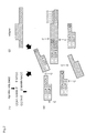
- the positions of the thus obtained spots are expressed as distances from an origin point in the X and Y directions such as (X1, Y1), (X2, Y2), ... (Xn, Yn), then the X coordinate reflects the distance from the recognition site of restriction enzyme A to that of restriction enzyme B (A-B fragment), and the Y coordinate reflects the distance from the recognition site of restriction enzyme A to that of restriction enzyme C (A-C fragment) ( Figure 1(6)). Accordingly, mutated points of a genome can be specified and mutation by DNA modification can be detected by analyzing the spot pattern.
- the density of a spot reflects the copy number of the detected fragments, it shows the duplication of specific regions, and where the position (X2, Y2) obtained after two-dimensional fraction in the spot pattern of figure 1(6) deviates or disappears, it shows that a portion of the nucleotide sequence of a DNA fragment is mutated (deleted, substituted or added).
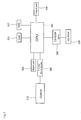
- Figure 3 is a block diagram showing a system configuration including a computer device which executes this program.
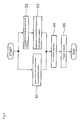
- Figure 4 is a flow chart showing the concrete processing of the program using this system.
- a computer executing this program is equipped with CPU 501 which controls the operation as a whole, RAM 503 which temporarily stores necessary program data and the like, input portion 504 into which an operator inputs various information, sending/receiving portion 505 which comprises a communication modem sending/receiving data to/from outside via the internet, output portion 505 which outputs data to be displayed, HDD 507 which records data and program etc., and CD drive 508 which reads data written onto CD-ROM 509. Furthermore, this system comprises database 510 which is connected to the computer by means of a communication line network such as the internet.
- This program can be recorded in any recording medium, as long as it is an information recording medium which is generally used as an external memory device of a computer, such as a tape-form or disk-form magnetic recording medium or an optical recording medium etc.
- CPU 501 controls the whole system and executes a process described later.
- Input portion 504 may be a scanner, a mouse and a key board etc., and this is operated when inputting conditions necessary for executing a processing.
- Output portion 506 may be a display and a printer etc., and this is able to display data and the like processed under the control of CPU 501.
- Sending/receiving portion 505 is able to take data which was recorded in database 510 into RAM 503 and HDD 507 etc. under the control of CPU 501.
- step S1 may be performed simultaneously with step S2 and step S3.
- step S1 may be performed prior to steps S2 and S3, or steps S2 and S3 may be performed prior to step S1.
- step S1 the two-dimensional pattern obtained from a target genomic DNA is input. Specifically, each spot position (Xn, Yn) obtained in "(7) Detection of spots" can be input as digital data. Furthermore, a two-dimensional electrophoresis pattern can be input as image data using a scanner etc.
- genomic nucleotide sequence information recorded in database 510 is obtained by means of a communication line network such as the internet.
- This genomic nucleotide sequence information is a nucleotide sequence which is registered as a wild type of a target genomic DNA.
- Examples of database 510 include Genbank, EMBL, DDBJ, TAIR and KAOS etc.
- the genomic nucleotide sequence information used for this program is not limited to that recorded in database 510, but those recorded in HDD 507, CD-ROM 509 and other information recording mediums etc. can also be used.
- step S2 abnormal information comprised in the genomic nucleotide sequence information is preferably detected. Specifically, in step S2, abnormal information comprised in the genomic nucleotide sequence information is detected and temporarily stored, for example, in RAM 503 as a sorting.
- abnormal information means that nucleotides located in a certain position are unidentified, and such information is comprised in a nucleotide sequence, for example, as literal information such as "m”, “r”, “w”, “s”, “y”, “k”, “v”, “h”, “d”, “b” and “n” etc.
- the character “m” represents that the position is A or C.
- the character “r” represents that the position is G or A.
- the character “w” represents that the position is A, T or U.
- the character “s” represents that the position is G or C.
- the character “y” represents that the position is T, U or C.
- the character “k” represents that the position is G, T or U.
- the character “v” represents that the position is A, G or C.
- the character “h” represents that the position is A, C, T or U.
- the character “d” represents that the position is A, G, T or U.
- the character “b” represents that the position is G, C, T or U.
- the character “n” represents that the position is A, G, C, T or U, or unknown.
- step S3 using the genomic nucleotide sequence information obtained from database 510, a control two-dimensional electrophoresis pattern is produced.
- the control two-dimensional electrophoresis pattern is produced, first, the recognition sequence of restriction enzyme A and that of restriction enzyme B are retrieved from genomic nucleotide sequence information in order to predict A-A fragments and A-B fragments. Then, where an adapter is used for a target two-dimensional electrophoresis pattern, the nucleotide sequence number of the adapter is added to estimated A-A fragments and A-B fragments. According to this, there can be estimated a plurality of estimated A-A fragments and estimated A-B fragments corresponding to a plurality of A-A fragments and A-B fragments obtained by "(3) Treatment for genomic DNA with a second restriction enzyme".
- an electrophoresis pattern which is presumably obtained when "(4) First-dimension fractionation" is performed.
- This electrophoresis pattern can be calculated by the method reported by Southern, E. M. (Measurement of DNA Length by Gel Electrophoresis, Analytical Biochemistry: 100, 319-323, 1979).
- the electrophoresis pattern can be estimated by inputting, as parameters, various conditions such as the concentration and type of gel, a voltage under which an electrophoresis is performed, electrophoresis period and temperature etc.
- a correction value is preferably provided in advance so that an electrophoresis pattern can be estimated using these conditions as parameters.
- the recognition sequence of restriction enzyme C in estimated A-A fragments and estimated A-B fragments is retrieved, and A-C fragments are estimated. According to this process, a plurality of estimated A-C fragments corresponding to a plurality of A-C fragments obtained in "(5) Treatment for genomic DNA with a third restriction enzyme" can be estimated.
- an electrophoresis pattern which is presumably obtained when "(6) Second-dimension fractionation" is performed, is estimated.
- this electrophoresis pattern can be estimated by inputting, as parameters, various conditions such as the type of gel, a voltage under which an electrophoresis is performed, electrophoresis period and temperature etc.
- a correction value is preferably provided in advance so that an electrophoresis pattern can be estimated using these conditions as parameters.
- an estimated spot position can be obtained as an estimated coordinate (Xn, Yn).
- X coordinate (Xn) represents a size-fraction position of A-B fragments to which the nucleotide sequence number of an adapter is added
- Y coordinate (Yn) represents a size-fraction position of A-C fragments to which the nucleotide sequence number of an adapter is added.
- a control two-dimensional electrophoresis pattern can also be obtained as image data based on the obtained estimated coordinates (Xn, Yn).
- an electrophoresis pattern is produced based on estimated A-B fragments.
- the electrophoresis pattern may be produced based on estimated A fragments which are estimated to be obtained by treatment with restriction enzyme A.
- the nucleotide sequence number of the adapter may not be considered.
- a control two-dimensional electrophoresis pattern is preferably produced as one which excludes a certain region from genomic nucleotide sequence information.
- regions to be excluded include a telomere region of genomic DNA and high frequency repeated nucleotide sequences located around a centromere of genomic DNA etc.
- fragments having the recognition sequence of restriction enzyme A only at one terminus thereof are not identified as spots, as they are derived from a telomere region of genomic DNA or a gap region during nucleotide sequencing.
- step S2 where abnormal information comprised in the genomic nucleotide sequence information is detected in step S2, a recognition sequence such as restriction enzyme A is detected, setting genomic nucleotide sequence information other than the abnormal information as a target. That is, a nucleotide which is located in a position detected as abnormal information is eliminated from a target, when detecting a recognition sequence such as restriction enzyme A.
- abnormal information comprised in the genomic nucleotide sequence information is detected in step S2
- the abnormal information is linked to an estimated spot.
- abnormal information such as whether or not abnormal information is found in a DNA fragment comprised in an estimated spot, and the type of abnormal information comprised in an estimated spot, is linked to each of the spots.
- step S4 the control two-dimensional electrophoresis pattern obtained based on genomic nucleotide sequence information is compared with a target two-dimensional electrophoresis pattern. Specifically, a coordinate (Xn, Yn) of a spot input in step S is compared with an estimated coordinate (Xn, Yn) of the spot obtained in step S3. Furthermore, in step S4, it is also possible that the image data of the two-dimensional electrophoresis pattern obtained in step S is compared with that obtained in step S3.
- step S5 the results of comparison performed in step S4 are displayed on a display.
- Such comparison results are provided by identifying spots in a control two-dimensional electrophoresis pattern, which do not match with a target two-dimensional electrophoresis pattern, and display the coordinates of these spots and nucleotide sequence information thereof. Since a control two-dimensional electrophoresis pattern is produced based on the genomic nucleotide sequence information obtained from database 510, the nucleotide sequence information can correspond to each of the spots.
- telomere region of genomic DNA or a high frequency repeated nucleotide sequence located around a centromere which shows unclear data during nucleotide sequencing is used as a control, it causes noise.
- the comparison of spots causing noise can be prevented by using a control two-dimensional electrophoresis pattern, from which a telomere region of genomic DNA and a high frequency repeated nucleotide sequence located around a centromere of genomic DNA are removed.
- both ends of each clone are marked at the initial stage to clearly distinguishing noise appearing in a pattern.
- step S5 where abnormal information comprised in the genomic nucleotide sequence information is detected in step S2, there is identified a spot in a control two-dimensional electrophoresis pattern, which does not match with a target two-dimensional electrophoresis pattern, and determined whether the spot comprises abnormal information or not.
- the abnormal information is corrected and the control two-dimensional electrophoresis pattern is reconstructed.
- abnormal information "m" is comprised in the identified spot, there are reconstructed both a control two-dimensional electrophoresis pattern in which the position of abnormal information is set at A, and another control two-dimensional electrophoresis pattern in which the position of abnormal information is set at C.
- step S5 the thus reconstructed control two-dimensional electrophoresis patterns are compared with a target two-dimensional electrophoresis pattern again.
- abnormal information "m" is comprised in the identified spot
- both control two-dimensional electrophoresis patterns in which the positions of abnormal information are set at A or C were reconstructed, these two types of control two-dimensional electrophoresis patterns are compared with a target two-dimensional electrophoresis pattern.
- the analysis of a target two-dimensional electrophoresis pattern can be carried out with higher precision, while reflecting abnormal information in genomic nucleotide sequence information. Where it is impossible to correct the obtained abnormal information, information regarding abnormal information comprised in the identified spot is linked to the spot.
- a control two-dimensional electrophoresis pattern is produced using genomic nucleotide sequence information recorded in database 510 etc., and a target two-dimensional electrophoresis pattern is analyzed using this control two-dimensional electrophoresis pattern.
- a two-dimensional electrophoresis pattern obtained from genomic DNA is analyzed, it is not necessary to produce a control two-dimensional electrophoresis pattern using genomic DNA extracted from wild type cell strains.
- enormous manpower and complex techniques are also not needed. Therefore, using to this program, target genomic DNA can be analyzed easily and rapidly.
- genomic DNA having a mutation such as a deletion of nucleotides induced by a physical mutagene is preferably used. That is to say, in the present program, it is preferable to use as a target, genomic DNA the length of which differs from a wild type genomic DNA.
- mutation-introduced genomic DNA is used as a target, applying a means involving induction of mutation by radiation of heavy ion beam (Japanese Patent Application Laying-Open (kokai) No. 9-28220).
- methylation insensitive restriction enzymes are preferably used as the first restriction enzymes. That is, where plant genomic DNA is used as a target, using methylation insensitive restriction enzymes, the DNA is reliably cleaved under no influence of methylation.
- methylation insensitive restriction enzymes used for the first restriction enzyme treatment include Acc III and Pac I etc.
- methylation insensitive restriction enzymes used for the second restriction enzyme treatment include Mbo I recognizing 4 nucleotides.
- methylation sensitive restriction enzymes may also be used.
- epigenetic mutated regions caused by methylation of DNA can be detected and analyzed.
- Arabidopsis thaliana chloroplast DNA is used as an analysis target.
- the nucleotide sequence of chloroplast DNA of Arabidopsis thaliana (Columbia-strain) has already been determined as one having a circular genomic DNA of 154,478bp (Sato, S. et al . 1999, DNA RESEARCH 6, 238-290), and the nucleotide sequence data can be obtained from DDBJ as Accession No. AP000423. Based on this nucleotide sequence data, a control two-dimensional electrophoresis pattern was produced by the above-stated program processes.
- a two-dimensional electrophoresis pattern was produced, using Acc III as the first restriction enzyme and Mbo I as the third restriction enzyme but not using the second restriction enzyme. These Acc III and Mbo I are insensitive to 5-methyl cytosine (5mC) which results from methylation of cytosine.
- the obtained control two-dimensional electrophoresis pattern is shown in Figure 5.
- the display of nucleotide number shows a mobility when a molecular weight marker was actually electrophoresed, and the horizontal axis shows development of a first-dimension fractionation and the longitudinal axis shows development of a second-dimension fractionation.
- the nucleotide sequence of spot CP3-a in the control two-dimensional electrophoresis pattern ( Figure 5) produced in this example is shown in SEQ ID NO: 2
- the nucleotide sequence of spot CP6-b is shown in SEQ ID NO: 3
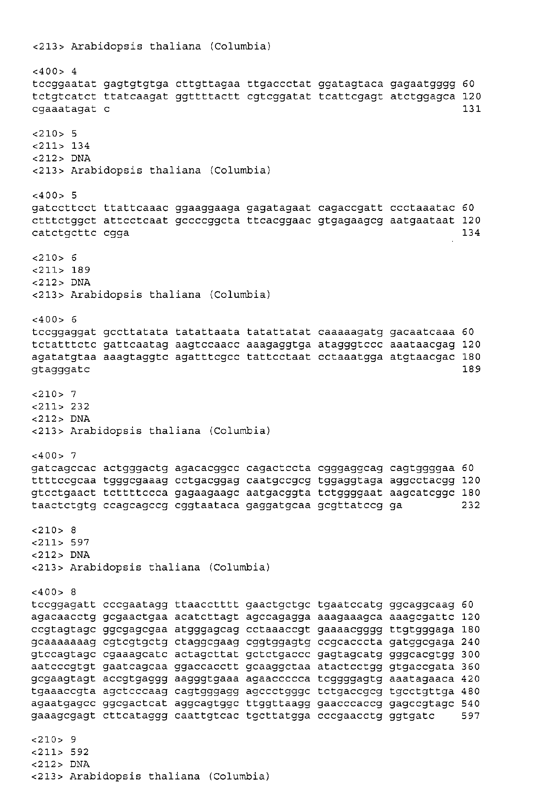
- the nucleotide sequence of spot CP16-a is shown in SEQ ID NO: 4
- the nucleotide sequence of spot CP16-b is shown in SEQ ID NO: 5
- the nucleotide sequence of spot CP19-a is shown in SEQ ID NO: 6
- the nucleotide sequence of spot CP19-b is shown in SEQ ID NO: 7
- the nucleotide sequence of spot CP22-a is shown in SEQ ID NO: 8
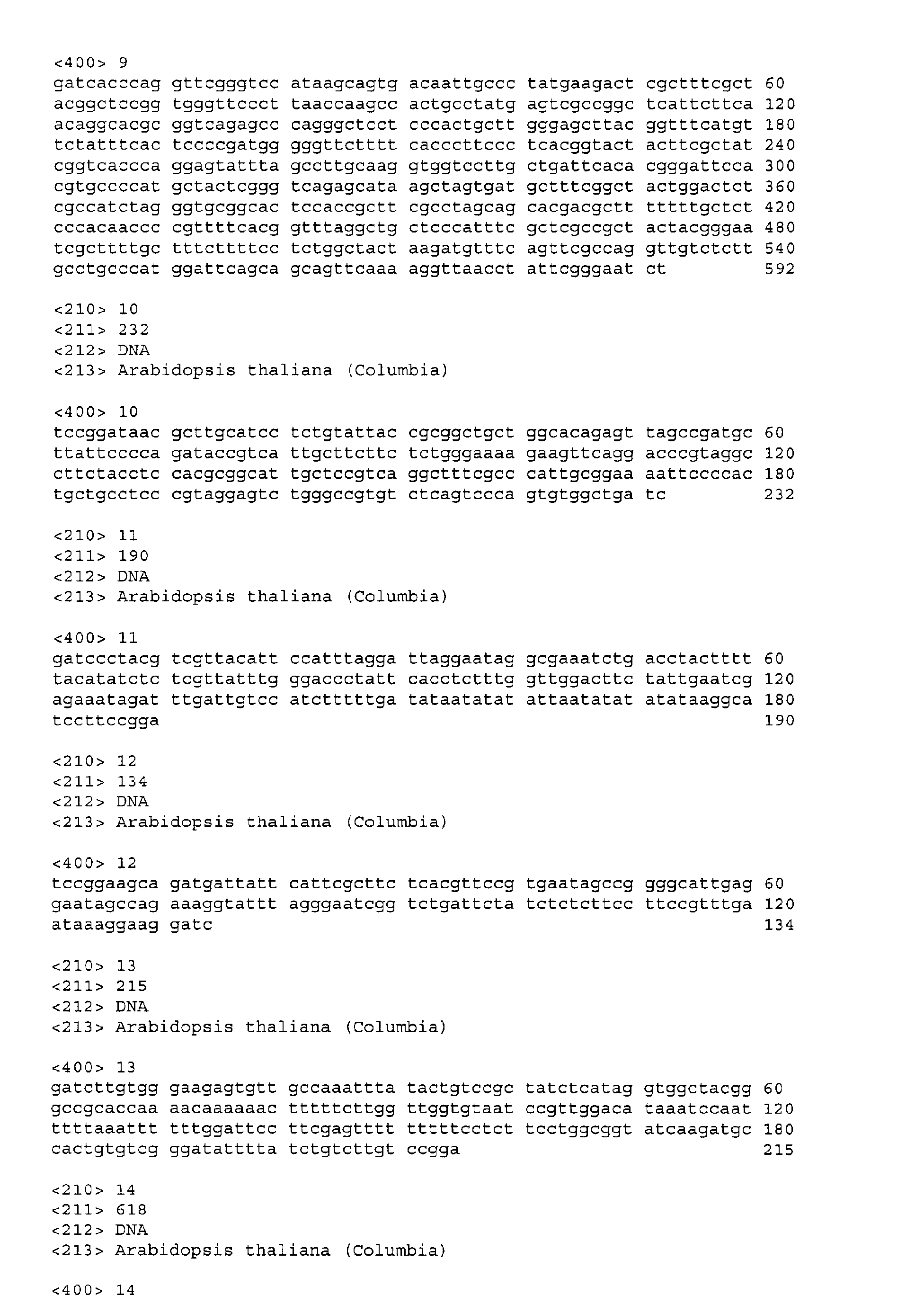
- the nucleotide sequence of spot CP25-b is shown in SEQ ID NO: 9
- the nucleotide sequence of spot CP28-a is shown in SEQ ID NO: 10
- Figure 5 shows the nucleotide sequence of a DNA fragment comprised in the selected spot CP25-b.
- a two-dimensional electrophoresis pattern was obtained by drying the gel, performing exposures at -80°C for 14 days, and developing.
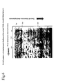
- a two-dimensional pattern obtained by analyzing chloroplast DNA is shown in Figure 6. In the figure, open triangles show the positions of spots observed.
- the target two-dimensional electrophoresis pattern shown in Figure 6 shows that a spot indicates a high signal strength when chloroplast DNA is used. This is because the copy numbers of chloroplast DNA are numerous. From this result, it was shown that the greater the amount of target genomic DNA, the higher the signal strength that can be displayed.
- Arabidopsis thaliana nuclear genomic DNA is used as an analysis target.
- nuclear genomic DNA of Arabidopsis thaliana Coldia-strain
- partial gap regions an undetermined region
- almost the entire nucleotide sequence about 130Mb
- the Arabidopsis Information resource referred to as TAIR, www.arabidopsis.org/some.html
- a two-dimensional electrophoresis pattern was produced, using Not I as the first restriction enzyme, Eco RV as the second restriction enzyme and Mbo I as the third restriction enzyme. These Eco RV and Mbo I are insensitive to 5-methyl cytosine (5mC) which results from methylation of cytosine, but Not I is sensitive.
- the obtained control two-dimensional electrophoresis pattern is shown in Figure 7.
- the display of nucleotide number shows mobility when a molecular weight marker was actually electrophoresed, and the horizontal axis shows development of a first-dimension fractionation and the longitudinal axis shows development of a second-dimension fractionation.
- each spot shows the name of gene locus comprising Not I site in genomic DNA.
- Each number from 1 to 5 located at the extreme left denotes chromosome number, and either "-a" or "-b" located at the extreme right denotes respectively the upstream or downstream side of Not I site.
- Character strings other than above stated denote the names of clones which were produced during determination of nucleotide sequences.
- a spot to which "4FCA1-b" is linked is derived from chromosome number 4, and it means a DNA fragment having a determined nucleotide sequence, comprising a DNA fragment being downstream of Not I site in FCA1 clone.
- a black circle ( ⁇ ) means a spot derived from chromosome number 1
- a black rhombus ( ⁇ ) means a spot derived from chromosome number 2
- a black triangle ( ⁇ ) means a spot derived from chromosome number 3
- a cross ( ⁇ ) means a spot derived from chromosome number 4
- a black square ( ⁇ ) means a spot derived from chromosome number 5.
- Control means a portion excerpted in part from a control two-dimensional electrophoresis pattern and enlarged.
- 4FCA1-b disappeared as a result of X-ray irradiation.
- spots of 4T15F16-b being upstream, and 4T16H5-a and 4T805-a being downstream could be confirmed, thereby suggesting that a deletion of at most about 2.4Mb occurred between 4FCA1-b and 4T16H5-a.
- abnormal information in the nucleotide sequence information regarding Arabidopsis thaliana nuclear genome was detected. Specifically, character strings other than A, G, C and T comprised in the nucleotide sequence information are detected, and the extracted character strings other than A, G, C and T are defined as abnormal information.
- abnormal information "r” was detected at position 27,421 of Accession No. U95973 (detected in DDBJ: http://ftp2.ddbj.nig.ac.jp: 8000/getstart-j.html), a T19D16 clone on the first chromosome.
- This case means that abnormal information is comprised in a spot derived from a T19D16 clone, in a control two-dimensional electrophoresis pattern shown in Figure 7. Accordingly, in advance of performing a comparison between the control two-dimensional electrophoresis pattern shown in Figure 7 and a target two-dimensional electrophoresis pattern, a spot derived from a T19D16 clone is linked to the abnormal information. According to this step, where a mismatch was observed regarding a spot derived from the T19D16 clone in the above comparison, it shows that abnormal information is linked to the spot derived from the T19D16 clone.
- the mismatch regarding a spot derived from a T19D16 clone is caused by abnormal information or not.
- the determination is carried out by correcting abnormal information "r" to "G” or "A” to prepare 2 types of control two-dimensional electrophoresis patterns, and comparing each of these 2 types of control two-dimensional electrophoresis pattern with a target two-dimensional electrophoresis pattern. Where a spot derived from a T19D16 clone in the target two-dimensional electrophoresis pattern matches with that in either of the 2 control two-dimensional electrophoresis patterns, it shows that the mismatch is caused by abnormal information.
- a target two-dimensional electrophoresis pattern was able to be analyzed more in detail by detecting abnormal information in the nucleotide sequence information regarding Arabidopsis thaliana nuclear genome and linking the abnormal information to a spot in a control two-dimensional electrophoresis pattern.
- a control two-dimensional electrophoresis pattern to analyze a target genomic DNA can easily be produced under various conditions. Therefore, the present invention provides a program for easily analyzing a two-dimensional electrophoresis pattern produced using genomic DNA.
- the present invention provides a method for quickly and reliably analyzing genomic DNA by producing a control two-dimensional electrophoresis pattern based on genomic nucleotide sequence information.
Landscapes
- Life Sciences & Earth Sciences (AREA)
- Health & Medical Sciences (AREA)
- Engineering & Computer Science (AREA)
- Physics & Mathematics (AREA)
- General Health & Medical Sciences (AREA)
- Chemical & Material Sciences (AREA)
- Analytical Chemistry (AREA)
- General Physics & Mathematics (AREA)
- Molecular Biology (AREA)
- Theoretical Computer Science (AREA)
- Medical Informatics (AREA)
- Spectroscopy & Molecular Physics (AREA)
- Biophysics (AREA)
- Biotechnology (AREA)
- Bioinformatics & Computational Biology (AREA)
- Bioinformatics & Cheminformatics (AREA)
- Proteomics, Peptides & Aminoacids (AREA)
- Chemical Kinetics & Catalysis (AREA)
- Electrochemistry (AREA)
- Biochemistry (AREA)
- Evolutionary Biology (AREA)
- Immunology (AREA)
- Pathology (AREA)
- Nuclear Medicine, Radiotherapy & Molecular Imaging (AREA)
- Radiology & Medical Imaging (AREA)
- Quality & Reliability (AREA)
- Computer Vision & Pattern Recognition (AREA)
- Measuring Or Testing Involving Enzymes Or Micro-Organisms (AREA)
Abstract
Description


Claims (25)
- A program which allows a computer to execute the processes of:producing a control two-dimensional electrophoresis pattern based on genomic nucleotide sequence information,comparing said control two-dimensional electrophoresis pattern and a target two-dimensional electrophoresis pattern obtained by performing two-dimensional electrophoresis using target genomic DNA, anddetecting a difference in spot position between said control two-dimensional electrophoresis pattern and said target two-dimensional electrophoresis pattern.
- The program according to claim 1, wherein, in said process of producing the control two-dimensional electrophoresis pattern, the control two-dimensional electrophoresis pattern is produced by detecting the recognition sequence of a first restriction enzyme and the recognition sequence of a second restriction enzyme in said genomic nucleotide sequence information, and basing on a nucleotide sequence flanked by the first restriction enzyme recognition sequence and the second restriction enzyme recognition sequence and another nucleotide sequence flanked by the two first restriction enzyme recognition sequences.
- The program according to claim 2, wherein said first restriction enzyme and said second restriction enzyme are selected from methylation insensitive restriction enzymes.
- The program according to claim 2, wherein said first restriction enzyme and said second restriction enzyme are selected from methylation sensitive restriction enzymes.
- The program according to any one of the preceding claims, which comprises a process of obtaining said genomic nucleotide sequence information by means of a communication line network.
- The program according to any one of the preceding claims, wherein, in said process of producing the control two-dimensional electrophoresis pattern, a plurality of spots are produced based on genomic nucleotide sequence information and these spots are linked to gene loci information on a genome.
- The program according to any one of the preceding claims, wherein, in said process of producing the control two-dimensional electrophoresis pattern based on a genomic nucleotide sequence information, abnormal information comprised in said genomic nucleotide sequence information is detected.
- The program according to claim 7, which links the detected abnormal information to a spot comprised in the produced two-dimensional electrophoresis pattern in order to memorize the information.
- A method for analyzing genomic DNA which comprises the steps of:producing a two-dimensional electrophoresis pattern by performing two-dimensional electrophoresis using a target genomic DNA,comparing said two-dimensional electrophoresis pattern and a control two-dimensional electrophoresis pattern produced based on genomic nucleotide sequence information, anddetecting a difference in spot position between said control two-dimensional electrophoresis pattern and said target two-dimensional electrophoresis pattern.
- The method for analyzing genomic DNA according to claim 9, which comprises a step of extracting said target genomic DNA from plant cells.
- The method for analyzing genomic DNA according to claim 9, wherein said target genomic DNA is derived from a higher organism.
- The method for analyzing genomic DNA according to any one of claims 9 to 11, wherein, in said step of producing the two-dimensional electrophoresis pattern of said target genomic DNA by the following steps (a) to (e):(a) treating genomic DNA with a first restriction enzyme,(b) adding a label to a site cleaved with said restriction enzyme,(c) performing a first-dimension fractionation on the obtained DNA fragment by electrophoresis,(d) after treating the DNA fragment fractioned in step (c) with a second restriction enzyme, performing a second-dimension fractionation, and(e) detecting a spot of the labeled DNA fragment fractioned in step (d).
- The method for analyzing genomic DNA according to claim 12, wherein, after said step (b), the obtained DNA fragment is treated with a restriction enzyme different from the first and second restriction enzymes and before said step (c) is performed.
- The method for analyzing genomic DNA according to claim 12 or claim 13, which comprises detecting the recognition sequences of said first and second restriction enzymes in genomic nucleotide sequence information, and producing the control two-dimensional electrophoresis pattern based on these cleavage sites.
- The method for analyzing genomic DNA according to any one of claims 12 to 14, wherein said step (b) is carried out by connecting one end of an adapter to the restriction enzyme cleavage site as well as adding said label to the other end of said adapter.
- The method for analyzing genomic DNA according to any one of claims 9 to 15, wherein said control two-dimensional electrophoresis pattern has a plurality of spots produced based on genomic nucleotide sequence information and links these spots to gene loci information on a genome.
- The method for analyzing genomic DNA according to any one of claims 9 to 16, which comprises detecting abnormal information comprised in genomic nucleotide sequence information before producing the control two-dimensional electrophoresis pattern based on said genomic nucleotide sequence information.
- The method for analyzing genomic DNA according to claim 17, which comprises linking the detected abnormal information to a spot comprised in the produced two-dimensional electrophoresis pattern.
- A method for identifying genes associated with mutant genes and/or mutant traits, which comprises detecting mutated sites in target genomic DNA by the method for analyzing genomic DNA according to any one of claims 9 to 18.
- A method for isolating genes associated with mutant genes and/or mutant traits, which comprises isolating a DNA fragment containing mutated sites detected by the identification method according to claim 19, from said two-dimensional electrophoresis pattern.
- A two-dimensional electrophoresis pattern, which has a plurality of spots produced based on genomic nucleotide sequence information and estimated from said genomic nucleotide sequence information.
- The two-dimensional electrophoresis pattern according to claim 21, wherein said plurality of spots are linked to gene loci information on a genome.
- The two-dimensional electrophoresis pattern according to claim 21, wherein said genomic nucleotide sequence information is nucoleotide sequence information obtained from a plant nuclear genome.
- The two-dimensional electrophoresis pattern according to claim 21, wherein abnormal information detected from said genomic nucleotide sequence information is linked to said plurality of spots.
- The two-dimensional electrophoresis pattern according to claim 21, wherein said genomic nucleotide sequence information is obtained from the genome of a higher organism.
Applications Claiming Priority (4)
| Application Number | Priority Date | Filing Date | Title |
|---|---|---|---|
| JP2000327632 | 2000-10-26 | ||
| JP2000327632 | 2000-10-26 | ||
| JP2001084370 | 2001-03-23 | ||
| JP2001084370 | 2001-03-23 |
Publications (2)
| Publication Number | Publication Date |
|---|---|
| EP1202211A2 true EP1202211A2 (en) | 2002-05-02 |
| EP1202211A3 EP1202211A3 (en) | 2004-12-22 |
Family
ID=26602866
Family Applications (1)
| Application Number | Title | Priority Date | Filing Date |
|---|---|---|---|
| EP01309062A Withdrawn EP1202211A3 (en) | 2000-10-26 | 2001-10-25 | Genomic DNA analysis computer program |
Country Status (2)
| Country | Link |
|---|---|
| US (1) | US20020147549A1 (en) |
| EP (1) | EP1202211A3 (en) |
Families Citing this family (3)
| Publication number | Priority date | Publication date | Assignee | Title |
|---|---|---|---|---|
| US20020183936A1 (en) * | 2001-01-24 | 2002-12-05 | Affymetrix, Inc. | Method, system, and computer software for providing a genomic web portal |
| US7704687B2 (en) * | 2002-11-15 | 2010-04-27 | The Johns Hopkins University | Digital karyotyping |
| JP7224207B2 (en) * | 2019-03-05 | 2023-02-17 | 株式会社日立ハイテク | Genotyping device and method |
Family Cites Families (3)
| Publication number | Priority date | Publication date | Assignee | Title |
|---|---|---|---|---|
| NL8801147A (en) * | 1988-05-02 | 1989-12-01 | Tno | METHOD FOR DETECTING GENETIC VARIATION, AND SUITABLE KIT. |
| US5776767A (en) * | 1995-12-12 | 1998-07-07 | Visible Genetics Inc. | Virtual DNA sequencer |
| US5858683A (en) * | 1996-08-30 | 1999-01-12 | Matritech, Inc. | Methods and compositions for the detection of cervical cancer |
-
2001
- 2001-10-25 EP EP01309062A patent/EP1202211A3/en not_active Withdrawn
- 2001-10-26 US US10/003,446 patent/US20020147549A1/en not_active Abandoned
Also Published As
| Publication number | Publication date |
|---|---|
| EP1202211A3 (en) | 2004-12-22 |
| US20020147549A1 (en) | 2002-10-10 |
Similar Documents
| Publication | Publication Date | Title |
|---|---|---|
| Dumschott et al. | Oxford Nanopore sequencing: new opportunities for plant genomics? | |
| Bentolila et al. | Comprehensive high-resolution analysis of the role of an Arabidopsis gene family in RNA editing | |
| Polanski et al. | Bioinformatics | |
| Usuka et al. | Optimal spliced alignment of homologous cDNA to a genomic DNA template | |
| JP6314091B2 (en) | DNA sequence data analysis | |
| US20220205034A1 (en) | Method for quickly identifying clean transgenic or gene-edited plants and insertion sites by using whole genome re-sequencing data | |
| Gregory et al. | Genome mapping by fluorescent fingerprinting | |
| US6195449B1 (en) | Method and apparatus for analyzing data files derived from emission spectra from fluorophore tagged nucleotides | |
| US5867402A (en) | Computational analysis of nucleic acid information defines binding sites | |
| Claverie | A streamlined random sequencing strategy for finding coding exons | |
| US5891632A (en) | Method and device for fragment trace data display in DNA base sequencing | |
| ATE296358T1 (en) | NEW GENETIC MAPPING TECHNOLOGY | |
| US20020147549A1 (en) | Genomic DNA analysis program | |
| CN118038979B (en) | Methods for detecting mutation patterns and transposition imprints of transposon insertion into human genome | |
| JP2005156361A (en) | Genomic DNA analysis program | |
| CN108774646A (en) | With the relevant SNP marker of the thick character of millet fringe and its detection primer and application | |
| JP5344670B2 (en) | Gene expression analysis method, gene expression analysis apparatus, and gene expression analysis program | |
| CN108796053A (en) | A kind of identification method of arabidopsis gene mutant | |
| JP2002350401A (en) | Genomic DNA analysis program | |
| JPH11118760A (en) | Analysis method of electrophoresis pattern of nucleic acid fragment | |
| Mathews | DNA Sequencing: A Brief History | |
| CN121171351B (en) | Method, device, equipment and medium for predicting site detection limitation in whole exome sequencing | |
| US20020102579A1 (en) | Method for analyzing genomic DNA | |
| Tenney | Basecalling for Traces Derived for Multiple Templates | |
| CN120690279A (en) | A method for sex identification of giant pandas based on short-read sequencing |
Legal Events
| Date | Code | Title | Description |
|---|---|---|---|
| PUAI | Public reference made under article 153(3) epc to a published international application that has entered the european phase |
Free format text: ORIGINAL CODE: 0009012 |
|
| AK | Designated contracting states |
Kind code of ref document: A2 Designated state(s): AT BE CH CY DE DK ES FI FR GB GR IE IT LI LU MC NL PT SE TR |
|
| AX | Request for extension of the european patent |
Free format text: AL;LT;LV;MK;RO;SI |
|
| PUAL | Search report despatched |
Free format text: ORIGINAL CODE: 0009013 |
|
| AK | Designated contracting states |
Kind code of ref document: A3 Designated state(s): AT BE CH CY DE DK ES FI FR GB GR IE IT LI LU MC NL PT SE TR |
|
| AX | Request for extension of the european patent |
Extension state: AL LT LV MK RO SI |
|
| 17P | Request for examination filed |
Effective date: 20050613 |
|
| AKX | Designation fees paid |
Designated state(s): AT BE CH CY DE DK ES FI FR GB GR IE IT LI LU MC NL PT SE TR |
|
| STAA | Information on the status of an ep patent application or granted ep patent |
Free format text: STATUS: THE APPLICATION IS DEEMED TO BE WITHDRAWN |
|
| 18D | Application deemed to be withdrawn |
Effective date: 20051125 |