EP1196081B1 - Bilderzeugungsgerät zum integrieren verschiedener bildmodalitäten - Google Patents
Bilderzeugungsgerät zum integrieren verschiedener bildmodalitäten Download PDFInfo
- Publication number
- EP1196081B1 EP1196081B1 EP00943361.6A EP00943361A EP1196081B1 EP 1196081 B1 EP1196081 B1 EP 1196081B1 EP 00943361 A EP00943361 A EP 00943361A EP 1196081 B1 EP1196081 B1 EP 1196081B1
- Authority
- EP
- European Patent Office
- Prior art keywords
- data
- images
- image
- sample
- spectral
- Prior art date
- Legal status (The legal status is an assumption and is not a legal conclusion. Google has not performed a legal analysis and makes no representation as to the accuracy of the status listed.)
- Expired - Lifetime
Links
Images
Classifications
-
- A—HUMAN NECESSITIES
- A61—MEDICAL OR VETERINARY SCIENCE; HYGIENE
- A61B—DIAGNOSIS; SURGERY; IDENTIFICATION
- A61B5/00—Measuring for diagnostic purposes; Identification of persons
- A61B5/44—Detecting, measuring or recording for evaluating the integumentary system, e.g. skin, hair or nails
- A61B5/441—Skin evaluation, e.g. for skin disorder diagnosis
- A61B5/442—Evaluating skin mechanical properties, e.g. elasticity, hardness, texture, wrinkle assessment
-
- A—HUMAN NECESSITIES
- A61—MEDICAL OR VETERINARY SCIENCE; HYGIENE
- A61B—DIAGNOSIS; SURGERY; IDENTIFICATION
- A61B5/00—Measuring for diagnostic purposes; Identification of persons
- A61B5/0059—Measuring for diagnostic purposes; Identification of persons using light, e.g. diagnosis by transillumination, diascopy, fluorescence
- A61B5/0071—Measuring for diagnostic purposes; Identification of persons using light, e.g. diagnosis by transillumination, diascopy, fluorescence by measuring fluorescence emission
-
- A—HUMAN NECESSITIES
- A61—MEDICAL OR VETERINARY SCIENCE; HYGIENE
- A61B—DIAGNOSIS; SURGERY; IDENTIFICATION
- A61B5/00—Measuring for diagnostic purposes; Identification of persons
- A61B5/0059—Measuring for diagnostic purposes; Identification of persons using light, e.g. diagnosis by transillumination, diascopy, fluorescence
- A61B5/0073—Measuring for diagnostic purposes; Identification of persons using light, e.g. diagnosis by transillumination, diascopy, fluorescence by tomography, i.e. reconstruction of 3D images from 2D projections
-
- A—HUMAN NECESSITIES
- A61—MEDICAL OR VETERINARY SCIENCE; HYGIENE
- A61B—DIAGNOSIS; SURGERY; IDENTIFICATION
- A61B5/00—Measuring for diagnostic purposes; Identification of persons
- A61B5/0059—Measuring for diagnostic purposes; Identification of persons using light, e.g. diagnosis by transillumination, diascopy, fluorescence
- A61B5/0077—Devices for viewing the surface of the body, e.g. camera, magnifying lens
-
- A—HUMAN NECESSITIES
- A61—MEDICAL OR VETERINARY SCIENCE; HYGIENE
- A61B—DIAGNOSIS; SURGERY; IDENTIFICATION
- A61B5/00—Measuring for diagnostic purposes; Identification of persons
- A61B5/01—Measuring temperature of body parts ; Diagnostic temperature sensing, e.g. for malignant or inflamed tissue
- A61B5/015—By temperature mapping of body part
-
- A—HUMAN NECESSITIES
- A61—MEDICAL OR VETERINARY SCIENCE; HYGIENE
- A61B—DIAGNOSIS; SURGERY; IDENTIFICATION
- A61B5/00—Measuring for diagnostic purposes; Identification of persons
- A61B5/145—Measuring characteristics of blood in vivo, e.g. gas concentration, pH value; Measuring characteristics of body fluids or tissues, e.g. interstitial fluid, cerebral tissue
- A61B5/14535—Measuring characteristics of blood in vivo, e.g. gas concentration, pH value; Measuring characteristics of body fluids or tissues, e.g. interstitial fluid, cerebral tissue for measuring haematocrit
-
- A—HUMAN NECESSITIES
- A61—MEDICAL OR VETERINARY SCIENCE; HYGIENE
- A61B—DIAGNOSIS; SURGERY; IDENTIFICATION
- A61B5/00—Measuring for diagnostic purposes; Identification of persons
- A61B5/145—Measuring characteristics of blood in vivo, e.g. gas concentration, pH value; Measuring characteristics of body fluids or tissues, e.g. interstitial fluid, cerebral tissue
- A61B5/1455—Measuring characteristics of blood in vivo, e.g. gas concentration, pH value; Measuring characteristics of body fluids or tissues, e.g. interstitial fluid, cerebral tissue using optical sensors, e.g. spectral photometrical oximeters
-
- A—HUMAN NECESSITIES
- A61—MEDICAL OR VETERINARY SCIENCE; HYGIENE
- A61B—DIAGNOSIS; SURGERY; IDENTIFICATION
- A61B5/00—Measuring for diagnostic purposes; Identification of persons
- A61B5/41—Detecting, measuring or recording for evaluating the immune or lymphatic systems
- A61B5/414—Evaluating particular organs or parts of the immune or lymphatic systems
- A61B5/415—Evaluating particular organs or parts of the immune or lymphatic systems the glands, e.g. tonsils, adenoids or thymus
-
- A—HUMAN NECESSITIES
- A61—MEDICAL OR VETERINARY SCIENCE; HYGIENE
- A61B—DIAGNOSIS; SURGERY; IDENTIFICATION
- A61B5/00—Measuring for diagnostic purposes; Identification of persons
- A61B5/41—Detecting, measuring or recording for evaluating the immune or lymphatic systems
- A61B5/414—Evaluating particular organs or parts of the immune or lymphatic systems
- A61B5/418—Evaluating particular organs or parts of the immune or lymphatic systems lymph vessels, ducts or nodes
-
- A—HUMAN NECESSITIES
- A61—MEDICAL OR VETERINARY SCIENCE; HYGIENE
- A61B—DIAGNOSIS; SURGERY; IDENTIFICATION
- A61B5/00—Measuring for diagnostic purposes; Identification of persons
- A61B5/44—Detecting, measuring or recording for evaluating the integumentary system, e.g. skin, hair or nails
- A61B5/441—Skin evaluation, e.g. for skin disorder diagnosis
- A61B5/444—Evaluating skin marks, e.g. mole, nevi, tumour, scar
-
- A—HUMAN NECESSITIES
- A61—MEDICAL OR VETERINARY SCIENCE; HYGIENE
- A61B—DIAGNOSIS; SURGERY; IDENTIFICATION
- A61B5/00—Measuring for diagnostic purposes; Identification of persons
- A61B5/44—Detecting, measuring or recording for evaluating the integumentary system, e.g. skin, hair or nails
- A61B5/441—Skin evaluation, e.g. for skin disorder diagnosis
- A61B5/445—Evaluating skin irritation or skin trauma, e.g. rash, eczema, wound, bed sore
-
- A—HUMAN NECESSITIES
- A61—MEDICAL OR VETERINARY SCIENCE; HYGIENE
- A61B—DIAGNOSIS; SURGERY; IDENTIFICATION
- A61B5/00—Measuring for diagnostic purposes; Identification of persons
- A61B5/44—Detecting, measuring or recording for evaluating the integumentary system, e.g. skin, hair or nails
- A61B5/441—Skin evaluation, e.g. for skin disorder diagnosis
Definitions
- the invention is directed to an imaging apparatus and methods for performing assessment and monitoring with interpreted imaging.
- Embodiments of the invention are particularly useful in surgery, clinical procedures, tissue assessment, diagnostic procedures, health monitoring, and medical evaluations.
- Spectroscopy is an enormously powerful tool for the analysis of biomedical samples.
- the medical community has a definite preference for imaging methods, as exemplified by methods such as MRI and CT scanning as well as standard X-ray photography and ultrasound imaging. This is entirely understandable as many factors need to be taken into account for a physician to make a clinical diagnosis. Imaging methods potentially can provide far more information to a physician than their non-imaging counterparts. With this medical reality in mind, there has been considerable effort put into combining the power and versatility of imaging method with the specificity of spectroscopic methods.
- Near-infrared (near-IR) spectroscopy and spectroscopic imaging can measure the balance between oxygen delivery and tissue oxygen utilization by monitoring the hemoglobin oxygen saturation in tissues ( Sowa, M.G. et al., 1998, Proc. SPIE 3252, pp. 199-207 ; Sowa, G.W. et al., 1999, Journal of Surgical Research, 86:62-29 ; Sow, G.W. et al., 1999, Journal of Biomedical Optics, 4:474-481 ; Mansfield, J.R., et al., 2000, International Society of Optical Engineers, 3920:99-197 ).
- Non-invasive monitoring of hemoglobin oxygenation exploits the differential absorption of HbO 2 and Hb, along with the fact that near-IR radiation can penetrate relatively deeply into tissues.
- Pulse oximetry routinely supplies a noninvasive measure of arterial hemoglobin oxygenation based on the differential red-visible and near infrared absorption of Hb and HbO 2 .
- Visible/near-IR multispectral imaging permits the regional variations in tissue perfusion to be mapped on macro and micro scale. Unlike infrared thermography, hyperspectral imaging alone does not map the thermal emission of the tissues.
- this imaging method relies on the differential absorption of light by a chromophore, such as, Hb and HbO 2 , resulting in differences in the wavelength dependence of the tissue reflectance depending on the hemoglobin oxygen saturation of the tissue.
- Spectroscopic imaging methodologies and data are becoming increasingly common in analytical laboratories, whether it be magnetic resonance (MRI), mid-IR, Raman, fluorescence and optical microscopy, or near-IR/visible-based imaging.
- MRI magnetic resonance
- Raman Raman
- fluorescence and optical microscopy or near-IR/visible-based imaging.
- the volume of information contained in spectroscopic images can make standard data processing techniques cumbersome.
- the objective of analyzing spectroscopic images is not only to determine what the spectrum is at any particular pixel in the sample, but also to determine which regions of the sample contain similar spectra; i.e., what regions of the sample contain chemically related compounds.
- Multivariate analysis methodologies can be used to determine both the spectral and spatial characteristics of a sample within a spectroscopic imaging data set. These techniques can also be used to analyze variations in the temporal shape of a time series of images either derived for extracted from a time series of spectroscopic images.
- Spectroscopic imaging provides the specificity of spectroscopy while at the same time relaying spatial information by providing images of the sample that convey some chemical meaning.
- the objective in analyzing heterogeneous systems is to identify not only the components present in the system, but their spatial distribution.
- the true power of this technique relative to traditional imaging methods lies in its inherent multivariate nature. Spatial relationships among many parameters can be assessed simultaneously.
- the chemical heterogeneity or regional similarity within a sample is captured in a high dimensional representation which can be projected onto a number of meaningful low dimensional easily interpretable representations which typically comprise a set of composite images each having a specific meaning.
- spectroscopic imaging data cube spectroscopic imaging data cube or just hypercube.
- This is a three dimensional array of data, consisting of two spatial dimensions (the imaging component), and one spectral dimension. It can be thought of as an array of spatially resolved individual spectra, with every pixel in the first image consisting of an entire spectrum, or as a series of spectrally resolved images.
- the 3D data cube can be treated as a single entity containing enormous amounts of spatial and spectral information about the sample from which it was acquired.
- Multi-modal image fusion is an important problem frequently addressed in medical image analysis. Registration is the process of aligning data that arise from different sources into one consistent coordinate frame. For example, various tissues appear more clearly in different types of imaging methods. Soft tissue, for example, is imaged well in MR scans, while bone is more easily discernible in CT scans. Blood vessels are often highlighted better in an MR angiogram than in a standard MR scan. Multiple scans of the same patient will generally be unregistered when acquired, as the patient may be in different positions in each scanner, and each scanner has its own coordinate system. In order to fuse the information from all scans into one coherent frame, the scans must be registered. The very reason why multiple scans are useful is what makes the registration process difficult. As each modality images tissue differently and has its own artifacts and noise characteristics, accurately modeling the intensity relationship between the scans, and subsequently aligning them, is difficult.
- the registration of two images consists of finding the transformation that best maps one image into the other. If I 1 and I 2 are two images of the same tissue and T is the correct transformation, then the voxel I 1 (x) corresponds to the same position in the sample as the voxel I 2 (T(x)).
- T is a rigid transformation consisting of three degrees of freedom of rotation and three degrees of freedom of translation. The need for rigid registration arises primarily from the patient being in different positions in the scanning devices used to image the anatomy. The information from all the images is best used when presented in one unified coordinate system. Without such image fusion, the clinician must mentally relate the information from the disparate coordinate frames.
- One method of aligning the two images is to define an intermediate, patient-centered coordinate system, instead of trying to directly register the images to one another.
- An example of a patient-centered reference frame is the use of fiducial markers attached to a patient throughout the various image acquisitions.
- the fiducial markers define a coordinate system specific to the patient, independent of the scanner or choice of imaging modality.
- the volumes can be registered by computing the best alignment of the corresponding fiducials ( Horn, B.K.P., 1987, Journal of the Optical Society of America A, 4:629-642 ; Mandava, V.R., et al., Proc SPIE, 1992, 1652:271-282 ; Haralick, R.M., et al., 1993, Computer and Robot Vision ).
- the main drawback of this method is that the markers must remain attached to the patient at the same positions throughout all image acquisitions. For applications such as change detection over months or years, this registration method is not suitable.
- Fiducial registration is typically used as ground-truth to evaluate the accuracy of other methods as careful placement and localization of the markers can provide very accurate alignment ( West, J. et al., 1996, Proc SPIE, Newport Beach, California ).
- corresponding anatomical feature points can be extracted from the images and used to compute the best alignment ( Maintz, J.B. Antione, et al., 1995 Computer Vision, Virtual Reality and Robotics in Medicine, pp. 219-228 ; Maguire, Jr., G., et al., 1991, IEEE Computer Graphics Applications, 11:20-29 ).
- This approach depends greatly on the ability to automatically and accurately extract reliable image features.
- methods of feature extraction such as intensity thresholding or edge detection do not work well on medical scans, due to non-linear gain fields and highly textured structures. Even manual identification of corresponding 3D anatomical points can be unreliable. Without the ability to accurately localize corresponding features in the images, alignment in this manner is difficult.
- a common method of registering MR and CT of the head involves extracting the skin (or skull) surfaces from both images, and aligning the 3D head models ( Jiang, H., et al., 1992 Proc. SPIE, 1808:196-213 ; Lemoine, D. et al., 1994, Proc. SPIE, 2164:46-56 ).
- the brain surface is typically used since the skull is not clearly visible in PET ( Pelizzari, C., et al., J Comput Assist. Tomogr., 1989, 13:20-26 ).
- the 3D models are then rigidly registered using surface-based registration techniques ( Ettinger, G., 1997, MIT Ph.D Thesis) .
- surface-based registration techniques Ettinger, G., 1997, MIT Ph.D Thesis.
- the success of such methods relies on the structures being accurately and consistently segmented across modalities and the surfaces having rich enough structure to be unambiguously registered.
- Voxel-based approaches to registration do not extract any features from the images, but use the intensities themselves to register the two images. Such approaches model the relationships between intensities of the two images when they are registered, and then search through the transformation space to find an alignment that best agrees with the model. Various intensity models are discussed, including correlation, mutual information. and joint intensity priors.
- Correlation is a measure commonly used to compare two images or regions of images for computer vision problems such as alignment or matching. Given the intensity values of two image patches stacked in the vectors u and v, the normalized correlation measure is the dot product of unit vectors in the directions of u and v: u • v / ⁇ u ⁇ ⁇ v ⁇
- correlation-based methods can be computed quite efficiently using convolution operators. Correlation is applicable when one expects a linear relationship between the intensities in the two images.
- normalized correlation provides some amount of robustness to lighting variation over a measure such as sum of square differences (SSD),
- SSD sum of square differences
- 2 The primary reason for acquiring more than one medical scan of a patient stems from the fact that each scan provides different information to the clinician. Therefore, two images that have a simple linear intensity relationship may be straightforward to register, but do not provide any additional information than one scan by itself. On the other hand, if the images are completely independent (e.g. no intensity relationship exists between them), then they cannot be registered using voxel-based methods. In general, there is some dependence between images of different modalities and each modality does provide additional information.
- an internal image is a rendering function R of underlying tissue properties, P(x), over positions x.
- An image of modality A could be represented as a function R A (P) and a registered image of modality B of the same patient would be another function, say R B (P).
- R A P
- R B P
- F F
- T * argmin T ⁇ ⁇ x ⁇ F R B P x - R A P x 2
- T is the transformation between the two sets of image coordinates.
- Van den Elsen et al. compute such a mapping that makes a CT image appear more like an MR, and then register the images using correlation ( van den Elsen, P., et al., 1994, "Visualization in Biomedical Computing," 1994 Proc SPIE, 2359:227-237 ).
- explicitly computing the function F that relates two imaging modalities is difficult and under-constrained.
- MI mutual information
- H U V H U + H V - H U V
- H(U) and H(V) are the entropies of the two variables
- H(U,V) is the joint entropy.
- H(U, V) H(U) + H(V), which implies the mutual information is zero.
- Derivatives of the entropies with respect to the pose parameters can be calculated and used to perform stochastic gradient ascent (Wells, 1996).
- West et al. compare many multi-modal registration techniques and find mutual information to be one of the most accurate across all pairs of modalities (West, 1996).
- Leventon et al. introduced an approach to multi-modal registration using statistical models derived from a training set of images ( Leventon, M., et al., 1998, Medical Image Computing and Computer-assisted Intervention ).
- the method involved building a prior model of the intensity relationship between the two scans being registered.
- the method requires a pair of registered training images of the same modalities as those to be registered in order to build the joint intensity model.
- To align a novel pair of images the likelihood of the two images given a certain pose based on our model by sampling the intensities at corresponding points is computed. This current hypothesis can be improved by ascending the log likelihood function. In essence, one computes a probabilistic estimate of the function F (that relates the two imaging modalities) based on intensity co-occurrence.
- F that relates the two imaging modalities
- the present invention is defined in claims 1 and 10 and overcomes problems and disadvantages associated with current strategies and designs and provides methods and apparatus for imaging using real-time or near real-time assessment and monitoring.
- Embodiments of the device are useful in a plurality of settings including surgery, clinical procedures, tissue assessment, diagnostic procedures, forensic, health monitoring and medical evaluations.
- One embodiment of the invention is directed to an imaging apparatus comprising integrating spatial, spectral and temporal features, and optionally other physiologic or relevant data, such as room temperature or ambient light, in a spectral and temporal multimodal imaging system for the evaluation of biological systems and stimuli and fusing one or more thermal images or other imaging modalities and hyperspectral data cube for assessment of biological processes.
- the integrated features may comprise two or more of visible or infrared hyperspectral images, visible or infrared brightfield images, thermal images, fluorescence images, Raman images and/or other relevant imaging modalities.
- the imaging apparatus may further comprise a specific UV, visible and/or infrared light source, and means for collecting two or more of visible or infrared hyperspectral images, visible or infrared brightfield images, thermal images, fluorescence images, Raman images, or standard video images.
- a specific UV, visible and/or infrared light source and means for collecting two or more of visible or infrared hyperspectral images, visible or infrared brightfield images, thermal images, fluorescence images, Raman images, or standard video images.
- Another embodiment of the invention is directed to methods for detecting a diseased condition comprising acquiring thermal images from a target, acquiring visible or infrared hyperspectral images from the same target, fusing the thermal images and visible or infrared hyperspectral images to analyze spatial distributions and/or feature determination of the target.
- Thermal images or hyperspectral images of the target and/or other data can be interlaced with a time dependent reference to determine changes which could influence and be correlated with results from other imaging modalities.
- Wavelengths can be selected to maximize diagnostic information for a specific tissue state or anticipated end diagnostic goal.
- the selection step involves performing multivariate image and spectral processing using multivariate image and spectral processing algorithms to extract information from the plurality of images and spectra for real-time or near real-time assessment.
- hyperspectral collection devices in a variety of wavelength regimens could be used simultaneously or sequentially or on an as needed basis.
- a visible hyperspectral images could be combined with a near infrared hyperspectral imager (plus or minus a broad band thermal camera) to provide combined information from both wavelength regions.
- tissue health mapping skin sebum level mapping; skin dryness, skin texture, skin feel or skin color mapping; skin damage detection and mapping (UV damage, frostbite, bums, cuts, abrasions) impact of cosmetics or other substances applied to the skin bruise age, force of impact, peripheral vascular disease diagnosis, extent, determination or regionalization of ischemia, varicose veins or hemorrhage detection, local detection and mapping, systemic infection detection, differentiation between viral, bacterial and fungal, and more specific identification, such as between gram negative and gram positive bacterial infection, venous occlusion increase in total hemoglobin, hematocrit, and change in deoxyhemoglobin/oxyhemoglobin ratio, differentiate between ischemia and hypoxia, burn depth and wound healing evaluation, non-invasive diagnosis of shock by imaging uninjured skin, hemorrhagic shock, septic shock, burn shock, changes in a dynamic system as a function of time or other parameter, vascular occlusion, vaso
- the present invention is directed to an imaging apparatus and methods for performing real-time or near real-time assessment and monitoring.
- Embodiments of the device are useful in a plurality of settings including surgery, clinical procedures, tissue assessment, diagnostic procedures, forensic, health monitoring and medical evaluations.
- ATR Automatic Target Recognition
- a technology developed within the military for automatic analysis and pattern recognition of signature data and gating of images relative to repetitive physiological parameters such as heart rate or respiration.
- an ATR is used to maintain image centering.
- the addition of such novel features as a common optical path optimizes data collection and minimizes processing requirements for a fused image.
- Image fusion between hyperspectral image datasets (also referred to as cubes) and other imaging modalities would allow for the extraction of more medically-relevant features and diagnostic information than any of the modalities alone.
- Addition of physiologically or medically related scalar variables to the data set of one or more hyperspectral imaging sets with or without formal image fusion being required allows for the enhancement of diagnostic algorithms.
- Thermal images or hyperspectral images may be used as an interlaced, time dependent reference to identify changes in the dynamic system. These changes may influence and be correlated with the results from all modalities.
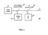
- signal beam 110 is acquired and IR Beam-splitter 160 is placed in the path of signal beam 110 and accordingly, splits or diverts a portion of the infra-red signal beam 110 to infra-red focal plane array 120.
- 90/10 Visible Beam-splitter 130 is placed in signal beam 110 behind IR Beamsplitter 160. Visible Beam-splitter 130 splits the visible spectrum of signal beam 110 into two portions, wherein one portion is received by video camera 150, and the other is received by visible camera 140.
- One or multiple mirrors can be used for the beam splitter. This allows for the simultaneous acquisition of data from multiple modalities.
- Fusion of broad band infrared and hyperspectral imaging methodologies may be useful to devise algorithms for wavelength selection that maximize the diagnostic information for a specific tissue state; employ various multivariate image processing algorithms to extract information from the hyperspectral images and spectra and the thermal images for real-time or near real-time assessment of tissue state; devise image processing algorithms to assess the size and shape of abnormal tissue regions or domains; acquire sequential hyperspectral imaging cubes, thermal images or other physiological data to examine changes in a dynamic system as a function of time. Utility is extended by pairing more superficial data from hyperspectral imaging cubes with deeper perfusion data.
- a method for determining a total hematocrit comprises measuring a spatial distribution of oxyhemoglobin, deoxyhemoglobin and methemoglobin using hyperspectral imaging methods within the visible range or infrared range of the electro-magnetic spectrum; determining total hematocrit by calculating the area under the oxyhemoglobin, deoxyhemoglobin and methemoglobin spectrum or the intensity at their respective wavelengths; and pairing this with perfusion data from broad band thermal camera to permit assessment of total blood volume.
- the invention may be used to determine blood flow within a patient.
- a thermal camera demonstrates a state of perfusion and a hyperspectral camera demonstrates a state of oxygen extraction.
- Spatial characteristics relative to blood vessel assist diagnosis, i.e., like mottling visible in skin, and can see more or less heterogeneity under certain thermal, neurohumoral, physiological or pathological circumstances and in specific spatial patterns.
- the present invention may be used to determine a static or dynamic response of tissue or musculature when applying an active stimulus, such as a thermal change, drug injection, and electromagnetic or mechanical stimulus.
- Venous occlusion causes an increase in total hemoglobin, hematocrit, and an increase in deoxyhemoglobin/oxyhemoglobin ratio. The time course also varies with arterial occlusion and oxyhemoglobin/deoxyhemoglobin ratios.
- Artery and vein measurements can be used as internal calibration on a given picture for tissue levels of oxyhemoglobin/deoxyhemoglobin or thermal image or signature. Further, one can add thermal data by fusing thermal image just as one of the wavelengths in series in hyperspectral cube, i.e., an extra plane. Alternatively, thermal images can be fused to each wavelength image in series. Alternatively or in addition, generic processed analysis of thermal image (degree of variation) weights an image of each wavelength plane or impacts hyperspectral algorithmic analysis. Scalar data presenting physiologic or other relevant data can be also incorporated as described above.
- correction for a patient's motion is done by tissue stabilization or in the case of repetitive motions by gating image frames with a patient's cardiac or respiration cycle.
- Frames at the specific wavelengths selected for a particular diagnostic module are acquired at the same position in sequential cardiac cycles.
- the timing of the cardiac cycle is provided by electrocardiogram or cardiac ultrasound or other method.
- the respiratory variation is timed with an external sensor of respiration or with either the ventilating mechanism or a sensor mechanism of an artificial respirator.
- the present invention may be used to provide signatures of tissue viability or cancer. Markers of cell viability include hyperspectral signatures of oxyhemoglobin and deoxyhemoglobin or other chromaphores, thermal signatures, or fused signatures.
- the present invention is used to determine drug impact on vasodilitation, neurohumoral response, physiology, and pathology.
- the present invention is used to identify and classify a large variety of chemical species, for example, those other than oxyhemoglobin and deoxyhemoglobin.
- Sensor/image fusion permits additional data acquisition and incorporation into diagnostic assessment. This is facilitated by the use of multiple optical paths properly aligned to optimize registration. Inclusion of simultaneous recording of standard video camera images facilitates registration and provides additional data.
- False color imaging may be added real-time to facilitate the rapid understanding of the data presented to the surgeon or other user.
- On board CCD chip filters can be provided to increase processing speed.
- Input for physiologic monitoring systems, such as blood pressure, heart rate, peripheral oxygenation, can be added to the data acquired and fed into diagnostic algorithms.
- a recording system can be included to log the real-time or near real-time output of imaging systems.
- a split frame video display is used to show all modes simultaneously.
- parameters of wound healing may be displayed, such as: oxyhemoglobin or deoxyhemoglobin independently or as a ratio; signatures associated with rapidly dividing cells or dead cells, or particular types of cells; fluid content; hydration/dehydration or edema of tissue; or tissue performance.
- Tissue perfusion data provided by a thermal camera increases accuracy, delivers information about underlying vascular, beds, and/or provides data that will minimize the hyperspectral data processing requirements.
- Thermal images are used provide a baseline to track oxygen extraction or signature changes induced by tissue exposure.
- Increased heterogeneity and spatial features can be important in a diagnosis. For example, in vasoconstriction, it allows identification of areas that are less well perfused small micro areas that manifest as heterogeneity, to be diagnosed. Differences in oxyhemoglobin and deoxyhemoglobin ratios with spatial characteristics provide an image of micromottling. If vasodilated are more uniform, the patterns of vasoconstriction are helpful in diagnosis of infection in general and can aid in the identification of specific infection. Other patterns of heterogeneity are seen with cancers, and for example are associated with areas of increased metabolism or necrosis.
- the present invention may be used to analyze tissue health mapping; skin sebum level mapping; skin dryness, skin texture, skin feel or skin color mapping; skin damage detection and mapping (UV damage, frostbite, bums, cuts, abrasions) impact of cosmetics or other substances applied to the skin bruise age, force of impact, peripheral vascular disease diagnosis, extent, determination or regionalization of ischemia, varicose veins or hemorrhage detection, local detection and mapping, systemic infection detection, differentiation between viral, bacterial and fungal, and more specific identification, such as between gram negative and gram positive bacterial infection, venous occlusion increase in total hemoglobin, hematocrit, and change in deoxyhemoglobin/oxyhemoglobin ratio, differentiate between ischemia and hypoxia, burn depth and wound healing evaluation, non-invasive diagnosis of shock by imaging uninjured skin, hemorrhagic shock, septic shock, burn shock, changes in a dynamic system as a function of time or other parameter, vascular occlusion, va
- motion artifacts of the measurements are used to measure heterogeneity.
- a homogeneous tissue will continue to produce the same spectral signature, whereas heterogeneous tissue will demonstrate a variety of different signatures.
- Extraneous motion artifacts can be reduced by mechanical stabilization of field of regard, for example, by clamping tissue or region of interest. Even in the absence of discrete spatial information, the simple range of spectra obtained, demonstrating the heterogeneity per se can be useful. Dilation makes thermal imaging more uniform and constriction more heterogeneous. The latter correlates with ischemia, microvascular mottling or the edge of larger vessels. Different changes would be detected in association with tumors, immunologic response to infection or other stimulus.
- Motion artifacts are used as an indicator of inhomogeneous distributions of oxygenation and perfusion. Increases or decreases in artifacts not related to motion are used to assess heterogeneity of oxygenation and perfusion, and, hence, viability.
- the present invention may be used to look for signs of perfusion vs. viability. Integration of spatial and spectral and temporal features allows for the diagnosis of viability by creating a perfusion viability matrix. Because blood flow has a temporal component, the amount of blood that gets to tissue may be measured. This can be useful in the assessment of viability, cancer or infection.
- images are correlated with pain and drug response to provide pain feedback with infusion; other drug levels, to provide positive/negative feedback.
- Surface heterogeneity is correlated with infection, to provide determine time of infection, severity, systemic vs. local infection, type of organism, bacterial vs. viral, gram positive versus gram negative
- the present invention is also used to detect drug usage.
- the present invention may also be used for the assessment of metabolism and nutrition. Tissue structure and function, and hence signature, are influenced by nutritional status.
- the present invention may also be used to define adequacy of regional anesthesia or evaluation of pain response and the response to drug therapy with or without an automatic feedback component. It may also be used to identify and evaluate the presence of a drug substance and evaluate the initial response and/or therapeutic efficacy of a variety of pharmaceuticals. It can be used to track die agents and quantify their presence in association with blood flow parameters.
Claims (19)
- Ein multimodaler bildgebender Apparat zum Untersuchen von Veränderungen in einem dynamischen System als eine Funktion der Zeit, umfassend:ein erstes Sammelgerät zum Sammeln spektraler Daten einer Probe und zum Schaffen eines Hyperspektraldatenwürfels;ein Mittel zum Registrieren und Analysieren multipler Hyperspektraldatenwürfel, die über die Zeit geschaffen werden;ein oder mehrere zusätzliche Sammelgeräte zum Sammeln von Wärmebilddaten oder anderen Bildmodalitätsdaten von genannter Probe; undein Mittel zum Fusionieren genannter multipler Hyperspektraldatenwürfel, die über die Zeit geschaffen wurden, mit genannten Wärmebilddaten oder anderen Bildmodalitätsdatenwobei die multiplen Hyperspektraldatenwürfel dreidimensionale Arrays von Daten umfassen, die Daten von zwei räumlichen Dimensionen und einer spektralen Dimension umfassen.
- Der bildgebende Apparat von Anspruch 1, wobei die anderen Bildmodalitätsdaten Daten von einem oder mehreren von sichtbaren oder infraroten Hyperspektralbildern, sichtbaren oder infraroten Hellfeldbildern, Fluoreszenzbildern, Raman-Bildern, und eine Kombination davon umfassen.
- Der bildgebende Apparat von Anspruch 1, weiter umfassend
ein Mittel zur Integration physiologischer Daten mit genannten multiplen Hyperspektraldatenwürfeln; und/oder
ein Mittel zur Integration von Kalibrierungsdaten mit genannten multiplen Hyperspektraldatenwürfeln. - Der bildgebende Apparat irgendeines der Ansprüche 1 bis 3, weiter umfassend eine Lichtquelle und, vorzugsweise, wobei die Lichtquelle eine Quelle sichtbaren Lichts oder eine Infrarotlichtquelle ist.
- Der bildgebende Apparat von Anspruch 4, wobei die Lichtquelle einen Signalstrahl mit einer oder mehreren vorbestimmten Wellenlängen emittiert und, vorzugsweise, wobei der bildgebende Apparat weiter einen oder mehrere Strahlenteiler zum Teilen genannten Signalstrahls in eine Vielzahl von Wellenlängen umfasst.
- Der bildgebende Apparat irgendeines der Ansprüche 1 bis 5, weiter umfassend eine Sammeloptik, die ein Endoskop ist.
- Der bildgebende Apparat irgendeines der Ansprüche 1 bis 6, weiter umfassend einen oder mehrere Inputs für Signale von einem oder mehreren Instrumenten, die ein oder mehrere biologische Funktionen eines Patienten überwachen und, vorzugsweise, wobei die eine oder mehreren biologischen Funktionen ausgewählt ist aus der Gruppe bestehend aus Atmung, Herzzyklus, Muskelkontraktion, Herzfrequenz, und eine Kombination davon.
- Der bildgebende Apparat irgendeines der Ansprüche 1 bis 7, weiter umfassend:eine Videokamera; und/odereinen On-Board-CCD-Chip-Filter, Prozessierungselektronik um Datenoperationen vor vor Signalübertragung durchzuführen, und ein oder mehrere Inputs für physiologische Überwachungssysteme; und/oderein oder mehrere Geräte zur Bildfusion und -ausrichtung, Mittel zum Ausrichten optischer Wege, ein Videodisplay zum Zeigen von Daten und zur Kameraausrichtung und -Positionierung, und ein Speichersystem zum Aufnehmen und Erfassen von Echtzeit- oder annähernd Echtzeit-Output..
- Der bildgebende Apparat irgendeines der Ansprüche 1 bis 8, weiter umfassend:Mittel zum Stabilisieren und Integrieren genannter Daten und genannter Bildmodalitätsdaten in einer temporär und geometrisch dynamischen Umgebung; und/oderein Bildregistrationsgerät um Bildausrichtung aufrechtzuerhalten; und/oder ein Referenzrahmengerät; und/oderein Bildverfolgungsgerät umfassend ein Mittel zum Schaffen eines vereinheitlichten räumlichen Mosaiks von Hyperspektralbildwürfeln, die an einer oder mehreren Positionen oder Zeiten aufgenommen werden.
- Ein Verfahren zum Analysieren einer Probe umfassend die Schritte von:Sammeln von multiplen Hyperspektraldatenwürfeln über Zeit, wobei jeder jeweilige Hyperspektraldatenwürfel in den multiplen Hyperspektraldatenwürfeln eine Vielzahl spektraler Bilder genannter Probe umfasst;Registrieren jedes jeweiligen spektralen Bildes in der Vielzahl spektraler Bilder; und Registrieren eines oder mehrerer Wärmebilder genannter Probe mit genannten multiplen Hyperspektraldatenwürfeln, die über die Zeit aufgenommen wurden,wobei die multiplen Hyperspektraldatenwürfel dreidimensionale Anordnungen von Daten umfassen, die Daten von zwei räumlichen Dimensionen und einer spektralen Dimension umfassen.
- Das Verfahren von Anspruch 10, wobei der Schritt von Registrieren eines oder mehrerer Wärmebilder genannter Probe mit genannten multiplen Hyperspektraldatenwürfeln Registrieren eines einzelnen Wärmebildes mit jedem jeweiligen Hyperspektraldatenwürfel in der Vielzahl von Hyperspektraldatenwürfeln umfasst.
- Das Verfahren von Anspruch 10, wobei der Schritt von Registrieren eines oder mehrerer Wärmebilder genannter Probe mit genannten multiplen Hyperspektraldatenwürfeln Registrieren eines Wärmebildes mit jedem jeweiligen spektralen Bild in der Vielzahl von spektralen Bildern umfasst.
- Das Verfahren irgendeines der Ansprüche 10 bis 12, weiter umfassend den Schritt von Auswählen einer Wellenlänge um diagnostische Information von genannter Probe zu erhalten und, vorzugsweise, wobei der Auswahlschritt durch einen oder mehrere multivariate Bild- und spektralprozessierende Algorithmen durchgeführt wird um Information von der Vielzahl spektraler Bilder für Echtzeit- oder annähernd Echtzeit-Bewertung zu extrahieren.
- Das Verfahren irgendeines der Ansprüche 10 bis 13, wobei die Probe eine biologische Probe ist und Haut, ein Organ, ein Gewebe, oder Kombination davon umfasst.
- Das Verfahren irgendeines der Ansprüche 10 bis 14, wobei totaler Hämatokrit bestimmt wird durch Messen einer räumlichen Verteilung von Oxyhämoglobin, Desoxyhämoglobin und Methämoglobin von genannter Probe und, vorzugsweise, wobei totales Blutvolumen weiter durch Messen von Perfusionsdaten bestimmt wird.
- Das Verfahren irgendeines der Ansprüche 13 bis 15, weiter umfassend den Schritt von Ansteuern jedes jeweiligen spektralen Bildes in der Vielzahl spektraler Bilder und des korrespondierenden einen oder mehreren Wärmebildern zu einer biologischen Funktion und, vorzugsweise, wobei die biologische Funktion ausgewählt ist aus der Gruppe bestehend aus Herzzyklus, Atmung, Puls, Muskelkontraktion, und eine Kombination davon.
- Das Verfahren irgendeines der Ansprüche 10 bis 16, wobei das Verfahren weiter Identifizieren einer chemischen Spezies außer Hämoglobin umfasst.
- Das Verfahren irgendeines der Ansprüche 10 bis 17, weiter umfassend den Schritt von Verwenden einer oder mehrerer automatischer Merkmalauszugstechniken um Orientierungspunkte in den multiplen Hyperspektraldatenwürfeln zu lokalisieren.
- Das Verfahren von Anspruch 10, weiter umfassend den Schritt von Registrieren eines dreidimensionalen räumlichen medizinischen Bildes mit genannten multiplen Hyperspektraldatenwürfeln, die über die Zeit aufgenommen wurden und, vorzugsweise, wobei das dreidimensionale räumliche medizinische Bild ausgewählt ist aus der Gruppe bestehend aus MR, CT, PET, SPECT, Ultraschall, und Kombinationen davon.
Applications Claiming Priority (3)
| Application Number | Priority Date | Filing Date | Title |
|---|---|---|---|
| US14206799P | 1999-07-02 | 1999-07-02 | |
| US142067P | 1999-07-02 | ||
| PCT/US2000/018221 WO2001001854A2 (en) | 1999-07-02 | 2000-07-03 | Integrated imaging apparatus |
Publications (2)
| Publication Number | Publication Date |
|---|---|
| EP1196081A2 EP1196081A2 (de) | 2002-04-17 |
| EP1196081B1 true EP1196081B1 (de) | 2013-08-21 |
Family
ID=22498432
Family Applications (1)
| Application Number | Title | Priority Date | Filing Date |
|---|---|---|---|
| EP00943361.6A Expired - Lifetime EP1196081B1 (de) | 1999-07-02 | 2000-07-03 | Bilderzeugungsgerät zum integrieren verschiedener bildmodalitäten |
Country Status (6)
| Country | Link |
|---|---|
| US (2) | US6640130B1 (de) |
| EP (1) | EP1196081B1 (de) |
| JP (1) | JP4849755B2 (de) |
| AU (1) | AU5783900A (de) |
| CA (1) | CA2374040C (de) |
| WO (1) | WO2001001854A2 (de) |
Cited By (2)
| Publication number | Priority date | Publication date | Assignee | Title |
|---|---|---|---|---|
| WO2015117084A1 (en) * | 2014-02-03 | 2015-08-06 | The Board Of Trustees Of The Leland Stanford Junior University | Contact-free physiological monitoring during simultaneous magnetic resonance imaging |
| DE102019123356A1 (de) * | 2019-08-30 | 2021-03-04 | Schölly Fiberoptic GmbH | Sensoranordnung, Verfahren zur Berechnung eines Farbbildes und eines hyperspektralen Bildes, Verfahren zur Durchführung eines Weißabgleichs und Verwendung der Sensoranordnung in der medizinischen Bildgebung |
Families Citing this family (143)
| Publication number | Priority date | Publication date | Assignee | Title |
|---|---|---|---|---|
| US7219086B2 (en) * | 1999-04-09 | 2007-05-15 | Plain Sight Systems, Inc. | System and method for hyper-spectral analysis |
| US6734962B2 (en) | 2000-10-13 | 2004-05-11 | Chemimage Corporation | Near infrared chemical imaging microscope |
| US6640132B1 (en) * | 1999-11-17 | 2003-10-28 | Hypermed, Inc. | Forensic hyperspectral apparatus and method |
| JP3807721B2 (ja) * | 2000-02-21 | 2006-08-09 | シャープ株式会社 | 画像合成装置 |
| JP2002123530A (ja) * | 2000-10-12 | 2002-04-26 | Hitachi Ltd | 多次元データの可視化方法及び装置 |
| US8360973B2 (en) * | 2000-11-29 | 2013-01-29 | L'oreal | Process for acquiring scanned image data relating to an external body portion and/or a product applied thereto |
| EP1475037B1 (de) * | 2002-02-14 | 2012-09-12 | Toshinori Kato | Gerät zur beurteilung einer biologischen funktion |
| US7117026B2 (en) * | 2002-06-12 | 2006-10-03 | Koninklijke Philips Electronics N.V. | Physiological model based non-rigid image registration |
| US20060078037A1 (en) * | 2003-04-16 | 2006-04-13 | Tzong-Sheng Lee | Thermometer with image display |
| JP2004317393A (ja) * | 2003-04-18 | 2004-11-11 | Shimadzu Corp | 二色放射温度計 |
| WO2005084360A2 (en) * | 2004-03-03 | 2005-09-15 | Advanced Biophotonics, Inc. | Integrated multi-spectral imaging systems and methods of tissue analyses using same |
| CN1311783C (zh) * | 2004-03-10 | 2007-04-25 | 刘忠齐 | 一种人体生理-心理状况调节手段的效果评价方法 |
| DE102004016435B4 (de) * | 2004-03-31 | 2009-05-28 | Imedos Gmbh | Verfahren zur spektralphotometrischen Ermittlung der Sauerstoffsättigung des Blutes in optisch zugänglichen Blutgefäßen |
| US20060098194A1 (en) * | 2004-11-08 | 2006-05-11 | David Tuschel | Method and apparatus for determining change in an attribute of a sample during nucleation, aggregation, or chemical interaction |
| US7580126B2 (en) * | 2004-06-30 | 2009-08-25 | Chemimage Corp. | Method and apparatus for producing a streaming Raman image of nucleation, aggregation, and chemical interaction |
| US7394542B2 (en) | 2004-08-18 | 2008-07-01 | Chemimage Corporation | Method and apparatus for chemical imaging in a microfluidic circuit |
| US7046359B2 (en) * | 2004-06-30 | 2006-05-16 | Chemimage Corporation | System and method for dynamic chemical imaging |
| EP1818016B1 (de) * | 2004-07-20 | 2019-02-20 | Toshinori Kato | Biofunktionsdiagnosegerät, biofunktionsdiagnoseverfahren, biosonde, biosonden-tragewerkzeug, biosonden-stützwerkzeug und biosondentragehilfswerkzeug |
| US7519210B2 (en) * | 2004-09-09 | 2009-04-14 | Raphael Hirsch | Method of assessing localized shape and temperature of the human body |
| US7525654B2 (en) * | 2004-10-20 | 2009-04-28 | Duquesne University Of The Holy Spirit | Tunable laser-based chemical imaging system |
| US7693564B2 (en) * | 2004-11-19 | 2010-04-06 | General Electric Company | System, apparatus and method for forensic facial approximation |
| US8374682B2 (en) | 2005-04-04 | 2013-02-12 | Hypermed Imaging, Inc. | Hyperspectral imaging in diabetes and peripheral vascular disease |
| US8224425B2 (en) * | 2005-04-04 | 2012-07-17 | Hypermed Imaging, Inc. | Hyperspectral imaging in diabetes and peripheral vascular disease |
| US8548570B2 (en) | 2004-11-29 | 2013-10-01 | Hypermed Imaging, Inc. | Hyperspectral imaging of angiogenesis |
| CA2631564A1 (en) | 2004-11-29 | 2006-06-01 | Hypermed, Inc. | Medical hyperspectral imaging for evaluation of tissue and tumor |
| AU2013202796B2 (en) * | 2004-12-28 | 2016-06-09 | Hypermed Imaging, Inc. | Hyperspectral/multispectral imaging in determination, assessment and monitoring of systemic physiology and shock |
| CA2592691C (en) | 2004-12-28 | 2017-07-04 | Hypermed, Inc. | Hyperspectral/multispectral imaging in determination, assessment and monitoring of systemic physiology and shock |
| EP1681015A1 (de) * | 2005-01-17 | 2006-07-19 | Imasys SA | Temperaturabbildung auf strukturellen Merkmalen |
| US8971984B2 (en) | 2005-04-04 | 2015-03-03 | Hypermed Imaging, Inc. | Hyperspectral technology for assessing and treating diabetic foot and tissue disease |
| US7738683B2 (en) * | 2005-07-22 | 2010-06-15 | Carestream Health, Inc. | Abnormality detection in medical images |
| WO2007014212A1 (en) * | 2005-07-25 | 2007-02-01 | Massachusetts Institute Of Technology | Multi modal spectroscopy |
| US8194946B2 (en) * | 2005-07-28 | 2012-06-05 | Fujifilm Corporation | Aligning apparatus, aligning method, and the program |
| AU2013200395B2 (en) * | 2005-08-12 | 2015-03-26 | Tcms Transparent Beauty Llc | System and method for applying a reflectance modifying agent to improve the visual attractiveness of human skin |
| CA2985955C (en) | 2005-08-12 | 2020-07-07 | Tcms Transparent Beauty Llc | System and method for medical monitoring and treatment through cosmetic monitoring and treatment |
| EP1920714A4 (de) * | 2005-09-02 | 2010-03-10 | Pola Chem Ind Inc | Verfahren zur beurteilung von hautleiden und verfahren zur schätzung der hautdicke |
| US20070081703A1 (en) * | 2005-10-12 | 2007-04-12 | Industrial Widget Works Company | Methods, devices and systems for multi-modality integrated imaging |
| US20070090310A1 (en) * | 2005-10-24 | 2007-04-26 | General Electric Company | Methods and apparatus for inspecting an object |
| US20070161922A1 (en) * | 2006-01-09 | 2007-07-12 | Medical Optical Imaging Systems Ltd. | Method of infrared tomography, active and passive, for earlier diagnosis of breast cancer |
| US7941199B2 (en) * | 2006-05-15 | 2011-05-10 | Masimo Laboratories, Inc. | Sepsis monitor |
| US8644911B1 (en) * | 2006-06-30 | 2014-02-04 | Hypermed Imaging, Inc. | OxyVu-1 hyperspectral tissue oxygenation (HTO) measurement system |
| US8942775B2 (en) * | 2006-08-14 | 2015-01-27 | Tcms Transparent Beauty Llc | Handheld apparatus and method for the automated application of cosmetics and other substances |
| US8184901B2 (en) * | 2007-02-12 | 2012-05-22 | Tcms Transparent Beauty Llc | System and method for applying a reflectance modifying agent to change a person's appearance based on a digital image |
| DE502006007337D1 (de) | 2006-12-11 | 2010-08-12 | Brainlab Ag | Mehrbandtracking- und Kalibrier-System |
| ATE472376T1 (de) * | 2007-02-12 | 2010-07-15 | Yeager Rick B | System und verfahren zur elektrostatischen anbringung eines wirkstoffs auf der menschlichen haut |
| US20090018414A1 (en) * | 2007-03-23 | 2009-01-15 | Mehrdad Toofan | Subcutanous Blood Vessels Imaging System |
| EP1982652A1 (de) * | 2007-04-20 | 2008-10-22 | Medicim NV | Verfahren zum Ableiten von Forminformationen |
| US8734440B2 (en) | 2007-07-03 | 2014-05-27 | St. Jude Medical, Atrial Fibrillation Division, Inc. | Magnetically guided catheter |
| US8206404B2 (en) | 2007-07-03 | 2012-06-26 | St. Jude Medical, Atrial Fibrillation Division, Inc. | Magnetically guided catheter |
| US10092082B2 (en) * | 2007-05-29 | 2018-10-09 | Tcms Transparent Beauty Llc | Apparatus and method for the precision application of cosmetics |
| WO2008154578A1 (en) * | 2007-06-11 | 2008-12-18 | Board Of Regents, The University Of Texas System | Characterization of a near-infrared laparoscopic hyperspectral imaging system |
| US8368741B2 (en) * | 2007-06-27 | 2013-02-05 | General Instrument Corporation | Apparatus and system for improving image quality |
| US9326715B1 (en) | 2007-07-02 | 2016-05-03 | Hypermed Imaging, Inc. | OxyVu-1 hyperspectral tissue oxygenation (HTO) measurement system |
| JP2009039280A (ja) * | 2007-08-08 | 2009-02-26 | Arata Satori | 内視鏡システム及び内視鏡システムを用いた被写体の検出方法 |
| WO2009117603A2 (en) | 2008-03-19 | 2009-09-24 | Hypermed, Inc. | Miniaturized multi-spectral imager for real-time tissue oxygenation measurement |
| JP5283415B2 (ja) * | 2008-03-28 | 2013-09-04 | 富士フイルム株式会社 | 撮影装置及び露出制御方法 |
| US9883833B2 (en) | 2008-05-13 | 2018-02-06 | Spectral Image, Inc. | Systems and methods for hyperspectral medical imaging using real-time projection of spectral information |
| TR201901658T4 (tr) | 2008-05-20 | 2019-02-21 | Univ Health Network | Floresan bazli görüntüleme ve i̇zleme i̇çi̇n ci̇haz ve metot |
| WO2009142758A1 (en) * | 2008-05-23 | 2009-11-26 | Spectral Image, Inc. | Systems and methods for hyperspectral medical imaging |
| US9117133B2 (en) | 2008-06-18 | 2015-08-25 | Spectral Image, Inc. | Systems and methods for hyperspectral imaging |
| US8473024B2 (en) * | 2008-08-12 | 2013-06-25 | Brainscope Company, Inc. | Flexible headset for sensing brain electrical activity |
| RU2011127444A (ru) * | 2008-12-05 | 2013-01-10 | Конинклейке Филипс Электроникс Н.В. | Устройство, система и способ для комбинированного оптического и термографического обнаружения состояния суставов |
| US20100311109A1 (en) * | 2009-06-03 | 2010-12-09 | Salaimeh Ahmad A | Non-contact method for quantifying changes in the dynamics of microbial populations |
| US8300880B2 (en) * | 2009-06-05 | 2012-10-30 | Ali Esmaili | System and method for temperature data acquisition |
| US8295548B2 (en) | 2009-06-22 | 2012-10-23 | The Johns Hopkins University | Systems and methods for remote tagging and tracking of objects using hyperspectral video sensors |
| CN102470251B (zh) | 2009-07-09 | 2015-08-05 | 皇家飞利浦电子股份有限公司 | 皮肤辐射设备和方法 |
| FR2949154B1 (fr) * | 2009-08-13 | 2011-09-09 | Snecma | Dispositif de controle de pieces d'un moteur d'avion par thermographie infrarouge |
| US8219247B2 (en) * | 2009-11-19 | 2012-07-10 | Air Products And Chemicals, Inc. | Method of operating a furnace |
| US8599264B2 (en) * | 2009-11-20 | 2013-12-03 | Fluke Corporation | Comparison of infrared images |
| CN102770071B (zh) | 2009-12-15 | 2015-03-25 | 爱默蕾大学 | 用于在诊断或治疗程序中提供实时解剖学指导的系统和方法 |
| US8634596B2 (en) * | 2009-12-22 | 2014-01-21 | Honeywell International Inc. | Three-dimensional multilayer skin texture recognition system and method |
| US8993964B2 (en) * | 2010-03-09 | 2015-03-31 | Chemimage Technologies Llc | System and method for detecting contaminants in a sample using near-infrared spectroscopy |
| WO2011127247A2 (en) * | 2010-04-07 | 2011-10-13 | Sanjay Krishna | Apparatus and techniques of non-invasive analysis |
| CA2797302C (en) | 2010-04-28 | 2019-01-15 | Ryerson University | System and methods for intraoperative guidance feedback |
| US8988680B2 (en) | 2010-04-30 | 2015-03-24 | Chemimage Technologies Llc | Dual polarization with liquid crystal tunable filters |
| US7884933B1 (en) | 2010-05-05 | 2011-02-08 | Revolutionary Business Concepts, Inc. | Apparatus and method for determining analyte concentrations |
| US9025850B2 (en) | 2010-06-25 | 2015-05-05 | Cireca Theranostics, Llc | Method for analyzing biological specimens by spectral imaging |
| WO2012051394A1 (en) * | 2010-10-14 | 2012-04-19 | The Arizona Board Of Regents On Behalf Of The University Of Arizona | Methods and apparatus for imaging detecting, and monitoring surficial and subdermal inflammation |
| CN102128817A (zh) * | 2010-12-09 | 2011-07-20 | 中国石油集团川庆钻探工程有限公司长庆录井公司 | 三维定量荧光光谱总体积积分方法 |
| WO2012128367A1 (ja) * | 2011-03-24 | 2012-09-27 | 株式会社ニコン | 光干渉断層観察装置、画像間の相対位置決定方法および画像間の相対位置決定プログラム |
| US10426356B2 (en) | 2011-07-09 | 2019-10-01 | Gauss Surgical, Inc. | Method for estimating a quantity of a blood component in a fluid receiver and corresponding error |
| US9652655B2 (en) | 2011-07-09 | 2017-05-16 | Gauss Surgical, Inc. | System and method for estimating extracorporeal blood volume in a physical sample |
| EP2753718A4 (de) * | 2011-09-08 | 2015-12-30 | Indicator Systems International Inc | Infektionsaktivierte wundpflegezusammensetzungen und vorrichtungen |
| JP5926806B2 (ja) | 2011-09-22 | 2016-05-25 | ザ・ジョージ・ワシントン・ユニバーシティThe George Washingtonuniversity | アブレーションされた組織を視覚化するシステムと方法 |
| ES2727868T3 (es) | 2011-09-22 | 2019-10-21 | Univ George Washington | Sistemas para visualizar el tejido ablacionado |
| US8868157B1 (en) | 2011-11-09 | 2014-10-21 | VisionQuest Biomedical LLC | Thermal optical imager system and method for detection of peripheral neuropathy |
| US8761476B2 (en) | 2011-11-09 | 2014-06-24 | The Johns Hopkins University | Hyperspectral imaging for detection of skin related conditions |
| EP2814803A4 (de) | 2011-12-14 | 2015-11-11 | Indicator Systems International Inc | Trisubstituierte methylalkohole und polymerisierbare derivate daraus |
| JP6021237B2 (ja) | 2012-05-14 | 2016-11-09 | ガウス サージカルGauss Surgical | 患者の失血を管理するシステム |
| CN104662559B (zh) | 2012-05-14 | 2019-02-05 | 高斯外科公司 | 用于估计液体罐中的血液成分的量的系统和方法 |
| US9329086B2 (en) | 2012-05-30 | 2016-05-03 | Chemimage Technologies Llc | System and method for assessing tissue oxygenation using a conformal filter |
| US10322194B2 (en) | 2012-08-31 | 2019-06-18 | Sloan-Kettering Institute For Cancer Research | Particles, methods and uses thereof |
| US20140092255A1 (en) * | 2012-10-03 | 2014-04-03 | Bae Systems Information And Electronic Systems Integration Inc. | Auto correlation between camera bands |
| US10105456B2 (en) | 2012-12-19 | 2018-10-23 | Sloan-Kettering Institute For Cancer Research | Multimodal particles, methods and uses thereof |
| US9107567B2 (en) | 2012-12-27 | 2015-08-18 | Christie Digital Systems Usa, Inc. | Spectral imaging with a color wheel |
| US11653874B2 (en) | 2013-02-01 | 2023-05-23 | Acceleritas Corporation | Method and system for characterizing tissue in three dimensions using multimode optical measurements |
| CA2900138A1 (en) * | 2013-02-01 | 2014-08-07 | Daniel L. Farkas | Method and system for characterizing tissue in three dimensions using multimode optical measurements |
| CA2900686A1 (en) | 2013-02-20 | 2014-08-28 | Sloan-Kettering Institute For Cancer Research | Wide field raman imaging apparatus and associated methods |
| WO2014139020A1 (en) * | 2013-03-15 | 2014-09-18 | Synaptive Medical (Barbados) Inc. | Surgical imaging systems |
| CN103491328A (zh) * | 2013-06-26 | 2014-01-01 | 苏州联科盛世科技有限公司 | 带图像校正的静脉投影仪及图像校正方法 |
| WO2015003727A1 (en) | 2013-07-08 | 2015-01-15 | Brainlab Ag | Single-marker navigation |
| US20150094600A1 (en) * | 2013-10-01 | 2015-04-02 | Yale University | System And Method For Imaging Myelin |
| JP6737705B2 (ja) | 2013-11-14 | 2020-08-12 | ザ・ジョージ・ワシントン・ユニバーシティThe George Washingtonuniversity | 損傷部位の深さを決定するシステムの動作方法及び心臓組織の画像を生成するシステム |
| US20150141847A1 (en) | 2013-11-20 | 2015-05-21 | The George Washington University | Systems and methods for hyperspectral analysis of cardiac tissue |
| US20170140528A1 (en) * | 2014-01-25 | 2017-05-18 | Amir Aharon Handzel | Automated histological diagnosis of bacterial infection using image analysis |
| US10912947B2 (en) | 2014-03-04 | 2021-02-09 | Memorial Sloan Kettering Cancer Center | Systems and methods for treatment of disease via application of mechanical force by controlled rotation of nanoparticles inside cells |
| WO2015143417A1 (en) * | 2014-03-21 | 2015-09-24 | Hypermed Imaging, Inc. | Systems and methods for measuring tissue oxygenation |
| US10010278B2 (en) | 2014-03-21 | 2018-07-03 | Hypermed Imaging, Inc. | Systems and methods for measuring tissue oxygenation |
| WO2015160997A1 (en) | 2014-04-15 | 2015-10-22 | Gauss Surgical, Inc. | Method for estimating a quantity of a blood component in a fluid canister |
| JP6451741B2 (ja) * | 2014-07-11 | 2019-01-16 | 株式会社ニコン | 画像解析装置、撮像システム、手術支援システム、及び画像解析プログラム |
| CN115919256A (zh) | 2014-07-24 | 2023-04-07 | 大学健康网络 | 用于诊断目的的数据的收集和分析 |
| US9968285B2 (en) * | 2014-07-25 | 2018-05-15 | Christie Digital Systems Usa, Inc. | Multispectral medical imaging devices and methods thereof |
| JP2017523181A (ja) | 2014-07-28 | 2017-08-17 | メモリアル スローン−ケタリング キャンサー センター | 医療用同位体のためのユニバーサル結合剤としての金属(メタロイド)カルコゲンナノ粒子 |
| AU2015306075C1 (en) * | 2014-08-18 | 2021-08-26 | Electronic Pain Assessment Technologies (epat) Pty Ltd | A pain assessment method and system |
| CN107427213B (zh) | 2014-11-03 | 2021-04-16 | 460医学股份有限公司 | 用于接触质量的评估的系统和方法 |
| EP3215002B1 (de) | 2014-11-03 | 2024-03-20 | The George Washington University | Systeme zur läsionsbeurteilung |
| EP3223685A1 (de) * | 2014-11-27 | 2017-10-04 | Koninklijke Philips N.V. | Bildgebungsvorrichtung und verfahren zur erzeugung eines bildes eines patienten |
| EP3317035A1 (de) | 2015-07-01 | 2018-05-09 | Memorial Sloan Kettering Cancer Center | Anisotrope partikel, verfahren und verwendungen dafür |
| US10779904B2 (en) | 2015-07-19 | 2020-09-22 | 460Medical, Inc. | Systems and methods for lesion formation and assessment |
| US10460439B1 (en) | 2015-08-12 | 2019-10-29 | Cireca Theranostics, Llc | Methods and systems for identifying cellular subtypes in an image of a biological specimen |
| CN105354851B (zh) * | 2015-11-20 | 2018-07-17 | 中国安全生产科学研究院 | 对距离自适应的红外与可见光视频融合方法 |
| US10114467B2 (en) | 2015-11-30 | 2018-10-30 | Photopotech LLC | Systems and methods for processing image information |
| US11217009B2 (en) | 2015-11-30 | 2022-01-04 | Photopotech LLC | Methods for collecting and processing image information to produce digital assets |
| US10778877B2 (en) | 2015-11-30 | 2020-09-15 | Photopotech LLC | Image-capture device |
| US10306156B2 (en) | 2015-11-30 | 2019-05-28 | Photopotech LLC | Image-capture device |
| US10706621B2 (en) | 2015-11-30 | 2020-07-07 | Photopotech LLC | Systems and methods for processing image information |
| EP3736771A1 (de) * | 2015-12-23 | 2020-11-11 | Gauss Surgical, Inc. | Verfahren zur schätzung von blutbestandteilmengen in chirurgischen textilien |
| EP3257947B1 (de) * | 2016-06-16 | 2018-12-19 | Biomérieux | Verfahren und system zur identifizierung des gramtyps einer bakterie |
| EP3481290A4 (de) * | 2016-07-06 | 2020-01-22 | ChemImage Corporation | System und verfahren zur erkennung von ödemen |
| CA3034761A1 (en) | 2016-08-24 | 2018-03-01 | Mimosa Diagnostics Inc. | Multispectral mobile tissue assessment |
| CN106361281B (zh) * | 2016-08-31 | 2018-06-19 | 北京数字精准医疗科技有限公司 | 荧光实时成像、融合方法及装置 |
| US11229368B2 (en) | 2017-01-13 | 2022-01-25 | Gauss Surgical, Inc. | Fluid loss estimation based on weight of medical items |
| US10733442B2 (en) | 2017-05-09 | 2020-08-04 | Vision Engineering Solutions, LLC | Optical surveillance system |
| US10746470B2 (en) | 2017-06-29 | 2020-08-18 | Air Products & Chemicals, Inc. | Method of operating a furnace |
| FR3071124B1 (fr) * | 2017-09-12 | 2019-09-06 | Carbon Bee | Dispositif de capture d'une image hyperspectrale |
| CN107991591A (zh) * | 2017-12-04 | 2018-05-04 | 云南电网有限责任公司普洱供电局 | 一种基于Kaiser窗FFT单峰插值修正的图像融合方法 |
| US10943092B2 (en) | 2018-05-23 | 2021-03-09 | ClairLabs Ltd. | Monitoring system |
| CN109034213B (zh) * | 2018-07-06 | 2021-08-03 | 华中师范大学 | 基于相关熵原则的高光谱图像分类方法和系统 |
| EP3823525A4 (de) | 2018-07-16 | 2022-04-20 | BBI Medical Innovations, LLC | Perfusions- und sauerstoffanreicherungsmessung |
| US11561294B2 (en) | 2018-07-27 | 2023-01-24 | Vision Engineering Solutions, LLC | Laser safety system |
| WO2020101635A1 (en) | 2018-11-12 | 2020-05-22 | Hewlett-Packard Development Company, L.P. | Multiple-pattern fiducial for heterogeneous imaging sensor systems |
| TWI664582B (zh) * | 2018-11-28 | 2019-07-01 | 靜宜大學 | 細胞檢測方法、裝置與系統 |
| EP3920775A1 (de) | 2019-02-04 | 2021-12-15 | Massachusetts Institute of Technology | Systeme und verfahren für die bildgebung von lymphknoten und gefässen |
| US20220132052A1 (en) * | 2020-10-26 | 2022-04-28 | Epilog Imaging Systems Inc. | Imaging method and device |
Family Cites Families (24)
| Publication number | Priority date | Publication date | Assignee | Title |
|---|---|---|---|---|
| US4751571A (en) * | 1987-07-29 | 1988-06-14 | General Electric Company | Composite visible/thermal-infrared imaging apparatus |
| US5553614A (en) | 1988-12-21 | 1996-09-10 | Non-Invasive Technology, Inc. | Examination of biological tissue using frequency domain spectroscopy |
| US5490516A (en) * | 1990-12-14 | 1996-02-13 | Hutson; William H. | Method and system to enhance medical signals for real-time analysis and high-resolution display |
| US5936731A (en) * | 1991-02-22 | 1999-08-10 | Applied Spectral Imaging Ltd. | Method for simultaneous detection of multiple fluorophores for in situ hybridization and chromosome painting |
| US5784162A (en) * | 1993-08-18 | 1998-07-21 | Applied Spectral Imaging Ltd. | Spectral bio-imaging methods for biological research, medical diagnostics and therapy |
| US5991028A (en) * | 1991-02-22 | 1999-11-23 | Applied Spectral Imaging Ltd. | Spectral bio-imaging methods for cell classification |
| US5441053A (en) | 1991-05-03 | 1995-08-15 | University Of Kentucky Research Foundation | Apparatus and method for multiple wavelength of tissue |
| JPH0556918A (ja) * | 1991-09-05 | 1993-03-09 | Olympus Optical Co Ltd | 内視鏡装置 |
| US5377003A (en) | 1992-03-06 | 1994-12-27 | The United States Of America As Represented By The Department Of Health And Human Services | Spectroscopic imaging device employing imaging quality spectral filters |
| US5528368A (en) | 1992-03-06 | 1996-06-18 | The United States Of America As Represented By The Department Of Health And Human Services | Spectroscopic imaging device employing imaging quality spectral filters |
| US5568384A (en) * | 1992-10-13 | 1996-10-22 | Mayo Foundation For Medical Education And Research | Biomedical imaging and analysis |
| US5782770A (en) * | 1994-05-12 | 1998-07-21 | Science Applications International Corporation | Hyperspectral imaging methods and apparatus for non-invasive diagnosis of tissue for cancer |
| US5871013A (en) * | 1995-05-31 | 1999-02-16 | Elscint Ltd. | Registration of nuclear medicine images |
| EP0830789A4 (de) * | 1995-06-07 | 1998-12-02 | Stryker Corp | Bildaufnahmesystem mit unabhängiger verarbeitung von sichtbarer und infrarotlichtenergie |
| JP2001518241A (ja) * | 1995-06-07 | 2001-10-09 | ストリカー・コーポレーション | 可視光エネルギーと赤外線光エネルギーを別個に処理する画像システム |
| JPH09178566A (ja) * | 1995-12-26 | 1997-07-11 | Tokai Carbon Co Ltd | 熱画像表示方法および表示装置 |
| GB9606124D0 (en) * | 1996-03-22 | 1996-05-22 | Rogers Gary | System for detecting cancers |
| JPH1073412A (ja) * | 1996-08-30 | 1998-03-17 | Tokimec Inc | 遠赤外線撮像装置 |
| US5760899A (en) | 1996-09-04 | 1998-06-02 | Erim International, Inc. | High-sensitivity multispectral sensor |
| JPH1189789A (ja) * | 1997-09-24 | 1999-04-06 | Olympus Optical Co Ltd | 蛍光画像装置 |
| CA2308375C (en) * | 1997-10-30 | 2013-04-09 | Edgar N. Lewis | Multispectral/hyperspectral medical instrument |
| US6198957B1 (en) * | 1997-12-19 | 2001-03-06 | Varian, Inc. | Radiotherapy machine including magnetic resonance imaging system |
| WO2000013578A1 (en) | 1998-09-03 | 2000-03-16 | Hypermed Imaging, Inc. | Infrared endoscopic balloon probes |
| US6173201B1 (en) * | 1999-02-22 | 2001-01-09 | V-Target Ltd. | Stereotactic diagnosis and treatment with reference to a combined image |
-
2000
- 2000-07-03 AU AU57839/00A patent/AU5783900A/en not_active Abandoned
- 2000-07-03 EP EP00943361.6A patent/EP1196081B1/de not_active Expired - Lifetime
- 2000-07-03 CA CA2374040A patent/CA2374040C/en not_active Expired - Fee Related
- 2000-07-03 US US09/609,544 patent/US6640130B1/en not_active Expired - Lifetime
- 2000-07-03 WO PCT/US2000/018221 patent/WO2001001854A2/en active Application Filing
- 2000-07-03 JP JP2001507361A patent/JP4849755B2/ja not_active Expired - Lifetime
-
2003
- 2003-10-06 US US10/678,651 patent/US20040236229A1/en not_active Abandoned
Cited By (2)
| Publication number | Priority date | Publication date | Assignee | Title |
|---|---|---|---|---|
| WO2015117084A1 (en) * | 2014-02-03 | 2015-08-06 | The Board Of Trustees Of The Leland Stanford Junior University | Contact-free physiological monitoring during simultaneous magnetic resonance imaging |
| DE102019123356A1 (de) * | 2019-08-30 | 2021-03-04 | Schölly Fiberoptic GmbH | Sensoranordnung, Verfahren zur Berechnung eines Farbbildes und eines hyperspektralen Bildes, Verfahren zur Durchführung eines Weißabgleichs und Verwendung der Sensoranordnung in der medizinischen Bildgebung |
Also Published As
| Publication number | Publication date |
|---|---|
| AU5783900A (en) | 2001-01-22 |
| WO2001001854A3 (en) | 2001-09-13 |
| CA2374040C (en) | 2010-10-19 |
| JP2003503135A (ja) | 2003-01-28 |
| WO2001001854A2 (en) | 2001-01-11 |
| US6640130B1 (en) | 2003-10-28 |
| EP1196081A2 (de) | 2002-04-17 |
| JP4849755B2 (ja) | 2012-01-11 |
| CA2374040A1 (en) | 2001-01-11 |
| US20040236229A1 (en) | 2004-11-25 |
Similar Documents
| Publication | Publication Date | Title |
|---|---|---|
| EP1196081B1 (de) | Bilderzeugungsgerät zum integrieren verschiedener bildmodalitäten | |
| US20020173723A1 (en) | Dual imaging apparatus | |
| EP2271901B1 (de) | Miniaturisierter multispektralbildgeber für echtzeit-gewebe-oxigenations-messungen | |
| JP2003503135A5 (de) | ||
| US11278220B2 (en) | Determining peripheral oxygen saturation (SpO2) and hemoglobin concentration using multi-spectral laser imaging (MSLI) methods and systems | |
| EP3243068B1 (de) | Verfahren und system zur multispektralen laserbildgebung (msli) zur blutfluss- und perfusionsbildgebung und -quantifizierung | |
| EP2840957B1 (de) | Medizinische vorrichtung mit kohärenter optischer bildgebung und verfahren | |
| WO2013163211A1 (en) | Method and system for non-invasive quantification of biological sample physiology using a series of images | |
| EP3298963B1 (de) | Systeme und verfahren zur messung der sauerstoffanreicherung von gewebe | |
| EP1931262B1 (de) | Einweg-kalibrationsbildkoordinaten-markierung für hyperspektrale darstellung | |
| US20200294228A1 (en) | Non-Contact Multispectral Imaging for Blood Oxygen Level and Perfusion Measurement and Related Systems and Computer Program Products | |
| CN115474930A (zh) | 一种基于高光谱图像重建的无创血红蛋白检测方法 | |
| Jüstel et al. | Spotlight on nerves: portable multispectral optoacoustic imaging of peripheral nerve vascularization and morphology | |
| RU2528087C1 (ru) | Устройство для определения концентрации гемоглобина и степени оксигенации крови в слизистых оболочках | |
| Meriaudeau et al. | 3D and multispectral imaging for subcutaneous veins detection | |
| CN113261953B (zh) | 一种多光谱面诊测量方法 | |
| US10258237B2 (en) | Photobiomedical measurement apparatus | |
| Jolivot et al. | Research Article Skin Parameter Map Retrieval from a Dedicated Multispectral Imaging System Applied to Dermatology/Cosmetology | |
| Vilaseca et al. | Asian Journal of Physics | |
| Giggin et al. | ADVANCEMENTS IN IMAGING FOR CUTANEOUS WOUND EVALUATION | |
| Borisova et al. | Chromatic Monitoring of Biological Tissues and Fluids |
Legal Events
| Date | Code | Title | Description |
|---|---|---|---|
| PUAI | Public reference made under article 153(3) epc to a published international application that has entered the european phase |
Free format text: ORIGINAL CODE: 0009012 |
|
| 17P | Request for examination filed |
Effective date: 20020128 |
|
| AK | Designated contracting states |
Kind code of ref document: A2 Designated state(s): AT BE CH CY DE DK ES FI FR GB GR IE IT LI LU MC NL PT SE |
|
| AX | Request for extension of the european patent |
Free format text: AL;LT;LV;MK;RO;SI |
|
| RIN1 | Information on inventor provided before grant (corrected) |
Inventor name: HOPMEIER, MICHAEL J. Inventor name: LEVENTON, MICHAEL Inventor name: LEWIS, EDGAR N. Inventor name: MANSFIELD, JAMES Inventor name: FREEMAN, JENNY, E. |
|
| RAP1 | Party data changed (applicant data changed or rights of an application transferred) |
Owner name: HYPERMED, INC. |
|
| 17Q | First examination report despatched |
Effective date: 20080911 |
|
| RAP1 | Party data changed (applicant data changed or rights of an application transferred) |
Owner name: HYPERSPECTRAL IMAGING, INC |
|
| RAP1 | Party data changed (applicant data changed or rights of an application transferred) |
Owner name: HYPERMED IMAGING, INC. |
|
| GRAP | Despatch of communication of intention to grant a patent |
Free format text: ORIGINAL CODE: EPIDOSNIGR1 |
|
| GRAS | Grant fee paid |
Free format text: ORIGINAL CODE: EPIDOSNIGR3 |
|
| GRAA | (expected) grant |
Free format text: ORIGINAL CODE: 0009210 |
|
| AK | Designated contracting states |
Kind code of ref document: B1 Designated state(s): AT BE CH CY DE DK ES FI FR GB GR IE IT LI LU MC NL PT SE |
|
| REG | Reference to a national code |
Ref country code: GB Ref legal event code: FG4D |
|
| REG | Reference to a national code |
Ref country code: CH Ref legal event code: EP |
|
| REG | Reference to a national code |
Ref country code: AT Ref legal event code: REF Ref document number: 627549 Country of ref document: AT Kind code of ref document: T Effective date: 20130915 |
|
| REG | Reference to a national code |
Ref country code: IE Ref legal event code: FG4D |
|
| REG | Reference to a national code |
Ref country code: DE Ref legal event code: R096 Ref document number: 60048214 Country of ref document: DE Effective date: 20131017 |
|
| REG | Reference to a national code |
Ref country code: AT Ref legal event code: MK05 Ref document number: 627549 Country of ref document: AT Kind code of ref document: T Effective date: 20130821 Ref country code: NL Ref legal event code: VDEP Effective date: 20130821 |
|
| PG25 | Lapsed in a contracting state [announced via postgrant information from national office to epo] |
Ref country code: PT Free format text: LAPSE BECAUSE OF FAILURE TO SUBMIT A TRANSLATION OF THE DESCRIPTION OR TO PAY THE FEE WITHIN THE PRESCRIBED TIME-LIMIT Effective date: 20131223 Ref country code: CY Free format text: LAPSE BECAUSE OF NON-PAYMENT OF DUE FEES Effective date: 20130610 Ref country code: AT Free format text: LAPSE BECAUSE OF FAILURE TO SUBMIT A TRANSLATION OF THE DESCRIPTION OR TO PAY THE FEE WITHIN THE PRESCRIBED TIME-LIMIT Effective date: 20130821 Ref country code: SE Free format text: LAPSE BECAUSE OF FAILURE TO SUBMIT A TRANSLATION OF THE DESCRIPTION OR TO PAY THE FEE WITHIN THE PRESCRIBED TIME-LIMIT Effective date: 20130821 |
|
| PG25 | Lapsed in a contracting state [announced via postgrant information from national office to epo] |
Ref country code: GR Free format text: LAPSE BECAUSE OF FAILURE TO SUBMIT A TRANSLATION OF THE DESCRIPTION OR TO PAY THE FEE WITHIN THE PRESCRIBED TIME-LIMIT Effective date: 20131122 Ref country code: FI Free format text: LAPSE BECAUSE OF FAILURE TO SUBMIT A TRANSLATION OF THE DESCRIPTION OR TO PAY THE FEE WITHIN THE PRESCRIBED TIME-LIMIT Effective date: 20130821 Ref country code: BE Free format text: LAPSE BECAUSE OF FAILURE TO SUBMIT A TRANSLATION OF THE DESCRIPTION OR TO PAY THE FEE WITHIN THE PRESCRIBED TIME-LIMIT Effective date: 20130821 |
|
| PG25 | Lapsed in a contracting state [announced via postgrant information from national office to epo] |
Ref country code: CY Free format text: LAPSE BECAUSE OF NON-PAYMENT OF DUE FEES Effective date: 20130821 |
|
| PG25 | Lapsed in a contracting state [announced via postgrant information from national office to epo] |
Ref country code: NL Free format text: LAPSE BECAUSE OF FAILURE TO SUBMIT A TRANSLATION OF THE DESCRIPTION OR TO PAY THE FEE WITHIN THE PRESCRIBED TIME-LIMIT Effective date: 20130821 Ref country code: DK Free format text: LAPSE BECAUSE OF FAILURE TO SUBMIT A TRANSLATION OF THE DESCRIPTION OR TO PAY THE FEE WITHIN THE PRESCRIBED TIME-LIMIT Effective date: 20130821 |
|
| PG25 | Lapsed in a contracting state [announced via postgrant information from national office to epo] |
Ref country code: IT Free format text: LAPSE BECAUSE OF FAILURE TO SUBMIT A TRANSLATION OF THE DESCRIPTION OR TO PAY THE FEE WITHIN THE PRESCRIBED TIME-LIMIT Effective date: 20130821 Ref country code: ES Free format text: LAPSE BECAUSE OF FAILURE TO SUBMIT A TRANSLATION OF THE DESCRIPTION OR TO PAY THE FEE WITHIN THE PRESCRIBED TIME-LIMIT Effective date: 20130821 |
|
| PLBE | No opposition filed within time limit |
Free format text: ORIGINAL CODE: 0009261 |
|
| STAA | Information on the status of an ep patent application or granted ep patent |
Free format text: STATUS: NO OPPOSITION FILED WITHIN TIME LIMIT |
|
| 26N | No opposition filed |
Effective date: 20140522 |
|
| REG | Reference to a national code |
Ref country code: DE Ref legal event code: R097 Ref document number: 60048214 Country of ref document: DE Effective date: 20140522 |
|
| PG25 | Lapsed in a contracting state [announced via postgrant information from national office to epo] |
Ref country code: LU Free format text: LAPSE BECAUSE OF FAILURE TO SUBMIT A TRANSLATION OF THE DESCRIPTION OR TO PAY THE FEE WITHIN THE PRESCRIBED TIME-LIMIT Effective date: 20140703 |
|
| REG | Reference to a national code |
Ref country code: CH Ref legal event code: PL |
|
| REG | Reference to a national code |
Ref country code: IE Ref legal event code: MM4A |
|
| REG | Reference to a national code |
Ref country code: FR Ref legal event code: ST Effective date: 20150331 |
|
| PG25 | Lapsed in a contracting state [announced via postgrant information from national office to epo] |
Ref country code: CH Free format text: LAPSE BECAUSE OF NON-PAYMENT OF DUE FEES Effective date: 20140731 Ref country code: LI Free format text: LAPSE BECAUSE OF NON-PAYMENT OF DUE FEES Effective date: 20140731 |
|
| PG25 | Lapsed in a contracting state [announced via postgrant information from national office to epo] |
Ref country code: FR Free format text: LAPSE BECAUSE OF NON-PAYMENT OF DUE FEES Effective date: 20140731 |
|
| PG25 | Lapsed in a contracting state [announced via postgrant information from national office to epo] |
Ref country code: IE Free format text: LAPSE BECAUSE OF NON-PAYMENT OF DUE FEES Effective date: 20140703 |
|
| PG25 | Lapsed in a contracting state [announced via postgrant information from national office to epo] |
Ref country code: MC Free format text: LAPSE BECAUSE OF FAILURE TO SUBMIT A TRANSLATION OF THE DESCRIPTION OR TO PAY THE FEE WITHIN THE PRESCRIBED TIME-LIMIT Effective date: 20130821 |
|
| PGFP | Annual fee paid to national office [announced via postgrant information from national office to epo] |
Ref country code: GB Payment date: 20170628 Year of fee payment: 18 |
|
| PGFP | Annual fee paid to national office [announced via postgrant information from national office to epo] |
Ref country code: DE Payment date: 20170627 Year of fee payment: 18 |
|
| REG | Reference to a national code |
Ref country code: DE Ref legal event code: R119 Ref document number: 60048214 Country of ref document: DE |
|
| GBPC | Gb: european patent ceased through non-payment of renewal fee |
Effective date: 20180703 |
|
| PG25 | Lapsed in a contracting state [announced via postgrant information from national office to epo] |
Ref country code: GB Free format text: LAPSE BECAUSE OF NON-PAYMENT OF DUE FEES Effective date: 20180703 Ref country code: DE Free format text: LAPSE BECAUSE OF NON-PAYMENT OF DUE FEES Effective date: 20190201 |