EP1187070A2 - Addition tomographic image producing method and X-ray CT apparatus - Google Patents
Addition tomographic image producing method and X-ray CT apparatus Download PDFInfo
- Publication number
- EP1187070A2 EP1187070A2 EP01307083A EP01307083A EP1187070A2 EP 1187070 A2 EP1187070 A2 EP 1187070A2 EP 01307083 A EP01307083 A EP 01307083A EP 01307083 A EP01307083 A EP 01307083A EP 1187070 A2 EP1187070 A2 EP 1187070A2
- Authority
- EP
- European Patent Office
- Prior art keywords
- addition
- data
- correction
- applying
- tomographic image
- Prior art date
- Legal status (The legal status is an assumption and is not a legal conclusion. Google has not performed a legal analysis and makes no representation as to the accuracy of the status listed.)
- Granted
Links
Images


Classifications
-
- A—HUMAN NECESSITIES
- A61—MEDICAL OR VETERINARY SCIENCE; HYGIENE
- A61B—DIAGNOSIS; SURGERY; IDENTIFICATION
- A61B6/00—Apparatus or devices for radiation diagnosis; Apparatus or devices for radiation diagnosis combined with radiation therapy equipment
-
- G—PHYSICS
- G06—COMPUTING OR CALCULATING; COUNTING
- G06T—IMAGE DATA PROCESSING OR GENERATION, IN GENERAL
- G06T12/00—Tomographic reconstruction from projections
- G06T12/10—Image preprocessing, e.g. calibration, positioning of sources or scatter correction
Definitions
- the present invention relates to an addition tomographic image producing method and an X-ray CT apparatus, and more particularly to an addition tomographic image producing method and an X-ray CT apparatus which, when an addition tomographic image is produced by adding a plurality of tomographic images, can produce an addition tomographic image not adversely affected by the partial volume effect even when the slice thicknesses of the tomographic images are different.
- Fig. 1 is a flow chart showing a first example of a conventional addition tomographic image producing process in a conventional X-ray CT apparatus comprising a multi X-ray detector having a plurality of detector rows.
- Step ST61 an image index counter j is initialized to "1".
- Step ST62 row data Ij is read from a j-th detector row in the multi X-ray detector.
- Step ST63 an offset correction is performed. Specifically, an offset is subtracted from the row data Ij of the j-th detector row. For brevity of notation, the row data from which the offset has been subtracted will also be designated as Ij.
- Step ST64 a detector sensitivity correction is performed. Specifically, the row data Ij of the j-th detector row is divided by an entering X-ray dose Ti ⁇ Io, wherein Tj is the slice thickness of the j-th detector row, and lo is the entering X-ray dose per unit thickness.
- Step ST65 a first correction is applied to the row data Ij/(Tj ⁇ Io) subjected to the detector sensitivity correction.
- the first correction is a reference correction.
- the row data subjected to the first correction will also be designated as Ij/(Tj ⁇ Io).
- Step ST66 if the value of a certain channel in the row data Ij/(Tj ⁇ Io) subjected to the first correction is not more than a predefined threshold, then the value is replaced by the average value of one or more adjacent channels. This processing will be referred to as Log filtering hereinbelow.
- Step ST67 logarithm processing is performed. Specifically, -Log ⁇ Ij/(Tj ⁇ Io) ⁇ is calculated.
- a second correction is applied to the row data subjected to the logarithm processing.
- the second correction is at least one of a beam hardening correction, a water correction, a body motion correction and a helical correction.
- Step ST69 filtering and a back projection operation are applied to the row data subjected to the second correction to obtain a CT image Gj.
- Step ST72 the CT images G1 ⁇ GJ are simply added to obtain an addition CT image G.
- Fig. 2 is a flow chart showing a second example of a conventional addition tomographic image producing process in a conventional X-ray CT apparatus comprising a multi X-ray detector having a plurality of detector rows.
- Step ST81 an image index counter j is initialized to "1".
- Step ST82 row data Ij is read from a j-th detector row in the multi X-ray detector.
- Step ST83 an offset correction is performed. Specifically, an offset is subtracted from the row data Ij of the j-th detector row. For brevity of notation, the row data from which the offset has been subtracted will also be designated as Ij.
- Step ST84 a detector sensitivity correction is performed. Specifically, the row data Ij of the j-th detector row is divided by an entering X-ray dose Tj ⁇ Io.
- Step ST85 a first correction is applied to the row data Ij/(Tj ⁇ Io) subjected to the detector sensitivity correction.
- the first correction is a reference correction.
- the row data subjected to the first correction will also be designated as Ij/(Tj ⁇ Io).
- Step ST 86 Log filtering is performed.
- Step ST87 logarithm processing is performed. Specifically, -Log ⁇ Ij/(Tj ⁇ Io) ⁇ is calculated.
- a second correction is applied to the row data subjected to the logarithm processing.
- the second correction is at least one of a beam hardening correction, a water correction, a body motion correction and a helical correction.
- row data I'1 ⁇ I'J subjected to the second correction are obtained.
- Step ST92 filtering and a back projection operation are applied to the addition data I to obtain an addition CT image G.
- the slice thicknesses are different, an adverse effect of the partial volume appears on the addition CT image G.
- the different slice thicknesses produce different partial volume effects, resulting in different CT values for the same substance.
- discontinuity arises in the CT values of the same substance.
- the CT values of an object which should appear to be uniform appear to partially vary on the addition CT image G.
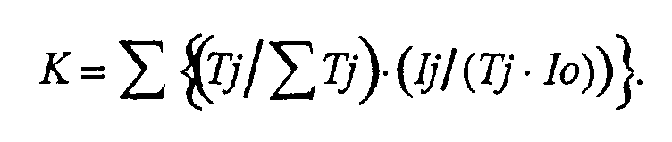
- the addition data is generated by performing a weighted addition on the data Kj subjected to the sensitivity correction with a weight Tj/ ⁇ Tj corresponding to the slice thickness Tj.
- the addition data is represented as K,
- K is equivalent to sensitivity-corrected data of one slice having a slice thickness of ⁇ Tj . Therefore, no adverse effect of the partial volume will appear on the addition tomographic image G produced by adding a plurality of tomographic images Gj having different slice thicknesses.
- the present invention provides the addition tomographic image producing method of the aforementioned configuration, wherein the tomographic image Gj is an X-ray CT image, and the data Ij is row data.
- the addition data K is generated by performing a weighted addition on the row data Kj subjected to the sensitivity correction with a weight Tj / ⁇ Tj corresponding to the slice thickness Tj.
- K is equivalent to sensitivity-corrected row data of one slice having a thickness of ⁇ Tj . Therefore, no adverse effect of the partial volume will appear on the addition tomographic image G produced by adding a plurality of X-ray CT images Gj having different slice thicknesses.
- the present invention provides the addition tomographic image producing method of aforementioned configuration, wherein the row data is that obtained by each detector row in a multi X-ray detector.
- a tomographic image can be produced from row data obtained by detector rows in a multi X-ray detector as if it were reconstructed from row data obtained by one detector row which is a combination of those detector rows.
- the present invention provides the addition tomographic image producing method of aforementioned configuration, comprising the step of applying a reference correction to the row data before or after the sensitivity correction, or to the addition data before the logarithm processing
- the reference correction when applied to the row data before or after the sensitivity correction, the reference correction may be performed in an ordinary way. When applied to the addition data before the logarithm processing, the reference correction needs to be performed only once.
- the present invention provides the addition tomographic image producing method of aforementioned configuration, comprising the step of applying at least one of a beam hardening correction, a water correction, a body motion correction and a helical correction to the addition data subjected to the logarithm processing.
- the corrections need to be performed only once.
- the present invention provides the addition tomographic image producing method of aforementioned configuration, comprising the step of, when a value of a certain channel in the row data before the logarithm processing is not more than a predefined threshold, replacing the value by an average value of one or more adjacent channels.
- the value when an abnormal value is contained in the row data, the value can be removed before the logarithm processing.
- the present invention provides the addition tomographic image producing method of aforementioned configuration, wherein the step of performing reconstruction comprises filtering and a back projection operation.
- a tomographic image can be reconstructed by a filtered back projection technique.
- I is equivalent to data of one slice having a slice thickness of ⁇ Tj . Therefore, no adverse effect of the partial volume will appear on the addition image G produced by adding a plurality of tomographic images Gj having different slice thicknesses.
- the processing may be the same as the ordinary one.
- I is equivalent to data of one slice having a slice thickness of ⁇ Tj. Therefore, no adverse effect of the partial volume will appear on the addition tomographic image G produced by adding a plurality of tomographic images Gj having different slice thicknesses.
- the offset correction since the offset correction is applied to the addition data I, the offset correction needs to be performed only once, although offset values for data Ij need to be added beforehand.
- the present invention provides the addition tomographic image producing method of aforementioned configuration, wherein the data Ij is row data obtained by each detector row in a multi X-ray detector.
- a tomographic image can be produced from row data obtained by detector rows in a multi X-ray detector as if it were reconstructed from row data obtained by one detector row which is a combination of those detector rows.
- the present invention provides the addition tomographic image producing method of aforementioned configuration, comprising the step of applying a reference correction to the addition data I before the sensitivity correction, or after the sensitivity correction and before the logarithm processing.
- the reference correction when applied to the row data before or after the sensitivity correction, the reference correction may be performed in an ordinary way.
- the reference correction needs to be performed only once.
- the present invention provides the addition tomographic image producing method of aforementioned configuration, comprising the step of applying at least one of a beam hardening correction, a water correction, a body motion correction and a helical correction to the addition data subjected to the logarithm processing.
- the corrections need to be performed only once.
- the present invention provides the addition tomographic image producing method of aforementioned configuration, comprising the step of, when a value of a certain channel in the addition data before the logarithm processing is not more than a predefined threshold, replacing the value by an average value of one or more adjacent channels.
- the value when an abnormal value is contained in the row data, the value can be removed before the logarithm processing.
- the present invention provides the addition tomographic image producing method of aforementioned configuration, wherein the step of performing reconstruction comprises filtering and a back projection operation.
- a tomographic image can be reconstructed by a filtered back projection technique.
- the addition tomographic image producing method of the second aspect can be suitably practiced.
- the present invention provides the X-ray CT apparatus of the aforementioned configuration, comprising first correcting means for applying a reference correction to the row data before or after the sensitivity correction, or to the addition data I before the logarithm processing.
- the addition tomographic image producing method of the fourth aspect can be suitably practiced.
- the present invention provides the X-ray CT apparatus of the aforementioned configuration, comprising second correcting means for applying at least one of a beam hardening correction, a water correction, a body motion correction and a helical correction to the addition data subjected to the logarithm processing.
- the addition tomographic image producing method of the fifth aspect can be suitably practiced.
- the addition tomographic image producing method of the eighth aspect can be suitably practiced.
- the addition tomographic image producing method of the ninth aspect can be suitably practiced.
- the present invention provides the X-ray CT apparatus of the aforementioned configuration, comprising first correcting means for applying a reference correction to the addition data before the sensitivity correction, or after the sensitivity correction and before the logarithm processing.
- the addition tomographic image producing method of the eleventh aspect can be suitably practiced.
- the present invention provides the X-ray CT apparatus of the aforementioned configuration, comprising second correcting means for applying at least one of a beam hardening correction, a water correction, a body motion correction and a helical correction to the addition data subjected to the logarithm processing.
- the addition tomographic image producing method of the twelfth aspect can be suitably practiced.
- an addition tomographic image when an addition tomographic image is produced by adding a plurality of tomographic images, an addition tomographic image can be produced which is not adversely affected by the partial volume effect even when the slice thicknesses of the tomographic images are different.
- Fig. 3 is a block diagram of an X-ray CT apparatus 100 in accordance with a first embodiment of the present invention.
- the X-ray CT apparatus 100 comprises an operating console 1, an imaging table 10 and a scan gantry 20.
- the operating console 1 comprises an input device 2 for receiving an instruction or information input supplied by a human operator; a central processing apparatus 3 for executing a scan process, image reconstruction process, addition tomographic image producing process and the like; a control interface 4 for exchanging control signals and the like with the imaging table 10 and the scan gantry 20; a data collection buffer 5 for collecting data acquired at the scan gantry 20; a CRT 6 for displaying an X-ray image reconstructed from the data; and a storage device 7 for storing programs, data and the X-ray image.
- the scan gantry 20 comprises an X-ray tube 21, an X-ray controller 22, a collimator 23, a collimator controller 24, a rotation controller 26 for rotating the X-ray tube 21 etc. around an isocenter (designated as IC in Fig. 4), and a twin X-ray detector 27 having two rows of detectors.
- Fig. 4 is a schematic view illustrating the X-ray tube 21, collimator 23 and twin X-ray detector 27.
- X-rays lo emitted from the X-ray tube 21 are formed into a flat X-ray beam Xr after passing through an aperture S of the collimator 23, and impinge upon first and second detector rows D1 and D2 of the twin X-ray detector 27.
- the opening width and position of the aperture S of the collimator 23 is regulated by the collimator controller 24 based on instructions from the central processing apparatus 3.
- Reference symbol Po designates an imaginary boundary line between a portion of the X-ray beam Xr which impinges upon the first detector row D1 and a portion which impinges upon the second detector row D2.
- the width of the X-ray beam Xr at the isocenter IC is referred to as X-ray beam width Xo.
- the width of a portion of the X-ray beam width Xo which impinges upon the first detector row D1 is a first slice thickness T1 and the width of a portion which impinges upon the second detector row D2 is a second slice thickness T2.
- Fig. 5 is a flow chart showing an addition tomographic image producing process conducted in the X-ray CT apparatus 100.
- Step ST1 an image index counter j is initialized to "1".
- Step ST2 row data Ij is read from a j-th detector row Dj in the multi X-ray detector.
- Step ST3 an offset correction is performed. Specifically, an offset is subtracted from the row data Ij of the j-th detector row Dj. For brevity of notation, the row data from which the offset has been subtracted will also be designated as Ij.
- Step ST4 a detector sensitivity correction is performed. Specifically, the row data Ij of the j-th detector row Dj is divided by an entering X-ray dose Tj ⁇ Io, wherein Tj is the slice thickness of the j-th detector row Dj, and Io is the entering X-ray dose per unit thickness.
- Step ST5 a first correction is applied to the row data Ij/(Tj ⁇ Io) subjected to the detector sensitivity correction.
- the first correction is a reference correction.
- the row data subjected to the first correction will also be designated as Ij/(Tj ⁇ Io).
- Step ST6 Log filtering is performed. Specifically, if the value of a certain channel in the row data Ij/(Tj ⁇ Io) subjected to the first correction is not more than a predefined threshold, then the value is replaced by the average value of one or more adjacent channels. Particularly, the value is replaced by the value of an adjacent channel; by the average value of adjacent channels on both sides; by the average value of the channels other than the current channel; or by the average value of two or more arbitrarily selected channels.
- row data I1/(T1 ⁇ Io) ⁇ IJ/(TJ ⁇ Io) subjected to the first correction are obtained.
- J 2
- I1/(T1 ⁇ Io) ⁇ I2/(T2 ⁇ Io) are obtained.
- Step ST9 a weighted addition is performed on the row data I1/(T1 ⁇ Io) ⁇ IJ/(TJ ⁇ Io) with a weight Tj/ ⁇ Tj corresponding to the slice thickness Tj to generate addition data K:
- Step ST10 logarithm processing is performed. Specifically, -Log ⁇ K ⁇ is calculated.
- a second correction is applied to the addition data -Log ⁇ K ⁇ subjected to the logarithm processing.
- the second correction is at least one of a beam hardening correction, a water correction, a body motion correction and a helical correction.
- the addition data subjected to the second correction will also be designated as -Log ⁇ K ⁇ .
- Steps ST12 and ST 13 filtering and a back projection operation are applied to the addition data -Log ⁇ K ⁇ subjected to the second correction to obtain an addition CT image G.
- Fig. 6 is a model diagram for explaining the physical meaning of the addition data K.
- an intervening substance H having an absorption coefficient of ⁇ , a path distance of x and a length of (L1+L2) is present in the air.
- the intervening substance H is present in the slice thickness T1 of the first detector row D1 by a length of L1, and in the slice thickness T2 of the second detector row D2 by a length of L2.
- the entering X-ray dose per unit thickness is represented as Io.
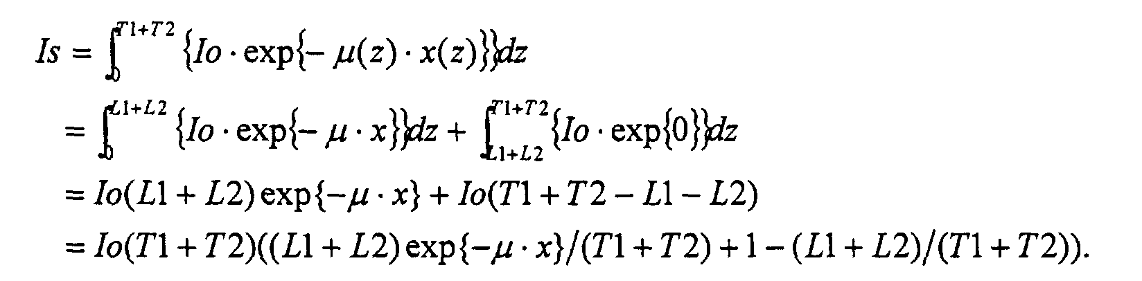
- Row data I1 obtained by the first detector row D1 is:
- the resulting row data Is will be:
- addition data K is equivalent to the row data derived by obtaining row data Is by one detector row Ds having a slice thickness of (T1+T2) and applying the sensitivity correction to the row data Is. Therefore, no adverse effect of the partial volume will appear on the addition tomographic image G.
- the first correction at Step ST5 may be applied to the row data Ij before the sensitivity correction (i.e., before Step ST3 or ST4), or to the addition data K before the logarithm processing (i.e., before Step ST10).
- Fig. 7 is a flow chart showing an addition tomographic image producing process in the X-ray CT apparatus in accordance with a second embodiment.
- Step ST21 an image index counter j is initialized to "1".
- Step ST22 row data Ij is read from a j-th detector row Dj in the multi X-ray detector.
- Step ST23 an offset correction is performed. Specifically, an offset is subtracted from the row data Ij of the j-th detector row Dj. For brevity of notation, the row data from which the offset has been subtracted will also be designated as Ij.
- the offset-corrected row data I1 ⁇ IJ are obtained.
- J 2
- row data I1 and I2 are obtained.
- Step ST27 a detector sensitivity correction is performed. Specifically, the addition data I is divided by an entering X-ray dose ⁇ Tj ⁇ Io , wherein Tj is the slice thickness of the j-th detector row Dj, and Io is the entering X-ray dose per unit thickness.
- Step ST28 a first correction is applied to the addition data I / ⁇ ( Tj ⁇ Io ) subjected to the detector sensitivity correction.
- the first correction is a reference correction.
- the addition data subjected to the first correction will also be designated as I / ⁇ ( Tj ⁇ Io ).
- Step ST29 Log filtering is performed. Specifically, if the value of a certain channel in the addition data I / ⁇ ( Tj ⁇ Io ) subjected to the first correction is not more than a predefined threshold, then the value is replaced by a filtered value of proximate channels.
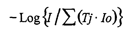
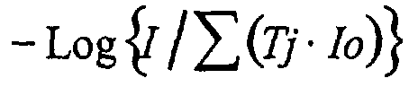
- Step ST30 logarithm processing is performed. Specifically, is calculated.
- a second correction is applied to the addition data subjected to the logarithm processing.
- the second correction is at least one of a beam hardening correction, a water correction, a body motion correction and a helical correction.
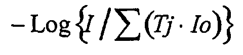
- the addition data subjected to the second correction will also be designated as
- Steps ST32 and ST 33 filtering and a back projection operation are applied to the addition data subjected to the second correction to obtain an addition CT image G.
- I1 Io ⁇ T 1( L 1 ⁇ exp ⁇ - ⁇ x ⁇ / T 1+1- L 1/ T 1)
- 12 Io ⁇ T 2( L 2 ⁇ exp ⁇ - ⁇ ⁇ x ⁇ l T 2+1 -L 2/ T 2)
- addition data I is equivalent to the row data Is obtained by one detector row Ds having a slice thickness of (T1+T2). Therefore, no adverse effect of the partial volume will appear on the addition tomographic image G.
- the offset correction at Step ST23 may be applied to the addition data I before the sensitivity correction (i.e., before Step ST27).
- respective offset values for the row data Ij are simply added beforehand, and the added value can be subtracted from the addition data I.
- the first correction at Step 28 may be applied to the addition data I before the sensitivity correction (i.e., before Step ST27).
Landscapes
- Engineering & Computer Science (AREA)
- Health & Medical Sciences (AREA)
- Life Sciences & Earth Sciences (AREA)
- Physics & Mathematics (AREA)
- Medical Informatics (AREA)
- General Physics & Mathematics (AREA)
- Theoretical Computer Science (AREA)
- Optics & Photonics (AREA)
- Heart & Thoracic Surgery (AREA)
- Nuclear Medicine, Radiotherapy & Molecular Imaging (AREA)
- Biophysics (AREA)
- Pathology (AREA)
- Radiology & Medical Imaging (AREA)
- Biomedical Technology (AREA)
- High Energy & Nuclear Physics (AREA)
- Molecular Biology (AREA)
- Surgery (AREA)
- Animal Behavior & Ethology (AREA)
- General Health & Medical Sciences (AREA)
- Public Health (AREA)
- Veterinary Medicine (AREA)
- Apparatus For Radiation Diagnosis (AREA)
- Image Processing (AREA)
Abstract
Description





Claims (10)
- An addition tomographic image producing method comprising the steps of: applying an offset correction and a sensitivity correction to data Ij (j = 1 ― J) from which respective tomographic images Gj can be reconstructed; generating addition data by performing a weighted addition on the data Ij with a weight Tj/ΣTj corresponding to the slice thickness Tj; applying logarithm processing to the addition data; and performing reconstruction on the addition data to produce an addition tomographic image G.
- The addition tomographic image producing method as defined by claim 1, wherein the tomographic image Gj is an X-ray CT image, and the data Ij is row data, and comprising the step of applying a reference correction to the row data before or after the sensitivity correction, or to the addition data before the logarithm processing.
- The addition tomographic image producing method as defined by claim 1 or claim 2, wherein the step of performing reconstruction comprises filtering and a back projection operation.
- An addition tomographic image producing method comprising the steps of: applying an offset correction to data Ij (j = 1 ― J) from which respective tomographic images Gj can be reconstructed; generating addition data I by simply adding the data Ij; applying a sensitivity correction and logarithm processing to the addition data I; and performing reconstruction on the addition data to produce an addition tomographic image G.
- An addition tomographic image producing method comprising the steps of: generating addition data I by simply adding data Ij (j = 1 ― J) from which respective tomographic images Gj can be reconstructed; applying an offset correction, a sensitivity correction and logarithm processing to the addition data I; and performing reconstruction on the addition data to produce an addition tomographic image G.
- An X-ray CT apparatus comprising a multi X-ray detector having a plurality of detector rows, comprising:sensitivity correction means for applying an offset correction and a sensitivity correction to row data Ij obtained by the detector rows; addition data generating means for generating addition data I by performing a weighted addition on the row data subjected to the sensitivity correction with a weight Tj/ΣTj corresponding to the slice thickness Tj; logarithm processing means for applying logarithm processing to the addition data I; and reconstruction means for applying filtering and a back projection operation to the addition data subjected to the logarithm processing.
- The X-ray CT apparatus as defined by claim 6, comprising first correcting means for applying a reference correction to the row data before or after the sensitivity correction, or to the addition data I before the logarithm processing.
- An X-ray CT apparatus comprising a multi X-ray detector having a plurality of detector rows, comprising:offset correcting means for applying an offset correction to row data Ij obtained by the detector rows; addition data generating means for generating addition data I by simply adding the row data subjected to the offset processing; sensitivity correcting means for applying a sensitivity correction to the addition data I; logarithm processing means for applying logarithm processing to the sensitivity-corrected addition data; and reconstruction means for applying filtering and a back projection operation to the addition data subjected to the logarithm processing.
- An X-ray CT apparatus comprising a multi X-ray detector having a plurality of detector rows, comprising:addition data generating means for generating addition data I by simply adding row data Ij obtained by the detector rows; offset correcting means for applying an offset correction to the addition data I; sensitivity correcting means for applying a sensitivity correction to the addition data subjected to the offset processing; logarithm processing means for applying logarithm processing to the sensitivity-corrected addition data; and reconstruction means for applying filtering and a back projection operation to the addition data subjected to the logarithm processing.
- The X-ray CT apparatus as defined by claim 8 or 9, comprising first correcting means for applying a reference correction to the addition data before the sensitivity correction, or after the sensitivity correction and before the logarithm processing.
Applications Claiming Priority (2)
| Application Number | Priority Date | Filing Date | Title |
|---|---|---|---|
| JP2000262057 | 2000-08-31 | ||
| JP2000262057A JP4130055B2 (en) | 2000-08-31 | 2000-08-31 | Addition tomographic image creation method and X-ray CT apparatus |
Publications (3)
| Publication Number | Publication Date |
|---|---|
| EP1187070A2 true EP1187070A2 (en) | 2002-03-13 |
| EP1187070A3 EP1187070A3 (en) | 2004-12-15 |
| EP1187070B1 EP1187070B1 (en) | 2006-11-29 |
Family
ID=18749792
Family Applications (1)
| Application Number | Title | Priority Date | Filing Date |
|---|---|---|---|
| EP01307083A Expired - Lifetime EP1187070B1 (en) | 2000-08-31 | 2001-08-20 | Addition tomographic image producing method and X-ray CT apparatus |
Country Status (6)
| Country | Link |
|---|---|
| US (1) | US6470206B2 (en) |
| EP (1) | EP1187070B1 (en) |
| JP (1) | JP4130055B2 (en) |
| KR (1) | KR100462954B1 (en) |
| CN (1) | CN1303941C (en) |
| DE (1) | DE60124830T2 (en) |
Cited By (3)
| Publication number | Priority date | Publication date | Assignee | Title |
|---|---|---|---|---|
| NL1032581C2 (en) * | 2005-09-27 | 2007-10-16 | Ge Med Sys Global Tech Co Llc | X-ray CT equipment. |
| NL1032638C2 (en) * | 2005-10-07 | 2007-11-23 | Ge Med Sys Global Tech Co Llc | Image reconstruction method and X-ray CT equipment. |
| US10354349B2 (en) | 2014-04-01 | 2019-07-16 | Heartflow, Inc. | Systems and methods for using geometry sensitivity information for guiding workflow |
Families Citing this family (12)
| Publication number | Priority date | Publication date | Assignee | Title |
|---|---|---|---|---|
| JP3802869B2 (en) * | 2002-12-02 | 2006-07-26 | ジーイー・メディカル・システムズ・グローバル・テクノロジー・カンパニー・エルエルシー | Beam hardening post-processing method and X-ray CT apparatus |
| US6845144B2 (en) * | 2003-02-08 | 2005-01-18 | Ge Medical Systems Global Technology Company, Llc | Three dimensional back projection method and an X-ray CT apparatus |
| JP2004347384A (en) * | 2003-05-21 | 2004-12-09 | Hitachi Ltd | X-ray CT apparatus and image creation method using X-ray CT apparatus |
| KR100775695B1 (en) * | 2004-06-24 | 2007-11-09 | 가부시끼가이샤 이시다 | X-ray inspection apparatus, and method for creating an image processing procedure of the x-ray inspection apparatus |
| JP2006102299A (en) * | 2004-10-07 | 2006-04-20 | Ge Medical Systems Global Technology Co Llc | X-ray dose correcting method and x-ray ct apparatus |
| CN101300600B (en) * | 2005-07-08 | 2016-01-20 | 威斯康星校友研究基金会 | For the backprojection reconstruction method of CT imaging |
| US7961839B2 (en) * | 2005-12-15 | 2011-06-14 | Koninklijke Philips Electronics N.V. | Advanced convergence for multiple iterative algorithm |
| JP5041751B2 (en) * | 2006-07-24 | 2012-10-03 | 株式会社イシダ | X-ray inspection apparatus and X-ray inspection program |
| AU2013286807A1 (en) * | 2012-07-03 | 2015-01-29 | The State Of Queensland Acting Through Its Department Of Health | Movement correction for medical imaging |
| CN103325087B (en) * | 2013-06-25 | 2016-06-01 | 上海交通大学 | The interpolation method of CT image slices data |
| US10255697B2 (en) * | 2014-11-20 | 2019-04-09 | Koninklijke Philips N.V. | Method for generation of synthetic mammograms from tomosynthesis data |
| JP7099238B2 (en) * | 2018-10-09 | 2022-07-12 | 富士通株式会社 | Medical image processing program, medical image processing device and medical image processing method |
Family Cites Families (25)
| Publication number | Priority date | Publication date | Assignee | Title |
|---|---|---|---|---|
| US6203777B1 (en) * | 1983-12-21 | 2001-03-20 | Nycomed Imaging As | Method of contrast enhanced magnetic resonance imaging using carbohydrate particles |
| DE3586203T2 (en) * | 1984-03-15 | 1993-01-28 | Yokogawa Medical Syst | IMAGE PROCESSING DEVICE FOR X-RAY GENTOMOGRAPHY. |
| JPS63147440A (en) * | 1986-12-12 | 1988-06-20 | 横河メディカルシステム株式会社 | Deconvolution processing method of x-ray tomographic imaging apparatus |
| US5043890A (en) * | 1989-06-12 | 1991-08-27 | General Electric | Compensation of computed tomography data for objects positioned outside the field of view of the reconstructed image |
| DE69232000D1 (en) * | 1991-05-21 | 2001-09-20 | Canon Kk | Image processing method and device |
| US5262945A (en) * | 1991-08-09 | 1993-11-16 | The United States Of America As Represented By The Department Of Health And Human Services | Method for quantification of brain volume from magnetic resonance images |
| US5247934A (en) * | 1991-08-09 | 1993-09-28 | Trustees Of The University Of Pennsylvania | Method and apparatus for diagnosing osteoporosis with MR imaging |
| US5459524A (en) * | 1991-11-18 | 1995-10-17 | Cooper; J. Carl | Phase modulation demodulator apparatus and method |
| US5241576A (en) * | 1991-12-23 | 1993-08-31 | General Electric Company | Segmented detector containing sub-elements for separate measuring of a fan beam |
| US5377250A (en) * | 1992-08-07 | 1994-12-27 | General Electric Company | Reconstruction method for helical scanning computed tomography apparatus with multi-row detector array |
| US5550582A (en) * | 1993-03-19 | 1996-08-27 | Olympus Optical Co., Ltd. | Endoscope-image processing apparatus for performing image processing of emphasis in endoscope image by pigment concentration distribution |
| US5473654A (en) * | 1994-06-24 | 1995-12-05 | General Electric Company | Backprojection for x-ray CT system |
| US5473656A (en) * | 1994-09-15 | 1995-12-05 | General Electric Company | Computed tomography system with correction for z-axis detector non-uniformity |
| US5984881A (en) * | 1995-03-31 | 1999-11-16 | Kabushiki Kaisha Toshiba | Ultrasound therapeutic apparatus using a therapeutic ultrasonic wave source and an ultrasonic probe |
| US5974108A (en) * | 1995-12-25 | 1999-10-26 | Kabushiki Kaisha Toshiba | X-ray CT scanning apparatus |
| DE19625863C2 (en) * | 1996-06-27 | 2002-06-27 | Siemens Ag | Image reconstruction method for a spiral tomograph |
| JP3310677B2 (en) * | 1996-09-12 | 2002-08-05 | アンリツ株式会社 | Highly sensitive foreign matter detection device by highly selective image processing |
| JP3679892B2 (en) * | 1997-04-10 | 2005-08-03 | 株式会社東芝 | Magnetic resonance imaging system |
| US6038265A (en) * | 1997-04-21 | 2000-03-14 | Motorola, Inc. | Apparatus for amplifying a signal using digital pulse width modulators |
| US5953444A (en) * | 1997-10-22 | 1999-09-14 | University Of Pennsylvania | Method for improved correction of spectrum hardening artifacts in computed tomography images |
| KR100654589B1 (en) * | 1998-09-21 | 2007-03-02 | 지이 요꼬가와 메디칼 시스템즈 가부시끼가이샤 | Radiation tomography method and apparatus |
| US6389310B1 (en) * | 1999-03-02 | 2002-05-14 | Georges Demonceau | Method and apparatus for analyzing heart function using 4D ECG synchronized heart cavity tomoscintigraphy |
| US6400978B1 (en) * | 1999-10-29 | 2002-06-04 | The Mclean Hospital Corporation | Method and apparatus for detecting mental disorders |
| JP3977972B2 (en) * | 1999-12-13 | 2007-09-19 | ジーイー・メディカル・システムズ・グローバル・テクノロジー・カンパニー・エルエルシー | Scanning condition determination method for tomography, tomography method and X-ray CT apparatus |
| JP2001187046A (en) * | 1999-12-27 | 2001-07-10 | Ge Medical Systems Global Technology Co Llc | Multi-slice x-ray ct device and method for controlling the same |
-
2000
- 2000-08-31 JP JP2000262057A patent/JP4130055B2/en not_active Expired - Fee Related
-
2001
- 2001-07-03 US US09/898,227 patent/US6470206B2/en not_active Expired - Fee Related
- 2001-08-20 DE DE60124830T patent/DE60124830T2/en not_active Expired - Fee Related
- 2001-08-20 EP EP01307083A patent/EP1187070B1/en not_active Expired - Lifetime
- 2001-08-30 KR KR10-2001-0052941A patent/KR100462954B1/en not_active Expired - Fee Related
- 2001-08-31 CN CNB011325151A patent/CN1303941C/en not_active Expired - Fee Related
Cited By (4)
| Publication number | Priority date | Publication date | Assignee | Title |
|---|---|---|---|---|
| NL1032581C2 (en) * | 2005-09-27 | 2007-10-16 | Ge Med Sys Global Tech Co Llc | X-ray CT equipment. |
| US7428290B2 (en) | 2005-09-27 | 2008-09-23 | Ge Medical Systems Global Technology Company, Llc | X-ray CT apparatus |
| NL1032638C2 (en) * | 2005-10-07 | 2007-11-23 | Ge Med Sys Global Tech Co Llc | Image reconstruction method and X-ray CT equipment. |
| US10354349B2 (en) | 2014-04-01 | 2019-07-16 | Heartflow, Inc. | Systems and methods for using geometry sensitivity information for guiding workflow |
Also Published As
| Publication number | Publication date |
|---|---|
| CN1303941C (en) | 2007-03-14 |
| JP2002085397A (en) | 2002-03-26 |
| US20020075991A1 (en) | 2002-06-20 |
| EP1187070A3 (en) | 2004-12-15 |
| US6470206B2 (en) | 2002-10-22 |
| KR20020018160A (en) | 2002-03-07 |
| KR100462954B1 (en) | 2004-12-23 |
| CN1340329A (en) | 2002-03-20 |
| DE60124830T2 (en) | 2007-06-28 |
| EP1187070B1 (en) | 2006-11-29 |
| JP4130055B2 (en) | 2008-08-06 |
| DE60124830D1 (en) | 2007-01-11 |
Similar Documents
| Publication | Publication Date | Title |
|---|---|---|
| EP1187070B1 (en) | Addition tomographic image producing method and X-ray CT apparatus | |
| Kinahan et al. | X-ray-based attenuation correction for positron emission tomography/computed tomography scanners | |
| CN102048552B (en) | Beam hardening correction for ct perfusion measurements | |
| EP1846893B1 (en) | Radial adaptive filter for metal artifact correction | |
| US8750582B2 (en) | Method for recording and reconstructing a three-dimensional image dataset and X-ray apparatus | |
| US8768045B2 (en) | Method for acquiring a 3D image dataset freed of traces of a metal object | |
| DE102005027436B4 (en) | Method for calculating absorber-specific weighting coefficients and method for improving an absorber-dependent contrast-to-noise ratio in an X-ray image of an object to be examined, which is generated by an X-ray device | |
| CN105902279B (en) | A scanning image reconstruction method and device | |
| JP2007530132A (en) | Method, computer program product and apparatus for enhancing computed tomography images | |
| CN111402150B (en) | CT image metal artifact removal method and device | |
| JP2019058480A (en) | Tomography imaging apparatus and tomography imaging method | |
| Choi et al. | Self‐supervised denoising of projection data for low‐dose cone‐beam CT | |
| US8121192B2 (en) | Method and system for treating noise in a generated image | |
| US7599464B2 (en) | Method and computed tomography system for producing tomograms of an object | |
| US6845143B2 (en) | CT image reconstruction | |
| CN113313649B (en) | Image reconstruction method and device | |
| CN102096905A (en) | Noise reduction in dual-energy computer tomography shooting | |
| US10363002B2 (en) | X-ray CT image reconstruction device, X-ray CT image reconstruction method, and X-ray CT apparatus | |
| KR102590461B1 (en) | Method and apparatus for decomposing multi-material based on dual-energy technique | |
| CN111127369A (en) | Image correction method, device, equipment and medium | |
| JP5117792B2 (en) | Storage medium and X-ray CT system | |
| EP4087487A1 (en) | X-ray imaging systems for reducing artefacts associated with anti-scatter grits and methods of operating the same | |
| US20060251313A1 (en) | Method of producing a cross-sectional image | |
| JP2000342570A (en) | Slice thickness setting method for twin asymmetrical scanning and x-ray ct device | |
| Jäger | Data-driven Image Quality Improvements for Cone-beam Computed Tomography in Radiation Therapy |
Legal Events
| Date | Code | Title | Description |
|---|---|---|---|
| PUAI | Public reference made under article 153(3) epc to a published international application that has entered the european phase |
Free format text: ORIGINAL CODE: 0009012 |
|
| AK | Designated contracting states |
Kind code of ref document: A2 Designated state(s): AT BE CH CY DE DK ES FI FR GB GR IE IT LI LU MC NL PT SE TR |
|
| AX | Request for extension of the european patent |
Free format text: AL;LT;LV;MK;RO;SI |
|
| PUAL | Search report despatched |
Free format text: ORIGINAL CODE: 0009013 |
|
| AK | Designated contracting states |
Kind code of ref document: A3 Designated state(s): AT BE CH CY DE DK ES FI FR GB GR IE IT LI LU MC NL PT SE TR |
|
| AX | Request for extension of the european patent |
Extension state: AL LT LV MK RO SI |
|
| 17P | Request for examination filed |
Effective date: 20050615 |
|
| AKX | Designation fees paid |
Designated state(s): DE FR GB |
|
| GRAP | Despatch of communication of intention to grant a patent |
Free format text: ORIGINAL CODE: EPIDOSNIGR1 |
|
| GRAS | Grant fee paid |
Free format text: ORIGINAL CODE: EPIDOSNIGR3 |
|
| GRAA | (expected) grant |
Free format text: ORIGINAL CODE: 0009210 |
|
| AK | Designated contracting states |
Kind code of ref document: B1 Designated state(s): DE FR GB |
|
| REG | Reference to a national code |
Ref country code: GB Ref legal event code: FG4D |
|
| REF | Corresponds to: |
Ref document number: 60124830 Country of ref document: DE Date of ref document: 20070111 Kind code of ref document: P |
|
| EN | Fr: translation not filed | ||
| PLBE | No opposition filed within time limit |
Free format text: ORIGINAL CODE: 0009261 |
|
| STAA | Information on the status of an ep patent application or granted ep patent |
Free format text: STATUS: NO OPPOSITION FILED WITHIN TIME LIMIT |
|
| 26N | No opposition filed |
Effective date: 20070830 |
|
| GBPC | Gb: european patent ceased through non-payment of renewal fee |
Effective date: 20070820 |
|
| PG25 | Lapsed in a contracting state [announced via postgrant information from national office to epo] |
Ref country code: FR Free format text: LAPSE BECAUSE OF FAILURE TO SUBMIT A TRANSLATION OF THE DESCRIPTION OR TO PAY THE FEE WITHIN THE PRESCRIBED TIME-LIMIT Effective date: 20070720 |
|
| PG25 | Lapsed in a contracting state [announced via postgrant information from national office to epo] |
Ref country code: DE Free format text: LAPSE BECAUSE OF NON-PAYMENT OF DUE FEES Effective date: 20080301 |
|
| PG25 | Lapsed in a contracting state [announced via postgrant information from national office to epo] |
Ref country code: GB Free format text: LAPSE BECAUSE OF NON-PAYMENT OF DUE FEES Effective date: 20070820 Ref country code: FR Free format text: LAPSE BECAUSE OF FAILURE TO SUBMIT A TRANSLATION OF THE DESCRIPTION OR TO PAY THE FEE WITHIN THE PRESCRIBED TIME-LIMIT Effective date: 20061129 |