FIELD OF THE INVENTION
The present invention relates to a novel protein
containing an amino acid sequence, which is the same or
substantially the same as the amino acid sequence
represented by SEQ ID NO:1 (preferably a protein having
a GABA transporter activity; hereinafter sometimes
referred to as the protein of the present invention) or
its salts, or a DNA encoding the same.
BACKGROUND ART
γ-Aminobutyric acid (GABA), which is an inhibitory
neurotransmitter, is released from GABAergic neurons
and then removed from extracellular fluids by the
action of GABA transporters located in neuronal
terminals and glia cells. The mechanism of this active
uptake of GABA transporters is most important for
determining when neurotransmission has been
accomplished.
It is known that a GABA transporter has four
subtypes (GAT-1. GAT-2, GAT-3 and BGT-1). GAT-1 and
GAT-3 are located in brain and retina, GAT-2 in almost
all organs, and BGT.l in kidney and brain. The GAT-1
gene was cloned from mouse (Gene Bank accession No.
M92378), rat (Gene Bank accession No. M33003) and human
(Gene Bank accession No. X54673); the GAT-2 gene from
mouse (Gene Bank accession No. L04663) and rat (Gene
Bank accession No. M95762); the GAT-3 gene from mouse
(Gene Bank accession No. L04662) and rat (Gene Bank
accession No. M95763); and the BGT-1 gene from mouse
(Gene Bank accession No. M97632) and dog (Gene Bank
accession No. M80403). However, a human GAT-2 gene is
yet unknown so far.
It is an object of the present invention to
provide the following:
DISCLOSURE OF THE INVENTION
The present inventors have made extensive studies
and as a result, succeeded in isolating cDNA encoding
human-derived GABA transporter protein having a high
homology to mouse- or rat-derived GAT-2, sequencing the
entire base sequence and then expressing cDNA in cells.
Based on these findings, the present inventors have
come to accomplish the present invention.
That is, the present invention provides the
following:
BRIEF DESCRIPTION OF THE DRAWINGS
FIG. 1 shows hydrophobic plotting prepared based
on the amino acid sequence of the human-derived protein
of the present invention, obtained in EXAMPLES.
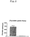
FIG. 2 shows the results of increased amounts of
labeled GABA uptake assayed after adding [3H]-GABA
to hGAT2/LM(TK-) cells where the protein of the
present invention was expressed, in a buffer
containing sodium ions and chlorine ions. In the left
graph, hGAT2/LM(TK-) designates the LM(TK-) cell
wherein the human-derived protein of the present
invention was expressed, and in the right graph,
Mock/LM(TK-) designates the LM(TK-) cell inserted
with plasmid pMCMVneo. The numerical value on the
left side denotes the assay results of amounts of
labeled GABA uptake measured after adding 50 nM
[3H]-GABA to hGAT2/LM(TK-) cells where the protein
of the present invention was expressed, in a
buffer containing sodium ions and chlorine ions. The
data is shown as a mean value ± standard error.
BEST MODE FOR CARRYING OUT THE INVENTION
The protein of the present invention is a protein
which has the same or substantially the same as the
amino acid sequence shown by SEQ ID NO:1.
The protein of the present invention may be any
protein derived from any cells of human and other warm-blooded
animals (e.g., guinea pig, rat, mouse, rabbit,
swine, sheep, bovine, monkey, etc.) such as splenocyte,
nerve cells, glial cells, β cells of pancreas, bone
marrow cells, mesangial cells, Langerhans' cells,
epidermic cells, epithelial cells, endothelial cells,
fibroblasts, fibrocytes, myocytes, fat cells, immune
cells (e.g., macrophage, T cells, B cells, natural
killer cells, mast cells, neutrophils, basophils,
eosinophils, monocytes), megakaryocytes, synovial cells,
chondrocytes, bone cells, osteoblasts, osteoclasts,
mammary gland cells, hepatocyte or interstitial cell;
or the corresponding precursor cells, stem cells,
cancer cells, etc.; or any tissues where such cells are
present, such as brain or any of brain regions (e.g.,
olfactory bulb, amygdaloid nucleus, basal ganglia,
hippocampus, thalamus, hypothalamus, subthalamic
nucleus, cerebral cortex, medulla oblongata, cerebellum,
occipital lobes, frontal lobe, lateral lobe, putamen,
caudate nucleus, corpus callosum, substantia nigra),
spinal cord, hypophysis, stomach, pancreas, kidney,
liver, gonad, thyroid, gall-bladder, bone marrow,
adrenal gland, skin, muscle, lung, gastrointestinal
tract (e.g., large intestine and small intestine),
blood vessel, heart, thymus, spleen, submandibular
gland, peripheral blood, peripheral hemocytes, prostate,
testis, ovary, placenta, uterus, bone, joint, skeletal
muscle, etc. (especially, brain or any of brain
regions); the proteins may also be synthetic proteins.
The amino acid sequence which is substantially the
same as the amino acid sequence represented by SEQ ID
NO:1 includes, for example, an amino acid sequence
having at least about 95% homology, preferably at least
about 98% homology, to the amino acid sequence
represented by SEQ ID NO:1.
Examples of the protein containing the amino acid
sequence which is substantially the same as the amino
acid sequence represented by SEQ ID NO:1 include a
protein containing the amino acid sequence which is
substantially the same as the amino acid sequence
represented by SEQ ID NO:1 and has an activity
substantially equivalent to that of the amino acid
sequence represented by SEQ ID NO:1, etc.
The term "substantially equivalent" is used to
mean that the activities of the protein, for example, a
transporter activity (GABA transporter activity),
physiological properties, etc. are substantially
equivalent. Substitution, deletion, addition or
insertion of an amino acid(s) do not often cause
significant changes in physiological or chemical
properties of polypeptides; in such a case, proteins
that have undergone such substitution, deletion,
addition or insertion will be considered to be
substantially equivalent to intact proteins that do not
receive undergo the substitution, deletion, addition or
insertion. Substantially the same substituent(s) of an
amino acid(s) in the amino acid sequence can be
selected from, e.g., other amino acids of the class to
which the amino acid(s) belong. Examples of non-polar
(hydrophobic) amino acids include alanine, leucine,
isoleucine, valine, proline, phenylalanine, tryptophan,
methionine and the like. Examples of polar (neutral)
amino acids are glycine, serine, threonine, cysteine,
tyrosine, asparagine, glutamine, and the like Examples
of positively charged (basic) amino acids are arginine,
lysine, histidine, and the like. Examples of negatively
charged (acidic) amino acids include aspartic acid,
glutamic acid, and the like.
The substantially equivalent activities are used
to mean that these activities are equivalent in terms
of its nature (physiolochemically or pharmacologically).
Therefore, it is preferred that these activities are
equivalent to each other, but it is allowable that
differences among grades such as the level of these
activities and molecular weight of the protein may be
present.
The transporter activity can be determined
according to a publicly known method, for example, by
means of screening which will be later described.
As the protein of the present invention, there may
be employed proteins containing (i) the amino acid
sequence represented by SEQ ID NO:1, of which at least
1 or 2 (preferably approximately 1 to 30, more
preferably approximately 1 to 9 and most preferably
several (1 or 2)) amino acids are deleted; (ii) the
amino acid sequence represented by SEQ ID NO:1, to
which at least 1 or 2 (preferably approximately 1 to 30,
more preferably approximately 1 to 9 and most
preferably several (1 or 2)) amino acids are added;
(iii) the amino acid sequence represented by SEQ ID
NO:1, in which at least 1 or 2 (preferably
approximately 1 to 30, more preferably approximately 1
to 9 and most preferably several (1 or 2)) amino acids
are substituted with other amino acids; and (iv) a
combination of the above amino acid sequences.
Furthermore, examples of the proteins of the present
invention include variants of the proteins described
above, wherein the amino group at the N-terminal
methionine residue of the proteins is protected with a
protecting group (e.g., a C1-6 acyl group such as a C2-6
alkanoyl group, e.g., formyl group, acetyl group,
etc.); those wherein the N-terminal region is cleaved
in vivo and Gln thus formed is pyroglutaminated; those
wherein a substituent (e.g., -OH, -SH, amino group,
imidazole group, indole group, guanidino group, etc.)
on the side chain of an amino acid in the molecule is
protected with a suitable protecting group (e.g., a C1-6
acyl group such as a C2-6 alkanoyl group, e.g., formyl
group, acetyl group, etc.), or conjugated proteins such
as glycoproteins having sugar chains.
The partial peptides of the proteins of the
present invention (hereinafter sometimes merely
referred to as the partial peptides) may be any partial
peptides so long as they are partial peptides of the
proteins of the present invention described above, but
exposed extracellularly sites, etc. in the proteins of
the present invention may be used.
Specifically, the partial peptides of the proteins
containing the amino acid sequence represented by SEQ
ID NO:1 are peptides containing the region determined
to be an extracellular region (hydrophilic site) in the
hydrophilic plotting shown in (FIG. 1]. Peptides
containing in part the hydrophobic site may also be
employed similarly. Peptides containing respective
domains separately may also be employed, and peptides
containing the site bearing a plurality of the domains
at the same time may be used as well.
Also the partial peptides of the present invention,
may be those containing the amino acid sequences
described above, of which at least 1 or 2 (preferably
approximately 1 to 30, more preferably approximately 1
to 9 and most preferably several (1 or 2)) amino acids
are deleted; to which at least 1 or 2 (preferably
approximately 1 to 30, more preferably approximately 1
to 9 and most preferably several (1 or 2)) amino acids
are added; or, in which at least 1 or 2 (preferably
approximately 1 to 30, more preferably approximately 1
to 9 and most preferably several (1 or 2)) amino acids
are substituted with other amino acids.
In the partial proteins of the present invention,
the C-terminus is usually in the form of a carboxyl
group (-COOH) or a carboxylate (-COO-) but may be in
the form of an amide (-CONH2) or an ester (-COOR)
(wherein R has the same significance as defined above).
The partial peptides of the present invention
further include those containing substituents on the
side chains of intramolecular amino acids, which are
protected with appropriate protecting groups,
conjugated peptides such as so-called glycoproteins
having sugar chains bound thereto, and the like.
The partial peptide of the present invention can
be employed as an antigen for producing an antibody and
therefore, does not necessarily possess the GABA
transporter activity.
As salts of the protein of the present invention
or its partial peptide, physiologically acceptable acid
addition salts are particularly preferred. Examples of
such salts employed are salts with, e.g., inorganic
acids (e.g., hydrochloric acid, phosphoric acid,
hydrobromic acid, sulfuric acid), or salts with organic
acids (e.g., acetic acid, formic acid, propionic acid,
fumaric acid, maleic acid, succinic acid, tartaric acid,
citric acid, malic acid, oxalic acid, benzoic acid,
methanesulfonic acid, benzenesulfonic acid) and the
like.
The protein of the present invention or its salts
may be manufactured by a publicly known purification
method from human or the other warm-blooded animal
cells or tissues described above. Alternatively, the
protein of the present invention or its salts may also
be manufactured by culturing a transformant containing
DNA encoding the protein of the present invention,
which will be later described. Furthermore, the protein
of the present invention or its salts may also be
manufactured by the methods for synthesizing proteins,
which will also be described hereinafter, or by a
modification of such methods.
Where the protein or its salts are manufactured
from human or mammalian tissues or cells, the human or
mammalian tissues or cells are homogenized, then
extracted with an acid or the like, and the extract is
isolated and purified by a combination of
chromatography techniques such as reverse phase
chromatography, ion exchange chromatography, and the
like.
The partial peptide of the protein of the present
invention or its salts can be manufactured by publicly
known methods for peptide synthesis, or by cleaving the
protein of the present invention with an appropriate
peptidase. For the methods for peptide synthesis, for
example, either solid phase synthesis or liquid phase
synthesis may be used. That is, the partial peptide or
amino acids that can construct the protein of the
present invention are condensed with the remaining part
of the partial peptide of the present invention. Where
the product contains protecting groups, these
protecting groups are removed to give the desired
peptide. Publicly known methods for condensation and
elimination of the protecting groups are described in
1) - 5) below.
After completion of the reaction, the product may
be purified and isolated by a combination of
conventional purification methods such as solvent
extraction, distillation, column chromatography, liquid
chromatography and recrystallization to give the
partial peptide of the present invention. When the
partial peptide obtained by the above methods is in a
free form, the peptide can be converted into an
appropriate salt by a publicly known method or a
modification of the known method; conversely when the
protein is obtained in a salt form, it can be converted
into a free form or a different salt form by a publicly
known method or a modification of the known method.
The DNA encoding the protein of the present
invention may be any DNA so long as it contains the
base sequence encoding the protein of the present
invention described above. Such a DNA may also be any
one of genomic DNA, genomic DNA library, cDNA derived
from the cells or tissues described above, cDNA library
derived from the cells or tissues described above and
synthetic DNA. The vector to be used for the library
may be any of bacteriophage, plasmid, cosmid, phagemid
and the like. In addition, the DNA can be directly
amplified by reverse transcriptase polymerase chain
reaction (hereinafter abbreviated as RT-PCR) with total
RNA or mRNA fraction prepared from the above-described
cells or tissues.
Specifically, the DNA encoding the protein of the
present invention may be any one of, for example, DNA
containing the base sequence represented by SEQ ID NO:2,
or any DNA having a base sequence hybridizable to the
base sequence represented by SEQ ID NO:2 under high
stringent conditions and encoding a protein which has
the activities substantially equivalent to those of the
protein of the present invention (e.g., a GABA
transporter activity, etc.).
Specific examples of the DNA that is hybridizable
to the base sequence represented by SEQ ID NO:2 include
DNA having at least about 90% homology, preferably at
least about 95% homology, and more preferably at least
about 98% homology, to the base sequence represented by
SEQ ID NO:2.
The hybridization can be carried out by publicly
known methods or by a modification thereof, for example,
according to the method described in Molecular Cloning,
2nd Ed., J. Sambrook et al., Cold Spring Harbor Lab.
Press, (1989). A commercially available library may
also be used according to the instructions of the
attached manufacturer's protocol. Preferably, the
hybridization can be carried out under high stringent
conditions.
The high stringent conditions used herein are, for
example, those in a sodium concentration at about 19 mM
to about 40 mM, preferably about 19 mM to about 20 mM
at a temperature of about 50°C to about 70°C,
preferably about 60°C to about 65°C. In particular,
hybridization conditions in a sodium concentration at
about 19 mM at a temperature of about 65°C are most
preferred.
More specifically, for the DNA encoding the
protein having the amino acid sequence represented by
SEQ ID NO:1, there may be employed DNA having the base
sequence represented by SEQ ID NO:2, etc.
The DNA encoding the partial peptide of the
present invention may be any DNA so long as it contains
the base sequence encoding the partial peptide of the
present invention described above. The DNA may also be
any of genomic DNA, genomic DNA library, cDNA derived
from the cells and tissues described above, cDNA
library derived from the cells and tissues described
above and synthetic DNA. The vector to be used for the
library may be any of bacteriophage, plasmid, cosmid,
phagemid and the like. In addition, the DNA can be
directly amplified by reverse transcriptase polymerase
chain reaction (hereinafter abbreviated as RT-PCR) with
total RNA or mRNA fraction prepared from the above-described
cells or tissues.
Specifically, the DNA encoding the partial peptide
of the present invention includes, for example, DNA
containing a partial base sequence of the DNA having
the base sequence represented by SEQ ID NO:2, or (ii)
DNA which has a base sequence hybridizable to the base
sequence represented by SEQ ID NO:2 under high
stringent conditions and contains a partial base
sequence of the DNA encoding the protein having the
activities substantially equivalent to those of the
protein of the present invention (e.g., a GABA
transporter activity, etc.).
Specific examples of the DNA that is hybridizable
to the base sequence represented by SEQ ID NO:2 include
DNA having at least about 95% homology, preferably at
least about 98% homology, to the base sequence
represented by SEQ ID NO:2. The hybridization and the
high stringent conditions used are those as described
above.
For cloning of the DNA that completely encodes the
protein or its partial peptide of the present invention
(hereinafter collectively referred to as the protein of
the present invention), (1) the DNA may be amplified by
PCR using synthetic DNA primers containing a part of
the base sequence of the protein of the present
invention, or (2) the DNA inserted into an appropriate
vector can be selected by hybridization with a labeled
DNA fragment or synthetic DNA that encodes a part or
the entire region of the protein of the present
invention, etc. The hybridization can be carried out,
for example, according to the method described in
Molecular Cloning, 2nd (J. Sambrook et al., Cold Spring
Harbor Lab. Press, 1989) or the like. The hybridization
may also be performed using commercially available
library in accordance with the protocol described in
the attached instructions.
Conversion (deletion, addition or substitution) of
the base sequence for DNA can be effected by publicly
known methods such as the Gapped duplex method or the
Kunkel method or modifications thereof, using a
publicly known kit available as MutanTM-G or MutanTM-K
(both Takara Shuzo Co., Ltd.).
The cloned DNA encoding the protein of the present
invention can be used as it is, or, if desired, after
digestion with restriction enzymes or after addition of
linkers thereto, depending upon purposes. The DNA may
contain ATG as a translation initiation codon at the 5'
end thereof and TAA, TGA or TAG as a translation
termination codon at the 3' end thereof. These
translation initiation and termination codons may also
be added by using an appropriate synthetic DNA adapter.
The expression vector of the protein of the
present invention can be manufactured, for example, by
(a) excising the desired DNA fragment from the DNA
encoding the protein of the present invention, (b) and
then ligating the DNA fragment with an appropriate
expression vector downstream a promoter in the
expression vector.
Examples of the vector include plasmids derived
form E. coli (e.g., pBR322, pBR325, pUC12. pUC13).
plasmids derived from Bacillus subtilis (e.g., pUB110,
pTP5, pC194), plasmids derived from yeast (e.g., pSH19,
pSHl5). bacteriophages such as λ phage, etc., animal
viruses such as retrovirus, vaccinia virus, baculovirus,
etc. as well as pA1-11, pXT1, pRc/CMV, pRc/RSV,
pcDNAI/Neo, etc.
The promoter used in the present invention may be
any promoter if it matches well with a host to be used
for gene expression. In the case of using animal cells
as the host, examples of the promoter include SRα
promoter, SV40 promoter, HIV·LTR promoter, CMV promoter,
HSV-TK promoter, etc. Among them, CMV promoter or SRα
promoter is preferably used. Where the host is bacteria
belonging to the genus Escherichia, preferred examples
are trp promoter, lac promoter, recA promoter, λPL
promoter, lpp promoter, etc. In the case of using
bacteria of the genus Bacillus as the host, preferred
example are SPO1 promoter, SPO2 promoter, penP promoter,
etc. When yeast is used as the host, preferred examples
are PHO5 promoter, PGK promoter, GAP promoter, ADH
promoter, etc. When insect cells are used as the host,
preferred examples are polyhedrin prompter, P10
promoter, etc.
In addition to the foregoing examples, the
expression vector may further optionally contain an
enhancer, a splicing signal, a poly A addition signal,
a selection marker, SV40 replication origin
(hereinafter sometimes abbreviated as SV40ori), etc.
Examples of the selection marker include dihydrofolate
reductase (hereinafter sometimes abbreviated as dhfr)
gene [methotrexate (MTX) resistance], ampicillin
resistant gene (hereinafter sometimes abbreviated as
Ampr), neomycin resistant gene (hereinafter sometimes
abbreviated as Neo, G418 resistance), etc. If necessary,
a signal sequence that matches with a host is added to
the N-terminus of the protein of the present invention.
Examples of the signal sequence that can be used are
PhoA signal sequence, OmpA signal sequence, etc. in the
case of using bacteria of the genus Escherichia as the
host; α-amylase signal sequence, subtilisin signal
sequence, etc. in the case of using bacteria of the
genus Bacillus as the host; MFα signal sequence, SUC2
signal sequence, etc. in the case of using yeast as the
host; and insulin signal sequence, α-interferon signal
sequence, antibody molecule signal sequence, etc. in
the case of using animal cells as the host,
respectively.
Using the vector containing the DNA encoding the
protein of the present invention thus constructed,
transformants can be manufactured.
Examples of the host, which may be employed, are
bacteria belonging to the genus Escherichia, bacteria
belonging to the genus Bacillus, yeast, insect cells,
insects and animal cells, etc.
Specific examples of the bacteria belonging to the
genus Escherichia include Escherichia coli K12 DH1
(Proc. Natl. Acad. Sci. U.S.A., 60, 160 (1968)), JM103
(Nucleic Acids Research, 9, 309 (1981)), JA221 (Journal
of Molecular Biology, 120, 517 (1978)), HB101 (Journal
of Molecular Biology, 41, 459 (1969)), C600 (Genetics,
39, 440 (1954)), etc.
Examples of the bacteria belonging to the genus
Bacillus include Bacillus subtilis MI114 (Gene, 24, 255
(1983)), 207-21 (Journal of Biochemistry, 95, 87
(1984)), etc.
Examples of yeast employed are Saccharomyces
cerevisiae AH22, AH22R-, NA87-11A, DKD-5D, 20B-12,
Schizosaccharomyces pombe NCYC1913, NCYC2036, Pichia
pastoris KM71, etc.
As insect cells, there may be employed, Spodoptera
frugiperda cells (Sf cells), MG1 cells derived from
mid-intestine of Trichoplusia ni, High FiveTM cells
derived from egg of Trichoplusia ni, cells derived from
Mamestra brassicae, cells derived from Estigmena acrea,
etc., when the virus is AcNPV; when the virus is BmNPV,
Bombyx mori N cells (BmN cells), etc. are used.
Examples of the Sf cells which can be used are Sf9
cells (ATCC CRL1711) and Sf21 cells (both cells are
described in Vaughn, J. L. et al., In Vivo, 13, 213-217
(1977)).
As the insect, for example, a larva of Bombyx mori
can be used (Maeda et al., Nature, 315, 592 (1985)).
Examples of animal cells include monkey cell COS-7,
Vero, Chinese hamster cell CHO (hereinafter abbreviated
as CHO cells), dhfr gene deficient Chinese hamster cell
CHO (hereinafter abbreviated as CHO (dhfr-) cells),
mouse L cells, mouse AtT-20, mouse myeloma cells, rat
GH 3, human FL cells, HEK293 cells, C127 cells, BALB3T3
cells, Sp-2 cells, etc. Among them, CHO cells, CHO
(dhfr-) cells, HEK293 cells, LM cells, etc. are
preferred.
Bacteria belonging to the genus Escherichia can be
transformed, for example, by the method described in
Proc. Natl. Acad. Sci. U.S.A., 69, 2110 (1972), Gene,
17, 107 (1982), etc.
Bacteria belonging to the genus Bacillus can be
transformed, for example, by the method described in
Molecular & General Genetics, 168, 111 (1979).
Yeast can be transformed, for example, by the
method described in Methods in Enzymology, 194, 182-187
(1991), Proc. Natl. Acad. Sci. U.S.A., 75, 1929 (1978),
etc.
Insect cells or insects can be transformed, for
example, according to the method described in
Bio/Technology, 6, 47-55(1988).
Methods for introducing the expression vectors
into animal cells include, for example, the calcium
phosphate method [Graham, F. L. and van der Eb, A., J.
Virology, 52, 456-467 (1973)], the electroporation
method [Nuemann, E. et al., EMBO J., 1, 841-845 (1982)],
etc.
As described above, transformants transformed with
the expression vector containing the DNA encoding the
protein of the present invention can be obtained.
For stably expressing the protein of the present
invention using animal cells, there is applicable a
method of selecting the cells by clone selection in
which the aforesaid expression vectors transfected to
animal cells are introduced into chromosomes.
Specifically, transformants are selected using as an
index the selection marker described above. Further by
repeated clone selections on the transformants thus
obtained using the selection marker, stable animal cell
line capable of highly expressing the protein of the
present invention can be obtained. Furthermore, when
the dhfr gene is used as a selection marker,
cultivation can be performed by gradually increasing a
level of MTX, resistant cells are selected thereby to
amplify the DNA encoding the protein of the present
invention in the cells together with the dhfr gene.
Thus, the animal cell line of higher expression can be
obtained.
The transformant described above is cultivated
under conditions that the DNA encoding the protein of
the present invention can express, to produce and
accumulate the protein of the present invention. Thus,
the protein of the present invention or its salts can
be produced.
Where the host is bacteria belonging to the genus
Escherichia or the genus Bacillus, the transformant can
be appropriately incubated in a liquid medium, which
contains materials required for growth of the
transformant such as carbon sources, nitrogen sources,
inorganic materials, etc. Examples of the carbon
sources include glucose, dextrin, soluble starch,
sucrose, etc. Examples of the nitrogen sources include
inorganic or organic materials such as ammonium salts,
nitrate salts, corn steep liquor, peptone, casein, meat
extract, soybean cake, potato extract, etc. Examples of
the inorganic materials are calcium chloride, sodium
dihydrogenphosphate, magnesium chloride, etc. In
addition, yeast extract, vitamins, growth promoting
factors, etc. may also be added to the medium.
Preferably, pH of the medium is adjusted to about 5 to
about 8.
A preferred example of the medium for incubation
of the bacteria belonging to the genus Escherichia is
M9 medium supplemented with glucose and Casamino acids
(Miller, Journal of Experiments in Molecular Genetics,
431-433, Cold Spring Harbor Laboratory, New York, 1972).
If necessary, a chemical such as 3β-indolylacrylic acid
can be added to the medium thereby to activate the
promoter efficiently.
Where the bacteria belonging to the genus
Escherichia are used as the host, the transformant is
usually cultivated at about 15°C to about 43°C for
about 3 hours to about 24 hours. If necessary, the
culture may be aerated or agitated.
Where the bacteria belonging to the genus Bacillus
are used as the host, the transformant is cultivated
generally at about 30°C to about 40°C for about 6 hours
to about 24 hours. If necessary, the culture can be
aerated or agitated.
Where yeast is used as the host, the transformant
is cultivated, for example, in Burkholder's minimal
medium (Bostian, K. L. et al., Proc. Natl. Acad. Sci.
U.S.A., 77, 4505 (1980)) or in SD medium supplemented
with 0.5% Casamino acids (Bitter, G. A. et al., Proc.
Natl. Acad. Sci. U.S.A., 81, 5330 (1984)). Preferably,
pH of the medium is adjusted to about 5 to about 8. In
general, the transformant is cultivated at about 20°C
to about 35°C for about 24 hours to about 72 hours. If
necessary, the culture can be aerated or agitated.
Where insect cells or insects are used as the host,
the transformant is cultivated in, for example, Grace's
Insect Medium (Grace, T. C. C., Nature, 195, 788
(1962)) to which an appropriate additive such as
immobilized 10% bovine serum is added. Preferably, pH
of the medium is adjusted to about 6.2 to about 6.4.
Normally, the transformant is cultivated at about 27°C
for about 3 days to about 5 days and, if necessary, the
culture can be aerated or agitated.
Where animal cells are employed as the host, the
transformant is cultivated in, for example, MEM medium
containing about 5% to about 20% fetal bovine serum
(Science, 122, 501 (1952)), DMEM medium (Virology, 8,
396 (1959)), RPMI 1640 medium (The Journal of the
American Medical Association, 199, 519 (1967)), 199
medium (Proceeding of the Society for the Biological
Medicine, 73, 1 (1950)), etc. Preferably, pH of the
medium is adjusted to about 6 to about 8. The
transformant is usually cultivated at about 30°C to
about 40°C for about 15 hours to about 72 hours and, if
necessary, the culture can be aerated or agitated.
As described above, the protein of the present
invention can be produced in the cell membrane of the
transformant, etc.
The protein of the present invention can be
separated and purified from the culture described above
by the following procedures.
When the protein of the present invention is
extracted from the culture or cells, after cultivation
the transformant or cell is collected by a publicly
known method and suspended in a appropriate buffer. The
transformant or cell is then disrupted by publicly
known methods such as ultrasonication, a treatment with
lysozyme and/or freeze-thaw cycling, followed by
centrifugation, filtration, etc. Thus, the crude
extract of the protein of the present invention can be
obtained. The buffer used for the procedures may
contain a protein modifier such as urea or guanidine
hydrochloride, or a surfactant such as Triton X-l00TM,
etc.
The protein of the present invention contained in
the extract thus obtained can be purified by
appropriately combining the publicly known methods for
separation and purification. Such publicly known
methods for separation and purification include a
method utilizing difference in solubility such as
salting out, solvent precipitation, etc.; a method
mainly utilizing difference in molecular weight such as
dialysis, ultrafiltration, gel filtration, SDS-polyacrylamide
gel electrophoresis, etc.; a method
utilizing difference in electric charges such as ion
exchange chromatography, etc.; a method utilizing
difference in specific affinity such as affinity
chromatography, etc.; a method utilizing difference in
hydrophobicity such as reverse phase high performance
liquid chromatography, etc.; a method utilizing
difference in isoelectric point such as
isoelectrofocusing electrophoresis; and the like.
When the protein of the present invention thus
obtained is in a free form, it can be converted into
the salt by publicly known methods or modifications
thereof. On the other hand, when the protein is
obtained in the form of a salt, it can be converted
into the free form or in the form of a different salt
by publicly known methods or modifications thereof.
The protein of the present invention produced by
the recombinant can be treated, prior to or after the
purification, with an appropriate protein modifying
enzyme so that the protein can be appropriately
modified to partially remove a polypeptide. Examples of
the protein-modifying enzyme include trypsin,
chymotrypsin, arginyl endopeptidase, protein kinase,
glycosidase and the like.
The thus produced protein of the present invention
or salts thereof can be assayed by an enzyme
immunoassay using a specific antibody, or the like.
Antibodies to the protein of the present invention,
its partial peptide, or salts thereof may be any of
polyclonal antibodies and monoclonal antibodies, as
long as they are capable of recognizing the protein of
the present invention, its partial peptide, or salts
thereof.
The antibodies to the protein of the present
invention, its partial peptide, or salts thereof
(hereinafter merely referred to as the protein of the
present invention) may be manufactured by publicly
known methods for manufacturing antibodies or antisera,
using as antigens the protein of the present invention.
[Preparation of monoclonal antibody]
(a) Preparation of monoclonal antibody-producing cells
The protein of the present invention is
administered to mammal either solely or together with
carriers or diluents to the site where the production
of antibody is possible by the administration. In order
to potentiate the antibody productivity upon the
administration, complete Freund's adjuvants or
incomplete Freund's adjuvants may be administered. The
administration is usually carried out once every two to
six weeks and about 2 to about 10 times in total.
Examples of the applicable mammals are monkeys, rabbits,
dogs, guinea pigs, mice, rats, sheep and goats, with
the use of mice and rats being preferred.
In the preparation of monoclonal antibody-producing
cells, a warm-blooded animal, e.g., mice,
immunized with an antigen wherein the antibody titer is
noted is selected, then spleen or lymph node is
collected after two to five days from the final
immunization and antibody-producing cells contained
therein are fused with myeloma cells from homozoic or
heterozoic animal to give monoclonal antibody-producing
hybridomas. Assay of the antibody titer in antisera may
be carried out, for example, by reacting a labeled form
of the protein of the present invention, which will be
described later, with the antiserum followed by
assaying the binding activity of the labeling agent
bound to the antibody. The fusion may be carried out,
for example, through the known method by Koehler and
Milstein (Nature, 256, 495, 1975). Examples of the
fusion accelerator are polyethylene glycol (PEG),
Sendai virus, etc., of which PEG is preferably employed.
Examples of the myeloma cells are those collected
from warm-blooded animals such as NS-1, P3U1, SP2/0,
AP-a, etc. In particular, P3U1 is preferably employed.
A preferred ratio of the count of the antibody-producing
cells used (spleen cells) to the count of
myeloma cells is within a range of approximately 1:1 to
20:1. When PEG (preferably, PEG 1000 to PEG 6000) is
added in a concentration of approximately 10 to 80%
followed by incubating at about 20 to about 40)C,
preferably at about 30 to about 37°C for about 1 to
about 10 minutes, an efficient cell fusion can be
carried out.
Various methods can be used for screening of a
monoclonal antibody-producing hybridoma. Examples of
such methods include a method which comprises adding
the supernatant of hybridoma to a solid phase (e.g.,
microplate) adsorbed with the protein of the present
invention as an antigen directly or together with a
carrier, adding an anti-immunoglobulin antibody (where
mouse cells are used for the cell fusion, anti-mouse
immunoglobulin antibody is used) labeled with a
radioactive substance or an enzyme, or Protein A, and
detecting the monoclonal antibody bound to the solid
phase; a method which comprises adding the supernatant
of hybridoma to a solid phase adsorbed with an anti-immunoglobulin
antibody or Protein A, adding the
protein of the present invention labeled with a
radioactive substance or an enzyme and detecting the
monoclonal antibody bound to the solid phase; and the
like.
The monoclonal antibody can be selected according
to publicly known methods or their modifications. In
general, the selection can be effected in a medium for
animal cells supplemented with HAT (hypoxanthine,
aminopterin and thymidine). Any selection and growth
medium can be employed as far as the hybridoma can grow
there. For example, RPMI 1640 medium containing 1% to
20%, preferably 10% to 20% fetal bovine serum, GIT
medium (Wako Pure Chemical Industries, Ltd.) containing
1% to 10% fetal bovine serum, a serum free medium for
hybridoma cultivation (SFM-101, Nissui Seiyaku Co.,
Ltd.) and the like can be used. The cultivation is
carried out generally at 20°C to 40°C, preferably at
about 37°C, for about 5 days to about 3 weeks,
preferably 1 to 2 weeks, normally in 5% CO2. The
antibody titer of the culture supernatant of a
hybridoma can be determined as in the assay for the
antibody titer in antisera described above.
(b) Purification of monoclonal antibody
Separation and purification of a monoclonal
antibody can be carried out by publicly known methods,
such as separation and purification of immunoglobulins
(for example, salting-out, alcohol precipitation,
isoelectric point precipitation, electrophoresis,
adsorption and desorption with ion exchangers (e.g.,
DEAE), ultracentrifugation, gel filtration, or a
specific purification method which comprises collecting
only an antibody with an activated adsorbent such as an
antigen-binding solid phase, Protein A, Protein G, etc.,
and dissociating the binding to obtain the antibody.
[Preparation of polyclonal antibody]
The polyclonal antibody of the present invention
can be manufactured by publicly known methods or
modifications thereof. For example, a mammal is
immunized with an immunogen (the protein antigen of the
present invention) per se, or a complex of immunogen
and a carrier protein is formed, and a mammal is
immunized with the complex in a manner similar to the
method described above for the manufacture of
monoclonal antibodies. The product containing the
antibody to the protein of the present invention is
collected from the immunized animal followed by
separation and purification of the antibody.
In the complex of immunogen and carrier protein
used to immunize a mammal, the type of carrier protein
and the mixing ratio of a carrier to a hapten may be
any type and in any ratio, as long as the antibody is
efficiently produced to the hapten immunized by
crosslinking to the carrier. For example, bovine serum
albumin, bovine thyroglobulin or keyhole limpet
hemocyanin is coupled to one hapten in a carrier-to-hapten
weight ratio of approximately 0.1 to 20,
preferably about 1 to about 5.
A variety of condensation agents can be used for
the coupling of carrier to hapten. Glutaraldehyde,
carbodiimide, maleimide activated esters and activated
ester reagents containing thiol group or dithiopyridyl
group are used for the coupling.
The condensation product is administered to warm-blooded
animals either solely or together with carriers
or diluents to the site that can produce the antibody
by the administration. In order to potentiate the
antibody productivity upon administration, complete
Freund's adjuvant or incomplete Freund's adjuvant may
be administered. The administration is usually made
once approximately every 2 to 6 weeks and about 3 to
about 10 times in total.
The polyclonal antibody can be collected from the
blood, ascites, etc., preferably from the blood of
mammal immunized by the method described above.
The polyclonal antibody titer in antiserum can be
assayed by the same procedure as that for the
determination of serum antibody titer described above.
The separation and purification of the polyclonal
antibody can be carried out, following the method for
the separation and purification of immunoglobulins
performed as in the separation and purification of
monoclonal antibodies described hereinabove.
The protein of the present invention, the partial
peptide, its amides or esters, or salts thereof and the
DNA encoding the same can be used for preparation of
antibodies and antisera; construction of systems for
expressing the protein of the present invention;
construction of systems for assaying the GABA
transporter activity using the expression systems and
screening of pharmaceutical candidate compounds;
effecting drug design based on the steric structure of
GABA transporters; reagents for preparation of probes
and PCR primers for gene diagnosis; production of
transgenic animals; or pharmaceutical drugs for the
gene prophylaxis and gene therapy.
In particular, by using the system assaying the
GABA transporter activity using the expression system
of the protein of the present invention, a compound
that alters the GABA transporter activity specific to
human or mammal can be screened, and the compound can
be used as an agent for the prevention/treatment of
various diseases.
The protein of the present invention, the partial
peptide, its amide or ester, or salts thereof
(hereinafter sometimes merely referred to as the
protein of the present invention), the DNA encoding the
protein of the present invention or its partial peptide
(hereinafter sometimes merely referred to as the DNA of
the present invention) and the cells that express the
protein of the present invention and the antibody to
the protein of the present invention (hereinafter
sometimes merely referred to as the antibody of the
present invention) are specifically explained below,
with respect to their applications.
(1) Method of screening the compound that alters the
activities of the protein of the present invention
Using the protein of the present invention, or by
applying the GABA transporter activity assay system
using the constructed expression system of the protein
of the present invention, the compound (e.g., a peptide,
protein, non-peptide compound, synthetic compound,
fermentation product, etc.) or its salt that alters the
GABA transporter activity of the protein in accordance
with the present invention can be efficiently screened.
Such compounds include compounds that potentiate
the GABA transporter activity of the protein of the
present invention, compounds that inhibit the GABA
transporter activity of the protein of the present
invention, and the like.
That is, the present invention provides a method
of screening a compound or its salt that alters the
function (specifically, a GABA transporter activity) of
the protein of the present invention, which comprises
comparing the case wherein a test compound is contacted
with the protein of the present invention, and the case
wherein the test compound is not contacted with the
protein of the present invention.
More specifically, the present invention provides:
Before the proteins of the present invention were
obtained, for screening a compound that alters, e.g.,
the GABA transporter activity, first, candidate
compounds were obtained using cells or tissues
containing a GABA transporter protein from rats or
other animals (primary screening), and then, the
candidate compounds needed to be examined whether the
compounds actually alter the GABA transporter activity
(secondary screening). When cells or tissues were
directly used, other transporter proteins were also
intermingled, and it was practically difficult to
screen such a compound to alter the objective GABA
transporter activity.
However, for example, using the cell capable of
expressing the human-derived protein of the present
invention, the primary screening becomes unnecessary,
and the compound that alters the GABA transporter
activity can be efficiently screened.
The screening method of the present invention is
specifically described below.
First, the protein of the present invention used
for the screening method of the present invention may
be any material so long as it contains the protein of
the present invention described above. Preferred are
cells containing the protein of the present invention
derived from mammalian organs. Since it is very
difficult to obtain human-derived organs in particular,
however, cells in which the human-derived protein is
expressed are suitable for use in the screening.
The cells in which the protein of the present
invention is expressed can be constructed by the
methods described above, preferably by expressing the
DNA of the present invention in mammalian or insect
cells. The DNA fragments encoding the objective protein
portion include, but are not necessarily limited to,
complementary DNA. For example, gene fragments or
synthetic DNA may also be used. For introducing DNA
fragments encoding the protein of the present invention
into host animal cells and efficiently expressing these
fragments, it is preferred to insert the DNA fragments
downstream the polyhedrin promoter of nuclear
polyhedrosis virus (NPV), which is a baculovirus having
insect as a host, an SV40-derived promoter, a
retrovirus promoter, a metallothionein promoter, a
human heat shock promoter, a cytomegalovirus promoter,
an SRα promoter or the like. Any cell is usable for
expression as long as the protein of the present
invention can be surely expressed in the cells, but it
is preferred to use cells having a low GABA transporter
activity. The amount and quality of the GABA
transporter expressed can be determined by publicly
known methods. For example, the determination can be
made in accordance with the method for assaying the
uptake activity of labeled GABA into the cells (The
Journal of Biological Chemistry, 267 (29), 21098-21104
(1992)) or by the method for assaying the activity of
binding a compound for inhibiting labeled GABA uptake
to the cells (Brain Research, 647, 231-241 (1994)).
More specifically, the cells containing the
protein of the present invention are first cultured on
a multi-well plate, etc. After the medium is replaced
with a buffer suitable for uptake of GABA, a test
compound and labeled GABA are added, followed by
incubation for a given period of time and then washing
with a buffer suitable for uptake of GABA. Subsequently,
the amount of labeled GABA uptake into the cells is
determined so that screening can be effected.
Examples of test compounds include a peptide, a
protein, a non-peptide compound, a synthetic compound,
a fermentation product, a cell extract, a plant extract,
an animal tissue extract and the like. These compounds
may be novel compounds or publicly known compounds.
The compounds that inhibit the activity of the
protein of the present invention are useful as safe and
low-toxic medicaments for, e.g., anxiety, spasm,
epilepsy, schizophrenia, cerebrovascular disorders,
cerebral palsy, spastic spinal paralysis, spondylosis
deformans, spinocerebellar degeneration, spastic
paralysis accompanied by multiple sclerosis, tension
headache, lumbago, neck, shoulder and arm syndrome, etc.
On the other hand, the compounds that accelerate
the activity of the protein of the present invention
are useful as safe and low-toxic medicaments for, e.g.,
dementia including Alzheimer disease.
Since the protein of the present invention also
has an osmotic pressure regulating action, the
compounds that accelerate or inhibit the activity of
the protein of the present invention can thus be used
as a diuretic in renal ureterolithiasis, as a
medicament for the prevention/treatment of intracranial
hypotension in brain abscess, ocular hypotension in
glaucoma and cerebral edema, as a regulator of cerebral
pressure and ocular hypertensive conditions, as a
medicament for the treatment of oliguria, edema
intracranial hypertension and intracranial edema, an
agent for post-treatment after brain surgery, and as a
medicament for the prevention/treatment of oliguria or
anuria in acute renal insufficiency.
The screening kit of the present invention
comprises the protein of the present invention
Protein-expressing cell (specifically, LM (TK- cell,
etc.) used in EXAMPLE 5 later described).
Examples of the screening kit according to the
present invention include the following:
[Reagents for screening]
(1) Buffer for assay
A buffer for GABA uptake (150 mM NaCl, 20 mM HEPES,
1 mM CaCl2, 10 mM glucose, 5 mM KCl, 1 mM MgCl2, pH was
adjusted to 7.4)
(2) [3H]-GABA
(3) Protein preparation of the present invention
When the compound or salts thereof obtainable by
the screening method or the screening kit of the
present invention are used as the pharmaceutical
compositions (agents for the prevention/treatment)
described above, the pharmaceutical compositions may be
prepared in a conventional manner. For example, the,
compound or its salts may be prepared into tablets,
capsules, elixirs, microcapsules, sterile solutions,
suspensions, etc.
Since the thus obtained preparations are safe and
low toxic, the preparations can be administered to
human or mammal (e.g., rat, rabbit, sheep, swine,
bovine, cat, dog, monkey, etc.) orally or parenterally.
The dose of the compound or its salt varies
depending on, target disease, subject to be
administered, route for administration, etc.; when an
inhibitor to the protein of the present invention is
orally administered for the treatment of, e.g., anxiety,
epilepsy, schizophrenia, etc., the inhibitor is
normally administered in a dose of about 0.1 mg to
about 100 mg, preferably about 1.0 to about 50 mg, more
preferably about 1.0 to about 20 mg per day for adult
(as 60 kg body weight). In parenteral administration,
the single dose of the compound varies depending on
subject to be administered, target disease, etc. but
for the purpose of treating, e.g., anxiety, epilepsy,
schizophrenia, etc., it is advantageous to administer
the inhibitor in the form of injection intravenously at
a daily dose of about 0.01 to about 30 mg, preferably
about 0.1 to about 20 mg, more preferably about 0.1 to
about 10 mg for adult (as 60 kg body weight). For other
animal species, the corresponding dose as converted per
60 kg weight can be administered.
(1a Method of screening the compound that alters the
GABA transporter activity
Using the protein of the present invention, or by
applying the GABA transporter activity assay system
using the constructed expression system of the protein
of the present invention together with the GABA
transporter activity assay system suitably using the
other proteins having a GABA transporter activity (GAT-1,
GAT-3, BGT-1, etc.), the compound (e.g., a peptide,
protein, non-peptide compound, synthetic compound,
fermentation product, etc.) or its salt that alters the
GABA transporter activity specific to a particular
subtype of the GABA transporter can be efficiently
screened by comparing the respective activities.
That is, the present invention provides a method
of screening a compound or its salt that promotes or
inhibits the GABA transporter activity, which comprises
assaying the GABA transporter activity using the
protein of the present invention.
Such compounds include the compounds that
potentiate the GABA transporter activity of the protein
of the present invention but do not potentiate (or do
not inhibit) the GABA transporter activity of other
subtypes (GAT-1. GAT-3, BTG-1, etc.), the compounds
that , the compounds that do not potentiate (or do not
inhibit) the GABA transporter activity of the protein
of the present invention but potentiate the GABA
transporter activity of other subtypes (GAT-1. GAT-3,
BTG-1, etc.), and the like.
It is particularly preferred to use the GABA
transporter activity assay system using the expression
system of the protein of the present invention in
combination with the GABA transporter activity assay
system using the expression system of GAT-1 or GAT-3
protein.
The GABA transporter activity assay system using
other subtypes (GAT-1. GAT-3, BTG-1, etc.) can be
constructed as in the GABA transporter activity assay
system using the expression system of the protein of
the present invention.
The thus obtained compounds that inhibit the GABA
transporter activity are useful as safe and low-toxic
medicaments for, e.g., anxiety, spasm, epilepsy,
schizophrenia, cerebrovascular disorders, cerebral
palsy, spastic spinal paralysis, spondylosis deformans,
spinocerebellar degeneration, spastic paralysis
accompanied by multiple sclerosis, tension headache,
lumbago, neck, shoulder and arm syndrome, etc.
On the other hand, the compounds that accelerate
the GABA transporter activity are useful as safe and
low-toxic medicaments for, e.g., dementia including
Alzheimer disease.
Since the protein of the present invention also
has an osmotic pressure regulating action, the
compounds that accelerate or inhibit the GABA
transporter activity can therefore be used as a
diuretic in renal ureterolithiasis, as a medicament for
the prevention/treatment of intracranial hypotension in
brain abscess, ocular hypotension in glaucoma and
cerebral edema, as a regulator of cerebral pressure
and ocular hypertensive conditions, as a medicament for
the treatment of oliguria, edema intracranial
hypertension and intracranial edema, an agent for post-treatment
after brain surgery, and as a medicament for
the prevention/treatment of oliguria or anuria in acute
renal insufficiency.
The screening kit of the present invention
comprises the protein of the present invention
(preferably, the cell capable of expressing the protein
of the present invention and other subtypes (GAT-1,
GAT-3, BGT-1, etc.) proteins (preferably, other
subtypes (GAT-1 GAT-3, BGT-1, etc.) protein-expressing
cells (specifically, LM (TK- cell, etc.) used in
EXAMPLE 5 later described).
Examples of the screening kit according to the
present invention include the following:
[Reagents for screening]
(1) Buffer for assay
A buffer for GABA uptake (150 mM NaCl, 20 mM HEPES,
1 mM CaCl2, 10 mM glucose, 5 mM KCl, 1 mM MgCl2, pH was
adjusted to 7.4)
(2) [3H]-GABA
(3) Protein preparation of the invention and other
subtypes (GAT-1 GAT-3, BGT-1, etc.) protein
preparations
Cells capable of expressing the protein of the
present invention and other subtypes (GAT-1 GAT-3,
BGT-1, etc.) proteins
When the compound or its salts obtainable by the
screening method or the screening kit of the present
invention are used as the pharmaceutical compositions
described above, the compositions may be prepared in a
conventional manner. For example, the compound or its
salts may be prepared into tablets, capsules, elixirs,
microcapsules, sterile solutions, suspensions, etc.
The thus obtained preparations are safe and low
toxic, and can be administered to human or mammal (e.g.,
rat, rabbit, sheep, swine, bovine, cat, dog, monkey,
etc.) orally or parenterally.
The dose of the compound or its salt varies
depending on, target disease, subject to be
administered, route for administration, etc.; when an
inhibitor to the protein of the present invention is
orally administered for the treatment of, e.g., anxiety,
epilepsy, schizophrenia, etc., the inhibitor is
normally administered in a dose of about 0.1 mg to
about 100 mg, preferably about 1.0 to about 50 mg, more
preferably about 1.0 to about 20 mg per day for adult
(as 60 kg body weight). In parenteral administration,
the single dose of the compound varies depending on
subject to be administered, target disease, etc. but
for the purpose of treating, e.g., anxiety, epilepsy,
schizophrenia, etc., it is advantageous to administer
the inhibitor in the form of injection intravenously at
a daily dose of about 0.01 to about 30 mg, preferably
about 0.1 to about 20 mg, more preferably about 0.1 to
about 10 mg for adult (as 60 kg body weight). For other
animal species, the corresponding dose as converted per
60 kg weight can be administered.
(2) Quantification for the protein of the present
invention, its partial peptide, or salts thereof
The antibody of the present is capable of
specifically recognizing the protein of the present
invention, and can thus be used for quantification of
the protein of the present invention in a test sample
fluid, in particular, for quantification by sandwich
immunoassay. That is, the present invention provides:
In the method (ii) described above, it is
preferred that one antibody is capable of recognizing
the N-terminal region of the protein of the present
invention, while another antibody is capable of
recognizing the C-terminal region of the protein of the
present invention.
The monoclonal antibody to the protein of the
present invention (hereinafter sometimes referred to as
the monoclonal antibody of the present invention) may
be used to assay the protein of the present invention.
Moreover, the protein of the present invention can be
detected by means of a tissue staining as well. For
these purposes, the antibody molecule per se may be
used, or F(ab')2, Fab' or Fab fractions of the antibody
molecule may also be used. Any particular limitation
should not be posed on the assay method using the
antibody to the protein of the present invention; any
method may be used so far as it is based on a method in
which the amount of antibody, antigen or antibody-antigen
complex can be detected by a chemical or a
physical means, depending on or corresponding to the
amount of antigen (e.g., the amount of the protein of
the present invention) in a test sample fluid to be
assayed, and then calculated using a standard curve
prepared by a standard solution containing the known
amount of antigen. Advantageously used are, for example,
nephrometry, competitive method, immunometric method
and sandwich method; in terms of sensitivity and
specificity, the sandwich method, which will be
described later, is particularly preferred.
Examples of labels used in the assay method using
labeling substances are radioisotopes, enzymes,
fluorescent substances, luminescent substances, and the
like. Examples of the radioisotope are [125I], [131I],
[3H], [14C], etc. Preferred examples of the enzymes are
those that are stable and have a high specific activity,
which include β-galactosidase, β-glucosidase, alkaline
phosphatase, peroxidase and malate dehydrogenase.
Examples of the fluorescent substances are
fluorescamine, fluorescein isothiocyanate, etc.
Examples of the luminescent substances are luminol,
luminol derivatives, luciferin, lucigenin, etc.
Furthermore, the biotin-avidin system may also be used
for binding of an antibody or antigen to a labeling
agent.
In the immobilization of antigens or antibodies,
physical adsorption may be used. Alternatively,
chemical binding that is conventionally used for
immobilization of proteins or enzymes may be used as
well. Examples of the carrier include insoluble
polysaccharides such as agarose, dextran, cellulose,
etc.; synthetic resins such as polystyrene,
polyacrylamide, silicone, etc.; glass; etc.
In the sandwich method, a test sample fluid is
reacted with an immobilized monoclonal antibody of the
present invention (first reaction), then reacted with
another labeled monoclonal antibody of the present
invention (second reaction) and the activity of the
labeling agent on the insoluble carrier is assayed,
whereby the amount of the protein of the present
invention in the test sample fluid can be quantified.
The first and second reactions may be carried out in a
reversed order, simultaneously or sequentially with an
interval. The type of the labeling agent and the method
for immobilization may be performed in a manner similar
to those described hereinabove. In the immunoassay by
the sandwich method, it is not always necessary that
the antibody used for the labeled antibody and for the
solid phase should be one type or one species but a
mixture of two or more antibodies may also be used for
the purpose of improving the measurement sensitivity,
etc.
In the method for assaying the protein of the
present invention by the sandwich method according to
the present invention, antibodies, which binding sites
to the protein of the present invention are different
from one another, are preferably employed as the
monoclonal antibodies of the present invention used for
the first and the second reactions. Thus, the
antibodies used in the first and the second reactions
are those wherein, when the antibody used in the second
reaction recognizes the C-terminal region of the
protein of the present invention or the receptor
protein, the antibody recognizing the site other than
the C-terminal regions, e.g., recognizing the N-terminal
region, is preferably used in the first
reaction.
The monoclonal antibody of the present invention
may be used in an assay system other than the sandwich
method, such as a competitive method, an immunometric
method and a nephrometry. In the competitive method, an
antigen in a test sample fluid and a labeled antigen
are competitively reacted with an antibody, then the
unreacted labeled antigen (F) and the labeled antigen
bound to the antibody (B) are separated (B/F
separation) and the labeled amount of either B or F is
measured to determine the amount of the antigen in the
test sample fluid. In the reactions for such a method,
there are a liquid phase method in which a soluble
antibody is used as the antibody and the B/F separation
is effected by polyethylene glycol while a second
antibody to the antibody is used, and a solid phase
method in which an immobilized antibody is used as the
first antibody or a soluble antibody is used as the
first antibody while an immobilized antibody is used as
the second antibody.
In the immunometric method, an antigen in a test
sample fluid and an immobilized antigen are
competitively reacted with a given amount of a labeled
antibody followed by separating the solid phase from
the liquid phase; or an antigen in a test sample fluid
and an excess amount of labeled antibody are reacted,
then an immobilized antigen is added to bind an
unreacted labeled antibody to the solid phase and the
solid phase is separated from the liquid phase.
Thereafter, the labeled amount of any of the phases is
measured to determine the antigen amount in the test
sample fluid.
In the nephrometry, the amount of insoluble
sediment, which is produced as a result of the antigen-antibody
reaction in a gel or in a solution, is
measured. Even when the amount of an antigen in a test
sample fluid is small and only a small amount of the
sediment is obtained, a laser nephrometry utilizing
laser scattering can be suitably used.
In applying each of those immunoassays to the
assay method for the present invention, any special
conditions or operations are not required to set forth.
The assay system for the protein of the present
invention may be constructed in addition to conditions
or operations conventionally used for each of the
methods, taking the technical consideration of one
skilled in the art into account consideration. For the
details of such conventional technical means, a variety
of reviews, reference books, etc. may be referred to
(for example, Hiroshi Irie (ed.): "Radioimmunoassay"
(published by Kodansha, 1974); Hiroshi Irie (ed.):
"Radioimmunoassay; Second Series" (published by
Kodansha, 1979); Eiji Ishikawa, et al. (ed.): "Enzyme
Immunoassay" (published by Igaku Shoin, 1978); Eiji
Ishikawa, et al. (ed.): "Enzyme Immunoassay" (Second
Edition) (published by Igaku Shoin, 1982); Eiji
Ishikawa, et al. (ed.): "Enzyme Immunoassay" (Third
Edition) (published by Igaku Shoin, 1987); "Methods in
Enzymology" Vol. 70 (Immuochemical Techniques (Part
A)); ibid., Vol. 73 (Immunochemical Techniques (Part
B)); ibid., Vol. 74 (Immunochemical Techniques (Part
C)); ibid., Vol. 84 (Immunochemical Techniques (Part D:
Selected Immunoassays)); ibid., Vol. 92 (Immunochemical
Techniques (Part E: Monoclonal Antibodies and General
Immunoassay Methods)); ibid., Vol. 121 (Immunochemical
Techniques (Part I: Hybridoma Technology and Monoclonal
Antibodies)) (published by Academic Press); etc.)
As described above, the protein of the present
invention can be quantified with high sensitivity,
using the antibody of the present invention.
(3) Neutralizing antibody
In the antibody of the present invention, the
neutralizing antibody that can bind to the
extracellular region of the protein of the present
invention and suppress the function (e.g., the GABA
transporter activity) of the protein of the present
invention may be used as therapeutic/prophylactic
agents for anxiety, spasm, epilepsy, schizophrenia,
cerebrovascular disorders, cerebral palsy, spastic
spinal paralysis, spondylosis deformans,
spinocerebellar degeneration, spastic paralysis
accompanied by multiple sclerosis, tension headache,
lumbago, neck, shoulder and arm syndrome, etc. The
antibody of the present invention can be prepared into
a pharmaceutical composition solely or together with
physiologically acceptable carriers such as an aid for
promote its uptake, and the composition may be
administered to human or other warm-blooded animal.
(4) Antisense DNA
Antisense DNA that can bind to the DNA of the
present invention complementarily and suppress
expression of the DNA can suppress the function of the
protein or DNA of the present invention, and can thus
be used as a therapeutic/prophylactic agent for anxiety,
spasm, epilepsy, schizophrenia, cerebrovascular
disorders, cerebral palsy, spastic spinal paralysis,
spondylosis deformans, spinocerebellar degeneration,
spastic paralysis accompanied by multiple sclerosis,
tension headache, lumbago, neck, shoulder and arm
syndrome, etc.
When the antisense DNA described above is used as
the therapeutic/prophylactic agent above, the antisense
DNA is administered directly, or the antisense DNA is
inserted into an appropriate vector such as retrovirus
vector, adenovirus vector, adenovirus-associated virus
vector, etc., which may then be administered as it
stands, or with a physiologically acceptable carrier to
assist its uptake by gene gun or through a catheter
such as a catheter with a hydrogel.
As the antisense DNA having a base sequence
substantially complementary to the DNA encoding the
protein of the present invention or its partial peptide
(hereinafter sometimes simply referred to as DNA of the
present invention), any antisense DNA is usable as long
as it has a base sequence substantially complementary
to the DNA of the present invention and has the action
capable of suppressing expression of the DNA.
The base sequence substantially complementary to
the DNA of the present invention may, for example,-be a
base sequence having at least about 95% homology,
preferably at least about 98% homology, to the full-length
base sequence or partial base sequence of the
base sequence complementary to the DNA of the present
invention (i.e., complementary strand to the DNA of the
present invention). Particularly in the entire base
sequence of the complementary strand to the DNA of the
present invention, an antisense DNA having at least
about 95% homology, preferably at least about 98%
homology, to the complementary strand of the base
sequence which encodes the N-terminal region of the
protein or its partial protein of the present invention
(e.g., the base sequence around the initiation codon).
These antisense DNAs can be synthesized using a
publicly known DNA synthesizer, etc.
(5) Preparation of non-human animal bearing the DNA
encoding the protein of the present invention
Transgenic non-human mammals capable of expressing
the protein of the present invention can be prepared
using the DNA of the present invention. Examples of the
non-human mammals include mammals (e.g., rats, mice,
rabbits, sheep, swine, bovine, cats, dogs, monkeys and
the like) (hereinafter referred to as animal). Above
all, preferred are mice and rats.
To introduce the DNA of the present invention to a
target animal, it is generally advantageous to employ
the DNA in a gene construct ligated downstream a
promoter capable of expressing the DNA in an animal
cell. For example, when the mouse-derived DNA of the
present invention is transferred, for example, the gene
construct, in which the DNA is ligated downstream
various promoters capable of expressing the DNA of the
present invention derived from an animal that is highly
homologous to the DNA of the present invention, is
microinjected to mouse fertilized ova. Thus, the DNA-transferred
animal capable of producing a high level of
the protein of the present invention can be prepared.
Examples of the promoters that can be used are a virus-derived
promoter and a ubiquitous expression promoter
such as metallothionein.
The transfer of the DNA of the present invention
at the fertilized egg cell stage secures the presence
of DNA in all germ and somatic cells in the target
animal. The presence of the protein of the present
invention in the germ cells in the DNA-transferred
animal means that all germ and somatic cells contain
the protein of the present invention in all progenies
of the animal. The progenies of the animal that took
over the gene contain the protein of the present
invention in all germ and somatic cells.
The transgenic animal to which the DNA of the
present invention has been transferred can be subjected
to a mating and a breeding for generations under common
breeding circumstance, as the DNA-carrying animal,
after confirming that the gene can be stably retained.
Moreover, male and female animals having the desired
DNA are mated to give a homozygote having the
transduced gene in both homologous chromosomes and then
the male and female animals are mated so that such
breeding for generations that progenies contain the DNA
can be performed.
The transgenic animal to which the DNA of the
present invention has been transferred is useful as the
animal for screening of drugs that act on the protein
of the present invention, since the protein of the
present invention is abundantly expressed.
The transgenic animal to which the DNA of the
present invention has been transferred can also be used
as the cell sources for tissue culture. The protein of
the present invention can be analyzed by, for example,
direct analysis of the DNA or RNA in tissues of the
DNA-transferred mice of the present invention, or by
analysis of tissues containing the protein of the
present invention expressed from the gene. Cells from
tissues containing the protein of the present invention
are cultured by the standard tissue culture technique.
Using these cells, the function of the cells from
tissues that are generally difficult to culture, for
example, cells derived from the brain and peripheral
tissues can be studied. Using these cells it is also
possible to screen pharmaceuticals, for example, that
increase the function of various tissues. Where a
highly expressing cell line is available, the protein
of the present invention can be isolated and purified
from the cell line.
In the specification and drawings, the codes of
bases and amino acids are denoted in accordance with
the IUPAC-IUB Commission on Biochemical Nomenclature or
by the common codes in the art, examples of which are
shown below. For amino acids that may have the optical
isomer, L form is presented unless otherwise indicated.
- DNA :
- deoxyribonucleic acid
- cDNA :
- complementary deoxyribonucleic acid
- A :
- adenine
- T :
- thymine
- G :
- guanine
- C :
- cytosine
- RNA :
- ribonucleic acid
- mRNA :
- messenger ribonucleic acid
- dATP :
- deoxyadenosine triphosphate
- dTTP :
- deoxythymidine triphosphate
- dGTP :
- deoxyguanosine triphosphate
- dCTP :
- deoxycytidine triphosphate
- ATP :
- adenosine triphosphate
- EDTA :
- ethylenediaminetetraacetic acid
- SDS :
- sodium dodecyl sulfate
- Gly :
- glycine
- Ala :
- alanine
- Val :
- valine
- Leu :
- leucine
- Ile :
- isoleucine
- Ser :
- serine
- Thr :
- threonine
- Cys :
- cysteine
- Met :
- methionine
- Glu :
- glutamic acid
- Asp :
- aspartic acid
- Lys :
- lysine
- Arg :
- arginine
- His :
- histidine
- Phe :
- phenylalanine
- Tyr :
- tyrosine
- Trp :
- tryptophan
- Pro :
- proline
- Asn :
- asparagine
- Gln :
- glutamine
- pGlu :
- pyroglutamic acid
- Me :
- methyl group
- Et :
- ethyl group
- Bu :
- butyl group
- Ph :
- phenyl group
- TC :
- thiazolidine-4(R)-carboxamide group
Substituents, protecting groups, and reagents
frequently used in the specification are denoted by the
codes below.
- Tos :
- p-toluenesulfonyl
- CHO :
- formyl
- Bzl :
- benzyl
- Cl2Bzl :
- 2,6-dichlorobenzyl
- Bom :
- benzyloxymethyl
- Z :
- benzyloxycarbonyl
- Cl-Z :
- 2-chlorobenzyloxycarbonyl
- Br-Z :
- 2-bromobenzyloxycarbonyl
- Boc :
- t-butoxycarbonyl
- DNP :
- dinitrophenol
- Trt :
- trityl
- Bum :
- t-butoxymethyl
- Fmoc :
- N-9-fluorenylmethoxycarbonyl
- Host :
- 1-hydroxybenztriazole
- HOOBt :
- 3,4-dihydro-3-hydroxy-4-oxo-1,2,3-benzotriazine
- HONB :
- 1-hydroxy-5-norbornene-2,3-dicarboximide
- DCC :
- N.N'-dichlorohexylcarbodiimide
The sequence identification numbers (SEQ ID NO:) in
the sequence listing of the specification indicates the
following sequence, respectively.
EXAMPLES
Genetic manipulation described in the following
EXAMPLES was performed in accordance with methods
described in the textbook (Maniatis et al., Molecular
Cloning, Cold Spring Harbor Laboratory, 1989) or by the
procedures described in the protocols attached to
reagents.
EXAMPLE 1 Partial cloning of human GAT2 gene
Using as a template human retina-derived cDNA
(Toyobo, QUICK-Clone cDNA), partial cloning of human
GAT2 gene was carried out by PCR using the primer set
below prepared with reference to the base sequence of
rat GAT2 gene reported by Borden et al. (J. Biol. Chem.,
267, 21098-21104 (1992)).
The PCR procedure was performed by a Hot Start
method using AmpliWax PCR Gem 100 (TAKARA SHUZO CO.,
LTD.). First, 2 ml of 10 x LA PCR Buffer, 3 ml of 2.5
mM dNTP solution, 2 ml of 25 mM MgCl2, 2.5 ml each of
primer solutions ([SEQ ID NO:3] and [SEQ ID NO:4]) and
8 ml of sterilized distilled water were mixed to obtain
a bottom layer solution mixture. One milliliter of
human retina cDNA (1 ng/ml) as a template, 3 ml of 10 x
LA PCR Buffer, 1 ml of 2.5 mM dNTP solution, 3 ml of 25
mM MgCl2, 0.5 ml of TaKaRa LA Taq DNA polymerase
(TAKARA SHUZO CO., LTD.) and 21.5 ml of sterilized
distilled water were mixed to obtain a top layer
solution mixture. The bottom solution mixture thus
prepared received one unit of AmpliWax PCR Gem 100
(TAKARA SHUZO CO., LTD.), and was treated at 70°C for 5
minutes and then in ice for 5 minutes. Thereafter, the
top layer solution mixture was added to prepare the
reaction mixture of PCR. A tube containing the reaction
mixture was set on a thermal cycler (Perkin Elmer) and
treated at 95°C for 2 minutes. After repeating the
cycle of 98°C for 10 seconds followed by 60°C for 30
seconds and then 72°C for 2 minutes 45 times, the tube
was further treated at 72°C for 8 minutes. The PCR
product thus obtained was subjected to electrophoresis
on agarose gel (1%) to recover 1.8 kb DNA fragment
containing GAT2 gene from the gel. Then, the fragment
was inserted into pT7 Blue vector (TAKARA SHUZO CO.,
LTD.) to obtain a plasmid designated as pT7-GAT2.
Confirmation of the base sequence for the PCR
fragment part of pT7-GAT2 shows that the base sequence
contained a sequence similar to rat-derived GAT2 gene.
EXAMPLE 2
Cloning of human GAT2 gene at the 5' and 3' regions
The 5' and 3' regions in the sequence of the PCR
fragment obtained in EXAMPLE 1 are sequences of rat
GAT2 gene. Thus, for cloning human GAT2 gene at the 5'
and 3' regions, the cloning was carried out using as
templates human retina- and kidney- derived cDNAs
(Toyobo, Marathon Ready cDNA), by nested PCR procedures
of a RACE (Rapid Amplification of cDNA End) method
using the following specific primer set prepared with
reference to the base sequence of pT7-GAT2 gene:
and the primer set attached to Marathon Ready cDNA
The first PCR procedure for the 5' region was
performed by a Hot Start method using AmpliWax PCR Gem
100 (TAKARA SHUZO CO., LTD.). A bottom layer solution
mixture was prepared by mixing 2 ml of 10 x pyrobest
DNA polymerase Buffer, 3 ml of 2.5 mM dNTP solution, 1
ml each of primer solutions (HGA2-247L [SEQ ID NO:6]
and AP-1 [SEQ ID NO:9]) and 13 ml of sterilized
distilled water. Five milliliters of human retina- and
kidney-derived cDNAs as a template, 3 ml of 10 x
pyrobest DNA polymerase Buffer, 1 ml of 2.5 mM dNTP
solution, 0.5 ml of pyrobest DNA polymerase (TAKARA
SHUZO CO., LTD.) and 20.5 ml of sterilized distilled
water were mixed to obtain a top layer solution mixture.
The bottom solution mixture thus prepared received one
unit of AmpliWax PCR Gem 100 (TAKARA SHUZO CO., LTD.),
and was treated at 70°C for 5 minutes and then in ice
for 5 minutes. Thereafter, the top layer solution
mixture was added to prepare the reaction mixture of
PCR. A tube charged with the reaction mixture was set
on a thermal cycler (Perkin Elmer) and treated at 94°C
for 2 minutes. The cycle of 94°C for 5 seconds and 72°C
for 3 minutes was repeated 5 times, then the cycle of
94°C for 5 seconds and 70°C for 3 minutes 5 times,
followed by the cycle of 94°C for 5 seconds and then
68°C for 3 minutes 25 times. Then, the nested PCR was
performed using the PCR product obtained as a template.
That is, 2 ml of 10 x LA PCR Buffer, 3 ml of 2.5 mM
dNTP solution, 2 ml of 25 mM MgCl2. 1 ml each of primer
solutions (HGA2-198L [SEQ ID NO:5] and AP-2 [SEQ ID
NO: and 11 ml of sterilized distilled water were
mixed to obtain a bottom layer solution mixture. One
milliliter of the PCR products of HGA2-247L and AP-1 at
the 5' region as templates, 3 ml of 10 x LA PCR Buffer,
1 ml of 2.5 mM dNTP solution, 3 ml of 25 mM MgCl2, 0.5
ml of TaKaRa LA Taq DNA polymerase (TAKARA SHUZO CO.,
LTD.) and 21.5 ml of sterilized distilled water were
mixed to obtain a top layer solution mixture. The
bottom solution mixture thus prepared received one unit
of AmpliWax PCR Gem 100 (TAKARA SHUZO CO., LTD.), and
was treated at 70°C for 5 minutes and then in ice for 5
minutes. Thereafter, the top layer solution mixture was
added to prepare the reaction mixture of PCR. A tube
charged with the reaction mixture was set on a thermal
cycler (Perkin Elmer) and treated at 94°C for 2 minutes.
The cycle of 94°C for 5 seconds and 72°C for 3 minutes
was repeated 5 times, then the cycle of 94°C for 5
seconds and 70°C for 3 minutes 5 times, followed by the
cycle of 94°C for 5 seconds and then 68°C for 3 minutes
25 times.
The first PCR procedure for the 3' region was
performed by a Hot Start method using AmpliWax PCR Gem
100 (TAKARA SHUZO CO., LTD.). A bottom layer solution
mixture was prepared by mixing 2 ml of 10 x pyrobest
DNA polymerase Buffer, 3 ml of 2.5 mM dNTP solution, 1
ml each of primer solutions (HGA2-1613U [SEQ ID NO:8]
and AP-1 [SEQ ID NO:9]) and 13 ml of sterilized
distilled water. Five milliliters of human retina- and
kidney-derived cDNAs as a template, 3 ml of 10 x
pyrobest DNA polymerase Buffer, 1 ml of 2.5 mM dNTP
solution, 0.5 ml of pyrobest DNA polymerase (TAKARA
SHUZO CO., LTD.) and 20.5 ml of sterilized distilled
water were mixed to obtain a top layer solution mixture.
The bottom solution mixture thus prepared received one
unit of AmpliWax PCR Gem 100 (TAKARA SHUZO CO., LTD.),
and was treated at 70°C for 5 minutes and then in ice
for 5 minutes. Thereafter, the top layer solution
mixture was added to prepare the reaction mixture of
PCR. A tube charged with the reaction mixture was set
on a thermal cycler (Perkin Elmer) and treated at 94°C
for 2 minutes. The cycle of 94°C for 5 seconds and 72°C
for 3 minutes was repeated 5 times, then the cycle of
94°C for 5 seconds and 70°C for 3 minutes 5 times,
followed by the cycle of 94°C for 5 seconds and then
68°C for 3 minutes 25 times. Then, the nested PCR was
performed using the PCR product obtained as a template.
That is, 2 ml of 10 x pyrobest DNA polymerase Buffer, 3
ml of 2.5 mM dNTP solution, 1 ml each of primer
solutions (HGA2-1665U [SEQ ID NO:7] and AP-2 [SEQ ID
NO:10]) and 13 ml of sterilized distilled water were
mixed to obtain a bottom layer solution mixture. One
milliliter of the PCR products of HGA2-1613U and AP-1
at the 3' region as templates, 3 ml of 10 x pyrobest
DNA polymerase Buffer, 1 ml of 2.5 mM dNTP solution,
0.5 ml of pyrobest DNA polymerase (TAKARA SHUZO CO.,
LTD.) and 24.5 ml of sterilized distilled water were
mixed to obtain a top layer solution mixture. The
bottom solution mixture thus prepared received one unit
of AmpliWax PCR Gem 100 (TAKARA SHUZO CO., LTD.), and
was treated at 70°C for 5 minutes and then in ice for 5
minutes. Thereafter, the top layer solution mixture was
added to prepare the reaction mixture of PCR. A tube
charged with the reaction mixture was set on a thermal
cycler (Perkin Elmer) and treated at 94°C for 2 minutes.
The cycle of 94°C for 5 seconds and 72°C for 3 minutes
was repeated 5 times, then the cycle of 94°C for 5
seconds and 70°C for 3 minutes 5 times, followed by the
cycle of 94°C for 5 seconds and then 68°C for 3 minutes
25 times.
Direct sequencing of the respective PCR products
thus obtained shows that the products contained
sequences similar to rat-derived GAT2 gene at the 5'
and 3' regions.
EXAMPLE 3
Cloning of the full-length of human GAT2 gene
Using three different templates of human kidney
QUICK-Clone CDNA (Toyobo) and human retina and kidney
Marathon Ready cDNAs (Toyobo), cloning of the full-length
human GAT2 gene was carried out by PCR using the
specific primer set below prepared with reference to
the base sequences of 5' and 3' regions obtained in
EXAMPLE 2.
The PCR procedure was performed by a Hot Start
method using AmpliWax PCR Gem 100 (TAKARA SHUZO CO.,
LTD.). When human kidney QUICK-Clone cDNA (Toyobo) was
used as a template, a bottom layer solution mixture was
prepared by mixing 2 ml of 10 x pyrobest DNA polymerase
Buffer, 3 ml of 2.5 mM dNTP solution, 1 ml each of
primer solutions ([SEQ ID NO:11] and [SEQ ID NO:12])
and 13 ml of sterilized distilled water. One milliliter
of the template, 3 ml of 10 x pyrobest DNA polymerase
Buffer, 1 ml of 2.5 mM dNTP solution, 0.5 ml of
pyrobest DNA polymerase (TAKARA SHUZO CO., LTD.) and
24.5 ml of sterilized distilled water were mixed to
obtain a top layer solution mixture. The bottom
solution mixture thus prepared received one unit of
AmpliWax PCR Gem 100 (TAKARA SHUZO CO., LTD.), and was
treated at 70°C for 5 minutes and then in ice for 5
minutes. Thereafter, the top layer solution mixture was
added to prepare the reaction mixture of PCR. When
human retina- and kidney Marathon Ready cDNAs (Toyobo)
were used as templates, a bottom layer solution mixture
was prepared by mixing 2 ml of 10 x pyrobest DNA
polymerase Buffer, 3 ml of 2.5 mM dNTP solution, 1 ml
each of the primer solutions and 13 ml of sterilized
distilled water. Five milliliters of the template, 3 ml
of 10 x pyrobest DNA polymerase Buffer, 1 ml of 2.5 mM
dNTP solution, 0.5 ml of pyrobest DNA polymerase
(TAKARA SHUZO CO., LTD.) and 20.5 ml of sterilized
distilled water were mixed to obtain a top layer
solution mixture. The bottom solution mixture thus
prepared received one unit of AmpliWax PCR Gem 100
(TAKARA SHUZO CO., LTD.), and was treated at 70°C for 5
minutes and then in ice for 5 minutes. Thereafter, the
top layer solution mixture was added to prepare the
reaction mixture of PCR. A tube charged with the
reaction mixture was set on a thermal cycler (Perkin
Elmer) and treated at 95°C for 2 minutes. The cycle of
98°C for 10 seconds, 60°C for 30 seconds and 72°C for 2
minutes was repeated 45 times, then the mixture was
treated at 72°C for 8 minutes. The respective PCR
products thus obtained were subjected to
electrophoresis on agarose gel (1%) to recover 1.8 kb
DNA fragment containing human GAT2 gene from the gel.
Then, the fragment was inserted into.pT7 Blue vector
(TAKARA SHUZO CO., LTD.) to produce pT7-hGAT2 No. 1-11
derived from human kidney QUICK-Clone cDNA, pT7-hGAT2
No. 3-6 derived from human kidney Marathon Ready cDNA,
and pT7-hGAT2 No. 4-13 derived from human retina
Marathon Ready cDNA.
Since the base sequences for the PCR fragment
parts derived from the three different templates all
coincided with each other, it was confirmed that human
GAT2 gene [SEQ ID NO:2] was acquired.
EXAMPLE 4
Preparation of plasmid for human GAT 2 expression
By ligating 5.6 Kb NheI-SalI fragment from plasmid
pMCMVneo with 1.8 Kb NheI-SalI fragment bearing human
GAT2 gene of plasmid pT7-hGAT2 No. 4-13, plasmid pMCMV-hGAT2
was produced.
Using plasmid pMCMV-hGAT2, Escherichia coli DH5α
strain was transfected to acquire Escherichia coli
DH5a/pMCMV-hGAT2.
EXAMPLE 5
Introduction of human GAT2-expressing plasmid into
LM(TK-) cell and acquisition of the expressed cell
A LM(TK-) cell cultured in a 750 ml tissue culture
flask (Corning) containing DMEM medium (Lifetech
Oriental) supplemented with 10% fetal bovine serum
(Lifetech Oriental) was scraped by treating with 0.5
g/L trypsin-0.2 g/L EDTA (Lifetech Oriental). The cell
was washed with PBS (Lifetech Oriental), centrifuged
(1000 rpm, 5 minutes) and then suspended in PBS.
Subsequently, a DNA was introduced into the cell under
the conditions shown below using GENE PULSER (Bio-Rad
Laboratories). That is, a cuvette having a 0.4 cm gap
received 8 x 10⁶ cells and 10 mg of plasmid pMCMV-hGAT2
for expression and was then subjected to
electroporation at the voltage of 0.25 kV under the
capacitance of 960 mF. Subsequently, the cell was
transferred to a DMEM medium containing 10% fetal
bovine serum and cultured for 24 hours. Thereafter, the
cell was scraped again, centrifuged, and then suspended
in DMEM medium containing 10% fetal bovine serum
supplemented with 500 mg/ml of GENETICIN (Lifetech
Oriental) and diluted to a level of 104 cells/ml upon
inoculation onto a 96-well plate (Becton-Dickinson),
followed by incubation in a carbonate gas incubator at
37°C. Thus, a GENETICIN-resistant transformant was
acquired.
Subsequently, GAT2 expression was confirmed by the
following procedures. That is, after the transformant
cell line thus obtained was cultured in a 96-well plate
(Corning), the medium was replaced by GABA uptake
Buffer (150 mM NaCl, 20 mM HEPES, 1 mM CaCl2, 10 mM
Glucose, 5 mM KCl, 1 mM MgCl2; pH was adjusted to 7.4)
and then 50 nM [3H]-GABA was added thereby to screen a
cell line in which the l[3H]-GABA uptake was induced,
i.e., hGAT2/LM(TK-) (FIG. 2).
INDUSTRIAL APPLICABILITY
The protein or its salt, the partial protein, its
amide or ester, or salts thereof, and DNA encoding the
same can be employed for obtaining antibodies and
antisera, constructing the expression system of the
protein of the present invention, for constructing the
assay system for the GABA transporter activity and
screening medicament candidate compounds using the
expression system, for making drug design based on the
steric structure of GABA transporters, as agents for
producing probes or PCR primers in gene diagnosis, for
preparing transgenic animals or as pharmaceutical
compositions for the prevention/treatment of gene, etc.