EP1185639B1 - Prostate-specific gene, pcgem1, and methods of using pcgem1 to detect, treat, and prevent prostate cancer - Google Patents
Prostate-specific gene, pcgem1, and methods of using pcgem1 to detect, treat, and prevent prostate cancer Download PDFInfo
- Publication number
- EP1185639B1 EP1185639B1 EP00918373A EP00918373A EP1185639B1 EP 1185639 B1 EP1185639 B1 EP 1185639B1 EP 00918373 A EP00918373 A EP 00918373A EP 00918373 A EP00918373 A EP 00918373A EP 1185639 B1 EP1185639 B1 EP 1185639B1
- Authority
- EP
- European Patent Office
- Prior art keywords
- seq
- pcgem1
- prostate
- prostate cancer
- sequence
- Prior art date
- Legal status (The legal status is an assumption and is not a legal conclusion. Google has not performed a legal analysis and makes no representation as to the accuracy of the status listed.)
- Expired - Lifetime
Links
Images










Classifications
-
- C—CHEMISTRY; METALLURGY
- C07—ORGANIC CHEMISTRY
- C07K—PEPTIDES
- C07K14/00—Peptides having more than 20 amino acids; Gastrins; Somatostatins; Melanotropins; Derivatives thereof
- C07K14/435—Peptides having more than 20 amino acids; Gastrins; Somatostatins; Melanotropins; Derivatives thereof from animals; from humans
- C07K14/46—Peptides having more than 20 amino acids; Gastrins; Somatostatins; Melanotropins; Derivatives thereof from animals; from humans from vertebrates
- C07K14/47—Peptides having more than 20 amino acids; Gastrins; Somatostatins; Melanotropins; Derivatives thereof from animals; from humans from vertebrates from mammals
- C07K14/4701—Peptides having more than 20 amino acids; Gastrins; Somatostatins; Melanotropins; Derivatives thereof from animals; from humans from vertebrates from mammals not used
- C07K14/4748—Tumour specific antigens; Tumour rejection antigen precursors [TRAP], e.g. MAGE
-
- A—HUMAN NECESSITIES
- A61—MEDICAL OR VETERINARY SCIENCE; HYGIENE
- A61P—SPECIFIC THERAPEUTIC ACTIVITY OF CHEMICAL COMPOUNDS OR MEDICINAL PREPARATIONS
- A61P35/00—Antineoplastic agents
-
- A—HUMAN NECESSITIES
- A61—MEDICAL OR VETERINARY SCIENCE; HYGIENE
- A61K—PREPARATIONS FOR MEDICAL, DENTAL OR TOILETRY PURPOSES
- A61K48/00—Medicinal preparations containing genetic material which is inserted into cells of the living body to treat genetic diseases; Gene therapy
Definitions
- the present invention relates to nucleic acids, as defined in the claims, that are expressed in prostate tissue. More particularly, the present invention relates to the first of a family of novel, androgen-regulated, prostate-specific genes, PCGEM1, that is over-expressed in prostate cancer, and methods of using the PCGEM1 sequence and fragments thereof to measure the hormone responsiveness of prostate cancer cells and to detect or diagnose prostate cancer and other prostate related diseases.
- PCGEM1 novel, androgen-regulated, prostate-specific genes
- Prostate cancer is the most common solid tumor in American men (1).
- Accompanying the rise in surgical intervention is the frustrating realization of the inability to predict organ-confined disease and clinical outcome for a given patient (5,6).
- Traditional prognostic markers, such as grade, clinical stage, and pretreatment PSA have limited prognostic value for individual men.
- prostatic intraepithelial neoplasia PIN
- Bostwick defines this spectrum as a histopathologic continuum that culminates in high grade PIN and early invasive cancer (8).
- the morphologic and molecular changes include the progressive disruption of the basal cell-layer, changes in the expression of differentiation markers of the prostatic secretory epithelial cells, nuclear and nucleolar abnormalities, increased cell proliferation, DNA content alterations, and chromosomal and allelic losses (8, 9).
- These molecular and genetic biomarkers particularly their progressive gain or loss, can be followed to trace the etiology of prostate carcinogenesis. Foremost among these biomarkers would be the molecular and genetic markers associated with histological phenotypes in transition between normal prostatic epithelium and cancer.
- the invasive carcinoma more often reflects a magnification of some of the events already manifest in PIN.
- RT-PCR reverse transcriptase - polymerase chain reaction
- PSA is not an ideal screening tool for prostate cancer. More reliable tumor-specific biomarkers are needed that can distinguish between normal and hyperplastic epithelium, and the preneoplastic and neoplastic stages of prostate cancer.
- the currently available TNM staging system assigns the original primary tumor (T) to one of four stages (14).
- the first stage, T1 indicates that the tumor is microscopic and cannot be felt on rectal examination.
- T2 refers to tumors that are palpable but fully contained within the prostate gland.
- a T3 designation indicates the cancer has spread beyond the prostate into surrounding connective tissue or has invaded the neighboring seminal vesicles.
- T4 cancer has spread even further.
- the TNM staging system also assesses whether the cancer has metastasized to the pelvic lymph nodes (N) or beyond (M). Metastatic tumors result when cancer cells break away from the original tumor, circulate through the blood or lymph, and proliferate at distant sites in the body.
- Androgen ablation also referred to as "hormonal therapy” is a common treatment for prostate cancer, particularly in patients with metastatic disease (14). Hormonal therapy aims to inhibit the body from making androgens or to block the activity of androgen.
- One way to block androgen activity involves blocking the androgen receptor; however, that blockage is often only successful initially. For example, 70-80% of patients with advanced disease exhibit an initial subjective response to hormonal therapy, but most tumors progress to an androgen-independent state within two years (16).
- One mechanism proposed for the progression to an androgen-independent state involves constitutive activation of the androgen signaling pathway, which could arise from structural changes in the androgen receptor protein (16).
- prostate cancer despite the growing body of knowledge regarding prostate cancer, there is still a need in the art to uncover the identity and function of the genes involved in prostate cancer pathogenesis. There is also a need for reagents and assays to accurately detect cancerous cells, to define various stages of prostate cancer progression, to identify and characterize genetic alterations defining prostate cancer onset and progression, to detect micro-metastasis of prostate cancer, and to treat and prevent prostate cancer.
- PC-GEM1 prostate specific gene
- EMBL Acc. No AC013401, 11 Nov. 1999 provides homo sapiens chromosome 5 chromosome 5 clone RP11-98N11 (unordered pieces).
- EMBL Acc. No. AF099810 28 Oct. 1998, provides the homo sapiens neurexin III-alpha gene (chromosome 14 from 14q24.3-14q 32).
- WO 99/00498 describes a human NK-3 related prostate specific gene-1.
- WO 95/19434 describes a tissue-specific enhancer active in the prostate.
- the present invention relates to the identification and characterization of a novel gene, the first of a family of genes, designated PCGEM1, for Prostate Cancer Gene Expression Marker 1.
- PCGEM1 is specific to prostate tissue, is androgen-regulated, and appears to be over-expressed in prostate cancer. More recent studies associate PCGEM1 cDNA with promoting cell growth.
- the invention provides the isolated nucleotide sequence of PCGEM 1 or fragments thereof and nucleic acid sequences that hybridize to PCGEM1, as defined in the claims. These sequences have utility, for example, as markers of prostate cancer and other prostate related diseases, and as targets for therapeutic intervention in prostate cancer and other prostate related diseases.
- the invention further provides a vector that directs the expression of PCGEM1, and a host cell transfected or transduced with this vector, as defined in the claims.
- the invention provides a method of detecting prostate cancer cells in a biological sample, as defined in the claims, such as by using nucleic acid amplification techniques with primers and probes selected to bind specifically to the PCGEM1 sequence.
- the invention further comprises a method of identifying an androgen responsive cell line, and a method of measuring responsiveness of a cell line to hormone-ablation therapy, as defined in the claims.
- the specification also describes an isolated polypeptide encoded by the PCGEM1 gene or a fragment thereof, and antibodies generated against the PCGEM1 polypeptide, peptides, or portions thereof, which can be used to detect, treat, and prevent prostate cancer.
- an isolated nucleic acid molecule selected from the group consisting of:
- an in vitro method of detecting prostate cancer in a patient comprising:
- the invention also provides an in vitro method of identifying an androgen-responsive cell line, the method comprising:
- an isolated nucleic acid molecule consisting of a fragment of SEQ ID NO: 1, wherein the fragment comprises at least 20 contiguous nucleotides of SEQ ID NO:1, as defined in the claims.
- the present invention relates to PCGEM1, the first of a family of genes, and its related nucleic acids, as defined in the claims, for use in the detection, of prostate cancer (e.g ., prostatic intraepithelial neoplasia (PIN), adenocarcinomas, nodular hyperplasia, and large duct carcinomas) and prostate related diseases (e,g., benign prostatic hyperplasia), as recited in the claims.
- prostate cancer e.g ., prostatic intraepithelial neoplasia (PIN), adenocarcinomas, nodular hyperplasia, and large duct carcinomas
- prostate related diseases e,g., benign prostatic hyperplasia
- PCGEM1 nucleotide sequence may be related to a family of non-coding poly A+RNA that may be implicated in processes relating to growth and embryonic development (40-44).
- Evidence presented herein supports this hypothesis.
- PCGEM1 cDNA may encode a small peptide.
- nucleotide-sequence refers to a polynucleotide molecule in the form of a separate fragment or as a component of a larger nucleic acid construct.
- the nucleic acid molecule has been derived from DNA or RNA isolated at least once in substantially pure form and in a quantity or concentration enabling identification, manipulation, and recovery of its component nucleotide sequences by standard biochemical methods (such as those outlined in Sambrook et al., Molecular Cloning: A Laboratory Manual, 2nd ed., Cold Spring Harbor Laboratory, Cold Spring Harbor, NY (1989 )).
- Nucleic acid molecules include DNA in both single-stranded and double-stranded form, as well as the RNA complement thereof.
- DNA includes, for example, cDNA, genomic DNA, chemically synthesized DNA, DNA amplified by PCR, and combinations thereof Genomic DNA may be isolated by conventional techniques, e.g ., using the cDNA of SEQ ID NO: 1, SEQ ID NO:2, or suitable fragments thereof as a probe.
- the DNA molecules of the invention include fun length genes as well as polynucleotides and fragments thereof, as defined in the claims.
- the full length gene may include the N-terminal signal peptide.
- PCGEM1 a non-coding role of PCGEM1 appears likely, the possibility of a protein product cannot presently be ruled out. Therefore, other embodiments may include DNA encoding a soluble form, e.g ., encoding the extracellular domain of the protein, either with or without the signal peptide.
- nucleic acids of the invention are preferentially derived from human sources, but the specification describes those derived from non-human species, as well.
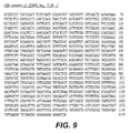
- nucleotide sequences of the invention are SEQ ID NO:1 and SEQ ID NO:2, as set forth in Figures 8 and 9 , respectively.
- this invention provides an isolated nucleic acid molecule selected from the group consisting of (a) the polynucleotide sequence consisting of SEQ ID NO:1 or SEQ ID NO:2 (b) the complement of (a) and (c) a nucleic acid sequence as defined in the claims.
- conditions of moderate stringency can be readily determined by those having ordinary skill in the art based on, for example, the length of the DNA.
- the basic conditions are set forth by Sambrook et al. Molecular Cloning: A Laboratory Manual, 2d ed. Vol. 1, pp.
- hybridization conditions are defined as above, and with washing at approximately 68°C, about 0.2X SSC, and about 0.1% SDS.
- temperature and wash solution salt concentration can be adjusted as necessary according to factors such as the length of the probe.
- a DNA sequence can vary from that shown in SEQ ID NO:1 or SEQ ID NO:2, and still encode PCGEM1.
- Such variant DNA sequences can result from silent mutations ( e.g ., occurring during PCR amplification), or can be the product of deliberate mutagenesis of a native sequence.
- the invention thus relates to isolated DNA sequences of the invention selected from: (a) DNA consisting of the nucleotide sequence ofSEQ ID NO:1 or SEQ ID NO:2; (b) the complement thereof; or (c) a fragment thereof as defined in the claims.
- sequences are preferably provided and/or constructed in the form of an open reading frame uninterrupted by internal non-translated sequences, or introns, that are typically present in eukaryotic genes. Sequences of non-translated DNA can be present 5' or 3' from an open reading frame, where the same do not interfere with manipulation or expression of the coding region.
- PCCEM1 encode a polypeptide
- polypeptides are encoded by such DNA sequences.
- Percent identity may be determined by visual inspection and mathematical calculation. Alternatively, percent identity of two nucleic acid sequences may be determined by comparing sequence information using the GAP computer program, version 6.0 described by Devereux et al. (Nucl. Acids Res. 12:387,1984 ) and available from the University of Wisconsin Genetics Computer Group (UWGCG).
- the preferred default parameters for the GAP program include: (1) a unary comparison matrix (containing a value of 1 for identities and 0 for non-identities) for nucleotides, and the weighted comparison matrix of Gribskov and Burgess, Nucl. Acids Res.
- Isolated nucleic acids may be useful in the production of polypeptides.
- polypeptides may be prepared by any of a number of conventional techniques.
- a DNA sequence of this invention or desired fragment thereof may be subcloned into an expression vector for production of the polypeptide or fragment.
- the DNA sequence advantageously is fused to a sequence encoding a suitable leader or signal peptide.
- the desired fragment may be chemically synthesized using known techniques.
- DNA fragments also may be produced by restriction endonuclease digestion of a full length cloned DNA sequence, and isolated by electrophoresis on agarose gels.
- oligonucleotides that reconstruct the 5' or 3' terminus to a desired point may be ligated to a DNA fragment generated by restriction enzyme digestion.
- Such oligonucleotides may additionally contain a restriction endonuclease cleavage site upstream of the desired coding sequence, and position an initiation codon (ATG) at the N-terminus of the coding sequence.
- PCR polymerase chain reaction
- Oligonucleotides that define the desired termini of the DNA fragment are employed as 5' and 3' primers.
- the oligonucleotides may additionally contain recognition sites for restriction endonucleases, to facilitate insertion of the amplified DNA fragment into an expression vector.
- PCR techniques are described in Saiki et al., Science 239:487 (1988 ); Recombinant DNA Methodology, Wu et al., eds., Academic Press, Inc., San Diego (1989), pp. 189-196 ; and PCR Protocols: A Guide to Methods and Applications, Innis et al., eds., Academic Press, Inc. (1990 ).
- the invention relates to PCGEM1 nucleotide sequences isolated from human prostate cells, including two full length cDNAs: SEQ ID NO:1 ( Figure 8 ) and SEQ ID NO:2 ( Figure 9 ), and fragments thereof as defined in the claims.
- the nucleic acids of the invention including DNA, RNA, mRNA and oligonucleotides thereof, are useful in a variety of applications in the detection, diagnosis, prognosis, and treatment of prostate cancer. Examples of applications envisaged include, but are not limited to:
- the present invention provides an in vitro method of detecting prostate cancer in a patient, which comprises (a) detecting PCGEM1 mRNA in a biological sample from the patient, as defined in the claims, and (b) correlating the amount of PCGEM1 mRNA in the sample with the presence of prostate cancer in the patient.
- detecting PCGEM1 mRNA in a biological sample includes: (a) isolating am RNA from a biological sample from the patient, (b) amplifying a cDNA molecule comprising SEQ ID NO:1, or a fragment thereof; (c) incubating the amplified cDNA with a nucleic acid probe that hybridises to SEQ ID No:1 to form a duplex molecule that is both stable and selective; and (d) detecting hybridization between the amplified cDNA and the probe.
- the biological sample can be selected from the group consisting of blood, urine, and tissue, for example, from a biopsy.
- the biological sample is blood. This method is useful in both the initial diagnosis of prostate cancer, and the later prognosis of disease. This method allows for testing prostate tissue in a biopsy, and after removal of a cancerous prostate, continued monitoring of the blood for micrometastases.
- the amount of PCGEM1 mRNA in a biological sample from a patient is correlated with the presence of prostate cancer in the patient Those of ordinary skill in the art can readily assess the level of over-expression that is correlated with the presence of prostate cancer.
- This invention also provides a method of identifying an androgen-responsive cell line, which comprises (a) incubating a cell line suspected of being androgen-responsive with an androgen; and (b) detecting a mRNA sequence in the cell line, by hybridising said mRNA sequence to SEQ ID NO: 1 or 2 under conditions of high stringency; wherein an increase in PCGEM1 mRNA, as compared to an untreated cell line, correlates with the cell line being androgen-responsive.
- the invention further provides an in vitro method of measuring the responsiveness of a prostatic tissue to hormone-ablation therapy, which comprises measuring a mRNA sequence in the prostatic tissue following hormone-ablation therapy, by hybridising said mRNA sequence to SEQ ID NO: 1 or 2 under conditions of high stringency; wherein a decrease in PCGEM1 mRNA, as compared to an untreated cell line, correlates with the cell line responding to hormone-ablation therapy.
- nucleic acid molecules may be introduced into a recombinant vector, such as a plasmid, cosmid, or virus, which can be used to transfect or transduce a host cell.
- Nucleic acids may be combined with other DNA sequences, such as promoters, polyadenylation signals, restriction enzyme sites, multiple cloning, sites, and other coding sequences.
- nucleic acids of the invention are used as probes or primers.
- probes or primers of the invention include those of SEQ ID NO: 6, and SEQ ID NO: 7, as well as those disclosed in Table I.
- Table I Primer Sequence (5' ⁇ 3') S/AS Starting Base # SEQ ID NO.
- a particularly preferred probe is derived from PCGEM1 (SEQ ID NO: 1) and comprises nucleotides 116 to 1140 of that sequence. It has been designated SEQ ID NO: 4 and is set forth in Figure 12 .
- hybridization probe When a hybridization probe binds to a target sequence, it forms a duplex molecule that is both stable and selective.
- nucleic acid molecules may be readily prepared, for example, by chemical synthesis or by recombinant techniques. A wide variety of methods are known in the art for detecting hybridization, including fluorescent, radioactive, or enzymatic means, or other ligands such as avidin/biotin.
- these nucleic acid molecules may be introduced into a recombinant vector, such as a plasmid, cosmid, or virus, which can be used to transfect or transduce a host cell.
- a recombinant vector such as a plasmid, cosmid, or virus
- the nucleic acids of the present invention may be combined with other DNA sequences, such as promoters, polyadenylation signals, restriction enzyme sites, multiple cloning sites, and other coding sequences.
- Probes based on the human DNA sequence of SEQ ID NO:1 or SEQ ID NO:2 may be used to screen cDNA libraries derived from other mammalian species, using conventional cross-species hybridization techniques.
- oligonucleotides are useful as primers, e.g., in polymerase chain reactions (PCR), whereby DNA fragments are isolated and amplified.
- PCR polymerase chain reactions
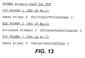
- Particularly preferred primers are set forth in Figures 13 and Table I and are designated SEQ ID NOS: 6-7 and 9-22, respectively.
- a particularly preferred primer pair is p518 (SEQ ID NO: 15) and p839 (SEQ ID NO: 22), which when used in PCR, preferentially amplifies mRNA, thereby avoiding less desirable cross-reactivity with genomic DNA.
- the PCGEM1 gene has been mapped by fluorescent in situ hybridization to the 2q32 region of chromosome 2 using a bacterial artificial chromosome (BAC) clone containing PCGEU1 genomic sequence.
- BAC bacterial artificial chromosome
- all or a portion of the nucleic acid molecule of SEQ ID NO:1 and SEQ ID NO:2 can be used by those skilled in the art using well-known techniques to identify human chromosome 2, and the specific locus thereof, that contains the PCGEM1 DNA.
- Useful techniques include, but are not limited to, using the nucleotide sequence of SEQ ID NO:1 or SEQ ID NO:2, or fragments thereof, including oligonucleotides, as a probe in various well-known techniques such as radiation hybrid mapping (high resolution), in situ hybridization to chromosome spreads (moderate resolution), and Southern blot hybridization to hybrid cell lines containing individual human chromosomes (low resolution).
- chromosomes can be mapped by radiation hybridization.
- PCR is performed using the Whitehead Institute/MIT Center for Genome Research Genebridge4 panel of 93 radiation hybrids
- PCGEM1 has been mapped to the 2q32 region of chromosome 2. This region is associated with specific diseases, which include but are not limited to diabetes mellitus (insulin dependent), and T cell leukemia/lymphoma.
- diseases include but are not limited to diabetes mellitus (insulin dependent), and T cell leukemia/lymphoma.
- the nucleic acids of SEQ ID NO: 1 or SEQ ID NO: 2, or fragments thereof, as defined in the claims can be used by one skilled in the art using well-known techniques to analyze abnormalities associated with gene mapping to chromosome 2. This enables one to distinguish conditions in which this marker is rearranged or deleted.
- nucleotides of SEQ ID NO:1 or SEQ ID NO:2, or fragments thereof as defined in the claims can be used as a positional marker to map other genes of unknown location.
- the DNA may be used in developing treatments for any disorder mediated (directly or indirectly) by defective, or insufficient amounts of PCGEM1, including prostate cancer. Disclosure herein of native nucleotide sequences permits the detection of defective genes, and the replacement thereof with normal genes. Defective genes may be detected in in vitro diagnostic assays, and by comparison of a native nucleotide sequence disclosed herein with that of a gene derived from a person suspected of harboring a defect in this gene.
- nucleic acids include antisense or sense oligonucleotides comprising a single-stranded nucleic acid sequence (either RNA or DNA) capable of binding to target mRNA (sense) or DNA (antisense) sequences.
- Antisense or sense oligonucleotides comprise a fragment of DNA (SEQ ID NO:1 or SEQ ID NO:2). Such a fragment generally comprises at least about 14 nucleotides, preferably from about 14 to about 30 nucleotides.
- Stein and Cohen Cancer Res. 48:2659, 1988
- van der Krol et al. BioTechniques 6:958, 1988 ).
- PCGEM1 biologic activity of PCGEM1 in assay cells and the over expression of PCGEM1 in prostate cancer tissues suggest that elevated levels of PCGEM1 promote prostate cancer cell growth.
- the antisense oligonucleotides to PCGEM1 may be used to reduce the expression of PCGEM1 and, consequently, inhibit the growth of the cancer cells.
- antisense or sense oligonucleotides Binding of antisense or sense oligonucleotides to target nucleic acid sequences results in the formation of duplexes.
- the antisense oligonucleotides thus may be used to block expression of proteins or to inhibit the function of RNA.
- Antisense or sense oligonucleotides further comprise oligonucleotides having modified sugar-phosphodiester backbones (or other sugar linkages, such as those described in WO91/06629 ) and wherein such sugar linkages are resistant to endogenous nucleases.
- Such oligonucleotides with resistant sugar linkages are stable in vivo ( i.e., capable of resisting enzymatic degradation) but retain sequence specificity to be able to bind to target nucleotide sequences.
- sense or antisense oligonucleotides include those oligonucleotides which are covalently linked to organic moieties, such as those described in WO 90/10448 , and other moieties that increases affinity of the oligonucleotide for a target nucleic acid sequence, such as poly-(L-lysine).
- intercalating agents such as ellipticine, and alkylating agents or metal complexes may be attached to sense or antisense oligonucleotides. Such modifications may modify binding specificities of the antisense or sense oligonucleotide for the target nucleotide sequence.
- Antisense or sense oligonucleotides may be introduced into a cell containing the target nucleic acid sequence by any gene transfer method, including, for example, lipofection, CaPO 4 -mediated DNA transfection, electroporation, or by using gene transfer vectors such as Epstein-Barr virus or adenovirus.
- Sense or antisense oligonucleotides also may be introduced into a cell containing the target nucleotide sequence by formation of a conjugate with a ligand binding molecule, as described in WO 91/04753 .
- Suitable ligand binding molecules include, but are not limited to, cell surface receptors, growth factors, other cytokines, or other ligands that bind to cell surface receptors.
- conjugation of the ligand binding molecule does not substantially interfere with the ability of the ligand binding molecule to bind to its corresponding molecule or receptor, or block entry of the sense or antisense oligonucleotide or its conjugated version into the cell.
- a sense or an antisense oligonucleotide may be introduced into a cell containing the target nucleic acid sequence by formation of an oligonucleotide-lipid complex, as described in WO 90/10448 .
- the sense or antisense oligonucleotide-lipid complex is preferably dissociated within the cell by an endogenous lipase.
- polypeptides and fragments thereof in various forms, including those that are naturally occurring or produced through various techniques such as procedures involving recombinant DNA technology. Such forms include, but are not limited to, derivatives, variants, and oligomers, as well as fusion proteins or fragments thereof.
- polypeptides described herein include full length proteins encoded by the nucleic acid sequences set forth above.
- the polypeptides may be membrane bound or they may be secreted and thus soluble.
- the specification also describes the expression, isolation and purification of polypeptides and fragments accomplished by any suitable technique.
- Differential display provides a method to separate and clone individual messenger RNAs by means of the polymerase chain reaction, as described in Liang et al., Science, 257:967-71 (1992 ), which is hereby incorporated by reference. Briefly, the method entails using two groups of oligonucleotide primers. One group is designed to recognize the polyadenylate tail of messenger RNAs. The other group contains primers that are short and arbitrary in sequence and anneal to positions in the messenger RNA randomly distributed from the polyadenylate tail. Products amplified with these primers can be differentiated on a sequencing gel based on their size. If different cell populations are amplified with the same groups of primers, one can compare the amplification products to identify differentially expressed RNA sequences.
- DD Differential display
- RNAzol B Tel-Test, Inc., Friendswood, TX
- the epithelial nature of the RNA source was further confirmed using cytokeratin 18 expression (45) in reverse transcriptase-polymerase chain reaction (RT-PCR) assays.
- RNA-free total RNA was amplified by RT-PCR which was performed using ten anchored antisense primers and four arbitrary sense primers according to the protocol provided by HieroglyphTM RNA Profile Kit 1 (Genomyx Corporation, CA).
- the cDNA fragments produced by the RT-PCR assay were analyzed by high resolution gel electrophoresis, carried out by using GenomyxTM LR DNA sequencer and LR-OptimizedTM HR-1000TM gel formulations (Genomyx Corporation, CA).
- a partial DD screening of normal/tumor tissues revealed 30 differentially expressed cDNA fragments, with 53% showing reduced or no expression in tumor RNA specimens and 47% showing over expression in tumor RNA specimen ( Figure 2 ).
- These cDNAs were excised from the DD gels, reamplified using T7 and M13 primers and the RT PCR conditions recommended in HieroglyphTM RNA Profile Kit-1 (Genomyx Corp., CA), and sequenced.
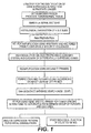
- the inclusion of T7 and M 13 sequencing primers in the DD primers allowed rapid sequencing and orientation of cDNAs ( Figure 1 ).
- cDNA fragments All the reamplified cDNA fragments were purified by Centricon-c-100 system (Amicon, USA). The purified fragments were sequenced by cycle sequencing and DNA sequence determination using an ABI 377 DNA sequencer. Isolated sequences were analyzed for sequence homology with known sequences by running searches through publicly available DNA sequence databases, including the National Center for Biotechnology Information and the Cancer Genome Anatomy Project. Approximately two-thirds of these cDNA sequences exhibited homology to previously described DNA sequences/genes e.g., ribosomal proteins, mitochondrial DNA sequences, growth factor receptors, and genes involved in maintaining the redox state in cells.
- DNA sequences/genes e.g., ribosomal proteins, mitochondrial DNA sequences, growth factor receptors, and genes involved in maintaining the redox state in cells.
- the PCGEM1 fragment obtained from the initial differential display screening represents a 530 base pair (nucleotides 410 to 940 of SEQ ID NO: 1) cDNA sequence which, in initial searches, did not exhibit any significant homology with sequences in the publicly available databases.
- PCGEM1 The full length of PCGEM1 was obtained by 5' and 3' RACE/PCR from the original 530 bp DD product (nucleotides 410 to 940 of PCGEM1 DNA SEQ ID NO:1) using a normal prostate cDNA library in lambda phage (Clontech, CA).
- the RACE/PCR products were directly sequenced.
- Lasergene and MacVector DNA analysis software were used to analyze DNA sequences and to define open reading frame regions.
- PCGEM1 DNA has been verified by several approaches including characterization of several clones of PCGEM1 and analysis of PCGEM1 cDNAs amplified from normal prostate tissue and prostate cancer cell lines. We have also obtained the genomic clones of PCGEM1, which has helped to confirm the PCGEM1 cDNA sequence.
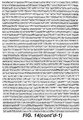
- the complete genomic DNA sequence of PCGEM1 (SEQ ID NO:8) is shown in Figure 14 .
- "Y" represents any one of the four nucleotide bases, cylosine, thymine, adenine, or guanine.
- Comparison of the cDNA and genomic sequences revealed the organization of the PCGEM1 transcription unit from three exons ( Figure 15 : E, Exon; B: BamHI; H: HindIII; X: XbaI; R: EcoRI).
- PCGEM1 genomic DNA was mapped to specific region 2q32 ( Figure 7A ).
- BAC Bacterial Artificial Chromosome
- PCGEM1-Bac clone 1 DNA was nick translated using spectrum orange (Vysis) as a direct label and flourescent in situ hybridization was done using this probe on normal human male metaphase chromosome spreads.
- DAPI inverted 4',6-diamidino-2-phenylindole
- Figure 7B a DAPI counter-stained chromosome 2 is shown on the left; an inverted DAPI stained chromosome 2 shown as G-bands is shown in the center; an ideogram of chromosome 2 showing the localization of the signal to band 2q32(bar) is shown on the right.
- NU200 image acquisition and registration software was used to create the digital images. More than 20 metaphases were analyzed.
- the expression of the PCGEM1 fragment was evaluated on a test panel of matched tumor and normal RNAs derived from the microdissected tissues of twenty prostate cancer patients.
- RNA-free RNA was prepared from these tissues and used in RT-PCR analysis to detect PCGEM1 expression.
- One hundred nanograms of total RNA was reverse transcribed into cDNA using RT-PCR kit (Perkin-Elmer, Foster, CA).
- the PCR was performed using Amplitaq Gold from Perkin-Elmer (Foster, CA). PCR cycles used were: 95°C for 10 minutes, 1 cycle; 95°C for 30 seconds, 55°C for 30 seconds, 72°C for 30 seconds, 42 cycles, and 72°C for 5 minutes, 1 cycle followed by a 4°C storage.
- Epithelial cell-associated cytokeratin 18 was used as an internal control.
- Expression of housekeeping genes, cytokeratin-18 ( Figure 3 ) and glyceraldehyde-3-phosphate dehydrogenase (GAPDH) (data not shown) remained constant in tumor and normal specimens of all the patients ( Figure 3 ).
- In situ hybridization was performed essentially as described by Wilkinson and Green (48). Briefly, OCT embedded tissue slides stored at -80°C were fixed in 4% PFA (paraformaldehyde), digested with proteinase K and then again fixed in 4% PFA. After washing in PBS, sections were treated with 0.25% acetic anhydride in 0.1M triethanolamine, washed again in PBS, and dehydrated in a graded ethanol series. Sections were hybridized with 35 S-labeled riboprobes at 52°C overnight. After washing and RNase A treatment, sections were dehydrated, dipped into NTB-2 emulsion and exposed for 11 days at 4°C.
- PFA parformaldehyde
- PCGEM1 expression was scored as percentage of cells showing 35 S signal: 1+, 1-25%; 2+, 25-50%; 3+, 50-75%, 4+, 75-100%.
- FIG. 17 shows an example of in situ hybridization of 35 S labeled PCGEM1 riboprobe to matched normal (A) versus tumor (B) sections of prostate cancer patients.
- the light gray areas are hematoxylin stained cell bodies, the black dots represent the PCGEM1 expression signal.
- the signal is background level in the normal (A), 2+ level in the tumor (B) section.
- the magnification is 40x.
- PCGEM1 gene expression was also evaluated in established prostate cancer cell lines: LNCaP, DU145, PC3 (all from ATCC), DuPro (available from Dr. David Paulson, Duke University, Durham, NC), and an E6/E7 - immortalized primary prostate cancer cell line, CPDR1 (47).
- CPDR1 is a primary CaP derived cell line immortalized by retroviral vector, LXSN 16 E6 E7, expressing E6 and E7 gene of the human papilloma virus 16.
- LNCaP is a well studied, androgen-responsive prostate cancer cell line, whereas DU145, PC3, DuPro and CPDR1 are androgen-independent and lack detectable expression of the androgen receptor.
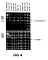
- PCGEM1 expression was easily detectable in LNCaP ( Figure 4 ). However, PCGEM1 expression was not detected in prostate cancer cell lines DU145, PC3, DuPro and CPDR. Thus, PCGEM1 was expressed in the androgen-responsive cell line but not in the androgen-independent cell lines. These results indicate that hormones, particularly androgen, may play a key role in regulating PCGEM1 expression in prostate cancer cells. In addition, the results suggest that PCGEM1 expression may be used to distinguish between hormone responsive tumor cells and more aggressive hormone refractory tumor cells.
- LNCaP cells were maintained in RPMI 1640 (Life Technologies, Inc., Gaithersburg, MD) supplemented with 10% fetal bovine serum (FBS, Life Technologies, Inc., Gaithersburg, MD) and experiments were performed on cells between passages 20 and 35.
- FBS fetal bovine serum
- cFBS charcoal/dextran stripped androgen-free FBS
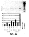
- LNCaP cells were cultured first in RPMI 1640 with 10% cFBS for 4 days and then stimulated with a non-metabolizable androgen analog R1881 (DUPONT, Boston, MA) at different concentrations for different times as shown in Figure 5A .
- RNAzol B Tel-Test, Inc, TX
- RNAzol B Tel-Test, Inc, TX
- RNAzol B Tel-Test, Inc, TX
- 2 ⁇ g/lane fractionated (2 ⁇ g/lane) by running on 1% formaldehyde-agarose gel and transferred to nylon membrane.
- Northern blots were analyzed for the expression of PCGEM1 using the nucleic acid molecule set forth in SEQ ID NO: 4 as a probe.
- the RNA from LNCaP cells treated with R1881 and RNA from control LNCaP cells were also analyzed by RT-PCR assays as described in Example 4.
- PCGEM1 expression increases in response to androgen treatment. This finding further supports the hypothesis that the PCGEM1 expression is regulated by androgens in prostate cancer cells.
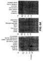
- RNA master blot analyses confirm the prostate tissue specificity of the PCGEM1 gene ( Figure 6b ).
- Northern blot analyses reveal that the prostate tissue specificity of PCGEM1 is comparable to the well known prostate marker PSA (77mer oligo probe) and far better than two other prostate specific genes PSMA (234 bp fragment from PCR product) and NKX3.1 (210 bp cDNA).
- PSMA is expressed in the brain (37) and in the duodenal mucosa and a subset of proximal renal tubules (38).
- NKX3.1 exhibits high levels of expression in adult prostate, it is also expressed in lower levels in testis tissue and several other tissues (39).
- PCGEM1 overexpression suggested that the increased expression of PCGEM1 may favor tumor cell proliferation.
- NIH3T3 cells have been extensively used to define cell growth promoting functions associated with a wide variety of genes (40-44).
- PCGEM1 expression vectors were constructed in sense and anti-sense orientations and were transfected into NIH3T3 cells, and hygromycin resistant colonies were counted 2-3 weeks later.
- Cells transfected with PCGEM1 sense construct formed about 2 times more colonies than vector alone in three independent experiments ( Figure 10 ). The size of the colonies in PCGEM1 sense construct transfected cells were significantly larger. No appreciable difference was observed in the number of colonies between anti-sense PCGEM1 constructs and vector controls.
- PCGEM1 The function of PCGEM1, however, does not appear to be due to protein expression.
- TestCode program GCG Wisconsin Package, Madison, WI
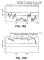
- Analysis of the PCGEM1 DNA sequence revealed that, at greater than 95% confidence level, the sequence does not contain any region with protein coding capacity ( Figure 16A ).
- Similar results were obtained when various published non-coding RNA sequences were analyzed with the TestCode program (data not shown), while known protein coding regions of similar size i.e., alpha actin ( Figure 16B ) can be detected with high fidelity.
- PCGEM1 characterization has been its apparent lack of protein coding capacity. Although we have not completely ruled out the possibility that PCGEM1 codes for a short unstable peptide, careful sequencing of PCGEM1 cDNA and genomic clones, computational analysis of PCGEM1 sequence, and in vitro transcription/translation experiments (data not shown) strongly suggest a non-coding nature of PCGEM1. It is interesting to note that an emerging group of novel mRNA-like non-coding RNAs are being discovered whose function and mechanisms of action remain poorly understood (49). Such RNA molecules have also been termed as "RNA riboregulators" because of their function(s) in development, differentiation, DNA damage, heat shock responses and tumorigenesis (40-42, 50).
- the H19, His -1 and Bic genes code for functional non-coding mRNAs (50).
- DD3 also appears to exhibit a tissue specific non-coding mRNA (51).
- PCGEM1 and DD3 may represent a new class of prostate specific genes.
- the recent discovery of a steroid receptor co-activator as an mRNA, lacking protein coding capacity further emphasizes the role of RNA riboregulators in critical biochemical function(s) (52).
- PCGEM1 expression in NIH3T3 cells caused a significant increase in the size of colonies in a colony forming assay and suggests that PCGEM1 cDNA confers cell proliferation and/or cell survival function(s). Elevated expression of PCGEM1 is prostate cancer cells may represent a gain in function favoring tumor cell proliferation/survival.
- PCGEM1 belongs to a novel class of prostate tissue specific genes with potential functions in prostate cell biology and the tumorigenesis of the prostate gland.
- PCGEM1 novel nucleotide sequence
Landscapes
- Health & Medical Sciences (AREA)
- Chemical & Material Sciences (AREA)
- Organic Chemistry (AREA)
- Life Sciences & Earth Sciences (AREA)
- General Health & Medical Sciences (AREA)
- Medicinal Chemistry (AREA)
- Veterinary Medicine (AREA)
- Biochemistry (AREA)
- Nuclear Medicine, Radiotherapy & Molecular Imaging (AREA)
- Animal Behavior & Ethology (AREA)
- General Chemical & Material Sciences (AREA)
- Public Health (AREA)
- Chemical Kinetics & Catalysis (AREA)
- Immunology (AREA)
- Toxicology (AREA)
- Zoology (AREA)
- Gastroenterology & Hepatology (AREA)
- Pharmacology & Pharmacy (AREA)
- Biophysics (AREA)
- Genetics & Genomics (AREA)
- Molecular Biology (AREA)
- Proteomics, Peptides & Aminoacids (AREA)
- Measuring Or Testing Involving Enzymes Or Micro-Organisms (AREA)
- Micro-Organisms Or Cultivation Processes Thereof (AREA)
- Medicines That Contain Protein Lipid Enzymes And Other Medicines (AREA)
- Medicines Containing Material From Animals Or Micro-Organisms (AREA)
- Medicines Containing Plant Substances (AREA)
Abstract
Description
- The present invention relates to nucleic acids, as defined in the claims, that are expressed in prostate tissue. More particularly, the present invention relates to the first of a family of novel, androgen-regulated, prostate-specific genes, PCGEM1, that is over-expressed in prostate cancer, and methods of using the PCGEM1 sequence and fragments thereof to measure the hormone responsiveness of prostate cancer cells and to detect or diagnose prostate cancer and other prostate related diseases.
- Prostate cancer is the most common solid tumor in American men (1). The wide spectrum of biologic behavior (2) exhibited by prostatic neoplasms poses a difficult problem in predicting the clinical course for the individual patient (3, 4). Public awareness of prostate specific antigen (PSA) screening efforts has led to an increased diagnosis of prostate cancer. The increased diagnosis and greater number of patients presenting with prostate cancer has resulted in wider use of radical prostatectomy for localized disease (5). Accompanying the rise in surgical intervention is the frustrating realization of the inability to predict organ-confined disease and clinical outcome for a given patient (5,6). Traditional prognostic markers, such as grade, clinical stage, and pretreatment PSA have limited prognostic value for individual men. There is clearly a need to recognize and develop molecular and genetic biomarkers to improve prognostication and the management of patients with clinically localized prostate cancer. As with other common human neoplasia (7), the search for molecular and genetic biomarkers to better define the genesis and progression of prostate cancer is the key focus for cancer research investigations worldwide.
- The new wave of research addressing molecular genetic alterations in prostate cancer is primarily due to increased awareness of this disease and the development of newer molecular technologies. The search for the precursor of prostatic adenocarcinoma has focused largely on the spectrum of microscopic changes referred to as "prostatic intraepithelial neoplasia" (PIN). Bostwick defines this spectrum as a histopathologic continuum that culminates in high grade PIN and early invasive cancer (8). The morphologic and molecular changes include the progressive disruption of the basal cell-layer, changes in the expression of differentiation markers of the prostatic secretory epithelial cells, nuclear and nucleolar abnormalities, increased cell proliferation, DNA content alterations, and chromosomal and allelic losses (8, 9). These molecular and genetic biomarkers, particularly their progressive gain or loss, can be followed to trace the etiology of prostate carcinogenesis. Foremost among these biomarkers would be the molecular and genetic markers associated with histological phenotypes in transition between normal prostatic epithelium and cancer. Most studies so far seem to agree that PIN and prostatic adenocarcinoma cells have a lot in common with each other. The invasive carcinoma more often reflects a magnification of some of the events already manifest in PIN.
- Early detection of prostate cancer is possible today because of the widely propagated and recommended blood PSA test that provides a warning signal for prostate cancer if high levels of serum PSA are detected. However, when used alone, PSA is not sufficiently sensitive or specific to be considered an ideal tool for the early detection or staging of prostate cancer (10). Combining PSA levels with clinical staging and Gleason scores is more predictive of the pathological stage of localized prostate cancer (11). In addition, new molecular techniques are being used for improved molecular staging of prostate cancer (12, 13). For instance, reverse transcriptase - polymerase chain reaction (RT-PCR) can measure PSA of circulating prostate cells in blood and bone marrow of prostate cancer patients.
- Despite new molecular techniques, however, as many as 25 percent of men with prostate cancer will have normal PSA levels - usually defined as those equal to or below 4 nanograms per milliliter of blood (14). In addition, more than 50 percent of the men with higher PSA levels are actually cancer free (14). Thus, PSA is not an ideal screening tool for prostate cancer. More reliable tumor-specific biomarkers are needed that can distinguish between normal and hyperplastic epithelium, and the preneoplastic and neoplastic stages of prostate cancer.
- Identification and characterization of genetic alterations defining prostate cancer onset and progression is important in understanding the biology and clinical course of the disease. The currently available TNM staging system assigns the original primary tumor (T) to one of four stages (14). The first stage, T1, indicates that the tumor is microscopic and cannot be felt on rectal examination. T2 refers to tumors that are palpable but fully contained within the prostate gland. A T3 designation indicates the cancer has spread beyond the prostate into surrounding connective tissue or has invaded the neighboring seminal vesicles. T4 cancer has spread even further. The TNM staging system also assesses whether the cancer has metastasized to the pelvic lymph nodes (N) or beyond (M). Metastatic tumors result when cancer cells break away from the original tumor, circulate through the blood or lymph, and proliferate at distant sites in the body.
- Recent studies of metastatic prostate cancer have shown a significant heterogeneity of allelic losses of different chromosome regions between multiple cancer foci (21-23). These studies have also documented that the metastatic lesion can arise from cancer foci other than dominant tumors (22). Therefore, it is critical to understand the molecular changes which define the prostate cancer metastasis especially when prostate cancer is increasingly detected in early stages (15-21).
- Moreover, the multifocal nature of prostate cancer needs to be considered (22-23) when analyzing biomarkers that may have potential to predict tumor progression or metastasis. Approximately 50-60% of patients treated with radical prostatectomy for localized prostate carcinomas are found to have microscopic disease that is not organ confined, and a significant portion of these patients relapse (24). Utilizing biostatistical modeling of traditional and genetic biomarkers such as p53 and bcl-2, Bauer et al. (25-26) were able to identify, patients at risk of cancer recurrence after surgery. Thus, there is clearly a need to develop biomarkers defining various stages of the prostate cancer progression.
- Another significant aspect of prostate cancer is the key role that androgens play in the development of both the normal prostate and prostate cancer. Androgen ablation, also referred to as "hormonal therapy," is a common treatment for prostate cancer, particularly in patients with metastatic disease (14). Hormonal therapy aims to inhibit the body from making androgens or to block the activity of androgen. One way to block androgen activity involves blocking the androgen receptor; however, that blockage is often only successful initially. For example, 70-80% of patients with advanced disease exhibit an initial subjective response to hormonal therapy, but most tumors progress to an androgen-independent state within two years (16). One mechanism proposed for the progression to an androgen-independent state involves constitutive activation of the androgen signaling pathway, which could arise from structural changes in the androgen receptor protein (16).
- As indicated above, the genesis and progression of cancer cells involve multiple genetic alterations as well as a complex interaction of several gene products. Thus, various strategies are required to fully understand the molecular genetic alterations in a specific type of cancer. In the past, most molecular biology studies had focused on mutations of cellular proto-oncogenes and tumor suppressor genes (TSGs) associated with prostate cancer (7). Recently, however, there has been an increasing shift toward the analysis of "expression genetics" in human cancer (27-31), i.e., the under-expression or over-expression of cancer-specific genes. This shift addresses limitations of the previous approaches including: 1) labor intensive technology involved in identifying mutated genes that are associated with human cancer; 2) the limitations of experimental models with a bias toward identification of only certain classes of genes, e.g., identification of mutant ras genes by transfection of human tumor DNAs utilizing NIH3T3 cells; and 3) the recognition that the human cancer associated genes identified so far do not account for the diversity of cancer phenotypes.
- A number of studies are now addressing the alterations of prostate cancer-associated gene expression in patient specimens (32-36). It is inevitable that more reports on these lines are to follow.
- Thus, despite the growing body of knowledge regarding prostate cancer, there is still a need in the art to uncover the identity and function of the genes involved in prostate cancer pathogenesis. There is also a need for reagents and assays to accurately detect cancerous cells, to define various stages of prostate cancer progression, to identify and characterize genetic alterations defining prostate cancer onset and progression, to detect micro-metastasis of prostate cancer, and to treat and prevent prostate cancer.
- EMBL Acc. No. AC003046, 8 Nov. 1997, provides homo sapiens Xp22 PACs RPC11-263P4 and RPC11-164K3.
- Proceedings of the American Ass for Cancer Res. Ann. 40: 37, March 1999, describes the structure and expression of a prostate specific gene, PC-GEM1.
- EMBL Acc. No AC013401, 11 Nov. 1999, provides homo
sapiens chromosome 5chromosome 5 clone RP11-98N11 (unordered pieces). - EMBL Acc. No. AF099810, 28 Oct. 1998, provides the homo sapiens neurexin III-alpha gene (
chromosome 14 from 14q24.3-14q 32). - EMBL Acc. No. AC006925, 11 Mar. 1999, provides homo sapiens chromosome 17, clone hRPK.81_I_9.
-
WO 99/00498 - Bussemakers M.J.G. et al., Urological Research, Vol.25, No.1, , describes a prostate-specific marker, strongly overexpressed in prostatic tumours.
- Wang Zhou et al., PNAS USA, Vol. 94, No.24, pgs 12999-13004, 25 Nov 1997, describes genes regulated by androgen in the rat ventral prostate.
-
WO 95/19434 - The present invention relates to the identification and characterization of a novel gene, the first of a family of genes, designated PCGEM1, for Prostate Cancer
Gene Expression Marker 1. PCGEM1 is specific to prostate tissue, is androgen-regulated, and appears to be over-expressed in prostate cancer. More recent studies associate PCGEM1 cDNA with promoting cell growth. The invention provides the isolated nucleotide sequence ofPCGEM 1 or fragments thereof and nucleic acid sequences that hybridize to PCGEM1, as defined in the claims. These sequences have utility, for example, as markers of prostate cancer and other prostate related diseases, and as targets for therapeutic intervention in prostate cancer and other prostate related diseases. The invention further provides a vector that directs the expression of PCGEM1, and a host cell transfected or transduced with this vector, as defined in the claims. - In another embodiment, the invention provides a method of detecting prostate cancer cells in a biological sample, as defined in the claims, such as by using nucleic acid amplification techniques with primers and probes selected to bind specifically to the PCGEM1 sequence. The invention further comprises a method of identifying an androgen responsive cell line, and a method of measuring responsiveness of a cell line to hormone-ablation therapy, as defined in the claims.
- The specification also describes an isolated polypeptide encoded by the PCGEM1 gene or a fragment thereof, and antibodies generated against the PCGEM1 polypeptide, peptides, or portions thereof, which can be used to detect, treat, and prevent prostate cancer.
- Thus, according to a first aspect of the invention, there is provided an isolated nucleic acid molecule selected from the group consisting of:
- (a) the polynucleotide sequence consisting SEQ ID NO:1 or SEQ ID NO:2;
- (b) the complement of (a); and
- (c) a nucleic acid sequence consisting of SEQ ID NO:4, SEQ ID NO:6, SEQ ID NO:7, SEQ ID NO:9, SEQ ID NO:10, SEQ ID NO:11, SEQ ID NO:12, SEQ ID NO:13, SEQ ID NO:14, SEQ 113 NO:15, SEQ ID NO: 16, SEQ ID NO: 17, SEQ ID NO: 18, SEQ ID NO:19, SEQ ID NO:20, SEQ ID NO:21 or SEQ ID NO:22.
- In another aspect of the invention there is provided an in vitro method of detecting prostate cancer in a patient, the method comprising:
- (a) detecting a mRNA sequence in a biological sample from the patient, by hybridising the mRNA sequence to SEQ ID NO:1 or SEQ ID NO:2 under conditions of high stringency; and
- (b) correlating the amount of the mRNA sequence in the sample with the presence of prostate cancer in the patient.
- The invention also provides an in vitro method of identifying an androgen-responsive cell line, the method comprising:
- (a) incubating a cell line suspected of being androgen responsive with an androgen; and
- (b) detecting a mRNA sequence in the cell line, by hybridising the mRNA sequence to SEQ ID NO:1 or SEQ ID NO:2 under conditions of high stringency;
- In another aspect there is provided an in vitro method of measuring the responsiveness of a prostate tissue to hormone-ablation therapy, as defined in the claims.
- In further embodiments of the invention there are also provided an isolated nucleic acid molecule consisting of a fragment of SEQ ID NO: 1, wherein the fragment comprises at least 20 contiguous nucleotides of SEQ ID NO:1, as defined in the claims.
- The invention will now be described in more detail, with particular reference to the accompanying drawings.
-
-
Figure 1 depicts the scheme for the identification of differentially expressed genes in prostate tumor and normal tissues. -
Figure 2 depicts a differential display pattern of mRNA obtained from matched tumor and normal tissues of a prostate cancer patient. Arrows indicate differentially expressed cDNAs. -
Figure 3 depicts the analysis of PCGEM1 expression in primary prostate cancers. PCGEM1 specific PCR primers were designed for RT-PCR analysis. Microdissected tissue derived 100 nanograms of genomic DNA-free RNA was used for RT-PCR. The PCR conditions were optimized to be within the logarithmic phase of amplication for all primers used. Epitheal cell associated cytokeratin-18 was used as an internal control The PCR was performed using Amplitaq Gold. PCR cycles included: 95°C for 10 minutes, 1 cycle followed by 95°C for 30 seconds, 55°C for 30 seconds, 72°C for 30 seconds, 42 cycles (PCGEM1) or 35 cycles (cytokertain-18) and 72°C for 5 minutes final extension followed by 4°C chilling. Three independent experiments showed the same results. -
Figure 4 depicts the expression pattern of PCCEM1 in prostate cancer cell lines. Prostate cancer cell lines: LNCaP, DU145, PC-3, DuPro and CPDR-1 were analyzed for PCGEM1 expression utilizing RT-PCR conditions as described inFigure 3 . -
Figure 5a depicts the androgen regulation of PCOEM1 expression in LNCaP cells, as measured by reverse transcriptase PCR. LNCaP cells were cultured in RPMI medium containing 10% charcoal stripped fetal bovine serum for 4 days followed by treatment with synthetic androgen: R1881 for 12 hours and 24 hours at 0.1 nanomolar and 10 nanomolar concentrations. Ply-A+RNA from treated and untreated cells were analyzed for PCGEM1 expression analysis by RT-PCR as described inFigure 3 . -
Figure 5b depicts the androgen regulation of PCGEM1 expression in LNCaP cells, as measured by Northern blot hybridization. LNCaP cells were cultured in RPMI medium containing 10% charcoal stripped fetal bovine serum for 4 days followed by treatment with synthetic androgen: R1881 for 24 hours at 0.1 nanomolar concentration. Poly-A+RNA from treated and untreated cells were analyzed for PCGEM1 expression by Northern blot hybridization. -
Figure 6a depicts the prostate tissue specific expression pattern of PCGEM1. Multiple tissue Northern blots (Clontech, CA) were probed with a 530bp PCGEM1 cDNA probe. Prostate tissue specific expression of a 1.7kb mRNA band was detected. A recent independent experiment confirmed these results. -
Figure 6b depicts a RNA master blot (Clontech, CA) showing the prostate tissue specificity of PCGEM1. -
Figure 7A depicts the chromosomal localization of PCGEM1 by fluorescent in situ hybridization analysis. FISH analysis was done using genomic DNA from PCGEM1 Bac clone. The DNA was nick translated using biotin labeled kit or digoxinin labeled kit. The metaphase chromosome preparation was hybridized and analyzed. -
Figure 7B depicts a DAPI counter-stained chromosome 2 (left), an inverted DAPI stainedchromosome 2 shown as G-bands (center), and an ideogram ofchromosome 2 showing the localization of the signal to band 2q32(bar). -
Figure 8 depicts a cDNA sequence of PCGEM1 (SEQ ID NO:1). Full length cDNA sequence of PCGEM1 was obtained by 5 prime and 3 prime RACE Marathon ready cDNA kit (Clontech, CA) and screening of normal human prostate cDNA library (Clontech, CA) using original PCGEM1 fragment as probe. The DNA sequencing was performed on ABI-310 sequence analyzer. -
Figure 9 depicts an additional cDNA sequence of PCGEMl1 (SEQ ID NO:2). Full.length cDNA sequence of PCGEM1 was obtained by a 5 prime and 3 prime RACE Marathon ready cDNA kit (Clontech, CA) and screening of normal human Prostate cDNA library (Clontech, CA) using original PCGEM1 fragment as probe. The DNA sequencing was performed on ABI-310 sequence analyzer. -
Figure 10 depicts the colony formation of NIH3T3 cell lines expressing various PCGEM1 constructs. PCGEM1 expression vector in sense and antisense orientations [PCDNA3.1/Hygro(+/-) from (Invitrogen, CA)] were transfected into NIH3T3 cells. Hygromycin resistant colonies were counted 2-3 weeks after staining with crystal violet -
Figure 11 depicts the cDNA sequence of the promoter region of PCGEM1 SEQ ID NO:3. The promoter region of PCGEM1 gene was obtained from human placenta genomic library (Stratagene, CA) by PCR using a PCGEM1 specific primer (underlined sequence) and a T7 promoter primer. The PCR product was cloned into TA-Cloning vector (Invitrogen, CA) and sequenced by 310 DNA sequence analyzer (Perkin-Elmer, CA). The sequence was further verified by direct sequencing of PCR product from placenta DNA. The triangle indicates the putative transcription start site. -
Figure 12 depicts the cDNA of a probe, designated SEQ ID NO:4. -
Figure 13 depicts the cDNAs of primers 1-3, designated SEQ ID NOs:5-7, respectively. -
Figure 14 depicts the genomic DNA sequence of PCGEM1, designated SEQ ID NO:8. -
Figure 15 depicts the structure of the PCGEM1 transcription unit. -
Figure 16 depicts a graph of the hypothetical coding capacity of PCGEM1. -
Figure 17 depicts a representative example of in situ hybridization results showing PCGEM1 expression in normal and tumor areas of prostate cancer tissues. - The present invention relates to PCGEM1, the first of a family of genes, and its related nucleic acids, as defined in the claims, for use in the detection, of prostate cancer (e.g., prostatic intraepithelial neoplasia (PIN), adenocarcinomas, nodular hyperplasia, and large duct carcinomas) and prostate related diseases (e,g., benign prostatic hyperplasia), as recited in the claims.
- Although we do not wish to be limited by any theory or hypothesis, preliminary data suggest that the PCGEM1 nucleotide sequence may be related to a family of non-coding poly A+RNA that may be implicated in processes relating to growth and embryonic development (40-44). Evidence presented herein supports this hypothesis. Alternatively, PCGEM1 cDNA may encode a small peptide.
- In a particular embodiment, the invention relates to certain isolated nucleotide sequences that are substantially free from contaminating endogenous material, as defined in the claims. A "nucleotide-sequence" refers to a polynucleotide molecule in the form of a separate fragment or as a component of a larger nucleic acid construct. The nucleic acid molecule has been derived from DNA or RNA isolated at least once in substantially pure form and in a quantity or concentration enabling identification, manipulation, and recovery of its component nucleotide sequences by standard biochemical methods (such as those outlined in Sambrook et al., Molecular Cloning: A Laboratory Manual, 2nd ed., Cold Spring Harbor Laboratory, Cold Spring Harbor, NY (1989)).
- Nucleic acid molecules include DNA in both single-stranded and double-stranded form, as well as the RNA complement thereof. DNA includes, for example, cDNA, genomic DNA, chemically synthesized DNA, DNA amplified by PCR, and combinations thereof Genomic DNA may be isolated by conventional techniques, e.g., using the cDNA of SEQ ID NO: 1, SEQ ID NO:2, or suitable fragments thereof as a probe.
- The DNA molecules of the invention include fun length genes as well as polynucleotides and fragments thereof, as defined in the claims. The full length gene may include the N-terminal signal peptide. Although a non-coding role of PCGEM1 appears likely, the possibility of a protein product cannot presently be ruled out. Therefore, other embodiments may include DNA encoding a soluble form, e.g., encoding the extracellular domain of the protein, either with or without the signal peptide.
- The nucleic acids of the invention are preferentially derived from human sources, but the specification describes those derived from non-human species, as well.
- Particularly preferred nucleotide sequences of the invention are SEQ ID NO:1 and SEQ ID NO:2, as set forth in
Figures 8 and9 , respectively. Two cDNA clones having the nucleotide sequences of SEQ ID NO:1 and SEQ ID NO:2, and the genomic DNA having the nucleotide sequence of SEQ ID NO: 8, were isolated as described in Example 2. - Thus, in a particular embodiment, this invention provides an isolated nucleic acid molecule selected from the group consisting of (a) the polynucleotide sequence consisting of SEQ ID NO:1 or SEQ ID NO:2 (b) the complement of (a) and (c) a nucleic acid sequence as defined in the claims.
- As used herein, conditions of moderate stringency can be readily determined by those having ordinary skill in the art based on, for example, the length of the DNA. The basic conditions are set forth by Sambrook et al. Molecular Cloning: A Laboratory Manual, 2d ed. Vol. 1, pp. 1.101-104, Cold Spring Harbor Laboratory Press, (1989), and include use of a prewashing solution for the nitrocellulose filters of about 5X SSC, about 0.5% SDS, and about 1.0 mM EDTA (pH 8.0), hybridization conditions of about 50% formamide, about 6X SSC at about 42°C (or other similar hybridization solution, such as Stark's solution, in about 50% formamide at about 42°C), and washing conditions of about 60°C, about 0.5X SSC, and about 0.1% SDS. Conditions of high stringency can also be readily determined by the skilled artisan based on, for example, the length of the DNA. Generally, such conditions are defined as hybridization conditions as above, and with washing at approximately 68°C, about 0.2X SSC, and about 0.1% SDS. The skilled artisan will recognize that the temperature and wash solution salt concentration can be adjusted as necessary according to factors such as the length of the probe.
- Due to the known degeneracy of the genetic code, wherein more than one codon can encode the same amino acid, a DNA sequence can vary from that shown in SEQ ID NO:1 or SEQ ID NO:2, and still encode PCGEM1. Such variant DNA sequences can result from silent mutations (e.g., occurring during PCR amplification), or can be the product of deliberate mutagenesis of a native sequence.
- The invention thus relates to isolated DNA sequences of the invention selected from: (a) DNA consisting of the nucleotide sequence ofSEQ ID NO:1 or SEQ ID NO:2; (b) the complement thereof; or (c) a fragment thereof as defined in the claims. Such sequences are preferably provided and/or constructed in the form of an open reading frame uninterrupted by internal non-translated sequences, or introns, that are typically present in eukaryotic genes. Sequences of non-translated DNA can be present 5' or 3' from an open reading frame, where the same do not interfere with manipulation or expression of the coding region. Of course, should PCCEM1 encode a polypeptide, polypeptides are encoded by such DNA sequences.
- Percent identity may be determined by visual inspection and mathematical calculation. Alternatively, percent identity of two nucleic acid sequences may be determined by comparing sequence information using the GAP computer program, version 6.0 described by Devereux et al. (Nucl. Acids Res. 12:387,1984) and available from the University of Wisconsin Genetics Computer Group (UWGCG). The preferred default parameters for the GAP program include: (1) a unary comparison matrix (containing a value of 1 for identities and 0 for non-identities) for nucleotides, and the weighted comparison matrix of Gribskov and Burgess, Nucl. Acids Res. 14:6745, 1986, as described by Schwartz and Dayhoff, eds., Atlas of Protein Sequence and Structure, National Biomedical Research Foundation, pp. 353-358, 1979; (2) a penalty of 3.0 for each gap and an additional 0.10 penalty for each symbol in each gap; and (3) no penalty for end gaps. Other programs used by one skilled in the art of sequence comparison may also be used.
- Isolated nucleic acids may be useful in the production of polypeptides. Such polypeptides may be prepared by any of a number of conventional techniques. A DNA sequence of this invention or desired fragment thereof may be subcloned into an expression vector for production of the polypeptide or fragment. The DNA sequence advantageously is fused to a sequence encoding a suitable leader or signal peptide. Alternatively, the desired fragment may be chemically synthesized using known techniques. DNA fragments also may be produced by restriction endonuclease digestion of a full length cloned DNA sequence, and isolated by electrophoresis on agarose gels. If necessary, oligonucleotides that reconstruct the 5' or 3' terminus to a desired point may be ligated to a DNA fragment generated by restriction enzyme digestion. Such oligonucleotides may additionally contain a restriction endonuclease cleavage site upstream of the desired coding sequence, and position an initiation codon (ATG) at the N-terminus of the coding sequence.
- The well-known polymerase chain reaction (PCR) procedure also may be employed to isolate and amplify a DNA sequence encoding a desired protein fragment. Oligonucleotides that define the desired termini of the DNA fragment are employed as 5' and 3' primers. The oligonucleotides may additionally contain recognition sites for restriction endonucleases, to facilitate insertion of the amplified DNA fragment into an expression vector. PCR techniques are described in Saiki et al., Science 239:487 (1988); Recombinant DNA Methodology, Wu et al., eds., Academic Press, Inc., San Diego (1989), pp. 189-196; and PCR Protocols: A Guide to Methods and Applications, Innis et al., eds., Academic Press, Inc. (1990).
- In a particular embodiment, the invention relates to PCGEM1 nucleotide sequences isolated from human prostate cells, including two full length cDNAs: SEQ ID NO:1 (
Figure 8 ) and SEQ ID NO:2 (Figure 9 ), and fragments thereof as defined in the claims. The nucleic acids of the invention, including DNA, RNA, mRNA and oligonucleotides thereof, are useful in a variety of applications in the detection, diagnosis, prognosis, and treatment of prostate cancer. Examples of applications envisaged include, but are not limited to: - amplifying PCGEM1 sequences;
- detecting a PCGEM1-derived marker of prostate cancer by hybridization with an oligonucleotide probe;
- identifying
chromosome 2; - mapping genes to
chromosome 2; - identifying genes associated with certain diseases, syndromes, or other conditions associated with
human chromosome 2; - constructing vectors having PCGEM1 sequences;
- expressing vector-associated PCGEM1 sequences as RNA and protein;
- detecting defective genes in an individual;
- developing gene therapy;
- developing immunologic reagents corresponding to PCGEM1-encoded products; and
- treating prostate cancer using antibodies, antisense nucleic acids, or other inhibitors specific for PCGEM1 sequences.
- The present invention provides an in vitro method of detecting prostate cancer in a patient, which comprises (a) detecting PCGEM1 mRNA in a biological sample from the patient, as defined in the claims, and (b) correlating the amount of PCGEM1 mRNA in the sample with the presence of prostate cancer in the patient. In one embodiment, detecting PCGEM1 mRNA in a biological sample includes: (a) isolating am RNA from a biological sample from the patient, (b) amplifying a cDNA molecule comprising SEQ ID NO:1, or a fragment thereof; (c) incubating the amplified cDNA with a nucleic acid probe that hybridises to SEQ ID No:1 to form a duplex molecule that is both stable and selective; and (d) detecting hybridization between the amplified cDNA and the probe. The biological sample can be selected from the group consisting of blood, urine, and tissue, for example, from a biopsy. In a preferred embodiment, the biological sample is blood. This method is useful in both the initial diagnosis of prostate cancer, and the later prognosis of disease. This method allows for testing prostate tissue in a biopsy, and after removal of a cancerous prostate, continued monitoring of the blood for micrometastases.
- According to this method of diagnosing and prognosticating prostate cancer in a patient, the amount of PCGEM1 mRNA in a biological sample from a patient is correlated with the presence of prostate cancer in the patient Those of ordinary skill in the art can readily assess the level of over-expression that is correlated with the presence of prostate cancer.
- This invention also provides a method of identifying an androgen-responsive cell line, which comprises (a) incubating a cell line suspected of being androgen-responsive with an androgen; and (b) detecting a mRNA sequence in the cell line, by hybridising said mRNA sequence to SEQ ID NO: 1 or 2 under conditions of high stringency; wherein an increase in PCGEM1 mRNA, as compared to an untreated cell line, correlates with the cell line being androgen-responsive.
- The invention further provides an in vitro method of measuring the responsiveness of a prostatic tissue to hormone-ablation therapy, which comprises measuring a mRNA sequence in the prostatic tissue following hormone-ablation therapy, by hybridising said mRNA sequence to SEQ ID NO: 1 or 2 under conditions of high stringency; wherein a decrease in PCGEM1 mRNA, as compared to an untreated cell line, correlates with the cell line responding to hormone-ablation therapy.
- These nucleic acid molecules may be introduced into a recombinant vector, such as a plasmid, cosmid, or virus, which can be used to transfect or transduce a host cell. Nucleic acids may be combined with other DNA sequences, such as promoters, polyadenylation signals, restriction enzyme sites, multiple cloning, sites, and other coding sequences.
- Among the uses of nucleic acids of the invention is the use of fragments as defined in the claims as probes or primers. Examples of probes or primers of the invention include those of SEQ ID NO: 6, and SEQ ID NO: 7, as well as those disclosed in Table I.
Table I Primer Sequence (5'→3') S/AS Starting Base # SEQ ID NO. p413 TGGCAACAGGCAAGCAGAG S 510 SEQ ID NO: 9 p414 GGCCAAAATAAAACCAAACAT AS 610 SEQ ID NO: 10 p489 GCAAATATGATTTAAAGATACAAC S 752 SEQ ID NO: 11 p490 GGTTGTATCTTTAAATCATATTTGC AS 776 SEQ ID NO: 12 p491 ACTGTCTTTTCATATATTTCTCAATGC S 559 SEQ ID NO: 13 p517 AAGTAGTAATTTTAAACATGGGAC AS 1516 SEQ ID NO: 14 p518 TTTTTCAATTAGGCAGCAACC S 131 SEQ ID NO: 15 p519 GAATTGTCTTTGTGATTGTTTTTAG S 1338 SEQ ID NO: 16 p560 CAATTCACAAAGACAATTCAGTTAAG AS 1355 SEQ ID NO: 17 p561 ACAATTAGACAATGTCCAGCTGA AS 1154 SEQ ID NO: 18 p562 CTTTGGCTGATATCATGAAGTGTC AS 322 SEQ ID NO: 19 p623 AACCTTTTGCCCTATGCCGTAAC S 148 SEQ ID NO: 20 p624 GAGACTCCCAACCTGATGATGT AS 376 SEQ ID NO: 21 p839 GGTCACGTTGAGTCCCAGTG AS 270 SEQ ID NO: 22 S/AS indicates whether the primer is Sense or AntiSense
Starting Base # indicates the starting base number with respect to the sequence of SEQ ID NO:1. - However, even larger probes may be used. For example, a particularly preferred probe is derived from PCGEM1 (SEQ ID NO: 1) and comprises nucleotides 116 to 1140 of that sequence. It has been designated SEQ ID NO: 4 and is set forth in
Figure 12 . - When a hybridization probe binds to a target sequence, it forms a duplex molecule that is both stable and selective. These nucleic acid molecules may be readily prepared, for example, by chemical synthesis or by recombinant techniques. A wide variety of methods are known in the art for detecting hybridization, including fluorescent, radioactive, or enzymatic means, or other ligands such as avidin/biotin.
- In another aspect of the invention, these nucleic acid molecules may be introduced into a recombinant vector, such as a plasmid, cosmid, or virus, which can be used to transfect or transduce a host cell. The nucleic acids of the present invention may be combined with other DNA sequences, such as promoters, polyadenylation signals, restriction enzyme sites, multiple cloning sites, and other coding sequences. Probes based on the human DNA sequence of SEQ ID NO:1 or SEQ ID NO:2 may be used to screen cDNA libraries derived from other mammalian species, using conventional cross-species hybridization techniques.
- One can use the knowledge of the genetic code in combination with the sequences set forth herein to prepare sets of degenerate oligonucleotides. Such oligonucleotides are useful as primers, e.g., in polymerase chain reactions (PCR), whereby DNA fragments are isolated and amplified. Particularly preferred primers are set forth in
Figures 13 and Table I and are designated SEQ ID NOS: 6-7 and 9-22, respectively. A particularly preferred primer pair is p518 (SEQ ID NO: 15) and p839 (SEQ ID NO: 22), which when used in PCR, preferentially amplifies mRNA, thereby avoiding less desirable cross-reactivity with genomic DNA. - As set forth in Example 3, the PCGEM1 gene has been mapped by fluorescent in situ hybridization to the 2q32 region of
chromosome 2 using a bacterial artificial chromosome (BAC) clone containing PCGEU1 genomic sequence. Thus, all or a portion of the nucleic acid molecule of SEQ ID NO:1 and SEQ ID NO:2 , including oligonucleotides, can be used by those skilled in the art using well-known techniques to identifyhuman chromosome 2, and the specific locus thereof, that contains the PCGEM1 DNA. Useful techniques include, but are not limited to, using the nucleotide sequence of SEQ ID NO:1 or SEQ ID NO:2, or fragments thereof, including oligonucleotides, as a probe in various well-known techniques such as radiation hybrid mapping (high resolution), in situ hybridization to chromosome spreads (moderate resolution), and Southern blot hybridization to hybrid cell lines containing individual human chromosomes (low resolution). - For example, chromosomes can be mapped by radiation hybridization. First, PCR is performed using the Whitehead Institute/MIT Center for Genome Research Genebridge4 panel of 93 radiation hybrids
- human_STS_releases/july97/rhmap/genebridge4.html). Primers are used which lie within a putative exon of the gene of interest and which amplify a product from human genomic DNA, but do not amplify hamster genomic DNA. The results of the PCRs are converted into a data vector that is submitted to the Whitehead/MIT Radiation Mapping site on the internet (http://www-seq.wi.mit.edu). The data is scored and the chromosomal assignment and placement relative to known Sequence Tag Site (STS) markers on the radiation hybrid map is provided. (The following web site provides additional information about radiation hybrid mapping: http://www-genome.wi.mit.edu/ftp/distribution/human_STS_releases/july97/ 07-97.INTRO.html).
- As noted above, PCGEM1 has been mapped to the 2q32 region of
chromosome 2. This region is associated with specific diseases, which include but are not limited to diabetes mellitus (insulin dependent), and T cell leukemia/lymphoma. Thus, the nucleic acids of SEQ ID NO: 1 or SEQ ID NO: 2, or fragments thereof, as defined in the claims, can be used by one skilled in the art using well-known techniques to analyze abnormalities associated with gene mapping tochromosome 2. This enables one to distinguish conditions in which this marker is rearranged or deleted. In addition, nucleotides of SEQ ID NO:1 or SEQ ID NO:2, or fragments thereof as defined in the claims, can be used as a positional marker to map other genes of unknown location. - The DNA may be used in developing treatments for any disorder mediated (directly or indirectly) by defective, or insufficient amounts of PCGEM1, including prostate cancer. Disclosure herein of native nucleotide sequences permits the detection of defective genes, and the replacement thereof with normal genes. Defective genes may be detected in in vitro diagnostic assays, and by comparison of a native nucleotide sequence disclosed herein with that of a gene derived from a person suspected of harboring a defect in this gene.
- Other useful fragments of nucleic acids include antisense or sense oligonucleotides comprising a single-stranded nucleic acid sequence (either RNA or DNA) capable of binding to target mRNA (sense) or DNA (antisense) sequences. Antisense or sense oligonucleotides comprise a fragment of DNA (SEQ ID NO:1 or SEQ ID NO:2). Such a fragment generally comprises at least about 14 nucleotides, preferably from about 14 to about 30 nucleotides. The ability to derive an antisense or a sense oligonucleotide, based upon a cDNA sequence encoding a given protein is described in, for example, Stein and Cohen (Cancer Res. 48:2659, 1988) and van der Krol et al. (BioTechniques 6:958, 1988).
- The biologic activity of PCGEM1 in assay cells and the over expression of PCGEM1 in prostate cancer tissues suggest that elevated levels of PCGEM1 promote prostate cancer cell growth. Thus, the antisense oligonucleotides to PCGEM1 may be used to reduce the expression of PCGEM1 and, consequently, inhibit the growth of the cancer cells.
- Binding of antisense or sense oligonucleotides to target nucleic acid sequences results in the formation of duplexes. The antisense oligonucleotides thus may be used to block expression of proteins or to inhibit the function of RNA. Antisense or sense oligonucleotides further comprise oligonucleotides having modified sugar-phosphodiester backbones (or other sugar linkages, such as those described in
WO91/06629 - Other examples of sense or antisense oligonucleotides include those oligonucleotides which are covalently linked to organic moieties, such as those described in
WO 90/10448 - Antisense or sense oligonucleotides may be introduced into a cell containing the target nucleic acid sequence by any gene transfer method, including, for example, lipofection, CaPO4-mediated DNA transfection, electroporation, or by using gene transfer vectors such as Epstein-Barr virus or adenovirus.
- Sense or antisense oligonucleotides also may be introduced into a cell containing the target nucleotide sequence by formation of a conjugate with a ligand binding molecule, as described in
WO 91/04753 - Alternatively, a sense or an antisense oligonucleotide may be introduced into a cell containing the target nucleic acid sequence by formation of an oligonucleotide-lipid complex, as described in
WO 90/10448 - The specification describes polypeptides and fragments thereof in various forms, including those that are naturally occurring or produced through various techniques such as procedures involving recombinant DNA technology. Such forms include, but are not limited to, derivatives, variants, and oligomers, as well as fusion proteins or fragments thereof.
- Thus, the polypeptides described herein include full length proteins encoded by the nucleic acid sequences set forth above. The polypeptides may be membrane bound or they may be secreted and thus soluble. The specification also describes the expression, isolation and purification of polypeptides and fragments accomplished by any suitable technique.
- Using the differential display technique, we identified a novel gene that is over-expressed in prostate cancer cells. Differential display provides a method to separate and clone individual messenger RNAs by means of the polymerase chain reaction, as described in Liang et al., Science, 257:967-71 (1992), which is hereby incorporated by reference. Briefly, the method entails using two groups of oligonucleotide primers. One group is designed to recognize the polyadenylate tail of messenger RNAs. The other group contains primers that are short and arbitrary in sequence and anneal to positions in the messenger RNA randomly distributed from the polyadenylate tail. Products amplified with these primers can be differentiated on a sequencing gel based on their size. If different cell populations are amplified with the same groups of primers, one can compare the amplification products to identify differentially expressed RNA sequences.
- Differential display ("DD") kits from Genomyx (Foster City, California) were used to analyze differential gene expression. The steps of the differential display technique are summarized in
Figure 1 . Histologically well defined matched tumor and normal prostate tissue sections containing approximately similar proportions of epithelial cells were chosen from individual prostate cancer patients. - Genomic DNA-free total RNA was extracted from this enriched pool of cells using RNAzol B (Tel-Test, Inc., Friendswood, TX) according to manufacturer's protocol. The epithelial nature of the RNA source was further confirmed using cytokeratin 18 expression (45) in reverse transcriptase-polymerase chain reaction (RT-PCR) assays. Using arbitrary and anchored primers containing 5' M13 or T7 sequences (obtained from Biomedical Instrumentation Center, Uniformed Services University of the Health Sciences, Bethesda), the isolated DNA-free total RNA was amplified by RT-PCR which was performed using ten anchored antisense primers and four arbitrary sense primers according to the protocol provided by Hieroglyph™ RNA Profile Kit 1 (Genomyx Corporation, CA). The cDNA fragments produced by the RT-PCR assay were analyzed by high resolution gel electrophoresis, carried out by using Genomyx™ LR DNA sequencer and LR-Optimized™ HR-1000™ gel formulations (Genomyx Corporation, CA).
- A partial DD screening of normal/tumor tissues revealed 30 differentially expressed cDNA fragments, with 53% showing reduced or no expression in tumor RNA specimens and 47% showing over expression in tumor RNA specimen (
Figure 2 ). These cDNAs were excised from the DD gels, reamplified using T7 and M13 primers and the RT PCR conditions recommended in Hieroglyph™ RNA Profile Kit-1 (Genomyx Corp., CA), and sequenced. The inclusion of T7 and M 13 sequencing primers in the DD primers allowed rapid sequencing and orientation of cDNAs (Figure 1 ). - All the reamplified cDNA fragments were purified by Centricon-c-100 system (Amicon, USA). The purified fragments were sequenced by cycle sequencing and DNA sequence determination using an ABI 377 DNA sequencer. Isolated sequences were analyzed for sequence homology with known sequences by running searches through publicly available DNA sequence databases, including the National Center for Biotechnology Information and the Cancer Genome Anatomy Project. Approximately two-thirds of these cDNA sequences exhibited homology to previously described DNA sequences/genes e.g., ribosomal proteins, mitochondrial DNA sequences, growth factor receptors, and genes involved in maintaining the redox state in cells. About one-third of the cDNAs represented novel sequences, which did not exhibit similarity to the sequences available in publicly available databases. The PCGEM1 fragment, obtained from the initial differential display screening represents a 530 base pair (nucleotides 410 to 940 of SEQ ID NO: 1) cDNA sequence which, in initial searches, did not exhibit any significant homology with sequences in the publicly available databases. Later searching of the high throughput genome sequence (HTGS) database revealed perfect homology to a
chromosome 2 derived uncharacterized, unfinished genomic sequence (accession # AC 013401). - The full length of PCGEM1 was obtained by 5' and 3' RACE/PCR from the original 530 bp DD product (nucleotides 410 to 940 of PCGEM1 DNA SEQ ID NO:1) using a normal prostate cDNA library in lambda phage (Clontech, CA). The RACE/PCR products were directly sequenced. Lasergene and MacVector DNA analysis software were used to analyze DNA sequences and to define open reading frame regions. We also used the original DD product to screen a normal prostate cDNA library. Three overlapping cDNA clones were identified.
- Sequencing of the cDNA clones was performed on an ABI-310 sequence analyzer and a new dRhodamine cycle sequencing kit (PE-Applied Biosystem, CA). The longest PCGEM1 cDNA clone, SEQ ID NO:1 (
Figure 8 ), revealed 1643 nucleotides with a potential polyadenylation site, ATTAAA, close to the 3' end followed by a poly (A) tail. As noted above, although initial searching of PCGEM1 gene in publically available DNA databases (e.g., National Center for Biotechnology Information) using the BLAST program did not reveal any homology, a recent search of the HTGS database revealed perfect homology of PCGEM1 (using cDNA of SEQ ID NO: 1) to achromosome 2 derived uncharacterized, unfinished genomic sequence (accession # AC 013401). One of the cDNA clones, SEQ ID NO:2 (Figure 9 ), contained a 123 bp insertion at 278, and this inserted sequence showed strong homology (87%) to Alu sequence. It is likely that this clone represented the premature transcripts. Sequencing of several clones from RT-PCR further confirmed the presence of the two forms of transcripts. - Sequence analysis did not reveal any significant long open reading frame in both strands. The longest ORF in the sense strand was 105 nucleotides (572-679) encoding 35 amino acid peptides. However, the ATG was not in a strong context of initiation. Although we could not rule out the coding capacity for a very small peptide, it is possible that PCGEM1 may function as a non-coding RNA.
- The sequence of PCGEM1 DNA has been verified by several approaches including characterization of several clones of PCGEM1 and analysis of PCGEM1 cDNAs amplified from normal prostate tissue and prostate cancer cell lines. We have also obtained the genomic clones of PCGEM1, which has helped to confirm the PCGEM1 cDNA sequence. The complete genomic DNA sequence of PCGEM1 (SEQ ID NO:8) is shown in
Figure 14 . InFigure 14 (and in the accompanying Sequence Listing), "Y" represents any one of the four nucleotide bases, cylosine, thymine, adenine, or guanine. Comparison of the cDNA and genomic sequences revealed the organization of the PCGEM1 transcription unit from three exons (Figure 15 : E, Exon; B: BamHI; H: HindIII; X: XbaI; R: EcoRI). - Using fluorescent in situ hybridization and the PCGEM1 genomic DNA as a probe, we mapped the location of PCGEM1 on chromosome 2q to specific region 2q32 (
Figure 7A ). Specifically, a Bacterial Artificial Chromosome (BAC) clone containing the PCGEM1 genomic sequence was isolated by custom services of Genome Systems (St. Louis, Mo). PCGEM1-Bac clone 1 DNA was nick translated using spectrum orange (Vysis) as a direct label and flourescent in situ hybridization was done using this probe on normal human male metaphase chromosome spreads. Counterstaining was done and chromosomal localization was determined based on the G-band analysis of inverted 4',6-diamidino-2-phenylindole (DAPI) images. (Figure 7B : a DAPIcounter-stained chromosome 2 is shown on the left; an inverted DAPI stainedchromosome 2 shown as G-bands is shown in the center; an ideogram ofchromosome 2 showing the localization of the signal to band 2q32(bar) is shown on the right.) NU200 image acquisition and registration software was used to create the digital images. More than 20 metaphases were analyzed. - To further characterize the tumor specific expression of the PCGEM1 fragment, and also to rule out individual variations of gene expression alterations commonly observed in tumors, the expression of the PCGEM1 fragment was evaluated on a test panel of matched tumor and normal RNAs derived from the microdissected tissues of twenty prostate cancer patients.
- Using the PCGEM1 cDNA sequence (SEQ ID NO:1), specific PCR primers (Sense primer 1 (SEQ ID NO: 5): 5' TGCCTCAGCCTCCCAAGTAAC 3' and Antisense primer 2 (SEQ ID NO: 6): 5' GGCCAAAATAAAACCAAACAT 3') were designed for RT-PCR assays. Radical prostatectomy derived OCT compound (Miles Inc. Elkhart, IN) embedded fresh frozen normal and tumor tissues from prostate cancer patients were characterized for histopathology by examining hematoxylin and eosin stained sections (46). Tumor and normal prostate tissues regions representing approximately equal number of epithelial cells were dissected out of frozen sections. DNA-free RNA was prepared from these tissues and used in RT-PCR analysis to detect PCGEM1 expression. One hundred nanograms of total RNA was reverse transcribed into cDNA using RT-PCR kit (Perkin-Elmer, Foster, CA). The PCR was performed using Amplitaq Gold from Perkin-Elmer (Foster, CA). PCR cycles used were: 95°C for 10 minutes, 1 cycle; 95°C for 30 seconds, 55°C for 30 seconds, 72°C for 30 seconds, 42 cycles, and 72°C for 5 minutes, 1 cycle followed by a 4°C storage. Epithelial cell-associated cytokeratin 18 was used as an internal control.
- RT-PCR analysis of microdissected matched normal and tumor tissue derived RNAs from 23 CaP patients revealed tumor associated overexpression of PCGEM1 in 13 (56%) of the patients (
Figure 5 ). Six of twenty-three (26%) patients did not exhibit detectable PCGEM1 expression in either normal or tumor tissue derived RNAs. Three of twenty-three (13%) tumor specimens showed reduced expression in tumors. One of the patients did not exhibit any change. Expression of housekeeping genes, cytokeratin-18 (Figure 3 ) and glyceraldehyde-3-phosphate dehydrogenase (GAPDH) (data not shown) remained constant in tumor and normal specimens of all the patients (Figure 3 ). These results were further confirmed by another set of PCGEM1 specific primers (Sense Primer 3 (SEQ ID NO: 7): 5' TGGCAACAGGCAAGCAGAG 3' and Antisense Primer 2 (SEQ ID NO: 6): 5' GGCCAAAATAAAACCAAACAT 3'). Four of 16 (25%) patients did not exhibit detectable PCGEM1 expression in either normal or tumor tissue derived RNAs. Two of 16 (12.5%) tumor specimens showed reduced expression in tumors. These results of PCGEM1 expression in tumor tissues could be explained by the expected individual variations between tumors of different patients. Most importantly, initial DD observations were confirmed by showing that 45% of patients analyzed did exhibit over expression of PCGEM1 in tumor prostate tissues when compared to corresponding normal prostate tissue of the same individual. - In situ hybridization was performed essentially as described by Wilkinson and Green (48). Briefly, OCT embedded tissue slides stored at -80°C were fixed in 4% PFA (paraformaldehyde), digested with proteinase K and then again fixed in 4% PFA. After washing in PBS, sections were treated with 0.25% acetic anhydride in 0.1M triethanolamine, washed again in PBS, and dehydrated in a graded ethanol series. Sections were hybridized with 35S-labeled riboprobes at 52°C overnight. After washing and RNase A treatment, sections were dehydrated, dipped into NTB-2 emulsion and exposed for 11 days at 4°C. After development, slides were lightly stained with hematoxylin and mounted for microscopy. In each section, PCGEM1 expression was scored as percentage of cells showing 35S signal: 1+, 1-25%; 2+, 25-50%; 3+, 50-75%, 4+, 75-100%.
- Paired normal (benign) and tumor specimens from 13 patients were tested using in situ hybridization. A representative example is shown in
Figure 17 . In 11 cases (84%) tumor associated elevation of PCGEM1 expression was detected. In 5 of these 11 patients the expression of PCGEM1 increased to 1+ in the tumor area from an essentially undetectable level in the normal area (on the 0 to 4+ scale). Tumor specimens from 4 of 11 patients scored between 2+ (example shown in Figure 17B) and4 +. Two of 11 patients showed focal signals with 3+ score in the tumor area, and one of these patients had similar focal signal (2+) in an area pathologically designated as benign. In the remaining 2 of the 13 cases there was no detectable signal in any of the tissue areas tested. The results indicate that PCGEM1 expression appears to be restricted to glandular epithelial cells. (Figure 17 shows an example of in situ hybridization of 35S labeled PCGEM1 riboprobe to matched normal (A) versus tumor (B) sections of prostate cancer patients. The light gray areas are hematoxylin stained cell bodies, the black dots represent the PCGEM1 expression signal. The signal is background level in the normal (A), 2+ level in the tumor (B) section. The magnification is 40x.) - PCGEM1 gene expression was also evaluated in established prostate cancer cell lines: LNCaP, DU145, PC3 (all from ATCC), DuPro (available from Dr. David Paulson, Duke University, Durham, NC), and an E6/E7 - immortalized primary prostate cancer cell line, CPDR1 (47). CPDR1 is a primary CaP derived cell line immortalized by retroviral vector, LXSN 16 E6 E7, expressing E6 and E7 gene of the human papilloma virus 16. LNCaP is a well studied, androgen-responsive prostate cancer cell line, whereas DU145, PC3, DuPro and CPDR1 are androgen-independent and lack detectable expression of the androgen receptor. Utilizing the RT-PCR assay described above, PCGEM1 expression was easily detectable in LNCaP (
Figure 4 ). However, PCGEM1 expression was not detected in prostate cancer cell lines DU145, PC3, DuPro and CPDR. Thus, PCGEM1 was expressed in the androgen-responsive cell line but not in the androgen-independent cell lines. These results indicate that hormones, particularly androgen, may play a key role in regulating PCGEM1 expression in prostate cancer cells. In addition, the results suggest that PCGEM1 expression may be used to distinguish between hormone responsive tumor cells and more aggressive hormone refractory tumor cells. - To test if PCGEM1 expression is regulated by androgens, we performed experiments evaluating PCGEM1 expression in LNCaP cells (ATCC) cultured with and without androgens. Total RNA from LNCaP cells, treated with synthetic androgen R1881 obtained from (DUPONT, Boston, MA), were analyzed for PCGEM1 expression. Both RT-PCR analysis (
Figure 5a ) and Northern blot analysis (Figure 5b ) were conducted as follows. - LNCaP cells were maintained in RPMI 1640 (Life Technologies, Inc., Gaithersburg, MD) supplemented with 10% fetal bovine serum (FBS, Life Technologies, Inc., Gaithersburg, MD) and experiments were performed on cells between passages 20 and 35. For the studies of NKX3.1 gene expression regulation, charcoal/dextran stripped androgen-free FBS (cFBS, Gemini Bio-Products, Inc., Calabasas, CA) was used. LNCaP cells were cultured first in RPMI 1640 with 10% cFBS for 4 days and then stimulated with a non-metabolizable androgen analog R1881 (DUPONT, Boston, MA) at different concentrations for different times as shown in
Figure 5A . LNCaP cells identically treated but without R1881 served as control. Poly A+ RNA derived from cells treated with/without R1881 was extracted at indicated time points with RNAzol B (Tel-Test, Inc, TX) and fractionated (2µg/lane) by running on 1% formaldehyde-agarose gel and transferred to nylon membrane. Northern blots were analyzed for the expression of PCGEM1 using the nucleic acid molecule set forth in SEQ ID NO: 4 as a probe. The RNA from LNCaP cells treated with R1881 and RNA from control LNCaP cells were also analyzed by RT-PCR assays as described in Example 4. - As set forth in
Figures 5a and5b , PCGEM1 expression increases in response to androgen treatment. This finding further supports the hypothesis that the PCGEM1 expression is regulated by androgens in prostate cancer cells. - Multiple tissue Northern blots (Clontech, CA) conducted according to the manufacturer's directions revealed prostate tissue-specific expression of PCGEM1. Polyadenylate RNAs of 23 different human tissues (heart, brain, placenta, lung, liver skeletal muscle, kidney, pancreas, spleen, thymus, prostate, testis, ovary, small intestine, colon, peripheral blood, stomach, thyroid, spinal cord, lymph node, trachea, adrenal gland and bone marrow) were probed with the 530 base pair PCGEM1 DNA fragment (nucleotides 410 to 940 of SEQ ID NO:1). A 1.7 kilobase mRNA transcript hybridized to the PCGEM1 probe in prostate tissue (
Figure 6a ). Hybridization was not observed in any of the other human tissues (Figure 6a ). Two independent experiments revealed identical results. - Additional Northern blot analyses on an RNA master blot (Clontech, CA) conducted according to the manufacturer's directions confirm the prostate tissue specificity of the PCGEM1 gene (
Figure 6b ). Northern blot analyses reveal that the prostate tissue specificity of PCGEM1 is comparable to the well known prostate marker PSA (77mer oligo probe) and far better than two other prostate specific genes PSMA (234 bp fragment from PCR product) and NKX3.1 (210 bp cDNA). For instance, PSMA is expressed in the brain (37) and in the duodenal mucosa and a subset of proximal renal tubules (38). While NKX3.1 exhibits high levels of expression in adult prostate, it is also expressed in lower levels in testis tissue and several other tissues (39). - The tumor associated PCGEM1 overexpression suggested that the increased expression of PCGEM1 may favor tumor cell proliferation. NIH3T3 cells have been extensively used to define cell growth promoting functions associated with a wide variety of genes (40-44). Utilizing pcDNA3.1/Hygro(+/-)(Invitrogen, CA), PCGEM1 expression vectors were constructed in sense and anti-sense orientations and were transfected into NIH3T3 cells, and hygromycin resistant colonies were counted 2-3 weeks later. Cells transfected with PCGEM1 sense construct formed about 2 times more colonies than vector alone in three independent experiments (
Figure 10 ). The size of the colonies in PCGEM1 sense construct transfected cells were significantly larger. No appreciable difference was observed in the number of colonies between anti-sense PCGEM1 constructs and vector controls. These promising results document a cell growth promoting/cell survival function(s) associated with PCGEM1. - The function of PCGEM1, however, does not appear to be due to protein expression. To assess this hypothesis, we used the TestCode program (GCG Wisconsin Package, Madison, WI), which identifies potential protein coding sequences of longer than 200 bases by measuring the non-randomness of the composition at every third base, independently from the reading frames. Analysis of the PCGEM1 DNA sequence revealed that, at greater than 95% confidence level, the sequence does not contain any region with protein coding capacity (
Figure 16A ). Similar results were obtained when various published non-coding RNA sequences were analyzed with the TestCode program (data not shown), while known protein coding regions of similar size i.e., alpha actin (Figure 16B ) can be detected with high fidelity. (InFigure 16 , evaluation of the coding capacity of the PCGEM1 (A) and the human alpha actin (B), is performed independently from the reading frame, by using the TestCode program. The number of base pairs is indicated on the X- axis, the TestCode values are shown on the Y-axis. Regions of longer than 200 base pairs above the upper line (at 9.5 value) are considered coding, under the lower line (at 7.3 value) are considered non-coding, at a confidence level greater than 95%.) - The Codon Preference program (GCG Wisconsin Package, Madison, WI), which locates protein coding regions in a reading frame specific manner further suggested the absence of protein coding capacity in the PCGEM1 gene (see www.cpdr.org). In vitro transcription/translation of PCGEM1 cDNA did not produce a detectable protein/peptide. Although we can not unequivocally rule out the possibility that PCGEM1 codes for a short unstable peptide, at this time both experimental and computational approaches strongly suggest that PCGEM1 cDNA does not have protein coding capacity. (It should be recognized that conclusions regarding the role of PCGEM1 are speculative in nature, and should not be considered limiting in any way.
- The most intriguing aspect of PCGEM1 characterization has been its apparent lack of protein coding capacity. Although we have not completely ruled out the possibility that PCGEM1 codes for a short unstable peptide, careful sequencing of PCGEM1 cDNA and genomic clones, computational analysis of PCGEM1 sequence, and in vitro transcription/translation experiments (data not shown) strongly suggest a non-coding nature of PCGEM1. It is interesting to note that an emerging group of novel mRNA-like non-coding RNAs are being discovered whose function and mechanisms of action remain poorly understood (49). Such RNA molecules have also been termed as "RNA riboregulators" because of their function(s) in development, differentiation, DNA damage, heat shock responses and tumorigenesis (40-42, 50). In the context of tumorigenesis, the H19, His-1 and Bic genes code for functional non-coding mRNAs (50). In addition, a recently reported prostate cancer associated gene, DD3 also appears to exhibit a tissue specific non-coding mRNA (51). In this regard it is important to point out that PCGEM1 and DD3 may represent a new class of prostate specific genes. The recent discovery of a steroid receptor co-activator as an mRNA, lacking protein coding capacity further emphasizes the role of RNA riboregulators in critical biochemical function(s) (52). Our preliminary results showed that PCGEM1 expression in NIH3T3 cells caused a significant increase in the size of colonies in a colony forming assay and suggests that PCGEM1 cDNA confers cell proliferation and/or cell survival function(s). Elevated expression of PCGEM1 is prostate cancer cells may represent a gain in function favoring tumor cell proliferation/survival. On the basis of our first characterization of PCGEM1 gene, we propose that PCGEM1 belongs to a novel class of prostate tissue specific genes with potential functions in prostate cell biology and the tumorigenesis of the prostate gland.
- In summary, utilizing surgical specimens and rapid differential display technology, we have identified candidate genes of interest with differential expression profile in prostate cancer specimens. In particular, we have identified a novel nucleotide sequence, PCGEM1, with no match in the publicly available DNA databases (except for the homology shown in the high throughput genome sequence database, discussed above). A PCGEM1 cDNA fragment detected a 1.7 kb mRNA on Northern blots with selective expression in prostate tissue. Furthermore, this gene was found to be up-regulated by the synthetic androgen, R1881. Careful analysis of microdissected matched tumor and normal tissues further revealed PCGEM1 over-expression in a significant percentage of prostate cancer specimens. Thus, we have provided a gene with broad implications for the diagnosis, prevention, and treatment of prostate cancer.
-
- 1. Parker SL, Tong T, Bolden S, and Wingo PA: Cancer statistics. CA Cancer J. Clin., 46:5-27, 1996.
- 2. Visakorpi T, Kallioniemi OP, Koivula T and Isola J: New prognostic factors in prostate carcinoma. Eur. Uro., 24:438-449, 1993.
- 3. Mostofi FK: Grading of prostate carcinoma. Cancer Chemothera Rep., 59:111, 1975.
- 4. Lu-Yao GL, McLerran D, Wasson J, Wennberg JE: An assessment of radical prostatectomy. Time trends, Geographical Variations and Outcomes. JAMA, 269:2633-2636, 1993.
- 5. Partin AW and Oesterling JE: The clinical usefulness of prostate-specific antigen: update 1994, J. Urol., 152:1358-1368, 1994.
- 6. Wasson JH, Cushman CC, Bruskewitz RC, Littenberg B, Mulley AG, and Wennberg JE: A structured literature review of treatment for localized CaP. Arch. Fam. Med., 2:487-493, 1993.
- 7. Weinberg RA: How cancer arises. Sci. Amer., 9, 62-70, 1996.
- 8. Bostwick DG: High grade prostatic intraepithelial neoplasia: The most likely precursor of prostate cancer. Cancer, 75:1823-1836, 1995.
- 9. Bostwick DG, Pacelli A, Lopez-Beltran A: Molecular Biology of Prostatic Intraepithelial Neoplasia. The Prostate, 29:117-134, 1996.
- 10. Pannek J, Partin AW: Prostate specific antigen: What's new in 1997. Oncology, 11:1273-1278, 1997.
- 11. Partin AW, Kattan MW, Subong EN, Walsh PC, Wojno KJ, Oesterling JE, Scardino PT, Pearson JD: Combination of prostate specific antigen, clinical stage, and Gleason score to predict pathological stage of localized prostate cancer. A multi-institutional update. JAMA, 277:1445-1451, 1997.
- 12. Gomella LG, Raj GV, Moreno JG: Reverse transcriptase polymerase chain reaction for prostate specific antigen in management of prostate cancer. J Urol., 158:326-337, 1997.
- 13. Gao CL, Dean RC, Pinto A, Mooneyhan R, Connelly RR, McLeod DG, Srivastava, S, Moul JW: Detection of PSA-expressing prostatic cells in bone marrow of radical prostatectomy patients by sensitive reverse transcriptase-polymerase chain reaction (RT-PCR). 1998 International Symposium on Biology of Prostate growth, National Institutes of Health, p. 83, 1998.
- 14. Garnick MB, Fair WR: Prostate cancer. Sci. Amer., 75-83, 1998.
- 15. Moul JW, Gaddipati J, and Srivastava S: 1994. Molecular biology of CaP. Oncogenes and tumor suppressor genes. Current Clinical Oncology: CaP. (Eds. Dawson, N.A. and Vogelzang, N.J.), Wiley-Liss Publications, 19-46.
- 16. Lalani E-N, Laniado ME and Abel PD: Molecular and cellular biology of prostate cancer. Cancer and Mets. Rev. 16:29-66, 1997.
- 17. Shi XB, Gumerlock PH, deVere White RW: Molecular Biology of CaP World J. Urol; 14, 318-328, 1996.
- 18. Heidenberg HB, Bauer JJ, McLeod DG, Moul JW and Srivastava S: The role of p53 tumor suppressor gene in CaP: a possible biomarker? Urology, 48:971-979, 1996.
- 19. Bova GS and Issacs WB: Review of allelic loss and gain in prostate cancer. World J Urol., 14:338-346, 1996.
- 20. Issacs WB and Bova GS: Prostate Cancer: The Genetic Basis of Human Cancer. Eds. Vogelstein B, and Kinzler KW, McGraw-Hill Companies, Inc., pp. 653-660, 1998.
- 21. Srivastava S and Moul JW: Molecular Progression of Prostate Cancer. Advances in Oncobiology. (In Press) 1998.
- 22. Sakr WA, Macoska JA, Benson P, Benson DJ, Wolman SR, Pontes JE, and Crissman: Allelic loss in locally metastatic, multi-sampled prostate cancer. Cancer Res., 54:3273-3277, 1994.
- 23. Mirchandani D, Zheng J, Miller GL, Ghosh AK, Shibata DK, Cote RJ and Roy-Burman P: Heterogeneity in intratumor distribution of p53 mutations in human prostate cancer. Am. J. Path. 147:92-101, 1995.
- 24. Bauer JJ, Moul JW, and McLeod DG: CaP: Diagnosis, treatment, and experience at one tertiary medical center, 1989-1994. Military Medicine, 161:646-653,1996.
- 25. Bauer JJ, Connelly RR, Sesterhenn IA, Bettencourt MC, McLeod DG, Srivastava S, Moul JW: Biostatistical modeling using traditional variables and genetic biomarkers predicting the risk of prostate cancer recurrence after radical prostatectomy. Cancer, 79:952-962, 1997.
- 26. Bauer JJ, Connelly RR, Sesterhenn IA, DeAusen JD, McLeod DG, Srivastava S, Moul JW: Biostatistical modeling using traditional preoperative and pathological prognostic variables in the selection of men at high risk of disease recurrence after radical prostatectomy. J. Urol., 159(3):929-933, 1998
- 27. Sager R: Expression genetics in cancer: Shifting the focus from DNA to RNA. Proc Natl. Acad Sci. USA, 94:952-957, 1997
- 28. Strausberg RL, Dahl CA, and Klausner RD: New opportunities for uncovering the molecular basis of cancer. Nature Genetics, 15:415-16, 1997.
- 29. Liang, Peng, and Pardec AB: Differential display of eukaryotic messenger RNA by means of the polymerase chain reaction. Science 257:967-971, 1992.
- 30. Velculescu VE, Zhang L, Vogelstein B, and Kinzler KW: Serial analysis of gene expression Science, 270:484-487, 1995.
- 31. Chena M, Shalon DS, Davis RW, and Brown PO: Quantitative monitoring of gene expression patterns with a complementary DNA microarrays. Science, 270:467-470, 1995.
- 32. Liu AY, Corey E, Vessella RL, Lange PH, True LD, Huang GM, Nelson PS and Hood L: Identification of differentially expressed prostate genes: Increased expression of transcription factor ETS-2 in prostate cancer. The Prostate 30:145-153, 1997.
- 33. Chuaqui RF, Englert CR, Strup SE, Vocke CD, Zhuang Z, Duray PH, Bostwick DG, Linehan WM, Liotta LA and Emmert-Buck MR: Identification of a novel transcript up-regulated in a clinically aggressive prostate carcinoma. Urology, 50:302-307, 1997.
- 34. Thigpen AE, Cala KM, Guileyardo JM, Molberg KH, McConnell JD, and Russell DW: Increased expression of early growth response-1 messenger ribonucleic acid in prostate adenocarcinoma. J. Urol., 155:975-981, 1996.
- 35. Wang FL, Wang Y, Wong WK, Liu Y, Addivinola FJ, Liang P, Chen LB, Kantoff PW and Pardee AB: Two differentially expressed genes in normal human prostate tissues and in carcinoma. Cancer Res., 56:3634-3637, 1996.
- 36. Schleicher RL, Hunter SB, Zhang M, Zheng M, Tan W, Bandea CI, Fallon MT, Bostwick DG, and Varma VA: Neurofilament heavy chain-like messenger RNA and protein are present in benign prostate and down regulated in prostate carcinoma. Cancer Res., 57:3532-3536, 1997.
- 37. O'Keefe, DS, Su, SL, Bacich DJ, Horiguchi Y, Luo Y, Powell CT, Zandvliet D, Russell PJ, Molloy PL, Nowak, NJ, Shows, TB, Mullins, C, Vonder Haar RA, Fair WR, and Heston WD: Mapping, genomic organization and promoter analysis of the human prostate-specific membrane antigen gene. Biochim Biophys Acta, 1443(1-2):113-127, 1998.
- 38. Silver DA, Pellicer I, Fair WR, Heston, WD, and Cordon-Cardo C: Prostate-specific membrane antigen expression in normal and malignant human tissues. Clin Cancer Res, 3(1):81-85, 1997.
- 39. He WW, Sciavolino PJ, Wing J, Augustus M, Hudson P, Meissner PS, Curtis RT, Shell BK, Bostwick DG, Tindall DJ, Gelmann EP, Abate-Shen C, and Carter KC: A novel prostate-specific, androgen-regulated homeobox gene (NKX3.1) that maps to 8p21, a region frequently deleted in prostate cancer. Genomics 43(1):69-77, 1997.
- 40. Crespi MD, Jurkevitch E, Poiret M, d'Aubenton-Carafa Y, Petrovics G, Kondorosi E, and Kondorosi A: Enod 40, a gene expressed during nodule organogenesis, codes for a non-translatable RNA involved in plant growth. The EMBO J 13:5099-5112, 1994.
- 41. Velleca MA, Wallace MC and Merlie JP: A novel synapse-associated non-coding RNA. Mol. Cell Bio. 14:7095-7104, 1994.
- 42. Takeda K. Ichijoh, Fujii M, Mochida Y, Saitoh M, Nishitoh H, Sampath TK and Miyazonok: Identification of a novel bone morphogenetic protein responsive gene that may function as non-coding RNA. J. Biol. Chem. 273:17079-17085, 1998.
- 43. Van de Sande K, Pawlowski K, Czaja I, Wieneke U, et al: Modification of phytohormone response by a peptide encode by ENOD 40 of legumes and a non-legume. Science 273:370-373.
- 44. Hao Y, Crenshaw T, Moulton T, Newcomb E and Tycko B: Tumor suppressor activity of H19RNA. Nature. 365:764-767, 1993.
- 45. Neumaier M, Gerhard M, Wagener C: Diagnosis of micrometastases by the amplification of tissue specific genes. Gene. 159(1) :43-47, 1995.
- 46. Gaddipati J, McLeod D, Sesterhenn I, Hussussian C, Tong Y, Seth P, Dracopoli N, Moul J and Srivastava S: Mutations of the p16 gene product are rare in prostate cancer. The Prostate. 30:188-194, 1997.
- 47. Davis LD, Sesterhenn IA, Moul JW and Srivastava S: Characterization of prostate cancer cells immortalized with E6/E7 genes. Int. Symp. On Biol. Of Prost. Growth Proceedings, National Institutes of Health., 77, 1998.
- 48. Wilkinson, D., & Green, J. (1990) in Post implantation Mammalian Embryos, eds. Copp, A.J. & Cokroft, D.L. (Oxford University Press, London), pp. 155-171.
- 49. Erdmann, V.A., Szymanski, M., Hochberg, A., de Groot, N., & Barciszewski, J. (1999) Nucleic Acids Research 27, 192-195.
- 50. Askew, D.S., & Xu, F. (1999) Histol Histopatho. 14,235-241.
- 51. Bussemakers, M.J.H., Van Bokhoven, A., Verhaegh, G.W., Smit, F.P., Karthaus, H.F., Schalken, J.A., Debruyne, F.M., Ru, N., & Isaacs, W.B. (1999) Cancer Res. 59, 5975-5979.
- 52. Lanz, R.B., McKenna, N.J., Onate, S.A., Albrecht, U., Wong, J., Tsai, S.Y., Tsai, M.J., & O'Mally, B.W. (1999) Cell 97, 17-27.
- 53. Srikantan V, Zou Z, Petrovics G, Xu L, Augustus M, Davis L, Livezey JR, Connell T, Sesterhenn IA, Yoshino K, Buzard GS, Mostofi FK, McLeod DG, Moul JW, and Srivastava S: PCGEM 1: A Novel Prostate Specific Gene is Overexpressed in Prostate Cancer. Submitted to Proceedings of the National Academy of Sciences .
-
- <110> Srikantan, Vasantha
Zou, Zhiqiang
Moul , Judd W.
Srivastava, Shiv - <120> PROSTATE-SPECIFIC GENE, PCGEM1, AND METHODS OF USING
PCGEM1 TO DETECT, TREAT, AND PREVENT PROSTATE CANCER - <130> 4995.0053-003-04
- <140> >
<141> > - <150>
60/126,469 <151> 1999-03-26 - <160> 22
- <170> PatentIn Ver. 2.1
- <210> 1
<211> 1603
<212> DNA
<213> Homo sapiens - <400> 1


- <210> 2
<211> 1579
<212> DNA
<213> Homo sapiens - <400> 2

- <210> 3
<211> 1819
<212> DNA
<213> Homo sapiens - <400> 3

- <210> 4
<211> 1025
<212> DNA
<213> Homo sapiens - <400> 4


- <210> 5
<211> 21
<212> DNA
<213> Artificial Sequence - <220>
<223> Description of Artificial Sequence:Probe/Primer - <400> 5
tgcctcagcc tcccaagtaa c 21 - <210> 6
<211> 21
<212> DNA
<213> Artificial Sequence - <220>
<223> Description of Artificial Sequence:Probe/Primer - <400> 6
ggccaaaata aaaccaaaca t 21 - <210> 7
<211> 19
<212> DNA
<213> Artificial Sequence - <220>
<223> Description of Artificial Sequence:Probe/Primer - <400> 7
tggcaacagg caagcagag 19 - <210> 8
<211> 11801
<212> DNA
<213> Homo sapiens - <220>
<221> unsure
<222> (7470)
<223> Y may represent any of the four nucleotide bases - <400> 8


- <210> 9
<211> 19
<212> DNA
<213> Artificial Sequence - <220>
<223> Description of Artificial Sequence:Probe/Primer - <400> 9
tggcaacagg caagcagag 19 - <210> 10
<211> 21
<212> DNA
<213> Artificial Sequence - <220>
<223> Description of Artificial Sequence:Probe/Primer - <400> 10
ggccaaaata aaaccaaaca t 21 - <210> 11
<211> 24
<212> DNA
<213> Artificial Sequence - <220>
<223> Description of Artificial Sequence:Probe/Primer - <400> 11
gcaaatatga tttaaagata caac 24 - <210> 12
<211> 25
<212> DNA
<213> Artificial Sequence - <220>
<223> Description of Artificial Sequence:Probe/Primer - <400> 12
ggttgtatct ttaaatcata tttgc 25 - <210> 13
<211> 27
<212> DNA
<213> Artificial Sequence - <220>
<223> Description of Artificial Sequence:Probe/Primer - <400> 13
actgtctttt catatatttc tcaatgc 27 - <210> 14
<211> 24
<212> DNA
<213> Artificial Sequence - <220>
<223> Description of Artificial Sequence:Probe/Primer - <400> 14
aagtagtaat tttaaacatg ggac 24 - <210> 15
<211> 21
<212> DNA
<213> Artificial Sequence - <220>
<223> Description of Artificial Sequence:Probe/Primer - <400> 15
tttttcaatt aggcagcaac c 21 - <210> 16
<211> 25
<212> DNA
<213> Artificial Sequence - <220>
<223> Description of Artificial Sequence:Probe/Primer - <400> 16
gaattgtctt tgtgattgtt tttag 25 - <210> 17
<211> 26
<212> DNA
<213> Artificial Sequence - <220>
<223> Description of Artificial Sequence:Probe/Primer - <400> 17
caattcacaa agacaattca gttaag 26 - <210> 18
<211> 23
<212> DNA
<213> Artificial Sequence - <220>
<223> Description of Artificial Sequence:Probe/Primer - <400> 18
acaattagac aatgtccagc tga 23 - <210> 19
<211> 24
<212> DNA
<213> Artificial Sequence - <220>
<223> Description of Artificial Sequence:Probe/Primer - <400> 19
cttcggctga tatcatgaag tgtc 24 - <210> 20
<211> 23
<212> DNA
<213> Artificial Sequence - <220>
<223> Description of Artificial Sequence:Probe/Primer - <400> 20
aaccttttgc cctatgccgt aac 23 - <210> 21
<211> 22
<212> DNA
<213> Artificial Sequence - <220>
<223> Description of Artificial Sequence:Probe/Primer - <400> 21
gagactccca acctgatgat gt 22 - <210> 22
<211> 20
<212> DNA
<213> Artificial Sequence - <220>
<223> Description of Artificial Sequence:Probe/Primer - <400> 22
ggtcacgttg agtcccagtg 20
Claims (16)
- An isolated nucleic acid molecule selected from the group consisting of:(a) the polynucleotide sequence consisting of SEQ ID NO:1 or SEQ ID NO:2;(b) the complement of (a); and(c) a nucleic acid sequence consisting of SEQ ID NO: 4, SEQ ID NO: 6, SEQ ID NO: 7, SEQ ID NO: 9, SEQ ID NO: 10, SEQ ID NO: 11, SEQ ID NO: 12, SEQ ID NO: 13, SEQ ID NO: 14, SEQ ID NO: 15, SEQ ID NO: 16, SEQ ID NO: 17, SEQ ID NO: 18, SEQ ID NO: 19, SEQ ID NO: 20, SEQ ID NO: 21 or SEQ ID NO: 22.
- A recombinant vector comprising a nucleic acid molecule according to Claim 1, wherein the vector directs the expression of said nucleic acid molecule.
- A host cell comprising a vector according to Claim 2, wherein said host cell is selected from bacterial cells, yeast cells and animal cells, provided that when said animal cells are human cells, they are isolated.
- An in vitro method of detecting prostate cancer in a patient, the method comprising:(a) detecting a mRNA sequence in a biological sample from the patient, by hybridising the mRNA sequence to SEQ ID NO:1 or SEQ ID NO:2 under conditions of high stringency; and(b) correlating the amount of the mRNA sequence in the sample with the presence of prostate cancer in the patient.
- An in vitro method of detecting prostate cancer in a patient, the method comprising:(a) isolating a mRNA sequence from a sample from the patient;(b) amplifying a cDNA comprising SEQ ID NO: 1 or a fragment thereof from the mRNA isolated in (a);(c) incubating the amplified cDNA with a nucleic acid probe that hybridises to SEQ ID NO: 1 to form a duplex molecule that is both stable and selective;(d) detecting hybridisation between the amplified cDNA and the probe; and(e) correlating the amount of the mRNA sequence in the sample with the presence of prostate cancer in the patient.
- A method according to Claim 5, wherein the cDNA is amplified with at least two nucleotide sequences selected from: SEQ ID NO: 5, SEQ ID NO:6; SEQ ID NO:7, SEQ ID NO:9, SEQ ID NO:10, SEQ ID NO:11, SEQ ID NO:12; SEQ ID NO:13, SEQ ID NO:14, SEQ ID NO:15, SEQ ID NO:16, SEQ ID NO:17; SEQ ID NO:18, SEQ ID NO:19, SEQ ID NO:20, SEQ ID NO:21 and SEQ ID NO: 22.
- A method according to Claim 6, wherein the at least two nucleotide sequences are SEQ ID NO: 15 and SEQ ID NO: 22.
- A method according to Claim 4, wherein the biological sample is selected from blood, urine and prostate tissue.
- A method according to Claim 8, wherein the biological sample is blood.
- An in vitro method of measuring the responsiveness of prostate tissue to hormone-ablation therapy, wherein said method comprises:(a) detecting a mRNA sequence in a biological sample from the patient, by hybridising the mRNA sequence to SEQ ID NO:1 or SEQ ID NO:2 under conditions of high stringency; and(b) correlating the amount of the mRNA sequence in the sample with the presence of prostate cancer in the patient.
- An isolated nucleic acid molecule consisting of a fragment of SEQ ID NO: 1,
wherein the fragment comprises at least 20 contiguous nucleotides of SEQ ID NO: 1. - The isolated nucleic acid molecule according to Claim 11, wherein the nucleic acid comprises at least 30 contiguous nucleotides of SEQ ID NO: 1.
- The isolated nucleic acid molecule according to Claim 11, wherein the nucleic acid comprises at least 60 contiguous nucleotides of SEQ ID NO: 1.
- An in vitro method of identifying an androgen-responsive cell line, the method comprising:(a) incubating a cell line suspected of being androgen responsive with an androgen; and(b) detecting a mRNA sequence in the cell line, by hybridising the mRNA sequence to SEQ ID NO:1 or SEQ ID NO:2 under conditions of high stringency;wherein an increase in the mRNA sequence, as compared to an untreated cell line, correlates with the cell line being androgen responsive.
- An in vitro method of identifying an androgen-responsive cell line, the method comprising:(a) incubating a cell line suspected of being androgen responsive with an androgen;(b) isolating a mRNA sequence from said cell line;(c) amplifying a cDNA comprising SEQ ID NO: 1 or SEQ ID NO: 2 or a fragment thereof from the mRNA isolated in (b);(d) incubating the amplified cDNA with a nucleic acid probe that hybridises to SEQ ID NO: 1 or SEQ ID NO: 2 to form a duplex molecule that is both stable and selective; and(e) detecting hybridisation between the amplified cDNA and the probe;wherein an increase in the mRNA sequence, as compared to an untreated cell line, correlates with the cell line being androgen responsive.
- An in vitro method of measuring the responsiveness of a prostate tissue to hormone-ablation therapy, the method comprising:(a) isolating mRNA from prostate tissue following hormone ablation therapy;(b) amplifying a cDNA comprising SEQ ID NO: 1 or SEQ ID NO: 2 or a fragment thereof from the mRNA isolated in (a);(c) incubating the amplified cDNA with a nucleic acid probe that hybridises to SEQ ID NO: 1 to form a duplex molecule that is both stable and selective; and(d) detecting hybridisation between the amplified cDNA and the probe;wherein a decrease in the mRNA sequence, as compared to untreated prostate tissue, correlates with the prostate tissue responding to hormone ablation therapy.
Applications Claiming Priority (3)
| Application Number | Priority Date | Filing Date | Title |
|---|---|---|---|
| US12646999P | 1999-03-26 | 1999-03-26 | |
| US126469P | 1999-03-26 | ||
| PCT/US2000/007906 WO2000058470A1 (en) | 1999-03-26 | 2000-03-24 | Prostate-specific gene, pcgem1, and methods of using pcgem1 to detect, treat, and prevent prostate cancer |
Publications (2)
| Publication Number | Publication Date |
|---|---|
| EP1185639A1 EP1185639A1 (en) | 2002-03-13 |
| EP1185639B1 true EP1185639B1 (en) | 2010-05-05 |
Family
ID=22425000
Family Applications (1)
| Application Number | Title | Priority Date | Filing Date |
|---|---|---|---|
| EP00918373A Expired - Lifetime EP1185639B1 (en) | 1999-03-26 | 2000-03-24 | Prostate-specific gene, pcgem1, and methods of using pcgem1 to detect, treat, and prevent prostate cancer |
Country Status (7)
| Country | Link |
|---|---|
| EP (1) | EP1185639B1 (en) |
| JP (1) | JP4942248B2 (en) |
| AT (1) | ATE466938T1 (en) |
| AU (1) | AU778088B2 (en) |
| CA (1) | CA2368385C (en) |
| DE (1) | DE60044350D1 (en) |
| WO (1) | WO2000058470A1 (en) |
Families Citing this family (14)
| Publication number | Priority date | Publication date | Assignee | Title |
|---|---|---|---|---|
| AU7019498A (en) | 1997-04-10 | 1998-10-30 | Diagnocure Inc. | Pca3, pca3 genes, and methods of use |
| US6828429B1 (en) | 1999-03-26 | 2004-12-07 | Henry M. Jackson Foundation For The Advancement Of Military Medicine | Prostate-specific gene, PCGEM1, and the methods of using PCGEM1 to detect, treat, and prevent prostate cancer |
| ES2260059T3 (en) | 1999-09-29 | 2006-11-01 | Diagnocure Inc. | RNA MESSENGER OF PCA3 IN BENIGN AND MALIGNED PROSTATE FABRICS. |
| EP1344064A2 (en) * | 2000-11-01 | 2003-09-17 | Diadexus, Inc. | Compositions and methods relating to prostate specific genes and proteins |
| US20030175707A1 (en) * | 2000-11-06 | 2003-09-18 | Yongming Sun | Compositions and methods relating to prostate specific genes and proteins |
| AU2002235164A1 (en) * | 2000-11-06 | 2002-05-21 | Diadexus, Inc. | Compositions and methods relating to prostate specific genes and proteins |
| WO2002042329A2 (en) * | 2000-11-21 | 2002-05-30 | Diadexus, Inc. | Compositions and methods relating to prostate specific genes and proteins |
| AU2002217991A1 (en) * | 2000-11-21 | 2002-06-03 | Diadexus, Inc. | Compositions and methods relating to prostate specific genes and proteins |
| US20020155463A1 (en) * | 2000-12-01 | 2002-10-24 | Zairen Sun | Prostate polynucleotides and uses |
| GB0119852D0 (en) * | 2001-08-15 | 2001-10-10 | Univ York | Baculovirus |
| WO2003042370A2 (en) | 2001-11-14 | 2003-05-22 | The Government Of The United States Of America As Represented By The Secretary Of The Department Of Health And Human Services | New gene expressed in prostate cancer and methods of use |
| WO2003089596A2 (en) * | 2002-04-19 | 2003-10-30 | Schering Aktiengesellschaft | Novel prostate tumor-specific promoter |
| AU2004209578A1 (en) | 2003-02-07 | 2004-08-19 | Diagnocure Inc. | Method to detect prostate cancer in a sample |
| CA2491067A1 (en) | 2004-12-24 | 2006-06-24 | Stichting Katholieke Universiteit | Mrna rations in urinary sediments and/or urine as a prognostic marker for prostate cancer |
Family Cites Families (4)
| Publication number | Priority date | Publication date | Assignee | Title |
|---|---|---|---|---|
| DK0462145T3 (en) | 1989-03-07 | 1994-08-08 | Genentech Inc | Covalent conjugates between lipid and oligonucleotide |
| KR920702409A (en) | 1989-10-24 | 1992-09-04 | 원본미기재 | Oligonucleotide Analogues Having Novel Bindings |
| US5830686A (en) * | 1994-01-13 | 1998-11-03 | Calydon | Tissue-specific enhancer active in prostate |
| WO1999000498A1 (en) * | 1997-06-27 | 1999-01-07 | Human Genome Sciences, Inc. | Human nk-3 related prostate specific gene-1 |
-
2000
- 2000-03-24 JP JP2000608751A patent/JP4942248B2/en not_active Expired - Fee Related
- 2000-03-24 AT AT00918373T patent/ATE466938T1/en not_active IP Right Cessation
- 2000-03-24 WO PCT/US2000/007906 patent/WO2000058470A1/en not_active Ceased
- 2000-03-24 CA CA2368385A patent/CA2368385C/en not_active Expired - Fee Related
- 2000-03-24 DE DE60044350T patent/DE60044350D1/en not_active Expired - Lifetime
- 2000-03-24 AU AU39196/00A patent/AU778088B2/en not_active Ceased
- 2000-03-24 EP EP00918373A patent/EP1185639B1/en not_active Expired - Lifetime
Non-Patent Citations (2)
| Title |
|---|
| DATABASE EMBL [online] 11 March 1999 (1999-03-11), Database accession no. AC006925 * |
| DATABASE EMBL [online] 28 October 1998 (1998-10-28), Database accession no. AF099810 * |
Also Published As
| Publication number | Publication date |
|---|---|
| DE60044350D1 (en) | 2010-06-17 |
| AU3919600A (en) | 2000-10-16 |
| CA2368385A1 (en) | 2000-10-05 |
| JP4942248B2 (en) | 2012-05-30 |
| AU778088B2 (en) | 2004-11-18 |
| JP2003521236A (en) | 2003-07-15 |
| ATE466938T1 (en) | 2010-05-15 |
| EP1185639A1 (en) | 2002-03-13 |
| WO2000058470A1 (en) | 2000-10-05 |
| CA2368385C (en) | 2012-05-15 |
Similar Documents
| Publication | Publication Date | Title |
|---|---|---|
| US7807373B2 (en) | Prostate specific gene, PCGEM1, and methods of using PCEGM1 to detect, treat, and prevent prostate cancer | |
| US7510707B2 (en) | PAR, a novel marker gene for breast and prostate cancers | |
| JP4942248B2 (en) | Prostate specific gene, PCGEM1, and methods of using PCGEM1 for detecting, treating and preventing prostate cancer | |
| US6399328B1 (en) | Methods and compositions for diagnosis and treatment of breast cancer | |
| JPH11510393A (en) | Isolated nucleic acid molecules useful as leukemia markers and in breast cancer prognosis | |
| WO1995019369A1 (en) | Method for detection and treatment of breast cancer | |
| US5677125A (en) | Method of detection and diagnosis of pre-invasive cancer | |
| WO1999064594A9 (en) | Genes and gene expression products that are differentially regulated in prostate cancer | |
| US6361948B1 (en) | Prognostic compositions for prostate cancer and methods of use thereof | |
| US7776519B2 (en) | Use of the slug gene, or of the transcription or expression products thereof in the detection and/or treatment of cancerous cells | |
| US7220844B2 (en) | Liver tumor marker sequences | |
| US7129328B2 (en) | Liver tumor marker sequences | |
| WO1999058665A2 (en) | Isolation and use of fetal urogenital sinus expressed sequences | |
| US7883896B2 (en) | Marker molecules associated with lung tumors | |
| US20030104418A1 (en) | Diagnostic markers for breast cancer | |
| JP2004057003A (en) | Rb1 gene-induced protein (rb1cc1) and gene | |
| EP1062333B1 (en) | Chondrosarcoma associated genes | |
| EP1158001B1 (en) | A nucleic acid which is upregulated in human tumor cells, a protein encoded thereby and a process for tumor diagnosis | |
| JP2008194045A (en) | Nucleic acids that are up-regulated in human tumor cells, proteins encoded thereby, and tumor diagnostic methods | |
| US20030219761A1 (en) | Novel prostate-specific or test-specific nucleic acid molecules, polypeptides, and diagnostic and therapeutic methods | |
| EP0804453A1 (en) | Method for detection and treatment of breast cancer | |
| EP1466988A2 (en) | Genes and gene expression products that are differentially regulated in prostate cancer | |
| US20030166501A1 (en) | Mucin-related tumor marker | |
| EP1100903A1 (en) | Pb 39, a gene dysregulated in prostate cancer, and uses thereof | |
| JP2002532057A (en) | Nucleic acid encoding human TBC-1 protein and polymorphic marker thereof |
Legal Events
| Date | Code | Title | Description |
|---|---|---|---|
| PUAI | Public reference made under article 153(3) epc to a published international application that has entered the european phase |
Free format text: ORIGINAL CODE: 0009012 |
|
| 17P | Request for examination filed |
Effective date: 20011002 |
|
| AK | Designated contracting states |
Kind code of ref document: A1 Designated state(s): AT BE CH CY DE DK ES FI FR GB GR IE IT LI LU MC NL PT SE |
|
| RAP1 | Party data changed (applicant data changed or rights of an application transferred) |
Owner name: SRIVASTAVA, SHIV Owner name: MOUL, JUDD W. Owner name: HENRY M. JACKSON FOUNDATION FOR THE ADVANCEMENT OF Owner name: ZOU, ZHIQIANG |
|
| RAP1 | Party data changed (applicant data changed or rights of an application transferred) |
Owner name: HENRY M. JACKSON FOUNDATION FOR THE ADVANCEMENT OF |
|
| 17Q | First examination report despatched |
Effective date: 20041022 |
|
| GRAP | Despatch of communication of intention to grant a patent |
Free format text: ORIGINAL CODE: EPIDOSNIGR1 |
|
| GRAS | Grant fee paid |
Free format text: ORIGINAL CODE: EPIDOSNIGR3 |
|
| GRAA | (expected) grant |
Free format text: ORIGINAL CODE: 0009210 |
|
| AK | Designated contracting states |
Kind code of ref document: B1 Designated state(s): AT BE CH CY DE DK ES FI FR GB GR IE IT LI LU MC NL PT SE |
|
| REG | Reference to a national code |
Ref country code: GB Ref legal event code: FG4D |
|
| REG | Reference to a national code |
Ref country code: CH Ref legal event code: EP |
|
| REG | Reference to a national code |
Ref country code: IE Ref legal event code: FG4D |
|
| REF | Corresponds to: |
Ref document number: 60044350 Country of ref document: DE Date of ref document: 20100617 Kind code of ref document: P |
|
| REG | Reference to a national code |
Ref country code: NL Ref legal event code: VDEP Effective date: 20100505 |
|
| PG25 | Lapsed in a contracting state [announced via postgrant information from national office to epo] |
Ref country code: ES Free format text: LAPSE BECAUSE OF FAILURE TO SUBMIT A TRANSLATION OF THE DESCRIPTION OR TO PAY THE FEE WITHIN THE PRESCRIBED TIME-LIMIT Effective date: 20100816 Ref country code: SE Free format text: LAPSE BECAUSE OF FAILURE TO SUBMIT A TRANSLATION OF THE DESCRIPTION OR TO PAY THE FEE WITHIN THE PRESCRIBED TIME-LIMIT Effective date: 20100505 Ref country code: NL Free format text: LAPSE BECAUSE OF FAILURE TO SUBMIT A TRANSLATION OF THE DESCRIPTION OR TO PAY THE FEE WITHIN THE PRESCRIBED TIME-LIMIT Effective date: 20100505 |
|
| PG25 | Lapsed in a contracting state [announced via postgrant information from national office to epo] |
Ref country code: FI Free format text: LAPSE BECAUSE OF FAILURE TO SUBMIT A TRANSLATION OF THE DESCRIPTION OR TO PAY THE FEE WITHIN THE PRESCRIBED TIME-LIMIT Effective date: 20100505 Ref country code: AT Free format text: LAPSE BECAUSE OF FAILURE TO SUBMIT A TRANSLATION OF THE DESCRIPTION OR TO PAY THE FEE WITHIN THE PRESCRIBED TIME-LIMIT Effective date: 20100505 |
|
| PG25 | Lapsed in a contracting state [announced via postgrant information from national office to epo] |
Ref country code: CY Free format text: LAPSE BECAUSE OF FAILURE TO SUBMIT A TRANSLATION OF THE DESCRIPTION OR TO PAY THE FEE WITHIN THE PRESCRIBED TIME-LIMIT Effective date: 20100505 Ref country code: GR Free format text: LAPSE BECAUSE OF FAILURE TO SUBMIT A TRANSLATION OF THE DESCRIPTION OR TO PAY THE FEE WITHIN THE PRESCRIBED TIME-LIMIT Effective date: 20100806 |
|
| PG25 | Lapsed in a contracting state [announced via postgrant information from national office to epo] |
Ref country code: PT Free format text: LAPSE BECAUSE OF FAILURE TO SUBMIT A TRANSLATION OF THE DESCRIPTION OR TO PAY THE FEE WITHIN THE PRESCRIBED TIME-LIMIT Effective date: 20100906 Ref country code: DK Free format text: LAPSE BECAUSE OF FAILURE TO SUBMIT A TRANSLATION OF THE DESCRIPTION OR TO PAY THE FEE WITHIN THE PRESCRIBED TIME-LIMIT Effective date: 20100505 |
|
| PG25 | Lapsed in a contracting state [announced via postgrant information from national office to epo] |
Ref country code: BE Free format text: LAPSE BECAUSE OF FAILURE TO SUBMIT A TRANSLATION OF THE DESCRIPTION OR TO PAY THE FEE WITHIN THE PRESCRIBED TIME-LIMIT Effective date: 20100505 |
|
| PLBE | No opposition filed within time limit |
Free format text: ORIGINAL CODE: 0009261 |
|
| STAA | Information on the status of an ep patent application or granted ep patent |
Free format text: STATUS: NO OPPOSITION FILED WITHIN TIME LIMIT |
|
| PG25 | Lapsed in a contracting state [announced via postgrant information from national office to epo] |
Ref country code: IT Free format text: LAPSE BECAUSE OF FAILURE TO SUBMIT A TRANSLATION OF THE DESCRIPTION OR TO PAY THE FEE WITHIN THE PRESCRIBED TIME-LIMIT Effective date: 20100505 |
|
| 26N | No opposition filed |
Effective date: 20110208 |
|
| REG | Reference to a national code |
Ref country code: DE Ref legal event code: R097 Ref document number: 60044350 Country of ref document: DE Effective date: 20110207 |
|
| PG25 | Lapsed in a contracting state [announced via postgrant information from national office to epo] |
Ref country code: MC Free format text: LAPSE BECAUSE OF NON-PAYMENT OF DUE FEES Effective date: 20110331 |
|
| REG | Reference to a national code |
Ref country code: CH Ref legal event code: PL |
|
| REG | Reference to a national code |
Ref country code: IE Ref legal event code: MM4A |
|
| PG25 | Lapsed in a contracting state [announced via postgrant information from national office to epo] |
Ref country code: LI Free format text: LAPSE BECAUSE OF NON-PAYMENT OF DUE FEES Effective date: 20110331 Ref country code: CH Free format text: LAPSE BECAUSE OF NON-PAYMENT OF DUE FEES Effective date: 20110331 Ref country code: IE Free format text: LAPSE BECAUSE OF NON-PAYMENT OF DUE FEES Effective date: 20110324 |
|
| PG25 | Lapsed in a contracting state [announced via postgrant information from national office to epo] |
Ref country code: LU Free format text: LAPSE BECAUSE OF NON-PAYMENT OF DUE FEES Effective date: 20110324 |
|
| REG | Reference to a national code |
Ref country code: FR Ref legal event code: PLFP Year of fee payment: 16 |
|
| REG | Reference to a national code |
Ref country code: FR Ref legal event code: PLFP Year of fee payment: 17 |
|
| PGFP | Annual fee paid to national office [announced via postgrant information from national office to epo] |
Ref country code: FR Payment date: 20160328 Year of fee payment: 17 Ref country code: GB Payment date: 20160329 Year of fee payment: 17 |
|
| PGFP | Annual fee paid to national office [announced via postgrant information from national office to epo] |
Ref country code: DE Payment date: 20160331 Year of fee payment: 17 |
|
| REG | Reference to a national code |
Ref country code: DE Ref legal event code: R119 Ref document number: 60044350 Country of ref document: DE |
|
| GBPC | Gb: european patent ceased through non-payment of renewal fee |
Effective date: 20170324 |
|
| REG | Reference to a national code |
Ref country code: FR Ref legal event code: ST Effective date: 20171130 |
|
| PG25 | Lapsed in a contracting state [announced via postgrant information from national office to epo] |
Ref country code: FR Free format text: LAPSE BECAUSE OF NON-PAYMENT OF DUE FEES Effective date: 20170331 Ref country code: DE Free format text: LAPSE BECAUSE OF NON-PAYMENT OF DUE FEES Effective date: 20171003 |
|
| PG25 | Lapsed in a contracting state [announced via postgrant information from national office to epo] |
Ref country code: GB Free format text: LAPSE BECAUSE OF NON-PAYMENT OF DUE FEES Effective date: 20170324 |