EP1184468A2 - Method for sequencing using probe arrays - Google Patents
Method for sequencing using probe arrays Download PDFInfo
- Publication number
- EP1184468A2 EP1184468A2 EP01307442A EP01307442A EP1184468A2 EP 1184468 A2 EP1184468 A2 EP 1184468A2 EP 01307442 A EP01307442 A EP 01307442A EP 01307442 A EP01307442 A EP 01307442A EP 1184468 A2 EP1184468 A2 EP 1184468A2
- Authority
- EP
- European Patent Office
- Prior art keywords
- stranded nucleic
- nucleic acid
- probes
- base sequence
- pattern
- Prior art date
- Legal status (The legal status is an assumption and is not a legal conclusion. Google has not performed a legal analysis and makes no representation as to the accuracy of the status listed.)
- Granted
Links
- 239000000523 sample Substances 0.000 title claims abstract description 148
- 238000000034 method Methods 0.000 title claims abstract description 32
- 238000012163 sequencing technique Methods 0.000 title description 4
- 238000003491 array Methods 0.000 title description 3
- 108020004707 nucleic acids Proteins 0.000 claims abstract description 71
- 102000039446 nucleic acids Human genes 0.000 claims abstract description 71
- 150000007523 nucleic acids Chemical class 0.000 claims abstract description 71
- 239000000758 substrate Substances 0.000 claims abstract description 36
- 230000000295 complement effect Effects 0.000 claims abstract description 31
- 108020004711 Nucleic Acid Probes Proteins 0.000 claims abstract description 17
- 239000002853 nucleic acid probe Substances 0.000 claims abstract description 17
- FAPWRFPIFSIZLT-UHFFFAOYSA-M Sodium chloride Chemical compound [Na+].[Cl-] FAPWRFPIFSIZLT-UHFFFAOYSA-M 0.000 claims description 10
- 238000002360 preparation method Methods 0.000 claims description 9
- 239000011780 sodium chloride Substances 0.000 claims description 5
- 238000003505 heat denaturation Methods 0.000 claims description 4
- 239000012488 sample solution Substances 0.000 claims description 3
- 238000005755 formation reaction Methods 0.000 claims 2
- 108020004414 DNA Proteins 0.000 description 48
- 238000006243 chemical reaction Methods 0.000 description 23
- 238000009396 hybridization Methods 0.000 description 23
- 239000000243 solution Substances 0.000 description 11
- 239000003298 DNA probe Substances 0.000 description 8
- VLARLSIGSPVYHX-UHFFFAOYSA-N (2,5-dioxopyrrolidin-1-yl) 6-(2,5-dioxopyrrol-1-yl)hexanoate Chemical compound O=C1CCC(=O)N1OC(=O)CCCCCN1C(=O)C=CC1=O VLARLSIGSPVYHX-UHFFFAOYSA-N 0.000 description 7
- 239000011521 glass Substances 0.000 description 7
- 108090000623 proteins and genes Proteins 0.000 description 7
- XLYOFNOQVPJJNP-UHFFFAOYSA-N water Substances O XLYOFNOQVPJJNP-UHFFFAOYSA-N 0.000 description 7
- ZHNUHDYFZUAESO-UHFFFAOYSA-N Formamide Chemical compound NC=O ZHNUHDYFZUAESO-UHFFFAOYSA-N 0.000 description 6
- PEDCQBHIVMGVHV-UHFFFAOYSA-N Glycerine Chemical compound OCC(O)CO PEDCQBHIVMGVHV-UHFFFAOYSA-N 0.000 description 6
- 238000004458 analytical method Methods 0.000 description 5
- 125000005439 maleimidyl group Chemical group C1(C=CC(N1*)=O)=O 0.000 description 5
- LFQSCWFLJHTTHZ-UHFFFAOYSA-N Ethanol Chemical compound CCO LFQSCWFLJHTTHZ-UHFFFAOYSA-N 0.000 description 4
- 108091034117 Oligonucleotide Proteins 0.000 description 4
- 239000006087 Silane Coupling Agent Substances 0.000 description 4
- 239000007864 aqueous solution Substances 0.000 description 4
- 238000005859 coupling reaction Methods 0.000 description 4
- 238000012360 testing method Methods 0.000 description 4
- 102000053602 DNA Human genes 0.000 description 3
- 238000000018 DNA microarray Methods 0.000 description 3
- IAZDPXIOMUYVGZ-UHFFFAOYSA-N Dimethylsulphoxide Chemical compound CS(C)=O IAZDPXIOMUYVGZ-UHFFFAOYSA-N 0.000 description 3
- LYCAIKOWRPUZTN-UHFFFAOYSA-N Ethylene glycol Chemical compound OCCO LYCAIKOWRPUZTN-UHFFFAOYSA-N 0.000 description 3
- KFZMGEQAYNKOFK-UHFFFAOYSA-N Isopropanol Chemical compound CC(C)O KFZMGEQAYNKOFK-UHFFFAOYSA-N 0.000 description 3
- HEMHJVSKTPXQMS-UHFFFAOYSA-M Sodium hydroxide Chemical compound [OH-].[Na+] HEMHJVSKTPXQMS-UHFFFAOYSA-M 0.000 description 3
- XSQUKJJJFZCRTK-UHFFFAOYSA-N Urea Chemical compound NC(N)=O XSQUKJJJFZCRTK-UHFFFAOYSA-N 0.000 description 3
- JLCPHMBAVCMARE-UHFFFAOYSA-N [3-[[3-[[3-[[3-[[3-[[3-[[3-[[3-[[3-[[3-[[3-[[5-(2-amino-6-oxo-1H-purin-9-yl)-3-[[3-[[3-[[3-[[3-[[3-[[5-(2-amino-6-oxo-1H-purin-9-yl)-3-[[5-(2-amino-6-oxo-1H-purin-9-yl)-3-hydroxyoxolan-2-yl]methoxy-hydroxyphosphoryl]oxyoxolan-2-yl]methoxy-hydroxyphosphoryl]oxy-5-(5-methyl-2,4-dioxopyrimidin-1-yl)oxolan-2-yl]methoxy-hydroxyphosphoryl]oxy-5-(6-aminopurin-9-yl)oxolan-2-yl]methoxy-hydroxyphosphoryl]oxy-5-(6-aminopurin-9-yl)oxolan-2-yl]methoxy-hydroxyphosphoryl]oxy-5-(6-aminopurin-9-yl)oxolan-2-yl]methoxy-hydroxyphosphoryl]oxy-5-(6-aminopurin-9-yl)oxolan-2-yl]methoxy-hydroxyphosphoryl]oxyoxolan-2-yl]methoxy-hydroxyphosphoryl]oxy-5-(5-methyl-2,4-dioxopyrimidin-1-yl)oxolan-2-yl]methoxy-hydroxyphosphoryl]oxy-5-(4-amino-2-oxopyrimidin-1-yl)oxolan-2-yl]methoxy-hydroxyphosphoryl]oxy-5-(5-methyl-2,4-dioxopyrimidin-1-yl)oxolan-2-yl]methoxy-hydroxyphosphoryl]oxy-5-(5-methyl-2,4-dioxopyrimidin-1-yl)oxolan-2-yl]methoxy-hydroxyphosphoryl]oxy-5-(6-aminopurin-9-yl)oxolan-2-yl]methoxy-hydroxyphosphoryl]oxy-5-(6-aminopurin-9-yl)oxolan-2-yl]methoxy-hydroxyphosphoryl]oxy-5-(4-amino-2-oxopyrimidin-1-yl)oxolan-2-yl]methoxy-hydroxyphosphoryl]oxy-5-(4-amino-2-oxopyrimidin-1-yl)oxolan-2-yl]methoxy-hydroxyphosphoryl]oxy-5-(4-amino-2-oxopyrimidin-1-yl)oxolan-2-yl]methoxy-hydroxyphosphoryl]oxy-5-(6-aminopurin-9-yl)oxolan-2-yl]methoxy-hydroxyphosphoryl]oxy-5-(4-amino-2-oxopyrimidin-1-yl)oxolan-2-yl]methyl [5-(6-aminopurin-9-yl)-2-(hydroxymethyl)oxolan-3-yl] hydrogen phosphate Polymers Cc1cn(C2CC(OP(O)(=O)OCC3OC(CC3OP(O)(=O)OCC3OC(CC3O)n3cnc4c3nc(N)[nH]c4=O)n3cnc4c3nc(N)[nH]c4=O)C(COP(O)(=O)OC3CC(OC3COP(O)(=O)OC3CC(OC3COP(O)(=O)OC3CC(OC3COP(O)(=O)OC3CC(OC3COP(O)(=O)OC3CC(OC3COP(O)(=O)OC3CC(OC3COP(O)(=O)OC3CC(OC3COP(O)(=O)OC3CC(OC3COP(O)(=O)OC3CC(OC3COP(O)(=O)OC3CC(OC3COP(O)(=O)OC3CC(OC3COP(O)(=O)OC3CC(OC3COP(O)(=O)OC3CC(OC3COP(O)(=O)OC3CC(OC3COP(O)(=O)OC3CC(OC3COP(O)(=O)OC3CC(OC3COP(O)(=O)OC3CC(OC3CO)n3cnc4c(N)ncnc34)n3ccc(N)nc3=O)n3cnc4c(N)ncnc34)n3ccc(N)nc3=O)n3ccc(N)nc3=O)n3ccc(N)nc3=O)n3cnc4c(N)ncnc34)n3cnc4c(N)ncnc34)n3cc(C)c(=O)[nH]c3=O)n3cc(C)c(=O)[nH]c3=O)n3ccc(N)nc3=O)n3cc(C)c(=O)[nH]c3=O)n3cnc4c3nc(N)[nH]c4=O)n3cnc4c(N)ncnc34)n3cnc4c(N)ncnc34)n3cnc4c(N)ncnc34)n3cnc4c(N)ncnc34)O2)c(=O)[nH]c1=O JLCPHMBAVCMARE-UHFFFAOYSA-N 0.000 description 3
- 125000003277 amino group Chemical group 0.000 description 3
- 230000015572 biosynthetic process Effects 0.000 description 3
- 230000000903 blocking effect Effects 0.000 description 3
- 239000004202 carbamide Substances 0.000 description 3
- 238000001514 detection method Methods 0.000 description 3
- 239000012153 distilled water Substances 0.000 description 3
- QFXZANXYUCUTQH-UHFFFAOYSA-N ethynol Chemical compound OC#C QFXZANXYUCUTQH-UHFFFAOYSA-N 0.000 description 3
- 235000011187 glycerol Nutrition 0.000 description 3
- 238000003909 pattern recognition Methods 0.000 description 3
- PYWVYCXTNDRMGF-UHFFFAOYSA-N rhodamine B Chemical compound [Cl-].C=12C=CC(=[N+](CC)CC)C=C2OC2=CC(N(CC)CC)=CC=C2C=1C1=CC=CC=C1C(O)=O PYWVYCXTNDRMGF-UHFFFAOYSA-N 0.000 description 3
- YODZTKMDCQEPHD-UHFFFAOYSA-N thiodiglycol Chemical compound OCCSCCO YODZTKMDCQEPHD-UHFFFAOYSA-N 0.000 description 3
- 229950006389 thiodiglycol Drugs 0.000 description 3
- IJGRMHOSHXDMSA-UHFFFAOYSA-N Atomic nitrogen Chemical compound N#N IJGRMHOSHXDMSA-UHFFFAOYSA-N 0.000 description 2
- 108091003079 Bovine Serum Albumin Proteins 0.000 description 2
- 102100025064 Cellular tumor antigen p53 Human genes 0.000 description 2
- 108020005187 Oligonucleotide Probes Proteins 0.000 description 2
- 108020004682 Single-Stranded DNA Proteins 0.000 description 2
- 108700025716 Tumor Suppressor Genes Proteins 0.000 description 2
- 102000044209 Tumor Suppressor Genes Human genes 0.000 description 2
- 229940098773 bovine serum albumin Drugs 0.000 description 2
- 239000003153 chemical reaction reagent Substances 0.000 description 2
- 230000008878 coupling Effects 0.000 description 2
- 238000010168 coupling process Methods 0.000 description 2
- 239000003599 detergent Substances 0.000 description 2
- 238000003745 diagnosis Methods 0.000 description 2
- 229910001873 dinitrogen Inorganic materials 0.000 description 2
- 238000002474 experimental method Methods 0.000 description 2
- 239000012634 fragment Substances 0.000 description 2
- 238000010438 heat treatment Methods 0.000 description 2
- 239000002751 oligonucleotide probe Substances 0.000 description 2
- 230000035484 reaction time Effects 0.000 description 2
- 239000011347 resin Substances 0.000 description 2
- 229920005989 resin Polymers 0.000 description 2
- 150000003839 salts Chemical class 0.000 description 2
- 238000004506 ultrasonic cleaning Methods 0.000 description 2
- 108020003215 DNA Probes Proteins 0.000 description 1
- 241000156978 Erebia Species 0.000 description 1
- OKIZCWYLBDKLSU-UHFFFAOYSA-M N,N,N-Trimethylmethanaminium chloride Chemical compound [Cl-].C[N+](C)(C)C OKIZCWYLBDKLSU-UHFFFAOYSA-M 0.000 description 1
- GKXJWSZPLIKUPS-IUNAMMOKSA-N N-[(2Z,6Z)-2,6-bis(hydroxyimino)cyclohexylidene]hydroxylamine Chemical compound O\N=C1\CCC\C(=N\O)C1=NO GKXJWSZPLIKUPS-IUNAMMOKSA-N 0.000 description 1
- 108091028043 Nucleic acid sequence Proteins 0.000 description 1
- BLRPTPMANUNPDV-UHFFFAOYSA-N Silane Chemical compound [SiH4] BLRPTPMANUNPDV-UHFFFAOYSA-N 0.000 description 1
- 238000009825 accumulation Methods 0.000 description 1
- 150000001413 amino acids Chemical class 0.000 description 1
- 238000007664 blowing Methods 0.000 description 1
- 239000003795 chemical substances by application Substances 0.000 description 1
- 238000004140 cleaning Methods 0.000 description 1
- 150000001875 compounds Chemical class 0.000 description 1
- 238000007796 conventional method Methods 0.000 description 1
- 238000007405 data analysis Methods 0.000 description 1
- 230000007547 defect Effects 0.000 description 1
- 238000012217 deletion Methods 0.000 description 1
- 230000037430 deletion Effects 0.000 description 1
- 238000013461 design Methods 0.000 description 1
- 239000012847 fine chemical Substances 0.000 description 1
- 238000002073 fluorescence micrograph Methods 0.000 description 1
- 125000000524 functional group Chemical group 0.000 description 1
- 238000010191 image analysis Methods 0.000 description 1
- 230000002401 inhibitory effect Effects 0.000 description 1
- 238000003780 insertion Methods 0.000 description 1
- 230000037431 insertion Effects 0.000 description 1
- 238000002372 labelling Methods 0.000 description 1
- 239000011159 matrix material Substances 0.000 description 1
- 239000003607 modifier Substances 0.000 description 1
- PHQOGHDTIVQXHL-UHFFFAOYSA-N n'-(3-trimethoxysilylpropyl)ethane-1,2-diamine Chemical compound CO[Si](OC)(OC)CCCNCCN PHQOGHDTIVQXHL-UHFFFAOYSA-N 0.000 description 1
- 239000002773 nucleotide Substances 0.000 description 1
- 125000003729 nucleotide group Chemical group 0.000 description 1
- 108700025694 p53 Genes Proteins 0.000 description 1
- 239000008055 phosphate buffer solution Substances 0.000 description 1
- 238000012545 processing Methods 0.000 description 1
- 239000011541 reaction mixture Substances 0.000 description 1
- 238000011160 research Methods 0.000 description 1
- 230000035945 sensitivity Effects 0.000 description 1
- FZHAPNGMFPVSLP-UHFFFAOYSA-N silanamine Chemical compound [SiH3]N FZHAPNGMFPVSLP-UHFFFAOYSA-N 0.000 description 1
- 229910000077 silane Inorganic materials 0.000 description 1
- 239000000126 substance Substances 0.000 description 1
- 238000004381 surface treatment Methods 0.000 description 1
- 238000003786 synthesis reaction Methods 0.000 description 1
- 238000009210 therapy by ultrasound Methods 0.000 description 1
- 125000003396 thiol group Chemical group [H]S* 0.000 description 1
- 238000005406 washing Methods 0.000 description 1
Images


Classifications
-
- C—CHEMISTRY; METALLURGY
- C12—BIOCHEMISTRY; BEER; SPIRITS; WINE; VINEGAR; MICROBIOLOGY; ENZYMOLOGY; MUTATION OR GENETIC ENGINEERING
- C12Q—MEASURING OR TESTING PROCESSES INVOLVING ENZYMES, NUCLEIC ACIDS OR MICROORGANISMS; COMPOSITIONS OR TEST PAPERS THEREFOR; PROCESSES OF PREPARING SUCH COMPOSITIONS; CONDITION-RESPONSIVE CONTROL IN MICROBIOLOGICAL OR ENZYMOLOGICAL PROCESSES
- C12Q1/00—Measuring or testing processes involving enzymes, nucleic acids or microorganisms; Compositions therefor; Processes of preparing such compositions
- C12Q1/68—Measuring or testing processes involving enzymes, nucleic acids or microorganisms; Compositions therefor; Processes of preparing such compositions involving nucleic acids
- C12Q1/6869—Methods for sequencing
- C12Q1/6874—Methods for sequencing involving nucleic acid arrays, e.g. sequencing by hybridisation
Definitions
- the present invention relates to a method of identifying the base sequence of a nucleic acid by using a DNA chip for DNA diagnosis and medical treatment.
- USP 5,445,934 discloses a DNA array where 100,000 or more oligonucleotide probes are bonded in 1 inch square. Such a DNA array has an advantage that many characteristics can be examined at the same time with a very small sample amount.
- a fluorescence-labeled sample is poured onto such a DNA chip, DNA fragments in the sample bind to probes having a complementary sequence fixed on the DNA chip, and only that part can be discriminated by fluorescence to elucidate the sequence of the DNA fragment in the DNA sample.
- SBH Sequencing By Hybridization
- hybridization reaction is carried out with a probe having a complementary sequence, and the presence or absence of hybridization is detected.
- it is very difficult to judge the presence or absence of one sequence by using one complementary probe and hybridization because even when fully matched hybrids are compared, the fluorescence intensities of the hybrids differ from each other according to their sequence.
- GC content in the sequence greatly affects the stability of the hybrid.
- sequences not exactly complementary but containing one base mismatch also form a hybrid to emit fluorescence.
- Such a hybrid has lower stability and weaker fluorescence compared with a fully matched hybrid of the same sequence, but it is often observed that such a mismatch hybrid has a stronger fluorescence than a full-matched hybrid of different sequence.
- the stability of one mismatch hybrid greatly varies according to the location of the mismatch in the hybrid. When the mismatch locates at the terminus, a relatively stable hybrid is obtained. When the mismatch locates at the center of the hybrid, the hybrid becomes unstable because the consecutiveness of the complementary strand is broken.
- various factors are complexedly participating in the hybrid stability, and the absolute value (standard value) for the fluorescence intensity to judge whether the hybrid is fully matched or not is not obtained.
- conditions for detecting the fluorescence solely from the fully matched hybrid, eliminating fluorescence from one-base mismatched hybrids have not been determined.
- a method for judging whether a hybrid being a perfect match is described in Science vol. 274 p.610-614, 1996, in which a 15-mer oligonucleotide probe and 15-mer oligonucleotides having the same sequence except for one mismatching base at the center of the sequence are prepared, and the fluorescence intensity of the hybrid with the probe (perfect match) is compared with those of hybrids with other one-base mismatching oligonucleotides, and only when the intensity of the perfect match is stronger, it is judged positive.
- USP 5,733,729 discloses a method using a computer for more accurate determination, where the fluorescence intensities of the hybrids are compared by using a computer to know the base sequence of a sample.
- the SBH method may solve the above described problems and in principle, it may cope with any variation. Call, however, is rather difficult, since intensity of one-base mismatch in one sequence is stronger than that of a full match in another sequence and since stability of the hybrid differs considerably according to the position of the mismatch in the sequence even if it is an one-base mismatch. As a result, a full match, one-base and two-base mismatches (continuous or discontinuous) can not be simply called from the fluorescence intensities. Accordingly complex analyses including theoretical predictions, comparison between individual sequences and accumulation of empirical parameters are required.
- the present invention provides a method of accurate gene sequencing not requiring complex analyses.
- the fluorescence intensity of a hybrid is controlled by various factors.
- a probe having about 12 mer to 25 mer in length it is hard to exclude the fluorescence due to hybrids having one-base mismatch.
- the present invention has been achieved based on such a finding characterized in that spots of mismatch hybrids containing a predetermined number of mismatches are taken into account as well as a spot of a perfect match hybrid.
- a method for identifying an unknown base sequence present in a target single-stranded nucleic acid comprising the steps of:
- a method for identifying an unknown base sequence present in a target single-stranded nucleic acid comprising the steps of:
- patterns of positive spots on the substrate are taken as images, and, the unknown sequence can be analyzed by comparing the images with the predicted pattern to identify the unknown genetic sequence easily.
- Hybridization conditions which allow complete discrimination between one-base mismatch and two-base mismatch are also disclosed.
- One embodiment of the present invention is particularly effective when bases which may cause mismatching exist close to each other.
- This sequence includes a base sequence corresponding to the 248th and 249th amino acids (hereinafter AA248 and AA249) of tumor suppressor gene p53.
- AA248 and AA249 are base sequence corresponding to the 248th and 249th amino acids (hereinafter AA248 and AA249) of tumor suppressor gene p53.
- This example is only to explain this invention roughly, not to limit the present invention to a specific array form or probe arrangement.
- the concept of the present invention that the result is treated as an image is applicable to any form of arrays.
- the SBH method is naturally subjected to the analysis of the present invention.
- FIG. 1 An example of the arrangement when 64 types of probes are used is shown in FIG. 1.
- probes having the second N of A are positioned in the first column from the left, G, C and T for the second, third and fourth columns respectively.
- probes having the third N of A are positioned in the first row from the top in each region, G, C and T in the second, third and fourth rows respectively.
- the sequence of 5'GATGGGACTCAAGTTCAT3' corresponds to the upper left corner spot.
- a target nucleic acid being 5'ATGAACCGGAGGCCCATC3', which corresponds to the normal gene, is expected to form a hybrid with a probe DNA 5'GATGGGCCTCCGGTTCAT3' which is positioned at the crosspoint of the third column from the right and the third row from the top.
- one-base mismatching sequences to be called positive correspond to 9 points (shadowed circles), forming a pattern together with the perfect match point as shown in FIG. 2.
- the pattern change is observed with a target nucleic acid having a variant sequence to be identified, as shown in FIG. 3.
- images of the expected fluorescent patterns composed of such full match and one-base mismatch hybrids are input into a computer memory device or the like in beforehand, and the call is performed by comparing the fluorescent image obtained by a predetermined method with the memory.
- detailed quantitative data of the fluorescence intensity of positive spots are not required. Simple judgement on whether the fluorescence is stronger than the threshold value which has been determined experimentally enables simple and automatic calling using a computer etc.
- the threshold is preferably set between the fluorescence intensity of the one-base mismatch and that of the two-base mismatch.
- the fluorescence intensity depends on the sequence or the reaction conditions, 50% to 25%, more preferably 30% to 20%, of the highest fluorescent intensity (normally of the full match hybrid) may be used as the threshold.
- the threshold will be lower.
- Fluorescence of those having three-base mismatch will be below 10% of the maximum fluorescence, allowing complete discrimination.
- FIG. 4 shows the spots which fluoresce at an intensity higher than 10% of the maximum fluorescence corresponding to the full match and one-base mismatch hybrids.
- the hybridization reaction When the hybridization reaction is carried out very selectively, strong fluorescence appears only at one point (the full match).
- the one-base mismatch points When the sensitivity is increased gradually or the stringency in reaction conditions is reduced, as expected from FIG. 3 in the above arranged example, the one-base mismatch points will appear in a row and a column crossing at the full match point.
- the actual fluorescent image is not always such that three spots each align in a row and column around a strong fluorescent point. Since six points not always have similar fluorescence intensity due to the hybrid stability difference, not all of the spots can be detected. However, at least some spots would be seen on those lines. At the same time, the remaining one-base mismatches may fluoresce at the expected positions, although the intensity might be weaker than other spots.
- the full match hybrid and one-base mismatch hybrids may have similar fluorescence intensity to give a pattern consisting of the expected 10 spots of the full match and one-base mismatches.
- the method of the present invention where calling is performed by comparing the expected pattern with the actually obtained fluorescent image has a feature that the presence or absence of variation in the test gene can be easily determined and, at the same time, the nature of the variation (which base(s) is (are) changed to what base(s)) can be determined.
- the idea of pattern assessment has an advantage that calling is more reliable than with only one spot. Since the hybrids with 64 DNA probes differ in heat stability between individual sequences, it is not ensured that the full match hybrid is always far more stable and radiates stronger fluorescence. In addition, it is often impossible to determine the strongest and full match spot due to the foreign matters on the substrate or the artifacts during the hybridization reaction. In this point, calling by pattern can compensate certain variation of fluorescence intensity, if any.
- the probe length used for the present invention is approximately 8 mer to 30 mer, more preferably 12 mer to 25 mer. When it is shorter than 8 mer, stability of the hybrids having one-base mismatch is low and the fluorescence from the full match is superior, while when it is longer than 30 mer, the fluorescence of two-base mismatches sometimes is (for example, when mismatches locate at the both ends) stronger than that of one-base mismatches.
- Preferable hybridization conditions are as follows: A substrate is soaked completely in a sample solution and heated for heat-denaturing both the DNA probes on the substrate and the sample DNA, then the substrate and the solution are cooled slowly to perform the hybridization reaction.
- the salt concentration of the reaction mixture without formamide is desirably below 100 mM.
- Appropriate temperature for heat denaturation is 60°C or higher, preferably 80°C or higher.
- the temperature for heat denaturation is determined depending on the stability of the substrate itself, length and concentration of the test DNA, type of the labeling compound. For example, with such a substrate prepared by binding DNA to a resin layer formed on the surface of the substrate, sometimes the resin layer is destroyed by heating at a high temperature.
- substrates prepared using a silane coupling agent are rather heat-stable and can be heated to a higher temperature.
- the intramolecular double-stranded structure is molten at 70°C or more, while when the sample is a double-stranded DNA or long single-stranded DNA, it is necessary to melt the double-stranded structure by heating at a higher temperature or by adding a denaturing agent such as formamide. Time required for heat denaturation is 10 min or more, depending on the microassay size and the volume of the sample solution.
- the hybridization conditions are determined according to the conventional method where temperature and salt concentration are changed considering the length and sequence of the probes, and the type of the test sample.
- the suitable conditions for discriminating extremely similar sequences as in the present invention are 45°C for over 3 hours in a solution containing 100 mM of sodium chloride.
- the reaction time is greatly affected by the sample concentration, it is not limited to the above reaction conditions. With a sample of high concentration, calling within 3 hours is possible, while with a dilute sample, 10 hours or more of the reaction time is required. When formamide is added, the concentration of sodium chloride should be increased.
- the substrate preparation method is not specifically limited.
- DNA probes may be covalently bonded to the substrate by reacting the probes with functional groups on the substrate.
- the following is a method of coupling reaction between a maleimide group on the glass surface with an SH group at the end of DNA.
- Maleimide groups can be incorporated onto the surface of a substrate, first, by introducing amino groups with an amino silane coupler onto the substrate, and then reacting the amino groups with a reagent containing N-(6-maleimidocaproyloxy)succinimide (EMCS reagent: Dojin Co., Ltd.).
- EMCS reagent N-(6-maleimidocaproyloxy)succinimide
- Introduction of an SH group to DNA can be performed by use of 5'-Thiol-Modifier C6 (Glen Research Company) on a DNA-automatic synthesizer.
- a DNA solution suitable for ink jet ejection to the maleimide-substrate is one containing glycerin, urea, thiodiglycol or ethylene glycol, acetylenol EH (Kawaken Fine Chemical Company-made) and isopropyl alcohol. Particularly, a solution containing 7.5% of glycerin, 7.5% of urea, 7.5% of thiodiglycol and 1% of acetylenol EH is preferable.
- the array substrate to which DNA has been bonded is then soaked in an aqueous solution of 2% bovine serum albumin for 2 hours for blocking. Now it is ready for a hybridization reaction.
- the designed nucleic acid are 18-mer nucleic acids harboring variegated above mentioned six bases sandwiched between the common sequences, to be represented by 5'ATGAACNNGAGNCCCATC3' where N corresponds to any of 4 bases, A, G, C and T.
- Actual probes to detect the above sequence should be have a complementary sequence of 5'GATGGGNCTCNNGTTCAT3'.
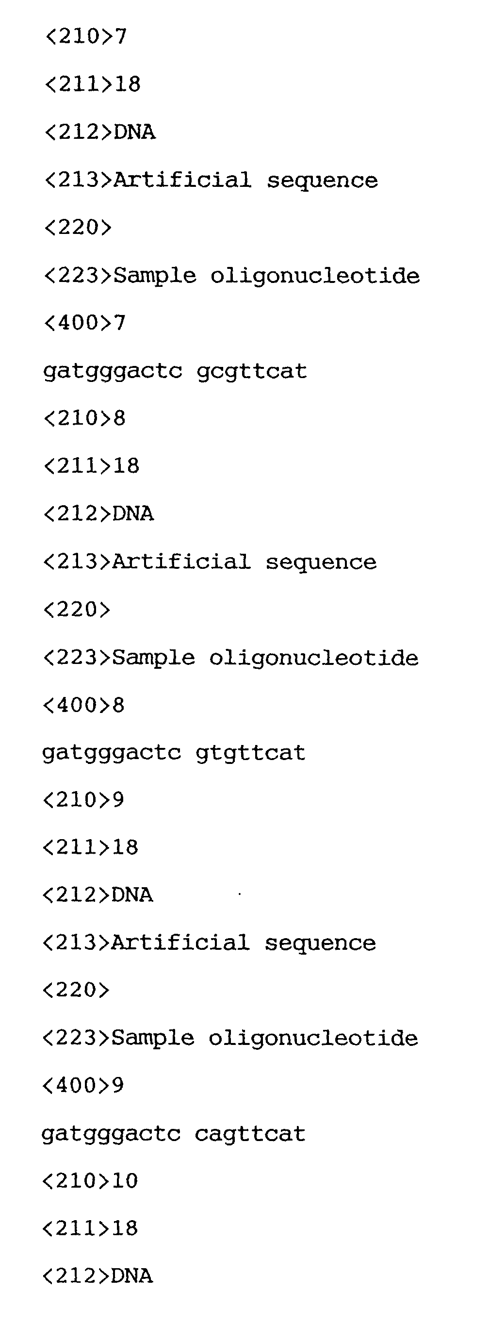
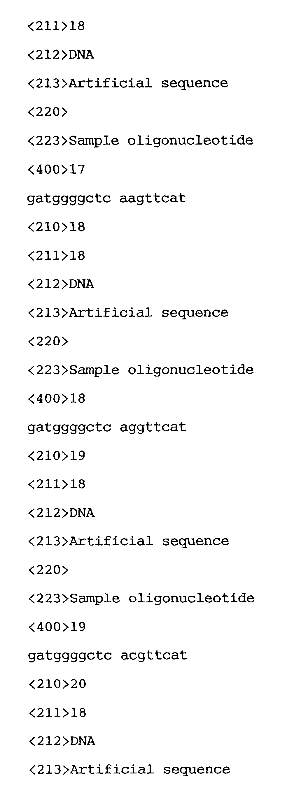
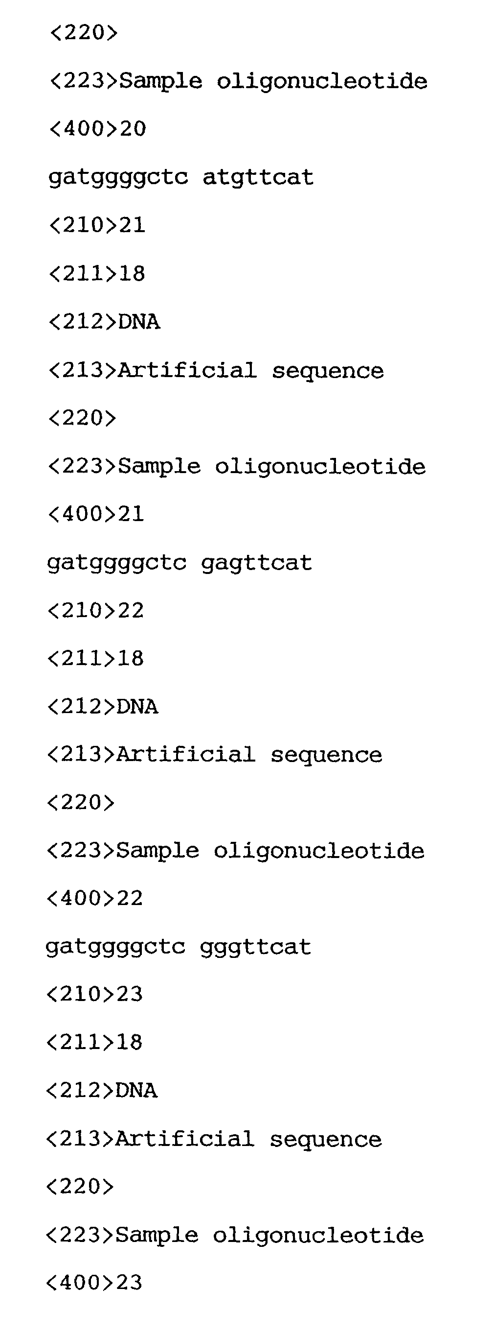
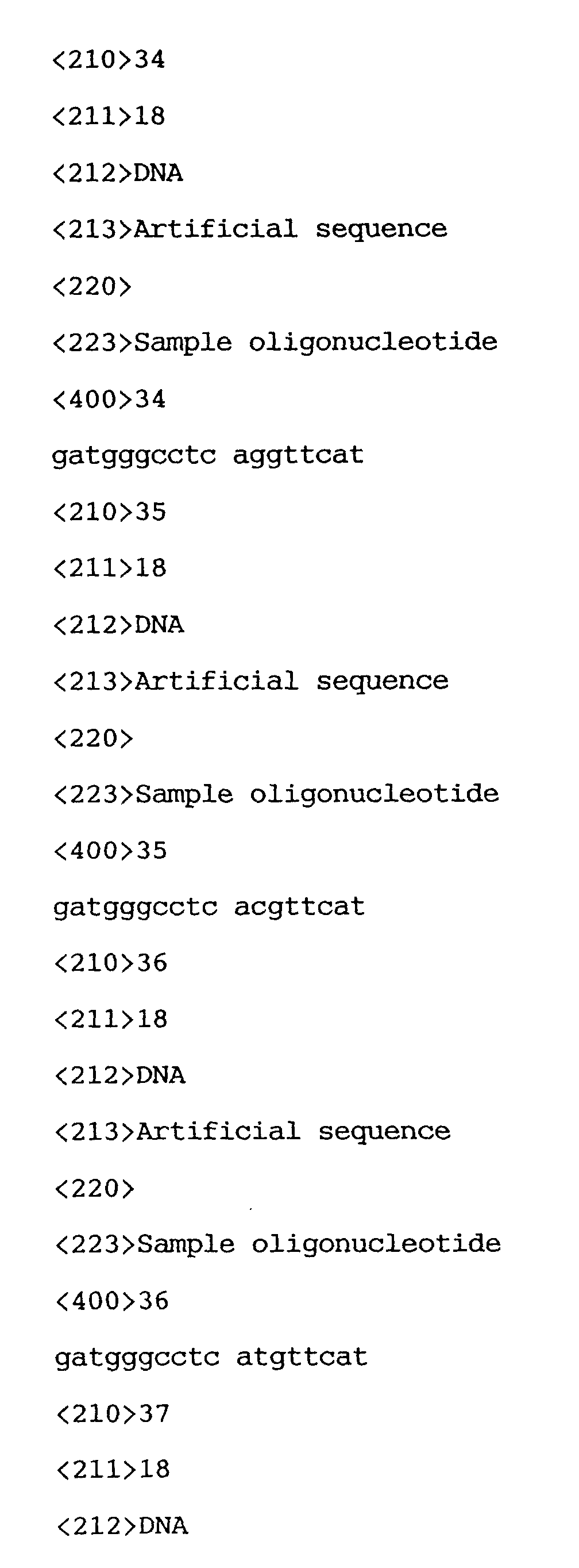
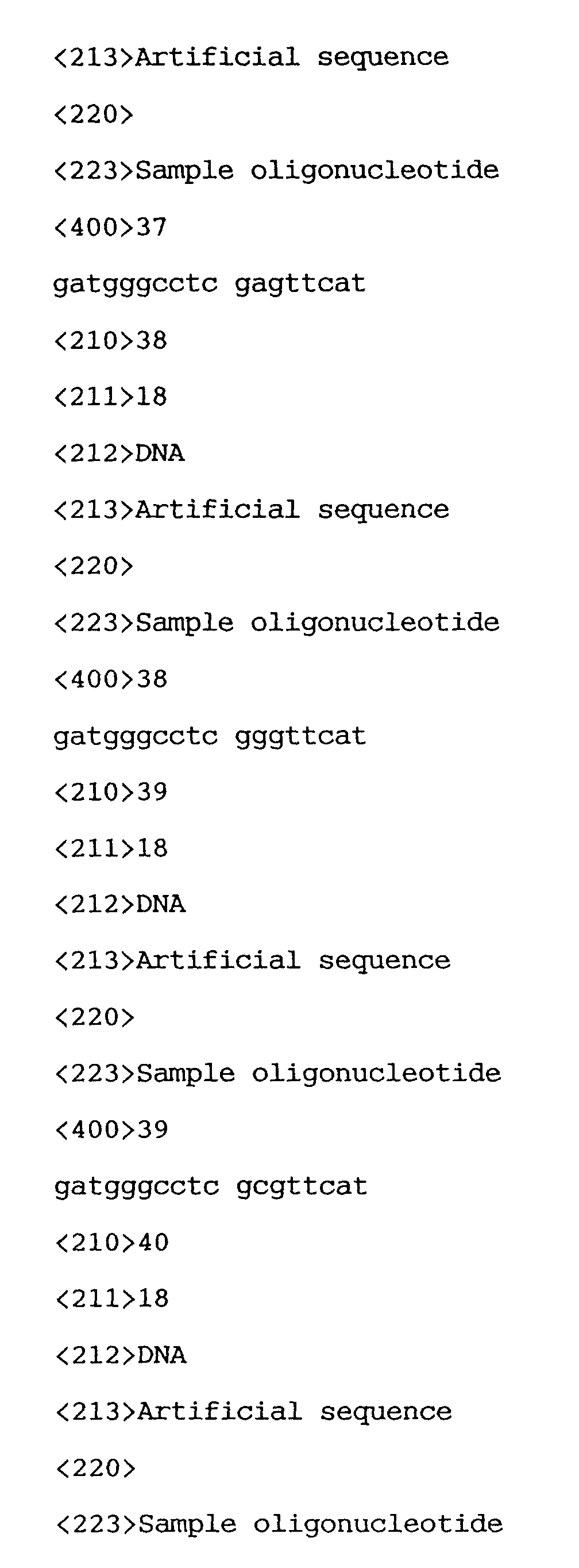
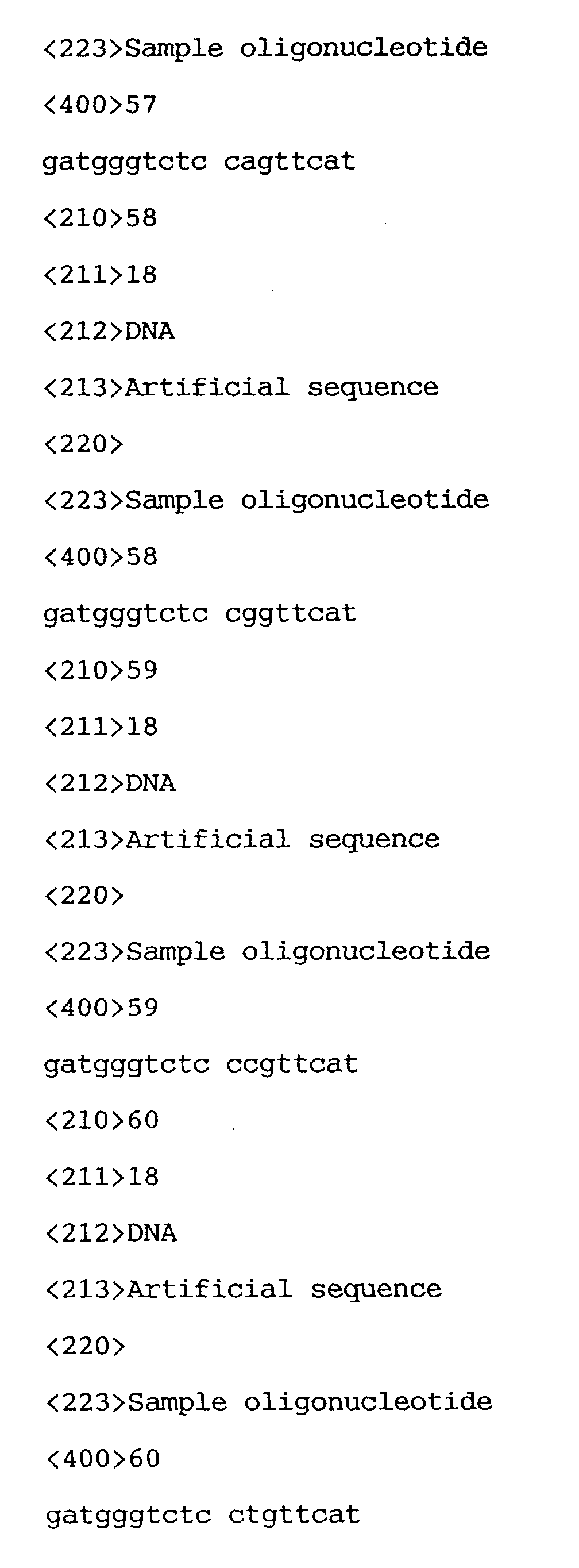
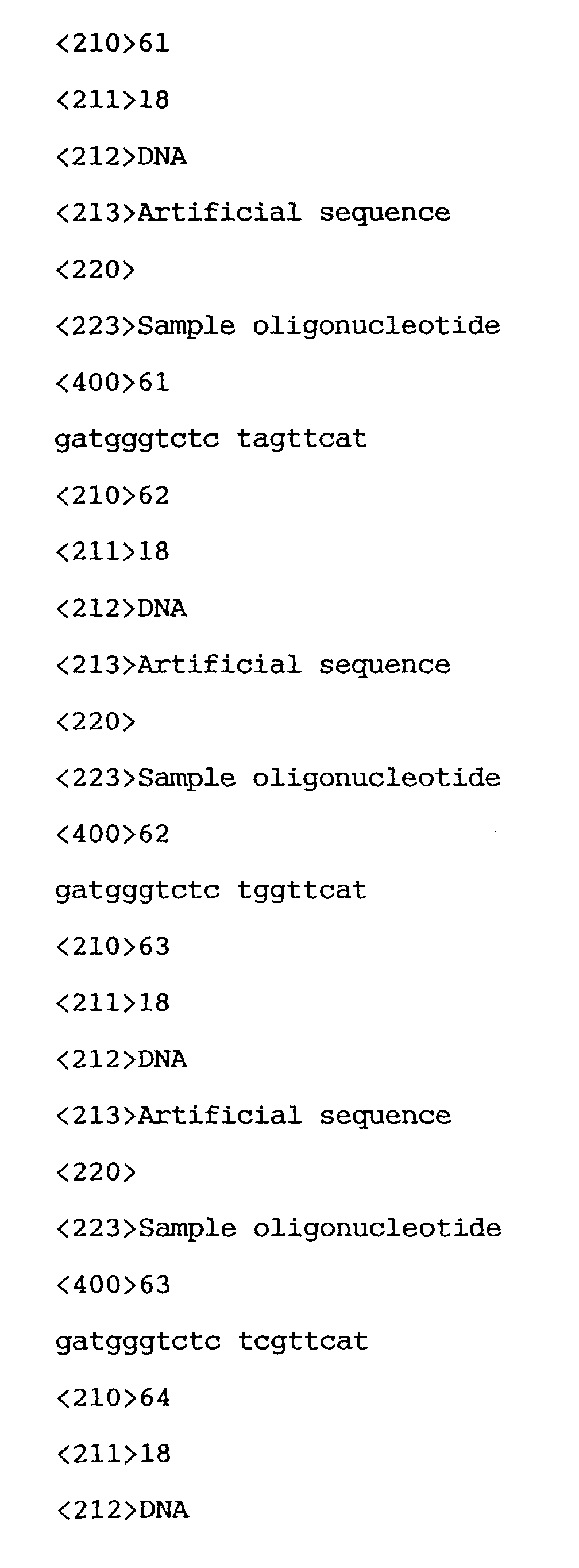
- FIG. 1 shows an arrangement of 64 types of DNA probes on a substrate. Each sequence (SEQ ID NOs: 1 to 64) is specifically shown in Table 1.
- 5' ATGAACCGGAGGCCCATC3' which is the sequence corresponding to the normal gene is expected to form a hybrid with the DNA probe 42 of 5'GATGGGCCTCCGGTTCAT3' located at the third point from the right and from the top.
- a glass plate of 1 inch square was placed in a rack and soaked in an ultrasonic cleaning detergent overnight. Then, after 20 min of ultrasonic cleaning, the detergent was removed by washing with water. After rinsing the plate with distilled water, ultrasonic treatment was repeated in a container filled with distilled water, for additional 20 min. Then the plate was soaked in a prewarmed 1N sodium hydroxide solution for 10 min, washed with water and then distilled water.
- silane coupling agent product of Shin-Etsu Chemical Industry: Trade name KBM 603
- nitrogen gas was blown on the both sides blowing off water to dryness.
- the silane coupling treatment was completed by baking the plate in an oven at 120°C for 1 hour.
- EMCS N-(6-maleimidocaproyloxy) succinimide: Dojin Company
- DMSO/ethanol final concentration: 0.3 mg/ml
- the glass substrate treated with the silane coupling agent was soaked in this EMCS solution for 2 hours to react the amino group of the silane coupling agent with the succimide group of EMCS.
- the maleimide group of EMCS is transferred to the glass surface.
- the glass plate was washed with ethanol, and dried with nitrogen gas to be used for a coupling reaction with DNA.
- the above 64 types of DNAs were ejected respectively as follows. Each DNA was dissolved in water and diluted with SG Clear (aqueous solution containing 7.5% of glycerin, 7.5% of urea, 7.5% of thiodiglycol and 1% of acetylenol EH), to a final concentration of 8 ⁇ M. Then 100 ⁇ l of this DNA solution was filled into a nozzle of a BJ printer Head BC 62 (Canon) modified to eject a small amount, and to eject six solutions per head. Two heads were used at a time so that 12 types of DNAs could be ejected at once, and the heads were changed 6 times so that 64 spots of 64 types of DNAs were formed on the glass plates independently.
- SG Clear aqueous solution containing 7.5% of glycerin, 7.5% of urea, 7.5% of thiodiglycol and 1% of acetylenol EH
- the substrate was washed with a 1 M NaCl/50 mM phosphate buffer solution (pH 7.0) to wash out thoroughly the DNA solution on the glass surface. Then, this was soaked in an aqueous solution of 2% bovine serum albumin and allowed to stand for 2 hours to carry out a blocking reaction.
- a 1 M NaCl/50 mM phosphate buffer solution pH 7.0
- Rhodamine labeled DNA No. 1 (SEQ ID NO: 65) of the same length as the probes but having the normal sequence of p53 gene was prepared. The sequence is shown below and rhodamine is bonded to the 5' terminus.
- the detection was performed by connecting an image analysis processing apparatus, ARGUS ( a product of Hamamatsu Photonics) to a fluorescence microscope (a product of Nicon).
- the fluorescence intensities obtained from the model hybridization reaction with the labeled DNA No. 1 (18-mer) are shown in FIG. 4.
- the maximum value of the fluorescence intensity was obtained at the spot of probe 42 which is fully complementary to DNA No. 1. Taking this intensity as the maximum value (1.0), the threshold is set at 10% of this value and the spots having higher intensity are painted dark.
- the spots of probes 10, 26, 41, 46 and 58 of one-base mismatch hybrids have fluorescence higher than the threshold, and it is understood that the location is well coincident with FIG. 2 of the expected pattern.
- the threshold By lowering the threshold further, in addition to the above 5 spots, the spots of other one-base mismatch probes appeared around the full matching probe in vertical and horizontal lines, in coincidence with the expected pattern.
- a DNA array of 64 types of probes was prepared in the same manner as in Example 1, and the hybridization reaction was performed using a rhodamine-labeled DNA No. 2 as a model sample.
- the DNA No. 2 (SEQ ID NO: 66) has a sequence complementary to the No. 46 probe of FIG. 1.
- FIG. 5 is an expected pattern consisting of the perfect match and one-base mismatch hybrids, and the resulting pattern obtained as in Example 1 is shown in FIG. 6.
- the threshold is set at 10% of the maximum value. When the detected spots are painted dark, the result corresponds well to the expectation.
- the threshold is set as 50%, fluorescence was detected at the positions (shaded parts) of Nos. 34 and 62 probes (one-base mismatch) in addition to No.46 (full match), and with further reduction of the threshold to 10%, the result was coincident with the expected pattern.
- Nos. 6, 22 and 54 of two-base mismatch probes were detected, but the two-base mismatch can be distinguished from the one-base mismatch as the deviation from the expected pattern of one-base mismatch, and No. 46 can be called as the full matched probe.
Landscapes
- Life Sciences & Earth Sciences (AREA)
- Proteomics, Peptides & Aminoacids (AREA)
- Chemical & Material Sciences (AREA)
- Organic Chemistry (AREA)
- Zoology (AREA)
- Wood Science & Technology (AREA)
- Health & Medical Sciences (AREA)
- Engineering & Computer Science (AREA)
- Microbiology (AREA)
- Biochemistry (AREA)
- Biotechnology (AREA)
- Molecular Biology (AREA)
- Biophysics (AREA)
- Analytical Chemistry (AREA)
- Physics & Mathematics (AREA)
- Immunology (AREA)
- Bioinformatics & Cheminformatics (AREA)
- General Engineering & Computer Science (AREA)
- General Health & Medical Sciences (AREA)
- Genetics & Genomics (AREA)
- Measuring Or Testing Involving Enzymes Or Micro-Organisms (AREA)
- Apparatus Associated With Microorganisms And Enzymes (AREA)
- Investigating Or Analysing Biological Materials (AREA)
Abstract
Description

Subsequently, 2.7 mg of EMCS (N-(6-maleimidocaproyloxy) succinimide: Dojin Company) was weighed and dissolved in a 1 : 1 solution of DMSO/ethanol (final concentration: 0.3 mg/ml). The glass substrate treated with the silane coupling agent was soaked in this EMCS solution for 2 hours to react the amino group of the silane coupling agent with the succimide group of EMCS. At this stage, the maleimide group of EMCS is transferred to the glass surface. After that, the glass plate was washed with ethanol, and dried with nitrogen gas to be used for a coupling reaction with DNA.









Claims (9)
- A method for identifying an unknown base sequence present in a target single-stranded nucleic acid comprising the steps of:(a) providing a probe array in which single-stranded nucleic acid probes of No. 1 to No. n (n≥2) are arranged as isolated spots on a substrate, the probes each having a base sequence complementary to one of plural base sequences expected to be the unknown base sequence;(b) reacting a single-stranded nucleic acid, which has a base sequence fully complementary to a base sequence of one of the single-stranded nucleic acid probes and is fluorescence-labeled, with the probe array under such conditions that single-stranded nucleic acids complementary to each other form a double-stranded nucleic acid;removing the unreacted labeled single-stranded nucleic acid, andmeasuring fluorescence intensity of each spot of the probe array to obtain a first template pattern showing a relationship between location of the probes and fluorescent characteristics;(c) performing the same operation as the step (b) for each of remaining single-stranded nucleic acid probes using a second to a nth single-stranded nucleic acid, and obtaining template patterns of No. 2 to No. n showing a relationship between location and fluorescent characteristics of the probes;(d) performing the same operation as the step (b) using a sample containing the target single-stranded nucleic acid of unknown base sequence to obtain a sample pattern showing relationship between a position and fluorescent characteristics; and(e) comparing the sample pattern obtained in the step (d) with n pieces of template patterns obtained in the steps (b) and (c), to identify a template pattern showing substantially the same pattern as the sample pattern and identifying the base sequence of the single-stranded nucleic acid used for the preparation of the identified template pattern as the unknown base sequence of the target single-stranded nucleic acid.
- A method for identifying an unknown base sequence present in a target single-stranded nucleic acid comprising the steps of:(a) providing a probe array in which single-stranded nucleic acid probes of No. 1 to No. n (n≥2) are arranged as isolated spots on a substrate, the probes each having a base sequence complementary to one of plural base sequences expected to be the unknown base sequence;(b) reacting a single-stranded nucleic acid which has a base sequence fully complementary to a base sequence of one of the single-stranded nucleic acid probes and is fluorescence-labeled, with the probe array under such conditions that single-stranded nucleic acids complementary to each other form a double-stranded nucleic acid;removing the unreacted labeled single-stranded nucleic acid, andmeasuring fluorescence intensity of each spot of the probe array to obtain a first template pattern showing a relationship between location of the probes and fluorescent characteristics;(c) analyzing the first template pattern to locate probes and to calculate a mean value of fluorescence intensities (Fi) of the double-stranded nucleic acids having i of mismatched base pairs, where i is an integer not less than 1;(d) calculating a difference (F1, 0) between the fluorescence intensity of the fully complementary double-stranded nucleic acid without mismatch (F0) and the mean value of the fluorescence intensities of the double-stranded nucleic acids having one-base mismatch (F1), further calculating a difference (Fi+1, i) between a fluorescence intensity of a double-stranded nucleic acid having (i+1) base mismatches (Fi+1) and a fluorescence intensity of a double-stranded nucleic acid having i-base mismatches (Fi), and identifying i being Fi+1, i << Fi, i-1;(e) assuming a target DNA which base sequence is complementary to the second probe sequence, then obtaining the second template pattern formed by the probe position where the number of mismatched base pairs to the target having the complementary sequence to the second probe sequence is not more than i;(f) performing the same operation as the step (e) for each of remaining single-stranded nucleic acid probes using a third to a nth single-stranded nucleic acid, and obtaining template patterns of No. 3 to No. n showing a relationship between location and fluorescent characteristics of the probes, wherein the template patterns are formed from the positions of the probes having a base sequence that forms mismatched base pairs in a number not more than i;(g) performing the same operation as the step (b) using a sample containing the target single-stranded nucleic acid of unknown base sequence to obtain a sample pattern showing relationship between a position and fluorescent characteristics; and(h) comparing the sample pattern obtained in the step (g) with n pieces of template patterns obtained in the steps (b), (c) and (e), to identify a template pattern showing essentially the same pattern as the sample pattern and identifying the base sequence of the single-stranded nucleic acid used for the preparation of the identified template pattern as the unknown base sequence of the target single-stranded nucleic acid.
- A method for identifying an unknown base sequence present in a target single-stranded nucleic acid comprising the steps of:wherein the 2nd to nth template patterns are formed from the positions of probes having a base sequence that forms mismatched base pairs in a number not more than i.(a) providing a probe array in which single-stranded nucleic acid probes of No. 1 to No. n (n≥2) are arranged as isolated spots on a substrate, the probes each having a base sequence complementary to one of a plurality of base sequences expected to be the unknown base sequence;(b) reacting a fluorescence-labelled single-stranded nucleic acid which has a base sequence fully complementary to a base sequence of one of the single-stranded nucleic acid probes with the probe array under such conditions that single-stranded nucleic acids complementary to each other form a double-stranded nucleic acid;removing the unreacted labeled single-stranded nucleic acid, andmeasuring fluorescence intensity of each spot of the probe array to obtain a first template pattern showing a relationship between location of the probes and fluorescent characteristics;(c) analyzing the first template pattern to locate probes and to calculate a mean value of fluorescence intensities (Fi) of the double-stranded nucleic acids having i of mismatched base pairs, where i is an integer not less than 1;(d) calculating a difference (F1, 0) between the fluorescence intensity of the fully complementary double-stranded nucleic acid without mismatch (F0) and the mean value of the fluorescence intensities of the double-stranded nucleic acids having one-base mismatch (F1), further calculating a difference (Fi+1, i) between a fluorescence intensity of a double-stranded nucleic acid having (i+1) base mismatches (Fi+1) and a fluorescence intensity of a double-stranded nucleic acid having i-base mismatches (Fi), and identifying i being Fi+1, i << Fi, i-1;(e) for a second fluorescence-labelled single-stranded nucleic acid, obtaining a second template pattern showing a relationship between location of the probes and fluorescent characteristics;(f) performing the same operation as step (e) for each of remaining single-stranded nucleic acid probes using a third to a nth single-stranded nucleic acid, and obtaining template patterns of No. 3 to No. n showing a relationship between location and fluorescent characteristics of the probes;(g) performing the same operation as the step (b) using a sample containing the target single-stranded nucleic acid of unknown base sequence to obtain a sample pattern showing relationship between a position and fluorescent characteristics; and(h) comparing the sample pattern obtained in the step (g) with n pieces of template patterns obtained in the steps (b), (c),(e) and (f) to identify a template pattern showing essentially the same pattern as the sample pattern and identifying the base sequence of the single-stranded nucleic acid used for the preparation of the identified template pattern as the unknown base sequence of the target single-stranded nucleic acid;
- A method according to claim 2 or 3, wherein the step (g) further comprises the substep of obtaining a two-valued pattern of the fluorescence intensity by using the threshold fluorescence intensity Fi.
- A method according to any preceding claim wherein length of the probe is 8 mer to 30 mer.
- A method according to claim 5, wherein the length of the probe is 12 mer to 25 mer.
- A method according to any one of claims 2 to 6, wherein the number of the mismatched base pairs (i) is 1.
- A method according to any preceding claim wherein the probes in the probe array are heat denatured in a solution containing the single-stranded nucleic acid, and cooled to a temperature suitable for a double-stranded formation reaction while the substrate is soaked in the solution.
- A method according to claim 8, wherein the length of the single-stranded nucleic acid probe is 18 mer, the temperature for performing the heat denaturation is 70°C or more, the temperature for the double-strand formation reaction is 40°C or more, and 100 mM sodium chloride is contained in the sample solution at that time.
Applications Claiming Priority (2)
| Application Number | Priority Date | Filing Date | Title |
|---|---|---|---|
| JP2000263506A JP3467004B2 (en) | 2000-08-31 | 2000-08-31 | Analysis method of nucleic acid base sequence |
| JP2000263506 | 2000-08-31 |
Publications (3)
| Publication Number | Publication Date |
|---|---|
| EP1184468A2 true EP1184468A2 (en) | 2002-03-06 |
| EP1184468A3 EP1184468A3 (en) | 2002-08-14 |
| EP1184468B1 EP1184468B1 (en) | 2012-01-25 |
Family
ID=18751046
Family Applications (1)
| Application Number | Title | Priority Date | Filing Date |
|---|---|---|---|
| EP01307442A Expired - Lifetime EP1184468B1 (en) | 2000-08-31 | 2001-08-31 | Method for sequencing using probe arrays |
Country Status (3)
| Country | Link |
|---|---|
| US (2) | US7273697B2 (en) |
| EP (1) | EP1184468B1 (en) |
| JP (1) | JP3467004B2 (en) |
Families Citing this family (13)
| Publication number | Priority date | Publication date | Assignee | Title |
|---|---|---|---|---|
| JP2002071687A (en) * | 2000-08-31 | 2002-03-12 | Canon Inc | Screening method for mutant gene |
| JP3467004B2 (en) * | 2000-08-31 | 2003-11-17 | キヤノン株式会社 | Analysis method of nucleic acid base sequence |
| US20050045063A1 (en) * | 2001-11-02 | 2005-03-03 | Matthias Niggemann | Marking solution for counterfeit-resistant identification of a valuable object, marking produced by the marking solution and method for marking a valuable object |
| US20070105100A1 (en) * | 2003-04-28 | 2007-05-10 | Canon Kabushiki Kaisha | Process for assay of nucleic acids by competitive hybridization using a dna microarray |
| JP4458327B2 (en) * | 2003-08-28 | 2010-04-28 | キヤノン株式会社 | Method for quantifying target substance and probe carrier used in the method |
| JP4541731B2 (en) * | 2004-03-12 | 2010-09-08 | キヤノン株式会社 | Nucleic acid detection method |
| JP2006094760A (en) * | 2004-09-29 | 2006-04-13 | Canon Inc | Cell screening method and oral cancer cell detection method |
| US8535914B2 (en) * | 2005-01-21 | 2013-09-17 | Canon Kabushiki Kaisha | Probe, probe set and information acquisition method using the same |
| JP2007010413A (en) * | 2005-06-29 | 2007-01-18 | Canon Inc | Method for measuring melting point of nucleic acid hybrid and apparatus therefor |
| US7642086B2 (en) * | 2005-08-09 | 2010-01-05 | Canon Kabushiki Kaisha | Labeled probe bound object, method for producing the same and method for using the same |
| US20090011413A1 (en) * | 2005-12-14 | 2009-01-08 | Canon Kabushiki Kaisha | Method for screening colon cancer cells and gene set used for examination of colon cancer |
| US20090035767A1 (en) * | 2006-11-28 | 2009-02-05 | Canon Kabushiki Kaisha | Primer for bacterium genome amplification reaction |
| JP5729904B2 (en) | 2009-06-02 | 2015-06-03 | キヤノン株式会社 | Methods for preparing proteins, DNA, RNA from cells |
Family Cites Families (14)
| Publication number | Priority date | Publication date | Assignee | Title |
|---|---|---|---|---|
| US5202231A (en) * | 1987-04-01 | 1993-04-13 | Drmanac Radoje T | Method of sequencing of genomes by hybridization of oligonucleotide probes |
| US5143854A (en) * | 1989-06-07 | 1992-09-01 | Affymax Technologies N.V. | Large scale photolithographic solid phase synthesis of polypeptides and receptor binding screening thereof |
| US5346811A (en) * | 1991-07-22 | 1994-09-13 | Cerveceria Polar | Method and products for human papillomavirus detection |
| WO1995011995A1 (en) * | 1993-10-26 | 1995-05-04 | Affymax Technologies N.V. | Arrays of nucleic acid probes on biological chips |
| US6027880A (en) * | 1995-08-02 | 2000-02-22 | Affymetrix, Inc. | Arrays of nucleic acid probes and methods of using the same for detecting cystic fibrosis |
| US5795716A (en) | 1994-10-21 | 1998-08-18 | Chee; Mark S. | Computer-aided visualization and analysis system for sequence evaluation |
| US5733729A (en) | 1995-09-14 | 1998-03-31 | Affymetrix, Inc. | Computer-aided probability base calling for arrays of nucleic acid probes on chips |
| AU2189397A (en) * | 1996-02-08 | 1997-08-28 | Affymetrix, Inc. | Chip-based speciation and phenotypic characterization of microorganisms |
| US5773729A (en) * | 1996-09-17 | 1998-06-30 | Ncr Corporation | Overload protected loadcell |
| US5985567A (en) * | 1997-08-15 | 1999-11-16 | Beckman Coulter, Inc. | Hybridization detection by pretreating bound single-stranded probes |
| US20020012913A1 (en) | 1998-09-15 | 2002-01-31 | Kevin L. Gunderson | Nucleic acid analysis using complete n-mer arrays |
| JP3467004B2 (en) * | 2000-08-31 | 2003-11-17 | キヤノン株式会社 | Analysis method of nucleic acid base sequence |
| JP2002071687A (en) * | 2000-08-31 | 2002-03-12 | Canon Inc | Screening method for mutant gene |
| US20030190612A1 (en) * | 2000-08-31 | 2003-10-09 | Nobuko Yamamoto | Detecting method and detection substrate for use therein |
-
2000
- 2000-08-31 JP JP2000263506A patent/JP3467004B2/en not_active Expired - Fee Related
-
2001
- 2001-08-31 US US09/942,596 patent/US7273697B2/en not_active Expired - Fee Related
- 2001-08-31 EP EP01307442A patent/EP1184468B1/en not_active Expired - Lifetime
-
2007
- 2007-08-30 US US11/896,141 patent/US20080051293A1/en not_active Abandoned
Also Published As
| Publication number | Publication date |
|---|---|
| EP1184468B1 (en) | 2012-01-25 |
| EP1184468A3 (en) | 2002-08-14 |
| JP2002306166A (en) | 2002-10-22 |
| US20020168648A1 (en) | 2002-11-14 |
| US7273697B2 (en) | 2007-09-25 |
| US20080051293A1 (en) | 2008-02-28 |
| JP3467004B2 (en) | 2003-11-17 |
Similar Documents
| Publication | Publication Date | Title |
|---|---|---|
| US20080051293A1 (en) | Method for analyzing base sequence of nucleic acid | |
| US6924103B2 (en) | Screening method for gene variation | |
| US20220333192A1 (en) | Methods and devices for spatial assessment of rna quality | |
| US6232067B1 (en) | Adapter directed expression analysis | |
| KR100691043B1 (en) | Computer Controlled Fetal Cell Inclusion Rare Cell Diagnostic Methods and Devices | |
| US20020187508A1 (en) | Methods and products for analyzing nucleic acids using nick translation | |
| EP1469067A1 (en) | Method and apparatus for detecting nucleic acid data | |
| JP3872762B2 (en) | DNA chip quality control method | |
| JP2006511798A (en) | Biochip chip reader and related method | |
| JPH09149799A (en) | Nucleic acid analysis method or inspection method, and nucleic acid analysis apparatus or inspection apparatus | |
| JP2003130875A (en) | Quality inspection method of biochemical substance microarray chip | |
| US20030082602A1 (en) | Method for analyzing base sequence of nucleic acid | |
| JP2001054400A (en) | Genotype determining two allele marker | |
| JP4672234B2 (en) | Method and test kit for quantification of variation in polynucleotide content in a cell or tissue sample | |
| US20050074781A1 (en) | Nucleic acid braided J-probes | |
| US20090088328A1 (en) | Microarray Quality Control | |
| US20030129641A1 (en) | Method for determining biospecies contained in test specimen and kit used for the same | |
| US20260125746A1 (en) | Methods and compositions for substrate surface chemistry | |
| JP2004041198A (en) | Method for analyzing nucleic acid base sequence | |
| US20230099994A1 (en) | Digital polymerase chain reaction (dpcr) platforms on next generation sequencer patterned flow cell technology | |
| JP2004016132A (en) | Method for measuring nucleic acid and data analysis method obtained by the method | |
| JP4649312B2 (en) | Mutant gene screening method | |
| WO2024077047A1 (en) | Methods and compositions for substrate surface chemistry | |
| EP1284421A1 (en) | Method of constructing polynucleotide microarray, apparatus for the construction and polynucleotide microarray | |
| US20070154923A1 (en) | Method for Gridding and Quality Control of Polymer Arrays |
Legal Events
| Date | Code | Title | Description |
|---|---|---|---|
| PUAI | Public reference made under article 153(3) epc to a published international application that has entered the european phase |
Free format text: ORIGINAL CODE: 0009012 |
|
| AK | Designated contracting states |
Kind code of ref document: A2 Designated state(s): AT BE CH CY DE DK ES FI FR GB GR IE IT LI LU MC NL PT SE TR |
|
| AX | Request for extension of the european patent |
Free format text: AL;LT;LV;MK;RO;SI |
|
| PUAL | Search report despatched |
Free format text: ORIGINAL CODE: 0009013 |
|
| AK | Designated contracting states |
Kind code of ref document: A3 Designated state(s): AT BE CH CY DE DK ES FI FR GB GR IE IT LI LU MC NL PT SE TR |
|
| AX | Request for extension of the european patent |
Free format text: AL;LT;LV;MK;RO;SI |
|
| 17P | Request for examination filed |
Effective date: 20030109 |
|
| AKX | Designation fees paid |
Designated state(s): CH DE FR GB LI NL SE |
|
| 17Q | First examination report despatched |
Effective date: 20060310 |
|
| RIC1 | Information provided on ipc code assigned before grant |
Ipc: C12Q 1/68 20060101AFI20110608BHEP |
|
| GRAP | Despatch of communication of intention to grant a patent |
Free format text: ORIGINAL CODE: EPIDOSNIGR1 |
|
| GRAJ | Information related to disapproval of communication of intention to grant by the applicant or resumption of examination proceedings by the epo deleted |
Free format text: ORIGINAL CODE: EPIDOSDIGR1 |
|
| GRAP | Despatch of communication of intention to grant a patent |
Free format text: ORIGINAL CODE: EPIDOSNIGR1 |
|
| GRAS | Grant fee paid |
Free format text: ORIGINAL CODE: EPIDOSNIGR3 |
|
| GRAA | (expected) grant |
Free format text: ORIGINAL CODE: 0009210 |
|
| RAP1 | Party data changed (applicant data changed or rights of an application transferred) |
Owner name: CANON KABUSHIKI KAISHA |
|
| AK | Designated contracting states |
Kind code of ref document: B1 Designated state(s): CH DE FR GB LI NL SE |
|
| REG | Reference to a national code |
Ref country code: GB Ref legal event code: FG4D |
|
| REG | Reference to a national code |
Ref country code: CH Ref legal event code: EP |
|
| REG | Reference to a national code |
Ref country code: DE Ref legal event code: R096 Ref document number: 60146027 Country of ref document: DE Effective date: 20120322 |
|
| REG | Reference to a national code |
Ref country code: NL Ref legal event code: VDEP Effective date: 20120125 |
|
| PG25 | Lapsed in a contracting state [announced via postgrant information from national office to epo] |
Ref country code: NL Free format text: LAPSE BECAUSE OF FAILURE TO SUBMIT A TRANSLATION OF THE DESCRIPTION OR TO PAY THE FEE WITHIN THE PRESCRIBED TIME-LIMIT Effective date: 20120125 |
|
| PG25 | Lapsed in a contracting state [announced via postgrant information from national office to epo] |
Ref country code: SE Free format text: LAPSE BECAUSE OF FAILURE TO SUBMIT A TRANSLATION OF THE DESCRIPTION OR TO PAY THE FEE WITHIN THE PRESCRIBED TIME-LIMIT Effective date: 20120125 |
|
| PLBE | No opposition filed within time limit |
Free format text: ORIGINAL CODE: 0009261 |
|
| STAA | Information on the status of an ep patent application or granted ep patent |
Free format text: STATUS: NO OPPOSITION FILED WITHIN TIME LIMIT |
|
| 26N | No opposition filed |
Effective date: 20121026 |
|
| REG | Reference to a national code |
Ref country code: DE Ref legal event code: R097 Ref document number: 60146027 Country of ref document: DE Effective date: 20121026 |
|
| REG | Reference to a national code |
Ref country code: CH Ref legal event code: PL |
|
| GBPC | Gb: european patent ceased through non-payment of renewal fee |
Effective date: 20120831 |
|
| PG25 | Lapsed in a contracting state [announced via postgrant information from national office to epo] |
Ref country code: CH Free format text: LAPSE BECAUSE OF NON-PAYMENT OF DUE FEES Effective date: 20120831 Ref country code: LI Free format text: LAPSE BECAUSE OF NON-PAYMENT OF DUE FEES Effective date: 20120831 |
|
| REG | Reference to a national code |
Ref country code: FR Ref legal event code: ST Effective date: 20130430 |
|
| PG25 | Lapsed in a contracting state [announced via postgrant information from national office to epo] |
Ref country code: GB Free format text: LAPSE BECAUSE OF NON-PAYMENT OF DUE FEES Effective date: 20120831 |
|
| PG25 | Lapsed in a contracting state [announced via postgrant information from national office to epo] |
Ref country code: FR Free format text: LAPSE BECAUSE OF NON-PAYMENT OF DUE FEES Effective date: 20120831 |
|
| PGFP | Annual fee paid to national office [announced via postgrant information from national office to epo] |
Ref country code: DE Payment date: 20130831 Year of fee payment: 13 |
|
| REG | Reference to a national code |
Ref country code: DE Ref legal event code: R119 Ref document number: 60146027 Country of ref document: DE |
|
| REG | Reference to a national code |
Ref country code: DE Ref legal event code: R119 Ref document number: 60146027 Country of ref document: DE Effective date: 20150303 |
|
| PG25 | Lapsed in a contracting state [announced via postgrant information from national office to epo] |
Ref country code: DE Free format text: LAPSE BECAUSE OF NON-PAYMENT OF DUE FEES Effective date: 20150303 |