EP1184463A1 - Method of detecting protein-protein interaction - Google Patents
Method of detecting protein-protein interaction Download PDFInfo
- Publication number
- EP1184463A1 EP1184463A1 EP01912323A EP01912323A EP1184463A1 EP 1184463 A1 EP1184463 A1 EP 1184463A1 EP 01912323 A EP01912323 A EP 01912323A EP 01912323 A EP01912323 A EP 01912323A EP 1184463 A1 EP1184463 A1 EP 1184463A1
- Authority
- EP
- European Patent Office
- Prior art keywords
- protein
- cell
- reporter
- interaction
- gfp
- Prior art date
- Legal status (The legal status is an assumption and is not a legal conclusion. Google has not performed a legal analysis and makes no representation as to the accuracy of the status listed.)
- Withdrawn
Links
Images


Classifications
-
- C—CHEMISTRY; METALLURGY
- C40—COMBINATORIAL TECHNOLOGY
- C40B—COMBINATORIAL CHEMISTRY; LIBRARIES, e.g. CHEMICAL LIBRARIES
- C40B30/00—Methods of screening libraries
- C40B30/04—Methods of screening libraries by measuring the ability to specifically bind a target molecule, e.g. antibody-antigen binding, receptor-ligand binding
-
- G—PHYSICS
- G01—MEASURING; TESTING
- G01N—INVESTIGATING OR ANALYSING MATERIALS BY DETERMINING THEIR CHEMICAL OR PHYSICAL PROPERTIES
- G01N33/00—Investigating or analysing materials by specific methods not covered by groups G01N1/00 - G01N31/00
- G01N33/48—Biological material, e.g. blood, urine; Haemocytometers
- G01N33/50—Chemical analysis of biological material, e.g. blood, urine; Testing involving biospecific ligand binding methods; Immunological testing
- G01N33/5005—Chemical analysis of biological material, e.g. blood, urine; Testing involving biospecific ligand binding methods; Immunological testing involving human or animal cells
- G01N33/5008—Chemical analysis of biological material, e.g. blood, urine; Testing involving biospecific ligand binding methods; Immunological testing involving human or animal cells for testing or evaluating the effect of chemical or biological compounds, e.g. drugs, cosmetics
-
- G—PHYSICS
- G01—MEASURING; TESTING
- G01N—INVESTIGATING OR ANALYSING MATERIALS BY DETERMINING THEIR CHEMICAL OR PHYSICAL PROPERTIES
- G01N33/00—Investigating or analysing materials by specific methods not covered by groups G01N1/00 - G01N31/00
- G01N33/48—Biological material, e.g. blood, urine; Haemocytometers
- G01N33/50—Chemical analysis of biological material, e.g. blood, urine; Testing involving biospecific ligand binding methods; Immunological testing
- G01N33/5005—Chemical analysis of biological material, e.g. blood, urine; Testing involving biospecific ligand binding methods; Immunological testing involving human or animal cells
- G01N33/5008—Chemical analysis of biological material, e.g. blood, urine; Testing involving biospecific ligand binding methods; Immunological testing involving human or animal cells for testing or evaluating the effect of chemical or biological compounds, e.g. drugs, cosmetics
- G01N33/502—Chemical analysis of biological material, e.g. blood, urine; Testing involving biospecific ligand binding methods; Immunological testing involving human or animal cells for testing or evaluating the effect of chemical or biological compounds, e.g. drugs, cosmetics for testing non-proliferative effects
-
- G—PHYSICS
- G01—MEASURING; TESTING
- G01N—INVESTIGATING OR ANALYSING MATERIALS BY DETERMINING THEIR CHEMICAL OR PHYSICAL PROPERTIES
- G01N33/00—Investigating or analysing materials by specific methods not covered by groups G01N1/00 - G01N31/00
- G01N33/48—Biological material, e.g. blood, urine; Haemocytometers
- G01N33/50—Chemical analysis of biological material, e.g. blood, urine; Testing involving biospecific ligand binding methods; Immunological testing
- G01N33/53—Immunoassay; Biospecific binding assay; Materials therefor
- G01N33/531—Production of immunochemical test materials
- G01N33/532—Production of labelled immunochemicals
- G01N33/533—Production of labelled immunochemicals with fluorescent label
-
- G—PHYSICS
- G01—MEASURING; TESTING
- G01N—INVESTIGATING OR ANALYSING MATERIALS BY DETERMINING THEIR CHEMICAL OR PHYSICAL PROPERTIES
- G01N33/00—Investigating or analysing materials by specific methods not covered by groups G01N1/00 - G01N31/00
- G01N33/48—Biological material, e.g. blood, urine; Haemocytometers
- G01N33/50—Chemical analysis of biological material, e.g. blood, urine; Testing involving biospecific ligand binding methods; Immunological testing
- G01N33/68—Chemical analysis of biological material, e.g. blood, urine; Testing involving biospecific ligand binding methods; Immunological testing involving proteins, peptides or amino acids
- G01N33/6803—General methods of protein analysis not limited to specific proteins or families of proteins
- G01N33/6845—Methods of identifying protein-protein interactions in protein mixtures
-
- G—PHYSICS
- G01—MEASURING; TESTING
- G01N—INVESTIGATING OR ANALYSING MATERIALS BY DETERMINING THEIR CHEMICAL OR PHYSICAL PROPERTIES
- G01N2500/00—Screening for compounds of potential therapeutic value
- G01N2500/02—Screening involving studying the effect of compounds C on the interaction between interacting molecules A and B (e.g. A = enzyme and B = substrate for A, or A = receptor and B = ligand for the receptor)
-
- G—PHYSICS
- G01—MEASURING; TESTING
- G01N—INVESTIGATING OR ANALYSING MATERIALS BY DETERMINING THEIR CHEMICAL OR PHYSICAL PROPERTIES
- G01N2500/00—Screening for compounds of potential therapeutic value
- G01N2500/10—Screening for compounds of potential therapeutic value involving cells
Definitions
- the invention of this application relates to a detection method of protein-protein interaction. According to the method, it is possible to screen intracellular proteins to find out a specific protein that interacts with a target protein under intracellular condition. Therefore, the method is a promising means for clarifying the protein network in cells. Clarifying the protein network in cells is useful for developing new medicines. According to the method, it is possible to screen low-molecular substances for new medicines.
- the whole base sequence analysis of the genome in a cell is clarifying the primary structure of every protein constituting the cell.
- the next theme in the art is to analyze and clarify what kind of intracellular network is formed by the proteins in a cell and what kind of life activity of the cell is born by the proteins therein.
- the protein network in a cell if clarified, is helpful in clarification of the molecular mechanism of diseases to be caused by protein disorders, and in developing the medicines for the diseases.
- the other methods (7) to (9) are for detecting the reconstruction of reporter proteins or the approach of the two to each other on the basis of the transcription activity or the enzymatic activity of the reporter proteins or on the basis of the fluorescence resonance energy transfer between the reporter proteins, that results from the interaction of target proteins in a cell through intracellular expression of the cDNAs that separately code for the two interacting target proteins, to give a fused gene of each cDNA and a reporter cDNA for it.
- the two reporter proteins each separately fused to the two target proteins must be bound to each other by the binding of the target proteins, or they must be near to each other within a predetermined distance.
- the methods are not applicable to target proteins which are too large or which do not have a flexible structure.
- the method (7) is used most frequently as its limitations on the protein structure are relatively small as compared with those of the other methods.
- the method (7) is defective in that the yeast growth therein is slow and the efficiency of transformation therein is low.
- this system cannot be used for them.
- the invention of this application has been made in consideration of the prior art problems noted above, and its object is to provide a method for surely detecting the protein-protein interaction in a cell in a simplified manner.
- Another object of the invention of this application is to provide a method for detecting the action of a compound on the protein-protein interaction in a cell.
- the present application provides a method for detecting the interaction of a protein X with a protein Y having a reporter function in a eucaryotic cell, which comprises;
- the present application provides a method for detecting the inhibitory or promotive action of a compound Z on the interaction of a protein X with a protein Y having a reporter function in a eucaryotic cell, which comprises;
- the protein Y having a reporter function is a fusion protein Y-B with a reporter protein B
- the reporter protein B is a fluorescent protein
- still another preferred embodiment is that, in the step (1), the intracellular localization signal sequence of the protein X and/or the protein Y is mutated, or the protein X is fused with a protein A of which the intracellular localization signal sequence differs from that of the protein Y.
- the compound Z is, for example, a low-molecular compound, a peptide or a protein, and in case where the compound Z is a low-molecular compound, it coexists with the eucaryotic cell, and in case where the compound Z is a protein or a peptide, it is expressed in the eucaryotic cell.
- the proteins X, Y, A and B may be matured proteins, parts thereof, or peptides.
- the fusion protein X-A and the fusion protein Y-B may be a fusion protein A-X and a fusion protein B-Y, respectively, in which "-" indicates a peptide bond.
- the protein Y used is a fusion protein Y-B with a reporter B (protein or peptide).
- a reporter B protein or peptide
- an expression vector for expressing a fusion protein Y-B with a reporter B of being fused to the N-terminal or C-terminal of Y is prepared, and this is used together with an expression vector for a fusion protein X-A in the manner described below.
- the Y-B expression vector previously prepared is introduced into a eucaryotic cell together with the X-A expression vector prepared in the step 1.
- a cell with only the Y-B expression vector introduced thereinto is also prepared.
- the cells are cultured, and the site in these in which the reporter B is localized is detected by any optical means or biochemical means to thereby judge the presence or absence of the Y-B localization site difference between the cell with only the Y-B expression vector introduced thereinto and the cell with the two expression vectors introduced thereinto.
- the intracellular localization of Y-B is identical with that of X-A, then it means that X and Y bind to each other.
- Fig. 1 is a schematic view showing the principle of the first aspect of the invention, concretely illustrating one example in which a protein A having an intranuclear localization signal sequence is used and this is fused to form a fusion protein A-X.
- the fusion protein A-X is localized in the nucleus of the cell (Fig. 1a).
- a fusion protein Y-B does not have the localization signal which A has, it exists in a site of the cell in which A-X does not exist (in this example, Y-B exists everywhere in the cell including its cytoplasm and nucleus) (Fig. 1b).
- X has a localization signal sequence
- the localization signal sequence that X originally has may be deleted or mutated, and A may be fused to the resulting X.
- A may be a protein having a localization signal sequence or may also be a peptide containing a minimal unit that serves as a localization signal.
- the localization signal includes, for example, a nuclear localization signal, a mitochondrial localization signal, a Golgi localization signal, and a membrane localization signal. Of those, preferred are a nuclear localization signal and a mitochondrial signal that facilitate the identification of the intended localized sites.
- A may contain a site having a reporter function. Its reporter function, if used, makes it possible to confirm the site where the fusion protein X-A is localized. Needless-to-say, since the site where the fusion protein X-A is localized can be presumed with accuracy, depending on the type of the localization signal of A, the reporter function of A is not indispensable.
- Y has a reporter function
- it does not always require the reporter B to form such a fused protein Y-B.
- the reporter B includes, for example, green fluorescent protein (GFP), reciferase, ⁇ -galactosidase, chloramphenicol acetyltransferase, and various epitope tags.
- GFP green fluorescent protein
- reciferase ⁇ -galactosidase
- chloramphenicol acetyltransferase chloramphenicol acetyltransferase
- epitope tags for it, preferred are fluorescent proteins such as GFP, as facilitating the detection of the intended protein localization through microscopic cell observation.
- a method that comprises preparing fusion protein expression vectors followed by introducing the two types of vectors into the cell.
- a cell to produce the protein X that is localized in a specific site are used, only the Y-B expression vector may be introduced into the cell.
- fusion protein expression vector employable are all types of expression vectors for eucaryotic cell so far as they have a promoter, a splicing region and/or a poly(A) additional site.
- they include pKA1, pCDM8, pSVK3, pMSG, pSVL, pBK-CMV, pIND/V5-His, pFLAG-CMV-2, pEGFP-N1, pEGFP-C1, pBK-RSV, EBV vector, pRS, and pYES2.
- cDNA or DNA fragment each encoding any of X, Y, A and B are cloned to prepare the intended, fusion protein X-A or Y-B expression vectors.
- These cDNAs and DNA fragments may be prepared, for example, by fixing any one of X and Y while cloning the other in the DNA fragments derived from a cDNA library or a random DNA library, whereby the DNA fragment that encodes the protein capable of binding to the fixed protein can be screened from the library.
- the cell to express the fusion proteins may be all types of eucaryotic cells, including, for example, cultured mammal cells such as monkey kidney cells COS7, Chinese hamster ovarian cells CHO, various human tumor cell lines; and also budding yeasts, fission yeasts, silkworm cells, Xenopus laevis cells.
- cultured mammal cells such as monkey kidney cells COS7, Chinese hamster ovarian cells CHO, various human tumor cell lines; and also budding yeasts, fission yeasts, silkworm cells, Xenopus laevis cells.
- employable is any known method such as an electroporation method, a calcium phosphate method, a liposome method or a DEAE-dextran method.
- the cell containing it may be observed with a fluorescence microscope to confirm the site in which the reporter is localized.
- an enzyme used for the reporter
- the cell containing it is fixed and then a substrate is added thereto to lead to enzymatic reaction therein. With that, the site in the cell in which the reaction product exists is confirmed through microscopic observation.
- the cell containing the reporter protein is homogenized, and then the intracellular organ that contains the reporter protein is detected through enzymatic reaction to thereby determine the site in which the reporter protein is localized.
- an epitope tag is used for the reporter, the cell containing it is immunostained with an antibody against the reporter and the site in which the reporter is localized is confirmed through fluorescence microscopy.
- the second aspect of the invention of this application is a screening method for a compound Z that inhibits or promotes the interaction of X and Y.
- Z is a low-molecular compound
- it is added to the cell culture in which both the fusion protein X-A and the fusion protein Y-B have been co-expressed, and the localization change of Y-B in the cell is determined based on the reporter B that serves as the index thereof. With that, the influence of Z on the binding of X to Y can be determined.
- X is a peptide or a protein
- a Z expression vector is introduced into cell together with X-A and Y-B expression vectors, and the influence of Z on the localization change of Y-B in the cell is checked.
- Npw38 contains a nuclear localization signal sequence (from 176th arginine to 192th lysine), and it is known that the protein is localized in nucleus (Fig. 2a).
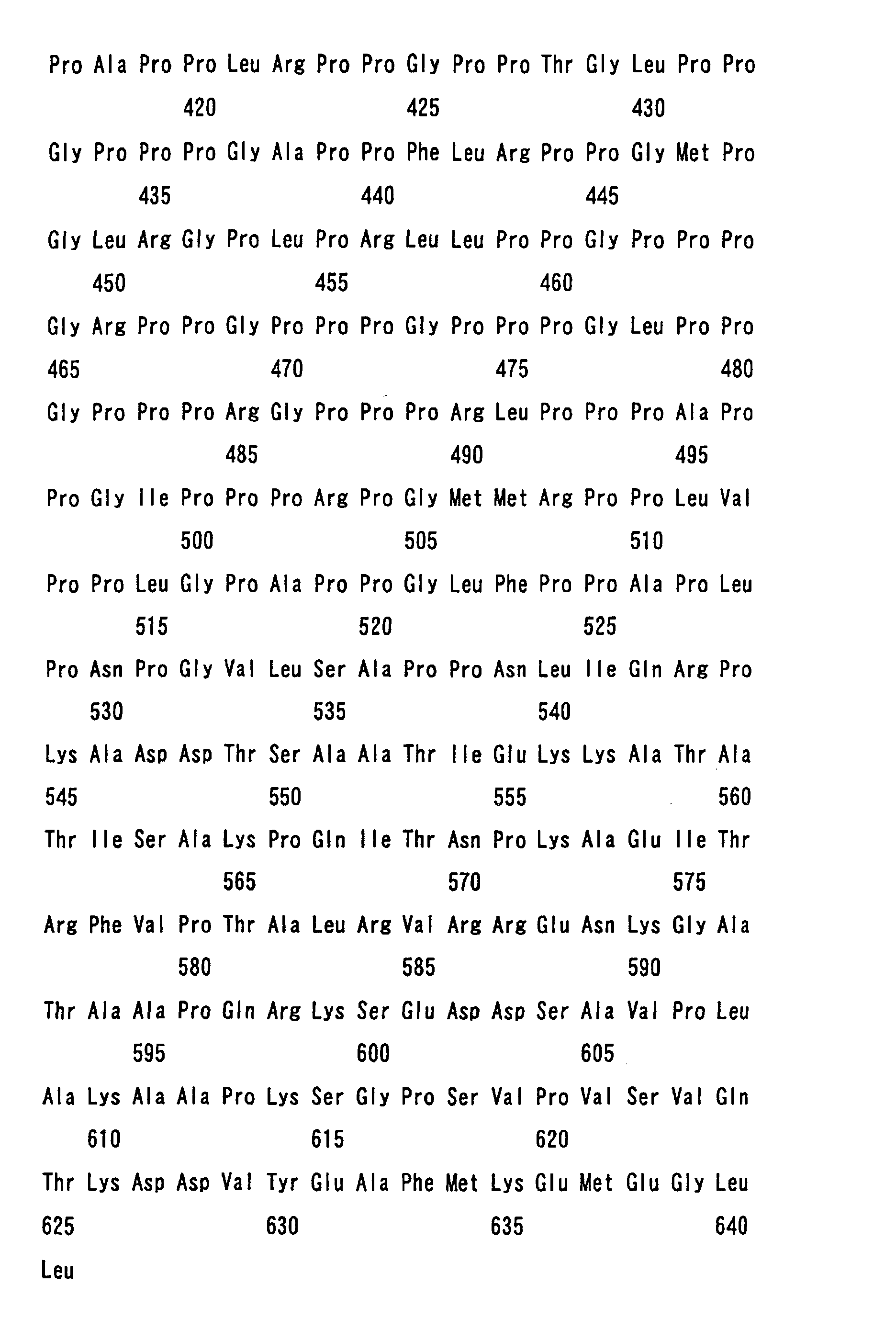
- NpwBP SEQ ID NO: 2 is a protein that binds to Npw38.
- Npw38 from 52nd tryptophan to 78th proline in SEQ ID NO: 1
- NpwBP proline-rich region of NpwBP
- P2 primer 1 SEQ ID NO: 5
- P2 primer 2 SEQ ID NO: 6
- the pKA1-Npw38 (A. Komuro et al., Nucl. Acids Res., 27:1957-1965, 1999) serving as a template was used along with two primers, primer 1 (SEQ ID NO: 7) and primer 2 (SEQ ID NO: 8) to prepare a PCR product. This was digested with EcoRI and SmaI, and then inserted into pKA1-GFP at its EcoRI and Smal to prepare pKA1-Npw38( ⁇ C)-GFP.
- Monkey kidney-derived, cultured cell COS7 was incubated in a 10% fetal calf serum-containing Dulbecco's modified Eagle's medium (DMEM) in the presence of 5 % CO 2 at 37°C.
- COS7 cells of 1 ⁇ 10 5 were placed in a 6-well plate (from Nunk, having a well diameter of 3 cm), and further incubated therein in the presence of 5 % CO 2 at 37°C for 22 hours. After the medium was removed, the cell surfaces were washed with a phosphate buffer solution (PBS), and then further washed with a 50 mM Tris-HCl (pH 7.5)-containing DMEM.
- PBS phosphate buffer solution
- Fusion protein HA-Npw38 (A. Komuro et al., J. Biol. Chem., 274: 36513-36519, 1999) was used. This is a fusion protein A-X in which X is Npw38 and A is HA tag. Since Npw38 contains a inuclear localization signal, A does not require a localization signal. Also used were fusion proteins NpwBP(P1)-GFP and NpwBP(P2)-GFP. These are fusion proteins Y-B in which Y is a proline-rich region of NpwBP and B is GFP.
- P1 contains amino acid residues of from 190th valine to 210th glutamine in SEQ ID NO: 2; and P2 contains amino acid residues of from 401st glutamine to 541st isoleucine in Seq ID NO: 2.
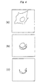
- pKA1-NpwBP(P1)-GFP or pKA1-NpwBP(P2)-GFP was singly introduced into COS7 cells, and the localization of these fusion proteins in the cells was checked through fluorescence microscopy based on the green fluorescence of GFP serving as the index thereof. In the two cases, the entire cells each including its cytoplasm and nucleus were found to gave the green fluorescence (Fig. 3a).
- an Npw38 expression vector pKA1-Npw38 was introduced into COS7 cells, and the green fluorescence was localized in the nucleus of each cell (Fig. 3b).
- NpwBP was used as a fusion protein X-A. Also in this case, since NpwBP contains a nuclear localization signal, the protein X does not require the additional protein A.
- Y-B used was a fusion protein Npw38( ⁇ C)-GFP in which the nuclear localization signal in Npw38 for Y was deleted, and B is GFP.
- the fusion protein Npw38( ⁇ C)-GFP was expressed in COS7 cells, and the green fluorescence was found to appear in the entire cells each including its cytoplasm and nucleus. In this system, when NpwBP was co-expressed, the green fluorescence was found to move in the nucleus of each cell.
- NpwBP of more than NpwBP(P2)-GFP was co-expressed in the cells along with Npw38 and NpwBP(P2)-GFP.
- the intranuclear localization of NpwBP(P2)-GFP was inhibited. This will be because the over-expressed NpwBP bound to Npw38 to inhibit the binding of Npw38 to NpwBP(P2)-GFP, and therefore NpwBP(P2)-GFP could not move to the nuclei of the cells.
- the binding-inhibitory substance is present in the above-mentioned system, it inhibits the localization of the GFP-fused protein. Taking advantage of this system, therefore, the binding-inhibitory factor can be searched out.
- fusion protein X-A herein used was NpwBP(P2)-Bcl-X L in which X is NpwBP(P2), and A is Bcl-X L , a type of mitochondrial protein.
- a full-length cDNA clone HPO3564 of Bcl-X L (its ORF is in SEQ ID NO: 9) was selected from a human full-length cDNA bank (Kato et al., Gene, 150: 243-250, 1994).
- the invention of this application provides a method of rapidly detecting any desired protein-protein interaction in an intracellular condition, not requiring a process of protein production and purification.
- the interaction of different types of proteins in a cell can be detected, and the intracellular protein network in the cell can be clarified.
- novel medicines can be created, and diseases can be clarified on the molecular level.
Landscapes
- Health & Medical Sciences (AREA)
- Life Sciences & Earth Sciences (AREA)
- Engineering & Computer Science (AREA)
- Immunology (AREA)
- Molecular Biology (AREA)
- Biomedical Technology (AREA)
- Chemical & Material Sciences (AREA)
- Urology & Nephrology (AREA)
- Hematology (AREA)
- Biochemistry (AREA)
- Physics & Mathematics (AREA)
- Medicinal Chemistry (AREA)
- General Health & Medical Sciences (AREA)
- Biotechnology (AREA)
- General Physics & Mathematics (AREA)
- Cell Biology (AREA)
- Microbiology (AREA)
- Pathology (AREA)
- Food Science & Technology (AREA)
- Analytical Chemistry (AREA)
- Bioinformatics & Cheminformatics (AREA)
- Tropical Medicine & Parasitology (AREA)
- Organic Chemistry (AREA)
- Toxicology (AREA)
- Chemical Kinetics & Catalysis (AREA)
- General Chemical & Material Sciences (AREA)
- Bioinformatics & Computational Biology (AREA)
- Biophysics (AREA)
- Proteomics, Peptides & Aminoacids (AREA)
- Peptides Or Proteins (AREA)
- Measuring Or Testing Involving Enzymes Or Micro-Organisms (AREA)
- Micro-Organisms Or Cultivation Processes Thereof (AREA)
Abstract
Description




Claims (12)
- A detection method of protein-protein interaction that detects the interaction of a protein X with a protein Y having a reporter function in a eucaryotic cell, which comprises;(1) modifying the protein X and/or the protein Y to be expressed with different localization patterns in the cell,(2) co-expressing the protein X and the protein Y in the eucaryotic cell, and(3) identifying the intracellular localization of the protein Y by its reporter function that serves as the index thereof.
- The method as claimed in claim 1, wherein the protein Y is a fusion protein Y-B with a reporter protein B.
- The method as claimed in claim 2, wherein the reporter protein B is a fluorescent protein.
- The method as claimed in claim 1, wherein, in the step (1), the intracellular localization signal sequence of the protein X and/or the protein Y is mutated.
- The method as claimed in claim 1, wherein, in the step (1), the protein X is fused with a protein A of which the intracellular localization signal sequence differs from that of the protein Y.
- A detection method of protein-protein interaction that detects the inhibitory or promotive action of a compound Z on the interaction of a protein X with a protein Y having a reporter function in a eucaryotic cell, which comprises;(1) modifying the protein X and/or the protein Y to be expressed with different localization patterns in the cell,(2) co-expressing the protein X and the protein Y in the eucaryotic cell,(3) introducing the compound Z into the eucaryotic cell, and(4) identifying the intracellular localization of the protein Y by its reporter function that serves as the index thereof.
- The method as claimed in claim 6, wherein the protein Y is a fusion protein Y-B with a reporter protein B.
- The method as claimed in claim 7, wherein the reporter protein B is a fluorescent protein.
- The method as claimed in claim 6, wherein, in the step (1), the intracellular localization signal sequence of the protein X and/or the protein Y is mutated.
- The method as claimed in claim 6, wherein, in the step (1), the protein X is fused with a protein A of which the intracellular localization signal sequence differs from that of the protein Y.
- The method as claimed in claim 6, wherein, in the step (3), the compound Z is coexisted with the eucaryotic cell.
- The method as claimed in claim 6, wherein, in the step (3), the compound Z is expressed in the eucaryotic cell.
Applications Claiming Priority (5)
| Application Number | Priority Date | Filing Date | Title |
|---|---|---|---|
| JP2000073095 | 2000-03-15 | ||
| JP2000073095 | 2000-03-15 | ||
| JP2000254418 | 2000-08-24 | ||
| JP2000254418A JP2001327296A (en) | 2000-03-15 | 2000-08-24 | Method for detecting protein-protein interaction |
| PCT/JP2001/001973 WO2001068885A1 (en) | 2000-03-15 | 2001-03-13 | Method of detecting protein-protein interaction |
Publications (2)
| Publication Number | Publication Date |
|---|---|
| EP1184463A1 true EP1184463A1 (en) | 2002-03-06 |
| EP1184463A4 EP1184463A4 (en) | 2005-01-19 |
Family
ID=26587632
Family Applications (1)
| Application Number | Title | Priority Date | Filing Date |
|---|---|---|---|
| EP01912323A Withdrawn EP1184463A4 (en) | 2000-03-15 | 2001-03-13 | METHOD FOR DETECTING PROTEIN-PROTEIN INTERACTION |
Country Status (5)
| Country | Link |
|---|---|
| US (1) | US20030040012A1 (en) |
| EP (1) | EP1184463A4 (en) |
| JP (1) | JP2001327296A (en) |
| CA (1) | CA2374155A1 (en) |
| WO (1) | WO2001068885A1 (en) |
Cited By (6)
| Publication number | Priority date | Publication date | Assignee | Title |
|---|---|---|---|---|
| WO2002086450A3 (en) * | 2001-04-20 | 2003-02-06 | Harvard College | Compositions and methods for the identification of protein interactions in vertebrate cells |
| EP1774330A4 (en) * | 2004-08-02 | 2008-10-15 | Cellumen Inc | Methods for the detection of molecular interactions within cells |
| US7772368B2 (en) | 2005-01-25 | 2010-08-10 | Ewha University-Industry Collaboration Foundation | Deletion forms of IGE-dependent histamine releasing factor having histamine releasing activity, HRF-binding peptides and the uses thereof |
| US8114615B2 (en) | 2006-05-17 | 2012-02-14 | Cernostics, Inc. | Method for automated tissue analysis |
| EP3156484A4 (en) * | 2014-06-10 | 2017-12-27 | Medical & Biological Laboratories Co., Ltd. | Method for judging protein interaction |
| US10018631B2 (en) | 2011-03-17 | 2018-07-10 | Cernostics, Inc. | Systems and compositions for diagnosing Barrett's esophagus and methods of using the same |
Families Citing this family (1)
| Publication number | Priority date | Publication date | Assignee | Title |
|---|---|---|---|---|
| US11922756B2 (en) | 2019-01-30 | 2024-03-05 | J.J. Mackay Canada Limited | Parking meter having touchscreen display |
Family Cites Families (1)
| Publication number | Priority date | Publication date | Assignee | Title |
|---|---|---|---|---|
| EP1088233B9 (en) * | 1998-06-16 | 2006-05-17 | PerkinElmer BioSignal Inc. | A bioluminescence resonance energy transfer (bret) system and its use |
-
2000
- 2000-08-24 JP JP2000254418A patent/JP2001327296A/en active Pending
-
2001
- 2001-03-13 CA CA002374155A patent/CA2374155A1/en not_active Abandoned
- 2001-03-13 WO PCT/JP2001/001973 patent/WO2001068885A1/en not_active Ceased
- 2001-03-13 US US09/959,987 patent/US20030040012A1/en not_active Abandoned
- 2001-03-13 EP EP01912323A patent/EP1184463A4/en not_active Withdrawn
Cited By (8)
| Publication number | Priority date | Publication date | Assignee | Title |
|---|---|---|---|---|
| WO2002086450A3 (en) * | 2001-04-20 | 2003-02-06 | Harvard College | Compositions and methods for the identification of protein interactions in vertebrate cells |
| EP1774330A4 (en) * | 2004-08-02 | 2008-10-15 | Cellumen Inc | Methods for the detection of molecular interactions within cells |
| US7772368B2 (en) | 2005-01-25 | 2010-08-10 | Ewha University-Industry Collaboration Foundation | Deletion forms of IGE-dependent histamine releasing factor having histamine releasing activity, HRF-binding peptides and the uses thereof |
| US8114615B2 (en) | 2006-05-17 | 2012-02-14 | Cernostics, Inc. | Method for automated tissue analysis |
| US8597899B2 (en) | 2006-05-17 | 2013-12-03 | Cernostics, Inc. | Method for automated tissue analysis |
| US10018631B2 (en) | 2011-03-17 | 2018-07-10 | Cernostics, Inc. | Systems and compositions for diagnosing Barrett's esophagus and methods of using the same |
| EP3156484A4 (en) * | 2014-06-10 | 2017-12-27 | Medical & Biological Laboratories Co., Ltd. | Method for judging protein interaction |
| US10761085B2 (en) | 2014-06-10 | 2020-09-01 | Medical & Biological Laboratories Co., Ltd. | Method for determining a protein-protein interaction |
Also Published As
| Publication number | Publication date |
|---|---|
| WO2001068885A1 (en) | 2001-09-20 |
| EP1184463A4 (en) | 2005-01-19 |
| US20030040012A1 (en) | 2003-02-27 |
| CA2374155A1 (en) | 2001-09-20 |
| JP2001327296A (en) | 2001-11-27 |
Similar Documents
| Publication | Publication Date | Title |
|---|---|---|
| Tie et al. | A novel imaging method for quantitative Golgi localization reveals differential intra-Golgi trafficking of secretory cargoes | |
| Rayala et al. | The mRNA export factor human Gle1 interacts with the nuclear pore complex protein Nup155 | |
| KR20020059370A (en) | Methods and compositions for the construction and use of fusion libraries | |
| US12529162B2 (en) | Method of screening for peptides capable of binding to a ubiquitin protein ligase (E3) | |
| US20100184619A1 (en) | Method for analyzing organelle-localized protein and material for analysis | |
| US20020151051A1 (en) | Compositions and methods for isolating genes comprising subcellular localization sequences | |
| US20050277116A1 (en) | Compositions and methods for the identification of protein interactions in vertebrate cells | |
| US20030040012A1 (en) | Method of detecting protein-protein interaction | |
| JP2008511289A (en) | Cell cycle phase marker | |
| US12264339B2 (en) | Compositions and methods involving engineered P27 | |
| Van Vliet et al. | A Rab1 interactome illuminates a dual role in autophagy and membrane trafficking | |
| EP1367125A1 (en) | Vectors for expression of biotinylated proteins in mammalian cells, and their use for identification of protein-nucleic acid interactions in vivo | |
| KR102317672B1 (en) | A method for detecting multiple post-translational modification in a single molecule protein | |
| KR20240123808A (en) | Method for screening signal peptides for efficient expression and secretion of heterologous polypeptides in mammalian cells | |
| KR100631484B1 (en) | Plk1 (Polo-like kinase) C-terminal-derived peptide and screening method of ピ lk1 target protein using the same | |
| US20080161199A1 (en) | Fusion Proteins and Methods for Determining Protein-Protein-Interactions in Living Cells and Cell Lysates, Nucleic Acids Encoding these Fusion Proteins, as well as Vectors and Kits Containing These | |
| AU2003299518A1 (en) | Process for determining target function and identifying drug leads | |
| CN113945714A (en) | Method for detecting neutralizing capacity of novel coronavirus neutralizing antibody drugs | |
| US20050214734A1 (en) | Cell population provided with identification codes and method of screening cell population | |
| Knuesel et al. | Identification of Novel Protein-Protein Interactions Using A Versatile Mammalian TAP | |
| Viola | Biochemical studies of human TBP-associated factor TAF2 and a TAF2 and a TAF2-8-10 subcomplex of human general transcription factor TFIID | |
| Chu | The identification and characterization of new components of the multivesicular body (MVB) sorting pathway | |
| Sengar | A functional analysis of the Ese proteins | |
| ES2549488A1 (en) | BIOSENSOR TO DETERMINE THE CRM1 EXPORT FUNCTION |
Legal Events
| Date | Code | Title | Description |
|---|---|---|---|
| PUAI | Public reference made under article 153(3) epc to a published international application that has entered the european phase |
Free format text: ORIGINAL CODE: 0009012 |
|
| 17P | Request for examination filed |
Effective date: 20011213 |
|
| AK | Designated contracting states |
Kind code of ref document: A1 Designated state(s): AT BE CH CY DE DK ES FI FR GB GR IE IT LI LU MC NL PT SE TR |
|
| RBV | Designated contracting states (corrected) |
Designated state(s): DE FR GB |
|
| RAP1 | Party data changed (applicant data changed or rights of an application transferred) |
Owner name: JAPAN SCIENCE AND TECHNOLOGY AGENCY |
|
| A4 | Supplementary search report drawn up and despatched |
Effective date: 20041206 |
|
| RIC1 | Information provided on ipc code assigned before grant |
Ipc: 7G 01N 33/50 B Ipc: 7C 07K 19/00 B Ipc: 7G 01N 33/68 B Ipc: 7G 01N 33/58 B Ipc: 7C 12P 21/02 B Ipc: 7C 12N 15/79 A |
|
| STAA | Information on the status of an ep patent application or granted ep patent |
Free format text: STATUS: THE APPLICATION HAS BEEN WITHDRAWN |
|
| 18W | Application withdrawn |
Effective date: 20050912 |