BACKGROUND OF THE INVENTION
This invention relates to novel compounds and reagent compositions useful for
non-viral introduction of biologically active molecules such as DNA, RNA, peptides and
proteins into eukaryotic cells, and more specifically, to compounds and compositions
useful in lipid transfection.
Gene transfer technology has become a field of considerable interest. Introduction
of an exogenous gene into a cell, i.e., transfection, bears many important scientific and
medical applications, from gene regulation and the production of recombinant proteins to
gene therapy.
Viruses have evolved to bypass the different cellular barriers to gene transfer and
have indeed become vectors of choice for transfection. Many viruses, including
retrovirus, adenovirus and herpes virus, are now engineered to carry therapeutic genes
and are used in human clinical trials for gene therapy. However, there remains a risk of
infectious and immunologic reaction, and the large scale production of viruses is difficult
and time consuming.
For these various reasons, non-viral systems have been developed to carry DNA
into cells, e.g., the transfection technique based on a cationic lipid, the dioleoyloxypropyl
trimethylammonium (Felgner et al., Proc. Natl. Acad. Sci. USA 84, 7413-7417, 1987)
commercialized as Lipofectin™. Since the discovery of this transfection technique, many
more cationic lipids have been synthesized, and some are commercially available as
transfecting reagents for laboratory use. Some examples are DOGS (Transfectam™),
DOSPA (Lipofectamine™) and DOTAP (DOTAP™).
Nevertheless, despite important progress in the formulation of non-viral gene
delivery systems, there remains a need for more efficient techniques since the
transfection efficiency of synthetic systems is usually below that of viral vectors.
Furthermore, many problems arise in vivo, and the poor stability of the non-viral systems
in biological fluids does not allow high and reproducible levels of transfection in vivo.
Transfection of cells with oligonucleotides such as DNA can be used, for
example, to express in a host cell or organism a protein which is not normally expressed
by that cell or organism. For example, a DNA molecule called a plasmid may be
introduced into a cell that does not normally contain the gene(s) encoded by that plasmid
in order to express a marker gene product in that cell, or to express a protein of interest
such as a recombinant protein which is later harvested from such cells (Sambrook et al.,
Molecular Cloning: A Laboratory Manual, 2nd ed., Cold Spring Harbor, 1989). The
transfection of oligonucleotides into cells can also be used therapeutically. For example,
antisense oligonucleotides, once in the cell or cell nucleus, bind to target single-stranded
nucleic acid molecules by Watson-Crick base pairing or to double stranded nucleic acids
by Hoogsteen base pairing, and in so doing, disrupt the function of the target by (a)
preventing the binding of factors required for normal transcription, splicing or translation,
(b) triggering the enzymatic degradation of mRNA by RNAse H, or (c) destroying the
target via reactive groups attached directly to the antisense oligonucleotide (Zamecnic et
al., Proc. Natl. Acad. Sci. USA 75, 280-284, 1978). Gene therapy or DNA-based
vaccination are other therapeutic applications.
Proteins and other macromolecules are transfected into cells for therapeutic and
screening purposes. For example, immunization is enhanced by introducing an
immunogenic protein into a cell so that it is more efficiently processed and presented on
the surface of the cell, thereby enhancing the immunogenic response. Negatively charged
macromolecules which act inside a cell are transported past the cell membrane into the
cytoplasm where they exert their effect. Factors which enhance or hinder transcription of
DNA can be used in a screening test to verify the transcription of a gene of interest.
These transcription assays are well known for use in screening compounds to determine
their effect against a particular macromolecule, for example, a cell receptor.
Cell-lytic antibacterial peptides that act by perturbing the barrier function of
membranes are reviewed in Saberwal et al., Biochim. Biophys. Acta 1197, 109-131, 1994.
Certain fatty acid-bearing basic peptides having antibacterial activity are disclosed in
Vogler et al., Helv. Chim. Acta 47, 526-543, 1964. Poly(lysine-serine) random polymers
for use as carriers to transport oligonucleotides into cells are disclosed in European Patent
Publication No. EP-A-727 223.
European Patent Publication No. EP-A-784 984 and Legendre et al., Bioconjugate
Chem. 8, 57-63, 1997, describe conjugates of a lipid and a basic, membrane-disturbing
peptide that bind polynucleotides and anionic macromolecules and that can be used for
transfection of cells. The peptide portion of the conjugate consists of natural amino acids
linked by a natural amide binding.
Nevertheless, despite important progress in the formulation of non-viral gene
delivery systems, there remains a need for more efficient techniques, since the
transfection efficiency of synthetic systems is usually below that of viral vectors.
Furthermore, many problems arise in vivo and in vitro due in part to the poor stability of
the non-viral systems in biological fluids, and culture media does not allow high and
reproducible levels of transfection.
Many known compounds used for transfection experiments are toxic for
eukaryotic cells. In addition problems may occur in scaling up where the conditioned
medium reduces the effect of known transfecting agents.
The present invention is directed to novel compositions which avoid
disadvantages associated with known transfection agents.
In one aspect, the invention is concerned with novel compounds which are
conjugates of lipids and a modified basic membrane-disturbing peptide, characterized in
that the peptides comprise a reversed amide backbone.
The term "conjugate" means a compound consisting of a lipid chemically bound
to a peptide, e.g., via a disulfide bond formed between a sulfur atom present in or
attached to the lipid and a sulfur atom present in or attached to the peptide, or an amide
bond formed between a carboxyl group present in or attached to the lipid and an amino
group of the peptide.
The term "lipid" as used herein comprises straight chain, branched-chain,
saturated or unsaturated aliphatic carboxylic acids and phospholipids. Examples of
aliphatic carboxylic acids are lauric acid, palmitic acid, stearic acid, oleic acid and
(CH3(CH2)n)2CHCOOH, where n is an integer from 3 to 19. Examples of phospholipids
are phosphatidylethanolamines such as dioleoylphosphatidylethanolamine.
The term "basic peptide" denotes a peptide containing at least one basic amino
acid. Examples of such basic amino acids are natural and non-natural diaminomonocarboxylic
acids such as alpha- and beta-diaminopropionic acid, alpha- and gamma-diaminobutyric
acid, lysine, arginine, ornithine and p-aminophenylalanine.
The term "membrane-disturbing peptide" denotes a cell-lytic or antibacterial
peptide that perturbs the barrier function of membranes (G. Saberwal and R. Nagaraj,
BBA 1197, 109-131, 1994). Examples of basic, cell-lytic peptides are melittin, hemolysin,
mastoparan, bombolitin, crabrolin and derivatives thereof. Examples of basic
antibacterial peptides are cecropins, magainins, gramicidin S, tyrocidine and derivatives
thereof.
The term "derivative" refers to a peptide wherein one or more amino acid residues
are either missing, have been added or have been replaced by another amino acid residue
without substantially changing the biological activity of the original peptide concerned,
i.e., it allows transfection of a macromolecule, preferably a polynucleotide, into a cell.
The term "derivative" also refers to a peptide wherein the terminal carboxyl group is
esterified, particularly to form lower alkyl esters such as the methyl and ethyl ester, or
converted into an amide, lower alkyl amide or di-lower alkyl amide or hydrazide. The
term "derivative" also relates to a peptide wherein the NH2 group of the N-terminus may
be acylated to form an amide or a lower alkyl amide. In particular, the NH2 group is
acetylated. The term "lower" denotes groups containing from 1-6 carbon atoms.
The term "reversed amide backbone" refers to a retro-peptide which
characteristically has the same composition as its parent peptide, but the sequence is
reversed, i.e., n, ... 3, 2, 1 instead of 1, 2, 3, ... n when reading in the N to C direction.
Both have normal peptide bonds.
Throughout this application, the following standard abbreviations are used to refer
to amino acids:
| Alanine | Ala | D-Alanine | D-Ala |
| Glutamate | Glu | D-Glutamate | D-Glu |
| Glutamine | Gln | D-Glutamine | D-Gln |
| Aspartate | Asp | D-Aspartate | D-Asp |
| Asparagine | Asn | D-Asparagine | D-Asn |
| Leucine | Leu | D-Leucine | D-Leu |
| Glycine | Gly |
| Lysine | Lys | D-Lysine | D-Lys |
| Serine | Ser | D-Serine | D-Ser |
| Valine | Val | D-Valine | D-Val |
| Arginine | Arg | D-Arginine | D-Arg |
| Threonine | Thr | D-Threonine | D-Thr |
| Proline | Pro | D-Proline | D-Pro |
| Isoleucine | Ile | D-Isoleucine | D-Ile |
| Methionine | Met | D-Methionine | D-Met |
| Phenylalanine | Phe | D-Phenylalanine | D-Phe |
| Tyrosine | Tyr | D-Tyrosine | D-Tyr |
| Cysteine | Cys | D-Cysteine | D-Cys |
| Tryptophan | Trp | D-Tryptophan | D-Trp |
| Histidine | His | D-Histidine | D-His |
| Ornithine | Orn | D-Ornithine | D-Orn |
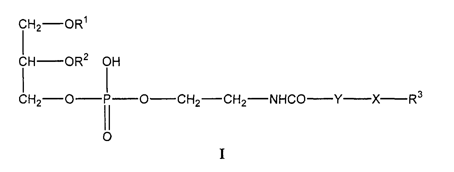
In a preferred embodiment, the novel compounds of the present invention are
compounds of the formula
wherein R
1 and R
2 are selected from the group consisting of hydrocarbyl moieties of
straight-chain and branched-chain, saturated and unsaturated aliphatic carboxylic acids
and phospholipid moieties, R
3 is a basic, membrane-disturbing peptide with a reversed
amide backbone, Y is a C
2-10 alkylene, and X is selected from the group consisting of
-C(O)-NH-, -S-S-, and salts thereof.
Particularly preferred compounds are those wherein R1 and R2 independently are
an acyl moiety of a C12-20 carboxylic acid. The term "C12-20" denotes a number of carbon
atoms of from 12 to 20. The acyl moieties R1 and R2 can be straight chain or branched-chain,
saturated or unsaturated moieties. Examples of such moieties are lauroyl,
palmitoyl, stearoyl and oleoyl. In a preferred aspect, R1 and R2 are oleoyl. Y is preferably
ethylene, propylene or decamethylene. X is preferably -S-S-.
R3 is a basic, membrane-disturbing peptide with a reversed amide backbone. For
example, R3 is Gln-Gln-Arg-Lys-Arg-Lys-Ile-Trp-Ser-Ile-Leu-Ala-Pro-Leu-Gly-Thr-Thr-Leu-Val-Lys-Leu-Val-Ala-Gly-Ile-NH-CH[CONH2]-(CH2)-.
The peptide has a
reversed amide backbone. Preferably, R3 comprises at least 50%, more preferably 65%,
and even more preferably 80% D-amino acids or derivatives thereof. In the most
preferred embodiment, all amino acids are D-amino acids. The term "D-amino acid"
refers to naturally as well as non-naturally occurring D-α-amino carbonic acids and
derivatives thereof. In addition, R3 also comprises membrane-disturbing peptide
sequences such as magainin, cecropin, defensins, etc., or chimers of such, e.g., cecropin-melittin
(Hancock et al., TIBTECH 16, 82-88, 1998) synthesized as the retro-inverso
peptides and derivatized to allow conjugation to lipids, analogous to the methods as
described below, especially analogous to Examples 1 and 2.
The term "retro-inverso peptide" means the all -D analog of an all -L retro-peptide.
In a further preferred embodiment, R3 is D-Gln-D-Gln-D-Arg-D-Lys-D-Arg-D-Lys-D-Ile-D-Trp-D-Ser-D-Ile-D-Leu-D-Ala-D-Pro-D-Leu-D-Gly-D-Thr-D-Thr-D-Leu-D-Val-D-Lys-D-Leu-D-Val-D-Ala-D-Gly-D-Ile-NH-CH[CONH2]-CH-(CH2)-.
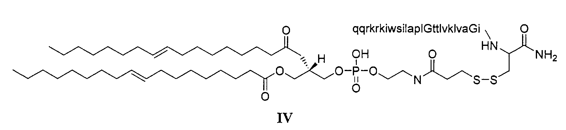
The most preferred compound is
A further embodiment of the present invention refers to the peptide portion of R3,
especially to the intermediate peptide Gln-Gln-Arg-Lys-Arg-Lys-Ile-Trp-Ser-Ile-Leu-Ala-Pro-Leu-Gly-Thr-Thr-Leu-Val-Lys-Leu-Val-Ala-Gly-Ile-Cys-NH2
and derivatives,
salts and solvates thereof having a reversed amide backbone and consisting of at least
50%, more preferably 65%, and even more preferably 80% of D-amino acids or
derivatives thereof. In a most preferred embodiment, all amino acids are D-amino acids.
The term "D-amino acid" refers to naturally as well as non-naturally occurring D-α-amino
carbonic acids and derivatives thereof.
In a preferred embodiment, the peptide is D-Gln-D-Gln-D-Arg-D-Lys-D-Arg-D-Lys-D-Ile-D-Trp-D-Ser-D-Ile-D-Leu-D-Ala-D-Pro-D-Leu-D-Gly-D-Thr-D-Thr-D-Leu-D-Val-D-Lys-D-Leu-D-Val-D-Ala-D-Gly-D-Ile-D-Cys-NH2.
In another aspect, this invention relates to a process for preparing the novel
compounds defined above, i.e., conjugates of lipids and basic, membrane-disturbing
peptides wherein the peptides comprise a reversed amide backbone, and compositions
comprising at least one such compound, a polynucleotide or any other anionic
macromolecule, and, optionally, a helper lipid, a short chain phospholipid, or a cationic
lipid and optionally an additional known transfection reagent other than a conjugate of
this invention, i.e., a compound of formula I or II. In still another aspect, this invention
relates to compositions comprising conjugates of lipids and basic, membrane-disturbing
peptides and a helper lipid, a short chain phospholipid, or a cationic lipid and optionally
an additional known transfection reagent other than a conjugate of this invention, e.g., a
compound of formula I. The invention further relates to the use of the novel compounds
as a carrier for transfecting a cell with a polynucleotide or any other anionic
macromolecule. Even further, the invention relates to the enhancement of transfection
efficiency when DNA complex formation is performed in the presence of additional
amino acids, preferably in the form of a protein hydrolysate.
DETAILED DESCRIPTION OF THE INVENTION
The compounds provided by this invention can be prepared by reacting a peptide
of the formula R
3NH
2 with a lipid of the formula
or a peptide of the formula R
3SH with a lipid of the formula
wherein R
1, R
2, R
3 and Y are as defined above and Z is a leaving group such as 2-pyridinethio.
These reactions can be carried out in a manner known per se.
Compounds of the formula R3NH2 wherein the peptide part comprises a reversed
amide backbone may be prepared by methods known in the art. The corresponding
methods have been described, for example, for the preparation of synthetic melittin, its
enantio-, retro- and retroenantio-isomers and derivatives thereof (Juvvadi et al., J. Am.
Chem. Soc. 118, 8989-8997, 1996).
The peptides may be prepared by the solid-phase synthesis technique. In this
technique, synthesis occurs while the peptide is attached to a polymeric support, thereby
allowing for the separation of product from byproduct by washing steps. The completion
of the acylation reaction is ensured by using large excesses of soluble reagents. Synthesis
involves the covalent anchorage of the first amino acid in the sequence to the solid
support followed by the deprotection of the protected amino function for subsequent
coupling to the incoming amino acid derivative. After n cycles of deprotection and
coupling, the peptide is released from the solid phase by a chemical cleavage reaction. In
a preferred embodiment, D-amino acids are used.
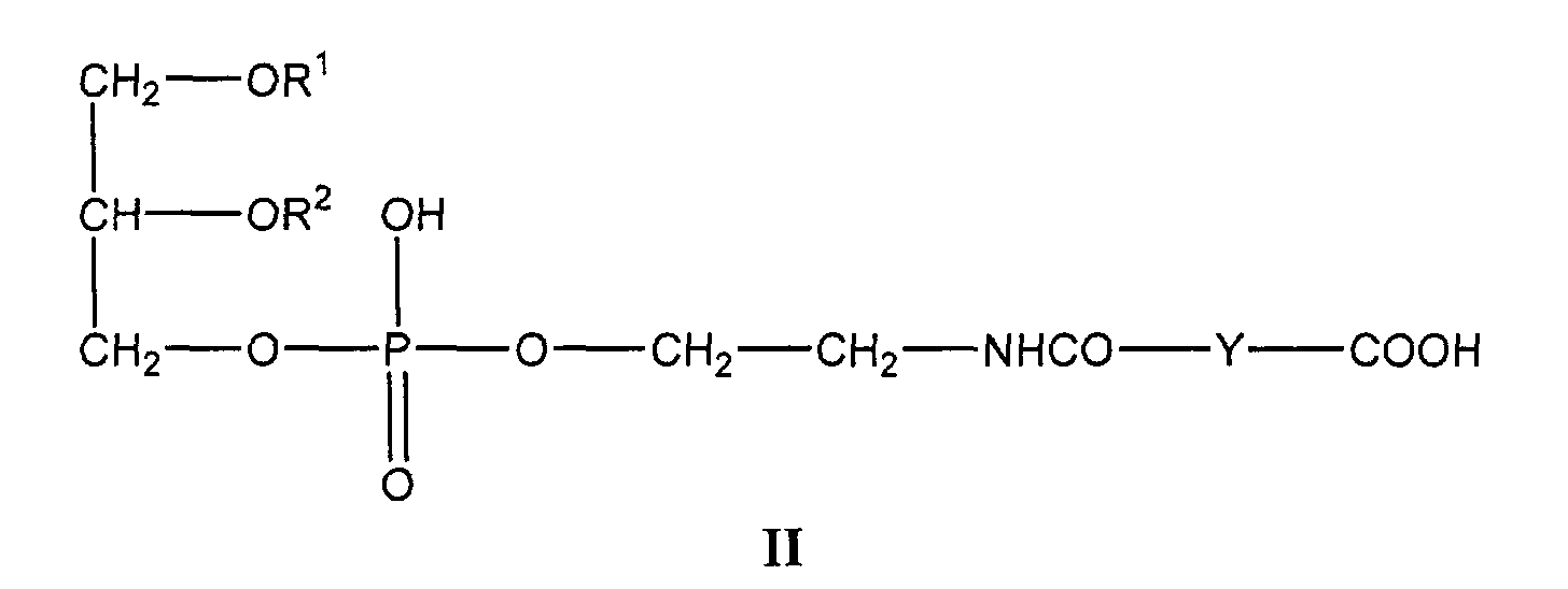
Thus, the coupling of a peptide of the formula R3NH2 with a lipid of the formula
II can be accomplished by reacting the compounds wherein an amino group other than
the amino group to be reacted are protected in a suitable solvent in the presence of a
condensation agent such as dicyclohexylcarbodiimide analogous to known methods for
producing peptide bonds.
The reaction of a peptide of the formula R3SH with a compound of the formula R-SZ
can be carried out in an appropriate solvent or solvent mixture which solubilizes both
reactants. The compound of the formula R-SZ can be dissolved in an organic solvent,
e.g., in chloroform. The peptide R3SH is suitably dissolved in an aqueous buffer solution,
such as phosphate buffer, that contains an appropriate amount of a water-miscible organic
solvent such as acetonitrile to accomplish the formation of a single phase reaction system.
Compounds of the formula II and III are known or can be prepared by known
methods, e.g. as described in
Biochim. Biophys. Acta 862, 435-439, 1986. For example,
compounds of the formula
wherein R
1 and R
2 are oleolyl, and Y is ethylene, propylene or decamethylene and
compounds of the formula
wherein R
1 and R
2 are oleolyl, Y is ethylene and Z is 2-pyridinethio are commercially
available as N-Succinyl-PE, N-Glutaryl-PE, N-Dodecanyl-PE and N-PDP-PE from
Avanti Polar Lipids, Alabaster, Alabama.
Any anionic macromolecule can be transfected into a cell using a compound of
formula I. An "anionic macromolecule" is a macromolecule which contains at least one
negative charge per molecule. Examples of anionic macromolecules which can be
transfected in accordance with this invention include polynucleotides, such as
deoxyribonucleic acids (DNA) and ribonucleic acids (RNA), and proteins, such as
ribonucleoproteins and proteins used for immunization, e.g., viral proteins. Examples of
DNA for use in the present invention are plasmids and genes, especially those for which
gene therapy protocols have been launched, such as cystic fibrosis transmembrane
regulator (CFTR), adenosine deaminase (ADA), thymidine kinase (TK) and HLA B7, as
well as reporter genes such as β-galactosidase, luciferase, green fluorescence protein
(GFP), chloramphenicol transferase and alpha- antitrypsin. Other examples of DNA are
oligodeoxynucleotides and their analogs used as antisense, aptamer or 'triple-helix'
agents. Examples of RNA are ribozymes and oligoribonucleotide antisense molecules.
The nature of the cell that is to be transfected is not narrowly crucial. The cell can
be a prokaryotic or eukaryotic cell, and a mammalian or a plant cell.
In transfecting a cell using a conjugate of this invention, e.g., a compound of
formula I, the cell is contacted with the anionic macromolecule in the presence of an
appropriate amount of such compound. The appropriate amount of the conjugate, e.g., a
compound of formula I, for a given amount of anionic macromolecule depends on their
respective charges. The +/- charge ratio between the conjugate and the molecule to be
transfected generally varies between 0.1-10, preferably between 0.5-5. The value of "+/-charge
ratio" is calculated by dividing the number of positively charged groups on the
amino acids in the group R3 by the number of negative charges of the molecule to be
transfected. When the molecule to be transfected is a polynucleotide, for example, the
number of negative charges means the number of negatively charged phosphates in the
backbone. The optimal ratio within these ranges depends on the cell to be transfected and
is readily ascertained by one of skill in the art to which this invention pertains.
The amount of anionic macromolecules to the number of cells is such that the
amount of anionic macromolecule for transfecting 104 cells is from 0.1 µg to 10 µg,
preferably from 0.2 µg to 2 µg. When the anionic macromolecule is DNA, the preferred
amount of DNA for transfecting 104 cells in vitro is from 0.1 µg to 10 µg. When cells are
being transfected in vivo, the preferred amount of DNA is from 0.1 µg to 1 µg.
In a preferred aspect of the invention, the transfection is further carried out in the
presence of a helper lipid, short chain phospholipid, cationic lipid or any other known
transfection-competent molecule other than a conjugate of this invention. Any
conventional helper lipid can be used. "Helper lipids" are phospholipids which are known
to increase delivery of macromolecules to cells when used together with known
transfection competent molecules. Examples of helper lipids are phospholipids, such as
phosphatidylcholines and phosphatidylethanolamines and mixtures thereof. Preferred
helper lipids are phosphatidylethanolamines, such as dioleoylphosphatidylethanolamine.
Any conventional short chain phospholipid can be used. "Short chain phospholipids" are
phospholipids containing fatty acid residues which contain from 6 to 12 carbon atoms in
their backbone. Examples of short chain phospholipids are phosphatidylcholines that
carry two C6-12 fatty acid residues. Preferred short chain phospholipids are dicapryl- and
dicapryloyl phosphatidylcholine.
Examples of transfection competent molecules include cationic lipids as described
by Behr in Bioconjugate Chem. 5, 382-389, 1994 and Gao et al. in Gene Ther. 2, 710-722,
1995, polycations as described by Kabanov et al. in Bioconjugate Chem. 6, 7-20,
1995, and peptides, polymers and other non-viral gene delivery systems as described by
Ledley in Human Gene Therapy 6, 1129-1144, 1995.
The helper lipid, short chain phospholipid, cationic lipid and optionally other
additional known transfection competent molecule other than a conjugate of this
invention is suitably in the form of a liposome, micelle, organic or aqueous dispersion, or
an organic or aqueous solution. The optimal molar ratio between the compound of
formula I and the helper lipid is 0.1:50, preferably 1:10. The optimal molar ratio between
helper lipid and short-chain phospholipid is 2:20. The optimal molar ratio between the
compound of formula I or II and additional transfection competent molecules is 0.1:10.
In an especially preferred aspect of the present invention, transfection efficiency
is significantly enhanced when DNA complex formation is performed in the presence of
a concentrated amino acid mixture in the form of a protein hydrolysate. A preferred
protein hydrolysate is a tryptic meat digest, Primatone RL (Sheffield Products, Quest
International, Germany). Primatone RL is a medium supplement of a complex nature
which serves as a source of amino acids, oligopeptides, iron salts, some lipids and other
trace low molecular weight substances. The addition of Primatone RL to mammalian and
insect cell culture media is known to improve the cell growth properties of many cell
lines. However, it was quite surprising to discover that the addition of Primatone RL to
the DNA complex formation medium of the present invention significantly enhanced
complex formation. This effect was surprising, in part, because it is well accepted in the
art to which the present invention belongs that it is essential that the lipid-nucleic acid
complex is formed in the absence of serum, a complex medium supplement containing
proteins, peptides, lipids, etc. which are known to interfere with complex formation
(FAQS for Transfections and Cationic Lipid Reagents, Life Technologies,
http://www.lifetech.com). Other preferred protein hydrolysates include Primatone RL-UF
(ultrafiltered) and Primatone HS.
The present invention also comprises the use of a composition as defined above
for transfecting a eukaryotic or prokaryotic cell in vivo or in vitro with an anionic
macromolecule, preferably with a polynucleotide.
The invention also comprises the use of compounds of formula I as defined above
for transfecting a eukaryotic or prokaryotic cell in vivo or in vitro with an anionic
macromolecule, preferably with a polynucleotide.
A further embodiment of the present invention is a process for transfecting a cell
in vivo or in vitro with an anionic macromolecule, preferably a polynucleotide,
comprising contacting a cell in vivo or in vitro with the anionic macromolecule in the
presence of a compound of formula I or with a composition as defined above.
In addition, the invention is directed to a process for introducing a biologically
active anionic molecule into a cell in vivo or in vitro with an anionic macromolecule,
comprising contacting a cell in vivo or in vitro with the anionic macromolecule in the
presence of a composition or a compound as defined above.
For transfection, an appropriate amount of a conjugate of this invention, e.g., a
compound of formula I, is added to the molecule to be transfected, e.g., plasmid DNA,
suitably in an aqueous solution. Optionally, a helper lipid and, if desired, a short chain
phospholipid, a cationic lipid and optionally another additional known transfection
competent molecule other than a conjugate of this invention are then added, either in the
form of a liposome, micelle, or as an organic solution or aqueous dispersion.
Alternatively, the molecule to be transfected may be added to a composition comprising a
compound in accordance with this invention, a helper lipid, and, if desired, a short chain
phospholipid, a cationic lipid and optionally another additional known transfection
competent molecule other than a conjugate of this invention. The composition may be in
solid, liquid, semisolid or aerosol form, suitably in the form of a liposome, micelle, or an
organic solution or aqueous dispersion.
For transfecting cells in an animal or human patient, the composition can be
administered by oral, parenteral, transdermal, pulmonary, nasal, rectal, ocular,
ventricular, vascular and intratumoral routes. Furthermore, the composition can be
administered by high velocity impaction administration to the skin surface. The progress
of transfection can be measured by appropriate testing protocols which are known to
those skilled in the art.
The present invention also relates to a composition comprising at least one
compound as defined above and a helper lipid, a short chain phospholipid, a cationic
lipid, or optionally another additional transfection competent molecule. In addition, the
composition may comprise an anionic macromolecule, preferably a polynucleotide.
These compositions may also comprise a polycationic polymer, preferably
polyethyleneimine (PEI). The components of these compositions may be in the form of
an aqueous or organic solution, an aqueous or organic dispersion, or a liposome or
micelle.
The term "polyethyleneimine (PEI)" refers to a synthetic organic, generally
branched or linear macromolecule with a high cationic charge/density potential,
preferably with a molecular weight of about 25 kDa.
The invention also relates to the use of a compound as above for transfecting a
cell with an anionic macromolecule, preferably a polynucleotide.
In accordance with the present invention, it has been found that conjugating a
lipid to a basic, membrane-disturbing peptide having a reversed amide bond results in
novel compounds that bind polynucleotides and other anionic macromolecules and that
can be used for transfection of cells. Thus, the invention is concerned with a process for
transfecting a cell with an anionic macromolecule comprising contacting the cell with the
anionic macromolecule in the presence of a compound as described above, so as to
transfect the cell with the anionic macromolecule.
The invention also relates to a process for production of large quantities of
recombinant proteins in PEI-mediated transfected cells. For example, high-level
expression of both G protein-coupled receptors and ligand-gated ion channels by the use
of Semliki Forest virus (SFV) has been realized. Generally, Bmax values of more than 50
pmol receptor per mg protein and receptor densities of more the 3 x 10⁶ receptors per cell
have been achieved. To further facilitate large-scale protein production, optimal
conditions for mammalian serum-free suspension cultures have been obtained.
Adaptation of BHK, CHO and HEK293 cells to these conditions has allowed efficient
infection with SFV vectors to produce large volumes of recombinant protein expressing
cell cultures.
Surprisingly, the growth temperature of the cell cultures can have a dramatic
effect on the duration and levels of recombinant protein expression. Expression of
recombinant luciferase is increased 5- to 10-fold by lowering the growth temperature of
BHK and CHO cells from 37 °C to 33 °C. The expression time is much longer at 33 °C,
with high expression 65 hours post-infection.
Moreover, the effect of the temperature on the expression of two 7TM receptors,
human neurokinin-1 receptor and rat metabotropic glutamate receptor-2 and the 5-HT3
ligand-gated ion channel could be shown. A similar effect as observed for luciferase has
also been obtained for the receptors. The receptor density is much higher in cells grown
at 33 °C compared to 37 °C. The expression time for receptors is usually restricted to 24
hours in SFV-infected cells, but can be prolonged to 65 hours when cells are cultured at
33 °C. This improvement can greatly facilitate production of large quantities of
recombinant proteins. Moreover, the utility of the inventive transfection for the rapid
expression of heterologous proteins in large scale for adherent cells (CFU) and
suspension cells (12 liter, 23 liter, 60 liter fermentors) has been realized. In particular,
helper lipids in combination with PEI exhibit a high transient gene expression in HEK293
cells and other mammalian cell lines at low DNA concentration. In addition, the
frequently occurring inhibitory effect of conditioned medium, which is a serious problem
for scale up expression methods, is significantly reduced.
The general procedure for protein expression comprises (a) adaptation and growth
of a cell line to suspension culture in an appropriate medium and expression vectors, (b)
washing of cells to remove heparin/dextran sulfate in preparation for transfection, (c)
counting of cells and resuspension in a culture medium appropriate for transfection, (d)
preparation of the transfection reagent:DNA complex, maturation of the complex and
addition to the cells, and (e) incubation and monitoring of protein expression over 2 to 10
days.
The following examples, which are not meant to be limiting, illustrate the
invention further.
Example 1. Preparation of the retro-inverso peptide R3SH
Continuous-flow solid-phase synthesis was performed on a Pioneer™ Peptide
Synthesis System, starting from Tenta Gel S RAM resin (0.25 mmol/g [Rapp Polymere
GmbH, Tübingen, Germany] according to the method described by Atherton and
Sheppard, Solid Phase Peptide Synthesis: A Practical Approach (IRL Press, Oxford,
1989). The base-labile Fmoc group was used for α-amino protection. Side chains were
protected with the following protection groups: D-Arg(Pbf), D-Cys(Trt), D-Gln(Trt), D-Lys(Boc),
D-Ser(But), D-Thr-(But) and D-Trp(Boc). Fmoc-amino acids (2.5 equivalents)
were activated with an equivalent amount of TATU (L.A. Carpino, J Am. Chem. Soc.
115, 4397-4398, 1993) and DIPEA. Fmoc deprotection was achieved with 20%
piperidine in DMF. D-Gln(Trt)-D-Gln(Trt)-D-Arg(Pbf)-D-Lys(Boc)-D-Arg(Pbf)-D-Lys(Boc)-D-Ile-D-Trp(Boc)-D-Ser(But)-D-Ile-D-Leu-D-Ala-D-Pro-D-Leu-Gly-D-Thr(But)-D-Thr(But)-D-Leu-D-Val-D-Lys(Boc)-D-Leu-D-Val-D-Ala-Gly-D-Ile-D-Cys(Trt)-amide
Tenta Gel S-resin (2.0 g) was treated with a mixture (100 ml) of 90%
TFA, 2% EDT, 5% H2O, 3% triisopropylsilane for 6 hours. The reaction mixture was
concentrated and poured into diethyl ether, and the precipitate was collected by filtration
and lyophilized from water. The crude peptide was purified by preparative RP-HPLC.
There was obtained homogenous D-Gln-D-Gln-D-Arg-D-Lys-D-Arg-D-Lys-D-Ile-D-Trp-D-Ser-D-Ile-D-Leu-D-Ala-D-Pro-D-Leu-D-Gly-D-Thr-D-Thr-D-Leu-D-Val-D-Lys-D-Leu-D-Val-D-Ala-D-Gly-D-Ile-D-Cys-NH2·6
TFA.
Example 2. Preparation of a compound of formula IV
The homogenous peptide obtained in Example 1 (24.2 mg, 7 mmol) was dissolved
in a mixture of 2 ml of 100 mM ammonium acetate buffer, pH 6.5, and 2 ml of
acetonitrile. To this solution there was added 7.4 mg of 1,2-dioleoyl-sn-glycero-3-phosphoethanolamine-N-[3-(2-pyridyldithio)propionate]
in 0.5 ml of chloroform. The
mixture was stirred at room temperature for 1 hour, and the organic solvent was removed
by evaporation. The remaining solution was washed 3 times with ethyl acetate, and the
aqueous phase was lyophilized. There was obtained 28.5 mg of a compound of formula
IV wherein R1 and R2 are oleoyl and D-Gln-D-Gln-D-Arg-D-Lys-D-Arg-D-Lys-D-Ile-D-Trp-D-Ser-D-Ile-D-Leu-D-Ala-D-Pro-D-Leu-D-Gly-D-Thr-D-Thr-D-Leu-D-Val-D-Lys-D-Leu-D-Val-D-Ala-D-Gly-D-Ile-NH-CH[CONH2]-CH-(CH2)-
is R3.
ISP-MS:M = 3722.
Example 3. Transfection using a compound of formula IV
1 mg of the compound obtained in Example 2 was dissolved in 500 µl
acetonitrile, diluted in 500 µl sterile pure water (1 mg/ml) and stored at 4 °C. 15 µl of a
plasmid solution (1 mg/ml) encoding the soluble human tumor necrosis factor receptor
p55 gene was transferred into 1.5 ml medium and mixed. Various amounts of the
compound obtained in Example 1 was added, mixed and after 10 minutes at room
temperature, the mixture was transferred to 15 ml HEK293-EBNA cells in suspension.
Table 1 shows the transfection efficiency of the compound as described in
Example 2 and the cell viability in serum-free suspension culture.
| Transient transfection of HEK293-EBNA cells |
| Compound (µg/ml) | 1 | 2 | 4 | 6 | 10 |
| Viability (%) | 90 - 95 | 90 - 95 | 90 - 95 | 90 - 95 | 90 - 95 |
| TNFRp55 (ng/ml) | 0.1 | 0.2 | 2.5 | 6.3 | 83 |
The data clearly indicate that compound-mediated transfection can be achieved in the
absence of detectable cell toxicity effects in serum-free suspension culture of HEK293-EBNA
cells.
1 mg of the compound obtained in Example 2 was dissolved in 500 µl acetonitrile
and diluted in 500 µl sterile pure water (1 mg/ml) and stored at 4 °C. 90 mg
polyethyleneimine (PEI), with a molecular weight of 25 kDa, were dissolved in sterile
pure water (0.9 mg/ml), neutralized with HCl, sterile filtered and stored at room
temperature.
3 µl of a plasmid solution (1 mg/ml) encoding the soluble human tumor necrosis
factor receptor p55 gene was diluted in 1.5 ml culture medium and mixed. Various
amounts of the compound (1 mg/ml) as described in Example 2 were added, mixed,
followed by 8.3 µl PEI (0.9 mg/ml). After mixing and incubation for 10 minutes at room
temperature, the mixture was transferred into 15 ml of HEK293-EBNA cells in
suspension. Cells which were grown in serum-free suspension culture were transferred
into fresh medium before adding the complexes. The released receptor protein was
measured 72 hours post-transfection in the culture medium and expressed as ng receptor
per milliliter culture.
| Transient transfection of HEK293-EBNA cells together with PEI |
| Compound + polyethyleneimine (µg/ml) | 0.02 + 0.5 | 0.05 + 0.5 | 0.1 + 0.5 | 0.15 + 0.5 | 0 + 0.5 |
| TNFRp55 (ng/ml) | 490 | 800 | 1120 | 1100 | 180 |
The results clearly indicate the synergistic enhancement of the compound together with
PEI. The sequence of adding first the compound followed by PEI to the DNA or vice
versa has no detectable influence on the transfection efficiency. No significant changes of
the expression levels were observed when the cells were cultured in the presence of 10%
serum.
1 mg of the compound obtained in Example 2 was dissolved in 500 µl sterile pure
water (1 mg/ml) and stored at 4 °C. 90 mg polyethyleneimine, with a molecular weight of
25 kDa, was dissolved in sterile pure water (0.9 mg/ml), neutralized with HCl, sterile
filtered and stored at room temperature.
3 µl of a plasmid solution (1 mg/ml) encoding the luciferase gene was diluted in
1.5 ml culture medium and mixed. 2.25 µl compound (1 mg/ml) as described in Example
2 was added, mixed and followed by 8.3 µl PEI (0.9 mg/ml). After mixing and incubation
for 10 minutes at room temperature, the mixture was transferred into 15 ml of various
cells. Cells which were grown in serum-free suspension culture were transferred into
fresh medium before adding the complexes. The luciferase activity was measured in the
soluble cell extract after 24 hours incubation and expressed as relative light unit (RLU)
per milligram protein.
Table 3 shows the transfection efficiency of the compound described in Example
2 in combination with PEI by measuring the luciferase activity in serum-free suspension
culture.
| Luciferase activity (RLU/mg protein) |
| | Compound | PEI | Compound + PEI |
| BHK 21 Cl | 5.2 x 10⁵ | 2.8 x 10⁷ | 1.8 x 10⁹ |
| CHO dhfr- | 1.4 x 10⁶ | 3.5 x 10⁷ | 5.3 x 10⁹ |
| HEK293-EBNA | 2.2 x 10⁶ | 3.2 x 10⁹ | 6.7 x 10¹⁰ |
| HEK293 | 1.8 x 10⁶ | 2.9 x 10⁹ | 6.3 x 10¹⁰ |
| Glioma C6 | 5.8 x 10⁶ | 6.5 x 10⁷ | 3.4 x 10⁹ |
The results clearly indicate the synergistic enhancement of the compound together with
PEI in various cell lines.
1 mg of the compound obtained in Example 2 was dissolved in 50 µl acetonitrile
and diluted in 500 µl sterile pure water (1 mg/ml) and stored at 4 °C. 90 mg
polyethyleneimine, with a molecular weight of 25 kDa, was dissolved in sterile pure
water (0.9 mg/ml), neutralized with HCl, sterile filtered and stored at room temperature.
3 ml of a plasmid solution (1 mg/ml) encoding the green fluorescence protein was
diluted in 1.5 ml culture medium and mixed. 2.25 µl compound (1 mg/ml) was added,
mixed and followed by 8.3 µl PEI (0.9 mg/ml). After mixing and incubation for 10
minutes at room temperature, the mixture was transferred into 15 ml of various cells.
Cells which were grown in serum-free suspension culture were transferred into fresh
medium before adding the complexes. The transfection efficiency was measured by
counting the number of fluorescent cells after 24 hours incubation and expressed as
percent transfected cells.
Table 4 shows the transfection efficiency of the compound described in Example
2 in combination with PEI by measuring the green fluorescence within various cell lines
in serum-free suspension culture.
| Amount of fluorescence cells (%) - compound + PEI |
| BHK 21 Cl | 25 (± 5) |
| CHO dhfr- | 15 (± 5) |
| HEK293-EBNA | 85 (± 5) |
1 mg of the compound obtained in Example 2 was dissolved in 500 µl acetonitrile
and diluted in 500 µl sterile pure water (1 mg/ml) and stored at 4 °C. 90 mg
polyethyleneimine, with a molecular weight of 25 kDa, was dissolved in sterile pure
water (0.9 mg/ml), neutralized with HCl, sterile filtered and stored at room temperature.
3 ml of a plasmid solution (1 mg/ml) encoding the luciferase gene was diluted in
1.5 ml culture medium and mixed. 2.25 µl compound (1 mg/ml) was added, mixed, and
followed by 8.3 µl PEI (0.9 mg/ml). After mixing and incubation for 10 minutes at room
temperature, 0.1 ml of the mixture was transferred into a well containing 1 ml medium of
a 12-well plate with various adherent cell lines. Cells which were grown in the presence
of 10% serum were incubated during 4 hours post-transfection in serum-free medium.
The luciferase activity was measured in the soluble cell extract after 24 hours incubation
and expressed as relative light unit (RLU) per milligram protein.
Table 5 shows the transfection efficiency of the compound described in Example
2 in combination with PEI by measuring the luciferase activity in various adherent cell
lines.
| Luciferase activity (RLU/mg protein) |
| | PEI | Compound + PEI |
| HEK293 | 5.4 x 10⁹ | 5.3 x 10¹⁰ |
| Glioma C6 | 3.1 x 10⁹ | 1.9 x 10¹⁰ |
| IMR32 | 2.2 x 10⁹ | 7.7 x 10⁹ |
| COS 7 | 1.1 | 6.3 x 10¹⁰ |
| HepG2 | <1 x 10⁸ | 8 x 10⁸ |
The results clearly indicate also the synergistic enhancement of the compound together
with PEI in various adherent cell lines.
In the examples that follow, the following materials and methods were employed:
Chemicals and expression vectors
Polyethylenimine (PEI), MW 25, was obtained from Aldrich (Buchs,
Switzerland). A 10 mM monomer aqueous stock solution was prepared by mixing 9 mg
PEI (100% wt/vol of 25 kDa) with 10 ml water, neutralizing with HCl and sterile filtering
(Boussif et al., Proc. Natl. Acad. Sci. 92, 7297-7301, 1995). The stock solutions were
stored at room temperature or at 4 °C for several months.1 mg of the synthetic retro-inverso
dioleylmelittin from Example 2 was dissolved in 500 µl acetonitrile (remained
turbid) and diluted in 500 µl sterile pure water (became almost clear). The stock reagent
was overlaid with nitrogen and stored for several months at 4 °C.
Firefly luciferase and secreted human placental alkaline phosphatase (SEAP)
were used as reporter genes. The pGL3-CMV plasmid (Promega) encoding the luciferase
was constructed as described recently (Legendre et al., 1996) and the pCMV/SEAP
plasmid was obtained from Tropix (USA). Plasmid preparations (E. coli DH5 in Terrific
Broth) were done using a commercially available miniprep kit (BIO-Rad Laboratories,
CA. USA,) and a maxiprep kit (Nucleobond AX, Macherry-Nagel AG, Switzerland). The
plasmid concentrations were determined spectrophotometrically and were estimated by
(1%) agarose gel electrophoresis calibrated with pUC18 DNA (Pharmacia Biotech,
Zürich, Switzerland).
Cell culture
Human embryonic kidney cells HEK293 (ATCC CRL 1573) and subclone 293-EBNA
(Invitrogen) were adapted to serum-free growth in a fortified HL medium
(Schumpp et al, J. Cell Science 97, 639-647, 1990). The calcium-free base HL medium is
a mixture of enforced DHI and RPMI 1640 medium (2:1 by weight) as described recently
(Schlaeger, J. Immunol. Methods 194, 191-199, 1996) and contains glutamine (5 mM),
glucose (3 g/l), insulin (5 mg/l, Sigma Chemie), human transferrin (15 mg/l) (Roche
Diagnostics, Rotkreuz, Switzerland), Primatone RL (0.3% v/v from stock solution 10%,
Sheffield Products/Quest International, The Netherlands), Pluronic F68 (0.01%, from
stock solution 10%) and heparin (30 U/ml, Sigma Chemie). The powder media, calcium-free
DHI and calcium-free RPMI 1640, were purchased from Life Technologies (Basel,
Switzerland). The feed solution was prepared as described recently (Schlaeger, 1996).
The cells were routinely cultured in spinner flasks at 105 rpm using 70-80% of the
recommended working volume (Bellco, Inotech AG, Dottikon, Switzerland). A
Variomag stirrer 4B was used in the CO2 incubator with the Biomodul 40B (Munich,
Germany).
Transfection procedure
HEK293-EBNA cells were routinely grown in the presence of heparin to avoid
cell aggregation. For transfection experiments, cells were cultured to a density of 8-12 x
105 cells/ml, centrifuged for 2 minutes at 1100 rpm (Labofuge 400, Heraeus AG,
Germany), washed once with heparin-free HL medium (room temperature) and
resuspended in heparin- and antibiotic-free medium. The cell concentration was adjusted
to 5.5-6 x 10⁵ cells/ml, and the culture was incubated in spinner flasks for at least 4 hours
before transfection took place. After addition of the transfection complexes, the cells
were incubated for 1-4 days without medium replacement. Two days post-transfection,
the cultures were generally fed once daily with a nutrient mixture.
Preparation of transfection complexes
The DNA complexes were formed in 1/10 of the culture volume in HL medium at
room temperature. Under optimized gene delivery conditions for 1 ml cells, 0.2 µg DNA
was added to 0.1 ml fresh medium (15 ml Falcon tube) and mixed gently. After two
minutes, 0.15 µg of the compound from Example 2 was added and mixed, and after
additional two minutes, 0.5 µg PEI was added and mixed. After 15-30 minutes incubation
at room temperature, the 0.1 ml transfection complexes were transferred to 1 ml prepared
cells, gently shaken and cultured at 37 °C.
Standard transfection experiments were performed using 15 ml cells in a 25 ml
spinner flask. 3 µg DNA were added 1.5 ml to HL medium followed by 2.25 µg of the
compound from Example 2 and 7.5 µg PEI. After mixing and incubation, the DNA
complexes were added to the 15 ml cells and cultured at 37 °C in a CO2 incubator.
Product analysis
The luciferase activity was measured generally 20-24 hours post transfection. At
each time point, 2 x 1 ml samples were taken and centrifuged for 5 minutes at 2000 rpm
at 4 °C, the supernatants decanted and the remaining liquid carefully removed. The cell
pellets were resuspended mechanically, and 1 ml ice-cold lysis buffer (Boehringer
Mannheim) was added. After mixing, the samples were left on ice for 13 minutes and
centrifuged at 12000 rpm for 10 minutes to remove cell debris. The supernatants were
frozen at -20 °C until measurement. The samples were thawed, diluted in lysis buffer and
analyzed (tube format) using the Lumat LB9501 (Berthold AG, Regensdorf, Switzerland)
as described (Legendre et al., 1996).
The protein content of the soluble cell extracts was analyzed using BCA protein
assay reagent (Pierce, Rockford, IL, USA).
The SEAP expression was determined in the cell culture supernatant using the
chemiluminescent SEAP reporter gene assay (microtiter plate format, Top Count 1 NXT,
Packard) according to the protocol of the supplier (Roche Diagnostics, Rotkreuz,
Switzerland).
Example 8. Effect of protein hydrolysate on luciferase expression
1 mg of the compound from Example 2 was dissolved in 0.5 ml acetonitrile mixed
for 3-5 minutes at 37 °C, diluted in 0.5 ml sterile, pure water (1 mg/ml) and stored at 4
°C. 90 mg polyethelenimine (PEI), with a molecular weight of 25 kDa, were dissolved in
sterile, pure water (0.9 mg/ml), neutralized with HCl and sterile filtered and stored at 4
°C. Primatone RL (sterilized 10% stock solution in pure water) was added to 1.5 ml HL
medium (without Primatone) and mixed. After 2-3 minutes, 3 µl of a plasmid solution (1
mg/ml) encoding the luciferase gene was added to the 1.5 ml HL medium and mixed.
After 2 minutes incubation at room temperature, 2.25 µl of the compound from Example
2 (1mg/ml) was added and mixed, followed by 8.3 µl PEI (0.9 mg/ml). After mixing and
incubation for 15-30 minutes at room temperature, the mixture was transferred into 15 ml
of HEK293-EBNA cells. Cells which were grown in serum-free suspension culture in HL
medium (containing 0.3% Primatone RL) were transferred into fresh HL medium
(containing 0.3% Primatone RL), then adjusted to 5.5-6 x 10⁶ cells/ml before adding the
DNA complexes.
The luciferase activity was measured in the soluble cell extract after 22 hrs
incubation time and expressed as relative light units per ml culture.
Table 6 shows the increase of the expression levels with increasing amounts of
protein hydrolysate in the DNA complex medium (HL) using the compound from
Example 2 and PEI.
| Increased protein hydrolysate concentrations in DNA complex medium (HL) |
| Protein hydrolysate (percent) | Luciferase activity (RLU x 109/ml) | Relative increase |
| 0 | 5.72 | 1 |
| 0.05 | 5.81 | 1.01 |
| 0.075 | 7.0 | 1.22 |
| 0.1 | 10.90 | 1.90 |
| 0.15 | 9.91 | 1.73 |
| 0.2 | 6.65 | 1.16 |
| 0.3 | 5.60 | 0.98 |
| 0.45 | 1.81 | 0.31 |
The results indicated an increase of the luciferase activity in the presence of protein
hydrolysate with an optimum of 0.1 % Primatone RL in the DNA complex HL medium.
1 mg of the compound from Example 2 was dissolved in 0.5 ml acetonitrile mixed
for 3-5 minutes at 37 °C and diluted in 0.5 ml sterile, pure water (1 mg/ml) and stored at
4 °C. 90 mg polyethelenimine (PEI), with a molecular weight of 25 kDa, were dissolved
in sterile, pure water (0.9 mg/ml), neutralized with HCl, sterile filtered and stored at 4 °C.
Primatone RL (sterilized 10% stock solution in pure water) was added to different 1.5 ml
medium (without Primatone) and mixed. After 2-3 minutes, 3 µl of a plasmid solution (1
mg/ml) encoding the luciferase gene was added to the 1.5 ml medium and mixed. After 2
minutes incubation at room temperature, 2.25 µl of the compound from Example 2
(1mg/ml) was added, mixed, followed by 8.3 µl PEI (0.9 mg/ml). After mixing and
incubation for 15-30 minutes at room temperature, the mixture was transferred into 15 ml
of HEK293-EBNA cells. Cells which were grown in serum-free suspension culture in HL
medium (containing 0.3% Primatone RL) were transferred into fresh HL medium
(containing 0.3% Primatone RL) then adjusted to 5.5-6 x 10⁶ cells/ml before adding the
DNA complexes. The luciferase activity was measured in the soluble cell extract after 20-22
hours incubation time and expressed as relative light units per ml culture.
Table 7 shows the luciferase activities in the presence and absence of protein
hydrolysate in different DNA complex media using the compound from Example 2 and
PEI.
| DNA complexes in different media |
| Medium | Supplier | Luciferase activity (RLU x 109/ml) |
| | | Without protein hydrolysate | With protein hydrolysate (0.2%) |
| HL | Roche | - | 12 |
| DHI | Roche | 6.5 | 10.5 |
| DMEM | Roche/LTI** | 6.4 | 6.4 |
| Dulbecco's Medium w/o phenol red | LTI | 6.42 | 6.5 |
| DMEM/HAM F12 | LTI | 0.025 | 12 |
| IMDM (ISCOVE) | Roche/LTI | 0.027 | 9.02 |
| MEM | LTI | 3.2 | 4.1 |
| MEM α | LTI | 2.32 | 5.8 |
| OPTI-MEM | LTI | 2.14 | 7.8 |
| RPMI 1640 | LTI | 4.2 | 6.7 |
| IS 293 | Irvine Scientific | 1.84 | 12 |
The results clearly indicated highly significant variations in the expression levels in
different complex media. HL and DHI medium showed the highest activity and relatively
low protein hydrolysate stimulation whereas DNA complexes formed in DMEM/HAM
F12 medium more then 100-fold increase in the presence of Primatone RL was achieved.
1 mg of the compound from Example 2 was dissolved in 0.5 ml acetonitrile mixed
for 3-5 minutes at 37°C, diluted in 0.5 ml sterile, pure water (1 mg/ml) and stored at 4 °C.
90 mg polyethelenimine (PEI), with a molecular weight of 25 kDa, were dissolved in
sterile, pure water (0.9 mg/ml), neutralized with HCl, sterile filtered and stored at 4 °C.
Primatone RL (sterilized 10% stock solution in pure water) was added to 1.5 ml
DMEM/HAM F12 medium (without Primatone) and mixed. After 2-3 minutes, 3 µl of a
plasmid solution (1 mg/ml) encoding the luciferase gene was further added and mixed.
After 2 minutes incubation at room temperature, 2.25 µl of the compound from Example
2 (1mg/ml) was added, mixed, followed by 8.3 µl PEI (0.9 mg/ml). After mixing and
incubation for 15-30 minutes at room temperature, the mixture was transferred into 15 ml
of HEK293-EBNA cells. Cells which were grown in serum-free suspension culture in HL
medium (containing 0.3% Primatone RL) were transferred into fresh HL medium
(containing 0.3% Primatone RL), then adjusted to 5.5-6 x 10⁶ cells/ml before adding the
DNA complexes. The luciferase activity was measured in the soluble cell extract after 20-22
hours incubation time and expressed as relative light units per ml culture.
Table 8 shows the increase of the expression levels with increasing amounts of
protein hydrolysate in DMEM/HAM F12 DNA complex medium using the compound
from Example 2 and PEI.
| Increased protein hydrolysate concentrations in DNA complex medium (DMEM/HAM F12) |
| Protein hydrolysate (percent) | Luciferase activity (RLU x 109/ml) | Relative increase |
| 0 | 0.029 | 1 |
| 0.1 | 9.47 | 326 |
| 0.2 | 11.6 | 400 |
| 0.3 | 7.95 | 274 |
| 0.4 | 5.92 | 204 |
The results indicated a significant increase of the expression levels with an optimum of
0.2% protein hydrolysate in DMEM/HAM F12 medium.
1 mg of the compound from Example 2 was dissolved in 0.5 ml acetonitrile,
mixed for 3-5 minutes at 37 °C, diluted in 0.5 ml sterile, pure water (1 mg/ml) and stored
at 4 °C. 90 mg polyethelenimine (PEI), with a molecular weight of 25 kDa, were
dissolved in sterile, pure water (0.9 mg/ml), neutralized with HCl, sterile filtered and
stored at 4 °C.
Different growth promotion compounds were added to 1.5 ml DMEM/HAM F12
medium (without any compounds) and mixed. After 2-3 minutes, 3 µl of a plasmid
solution (1 mg/ml) encoding the luciferase gene was added to the 1.5 ml HL medium and
mixed. After 2 minutes incubation at room temperature, 2.25 µl of the compound from
Example 2 (1mg/ml) was added and mixed, followed by 8.3 µl PEI (0.9 mg/ml). After
mixing and incubation for 15-30 minutes at room temperature, the mixture was
transferred into 15 ml of HEK293-EBNA cells. Cells which were grown in serum-free
suspension culture in HL medium (containing 0.3% Primatone RL) were transferred into
fresh HL medium (containing 0.3% Primatone RL) then adjusted to 5.5-6 x 10⁶ cells/ml
before adding the DNA complexes. The luciferase activity was measured in the soluble
cell extract after 20-22 hours incubation time and expressed as relative light units per ml
culture.
Table 9 shows the influence of different compounds on the DNA complex
formation in DMEM/HAM F12 medium using the compound from Example 2 and PEI.
| DNA complexes in DMEM/HAM F12 medium using different compounds |
| Compound | Final concentration (dilution) | Luciferase activity (RLU x 109/ml) | % Increase |
| Control (without addition) | | 0.019 | 0 |
| Primatone RL | 0.2% | 8.03 | 100 |
| Primatone RL-UF | 0.2% | 8.02 | 99.8 |
| Primatone HS | 0.2% | 8.04 | 100.2 |
| Lactalbumin | 0.3% | 3.64 | 45 |
| HySoy | 0.3% | 5.94 | 73 |
| Yeast extract | 0.3% | 0.028 | 0.1 |
| Glutamine 200 mM | 1:100 | 1.06 | 13 |
| Amino acids, nonessential | 1:50 | 0.31 | 3.6 |
| Amino acids, essential | 1:100 | 0.69 | 8.4 |
| Lipid mixture | 0.01% | 0.63 | 7.6 |
| FCS | 0.3% | 0.19 | 2.1 |
The results indicated that all 3 different Primatone batches (Primatone RL was set as 100
%) gave the best results, followed by HySoy and Lactalbumin. Primatone is a cell culture
product, a known source for amino acids and small peptides used for many years as a
medium supplement for many different cell lines as a serum replacement to increase cell
density and cell viability and also to increase yields of recombinant proteins. HySoy
(Sheffield Products, Quest International, 10% stock solution in pure water) is a protein
hydrolysate from Soja beans. Lactalbumin (LTI, Switzerland) was used as a 10% stock
solution in pure water. Yeast extract (Difco, Switzerland) was used as a 10% stock
solution in pure water. Glutamine was used as a 200 mM stock solution in pure water and
stored at -20 °C. Essential amino acids (LTI, Switzerland) were used as a 100-fold stock
solution. Non-essential amino acids (LTI, Switzerland) were used as a 50-fold stock
solution. To prepare the lipid mixture, different lipids were dissolved in ethanol and
diluted by rigorously mixing with Pluronic F68 according to the description for preparing
serum-free insect cell medium (Schlaeger et al, 1995).
1 mg of the compound from Example 2 was dissolved in 0.5 ml acetonitrile mixed
for 3-5 minutes at 37 °C and diluted in 0.5 ml sterile, pure water (1 mg/ml) and stored at
4 °C. 90 mg polyethelenimine (PEI), with a molecular weight of 25 kDa, were dissolved
in sterile, pure water (0.9 mg/ml), neutralized with HCl, sterile filtered and stored at 4 °C.
Primatone RL (sterilized 10% stock solution in pure water) was added to 1.5 ml IS 293
medium (without Primatone) and mixed. After 2-3 minutes, 3 µl of a plasmid solution (1
mg/ml) encoding the luciferase gene was added further and mixed. After 2 minutes
incubation at room temperature 2.25 µl of the compound from Example 2 (1mg/ml) was
added, mixed, followed by 8.3 µl PEI (0.9 mg/ml). After mixing and incubation for 15-30
minutes at room temperature the mixture was transferred into 15 ml of HEK293-EBNA
cells. Cells which were grown in serum-free suspension culture in HL medium
(containing 0.3% Primatone RL) were transferred into fresh HL medium (containing
0.3% Primatone RL) then adjusted to 5.5-6 x 10⁶ cells/ml before adding the DNA
complexes. The luciferase activity was measured in the soluble cell extract after 20-22
hours incubation time and expressed as relative light units per ml culture.
Table 10 shows the stability of the DNA complexes in the presence and absence
of protein hydrolysate in IS293 medium using the compound from Example 2 and PEI.
| Stability of DNA complexes in IS 293 medium |
| Incubation time | Temperature | Luciferase activity (RLU x 109/ml) |
| | | Without protein hydrolysate* | With protein hydrolysate (0.2%) |
| Omin | 21 °C | 0.18 | 0.76 |
| 5 min | 21 °C | 0.268 | 1.81 |
| 15 min | 21 °C | 1.61 | 6.12 |
| 30 min | 21 °C | 2.00 | 9.41 |
| 75 min | 21 °C | 3.41 | 7.82 |
| 20 hrs | 21 °C | 0.42 | 0.60 |
| 20 hrs | 4 °C | 1.45 | 5.08 |
The results clearly show that with 0.2% Primatone RL and 30 minutes incubation time,
the highest expression levels were achieved in the IS 293 medium.
1 mg of the compound from Example 2 was dissolved in 0.5 ml acetonitrile,
mixed for 3-5 minutes at 37 °C, diluted in 0.5 ml sterile, pure water (1 mg/ml) and stored
at 4 °C. 90 mg polyethelenimine (PEI), with a molecular weight of 25 kDa, were
dissolved in sterile, pure water (0.9 mg/ml), neutralized with HCl, sterile filtered and
stored at 4 °C. 3 µl of a plasmid solution (1 mg/ml) encoding the luciferase gene was
added to 1.5 ml HL medium (containing 0.3% Primatone RL) and mixed. After 2 minutes
incubation at room temperature, 2.25 µl of the compound from Example 2 (1mg/ml) was
added and mixed, followed by 8.3 µl PEI (0.9 mg/ml). After mixing and incubation for
15-30 minutes at room temperature, the mixture was transferred into 15 ml of HEK293-EBNA
cells containing different antibiotics. Cells which were grown in serum-free
suspension culture in HL medium (containing 0.3% Primatone RL and different
antibiotics) were transferred into fresh HL medium (containing 0.3% Primatone RL and
different antibiotics), then adjusted to 5.5-6 x 10⁶ cells/ml before adding the DNA
complexes. The luciferase activity was measured in the soluble cell extract after 20-22
hours incubation time and expressed as relative light units per ml culture.
Table 11 shows the expression levels in the presence of different antibiotics in HL
medium containing Primatone RL (0.3%).
| Influences of different antibiotics on transient transfection in HL medium |
| Antibiotic | Concentration (µg/ml) | Luciferase activity (percent) |
| Control (without) | - | 100 |
| Pen/Strep | 1:100 | 90.3 |
| Gentamycin | 50 | 80 |
| Geneticin (G-418) | 250 | 93 |
| Doxycycline | 2 | 89 |
The results indicated a slight decrease of the expression levels in the presence of different
antibiotics. Penicillin/Streptomycin (110-fold concentrated) and Gentamycin were from
LTI, and Geneticin and Doxycyclin were from Sigma.
Many different culture media were tested in the presence and in the absence of
protein hydrolysate for the formation of highly active transfection complexes. The results
clearly showed that a high variation in expression levels was observed in different media,
whereby the addition of protein hydrolysates led in several cases to a significantly
increased expression activity of 2- to 100-fold. The positive effect of protein hydrolysate
on the DNA complex formation was also observed when other transfection reagents were
tested. The enhancement of the transfection efficiency when the DNA complex formation
was performed in the presence of protein hydrolysate is not yet understood.
1 mg of the compound from Example 2 was dissolved in 0.5 ml acetonitrile,
mixed for 3-5 minutes at 37 °C, diluted in 0.5 ml sterile, pure water (1 mg/ml) and stored
at 4 °C. 90 mg polyethelenimine (PEI), with a molecular weight of 25 kDa, were
dissolved in sterile, pure water (0.9 mg/ml), neutralized with HCl, sterile filtered and
stored at 4 °C.
1 µl of a plasmid solution (1 mg/ml) encoding the luciferase gene was added to
0.5 ml different medium and mixed. After 2 minutes incubation at room temperature,
0.75 µl of the compound from Example 2 (1mg/ml) was added and mixed, followed by
different amounts of different transfection reagents. After mixing and incubation for 15-30
minutes at room temperature, the mixture was transferred into 1.5 ml of HEK293-EBNA
cells in Bellco incubation tubes. Cells which were grown in serum-free
suspension culture in HL medium (containing 0.3% Primatone RL) were transferred into
fresh HL medium (containing 0.3% Primatone RL), then adjusted to 5.5-6 x 10
6 cells/ml
before adding the DNA complexes. The tubes were rotated in a roller drum during
incubation. The luciferase activity was measured in the soluble cell extract after 20-22
hours incubation time and expressed as relative light units per ml culture.
| Sample | Medium | Transfection Reagent | Without protein hydrolysate RLU/ml [x 109] | With protein hydrolysate RLU/ml [x 109] |
| Blank | | | 0.00178 | 0.00178 |
| Control sample | | | 18.74 | 18.74 |
| 1 | D/F12 | LipofectAMINE | 0.50 | 0.97 |
| 2 | D/F12 | LipofectAMINE 2000 | 6.78 | 9.04 |
| 3 | D/F12 | DMRIE-C | 0.94 | 2.10 |
| 4 | D/F12 | Fugene 6 | 9.22 | 9.16 |
| 5 | D/F12 | PolyFect (Qiagen) | 1.97 | 1.79 |
| 6 | D/F12 | Transfectam | 2.80 | 2.60 |
| 7 | D/F12 | PEI (25 kDa) | 0.056 | 3.61 |
| 8 | D/F12 | Cpd. from Ex. 2/PEI | 0.045 | 12.36 |
| 9 | IS 293 | Cpd. from Ex. 2/PEI | 1.84 | 12.32 |
| 10 | IS 293 | Fugene 6 | 9.83 | 9.81 |
| 11 | MEM α (LTI) | Lipo 2000 | 4.88 | 6.29 |
| 12 | Optimem (LTI) | Lipo 2000 | 5.15 | 9.11 |
| 13 | IS 293 | Lipo 2000 | 4.62 | 5.31 |
| 14 | HL (8) control | Cpd. from Ex. 2/PEI | 5.05 | 12.00 |