EP1181939A1 - Method for evaluating matrix metalloprotease inhibitory activity - Google Patents
Method for evaluating matrix metalloprotease inhibitory activity Download PDFInfo
- Publication number
- EP1181939A1 EP1181939A1 EP00929826A EP00929826A EP1181939A1 EP 1181939 A1 EP1181939 A1 EP 1181939A1 EP 00929826 A EP00929826 A EP 00929826A EP 00929826 A EP00929826 A EP 00929826A EP 1181939 A1 EP1181939 A1 EP 1181939A1
- Authority
- EP
- European Patent Office
- Prior art keywords
- thin membrane
- mmp
- matrix metalloproteinase
- inhibitory activity
- specimen
- Prior art date
- Legal status (The legal status is an assumption and is not a legal conclusion. Google has not performed a legal analysis and makes no representation as to the accuracy of the status listed.)
- Granted
Links
- 238000000034 method Methods 0.000 title claims abstract description 45
- 239000011159 matrix material Substances 0.000 title claims abstract description 23
- 230000002401 inhibitory effect Effects 0.000 title description 10
- 102000005741 Metalloproteases Human genes 0.000 title 1
- 108010006035 Metalloproteases Proteins 0.000 title 1
- 230000000694 effects Effects 0.000 claims abstract description 39
- 238000007805 zymography Methods 0.000 claims abstract description 26
- 238000011065 in-situ storage Methods 0.000 claims abstract description 25
- 150000001875 compounds Chemical class 0.000 claims abstract description 22
- 241000124008 Mammalia Species 0.000 claims abstract description 15
- 230000001747 exhibiting effect Effects 0.000 claims abstract description 15
- 239000012528 membrane Substances 0.000 claims description 69
- 102000002274 Matrix Metalloproteinases Human genes 0.000 claims description 50
- 108010000684 Matrix Metalloproteinases Proteins 0.000 claims description 50
- 108010010803 Gelatin Proteins 0.000 claims description 32
- 239000008273 gelatin Substances 0.000 claims description 32
- 229920000159 gelatin Polymers 0.000 claims description 32
- 235000019322 gelatine Nutrition 0.000 claims description 32
- 235000011852 gelatine desserts Nutrition 0.000 claims description 32
- 239000003795 chemical substances by application Substances 0.000 claims description 27
- 239000000758 substrate Substances 0.000 claims description 27
- 238000001727 in vivo Methods 0.000 claims description 19
- 239000002246 antineoplastic agent Substances 0.000 claims description 11
- 102000008186 Collagen Human genes 0.000 claims description 9
- 108010035532 Collagen Proteins 0.000 claims description 9
- 229920001436 collagen Polymers 0.000 claims description 9
- 230000001093 anti-cancer Effects 0.000 claims description 8
- 238000010186 staining Methods 0.000 claims description 6
- 239000012530 fluid Substances 0.000 claims description 5
- 238000011534 incubation Methods 0.000 claims description 4
- 206010028980 Neoplasm Diseases 0.000 description 27
- 210000001519 tissue Anatomy 0.000 description 26
- 229940124761 MMP inhibitor Drugs 0.000 description 25
- 241000699666 Mus <mouse, genus> Species 0.000 description 20
- 201000011510 cancer Diseases 0.000 description 17
- 238000012360 testing method Methods 0.000 description 13
- 230000015556 catabolic process Effects 0.000 description 11
- 238000006731 degradation reaction Methods 0.000 description 11
- 102000035195 Peptidases Human genes 0.000 description 10
- 108091005804 Peptidases Proteins 0.000 description 10
- 239000004365 Protease Substances 0.000 description 10
- 239000003814 drug Substances 0.000 description 9
- 229940079593 drug Drugs 0.000 description 9
- 239000000243 solution Substances 0.000 description 8
- 238000011156 evaluation Methods 0.000 description 7
- 239000003112 inhibitor Substances 0.000 description 7
- QTBSBXVTEAMEQO-UHFFFAOYSA-N Acetic acid Chemical compound CC(O)=O QTBSBXVTEAMEQO-UHFFFAOYSA-N 0.000 description 6
- OKKJLVBELUTLKV-UHFFFAOYSA-N Methanol Chemical compound OC OKKJLVBELUTLKV-UHFFFAOYSA-N 0.000 description 6
- 210000004027 cell Anatomy 0.000 description 6
- 238000000386 microscopy Methods 0.000 description 6
- 102100026802 72 kDa type IV collagenase Human genes 0.000 description 5
- 102000004190 Enzymes Human genes 0.000 description 5
- 108090000790 Enzymes Proteins 0.000 description 5
- 241000282414 Homo sapiens Species 0.000 description 5
- 206010027476 Metastases Diseases 0.000 description 5
- 229940088598 enzyme Drugs 0.000 description 5
- 230000009401 metastasis Effects 0.000 description 5
- -1 vinyl sulfonyl Chemical group 0.000 description 5
- NLABJQQLMHAJIL-JOCHJYFZSA-N (2r)-3-(1h-indol-3-yl)-2-[[4-(2-phenyltetrazol-5-yl)phenyl]sulfonylamino]propanoic acid Chemical compound N([C@H](CC=1C2=CC=CC=C2NC=1)C(=O)O)S(=O)(=O)C(C=C1)=CC=C1C(=N1)N=NN1C1=CC=CC=C1 NLABJQQLMHAJIL-JOCHJYFZSA-N 0.000 description 4
- 101710151806 72 kDa type IV collagenase Proteins 0.000 description 4
- 206010019280 Heart failures Diseases 0.000 description 4
- 108010015302 Matrix metalloproteinase-9 Proteins 0.000 description 4
- 241001465754 Metazoa Species 0.000 description 4
- 241000699670 Mus sp. Species 0.000 description 4
- 238000001962 electrophoresis Methods 0.000 description 4
- 239000011521 glass Substances 0.000 description 4
- 230000005764 inhibitory process Effects 0.000 description 4
- 238000001000 micrograph Methods 0.000 description 4
- 238000011160 research Methods 0.000 description 4
- 210000001179 synovial fluid Anatomy 0.000 description 4
- HKJKONMZMPUGHJ-UHFFFAOYSA-N 4-amino-5-hydroxy-3-[(4-nitrophenyl)diazenyl]-6-phenyldiazenylnaphthalene-2,7-disulfonic acid Chemical compound OS(=O)(=O)C1=CC2=CC(S(O)(=O)=O)=C(N=NC=3C=CC=CC=3)C(O)=C2C(N)=C1N=NC1=CC=C([N+]([O-])=O)C=C1 HKJKONMZMPUGHJ-UHFFFAOYSA-N 0.000 description 3
- WVDDGKGOMKODPV-UHFFFAOYSA-N Benzyl alcohol Chemical compound OCC1=CC=CC=C1 WVDDGKGOMKODPV-UHFFFAOYSA-N 0.000 description 3
- 201000009030 Carcinoma Diseases 0.000 description 3
- 102000000380 Matrix Metalloproteinase 1 Human genes 0.000 description 3
- 108010016113 Matrix Metalloproteinase 1 Proteins 0.000 description 3
- 102100030412 Matrix metalloproteinase-9 Human genes 0.000 description 3
- 239000003435 antirheumatic agent Substances 0.000 description 3
- 239000005018 casein Substances 0.000 description 3
- BECPQYXYKAMYBN-UHFFFAOYSA-N casein, tech. Chemical compound NCCCCC(C(O)=O)N=C(O)C(CC(O)=O)N=C(O)C(CCC(O)=N)N=C(O)C(CC(C)C)N=C(O)C(CCC(O)=O)N=C(O)C(CC(O)=O)N=C(O)C(CCC(O)=O)N=C(O)C(C(C)O)N=C(O)C(CCC(O)=N)N=C(O)C(CCC(O)=N)N=C(O)C(CCC(O)=N)N=C(O)C(CCC(O)=O)N=C(O)C(CCC(O)=O)N=C(O)C(COP(O)(O)=O)N=C(O)C(CCC(O)=N)N=C(O)C(N)CC1=CC=CC=C1 BECPQYXYKAMYBN-UHFFFAOYSA-N 0.000 description 3
- 235000021240 caseins Nutrition 0.000 description 3
- 230000001419 dependent effect Effects 0.000 description 3
- 230000029087 digestion Effects 0.000 description 3
- 239000007788 liquid Substances 0.000 description 3
- 229920000139 polyethylene terephthalate Polymers 0.000 description 3
- 239000005020 polyethylene terephthalate Substances 0.000 description 3
- 238000004445 quantitative analysis Methods 0.000 description 3
- XLYOFNOQVPJJNP-UHFFFAOYSA-N water Substances O XLYOFNOQVPJJNP-UHFFFAOYSA-N 0.000 description 3
- CSCPPACGZOOCGX-UHFFFAOYSA-N Acetone Chemical compound CC(C)=O CSCPPACGZOOCGX-UHFFFAOYSA-N 0.000 description 2
- 229920000936 Agarose Polymers 0.000 description 2
- IJGRMHOSHXDMSA-UHFFFAOYSA-N Atomic nitrogen Chemical compound N#N IJGRMHOSHXDMSA-UHFFFAOYSA-N 0.000 description 2
- 102000011632 Caseins Human genes 0.000 description 2
- 108010076119 Caseins Proteins 0.000 description 2
- 102000029816 Collagenase Human genes 0.000 description 2
- 108060005980 Collagenase Proteins 0.000 description 2
- 102000016942 Elastin Human genes 0.000 description 2
- 108010014258 Elastin Proteins 0.000 description 2
- LFQSCWFLJHTTHZ-UHFFFAOYSA-N Ethanol Chemical compound CCO LFQSCWFLJHTTHZ-UHFFFAOYSA-N 0.000 description 2
- 102000016359 Fibronectins Human genes 0.000 description 2
- 108010067306 Fibronectins Proteins 0.000 description 2
- WZUVPPKBWHMQCE-UHFFFAOYSA-N Haematoxylin Chemical compound C12=CC(O)=C(O)C=C2CC2(O)C1C1=CC=C(O)C(O)=C1OC2 WZUVPPKBWHMQCE-UHFFFAOYSA-N 0.000 description 2
- 206010058467 Lung neoplasm malignant Diseases 0.000 description 2
- 241000282898 Sus scrofa Species 0.000 description 2
- 208000024770 Thyroid neoplasm Diseases 0.000 description 2
- 206010064390 Tumour invasion Diseases 0.000 description 2
- 239000002253 acid Substances 0.000 description 2
- 102000036639 antigens Human genes 0.000 description 2
- 108091007433 antigens Proteins 0.000 description 2
- 238000003556 assay Methods 0.000 description 2
- 210000000988 bone and bone Anatomy 0.000 description 2
- 230000009400 cancer invasion Effects 0.000 description 2
- 238000001514 detection method Methods 0.000 description 2
- 229920002549 elastin Polymers 0.000 description 2
- 238000007804 gelatin zymography Methods 0.000 description 2
- 238000000338 in vitro Methods 0.000 description 2
- 201000005202 lung cancer Diseases 0.000 description 2
- 208000020816 lung neoplasm Diseases 0.000 description 2
- 230000007246 mechanism Effects 0.000 description 2
- 239000000203 mixture Substances 0.000 description 2
- 230000035755 proliferation Effects 0.000 description 2
- 206010039073 rheumatoid arthritis Diseases 0.000 description 2
- 239000002356 single layer Substances 0.000 description 2
- 239000000126 substance Substances 0.000 description 2
- 201000002510 thyroid cancer Diseases 0.000 description 2
- 238000011282 treatment Methods 0.000 description 2
- VKUYLANQOAKALN-UHFFFAOYSA-N 2-[benzyl-(4-methoxyphenyl)sulfonylamino]-n-hydroxy-4-methylpentanamide Chemical compound C1=CC(OC)=CC=C1S(=O)(=O)N(C(CC(C)C)C(=O)NO)CC1=CC=CC=C1 VKUYLANQOAKALN-UHFFFAOYSA-N 0.000 description 1
- QWZOJDWOQYTACD-UHFFFAOYSA-N 2-ethenylsulfonyl-n-[2-[(2-ethenylsulfonylacetyl)amino]ethyl]acetamide Chemical compound C=CS(=O)(=O)CC(=O)NCCNC(=O)CS(=O)(=O)C=C QWZOJDWOQYTACD-UHFFFAOYSA-N 0.000 description 1
- QFVHZQCOUORWEI-UHFFFAOYSA-N 4-[(4-anilino-5-sulfonaphthalen-1-yl)diazenyl]-5-hydroxynaphthalene-2,7-disulfonic acid Chemical compound C=12C(O)=CC(S(O)(=O)=O)=CC2=CC(S(O)(=O)=O)=CC=1N=NC(C1=CC=CC(=C11)S(O)(=O)=O)=CC=C1NC1=CC=CC=C1 QFVHZQCOUORWEI-UHFFFAOYSA-N 0.000 description 1
- 241000283690 Bos taurus Species 0.000 description 1
- 206010006187 Breast cancer Diseases 0.000 description 1
- 208000026310 Breast neoplasm Diseases 0.000 description 1
- 229920002134 Carboxymethyl cellulose Polymers 0.000 description 1
- 241000725101 Clea Species 0.000 description 1
- 206010009944 Colon cancer Diseases 0.000 description 1
- 206010063045 Effusion Diseases 0.000 description 1
- 201000009273 Endometriosis Diseases 0.000 description 1
- 208000000461 Esophageal Neoplasms Diseases 0.000 description 1
- 102000010834 Extracellular Matrix Proteins Human genes 0.000 description 1
- 108010037362 Extracellular Matrix Proteins Proteins 0.000 description 1
- 102000013382 Gelatinases Human genes 0.000 description 1
- 108010026132 Gelatinases Proteins 0.000 description 1
- 241000282412 Homo Species 0.000 description 1
- 102000007547 Laminin Human genes 0.000 description 1
- 108010085895 Laminin Proteins 0.000 description 1
- 102100030417 Matrilysin Human genes 0.000 description 1
- 108090000855 Matrilysin Proteins 0.000 description 1
- 108010076557 Matrix Metalloproteinase 14 Proteins 0.000 description 1
- 108010016165 Matrix Metalloproteinase 2 Proteins 0.000 description 1
- 102100030216 Matrix metalloproteinase-14 Human genes 0.000 description 1
- 102000001776 Matrix metalloproteinase-9 Human genes 0.000 description 1
- 102100039364 Metalloproteinase inhibitor 1 Human genes 0.000 description 1
- 102100026262 Metalloproteinase inhibitor 2 Human genes 0.000 description 1
- 241000699660 Mus musculus Species 0.000 description 1
- 206010030155 Oesophageal carcinoma Diseases 0.000 description 1
- 102000016611 Proteoglycans Human genes 0.000 description 1
- 108010067787 Proteoglycans Proteins 0.000 description 1
- 208000005718 Stomach Neoplasms Diseases 0.000 description 1
- 102100030416 Stromelysin-1 Human genes 0.000 description 1
- 101710108790 Stromelysin-1 Proteins 0.000 description 1
- 108010031374 Tissue Inhibitor of Metalloproteinase-1 Proteins 0.000 description 1
- 108010031372 Tissue Inhibitor of Metalloproteinase-2 Proteins 0.000 description 1
- 102000004142 Trypsin Human genes 0.000 description 1
- 108090000631 Trypsin Proteins 0.000 description 1
- 241000251539 Vertebrata <Metazoa> Species 0.000 description 1
- 210000003815 abdominal wall Anatomy 0.000 description 1
- 239000003513 alkali Substances 0.000 description 1
- 238000004458 analytical method Methods 0.000 description 1
- 206010003246 arthritis Diseases 0.000 description 1
- 235000019445 benzyl alcohol Nutrition 0.000 description 1
- VVAVKBBTPWYADW-RVTJCSDESA-L biebrich scarlet Chemical compound [Na+].[Na+].OC1=CC=C2C=CC=CC2=C1\N=N\C(C(=C1)S([O-])(=O)=O)=CC=C1\N=N\C1=CC=C(S([O-])(=O)=O)C=C1 VVAVKBBTPWYADW-RVTJCSDESA-L 0.000 description 1
- 210000004369 blood Anatomy 0.000 description 1
- 239000008280 blood Substances 0.000 description 1
- 210000001124 body fluid Anatomy 0.000 description 1
- 239000010839 body fluid Substances 0.000 description 1
- 239000001768 carboxy methyl cellulose Substances 0.000 description 1
- 235000010948 carboxy methyl cellulose Nutrition 0.000 description 1
- 239000008112 carboxymethyl-cellulose Substances 0.000 description 1
- 229960002424 collagenase Drugs 0.000 description 1
- 208000029742 colonic neoplasm Diseases 0.000 description 1
- 238000004040 coloring Methods 0.000 description 1
- 238000007766 curtain coating Methods 0.000 description 1
- 230000003247 decreasing effect Effects 0.000 description 1
- 238000011161 development Methods 0.000 description 1
- 238000003618 dip coating Methods 0.000 description 1
- 231100000673 dose–response relationship Toxicity 0.000 description 1
- 238000001035 drying Methods 0.000 description 1
- 239000000975 dye Substances 0.000 description 1
- YQGOJNYOYNNSMM-UHFFFAOYSA-N eosin Chemical compound [Na+].OC(=O)C1=CC=CC=C1C1=C2C=C(Br)C(=O)C(Br)=C2OC2=C(Br)C(O)=C(Br)C=C21 YQGOJNYOYNNSMM-UHFFFAOYSA-N 0.000 description 1
- 201000004101 esophageal cancer Diseases 0.000 description 1
- 238000002474 experimental method Methods 0.000 description 1
- 238000007765 extrusion coating Methods 0.000 description 1
- 238000000799 fluorescence microscopy Methods 0.000 description 1
- 239000007850 fluorescent dye Substances 0.000 description 1
- 206010017758 gastric cancer Diseases 0.000 description 1
- 210000005003 heart tissue Anatomy 0.000 description 1
- 238000010562 histological examination Methods 0.000 description 1
- 238000012744 immunostaining Methods 0.000 description 1
- 230000002779 inactivation Effects 0.000 description 1
- 239000010410 layer Substances 0.000 description 1
- 230000003902 lesion Effects 0.000 description 1
- 229920002521 macromolecule Polymers 0.000 description 1
- 229910021645 metal ion Inorganic materials 0.000 description 1
- 230000009456 molecular mechanism Effects 0.000 description 1
- 229910052757 nitrogen Inorganic materials 0.000 description 1
- 238000011580 nude mouse model Methods 0.000 description 1
- 239000003960 organic solvent Substances 0.000 description 1
- 238000001050 pharmacotherapy Methods 0.000 description 1
- 239000002504 physiological saline solution Substances 0.000 description 1
- 239000000049 pigment Substances 0.000 description 1
- 239000002985 plastic film Substances 0.000 description 1
- 229920006255 plastic film Polymers 0.000 description 1
- 235000010482 polyoxyethylene sorbitan monooleate Nutrition 0.000 description 1
- 229920000053 polysorbate 80 Polymers 0.000 description 1
- 238000002360 preparation method Methods 0.000 description 1
- 239000003755 preservative agent Substances 0.000 description 1
- 238000007761 roller coating Methods 0.000 description 1
- 210000003296 saliva Anatomy 0.000 description 1
- 241000894007 species Species 0.000 description 1
- 238000003892 spreading Methods 0.000 description 1
- 239000003381 stabilizer Substances 0.000 description 1
- 201000011549 stomach cancer Diseases 0.000 description 1
- 230000008961 swelling Effects 0.000 description 1
- 208000024891 symptom Diseases 0.000 description 1
- 210000001258 synovial membrane Anatomy 0.000 description 1
- 238000002560 therapeutic procedure Methods 0.000 description 1
- 230000007704 transition Effects 0.000 description 1
- 239000012588 trypsin Substances 0.000 description 1
- 210000001635 urinary tract Anatomy 0.000 description 1
Classifications
-
- A—HUMAN NECESSITIES
- A61—MEDICAL OR VETERINARY SCIENCE; HYGIENE
- A61K—PREPARATIONS FOR MEDICAL, DENTAL OR TOILETRY PURPOSES
- A61K49/00—Preparations for testing in vivo
-
- C—CHEMISTRY; METALLURGY
- C12—BIOCHEMISTRY; BEER; SPIRITS; WINE; VINEGAR; MICROBIOLOGY; ENZYMOLOGY; MUTATION OR GENETIC ENGINEERING
- C12Q—MEASURING OR TESTING PROCESSES INVOLVING ENZYMES, NUCLEIC ACIDS OR MICROORGANISMS; COMPOSITIONS OR TEST PAPERS THEREFOR; PROCESSES OF PREPARING SUCH COMPOSITIONS; CONDITION-RESPONSIVE CONTROL IN MICROBIOLOGICAL OR ENZYMOLOGICAL PROCESSES
- C12Q1/00—Measuring or testing processes involving enzymes, nucleic acids or microorganisms; Compositions therefor; Processes of preparing such compositions
- C12Q1/34—Measuring or testing processes involving enzymes, nucleic acids or microorganisms; Compositions therefor; Processes of preparing such compositions involving hydrolase
- C12Q1/37—Measuring or testing processes involving enzymes, nucleic acids or microorganisms; Compositions therefor; Processes of preparing such compositions involving hydrolase involving peptidase or proteinase
-
- G—PHYSICS
- G01—MEASURING; TESTING
- G01N—INVESTIGATING OR ANALYSING MATERIALS BY DETERMINING THEIR CHEMICAL OR PHYSICAL PROPERTIES
- G01N2333/00—Assays involving biological materials from specific organisms or of a specific nature
- G01N2333/81—Protease inhibitors
- G01N2333/8107—Endopeptidase (E.C. 3.4.21-99) inhibitors
- G01N2333/8146—Metalloprotease (E.C. 3.4.24) inhibitors, e.g. tissue inhibitor of metallo proteinase, TIMP
-
- G—PHYSICS
- G01—MEASURING; TESTING
- G01N—INVESTIGATING OR ANALYSING MATERIALS BY DETERMINING THEIR CHEMICAL OR PHYSICAL PROPERTIES
- G01N2333/00—Assays involving biological materials from specific organisms or of a specific nature
- G01N2333/90—Enzymes; Proenzymes
- G01N2333/914—Hydrolases (3)
- G01N2333/948—Hydrolases (3) acting on peptide bonds (3.4)
- G01N2333/95—Proteinases, i.e. endopeptidases (3.4.21-3.4.99)
- G01N2333/964—Proteinases, i.e. endopeptidases (3.4.21-3.4.99) derived from animal tissue
- G01N2333/96425—Proteinases, i.e. endopeptidases (3.4.21-3.4.99) derived from animal tissue from mammals
- G01N2333/96427—Proteinases, i.e. endopeptidases (3.4.21-3.4.99) derived from animal tissue from mammals in general
- G01N2333/9643—Proteinases, i.e. endopeptidases (3.4.21-3.4.99) derived from animal tissue from mammals in general with EC number
- G01N2333/96486—Metalloendopeptidases (3.4.24)
-
- G—PHYSICS
- G01—MEASURING; TESTING
- G01N—INVESTIGATING OR ANALYSING MATERIALS BY DETERMINING THEIR CHEMICAL OR PHYSICAL PROPERTIES
- G01N2500/00—Screening for compounds of potential therapeutic value
- G01N2500/02—Screening involving studying the effect of compounds C on the interaction between interacting molecules A and B (e.g. A = enzyme and B = substrate for A, or A = receptor and B = ligand for the receptor)
Definitions
- the present invention relates to a method for evaluating a compound exhibiting matrix metalloproteinase-inhibitory activity for an in vivo matrix metalloproteinase-inhibitory activity displayed by the compound, and specifically to a method therefor which comprises utilizing in situ zymography.
- Matrix metalloproteinase is a common name of enzymes which are secreted by cancer cells or normal cells surrounding cancers, and degrade extracellular matrix proteins such as collagen in a divalent metal ions-dependent manner.
- type IV collagenases MMP-2, MMP-9 are responsible for vascularization accompanied by tumor proliferation, and for tumor invasion and metastasis.
- a gelatin zymography method As an assay system for measuring matrix metalloproteinase, a gelatin zymography method is known wherein enzyme activities are determined based on degradation degree of a substrate. The method requires conducting electrophoresis, and therefore it is troublesome in the procedures, and time-consuming in the detection.
- MMPs in particular type IV collagenase
- MMPs are believed to be responsible for vascularization accompanied by tumor proliferation, and for tumor invasion and metastasis. Accordingly, inhibitors of MMPs have been a focus of attention in view of development of anti-cancer agents, and, in fact, some of them are clinically being tested by some pharmaceutical companies.
- Pharmacotherapy is achieved on the prerequisite that a drug used in the therapy is effective and safe, and the prerequisite is confirmed by drug evaluation, which is largely classified into two tests, nonclinical test such as animal experimentation, and clinical test wherein human beings are tested. Even agents that are effective in the nonclinical test, or have an excellent action mechanism would not be drugs when the agents are not effective in patients. This is the reason why the clinical test is required to evaluate the drug efficacy in humans.
- MMP inhibitors In drug evaluation of MMP inhibitors by the clinical test, it is difficult to determine exactly the in vivo activity of MMP inhibitors because MMP is inherently controlled in vivo by endogenous inhibitors such as TIMP-1, and -2 [Woessner, J.F.Jr., FASEB J . 5, 2145-54 (1991)].
- Immunostaining method wherein an antibody specific to MMP is used has been known as a method for detecting MMP ( Int . J. Cancer., 56, 500-505 (1994)).
- the present inventors investigated for a method of checking the in vivo activity of MMP inhibitors, and found that in situ zymography makes it possible to determine conveniently and rapidly the in vivo MMP inhibitory activity of MMP inhibitors.
- the present invention relates to a method for evaluating a compound exhibiting matrix metalloproteinase-inhibitory activity for an in vivo matrix metalloproteinase-inhibitory activity displayed by the compound, which comprises subjecting to in situ zymography a specimen isolated from a mammal that has received the compound.
- the present invention relates to the method wherein the in situ zymography is conducted using a thin membrane which comprises gelatin or collagen together with a hardening agent, and which has a thickness of 1 ⁇ m to 20 ⁇ m. More specifically, the in situ zymography is conducted by
- the substrate of matrix metalloproteinase is gelatin or collagen; that the thin membrane comprises a hardening agent; that the thin membrane has a thickness of 1 ⁇ m to 20 ⁇ m; and/or that the specimen is a tissue section, preferably a lyophilized tissue section, having a thickness of 2 ⁇ m to 10 ⁇ m, and the incubation is conducted at 20°C to 50°C, preferably 36 to 38°C, for 2 to 18 hours.
- the present invention relates to a method for evaluating a compound exhibiting matrix metalloproteinase-inhibitory activity for an in vivo matrix metalloproteinase-inhibitory activity displayed by the compound, which comprises: (1) administering to a mammal a compound exhibiting matrix metalloproteinase-inhibitory activity; (2) isolating a tissue or a fluid from the mammal to prepare a specimen; (3) contacting the isolated specimen onto a thin membrane comprising a substrate of matrix metalloproteinase; (4) incubating the specimen-thin membrane sample; (5) staining and then decolorizing the thin membrane; and (6) verifying the tone developed on the thin membrane.
- the present invention relates to a method for evaluating an anti-cancer agent exhibiting matrix metalloproteinase-inhibitory activity for its anti-cancer activity, which comprises subjecting to in situ zymography a specimen isolated from a mammal that has received the anti-cancer agent, and evaluating for an in vivo matrix metalloproteinase-inhibitory activity displayed by the agent as an indicator of the anti-cancer activity.
- the substrate of matrix metalloproteinase is gelatin or collagen; that the thin membrane comprises a hardening agent; that the thin membrane has a thickness of 1 ⁇ m to 20 ⁇ m; and/or that the specimen is a tissue section, preferably a lyophilized tissue section, having a thickness of 2 ⁇ m to 10 ⁇ m, and the incubation is conducted at 20°C to 50°C, preferably 36 to 38°C, for 2 to 18 hours.
- in situ zymography means an in vitro method useful for determination of protease activities, which is not involved in electrophoresis. In the present invention, this is conducted by contacting onto a thin membrane comprising a substrate of matrix metalloproteinase an specimen isolated from a mammal receiving a compound exhibiting matrix metalloproteinase-inhibitory activity (MMP inhibitor), and observing the MMP substrate after the lapse of a defined period of time, so that the inhibitor is evaluated for "in vivo MMP inhibitory activity”.
- MMP inhibitor matrix metalloproteinase-inhibitory activity
- mammal in the phrase "specimen isolated from a mammal” means a higher vertebrate, preferably human being, which has been administered with a MMP inhibitor.
- the test object in this method is a specimen isolated from the living body, which provides a result exactly corresponding to an in vivo MMP inhibitory activity of a MMP inhibitor, which the inhibitor exerts in vivo.
- specimen isolated may be derived from various tissues and fluids, and is varied depending on efficacy or indication of MMP inhibitors to be administered into a mammal.
- a MMP inhibitor is for an anti-cancer agent
- a specimen is derived from cancerous tissue samples isolated and taken by surgical operation or histological examination from cancerous tissues such as breast cancer, endometrioma, cancer of buccal cavity, lung cancer, gastric cancer, esophageal cancer, epithelial cancer of urinary tract, colon cancer, squamous cancer, thyroid cancer.
- an anti-rheumatic agent synovial membranes or fluids, or bone tissues derived from patients with rheumatoid arthritis or arthritis are used, whereas for an agent for treating heart failure, heart tissues or effusions therefrom derived from patients with heart failure would be used.
- the present invention may be utilized as methods for evaluating anti-cancer agents, anti-rheumatic agents, and agents for treating heart failure.
- Figure 1 is a picture substitute for drawings which shows the micrographs under light microscopy (the upper) of the carcinoma stained with H.E. in the mouse receiving an vehicle (A), the mouse receiving (-)BPHA (B), and the mouse receiving BPHA (C), as well as the in situ zymography therein regarding gelatin degradation (the lower).
- Figure 2 is a picture substitute for drawings which shows the micrographs under light microscopy (the lower) of the carcinoma stained with H.E. in the mouse receiving an vehicle (a), and the mice receiving MMI-166 (b-e), as well as the in situ zymography therein regarding gelatin degradation (the upper).
- Contacting the specimen onto the file means that the tissue removed is placed or attached onto the thin membrane, or the fluid taken is dropped onto the thin membrane.
- Isolated specimen includes frozen sections having a thickness of 2 ⁇ m to 10 ⁇ m, preferably 4 ⁇ m to 8 ⁇ m, which are prepared from samples of tissues quickly frozen by use of for example liquid nitrogen with an apparatus for preparing frozen sections.
- body fluids such as blood, saliva, synovial fluid, and the like may be used.
- synovial fluid taken from patients with rheumatoid arthritis as a specimen, about 5 to 50 ⁇ l, preferably about 20 ⁇ l, of synovial fluid is dropped onto the thin membrane.
- MMP Substrate is a macromolecular compound that is degradable by MMP, and includes for example gelatin, collagen, proteoglycan, fibronectin, laminin, elastin, and casein. Preferred one is gelatin, collagen, fibronectin, elastin or casein, and more preferred one is gelatin or collagen. MMP is described for example in Takashi Tsuruo ed., "Molecular Mechanism for cancer metastasis" pp.92-107 (1993) Medical View Publisher, and include, but is not limited to, proteases such as interstitial collagenase (MMP-1), gelatinase A (MMP-2), and gelatinase B (MMP-9).
- MMP-1 interstitial collagenase
- MMP-2 gelatinase A
- MMP-9 gelatinase B
- Thin membrane comprising a MMP substrate means those prepared from a monolayer or a multilayer of the MMP substrate as described above which is formed on the surface of a support. The details are described in WO97/32035.
- the thin membrane is a monolayer or a multilayer having a thickness of 6 ⁇ m to 20 ⁇ m, particularly 8 ⁇ m to 10 ⁇ m, which layer is on a support, and the thin membrane preferably has the homogeneous surface.
- the thin membrane as used in the method of the present invention preferably comprises a hardening agent.
- Hardening agent promotes the hardening of the thin membrane of MMP substrate, and/or prevents the formed thin membrane from swelling.
- the details of the hardening agent are described in WO97/32035.
- Hardening agents include vinyl sulfonyl type of hardening agents.
- “Support” is preferably transparent or semitransparent so as to allow the observation of the presence of MMP in tissues under a microscopy.
- Those transparent or semitransparent supports include a glass, and a transparent or semitransparent plastic film composed of polyethylene terephthalate or the like.
- the thickness of the support is not particularly limited, a glass support having a thickness similar to slide glass (for example about 2 to 3 mm) is preferred, whereas a polyethylene terephthalate support having a thickness of about 100 to 250 ⁇ m may be used.
- a MMP substrate dispersed in water or an organic solvent such as acetone, methanol, ethanol or a mixture thereof for example is spread on the surface of a support, and then dried.
- an organic solvent such as acetone, methanol, ethanol or a mixture thereof for example
- dip coating method, roller coating method, curtain coating method, extrusion coating method, and the like all of which are known in the art can be used.
- gelatin is not limited to any particular species, and for example an alkali-treated bovine bone gelatin, an acid-treated swine cutis gelatin, or the like may be used.
- “Specimen-thin membrane sample” is one obtained after attaching the tissue section onto the thin membrane, or dropping the liquid specimen onto the thin membrane, so as to contact a MMP-containing specimen to the thin membrane.
- “Incubating” means that a sample is left stand at 20°C to 50°C, preferably 36°C to 38°C in a thermostat bath, and the incubation is preferably conducted in a humid bath in order to prevent the thin membrane from dryness.
- tissue sections are incubated at 37°C for 1 to 24 hours, preferably 2 to 18 hours, more preferably about 4 to 8 hours, whereas liquid samples are incubated for 0.5 to 12 hours, preferably 1 to 6 hours.
- the protease degrades the MMP substrate in the thin membrane to form the trace of digestion thereon.
- the specimen-thin membrane sample obtained in step (2) is stained preferably with a stain solution.
- Stain solutions are those that stain the enzyme substrate, and include Amido Black, Coomassie Blue, Ponceau (Wako Pure Chemical Industries), Biebrich Scarlet (Aldrich). It is preferred to use a 0.5 to 2% solution of Amido Black.
- the thin membrane stained is decolorized with an appropriate decolorizing agent, for example, a 70% methanol-10% acetic acid solution, and water.
- an appropriate decolorizing agent for example, a 70% methanol-10% acetic acid solution, and water.
- the thin membrane treated by the steps as shown above will develop stain tones of dark and light colorings (gradient) depending on the range of MMP substrate degradation, since an area where the MMP substrate is degraded should not be stained.
- grade is verified by the observation with naked eye or under a microscope, or by quantitative analysis using a slide scanner, a laser microscope, or the like.
- the fact that the area is stained shows that MMP is therein being inactivated by an action of a MMP inhibitor administered, or for other reasons. Determination whether the inactivation is caused by the MMP inhibitory action or by other reasons may be readily conducted by comparing it with a control wherein no MMP inhibitor is administered.
- the present invention is directed to a method for evaluating a MMP inhibitor for an in vivo MMP-inhibitory activity displayed by the compound, and encompasses a method of the present invention which comprises as the first step administering a MMP inhibitor to a mammal. This makes it possible to evaluate an in vivo activity of a MMP inhibitor at the time when the inhibitor is administered to a patient during clinical test. The next steps are usually conducted setting a time interval of 30 minutes to four hours after the administration of a MMP inhibitor.
- the present invention further encompasses a method for evaluating an anti-cancer agent exhibiting matrix metalloproteinase-inhibitory activity for its anti-cancer activity, which comprises subjecting to in situ zymography a specimen isolated from a mammal that has received the anti-cancer agent, and evaluating for an in vivo matrix metalloproteinase-inhibitory activity displayed by the agent as an indicator of the anti-cancer activity.
- Drug evaluation for anti-cancer agents in the clinical test has been conducted mainly based on a patient's life-extending effectiveness or subjective symptoms of patients, the presence or absence of tumor regression/metastasis, amounts of tumor markers, or the like.
- the evaluation method for anti-cancer activity of the present invention in the clinical test, enables to determine the dosage of agents or the schedule for administration, determine if the agent reaches cancer tissue, and provides a useful means to select types of cancers to be treated.
- Human lung cancer cell line Ma44 ( CANCER RESEARCH 59, 1231-35 (1999)) that was known to produce MMP-2 in a large amount was used.
- Nude mice (six weeks aged, female, CLEA Japan, Inc.) were transplanted intraperitoneally with 1x10 5 Ma44 cells, and, 14 days later, BPHA or (-)BPHA was administered orally to the animals at 200mg/kg.
- a vehicle a physiological saline containing 0.4% Tween80, 0.5% carboxymethyl cellulose, and 0.9% benzyl alcohol
- Two hours after the administration the cancer-bearing mice were autopsied to remove the tumor mass formed at the abdominal wall and the mesenterium, and the mass was subjected to the in situ zymography experiment.
- the tumor mass was embedded in OCT Compound (SAKURA), and then Microtone/Cryostat TISSUE TEK II (Miles-Sankyo) was used to prepare lyophilized sections having a thickness of 8 ⁇ m.
- the lyophilized sections were placed gently onto the gelatin thin membrane previously prepared to give samples, and then the membranes were placed into a humid bath to prevent them from drying, followed by leaving the membranes to stand in an incubator at 37°C. After reacted for 3.5 hours, the thin membranes were taken out, and stained with a 1% Amido Black solution (Wako Pure Chemical Industries) for 15 minutes. Subsequently, the membranes were treated with a decolorizing solution (70% methanol, 10% acetic acid) for 10 minutes.
- a decolorizing solution 70% methanol, 10% acetic acid
- the cancer tissue on the thin membrane was observed under light microscopy to determine the area where the gelatin degradation was recognized. Additionally, the slide scanner (Nikon 35 mm film scanner: COOLSCAN II) was used to analyze quantitatively the tone with the software Optimas Ver.6 (Optimas Corporation).
- the results show that the tissues observed contain the cancer cells.
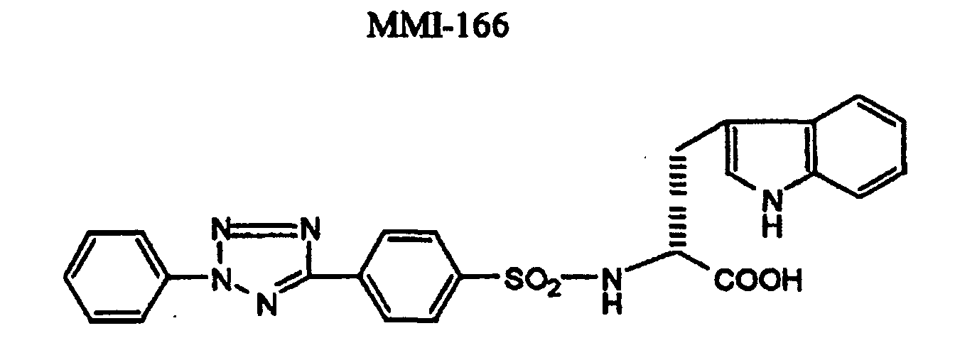
- MMI-166 of the formula was orally administered at dosages of 0.2, 2.0, 20.0, and 200.0 mg/kg in stead of the administrations of BPHA and (-)BPHA, treatments were conducted for in situ zymography.
- FIG. 2 The micrograph under light microscopy and the analysis of the slide scanner regarding gelatin degradation of the cancer tissues in the mouse receiving an vehicle and the mice receiving MMI-166 are shown at the lower lane and the upper lane in Figure 2, respectively.
- (a), (b), (c), (d) and (e) are each results of the vehicle-receiving mouse, the 0.2mg/kg-receiving mouse, the 2.0mg/kg-receiveing mouse, 20.0mg/kg-receiving mouse, and 200.0mg/kg-receiving mouse.
- the gelatin degradation was recognized coincidentally at the cancer area ( Figure 2a).
- the present invention makes in vitro determination of an in vivo activity of MMP inhibitors possible.
- the invention is useful to conduct drug evaluation in clinical test for anti-cancer agents, anti-rheumatic agents, and agents for treating heart failure, as well as elucidation of their action mechanism, specifically to determine the dosage or the administration schedule for the agents.
Landscapes
- Life Sciences & Earth Sciences (AREA)
- Health & Medical Sciences (AREA)
- Chemical & Material Sciences (AREA)
- Organic Chemistry (AREA)
- Proteomics, Peptides & Aminoacids (AREA)
- Zoology (AREA)
- Wood Science & Technology (AREA)
- General Health & Medical Sciences (AREA)
- Engineering & Computer Science (AREA)
- Immunology (AREA)
- Animal Behavior & Ethology (AREA)
- Analytical Chemistry (AREA)
- Physics & Mathematics (AREA)
- Biotechnology (AREA)
- Veterinary Medicine (AREA)
- Microbiology (AREA)
- Molecular Biology (AREA)
- Public Health (AREA)
- Biophysics (AREA)
- Epidemiology (AREA)
- Biochemistry (AREA)
- Bioinformatics & Cheminformatics (AREA)
- General Engineering & Computer Science (AREA)
- Genetics & Genomics (AREA)
- Measuring Or Testing Involving Enzymes Or Micro-Organisms (AREA)
- Investigating Or Analysing Biological Materials (AREA)
- Enzymes And Modification Thereof (AREA)
- Medicines Containing Antibodies Or Antigens For Use As Internal Diagnostic Agents (AREA)
Abstract
Description
- The present invention relates to a method for evaluating a compound exhibiting matrix metalloproteinase-inhibitory activity for an in vivo matrix metalloproteinase-inhibitory activity displayed by the compound, and specifically to a method therefor which comprises utilizing in situ zymography.
- Matrix metalloproteinase (MMP) is a common name of enzymes which are secreted by cancer cells or normal cells surrounding cancers, and degrade extracellular matrix proteins such as collagen in a divalent metal ions-dependent manner. In particular, it has been frequently reported that type IV collagenases (MMP-2, MMP-9) are responsible for vascularization accompanied by tumor proliferation, and for tumor invasion and metastasis.
- As an assay system for measuring matrix metalloproteinase, a gelatin zymography method is known wherein enzyme activities are determined based on degradation degree of a substrate. The method requires conducting electrophoresis, and therefore it is troublesome in the procedures, and time-consuming in the detection.
- As an assay system without electrophoresis, The FASEB Journal, vol.9, July, pp.974-980, 1995 has proposed a determination method based on the principle of zymography, which comprises incorporating, into agarose, casein or gelatin bound to a fluorescent compound useful as a substrate for the protease, forming the mixture into a thin membrane on slide glass, then placing a tissue section on the surface of a thin membrane, incubating the membrane, and observing the digestion of the substrate by a fluorescence microscopy. The system, however, encounters a trouble with decreased reproducibility, since the agarose affects the digestion of the substrate due to the protease.
- Recently, the applicants of the present application proposed a convenient and accurate method for measuring proteases, which comprises contacting a sample containing a protease to a thin membrane formed on the surface of a support, said thin membrane comprising a protease substrate and a hardening agent (WO97/32035), which have received some signs of confidence. For example, Proceedings of 5th Conference of the Japanese Association for Metastasis Research (1996) describes that in situ zymography based on our method was used to detect the gelatinase activity in the tissue and cells, thus observing that the protease destroyed tumor tissues. In situ zymography is described in The FEBS Journal, 974-980, Vol. 9 (1995) or the like.
- Further, H. Nakamura, et al. Cancer Research 59,467-473(1999) describes that in situ zymography similarly revealed that MMP inhibitors inhibited the MMP activity in human thyroid cancer.
- As described above, MMPs, in particular type IV collagenase, are believed to be responsible for vascularization accompanied by tumor proliferation, and for tumor invasion and metastasis. Accordingly, inhibitors of MMPs have been a focus of attention in view of development of anti-cancer agents, and, in fact, some of them are clinically being tested by some pharmaceutical companies.
- Pharmacotherapy is achieved on the prerequisite that a drug used in the therapy is effective and safe, and the prerequisite is confirmed by drug evaluation, which is largely classified into two tests, nonclinical test such as animal experimentation, and clinical test wherein human beings are tested. Even agents that are effective in the nonclinical test, or have an excellent action mechanism would not be drugs when the agents are not effective in patients. This is the reason why the clinical test is required to evaluate the drug efficacy in humans.
- In drug evaluation of MMP inhibitors by the clinical test, it is difficult to determine exactly the in vivo activity of MMP inhibitors because MMP is inherently controlled in vivo by endogenous inhibitors such as TIMP-1, and -2 [Woessner, J.F.Jr., FASEB J. 5, 2145-54 (1991)]. Immunostaining method wherein an antibody specific to MMP is used has been known as a method for detecting MMP (Int. J. Cancer., 56, 500-505 (1994)). Since the antibody used therein, however, recognizes MMP in a manner that MMP is merely an antigen protein, this method dose not allow to determine whether an antigen protein recognized by the antibody possesses the MMP activity or is inactivated by the inhibitors. Conventional gelatin zymography dissociates MMP from inhibitors during the electrophoresis, and therefore the method does not also allow the exact determination of the inhibitory activity of MMP inhibitors.
- In view of the above, the present inventors investigated for a method of checking the in vivo activity of MMP inhibitors, and found that in situ zymography makes it possible to determine conveniently and rapidly the in vivo MMP inhibitory activity of MMP inhibitors.
- In the first aspect, the present invention relates to a method for evaluating a compound exhibiting matrix metalloproteinase-inhibitory activity for an in vivo matrix metalloproteinase-inhibitory activity displayed by the compound, which comprises subjecting to in situ zymography a specimen isolated from a mammal that has received the compound.
- Specifically, the present invention relates to the method wherein the in situ zymography is conducted using a thin membrane which comprises gelatin or collagen together with a hardening agent, and which has a thickness of 1 µm to 20 µm. More specifically, the in situ zymography is conducted by
- (1) contacting the isolated specimen onto the thin membrane comprising a substrate of matrix metalloproteinase;
- (2) incubating the specimen-thin membrane sample;
- (3) staining and then decolorizing the thin membrane; and
- (4) verifying the tone developed on the thin membrane.
-
- In the evaluation method of the present invention, it is preferred that the substrate of matrix metalloproteinase is gelatin or collagen; that the thin membrane comprises a hardening agent; that the thin membrane has a thickness of 1 µm to 20 µm; and/or that the specimen is a tissue section, preferably a lyophilized tissue section, having a thickness of 2 µm to 10 µm, and the incubation is conducted at 20°C to 50°C, preferably 36 to 38°C, for 2 to 18 hours.
- In the second aspect, the present invention relates to a method for evaluating a compound exhibiting matrix metalloproteinase-inhibitory activity for an in vivo matrix metalloproteinase-inhibitory activity displayed by the compound, which comprises:
(1) administering to a mammal a compound exhibiting matrix metalloproteinase-inhibitory activity; (2) isolating a tissue or a fluid from the mammal to prepare a specimen; (3) contacting the isolated specimen onto a thin membrane comprising a substrate of matrix metalloproteinase; (4) incubating the specimen-thin membrane sample; (5) staining and then decolorizing the thin membrane; and (6) verifying the tone developed on the thin membrane. - In the third aspect, the present invention relates to a method for evaluating an anti-cancer agent exhibiting matrix metalloproteinase-inhibitory activity for its anti-cancer activity, which comprises subjecting to in situ zymography a specimen isolated from a mammal that has received the anti-cancer agent, and evaluating for an in vivo matrix metalloproteinase-inhibitory activity displayed by the agent as an indicator of the anti-cancer activity. In the method of the present invention, it is preferred that the substrate of matrix metalloproteinase is gelatin or collagen; that the thin membrane comprises a hardening agent; that the thin membrane has a thickness of 1 µm to 20 µm; and/or that the specimen is a tissue section, preferably a lyophilized tissue section, having a thickness of 2 µm to 10 µm, and the incubation is conducted at 20°C to 50°C, preferably 36 to 38°C, for 2 to 18 hours.
- It has been known that not only MMP but also many enzymes that degrade gelatin, such as trypsin, exist in the living body. For this reason, it was difficult to determine selectively the activity of MMP inhibitors by manes of in situ zymography using substrates of MMP such as gelatin, as used in the present invention. No method for determining an in vivo matrix metalloproteinase-inhibitory activity of MMP inhibitors has been known.
- As used herein, "in situ zymography" means an in vitro method useful for determination of protease activities, which is not involved in electrophoresis. In the present invention, this is conducted by contacting onto a thin membrane comprising a substrate of matrix metalloproteinase an specimen isolated from a mammal receiving a compound exhibiting matrix metalloproteinase-inhibitory activity (MMP inhibitor), and observing the MMP substrate after the lapse of a defined period of time, so that the inhibitor is evaluated for "in vivo MMP inhibitory activity".
- The term "mammal" in the phrase "specimen isolated from a mammal" means a higher vertebrate, preferably human being, which has been administered with a MMP inhibitor. The test object in this method is a specimen isolated from the living body, which provides a result exactly corresponding to an in vivo MMP inhibitory activity of a MMP inhibitor, which the inhibitor exerts in vivo.
- "Specimen isolated" may be derived from various tissues and fluids, and is varied depending on efficacy or indication of MMP inhibitors to be administered into a mammal. When, for example, a MMP inhibitor is for an anti-cancer agent, a specimen is derived from cancerous tissue samples isolated and taken by surgical operation or histological examination from cancerous tissues such as breast cancer, endometrioma, cancer of buccal cavity, lung cancer, gastric cancer, esophageal cancer, epithelial cancer of urinary tract, colon cancer, squamous cancer, thyroid cancer. For an anti-rheumatic agent, synovial membranes or fluids, or bone tissues derived from patients with rheumatoid arthritis or arthritis are used, whereas for an agent for treating heart failure, heart tissues or effusions therefrom derived from patients with heart failure would be used. In this manner, the present invention may be utilized as methods for evaluating anti-cancer agents, anti-rheumatic agents, and agents for treating heart failure.
- Figure 1 is a picture substitute for drawings which shows the micrographs under light microscopy (the upper) of the carcinoma stained with H.E. in the mouse receiving an vehicle (A), the mouse receiving (-)BPHA (B), and the mouse receiving BPHA (C), as well as the in situ zymography therein regarding gelatin degradation (the lower).
- Figure 2 is a picture substitute for drawings which shows the micrographs under light microscopy (the lower) of the carcinoma stained with H.E. in the mouse receiving an vehicle (a), and the mice receiving MMI-166 (b-e), as well as the in situ zymography therein regarding gelatin degradation (the upper).
- "In situ zymography" in the present invention specifically comprises the steps of:
- (1) contacting the isolated specimen onto the thin membrane comprising a substrate of matrix metalloproteinase;
- (2) incubating the specimen-thin membrane sample;
- (3) staining and then decolorizing the thin membrane; and
- (4) verifying the tone developed on the thin membrane.
-
- Each of the steps is further described below.
- "Contacting the specimen onto the file" means that the tissue removed is placed or attached onto the thin membrane, or the fluid taken is dropped onto the thin membrane.
- "Isolated specimen" includes frozen sections having a thickness of 2 µm to 10 µm, preferably 4 µm to 8 µm, which are prepared from samples of tissues quickly frozen by use of for example liquid nitrogen with an apparatus for preparing frozen sections. Alternatively, body fluids such as blood, saliva, synovial fluid, and the like may be used. In case of the use of synovial fluid taken from patients with rheumatoid arthritis as a specimen, about 5 to 50 µl, preferably about 20 µl, of synovial fluid is dropped onto the thin membrane.
- "MMP Substrate" is a macromolecular compound that is degradable by MMP, and includes for example gelatin, collagen, proteoglycan, fibronectin, laminin, elastin, and casein. Preferred one is gelatin, collagen, fibronectin, elastin or casein, and more preferred one is gelatin or collagen. MMP is described for example in Takashi Tsuruo ed., "Molecular Mechanism for cancer metastasis" pp.92-107 (1993) Medical View Publisher, and include, but is not limited to, proteases such as interstitial collagenase (MMP-1), gelatinase A (MMP-2), and gelatinase B (MMP-9).
- "Thin membrane comprising a MMP substrate" means those prepared from a monolayer or a multilayer of the MMP substrate as described above which is formed on the surface of a support. The details are described in WO97/32035. In brief, the thin membrane is a monolayer or a multilayer having a thickness of 6 µm to 20 µm, particularly 8 µm to 10 µm, which layer is on a support, and the thin membrane preferably has the homogeneous surface.
- The thin membrane as used in the method of the present invention preferably comprises a hardening agent. Hardening agent promotes the hardening of the thin membrane of MMP substrate, and/or prevents the formed thin membrane from swelling. The details of the hardening agent are described in WO97/32035. Hardening agents include vinyl sulfonyl type of hardening agents. In case of gelatin for example, it is advantageous to blend 0.1 to 20 mmol, preferably about 0.3 to 10 mmol, of a hardening agent to 100 g of gelatin in view of the detection capacity although amount of hardening agents is not particularly limited.
- In addition to the agents as shown above, other components such as dyes, pigments, preservatives, stabilizers or the like can be comprised as appropriate to prepare the thin membranes.
- "Support" is preferably transparent or semitransparent so as to allow the observation of the presence of MMP in tissues under a microscopy. Those transparent or semitransparent supports include a glass, and a transparent or semitransparent plastic film composed of polyethylene terephthalate or the like. Although the thickness of the support is not particularly limited, a glass support having a thickness similar to slide glass (for example about 2 to 3 mm) is preferred, whereas a polyethylene terephthalate support having a thickness of about 100 to 250 µm may be used.
- In order to prepare "thin membrane and support", a MMP substrate dispersed in water or an organic solvent such as acetone, methanol, ethanol or a mixture thereof for example is spread on the surface of a support, and then dried. As procedures for the spreading, dip coating method, roller coating method, curtain coating method, extrusion coating method, and the like, all of which are known in the art can be used. In case that gelatin is used as a MMP substrate, gelatin is not limited to any particular species, and for example an alkali-treated bovine bone gelatin, an acid-treated swine cutis gelatin, or the like may be used.
- "Specimen-thin membrane sample" is one obtained after attaching the tissue section onto the thin membrane, or dropping the liquid specimen onto the thin membrane, so as to contact a MMP-containing specimen to the thin membrane.
- "Incubating" means that a sample is left stand at 20°C to 50°C, preferably 36°C to 38°C in a thermostat bath, and the incubation is preferably conducted in a humid bath in order to prevent the thin membrane from dryness. For example, tissue sections are incubated at 37°C for 1 to 24 hours, preferably 2 to 18 hours, more preferably about 4 to 8 hours, whereas liquid samples are incubated for 0.5 to 12 hours, preferably 1 to 6 hours.
- When a specimen contains any MMP that is even inactivated by MMP inhibitors, the protease degrades the MMP substrate in the thin membrane to form the trace of digestion thereon.
- The specimen-thin membrane sample obtained in step (2) is stained preferably with a stain solution. Stain solutions are those that stain the enzyme substrate, and include Amido Black, Coomassie Blue, Ponceau (Wako Pure Chemical Industries), Biebrich Scarlet (Aldrich). It is preferred to use a 0.5 to 2% solution of Amido Black.
- Subsequently, the thin membrane stained is decolorized with an appropriate decolorizing agent, for example, a 70% methanol-10% acetic acid solution, and water.
- The thin membrane treated by the steps as shown above will develop stain tones of dark and light colorings (gradient) depending on the range of MMP substrate degradation, since an area where the MMP substrate is degraded should not be stained. Such tone is verified by the observation with naked eye or under a microscope, or by quantitative analysis using a slide scanner, a laser microscope, or the like. The fact that the area is stained shows that MMP is therein being inactivated by an action of a MMP inhibitor administered, or for other reasons. Determination whether the inactivation is caused by the MMP inhibitory action or by other reasons may be readily conducted by comparing it with a control wherein no MMP inhibitor is administered.
- The present invention is directed to a method for evaluating a MMP inhibitor for an in vivo MMP-inhibitory activity displayed by the compound, and encompasses a method of the present invention which comprises as the first step administering a MMP inhibitor to a mammal. This makes it possible to evaluate an in vivo activity of a MMP inhibitor at the time when the inhibitor is administered to a patient during clinical test. The next steps are usually conducted setting a time interval of 30 minutes to four hours after the administration of a MMP inhibitor.
- The present invention further encompasses a method for evaluating an anti-cancer agent exhibiting matrix metalloproteinase-inhibitory activity for its anti-cancer activity, which comprises subjecting to in situ zymography a specimen isolated from a mammal that has received the anti-cancer agent, and evaluating for an in vivo matrix metalloproteinase-inhibitory activity displayed by the agent as an indicator of the anti-cancer activity. Drug evaluation for anti-cancer agents in the clinical test has been conducted mainly based on a patient's life-extending effectiveness or subjective symptoms of patients, the presence or absence of tumor regression/metastasis, amounts of tumor markers, or the like. However, it is difficult that these indexes show whether the transition of drugs to the lesion is good or bad, and also provide fine selection of the dosage of drugs. On the other hand, the evaluation method for anti-cancer activity of the present invention, in the clinical test, enables to determine the dosage of agents or the schedule for administration, determine if the agent reaches cancer tissue, and provides a useful means to select types of cancers to be treated.
- Such method of this aspect of the present invention is also conducted by each of the steps as described above.
- According to the methods of the present invention, it can be checked if a cancer to be tested is dependent on MMP, thus providing a more appropriate method for the treatment for cancers.
- The present invention is further illustrated by the following examples, but is not restricted by these examples in any way.
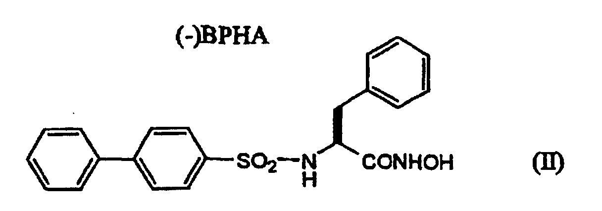
- In this test, compound BPHA of formula (I) exhibiting the MMP inhibitory activity, and compound (-)BPHA of formula (II) exhibiting no MMP inhibitory activity were used as test compounds for MMP inhibitors.

IC50 (nM) Enzymes BPHA (-)BPHA MMP-1 974 >1000 MMP-2 12 >1000 MMP-3 >1000 >1000 MMP-7 795 >1000 MMP-9 16 >1000 MMP-14 17 >1000 - Fifteen g of acid-treated swine cutis gelatin was dissolved in 123 g of pure water, and the solution was added with 1.2 ml of 1,2-bis(vinylsulfonylacetamido)ethane (4%) as a hardening agent. The solution was uniformly applied to a polyethylene terephthalate film undercoated with gelatin so as to obtain a dried membrane having a thickness of about 7 µm, and the membrane was dried to obtain a gelatin thin membrane. The gelatin thin membrane was stored at room temperature until the use.
- Human lung cancer cell line Ma44 (CANCER RESEARCH 59, 1231-35 (1999)) that was known to produce MMP-2 in a large amount was used. Nude mice (six weeks aged, female, CLEA Japan, Inc.) were transplanted intraperitoneally with 1x10⁵ Ma44 cells, and, 14 days later, BPHA or (-)BPHA was administered orally to the animals at 200mg/kg. As control, a vehicle (a physiological saline containing 0.4% Tween80, 0.5% carboxymethyl cellulose, and 0.9% benzyl alcohol) was similarly administered. Two hours after the administration, the cancer-bearing mice were autopsied to remove the tumor mass formed at the abdominal wall and the mesenterium, and the mass was subjected to the in situ zymography experiment.
- The tumor mass was embedded in OCT Compound (SAKURA), and then Microtone/Cryostat TISSUE TEK II (Miles-Sankyo) was used to prepare lyophilized sections having a thickness of 8 µm. The lyophilized sections were placed gently onto the gelatin thin membrane previously prepared to give samples, and then the membranes were placed into a humid bath to prevent them from drying, followed by leaving the membranes to stand in an incubator at 37°C. After reacted for 3.5 hours, the thin membranes were taken out, and stained with a 1% Amido Black solution (Wako Pure Chemical Industries) for 15 minutes. Subsequently, the membranes were treated with a decolorizing solution (70% methanol, 10% acetic acid) for 10 minutes.
- The cancer tissue on the thin membrane was observed under light microscopy to determine the area where the gelatin degradation was recognized. Additionally, the slide scanner (Nikon 35 mm film scanner: COOLSCAN II) was used to analyze quantitatively the tone with the software Optimas Ver.6 (Optimas Corporation).
- The micrographs under light microscopy of the cancer tissues stained with H.E. (hematoxylin and eosin) in the mouse receiving an vehicle, the mouse receiving (-)BPHA, and the mouse receiving BPHA, are shown at (A), (B), and (C) on the upper lane in Figure 1, respectively. The results show that the tissues observed contain the cancer cells.
- Additionally, the results obtained in the observation by the slide scanner are also shown at (A), (B), and (C) on the lower lane in Figure 1. These show that the gelatin degradation was observed in the mouse receiving an vehicle and the mouse receiving (-)BPHA (the compound without MMP inhibitory activity), whereas little gelatin degradation was observed in the mouse receiving BPHA. Quantitative analysis using the software Optimas Ver.6 (brightness in each tissue/size in each tissue) was estimated at 49.2, 40.1, and 8.6 in the mouse receiving an vehicle, the mouse receiving (-)BPHA, and the mouse receiving BPHA, respectively.
- The results are not inconsistent with those describing that BPHA exhibits the anti-cancer activity, whereas (-)BPHA does not in an animal experimentation (CANCER RESEARCH 59, 1231-35 (1999)).
- According to a similar manner to that of Example 1 except that MMI-166 of the formula:was orally administered at dosages of 0.2, 2.0, 20.0, and 200.0 mg/kg in stead of the administrations of BPHA and (-)BPHA, treatments were conducted for in situ zymography.
- The micrograph under light microscopy and the analysis of the slide scanner regarding gelatin degradation of the cancer tissues in the mouse receiving an vehicle and the mice receiving MMI-166 are shown at the lower lane and the upper lane in Figure 2, respectively. In Figure 2, (a), (b), (c), (d) and (e) are each results of the vehicle-receiving mouse, the 0.2mg/kg-receiving mouse, the 2.0mg/kg-receiveing mouse, 20.0mg/kg-receiving mouse, and 200.0mg/kg-receiving mouse. In the cancer tissue of the vehicle-receiving mouse, the gelatin degradation was recognized coincidentally at the cancer area (Figure 2a). On the other hand, the results in the MMI-166-receiving mice were that the complete inhibition of the gelatin degradation was provided at the highest dosage 200.0 mg/kg, that the inhibition was lowered in sequence of 20.0 to 2.0 mg/kg, and that the merely partial inhibition was recognized at the dosage of 0.2 mg/kg (Figure 2e-b). According to these results, it was found that the inhibition of the gelatin-degrading activity observed at the cancer tissue was dependent on the MMI-166 dosage. Quantitative analysis using the software Optimas Ver.6 was also estimated at 53.4, 41.3, 28.6, 13.7, and 5.3, respectively.
- The results as shown above suggest that the method of the present invention is useful to determine the dosage or the schedule for administration of compounds exhibiting MMP inhibitory activity required to inhibit actually gelatin degradation activity recognized at the cancer area.
- The present invention makes in vitro determination of an in vivo activity of MMP inhibitors possible. The invention is useful to conduct drug evaluation in clinical test for anti-cancer agents, anti-rheumatic agents, and agents for treating heart failure, as well as elucidation of their action mechanism, specifically to determine the dosage or the administration schedule for the agents.
Claims (9)
- A method for evaluating a compound exhibiting matrix metalloproteinase-inhibitory activity for an in vivo matrix metalloproteinase-inhibitory activity displayed by the compound, which comprises subjecting to in situ zymography a specimen isolated from a mammal that has received the compound.
- The method of claim 1, wherein the in situ zymography is conducted using a thin membrane which comprises gelatin or collagen together with a hardening agent, and which has a thickness of 1 µm to 20 µm.
- The method of claim 1, the in situ zymography is conducted by(1) contacting the isolated specimen onto the thin membrane comprising a substrate of matrix metalloproteinase;(2) incubating the specimen-thin membrane sample;(3) staining and then decolorizing the thin membrane; and(4) verifying the tone developed on the thin membrane.
- The method of claim 3, wherein the substrate of matrix metalloproteinase is gelatin or collagen.
- The method of claim 3 or 4, wherein the thin membrane comprises a hardening agent.
- The method of any one of claims 3 to 5, wherein the thin membrane has a thickness of 1 µm to 20 µm.
- The method of any one of claims 3 to 6, wherein the specimen is a tissue section having a thickness of 2 µm to 10 µm, and the incubation is conducted at 20°C to 50°C for 2 to 18 hours.
- A method for evaluating a compound exhibiting matrix metalloproteinase-inhibitory activity for an in vivo matrix metalloproteinase-inhibitory activity displayed by the compound, which comprises;(1) administering to a mammal a compound exhibiting matrix metalloproteinase-inhibitory activity;(2) isolating a tissue or a fluid from the mammal to prepare a specimen;(3) contacting the isolated specimen onto a thin membrane comprising a substrate of matrix metalloproteinase;(4) incubating the specimen-thin membrane sample;(5) staining and then decolorizing the thin membrane; and(6) verifying the tone developed on the thin membrane.
- A method for evaluating an anti-cancer agent exhibiting matrix metalloproteinase-inhibitory activity for its anti-cancer activity, which comprises subjecting to in situ zymography a specimen isolated from a mammal that has received the anti-cancer agent, and evaluating for an in vivo matrix metalloproteinase-inhibitory activity displayed by the agent as an indicator of the anti-cancer activity.
Applications Claiming Priority (5)
| Application Number | Priority Date | Filing Date | Title |
|---|---|---|---|
| JP14330899 | 1999-05-24 | ||
| JP14330899 | 1999-05-24 | ||
| JP21214599 | 1999-07-27 | ||
| JP21214599 | 1999-07-27 | ||
| PCT/JP2000/003270 WO2000071167A1 (en) | 1999-05-24 | 2000-05-23 | Method for evaluating matrix metalloprotease inhibitory activity |
Publications (4)
| Publication Number | Publication Date |
|---|---|
| EP1181939A1 true EP1181939A1 (en) | 2002-02-27 |
| EP1181939A9 EP1181939A9 (en) | 2002-04-24 |
| EP1181939A4 EP1181939A4 (en) | 2004-09-27 |
| EP1181939B1 EP1181939B1 (en) | 2006-05-31 |
Family
ID=26475073
Family Applications (1)
| Application Number | Title | Priority Date | Filing Date |
|---|---|---|---|
| EP00929826A Expired - Lifetime EP1181939B1 (en) | 1999-05-24 | 2000-05-23 | Method for evaluating in vivo matrix metalloproteinase inhibitory activity |
Country Status (9)
| Country | Link |
|---|---|
| US (1) | US6627411B2 (en) |
| EP (1) | EP1181939B1 (en) |
| JP (1) | JP4070409B2 (en) |
| AT (1) | ATE328111T1 (en) |
| AU (1) | AU4779800A (en) |
| DE (1) | DE60028372T2 (en) |
| ES (1) | ES2265345T3 (en) |
| TW (1) | TWI223599B (en) |
| WO (1) | WO2000071167A1 (en) |
Cited By (2)
| Publication number | Priority date | Publication date | Assignee | Title |
|---|---|---|---|---|
| EP1292700A4 (en) * | 2000-06-21 | 2005-07-13 | Novapharm Res Australia | Enzyme detection and measurement |
| WO2006010529A1 (en) * | 2004-07-28 | 2006-02-02 | F. Hoffmann-La Roche Ag | Timp-2 as target/marker of beta cell failure |
Families Citing this family (2)
| Publication number | Priority date | Publication date | Assignee | Title |
|---|---|---|---|---|
| JP2001054399A (en) * | 1999-08-18 | 2001-02-27 | Fuji Photo Film Co Ltd | Judgement of effectiveness of drug having protease inhibiting action |
| US20040225077A1 (en) | 2002-12-30 | 2004-11-11 | Angiotech International Ag | Drug delivery from rapid gelling polymer composition |
Family Cites Families (5)
| Publication number | Priority date | Publication date | Assignee | Title |
|---|---|---|---|---|
| WO1993023075A1 (en) * | 1992-05-14 | 1993-11-25 | Oncologix, Inc. | Treatment of vascular leakage syndrome and collagenase induced disease by administration of matrix metalloproteinase inhibitors |
| WO1994005807A1 (en) * | 1992-08-28 | 1994-03-17 | Dickson Robert B | Matrix-degrading metalloproteinase |
| DE69738933D1 (en) | 1996-02-29 | 2008-10-02 | Fujifilm Corp | Method of analyzing proteases and thin membrane for use in this method |
| JPH1045632A (en) | 1996-07-26 | 1998-02-17 | Meiji Milk Prod Co Ltd | Method for searching for peptides derived from proteins capable of inducing an immune response to the intestinal immune system |
| JPH11127896A (en) | 1997-10-24 | 1999-05-18 | Fuji Photo Film Co Ltd | Dry type multilayer analytic element |
-
2000
- 2000-05-23 DE DE60028372T patent/DE60028372T2/en not_active Expired - Lifetime
- 2000-05-23 ES ES00929826T patent/ES2265345T3/en not_active Expired - Lifetime
- 2000-05-23 TW TW089109933A patent/TWI223599B/en not_active IP Right Cessation
- 2000-05-23 AU AU47798/00A patent/AU4779800A/en not_active Abandoned
- 2000-05-23 WO PCT/JP2000/003270 patent/WO2000071167A1/en not_active Ceased
- 2000-05-23 JP JP2000619468A patent/JP4070409B2/en not_active Expired - Fee Related
- 2000-05-23 AT AT00929826T patent/ATE328111T1/en not_active IP Right Cessation
- 2000-05-23 US US09/979,510 patent/US6627411B2/en not_active Expired - Lifetime
- 2000-05-23 EP EP00929826A patent/EP1181939B1/en not_active Expired - Lifetime
Non-Patent Citations (2)
| Title |
|---|
| No further relevant documents disclosed * |
| See also references of WO0071167A1 * |
Cited By (3)
| Publication number | Priority date | Publication date | Assignee | Title |
|---|---|---|---|---|
| EP1292700A4 (en) * | 2000-06-21 | 2005-07-13 | Novapharm Res Australia | Enzyme detection and measurement |
| WO2006010529A1 (en) * | 2004-07-28 | 2006-02-02 | F. Hoffmann-La Roche Ag | Timp-2 as target/marker of beta cell failure |
| JP2008508502A (en) * | 2004-07-28 | 2008-03-21 | エフ.ホフマン−ラ ロシュ アーゲー | TIMP-2 as a target / marker of beta cell failure |
Also Published As
| Publication number | Publication date |
|---|---|
| TWI223599B (en) | 2004-11-11 |
| ES2265345T3 (en) | 2007-02-16 |
| EP1181939A9 (en) | 2002-04-24 |
| US6627411B2 (en) | 2003-09-30 |
| WO2000071167A1 (en) | 2000-11-30 |
| DE60028372T2 (en) | 2007-05-03 |
| DE60028372D1 (en) | 2006-07-06 |
| US20030157585A1 (en) | 2003-08-21 |
| EP1181939B1 (en) | 2006-05-31 |
| JP4070409B2 (en) | 2008-04-02 |
| ATE328111T1 (en) | 2006-06-15 |
| AU4779800A (en) | 2000-12-12 |
| EP1181939A4 (en) | 2004-09-27 |
Similar Documents
| Publication | Publication Date | Title |
|---|---|---|
| JP7395630B6 (en) | modified wound dressing | |
| JP2015057599A (en) | Identification of tissue for debridement | |
| US6627411B2 (en) | Method for evaluating matrix metalloprotease inhibitory activity | |
| Yuan et al. | A targeted activatable NIR-II nanoprobe for positive visualization of anastomotic thrombosis and sensitive identification of fresh fibrinolytic thrombus | |
| Simpson et al. | Raised levels of plasma big endothelin 1 in patients with colorectal cancer | |
| JP3302020B2 (en) | Method for measuring protease and thin film used in the method | |
| Zhang et al. | Neutrophil elastase specific fluorescent probe for early diagnosis of thyroiditis via serum sample testing and fluorescence imaging | |
| JPWO2000071167A1 (en) | Method for evaluating matrix metalloproteinase inhibitory activity | |
| JP2007509610A (en) | Non-invasive system for detecting skin cancer | |
| AU2002303908B2 (en) | Novel method and composition for inhibition of angiogenesis and tumor growth using compounds based on a sequence within MMP-2 | |
| US6413734B1 (en) | Method for judging effectiveness of drug having protease inhibitory activity | |
| JP2003533699A (en) | Proteolytic enzymes in the diagnosis of kidney damage | |
| Wollina et al. | Serum matrix metalloproteinase-2 in patients with malignant melanoma | |
| Griesbacher et al. | Involvement of tissue kallikrein but not plasma kallikrein in the development of symptoms mediated by endogenous kinins in acute pancreatitis in rats | |
| RU2605838C1 (en) | Method for prediction of risk of biochemical recurrent in prostate cancer patients after radiation and hormone therapy | |
| US20050064521A1 (en) | In vitro assay for evaluation of angiogenic effects | |
| EP1035213A1 (en) | Method for enzyme assay | |
| Vaganov et al. | Application of Intraoperative Fluorescence Spectroscopy in Classical Abdominoplasty in Patients with Diabetes Mellitus | |
| RU2612082C1 (en) | Method for prediction of pathological process direction in case of pancreas head cancer | |
| Yang et al. | Bioorthogonal Fluorogenic Reporters for Noninvasive Imaging and Urinalysis of Immunotherapeutic Response in Renal Cell Carcinoma | |
| TW201741331A (en) | Detecting microbial infection in wounds | |
| JP2001095599A (en) | Measurement of protease | |
| US20050136504A1 (en) | Method of measurement of protease and thin membranes used for said method | |
| Karl et al. | 892: Positve Cytology, but Negative White Light Endoscopy: An Indication for Fluorescence Cystoscopy in Bladder Cancer? | |
| Tokuda | Regulation of melanoma cell survival and function by matricellular signaling and microenvironmental factors |
Legal Events
| Date | Code | Title | Description |
|---|---|---|---|
| PUAI | Public reference made under article 153(3) epc to a published international application that has entered the european phase |
Free format text: ORIGINAL CODE: 0009012 |
|
| 17P | Request for examination filed |
Effective date: 20011129 |
|
| AK | Designated contracting states |
Kind code of ref document: A1 Designated state(s): AT BE CH CY DE DK ES FI FR GB GR IE IT LI LU MC NL PT SE |
|
| AX | Request for extension of the european patent |
Free format text: AL;LT;LV;MK;RO;SI |
|
| A4 | Supplementary search report drawn up and despatched |
Effective date: 20040927 |
|
| RIC1 | Information provided on ipc code assigned before grant |
Ipc: 7C 12Q 1/37 A Ipc: 7G 01N 33/50 B |
|
| 17Q | First examination report despatched |
Effective date: 20050308 |
|
| GRAP | Despatch of communication of intention to grant a patent |
Free format text: ORIGINAL CODE: EPIDOSNIGR1 |
|
| RTI1 | Title (correction) |
Free format text: METHOD FOR EVALUATING IN VIVO MATRIX METALLOPROTEINASE INHIBITORY ACTIVITY |
|
| GRAS | Grant fee paid |
Free format text: ORIGINAL CODE: EPIDOSNIGR3 |
|
| GRAA | (expected) grant |
Free format text: ORIGINAL CODE: 0009210 |
|
| RAP1 | Party data changed (applicant data changed or rights of an application transferred) |
Owner name: FUJI PHOTO FILM CO., LTD. Owner name: SHIONOGI & CO., LTD. |
|
| AK | Designated contracting states |
Kind code of ref document: B1 Designated state(s): AT BE CH CY DE DK ES FI FR GB GR IE IT LI LU MC NL PT SE |
|
| PG25 | Lapsed in a contracting state [announced via postgrant information from national office to epo] |
Ref country code: BE Free format text: LAPSE BECAUSE OF FAILURE TO SUBMIT A TRANSLATION OF THE DESCRIPTION OR TO PAY THE FEE WITHIN THE PRESCRIBED TIME-LIMIT Effective date: 20060531 Ref country code: FI Free format text: LAPSE BECAUSE OF FAILURE TO SUBMIT A TRANSLATION OF THE DESCRIPTION OR TO PAY THE FEE WITHIN THE PRESCRIBED TIME-LIMIT Effective date: 20060531 Ref country code: CH Free format text: LAPSE BECAUSE OF FAILURE TO SUBMIT A TRANSLATION OF THE DESCRIPTION OR TO PAY THE FEE WITHIN THE PRESCRIBED TIME-LIMIT Effective date: 20060531 Ref country code: AT Free format text: LAPSE BECAUSE OF FAILURE TO SUBMIT A TRANSLATION OF THE DESCRIPTION OR TO PAY THE FEE WITHIN THE PRESCRIBED TIME-LIMIT Effective date: 20060531 Ref country code: LI Free format text: LAPSE BECAUSE OF FAILURE TO SUBMIT A TRANSLATION OF THE DESCRIPTION OR TO PAY THE FEE WITHIN THE PRESCRIBED TIME-LIMIT Effective date: 20060531 Ref country code: NL Free format text: LAPSE BECAUSE OF FAILURE TO SUBMIT A TRANSLATION OF THE DESCRIPTION OR TO PAY THE FEE WITHIN THE PRESCRIBED TIME-LIMIT Effective date: 20060531 |
|
| REG | Reference to a national code |
Ref country code: GB Ref legal event code: FG4D Ref country code: CH Ref legal event code: EP |
|
| REG | Reference to a national code |
Ref country code: IE Ref legal event code: FG4D |
|
| REF | Corresponds to: |
Ref document number: 60028372 Country of ref document: DE Date of ref document: 20060706 Kind code of ref document: P |
|
| PG25 | Lapsed in a contracting state [announced via postgrant information from national office to epo] |
Ref country code: SE Free format text: LAPSE BECAUSE OF FAILURE TO SUBMIT A TRANSLATION OF THE DESCRIPTION OR TO PAY THE FEE WITHIN THE PRESCRIBED TIME-LIMIT Effective date: 20060831 Ref country code: DK Free format text: LAPSE BECAUSE OF FAILURE TO SUBMIT A TRANSLATION OF THE DESCRIPTION OR TO PAY THE FEE WITHIN THE PRESCRIBED TIME-LIMIT Effective date: 20060831 |
|
| PG25 | Lapsed in a contracting state [announced via postgrant information from national office to epo] |
Ref country code: PT Free format text: LAPSE BECAUSE OF FAILURE TO SUBMIT A TRANSLATION OF THE DESCRIPTION OR TO PAY THE FEE WITHIN THE PRESCRIBED TIME-LIMIT Effective date: 20061031 |
|
| NLV1 | Nl: lapsed or annulled due to failure to fulfill the requirements of art. 29p and 29m of the patents act | ||
| ET | Fr: translation filed | ||
| REG | Reference to a national code |
Ref country code: CH Ref legal event code: PL |
|
| REG | Reference to a national code |
Ref country code: ES Ref legal event code: FG2A Ref document number: 2265345 Country of ref document: ES Kind code of ref document: T3 |
|
| RAP2 | Party data changed (patent owner data changed or rights of a patent transferred) |
Owner name: FUJIFILM CORPORATION Owner name: SHIONOGI & CO., LTD. |
|
| PLBE | No opposition filed within time limit |
Free format text: ORIGINAL CODE: 0009261 |
|
| STAA | Information on the status of an ep patent application or granted ep patent |
Free format text: STATUS: NO OPPOSITION FILED WITHIN TIME LIMIT |
|
| 26N | No opposition filed |
Effective date: 20070301 |
|
| REG | Reference to a national code |
Ref country code: FR Ref legal event code: TQ |
|
| PG25 | Lapsed in a contracting state [announced via postgrant information from national office to epo] |
Ref country code: MC Free format text: LAPSE BECAUSE OF NON-PAYMENT OF DUE FEES Effective date: 20070531 |
|
| PG25 | Lapsed in a contracting state [announced via postgrant information from national office to epo] |
Ref country code: GR Free format text: LAPSE BECAUSE OF FAILURE TO SUBMIT A TRANSLATION OF THE DESCRIPTION OR TO PAY THE FEE WITHIN THE PRESCRIBED TIME-LIMIT Effective date: 20060901 |
|
| PG25 | Lapsed in a contracting state [announced via postgrant information from national office to epo] |
Ref country code: IE Free format text: LAPSE BECAUSE OF NON-PAYMENT OF DUE FEES Effective date: 20070523 |
|
| PG25 | Lapsed in a contracting state [announced via postgrant information from national office to epo] |
Ref country code: CY Free format text: LAPSE BECAUSE OF FAILURE TO SUBMIT A TRANSLATION OF THE DESCRIPTION OR TO PAY THE FEE WITHIN THE PRESCRIBED TIME-LIMIT Effective date: 20060531 Ref country code: LU Free format text: LAPSE BECAUSE OF NON-PAYMENT OF DUE FEES Effective date: 20070523 |
|
| REG | Reference to a national code |
Ref country code: DE Ref legal event code: R082 Ref document number: 60028372 Country of ref document: DE Representative=s name: VOSSIUS & PARTNER, DE |
|
| REG | Reference to a national code |
Ref country code: DE Ref legal event code: R081 Ref document number: 60028372 Country of ref document: DE Owner name: FUJIFILM CORPORATION, JP Free format text: FORMER OWNER: SHIONOGI & CO., LTD., FUJI PHOTO FILM CO., LTD., , JP Effective date: 20120328 Ref country code: DE Ref legal event code: R082 Ref document number: 60028372 Country of ref document: DE Representative=s name: VOSSIUS & PARTNER PATENTANWAELTE RECHTSANWAELT, DE Effective date: 20120328 Ref country code: DE Ref legal event code: R081 Ref document number: 60028372 Country of ref document: DE Owner name: FUJIFILM CORPORATION, TOKIO/TOKYO, JP Free format text: FORMER OWNER: SHIONOGI & CO., LTD., FUJI PHOTO FILM CO., LTD., , JP Effective date: 20120328 Ref country code: DE Ref legal event code: R081 Ref document number: 60028372 Country of ref document: DE Owner name: FUJIFILM CORPORATION, TOKIO/TOKYO, JP Free format text: FORMER OWNERS: SHIONOGI & CO., LTD., OSAKA, JP; FUJI PHOTO FILM CO., LTD., MINAMI-ASHIGARA, KANAGAWA, JP Effective date: 20120328 |
|
| REG | Reference to a national code |
Ref country code: FR Ref legal event code: TP Owner name: FUJIFILM CORPORATION, JP Effective date: 20120417 |
|
| REG | Reference to a national code |
Ref country code: ES Ref legal event code: PC2A Owner name: FUJIFILM CORPORATION Effective date: 20120516 |
|
| REG | Reference to a national code |
Ref country code: GB Ref legal event code: 732E Free format text: REGISTERED BETWEEN 20120607 AND 20120613 |
|
| REG | Reference to a national code |
Ref country code: FR Ref legal event code: PLFP Year of fee payment: 17 |
|
| REG | Reference to a national code |
Ref country code: FR Ref legal event code: PLFP Year of fee payment: 18 |
|
| REG | Reference to a national code |
Ref country code: FR Ref legal event code: PLFP Year of fee payment: 19 |
|
| PGFP | Annual fee paid to national office [announced via postgrant information from national office to epo] |
Ref country code: DE Payment date: 20190508 Year of fee payment: 20 Ref country code: IT Payment date: 20190527 Year of fee payment: 20 Ref country code: ES Payment date: 20190604 Year of fee payment: 20 |
|
| PGFP | Annual fee paid to national office [announced via postgrant information from national office to epo] |
Ref country code: FR Payment date: 20190410 Year of fee payment: 20 |
|
| PGFP | Annual fee paid to national office [announced via postgrant information from national office to epo] |
Ref country code: GB Payment date: 20190522 Year of fee payment: 20 |
|
| REG | Reference to a national code |
Ref country code: DE Ref legal event code: R071 Ref document number: 60028372 Country of ref document: DE |
|
| REG | Reference to a national code |
Ref country code: GB Ref legal event code: PE20 Expiry date: 20200522 |
|
| PG25 | Lapsed in a contracting state [announced via postgrant information from national office to epo] |
Ref country code: GB Free format text: LAPSE BECAUSE OF EXPIRATION OF PROTECTION Effective date: 20200522 |
|
| REG | Reference to a national code |
Ref country code: ES Ref legal event code: FD2A Effective date: 20200902 |
|
| PG25 | Lapsed in a contracting state [announced via postgrant information from national office to epo] |
Ref country code: ES Free format text: LAPSE BECAUSE OF EXPIRATION OF PROTECTION Effective date: 20200524 |