EP1180679B1 - A method for sensing fluorescent samples utilizing moment analysis - Google Patents
A method for sensing fluorescent samples utilizing moment analysis Download PDFInfo
- Publication number
- EP1180679B1 EP1180679B1 EP00117522A EP00117522A EP1180679B1 EP 1180679 B1 EP1180679 B1 EP 1180679B1 EP 00117522 A EP00117522 A EP 00117522A EP 00117522 A EP00117522 A EP 00117522A EP 1180679 B1 EP1180679 B1 EP 1180679B1
- Authority
- EP
- European Patent Office
- Prior art keywords
- cell
- soluble
- medium
- cells
- solid support
- Prior art date
- Legal status (The legal status is an assumption and is not a legal conclusion. Google has not performed a legal analysis and makes no representation as to the accuracy of the status listed.)
- Expired - Lifetime
Links
Images


Classifications
-
- G—PHYSICS
- G01—MEASURING; TESTING
- G01N—INVESTIGATING OR ANALYSING MATERIALS BY DETERMINING THEIR CHEMICAL OR PHYSICAL PROPERTIES
- G01N21/00—Investigating or analysing materials by the use of optical means, i.e. using sub-millimetre waves, infrared, visible or ultraviolet light
- G01N21/62—Systems in which the material investigated is excited whereby it emits light or causes a change in wavelength of the incident light
- G01N21/63—Systems in which the material investigated is excited whereby it emits light or causes a change in wavelength of the incident light optically excited
- G01N21/64—Fluorescence; Phosphorescence
- G01N21/645—Specially adapted constructive features of fluorimeters
- G01N21/6456—Spatial resolved fluorescence measurements; Imaging
-
- G—PHYSICS
- G01—MEASURING; TESTING
- G01N—INVESTIGATING OR ANALYSING MATERIALS BY DETERMINING THEIR CHEMICAL OR PHYSICAL PROPERTIES
- G01N21/00—Investigating or analysing materials by the use of optical means, i.e. using sub-millimetre waves, infrared, visible or ultraviolet light
- G01N21/62—Systems in which the material investigated is excited whereby it emits light or causes a change in wavelength of the incident light
- G01N21/63—Systems in which the material investigated is excited whereby it emits light or causes a change in wavelength of the incident light optically excited
- G01N21/64—Fluorescence; Phosphorescence
- G01N21/6428—Measuring fluorescence of fluorescent products of reactions or of fluorochrome labelled reactive substances, e.g. measuring quenching effects, using measuring "optrodes"
-
- G—PHYSICS
- G01—MEASURING; TESTING
- G01N—INVESTIGATING OR ANALYSING MATERIALS BY DETERMINING THEIR CHEMICAL OR PHYSICAL PROPERTIES
- G01N21/00—Investigating or analysing materials by the use of optical means, i.e. using sub-millimetre waves, infrared, visible or ultraviolet light
- G01N21/62—Systems in which the material investigated is excited whereby it emits light or causes a change in wavelength of the incident light
- G01N21/63—Systems in which the material investigated is excited whereby it emits light or causes a change in wavelength of the incident light optically excited
- G01N21/64—Fluorescence; Phosphorescence
- G01N21/6445—Measuring fluorescence polarisation
-
- G—PHYSICS
- G01—MEASURING; TESTING
- G01N—INVESTIGATING OR ANALYSING MATERIALS BY DETERMINING THEIR CHEMICAL OR PHYSICAL PROPERTIES
- G01N21/00—Investigating or analysing materials by the use of optical means, i.e. using sub-millimetre waves, infrared, visible or ultraviolet light
- G01N21/17—Systems in which incident light is modified in accordance with the properties of the material investigated
- G01N2021/178—Methods for obtaining spatial resolution of the property being measured
- G01N2021/1782—In-depth resolution
-
- G—PHYSICS
- G01—MEASURING; TESTING
- G01N—INVESTIGATING OR ANALYSING MATERIALS BY DETERMINING THEIR CHEMICAL OR PHYSICAL PROPERTIES
- G01N21/00—Investigating or analysing materials by the use of optical means, i.e. using sub-millimetre waves, infrared, visible or ultraviolet light
- G01N21/17—Systems in which incident light is modified in accordance with the properties of the material investigated
- G01N2021/178—Methods for obtaining spatial resolution of the property being measured
- G01N2021/1785—Three dimensional
Definitions
- G protein-coupled receptors belong to a large superfamily of multi-spanning membrane proteins which probably represent the single most important class of drug targets.
- GPCR's G protein-coupled receptors
- progress in the development of non-radioactive, homogeneous, miniaturized assays for GPCR's has so far been slow.
- genome sequencing is continually leading to the identification of new GPCR targets ( Uddhav, K. et al., Mol. Biol. Rep. 25: 27 - 43, 1998 ; Stadel et al.., Trends Pharmacol. Sci. 18: 430 - 437, 1997 ).
- Uddhav, K. et al., Mol. Biol. Rep. 25: 27 - 43, 1998 ; Stadel et al.., Trends Pharmacol. Sci. 18: 430 - 437, 1997 the development of suitable assays is often a critical factor in competitiveness for pharmaceutical companies.
- EP-A-0 601 714 describes a method and device for determining the location of a molecule group and the number of fluorescent molecules in the molecule group.
- the present invention relates to a method for sensing fluorescent samples of cells, or soluble or solid supports in a medium, said method comprising the steps of:
- the medium is a liquid or gel. It preferably comprises fluorescent ligands capable of binding to said cells, or soluble or solid supports and is calculated from cumulants where c ligand denotes the mean number of ligands per sensed volume, and q ligand denotes the mean number of photon counts detected per ligand by one or more photon detectors.
- values of those characteristic parameters of the cells, or soluble or solid supports which are expected to be constant through a series of samples are fixed to a single value while estimating further characteristic parameters from said series of moments M ⁇ i .
- the method further comprises the application of an image analysis procedure to determine one or more of the parameters of P cell , a cell , or a medium . It might additionally be preferred to monitor the intensity of emission scattered by said samples.
- Said characteristic parameters of said cells, or soluble or solid supports a cell are preferably chosen from the group consisting of concentration, specific brightness, width of a brightness distribution, and a function of intensities of different polarization components of emission or spectrally different components of emission.
- a plurality of sensed volumes might be monitored sequentially in time by scanning a sensed volume through the sample. However, it is also possible to monitor a plurality of sensed volumes in parallel. Said plurality of sensed volumes can be used to form a two-or three-dimensional image.
- a further embodiment of the invention relates to the use of non-photoncounting detectors.
- many imaging detectors such as CCD, CID, CMOS pixel arrays provide intensity values for each pixel, especially when combined with parallel confocal optics (e.g. applying a Nipkow disk embodiment; or a line scanner embodiment; or a Programmable Array Microscope embodiment (see e.g. EP 0 911 667 A1 "Programmable spatially light modulated microscope and microscopy method” and WO 98/286655 "Light modulated confocal optical instrument and method” ; or a Multiple Photon excitation embodiment, especially 2-photon excitation embodiment).
- a confocal optical device for monitoring intensity of emission.
- the diameter of said cells, or soluble or solid supports is greater than the diameter of the sensed volume.
- the use of near field optics for exciting fluorescence and/or collecting the fluorescence photons is preferred.
- a feedback mechanism for adjusting the z-height of the nearfield optics tip is preferably employed in this embodiment to adapt for the nonplanar surface of cells or particles, especially beads.
- the method is particularlay suited for use in diagnostics, high throughput drug screening, optimization of properties of molecules or identification of specific cell, or soluble or solid carrier populations.
- the invention also provides an assay for screening a single compound or preferably a plurality of compounds for inhibition between a ligand and a target molecule expressed by or attached to a cell, or soluble or solid support, said assay comprising the steps of:
- the invention also provides an assay for testing a compound to determine the degree of binding of said compound to a target molecule expressed by or attached to a cell, or soluble or solid support, said assay comprising the steps of:
- a further assay tests the influence of a compound on the degree of expression and/or secretion of an intrinsically fluorescent molecule or marker molecule, especially green fluorescenct proteine (GFP), said assay comprising the steps of:
- a plurality of different compounds and/or different targets is tested in a plurality of containers.
- Said container can e.g. be a micro- or nanoplate, preferably with a density of 96, 384, 768, 1536, 3456 or 9600 wells per plate.
- Other containers comprise microcapillaries or microfluidic networks, preferably with capillary electrophoretic or dielectrophoretic actuation of the liquid or hydrostatic pressure applied to the liquid.
- the invention provides an apparatus for working the above disclosed method and assay principles.
- the apparatus comprises:
- the apparatus preferably comprises a confocal optical element located between the light source and the detector, the confocal optical element being capable of transmitting light substantially exclusively from a sensed volume of the sample.
- the apparatus comprises an X, Y-axes adjustment device that automatically adjusts the relative positions of the sensed volume and the sample along the X and Y axes (wherein X, Y-axes are defined perpendicular to each other and parallel to the stage).
- the apparatus might further comprise a Z-axis adjustment device that automatically adjusts the relative positions of the sensed volume along the Z-axis (wherein a Z-axis is defined substantially perpendicular to the stage).
- the apparatus makes use of near field spectroscopy or is adapted for multiple photon excitation.
- the present example illustrates the determination of cells parameters from normalized photon count histograms recorded by confocal measurement.
- the determination of the membrane brightness of cells expressing the beta-2 adrenergic receptor in the presence of a flurorescently labelled ligand for this receptor, namely FL-CGP12177 (Molecular Probes) is described.
- FL-CGP12177 Molecular Probes
- the cells are incubated with different ligand concentrations for one hour at 37 °C and 5% CO 2 .
- different concentrations of non-fluorescent competitor CPG-12177 are added to a 2 nM solution of the bodipy-labelled ligand and incubated with the cells. Fluorescence is measured confocally utilizing either a confocal fluorescence reader equipped with a beamscanner and focussing means (see European patent application 98 124 314.0 ) at an excitation wavelength of 488 nm with a 535RDF45 emission filter or a Nipkow-device at an excitation wavelength of 488 without emission filter.
- Photon counts are recorded for each of several sensed volumes within the sample.
- the fluorescence intensity in the different sensed volumes is recorded either sequentially or in parallel (Nipkow embodiment), and the frequency with which a particular photon count is observed is plotted.
- Some of the sensed volumes contain only the liquid surrounding the cells and therefore the fluorescence intensity measured in them reflects the ligand molecules not bound to the cells.
- Other sensed volumes reside on or in the cells and thus exhibit fluorescence orginating from the ligand that is bound to its receptor in the cell membrane.
- the photon count distribution obtained from the latter describes the contribution of the free ligand and allows to calculate molecular brightness.
- the moments of this second distribution are weighted with 1-P and subtracted from those of the first distribution (P denotes the propability of a particular sensed volume to reside in or on a cell).
- P denotes the propability of a particular sensed volume to reside in or on a cell.
- the histogram of the cell-containing sample is corrected for the contribution of free ligand molecules.
- the residuals reflect the brightness of cell membranes, which can then be calculated from the moments.
- Membrane brightness can be plotted as a function of either ligand or competitor concentration, which allows to determine by non-linear least squares fitting K d and IC 50 values, respectively.
Landscapes
- Health & Medical Sciences (AREA)
- Chemical & Material Sciences (AREA)
- Immunology (AREA)
- Physics & Mathematics (AREA)
- Life Sciences & Earth Sciences (AREA)
- Analytical Chemistry (AREA)
- Biochemistry (AREA)
- General Health & Medical Sciences (AREA)
- General Physics & Mathematics (AREA)
- Nuclear Medicine, Radiotherapy & Molecular Imaging (AREA)
- Pathology (AREA)
- Chemical Kinetics & Catalysis (AREA)
- Optics & Photonics (AREA)
- Investigating, Analyzing Materials By Fluorescence Or Luminescence (AREA)
- Investigating Or Analysing Biological Materials (AREA)
Description
- Screening large numbers of chemical compounds against assays for macromolecular targets has become the major paradigm in drug discovery (Persidis A., Nat. Biotechnol., 16: 488 - 489, 1998). Whereas the widespread application of combinatorial chemistry techniques is already resulting in a quasi-exponential increase in the number of compounds available for testing, genome sequencing and analysis will inevitably feed even more targets into drug discovery. Rapid and cost-effective processing of these targets requires the development of novel assay formats and technologies. These not only have to allow screening at higher throughput in a miniaturized format thus reducing compound and reagent consumption, but also should overcome the waste-disposal problems associated with commonly used radioactive methods. In addition, they cannot depend on separation steps which hamper automation and miniaturisation.
- G protein-coupled receptors (GPCR's) belong to a large superfamily of multi-spanning membrane proteins which probably represent the single most important class of drug targets. However, in comparison to other targets such as soluble proteins or enzymes, progress in the development of non-radioactive, homogeneous, miniaturized assays for GPCR's has so far been slow. In contrast, genome sequencing is continually leading to the identification of new GPCR targets (Uddhav, K. et al., Mol. Biol. Rep. 25: 27 - 43, 1998; Stadel et al.., Trends Pharmacol. Sci. 18: 430 - 437, 1997). In this situation, the development of suitable assays is often a critical factor in competitiveness for pharmaceutical companies.
- Qian et al. (Proc. Natl. Acad. Sci. USA vol. 87, July 1990, p. 5479-5483) describe studies of molecular aggregation in simple model systems consisting of fluorescent particles by the analysis of fluctuation moments of the fluorescence intensity.
EP-A-0 601 714 describes a method and device for determining the location of a molecule group and the number of fluorescent molecules in the molecule group. - It was therefore the object of the present invention to provide assay formats fulfilling the demands described above. These assay formats should also be suitable tools for analyzing solid support structures for target molecules, or for soluble support structures such as beads.
- The object has been solved by the method according to the features of claim 1 and the apparatus according to the features of claim 16. The subclaims define preferred embodiments of the method and the apparatus of the present invention.
- The present invention relates to a method for sensing fluorescent samples of cells, or soluble or solid supports in a medium, said method comprising the steps of:
- (a) monitoring intensity of fluorescence emitted from said fluorescent samples by measuring with the help of one or more photon detectors numbers of photon counts n per counting time interval or digitized values n of fluorescence intensity per time interval in a plurality of sensed volumes,
- (b) calculating a series of moments M̂¡ of the measured photon count numbers or of the digitized values, defined as

where P̂(n) denotes a normalized histogram of photon count numbers or digitized values, and - (c) estimating characteristic parameters of the cells, or soluble or solid supports a cell and of the medium a medium from said series of moments M̂i using a set of equations corresponding to different orders of the moment i, of a general form

where
Pcell denotes the probability that the sensed volume is situated in or on the cell, or soluble or solid support,
denotes the expected i-th moment of the photon count number or digitized value provided the sensed volume is situated in or on the cell, or soluble or solid support, as a function of the parameters a cell and
denotes the expected i-th moment of the photon count number or digitized value provided the sensed volume is situated in the medium, as a function of the parameters a medium . The functionshould be a given one.
- Preferably, the medium is a liquid or gel. It preferably comprises fluorescent ligands capable of binding to said cells, or soluble or solid supports andis calculated from cumulants


where cligand denotes the mean number of ligands per sensed volume,
and q ligand denotes the mean number of photon counts detected per ligand by one or more photon detectors. - Preferably, values of those characteristic parameters of the cells, or soluble or solid supports which are expected to be constant through a series of samples are fixed to a single value while estimating further characteristic parameters from said series of moments M̂i .
- In another embodiment, the method further comprises the application of an image analysis procedure to determine one or more of the parameters of Pcell , a cell , or a medium. It might additionally be preferred to monitor the intensity of emission scattered by said samples.
- Said characteristic parameters of said cells, or soluble or solid supports a cell are preferably chosen from the group consisting of concentration, specific brightness, width of a brightness distribution, and a function of intensities of different polarization components of emission or spectrally different components of emission.
- In one embodiment, the number of photon counts is determined using at least two photon detectors monitoring emission of different wavelength and/or of different polarization and a series of at least two-dimensional moments M̂ij of photon count numbers n 1 and n 2 is calculated, defined as

- A plurality of sensed volumes might be monitored sequentially in time by scanning a sensed volume through the sample. However, it is also possible to monitor a plurality of sensed volumes in parallel. Said plurality of sensed volumes can be used to form a two-or three-dimensional image.
- A further embodiment of the invention relates to the use of non-photoncounting detectors. For example, many imaging detectors, such as CCD, CID, CMOS pixel arrays provide intensity values for each pixel, especially when combined with parallel confocal optics (e.g. applying a Nipkow disk embodiment; or a line scanner embodiment; or a Programmable Array Microscope embodiment (see
e.g. EP 0 911 667 A1 "Programmable spatially light modulated microscope and microscopy method" andWO 98/286655 - It is preferred to use a confocal optical device for monitoring intensity of emission. In a further embodiment, it is preferred to employ multiple photon excitation to stimulate fluorescence emission. Preferably, in each dimension X, Y and Z the diameter of said cells, or soluble or solid supports is greater than the diameter of the sensed volume. In another embodiment, the use of near field optics for exciting fluorescence and/or collecting the fluorescence photons is preferred. A feedback mechanism for adjusting the z-height of the nearfield optics tip is preferably employed in this embodiment to adapt for the nonplanar surface of cells or particles, especially beads.
- The method is particularlay suited for use in diagnostics, high throughput drug screening, optimization of properties of molecules or identification of specific cell, or soluble or solid carrier populations.
- In this respect, the invention also provides an assay for screening a single compound or preferably a plurality of compounds for inhibition between a ligand and a target molecule expressed by or attached to a cell, or soluble or solid support, said assay comprising the steps of:
- adding a liquid suspension of said target molecules expressed by or attached to said cell, or soluble or solid support to a plurality of containers;
- adding a plurality of compounds to be screened for said inhibition to said plurality of containers;
- adding fluorescently labelled ligand to said containers;
- incubating said target molecules expressed by or attached to said cell, or soluble or solid support, said compounds, and said fluorescently labelled ligand;
- individually measuring amounts of fluorescence associated with said cells, or soluble or solid supports; and
- determining the degree of inhibition by one or more of said compounds of binding of said ligand to said target molecule;
- The invention also provides an assay for testing a compound to determine the degree of binding of said compound to a target molecule expressed by or attached to a cell, or soluble or solid support, said assay comprising the steps of:
- adding a liquid suspension of said target molecule expressed by or attached to said cell, or soluble or solid support to a container;
- adding a fluorescent compound to be tested for its binding to said container;
- incubating said target molecule expressed by or attached to said cell, or soluble or solid support and said compound;
- measuring amounts of fluorescence associated with said cell, or soluble or solid support; and
- determining the degree of binding of said compound to said target molecule;
- A further assay tests the influence of a compound on the degree of expression and/or secretion of an intrinsically fluorescent molecule or marker molecule, especially green fluorescenct proteine (GFP), said assay comprising the steps of:
- adding a liquid suspension of cells expressing said fluorescent target molecule to a container;
- adding a compound to said container;
- incubating said cells and said compound;
- measuring amounts of fluorescence associated with said cell, ; and
- determining the degree of expression and/or secretion of said target molecule;
- In one embodiment of the above disclosed assay principles, a plurality of different compounds and/or different targets is tested in a plurality of containers. Said container can e.g. be a micro- or nanoplate, preferably with a density of 96, 384, 768, 1536, 3456 or 9600 wells per plate. Other containers comprise microcapillaries or microfluidic networks, preferably with capillary electrophoretic or dielectrophoretic actuation of the liquid or hydrostatic pressure applied to the liquid.
- In a further aspect, the invention provides an apparatus for working the above disclosed method and assay principles. The apparatus comprises:
- a stage for supporting a sample in an examination site, the sample comprising cells, or soluble or solid supports in a medium;
- a light source positioned to deliver light to the sample in the examination site;
- a detector positioned to receive fluorescence emitted from the sample in the examination site and constructed so that it determines numbers of photon counts n per counting time interval or digitized values n of fluorescence intensity per time interval in a plurality of sensed volumes and delivers the numbers n to an evaluation device;
- an evaluation device coupled to the detector for receiving and evaluating the numbers or values n; characterized in that
- the evaluation device is adapted to
- (i) calculate a series of moments M̂i of the measured photon count numbers or digitized values, defined as

where P̂(n) denotes a normalized histogram of photon count numbers or digitized values, and - (ii) estimate characteristic parameters of the cells, or soluble or solid supports a cell and of the medium a medium from said series of moments M̂ i using a set of equations corresponding to different orders of the moment i, of a general form

where
Pcell denotes the probability that the sensed volume is situated in or on the cell, or soluble or solid support,
denotes the expected i-th moment of the photon count number or digitized value provided the sensed volume is situated in or on the cell, or soluble or solid support, as a function of the parameters a cell and

- (i) calculate a series of moments M̂i of the measured photon count numbers or digitized values, defined as
- The apparatus preferably comprises a confocal optical element located between the light source and the detector, the confocal optical element being capable of transmitting light substantially exclusively from a sensed volume of the sample. In one embodiment, the apparatus comprises an X, Y-axes adjustment device that automatically adjusts the relative positions of the sensed volume and the sample along the X and Y axes (wherein X, Y-axes are defined perpendicular to each other and parallel to the stage). The apparatus might further comprise a Z-axis adjustment device that automatically adjusts the relative positions of the sensed volume along the Z-axis (wherein a Z-axis is defined substantially perpendicular to the stage). The use of diffraction limited confocal optics results in a hightly focused laser beam which effectively decouples detection and assay volumes and makes the readout from these methods insensitive to miniaturization. In a further embodiment, the apparatus makes use of near field spectroscopy or is adapted for multiple photon excitation.
- The accompanying figures illustrate the present invention. In the figures the following is shown:
- Fig. 1
- is a schematic drawing of a preferred embodiment of the principle of the assay performed according to example 1.
- Fig. 2
- depicts results of example 1 and shows the binding of fluorescently labelled ligand to the receptor by virtue of membrane brightness determined utilizing the method according to the present invention.
- Fig. 3
- depicts results of example 1 and shows the inhibition of binding by virtue of membrane brightness determined utilizing the method according to the present invention.
- The present example illustrates the determination of cells parameters from normalized photon count histograms recorded by confocal measurement. As an example, the determination of the membrane brightness of cells expressing the beta-2 adrenergic receptor in the presence of a flurorescently labelled ligand for this receptor, namely FL-CGP12177 (Molecular Probes) is described. Through determination of membrane brightness utilizing the moment analysis according to the invention, both the binding of ligand to receptor and inhibition thereof are followed.
- The cells are incubated with different ligand concentrations for one hour at 37 °C and 5% CO2. For the determination of the IC50, different concentrations of non-fluorescent competitor CPG-12177 are added to a 2 nM solution of the bodipy-labelled ligand and incubated with the cells. Fluorescence is measured confocally utilizing either a confocal fluorescence reader equipped with a beamscanner and focussing means (see European patent application
98 124 314.0 - Photon counts are recorded for each of several sensed volumes within the sample. The fluorescence intensity in the different sensed volumes is recorded either sequentially or in parallel (Nipkow embodiment), and the frequency with which a particular photon count is observed is plotted. Some of the sensed volumes contain only the liquid surrounding the cells and therefore the fluorescence intensity measured in them reflects the ligand molecules not bound to the cells. Other sensed volumes reside on or in the cells and thus exhibit fluorescence orginating from the ligand that is bound to its receptor in the cell membrane. These different contributions to the photon count histogram are distinguished by recording a comparable measurement from a sample in which only the fluorescently labelled ligand, but no cells are present. Thus, the photon count distribution obtained from the latter describes the contribution of the free ligand and allows to calculate molecular brightness. The moments of this second distribution are weighted with 1-P and subtracted from those of the first distribution (P denotes the propability of a particular sensed volume to reside in or on a cell). Thus, the histogram of the cell-containing sample is corrected for the contribution of free ligand molecules. The residuals reflect the brightness of cell membranes, which can then be calculated from the moments. Membrane brightness can be plotted as a function of either ligand or competitor concentration, which allows to determine by non-linear least squares fitting Kd and IC50 values, respectively. These parameters are generally used in biochemistry and pharmacology to characterize receptor-ligand interactions and their inhibition.
Claims (22)
- A method for sensing fluorescent samples of cells, or soluble or solid supports in a medium, said method comprising the steps of:(a) monitoring intensity of fluorescence emitted from said fluorescent samples by measuring with the help of one or more photon detectors numbers of photon counts n per counting time interval in a plurality of sensed volumes,(b) calculating a series of moments M̂ i, of the measured photon count numbers, defined as

where P̂(n) denotes a normalized histogram of photon count numbers, and(c) estimating characteristic parameters of the cells, or soluble or solid supports a cell and of the medium a medium from said series of moments M̂ i using a set of equations corresponding to different orders of the moment i, of a general form
where
Pcell denotes the probability that the sensed volume is situated in or on the cell, or soluble or solid support,
denotes the expected i-th moment of the photon count number provided the sensed volume is situated in or on the cell, or soluble or solid support, as a function of the parameters a cell and

denotes the expected i-th moment of the photon count number provided the sensed volume is situated in the medium, as a function of the parameters a medium . - The method according to claim 1 wherein the medium is a liquid or gel.
- The method according to claim 1 or 2 wherein the medium comprises fluorescent ligands capable of binding to said cells, or soluble or solid supports and whereinis calculated from cumulants


where cligand denotes the mean number of ligands per sensed volume,
and q ligand denotes the mean number of photon counts detected per ligand by one or more photon detectors. - The method according to any of claims 1 or 3 wherein values of those characteristic parameters of the cells, or soluble or solid supports which are expected to be constant through a series of samples are fixed to a single value while estimating further characteristic parameters from said series of moments M̂ i.
- The method according to any of claims 1 to 4, further comprising:applying an image analysis procedure to determine one or more of the parameters of Pcell, a cell, or a medium.
- The method according to any of claims 1 to 5, further comprising:monitoring intensity of emission scattered by said samples.
- The method according to any of claims 1 to 6 wherein said characteristic parameters of said cells, or soluble or solid supports a cell are chosen from the group consisting of concentration, specific brightness, width of a brightness distribution, and a function of intensities of different polarization components of emission or spectrally different components of emission.
- The method according to any of claims 1 to 7 wherein the number of photon counts is determined using at least two photon detectors monitoring emission of different wavelength and/or of different polarization and wherein a series of at least two-dimensional moments M̂ij of photon count numbers n 1 and n 2 is calculated, defined as

- The method according to any of claims 1 to 8 wherein said plurality of sensed volumes is monitored sequentially in time by scanning a sensed volume through the sample.
- The method according to any of claims 1 to 8 wherein said plurality of sensed volumes is monitored in parallel.
- The method according to claims 9 or 10 wherein said plurality of sensed volumes are used to form a two-or three-dimensional image.
- The method according to any of claims 1 to 11 wherein a confocal optical device is used for monitoring intensity of emission.
- The method according to any of claims 1 to 12 wherein multiple photon excitation is employed to stimulate fluorescence emission.
- The method according to any of claims 1 to 13 wherein in each dimension X, Y and Z the diameter of said cells, or soluble or solid supports is greater than the diameter of the sensed volume.
- The method according to any of claims 1 to 14 for use in diagnostics, high throughput drug screening, optimization of properties of molecules or identification of specific cell, or soluble or solid carrier populations.
- An apparatus for working the method according to claims 1 to 15 comprising:- a stage for supporting a sample in an examination site, the sample comprising cells, or soluble or solid supports in a medium;- a light source positioned to deliver light to the sample in the examination site;- a detector positioned to receive fluorescence emitted from the sample in the examination site and constructed so that it determines numbers of photon counts n per counting time interval in a plurality of sensed volumes and delivers the numbers n to an evaluation device;- an evaluation device coupled to the detector for receiving and evaluating the numbers n; characterized in that- the evaluation device is adapted to(i) calculate a series of moments M̂ i of the measured photon count numbers, defined as

where P̂(n) denotes a normalized histogram of photon count numbers, and(ii) estimate characteristic parameters of the cells, or soluble or solid supports a cell and of the medium a medium from said series of moments M̂i using a set of equations corresponding to different orders of the moment i, of a general form
where
Pcell denotes the probability that the sensed volume is situated in or on the cell, or soluble or solid support,

denotes the expected i-th moment of the photon count number provided the sensed volume is situated in the medium, as a function of the parameters a medium .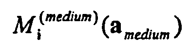
- The apparatus according to claim 16 further comprising a confocal optical element located between the light source and the detector, the confocal optical element being capable of transmitting light substantially exclusively from a sensed volume of the sample.
- The apparatus according to claim 16 or 17 wherein X, Y-axes are defined perpendicular to each other and parallel to the stage, further comprising an X, Y-axes adjustment device that automatically adjusts the relative positions of the sensed volume and the sample along the X and Y axes.
- The apparatus according to any of claims 16 to 18 wherein a Z-axis is defined substantially perpendicular to the stage, further comprising a Z-axis adjustment device that automatically adjusts the relative positions of the sensed volume along the Z-axis.
- An assay for screening a plurality of compounds for inhibition between a ligand and a target molecule expressed by or attached to a cell, or soluble or solid support, said assay comprising the steps of:- adding a liquid suspension of said target molecules expressed by or attached to said cell, or soluble or solid support to a plurality of containers;- adding a plurality of compounds to be screened for said inhibition to said plurality of containers;- adding fluorescently labelled ligand to said containers;- incubating said target molecules expressed by or attached to said cell, or soluble or solid support, said compounds, and said fluorescently labelled ligand;- individually measuring amounts of fluorescence associated with said cells, or soluble or solid supports; and- determining the degree of inhibition by one or more of saidwherein said amounts of fluorescence associated with said cells, or soluble or solid supports is estimated according to any of claims 1 to 15.
compounds of binding of said ligand to said target molecule; - An assay for testing a compound to determine the degree of binding of said compound to a target molecule expressed by or attached to a cell, or soluble or solid support, said assay comprising the steps of:- adding a liquid suspension of said target molecule expressed by or attached to said cell, or soluble or solid support to a container;- adding a fluorescent compound to be tested for its binding to said container;- incubating said target molecule expressed by or attached to said cell, or soluble or solid support and said compound;- measuring amounts of fluorescence associated with said cell, or soluble or solid support; and- determining the degree of binding of said compound to said target molecule;wherein said amounts of fluorescence associated with said cell, or soluble or solid support is estimated according to any of claims 1 to 15.
- The assay according to claim 21 wherein a plurality of different compounds and/or different targets is tested in a plurality of containers.
Priority Applications (2)
| Application Number | Priority Date | Filing Date | Title |
|---|---|---|---|
| DK00117522.3T DK1180679T3 (en) | 2000-08-14 | 2000-08-14 | Method for detecting fluorescent samples by moment analysis |
| EP00117522A EP1180679B1 (en) | 2000-08-14 | 2000-08-14 | A method for sensing fluorescent samples utilizing moment analysis |
Applications Claiming Priority (1)
| Application Number | Priority Date | Filing Date | Title |
|---|---|---|---|
| EP00117522A EP1180679B1 (en) | 2000-08-14 | 2000-08-14 | A method for sensing fluorescent samples utilizing moment analysis |
Publications (2)
| Publication Number | Publication Date |
|---|---|
| EP1180679A1 EP1180679A1 (en) | 2002-02-20 |
| EP1180679B1 true EP1180679B1 (en) | 2012-10-10 |
Family
ID=8169523
Family Applications (1)
| Application Number | Title | Priority Date | Filing Date |
|---|---|---|---|
| EP00117522A Expired - Lifetime EP1180679B1 (en) | 2000-08-14 | 2000-08-14 | A method for sensing fluorescent samples utilizing moment analysis |
Country Status (2)
| Country | Link |
|---|---|
| EP (1) | EP1180679B1 (en) |
| DK (1) | DK1180679T3 (en) |
Families Citing this family (15)
| Publication number | Priority date | Publication date | Assignee | Title |
|---|---|---|---|---|
| US20050142608A1 (en) * | 2003-09-22 | 2005-06-30 | Yokogawa Electric Corporation | Screening method and device, and new drug screening method and device |
| US7310147B2 (en) | 2005-01-27 | 2007-12-18 | Genetix Limited | Robotic apparatus for picking of cells and other applications with integrated spectroscopic capability |
| US8492098B2 (en) | 2006-02-21 | 2013-07-23 | The Trustees Of Tufts College | Methods and arrays for target analyte detection and determination of reaction components that affect a reaction |
| US11237171B2 (en) | 2006-02-21 | 2022-02-01 | Trustees Of Tufts College | Methods and arrays for target analyte detection and determination of target analyte concentration in solution |
| US20110195852A1 (en) | 2007-08-30 | 2011-08-11 | Trustees Of Tufts College | Methods for determining the concentration of an analyte in solution |
| US8222047B2 (en) | 2008-09-23 | 2012-07-17 | Quanterix Corporation | Ultra-sensitive detection of molecules on single molecule arrays |
| US9678068B2 (en) | 2010-03-01 | 2017-06-13 | Quanterix Corporation | Ultra-sensitive detection of molecules using dual detection methods |
| WO2011109379A1 (en) | 2010-03-01 | 2011-09-09 | Quanterix Corporation | Methods and systems for extending dynamic range in assays for the detection of molecules or particles |
| US8415171B2 (en) | 2010-03-01 | 2013-04-09 | Quanterix Corporation | Methods and systems for extending dynamic range in assays for the detection of molecules or particles |
| US8236574B2 (en) | 2010-03-01 | 2012-08-07 | Quanterix Corporation | Ultra-sensitive detection of molecules or particles using beads or other capture objects |
| US9952237B2 (en) | 2011-01-28 | 2018-04-24 | Quanterix Corporation | Systems, devices, and methods for ultra-sensitive detection of molecules or particles |
| WO2012142301A2 (en) | 2011-04-12 | 2012-10-18 | Quanterix Corporation | Methods of determining a treatment protocol for and/or a prognosis of a patients recovery from a brain injury |
| US9932626B2 (en) | 2013-01-15 | 2018-04-03 | Quanterix Corporation | Detection of DNA or RNA using single molecule arrays and other techniques |
| CN112461806B (en) * | 2020-11-18 | 2022-02-11 | 厦门大学 | Smartphone-based fluorescence spectroscopy detection method |
| CN117854656B (en) * | 2024-03-07 | 2024-05-10 | 四川大学 | A method for optimizing photon glass saturation by quantitatively adding broad-spectrum absorbers |
Family Cites Families (3)
| Publication number | Priority date | Publication date | Assignee | Title |
|---|---|---|---|---|
| JP2575270B2 (en) * | 1992-11-10 | 1997-01-22 | 浜松ホトニクス株式会社 | Method for determining base sequence of nucleic acid, method for detecting single molecule, apparatus therefor and method for preparing sample |
| EP0836090A1 (en) * | 1996-10-12 | 1998-04-15 | Evotec BioSystems GmbH | Method of analysis of samples by determination of the distribution of specific brightnesses of particles |
| EP0884583A1 (en) * | 1997-06-10 | 1998-12-16 | Evotec BioSystems GmbH | A method for characterizing samples in at least two dimensional space of specific physical properties |
-
2000
- 2000-08-14 EP EP00117522A patent/EP1180679B1/en not_active Expired - Lifetime
- 2000-08-14 DK DK00117522.3T patent/DK1180679T3/en active
Also Published As
| Publication number | Publication date |
|---|---|
| EP1180679A1 (en) | 2002-02-20 |
| DK1180679T3 (en) | 2013-02-04 |
Similar Documents
| Publication | Publication Date | Title |
|---|---|---|
| US20230280259A1 (en) | Parallel Flow Cytometer Using Radiofrequency Multiplexing | |
| EP1180679A1 (en) | A method for sensing fluorescent samples utilizing moment analysis | |
| EP0651879B1 (en) | Digital analyte detection system | |
| US6727071B1 (en) | System for cell-based screening | |
| US7027932B2 (en) | Biochemical examination method | |
| US6704104B2 (en) | Multi-wavelength array reader for biological assay | |
| US7117098B1 (en) | Machine-readable storage medium for analyzing distribution of macromolecules between the cell membrane and the cell cytoplasm | |
| EP0983498B1 (en) | A system for cell-based screening | |
| US7019310B2 (en) | Method of analysis of samples by determination of a function of specific brightness | |
| US20020195554A1 (en) | Scanning system with calibrated detection and method | |
| EP2717052A2 (en) | High-speed screening apparatus for a raman analysis-based high-speed multiple drug | |
| CN101542269A (en) | Multivariate detection of molecules in biossay | |
| US6556296B1 (en) | Method for characterizing samples by determination of a function of at least one specific property of particles in a sample | |
| JP2005513497A (en) | Method and / or apparatus for identification of fluorescent, luminescent and / or light-absorbing substances on and / or in a sample carrier | |
| EP1019687B1 (en) | A method for characterizing samples by determination of a function of at least one specific physical property of particles of said sample | |
| US20030008314A1 (en) | Fiber-optic sensor array | |
| CA2410688C (en) | A system for cell-based screening | |
| WO2025119804A1 (en) | Qualification standard and method utilizing raman activity of h2o | |
| Rufenach et al. | Fluorescence correlation spectroscopy (FCS) and FCS-related confocal fluorimetric methods (FCS+ plus): Multiple read-out options for miniaturized screening |
Legal Events
| Date | Code | Title | Description |
|---|---|---|---|
| PUAI | Public reference made under article 153(3) epc to a published international application that has entered the european phase |
Free format text: ORIGINAL CODE: 0009012 |
|
| AK | Designated contracting states |
Kind code of ref document: A1 Designated state(s): AT BE CH CY DE DK ES FI FR GB GR IE IT LI LU MC NL PT SE |
|
| AX | Request for extension of the european patent |
Free format text: AL;LT;LV;MK;RO;SI |
|
| 17P | Request for examination filed |
Effective date: 20020727 |
|
| AKX | Designation fees paid |
Free format text: AT BE CH CY DE DK ES FI FR GB GR IE IT LI LU MC NL PT SE |
|
| 17Q | First examination report despatched |
Effective date: 20031126 |
|
| 17Q | First examination report despatched |
Effective date: 20031126 |
|
| APBN | Date of receipt of notice of appeal recorded |
Free format text: ORIGINAL CODE: EPIDOSNNOA2E |
|
| APBR | Date of receipt of statement of grounds of appeal recorded |
Free format text: ORIGINAL CODE: EPIDOSNNOA3E |
|
| APBV | Interlocutory revision of appeal recorded |
Free format text: ORIGINAL CODE: EPIDOSNIRAPE |
|
| APAF | Appeal reference modified |
Free format text: ORIGINAL CODE: EPIDOSCREFNE |
|
| APBD | Information on interlocutory revision deleted |
Free format text: ORIGINAL CODE: EPIDOSDIRAPE |
|
| APBT | Appeal procedure closed |
Free format text: ORIGINAL CODE: EPIDOSNNOA9E |
|
| GRAP | Despatch of communication of intention to grant a patent |
Free format text: ORIGINAL CODE: EPIDOSNIGR1 |
|
| GRAS | Grant fee paid |
Free format text: ORIGINAL CODE: EPIDOSNIGR3 |
|
| RAP1 | Party data changed (applicant data changed or rights of an application transferred) |
Owner name: EVOTEC AG |
|
| GRAA | (expected) grant |
Free format text: ORIGINAL CODE: 0009210 |
|
| AK | Designated contracting states |
Kind code of ref document: B1 Designated state(s): AT BE CH CY DE DK ES FI FR GB GR IE IT LI LU MC NL PT SE |
|
| REG | Reference to a national code |
Ref country code: GB Ref legal event code: FG4D |
|
| REG | Reference to a national code |
Ref country code: AT Ref legal event code: REF Ref document number: 579170 Country of ref document: AT Kind code of ref document: T Effective date: 20121015 Ref country code: CH Ref legal event code: EP |
|
| REG | Reference to a national code |
Ref country code: IE Ref legal event code: FG4D |
|
| REG | Reference to a national code |
Ref country code: DE Ref legal event code: R096 Ref document number: 60047556 Country of ref document: DE Effective date: 20121206 |
|
| REG | Reference to a national code |
Ref country code: CH Ref legal event code: NV Representative=s name: SCHMAUDER AND PARTNER AG PATENT- UND MARKENANW, CH |
|
| REG | Reference to a national code |
Ref country code: DK Ref legal event code: T3 |
|
| REG | Reference to a national code |
Ref country code: NL Ref legal event code: VDEP Effective date: 20121010 |
|
| REG | Reference to a national code |
Ref country code: AT Ref legal event code: MK05 Ref document number: 579170 Country of ref document: AT Kind code of ref document: T Effective date: 20121010 |
|
| PG25 | Lapsed in a contracting state [announced via postgrant information from national office to epo] |
Ref country code: NL Free format text: LAPSE BECAUSE OF FAILURE TO SUBMIT A TRANSLATION OF THE DESCRIPTION OR TO PAY THE FEE WITHIN THE PRESCRIBED TIME-LIMIT Effective date: 20121010 Ref country code: FI Free format text: LAPSE BECAUSE OF FAILURE TO SUBMIT A TRANSLATION OF THE DESCRIPTION OR TO PAY THE FEE WITHIN THE PRESCRIBED TIME-LIMIT Effective date: 20121010 Ref country code: ES Free format text: LAPSE BECAUSE OF FAILURE TO SUBMIT A TRANSLATION OF THE DESCRIPTION OR TO PAY THE FEE WITHIN THE PRESCRIBED TIME-LIMIT Effective date: 20130121 Ref country code: SE Free format text: LAPSE BECAUSE OF FAILURE TO SUBMIT A TRANSLATION OF THE DESCRIPTION OR TO PAY THE FEE WITHIN THE PRESCRIBED TIME-LIMIT Effective date: 20121010 |
|
| PG25 | Lapsed in a contracting state [announced via postgrant information from national office to epo] |
Ref country code: BE Free format text: LAPSE BECAUSE OF FAILURE TO SUBMIT A TRANSLATION OF THE DESCRIPTION OR TO PAY THE FEE WITHIN THE PRESCRIBED TIME-LIMIT Effective date: 20121010 Ref country code: GR Free format text: LAPSE BECAUSE OF FAILURE TO SUBMIT A TRANSLATION OF THE DESCRIPTION OR TO PAY THE FEE WITHIN THE PRESCRIBED TIME-LIMIT Effective date: 20130111 Ref country code: CY Free format text: LAPSE BECAUSE OF FAILURE TO SUBMIT A TRANSLATION OF THE DESCRIPTION OR TO PAY THE FEE WITHIN THE PRESCRIBED TIME-LIMIT Effective date: 20121010 Ref country code: PT Free format text: LAPSE BECAUSE OF FAILURE TO SUBMIT A TRANSLATION OF THE DESCRIPTION OR TO PAY THE FEE WITHIN THE PRESCRIBED TIME-LIMIT Effective date: 20130211 |
|
| PG25 | Lapsed in a contracting state [announced via postgrant information from national office to epo] |
Ref country code: AT Free format text: LAPSE BECAUSE OF FAILURE TO SUBMIT A TRANSLATION OF THE DESCRIPTION OR TO PAY THE FEE WITHIN THE PRESCRIBED TIME-LIMIT Effective date: 20121010 |
|
| PLBE | No opposition filed within time limit |
Free format text: ORIGINAL CODE: 0009261 |
|
| STAA | Information on the status of an ep patent application or granted ep patent |
Free format text: STATUS: NO OPPOSITION FILED WITHIN TIME LIMIT |
|
| PG25 | Lapsed in a contracting state [announced via postgrant information from national office to epo] |
Ref country code: IT Free format text: LAPSE BECAUSE OF FAILURE TO SUBMIT A TRANSLATION OF THE DESCRIPTION OR TO PAY THE FEE WITHIN THE PRESCRIBED TIME-LIMIT Effective date: 20121010 |
|
| 26N | No opposition filed |
Effective date: 20130711 |
|
| REG | Reference to a national code |
Ref country code: DE Ref legal event code: R097 Ref document number: 60047556 Country of ref document: DE Effective date: 20130711 |
|
| PG25 | Lapsed in a contracting state [announced via postgrant information from national office to epo] |
Ref country code: MC Free format text: LAPSE BECAUSE OF FAILURE TO SUBMIT A TRANSLATION OF THE DESCRIPTION OR TO PAY THE FEE WITHIN THE PRESCRIBED TIME-LIMIT Effective date: 20121010 |
|
| REG | Reference to a national code |
Ref country code: IE Ref legal event code: MM4A |
|
| PG25 | Lapsed in a contracting state [announced via postgrant information from national office to epo] |
Ref country code: IE Free format text: LAPSE BECAUSE OF NON-PAYMENT OF DUE FEES Effective date: 20130814 |
|
| PG25 | Lapsed in a contracting state [announced via postgrant information from national office to epo] |
Ref country code: LU Free format text: LAPSE BECAUSE OF NON-PAYMENT OF DUE FEES Effective date: 20130814 |
|
| REG | Reference to a national code |
Ref country code: FR Ref legal event code: PLFP Year of fee payment: 16 |
|
| REG | Reference to a national code |
Ref country code: FR Ref legal event code: PLFP Year of fee payment: 17 |
|
| REG | Reference to a national code |
Ref country code: FR Ref legal event code: PLFP Year of fee payment: 18 |
|
| REG | Reference to a national code |
Ref country code: FR Ref legal event code: PLFP Year of fee payment: 19 |
|
| PGFP | Annual fee paid to national office [announced via postgrant information from national office to epo] |
Ref country code: DE Payment date: 20190830 Year of fee payment: 20 Ref country code: FR Payment date: 20190822 Year of fee payment: 20 Ref country code: DK Payment date: 20190822 Year of fee payment: 20 |
|
| PGFP | Annual fee paid to national office [announced via postgrant information from national office to epo] |
Ref country code: GB Payment date: 20190827 Year of fee payment: 20 |
|
| PGFP | Annual fee paid to national office [announced via postgrant information from national office to epo] |
Ref country code: CH Payment date: 20190826 Year of fee payment: 20 |
|
| REG | Reference to a national code |
Ref country code: DE Ref legal event code: R071 Ref document number: 60047556 Country of ref document: DE |
|
| REG | Reference to a national code |
Ref country code: DK Ref legal event code: EUP Expiry date: 20200814 |
|
| REG | Reference to a national code |
Ref country code: CH Ref legal event code: PL |
|
| REG | Reference to a national code |
Ref country code: GB Ref legal event code: PE20 Expiry date: 20200813 |
|
| PG25 | Lapsed in a contracting state [announced via postgrant information from national office to epo] |
Ref country code: GB Free format text: LAPSE BECAUSE OF EXPIRATION OF PROTECTION Effective date: 20200813 |