EP1180243B1 - Uses of notch related genes - Google Patents
Uses of notch related genes Download PDFInfo
- Publication number
- EP1180243B1 EP1180243B1 EP00935321A EP00935321A EP1180243B1 EP 1180243 B1 EP1180243 B1 EP 1180243B1 EP 00935321 A EP00935321 A EP 00935321A EP 00935321 A EP00935321 A EP 00935321A EP 1180243 B1 EP1180243 B1 EP 1180243B1
- Authority
- EP
- European Patent Office
- Prior art keywords
- sdrp
- seq
- nucleic acid
- protein
- expression
- Prior art date
- Legal status (The legal status is an assumption and is not a legal conclusion. Google has not performed a legal analysis and makes no representation as to the accuracy of the status listed.)
- Expired - Lifetime
Links
Images



Classifications
-
- A—HUMAN NECESSITIES
- A61—MEDICAL OR VETERINARY SCIENCE; HYGIENE
- A61K—PREPARATIONS FOR MEDICAL, DENTAL OR TOILETRY PURPOSES
- A61K38/00—Medicinal preparations containing peptides
- A61K38/16—Peptides having more than 20 amino acids; Gastrins; Somatostatins; Melanotropins; Derivatives thereof
- A61K38/43—Enzymes; Proenzymes; Derivatives thereof
- A61K38/53—Ligases (6)
-
- A—HUMAN NECESSITIES
- A61—MEDICAL OR VETERINARY SCIENCE; HYGIENE
- A61K—PREPARATIONS FOR MEDICAL, DENTAL OR TOILETRY PURPOSES
- A61K38/00—Medicinal preparations containing peptides
- A61K38/16—Peptides having more than 20 amino acids; Gastrins; Somatostatins; Melanotropins; Derivatives thereof
- A61K38/17—Peptides having more than 20 amino acids; Gastrins; Somatostatins; Melanotropins; Derivatives thereof from animals; from humans
- A61K38/1703—Peptides having more than 20 amino acids; Gastrins; Somatostatins; Melanotropins; Derivatives thereof from animals; from humans from vertebrates
- A61K38/1709—Peptides having more than 20 amino acids; Gastrins; Somatostatins; Melanotropins; Derivatives thereof from animals; from humans from vertebrates from mammals
-
- A—HUMAN NECESSITIES
- A61—MEDICAL OR VETERINARY SCIENCE; HYGIENE
- A61P—SPECIFIC THERAPEUTIC ACTIVITY OF CHEMICAL COMPOUNDS OR MEDICINAL PREPARATIONS
- A61P35/00—Antineoplastic agents
-
- A—HUMAN NECESSITIES
- A61—MEDICAL OR VETERINARY SCIENCE; HYGIENE
- A61K—PREPARATIONS FOR MEDICAL, DENTAL OR TOILETRY PURPOSES
- A61K48/00—Medicinal preparations containing genetic material which is inserted into cells of the living body to treat genetic diseases; Gene therapy
Definitions
- the present invention relates to the use of human homologues of a Drosophila Notch regulator gene and the encoded protein products thereof, as well as derivatives, fragments and analogues thereof and antibodies thereto in diagnosis and therapy.
- Notch is a transmembrane receptor protein which mediates a signal that controls the timing and outcome of cell differentiation decisions. Inappropriate activation of Notch has been associated with oncogenesis in humans and mice, causing T-Cell lymphoma and mammary gland tumours. In addition mutations in human Notch genes may result in the genetically inherited disease CADASIL, (Cerebral Autosomal Dominant Arteriopathy with Sub-cortical Infarcts and Leukoencephalopathy) which leads to a predisposition to multiple strokes and early onset dementia.
- CADASIL Cerebral Autosomal Dominant Arteriopathy with Sub-cortical Infarcts and Leukoencephalopathy
- Su(dx) Over expression of Drosophila Su(dx) results in over proliferation and cell differentiation phenotypes. It is likely that more than one target protein is recognised by Su(dx) and that multiple pathways are regulated by its activity leading to the regulation of cell differentiation and cell proliferation. Genetic data in Drosophila indicates that one of these regulated pathways is the Notch signalling pathway where Su(dx) acts as a negative regulator of the signal. In particular, genetic data indicates that Su(dx) regulates Notch signalling during the differentiation of Drosophila wing veins. The cell proliferation phenotype resulting from over expression of Su(dx) may reflect regulation of additional signals as well as or instead of Notch.
- the inventor has cloned the Drosophila Suppressor of deltex (Su(dx)) gene and the gene has been sequenced and the sequence of its protein product Su(dx) determined. Details of the cloning are provided in Cornell et al ., Genetics 152: 567-576 (June 1999). Such details were fully described in GB patent application no. 9912132.9, which serves as priority for this application.
- the inventor used his Drosophila Su(dx) clone to screen a DNA sequence database and the clone with the sequence with the closest homology with the Drosophila Su(dx) clone obtained (SEQ ID NO. 1) and used to screen a human foetal liver cDNA library using standard methods.
- the clone (SEQ ID NOS. 2 and 3) obtained by this screen was used to screen a human breast tumour cDNA library using standard methods and a clone (SEQ ID NO. 4) obtained that corresponds to the whole open reading frame expected for a sequence homologous to the Drosophila Su(dx) gene.
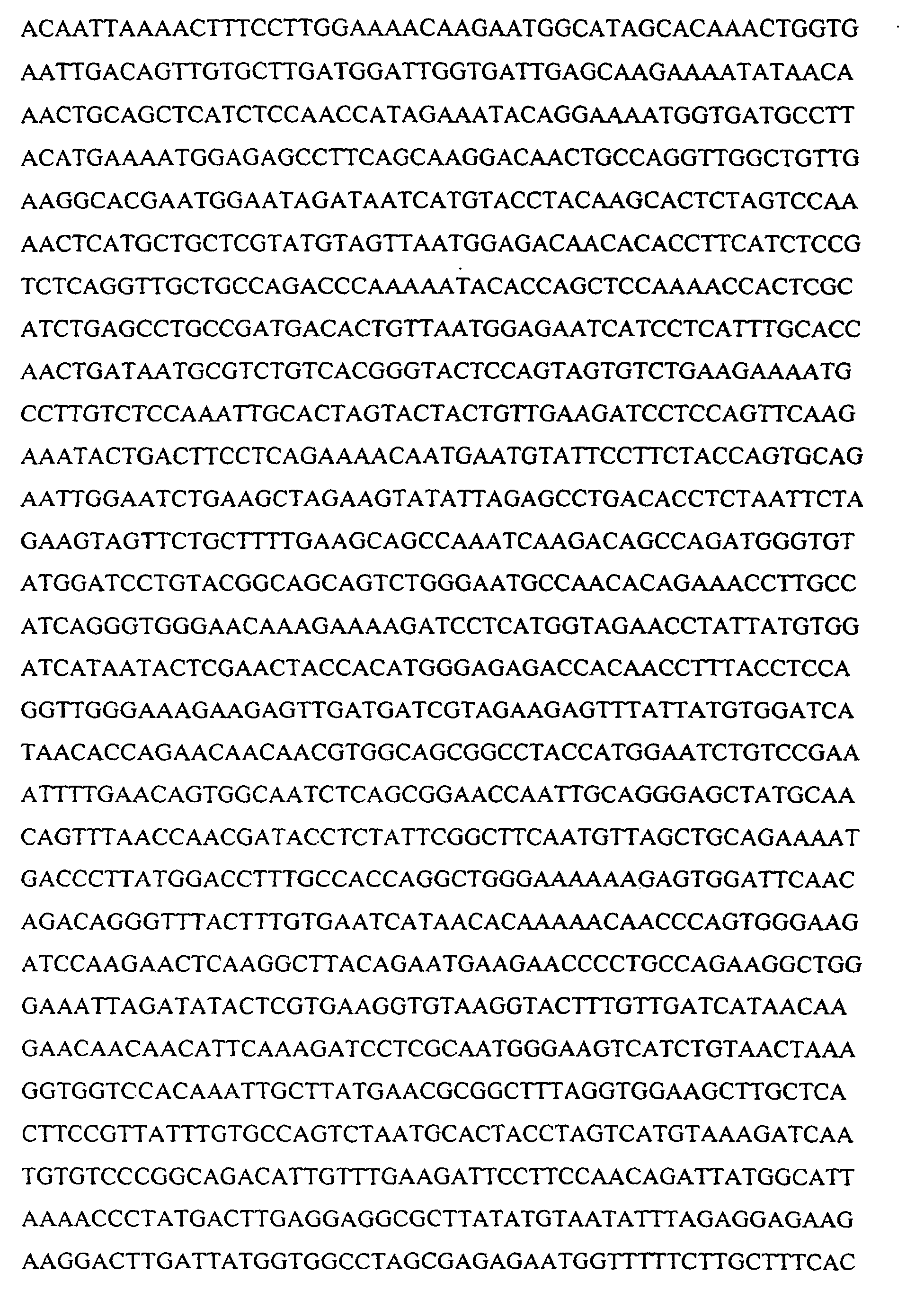
- the nucleic acid molecule having sequence homology to Drosophila Su(dx) obtained was termed clone h-SDRP-1 as is shown as SEQ ID No. 4.
- the protein sequence encoded by clone h-SDRP-1 is shown as SEQ ID No.5.
- the h-SDRP protein was analysed and found to have the same domain architecture as Drosophila Su(dx) in that it contains a C-terminal HECT domain (or E3 ubiquitin ligase domain) and four WW domains.
- PCT/US97/05547 A protein with substantial homology with h-SDRP is described in International application no. PCT/US97/05547 which described several polypeptides having WW domains, whose sequence was determined by screening cDNA libraries with a synthetic peptide based upon a consensus motif for WW proteins. PCT/US97/05547 does not suggest any function for the polypeptides described therein.
- Gray et al . discloses three human homologues of the Notch ligands Delta and Serrate, i.e. the genes HJ1, HJ2, and H-Delta-1 . (Gray et al . (1999), Human Ligands of the Notch Receptor, American Journal of Pathology , 154 (3):785-793). However, none of the three genes disclosed in this publication have any relationship with the human suppressor of deltex related protein. The DNA and protein sequences are totally dissimilar from each other
- the present invention in its first aspect provides for the use of h-SDRP nucleic acids, proteins and antibodies in diagnosis.
- h-SDRP nucleic acid and amino acid sequences and antibodies thereto may be used according to the invention for the detection and quantitation of h-SDRP mRNA and protein, to study the expression thereof, to produce h-SDRP proteins, fragments and other derivatives or analogues for use in the study, assay and manipulation of differentiation and other physiological processes in which Drosophila Su(dx) is postulated to be involved, e.g. diseases of cell fate and abnormal differentiation and cell proliferation.
- the h-SDRP nucleic acid has at least 60% sequence homology with the h-SDRP sequence provided as SEQ ID NO. 4(nucleic acid) or SEQ ID NO. 5 (protein), more preferably at least 70%, even more preferably at least 80% and most preferably at least 90% sequence homology with the h-SDRP nucleic acid sequence provided as SEQ ID NO. 4 or h-SDRP protein sequence provided as SEQ ID NO. 5 or subfragments or variants thereof specific to the h-SDRP sequence.
- the h-SDRP used according to the present invention preferably retain Drosophila Su(dx)-function, i.e. they are capable of displaying one or more known functions of the Drosophila Su(dx) protein, for example the protein can suppress deltex function, as shown by providing a transgenic Drosophila which expresses the vertebrate Su(dx) like protein and observing whether the expression can replace mutations that knock out endogenous Drosophila Su(dx).
- the phenotype of the Drosophila could be studied to see if over expression of the vertebrate Su(dx)-like nucleic acid causes epithelial cell over-proliferation or wing vein differentiation phenotypes similar to those found on over expression of Drosophila Su(dx).
- the h-SDRP nucleic acid molecules, proteins and antibodies may, but not necessarily disrupt Notch function by contributing to abnormal Notch signalling. Consequently, the h-SDRP nucleic acid molecules, proteins and antibodies may be used in methods of diagnosis and treatment of diseases of cell fate, differentiation and proliferation which may involve inappropriate Notch signalling.
- Suitable nucleotide variants of SEQ ID NO. 4 are those having a sequence altered by the substitution of different codons that encode the same or a functionally equivalent amino acid residue within the sequence, thus producing a silent change.
- Other suitable variants are those having nucleotide sequences homologous to SEQ ID NO. 4 but comprising all or portions of sequence which arc altered by the substitution of different codons that encode an amino acid of similar polarity to the amino acid it substitutes, to produce a silent change.
- Codons for an amino acid of a specific type or function may be substitutes to encode a different amino acid within the same class.
- non polar (hydrophobic amino acids include alanine, leucine, isoleucine, valine, proline, phenylalanine, tryptophan and methionine.
- the polar neutral amino acids include glycine, serine, threonine, cysteine, tyrosine, asparagine and glutamine.
- the positively charged (basic) amino acids include lysine, arginine and histidine.
- the negatively charged (acidic) amino acids include aspartic acid and glutamic acid.
- Suitable h-SDRP protein variants of SEQ ID NO. 5 are those having sequences in which substitutions of amino acids have been made with amino acids of the same function, thus producing a silent change.
- Amino acids of a specific type or function may be substitutes to encode a different amino acid within the same class.
- non polar (hydrophobic amino acids include alanine, leucine, isoleucine, valine, proline, phenylalanine, tryptophan and methionine.
- the polar neutral amino acids include glycine, serine, threonine, cysteine, tyrosine, asparagine and glutamine.
- the positively charged (basic) amino acids include lysine, arginine and histidine.
- the negatively charged (acidic) amino acids include aspartic acid and glutamic acid.
- PCT/US98/27610 describes several polynucleotides which are differentially expressed between cancerous and non-cancerous tissues and therefore which may be involved in abnormal cell differentiation. Although three of the sequences disclosed do contain one WW domain they are not homologous with h-SDRP.
- h-SDRP nucleic acid molecules are expressed more in breast tumour tissue than in tissues from healthy patients, resulting in more of the h-SDRP protein being present. Consequently the inventor proposes that the expression of h-SDRP may be used as a marker for breast cancer.
- h-SDRP nucleic acid molecules are expressed in a cell culture model of angiogenesis which suggests that h-SDRP is involved in the angiogenesis process. Consequently the inventor proposes that the over expression of h-SDRP may be used as a marker for increased angiogenesis. Such a marker may indicate the presence of tumour tissue as increased angiogenesis is involved in providing capillaries to tumours.
- a method of screening for breast cancer comprising assaying for the expression of h-SDRP nucleic acid.
- a method of screening for increased angiogenesis comprising assaying for the over expression of h-SDRP nucleic acid.
- the fourth aspect of the invention provides a method of screening for diseases associated with abnormal Notch signalling comprising assaying for the expression of h-SDRP nucleic acid.
- Such diseases include oncogenesis, dementia and CADASIL.
- cancerous and hyperproliferative conditions are said to exhibit upregulated Notch, such as colon cancer, cervical cancer, breast cancer, squamous adenocarcinoma, seminoma, melanoma, and lung cancer, as well as other hyperproliferative disorders and these may be diagnosed by the expression of Su(dx).
- Notch regulation include wound healing, rheumatoid arthritis and vascular diseases such as arteriosclerosis.
- h-SDRP nucleic acid may be an indication of a genetically inherited predisposition to a disease of cell fate, proliferation or differentiation, such as breast cancer.
- h-SDRP may be used to screen for cancers in biopsy tissues it is preferred that said screening is carried out in a non-invasive manner.
- h-SDRP as a tumour marker, particularly for breast cancer, allows classification of the tumour to allow different diagnosis and treatment of different degrees and types of cancer.
- Two classifications widely used in oncology that can benefit from identification of the expression levels of h-SDRP are staging of the cancerous disorder and grading the nature of the cancerous tissue. Details of both staging and grading in relation to cancer will be apparent to those skilled in the art.
- the h-SDRP nucleic acid molecules, proteins or antibodies may be used according to the invention to screen the blood or tissues of patients to determine if that patient has breast cancer. It is further proposed that the nucleic acid molecules, proteins or antibodies (or fragments or variants thereof) may be used to screen the blood or tissues of patients to determine if that patient has a disease or disorder associated with abnormal differentiation or cellular proliferation due to inappropriate Notch signalling.
- h-SDRP nucleic acid molecules including homologous nucleic acid sequences and subsequences, including complementary sequences, can be used in Northern assays, dot blots and in situ hybridisation assays.
- h-SDRP nucleic acid sequences or subsequences thereof comprising about at least 8 nucleotides specific to h-SDRP can be used as hybridisation probes.
- the probes comprise at least 12 nucleotides specific to h-SDRP. More preferably, the probes comprise at least 15 nucleotides specific to h-SDRP. Even more preferably, the probes comprise at least 18 nucleotides specific to h-SDRP. Most preferably, the probes comprise at least 22 nucleotides specific to h-SDRP.
- Hybridisation assays using these probes can be used to detect, prognose, diagnose or monitor breast tumours.
- hybridisation assays using the above probes can be used to detect, prognose, diagnose or monitor conditions, disorders or disease states associated with changes in Notch activity or the change in expression of h-SDRP.
- a hybridisation assay may be carried out by a method comprising contacting a sample containing nucleic acid with a h-SDRP nucleic acid probe capable of hybridising to substantially only h-SDRP DNA or mRNA under conditions such that hybridisation can occur and detecting or measuring any resultant hybridisation.
- an assay for the expression of h-SDRP nucleic acid is a PCR assay using PCR primers specific for h-SDRP to amplify expression of this nucleic acid molecule.
- the amplified DNA may then be assayed by size analysis, for example by electrophoresis.
- h-SDRP nucleic acid molecules particularly useful in the invention are the h-SDRP clone sequences provided as SEQ ID No. 1, SEQ ID No. 2 and SEQ ID No. 4. Any of these sequences or fragments thereof specific to h-SDRP may be used as hybridisation probes in the hybridisation method described above. Also the sequences may be used as targets against which specific PCR primers may be designed for use in the PCR assay described above.
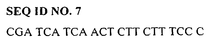
- PCR primers specific for h-SDRP nucleic acid are:
- a clone 8IIIA having the whole sequence provided as SEQ ID No. 2 was used to generate radiolabelled RNA for in situ hybridisation with tissue samples.
- a clone S112506 having the whole sequence provided as SEQ ID No. 1 was used a to screen the Southern blot of cDNA prepared from the 3' end of RNA expressed during angiogenesis.
- h-SDRP proteins, variants, derivatives and fragments thereof, and antibodies thereto can be used in assays, such as immunoassays, to detect, prognose, diagnose or monitor various conditions, diseases and disorders associated with abnormal Notch function or to monitor the treatment thereof.
- an immunoassay may be carried out by a method comprising contacting a sample derived from a patient with an anti-h-SDRP antibody under conditions such that immunospecific binding can occur and detecting or measuring the amount of immunospecific binding by the antibody.
- the immunoassays which can be used include but are not limited to competitive and non-competitive assay systems using techniques such as western-blots (followed by sodium dodecyl sulphate polyacrylamide gel electrophoresis), immunocytochemistry,, radioimmunoassays, ELISA (enzyme linked immunosorbant assay), "sandwich” immunoassays, immunoprecipitation assays and fluorescent immunoassays
- the assays according to the invention may be performed on patient tissue samples (e.g., from biopsy tissue), bodily fluids or on cell cultures derived from patient cells.
- patient tissue samples e.g., from biopsy tissue
- cell cultures derived from patient cells e.g., from breast tissue samples
- the assay may be performed on breast tissue biopsy sample to diagnose breast cancer.
- an agent that modulates expression or activity of h-SDRP nucleic acid may be used to treat breast cancer and inhibit or stimulate angiogenesis, and by extrapolation, conditions associated with abnormal Notch function.
- the inventor has cloned the Drosophila melangaster Suppressor of deltex (Su(dx)) gene and the gene has been sequenced and the sequence of its protein product Su(dx) determined. Details of the cloning are provided in Cornell et al ., Genetics 152: 567-576 (June 1999). Such details were fully described in GB patent application no. 9912132.9, which serves as priority for this application.
- the Drosophila Su(dx) clone was used to isolate a human Su(dx) clone.
- the human Su(dx) clone and fragments thereof was subsequently used to detect Su(dx) expression in both breast cancer biopsy tissue and endothelial cell cultures induced to have angiogenesis. Normal breast biopsy tissue and non-induced endothelial cells were shown not to express Su(dx) at detectable levels.
- the Drosophila Su(dx) gene clone sequence was used to search a DNA sequence database.
- the sequence with the closest homology with the Drosophila Su(dx) clone was a human expressed sequence tag clone S112506 (SEQ ID NO 1).
- the observed homology was to the HECT (or E3 ubiqutin liagase) domain region of Su(dx).
- the S112506 clone was freely available from the Washington University EST project and was obtained.
- a 1.3 kb insert from the S112506 clone was cut out, radiolabelled and used to screen a foetal liver cDNA library using standard methods.
- the library was a gift from G. Brady (School of Biological Sciences, University of Manchester).
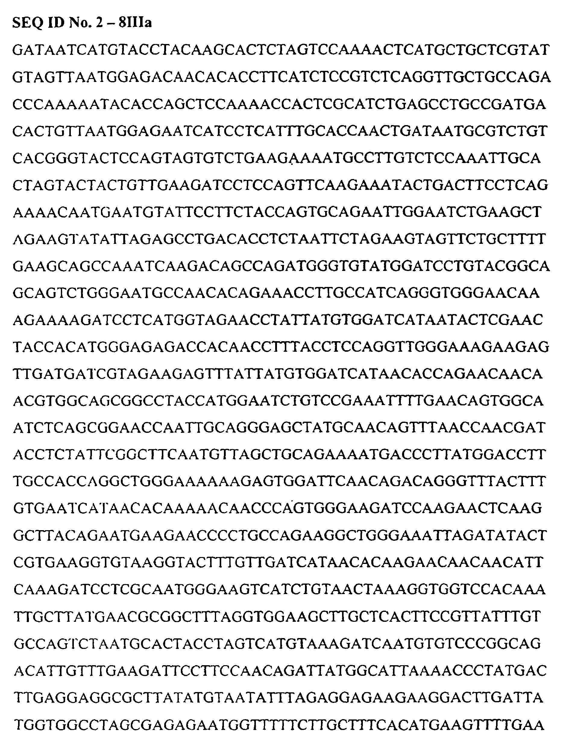
- clone 8IIIa A clone was detected and purified after 3 rounds of library screening. This clone was 8IIIa. This clone contains approximately 1.3 kb of sequence which overlaps that of S 112506. As expected, clone 8IIIa showed significant homology to Drosophila Su(dx) and is shown as SEQ ID No. 2. The protein sequence encoded by clone 8IIIa is shown as SEQ ID No.3.
- the radiolabelled 1.3 kb insert from the 8IIIa clone was also used to screen a breast tumour cDNA library using standard methods.
- the clone obtained contains approximately 4.4 kb of sequence which corresponds to the whole open reading frame expected for a sequence homologous to the Drosophila Su(dx) gene.
- the nucleic acid molecule of human origin having sequence homology to Drosophila Su(dx) obtained was termed clone h-SDRP-1 and is shown as SEQ ID No. 4.
- the protein sequence encoded by clone h-SDRP-1 is shown as SEQ ID No.5.
- h-SDRP was expressed during Drosophila wing development and the phenotypes compared with those resulting from expression of wild type and a mutant form of Drosophila Su(dx).
- transgenic line was constructed to express a truncated Drosophila Su(dx) molecule which lacked the HECT ubiquitin ligase domain.
- This construct was predicted to act as a dominant negative against wild type Su(dx) because it retains the WW protein specificity domains but lacks the capability to ubquitinate and down regulate the target. Thus by binding to the target the D-HECT form is predicted to protect it from the activity of wild type Su(dx).
- Radioactively labelled sense and anti-sense RNA was obtained by in-vitro transcription from T7 or T3 promoters of the cDNA. Radioactive labelling was by way of incorporation of 32 P labelled dUTP.
- In situ hybridisation was carried out according to standard techniques. Tissue sections from breast biopsies were first pre-hybridised to block non-specific hybridisation. All sections were washed in DPEC water for 10 minutes then immersed in 0.2 M HCl for 20 mins, rinsed in two fold standard saline buffer (2x SSC) and then treated with I ⁇ g/ml proteinase K in 0.05M tris, pH 7.4 at 37°C for 60 mins. The slides were washed twice in solutions of glycine in PBS, again in PBS for 3 mins and then incubated in 4% paraformaldehyde in PBS for 20 mins.
- the slides were then immersed in a solution of freshly prepared 0.25% acetic anhydride in 0.1 M triethanolamine, pH 8.0 for 10 mins followed by rinses in 70% and 90% ethanol for 5 min. After air-drying for 1 hour 50 ⁇ l of hybridisation solution was added to each section and left overnight at 37°C.
- the slides were exposed at 4°C for 10 days in a light sealed box, developed with Kodak D-19 developer for 5 min and fixed for 5 min. Haematoxylin and eosin were used to counterstain the sections.
- the fixed sections of tumour and non-tumour derived tissue were hybridised either with the sense or antisense 8IIIa RNA.
- the sense probe is a control for non-specific binding.
- the hybridisation of radioactive probes was detected after washing to remove unbound material, by overlaying the tissue with a photographic emulsion sensitive to the emitted radioactive particles. The presence and density of grains formed indicates the presence of the bound probe.
- the probe will bind where complementary RNA molecules are present. Thus the control sense strand probe should not bind.
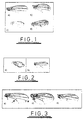
- the antisense probe hybridised strongly to tumour-derived cells but not to normal breast tissue (see Fig 4). Probing with the control sense strand showed little or no signal with both normal and tumour derived breast tissue. This indicates a strong up-regulation of expression of h-SDRP in the breast tumours.
- RT-PCR Reverse Transcriptase PCR
- Primers SEQ ID Nos. 6 and 7 were specific for the 5' end of h-SDRP protein sequence.
- RNA templates were obtained from Ann Canfield, Nigel Bundred and Richard Linforth (The Victoria University of Manchester).
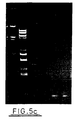
- Figure 5c shows RT-PCR detection of h-SDRP mRNA in RNA from a breast tumour cell line (T47D) (lane 3) but not from RNA derived from normal breast tissue (lane 4).
- Lanes 5 and 6 are GAP controls for the respective RNAs.
- Lanes 1 and 2 are DNA size markers (M).
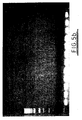
- the strength of the signal obtained reflects the proportion of h-SDRP mRNA molecules in the original extracted RNA sample and thus is semi-quantitative. It was observed that induction of angiogenesis by the fibroblast growth factor (FGF) induced the expression of h-SDRP - see Figure 6.
- FGF fibroblast growth factor
- Poly-A-PCR was performed on total RNA extracted from human cell lines or tissues. 1 ⁇ g of total RNA was reverse transcribed using random hexamers to prime cDNA synthesis in a total volume of 20 ⁇ l.
- cDNA was used as a template for PCR reactions.
- PCR reaction mix was prepared as follows: Component total volume added ( ⁇ l) 10 x TAQ Buffer (Boehringer Mannheim) 10 oligo (0.5-2.0 OD units/ml) Kvd or Not 1 1 25mM dNTPs 1 TAQ polymerase (Boehringer Mannheim) 1 cDNA template 1-5 ddH 2 O up to 100
- Standard sequence of cycles for Poly-A-PCR is as follows: 25 cycles 94°C for 1 minute, 42°C for 2 minutes, 72°C for 6 minutes.
- the gel was run according to standard methods and photographed. Next the gel was soaked in denaturation solution (0.5M NaOH, 1.5M NaCl) for 30 minutes. The gel was rinsed with distilled water, transferred to neutralising buffer (1.5M NaCl. 0.5M Tris HCl pH7.0) and soaked for 30 minutes at room temperature. Next the gel was soaked for 30 minutes at room temperature in 20X SSC buffer, with slow shaking. The gel then was carefully placed on a piece of Whatman 3MM paper, the same size as the gel and a pre-soaked nylon membrane (Hybond-N, Amersham) was put on top. On top of this were placed sheets of pre-wetted 3MM Whatman paper.
- the buffer tray was filled with transfer buffer (20x SSC) and transfer was started by connecting the gel stack with the buffer tray using the pre-cut "buffer wick", pre-soaked in transfer buffer. This was then left overnight or until all DNA from the gel was transferred onto the membrane. The membrane was then removed and gently washed in 2x SSC buffer for 10 minutes to remove agarose from the membrane. Next the membrane was exposed to a source of UV light for a total dose of 1200mJ/cm, to cross-link DNA to the filter.
- DNA probe S 112506 (SEQ ID No. 1) was performed using the Rediprime-Random Primer Labelling system from Amersham. To the premixed buffer and DNA polymerase solution, denatured DNA template (50ng) in a total volume of 12 ⁇ l was added. The sample of DNA was denatured by heating to 90-95°C in a boiling water bath. 2 ⁇ l of [ 32 P] (Amersham) was added to the probe/buffer mix and the samples were incubated at 37°C for 30 minutes. The reaction was stopped by adding 20 ⁇ l of 1x STE buffer (100mM NaCl, 20mM Tris HCl pH7.5, 10mM EDTA). The probe was purified by passing the reaction mixture through a NucTrap probe push column (Stratagene) according to the manufacturer's instructions.
- NucTrap probe push column (Stratagene) according to the manufacturer's instructions.
- the column was first equilibrated with 70 ⁇ l of STE buffer.
- the radiolabelled probe was made to a final volume of 60 ⁇ l with STE buffer and then passed through the column.
- the probe was eluted from the column with 70 ⁇ l of STE followed by a volume of air, and collected in an eppendorf tube.
- the probe was denatured by heating in a boiling water bath for 10 minutes and chilled immediately on ice.
- the radioactive probe was mixed with 10ml of hybridisation solution and added to the tube with the filter.
- the filter was hybridised overnight at 65°C.
- the membrane was washed twice for 10 minutes at room temperature in 2x SSPE, 0.1% (w/v) SDS solution. The membrane was then washed with 1x SSPE, 0.1% (w/v) SDS for 15 minutes at 65°C. Washings were repeated until the background diminished. If required the membrane was washed in 0.1x SSPE, 0.1% (w/v) SDS for 10 minutes at 65°C. The membrane was then covered with clingfilm and was subjected to autoradiography.
- Filters were exposed to Kodak X-ray film at -70°C. The film was developed using Kodak developing system. Alternatively filters were developed after exposition on phospho-imager plate.
- h-SDRP The expression of h-SDRP was investigated in bovine and human endothelial cells stimulated by different growth factors (figure 6).
- the figure shows the pattern of expression of the h-SDRP gene and a human osteonectin (a gene known to be expressed in endothelial cells) determined by the Poly-A PCR method using RNA extracted from bovine (baec) and human (heaec) endothelial cells.
- Cells in culture were treated, with appropriate non-treated controls, by different growth factors (basic fibroblast growth factor (bFGF), human growth factor (hGF) and VEGF) for different periods of time (24 and 48 hours) and RNA was extracted.
- bFGF basic fibroblast growth factor
- hGF human growth factor
- VEGF vascular endothelial growth factor
- Figure 6A shows the ethidium bromide stained gel with 10 ⁇ l of Poly-A PCR samples loaded.
- Figure 6B represents the equivalent Southern blot probed with 0.5 ⁇ g clone S 112506 (SEQ ID No. 1) and developed after 5 hour exposure on a phospho-imager.
- Figure 6C shows the ethidium bromide stained gel run with aliquots from the same Poly-A PCR samples as in (A).
- Figure 6D represents the equivalent Southern blot probed with human osteonectin and developed after I hour exposure.
- Endothelial cells were chosen because Notch has been shown to be involved in angiogenesis. Comparison of non-treated and treated samples following probing with 3' specific h-SDRP probe (S112506) showed a change of gene expression in the bovine endothelial cells stimulated by bFGF for 24 hours. No change in expression was observed in cells treated for longer periods with bFGF before harvesting, or for cells treated with hGF or VEGF (B).
- B hGF or VEGF
- D As a control for the Poly-A PCR amplification method, hybridisation of the same Poly-A PCR samples with human osteonectin (a gene known to be expressed in endothelial cells) was performed (D). In most of the samples hybridisation to the human osteonectin was observed. Only one sample - the bovine endothelial control cells, grown for 48 hours (baec -hGF 48 h) failed with detection of the osteonectin gene.
- the Poly-A PCR method was also applied to six human tumour cell line samples. The results indicated that only the rhabdomylosarcoma Rh18 represented a strong positive signal in response to the h-SDRP probe, despite comparable loadings. The rest of the tumour samples displayed only weak hybridisation signals. The lack of hybridisation to the negative control (sonicated ⁇ DNA) indicated that the hybridisation stringency was such that the level of non-specific background was minimal. Both the undifferentiated and differentiated mouse C2C12 skeletal muscle samples showed a strong positive hybridisation signal in response to the human Su(dx) radioactive probe.
- the pattern of the h-SDRP expression was also investigated in the human teratocarcinoma cell line samples. Cells were treated by retinoic acid to stimulate their differentiation. The Southern hybridisation result suggested that the h-SDRP gene is expressed in all the teratocarcinoma samples. A significant change of the levels of the h-SDRP expression on differentiation of these cells was not observed. The h-SDRP radioactive probe did not hybridise to the sonicated ⁇ DNA control confirming the specificity of the method.
Landscapes
- Health & Medical Sciences (AREA)
- Life Sciences & Earth Sciences (AREA)
- Medicinal Chemistry (AREA)
- Veterinary Medicine (AREA)
- Public Health (AREA)
- General Health & Medical Sciences (AREA)
- Chemical & Material Sciences (AREA)
- Animal Behavior & Ethology (AREA)
- Pharmacology & Pharmacy (AREA)
- Bioinformatics & Cheminformatics (AREA)
- Immunology (AREA)
- Proteomics, Peptides & Aminoacids (AREA)
- Epidemiology (AREA)
- Engineering & Computer Science (AREA)
- Gastroenterology & Hepatology (AREA)
- Organic Chemistry (AREA)
- Marine Sciences & Fisheries (AREA)
- Chemical Kinetics & Catalysis (AREA)
- General Chemical & Material Sciences (AREA)
- Nuclear Medicine, Radiotherapy & Molecular Imaging (AREA)
- Zoology (AREA)
- Measuring Or Testing Involving Enzymes Or Micro-Organisms (AREA)
- Investigating Or Analysing Biological Materials (AREA)
- Medicines That Contain Protein Lipid Enzymes And Other Medicines (AREA)
- Peptides Or Proteins (AREA)
- Saccharide Compounds (AREA)
- Pharmaceuticals Containing Other Organic And Inorganic Compounds (AREA)
- Seasonings (AREA)
- Polysaccharides And Polysaccharide Derivatives (AREA)
- Cosmetics (AREA)
Abstract
Description


| Component | total volume added (µl) |
| 10 x TAQ Buffer (Boehringer Mannheim) | 10 |
| oligo (0.5-2.0 OD units/ml) Kvd or Not 1 | 1 |
| 25mM dNTPs | 1 |
| TAQ polymerase (Boehringer Mannheim) | 1 |
| cDNA template | 1-5 |
| ddH2O | up to 100 |
| 25 cycles | 94°C for 1 minute, 42°C for 2 minutes, 72°C for 6 minutes. |
| 25 cycles | 94°C for 1 minute, 42°C for 1 minute, 72°C for 2 minutes. |








Claims (14)
- An in vitro method of screening for diseases caused by abnormal Notch signalling, the method comprising assaying for the expression of a nucleic acid encoding a human suppressor of deltex related protein, in a sample which has been obtained from an individual, wherein increased expression of the nucleic acid is associated with cell proliferation.
- A method according to claim 1, wherein the nucleic acid has at least 60% homology to the sequence provided as SEQ ID NO. 4.
- A method according to claim 1, wherein the nucleic acid has at least 70% homology to the sequence provided as SEQ ID NO. 4.
- A method according to claim 1, wherein the nucleic acid has at least 80% homology to the sequence provided as SEQ ID NO. 4.
- A method according to claim 1, wherein the nucleic acid has at least 90% homology to the sequence provided as SEQ ID NO. 4.
- A method according to claim 1, wherein the nucleic acid comprises a nucleic acid sequence shown in SEQ ID NO. 4.
- An in vitro method of screening for diseases caused by abnormal Notch signalling, the method comprising assaying for human suppressor of deltex related protein, in a sample which has been obtained from an individual, wherein an increased level of human suppressor of deltex related protein is associated with cell proliferation.
- A method according to claim 7, wherein the protein has at least 60% homology to the sequence provided as SEQ ID NO. 5.
- A method according to claim 7, wherein the protein has at least 70% homology to the sequence provided as SEQ ID NO. 5.
- A method according to claim 7, wherein the protein has at least 80% homology to the sequence provided as SEQ ID NO. 5.
- A method according to claim 7, wherein the protein has at least 90% homology to the sequence provided as SEQ ID NO. 5.
- A method according to claim 7, wherein the protein comprises amino acid sequence shown in SEQ ID NO. 5.
- A method according to any preceding claim, wherein the diseases which can be screened for comprise oncogenesis, colon cancer, cervical cancer, breast cancer, increased angiogenesis, squamous adenocarcinoma, seminoma, melanoma, lung cancer, dementia, CADASIL, wound healing, rheumatoid arthritis, and vascular diseases, such as arteriosclerosis.
- A method according to claim 13, wherein the diseases which can be screened for include increased angiogenesis and breast cancer.
Applications Claiming Priority (3)
| Application Number | Priority Date | Filing Date | Title |
|---|---|---|---|
| GB9912132 | 1999-05-26 | ||
| GBGB9912132.9A GB9912132D0 (en) | 1999-05-26 | 1999-05-26 | Notch regulator-related gene and uses thereof |
| PCT/GB2000/001990 WO2000073329A2 (en) | 1999-05-26 | 2000-05-23 | Uses of notch related genes |
Publications (2)
| Publication Number | Publication Date |
|---|---|
| EP1180243A2 EP1180243A2 (en) | 2002-02-20 |
| EP1180243B1 true EP1180243B1 (en) | 2005-07-20 |
Family
ID=10854121
Family Applications (1)
| Application Number | Title | Priority Date | Filing Date |
|---|---|---|---|
| EP00935321A Expired - Lifetime EP1180243B1 (en) | 1999-05-26 | 2000-05-23 | Uses of notch related genes |
Country Status (7)
| Country | Link |
|---|---|
| EP (1) | EP1180243B1 (en) |
| AT (1) | ATE300049T1 (en) |
| AU (1) | AU5087300A (en) |
| CA (1) | CA2375325A1 (en) |
| DE (1) | DE60021377D1 (en) |
| GB (1) | GB9912132D0 (en) |
| WO (1) | WO2000073329A2 (en) |
Families Citing this family (1)
| Publication number | Priority date | Publication date | Assignee | Title |
|---|---|---|---|---|
| CN100422345C (en) * | 2004-08-19 | 2008-10-01 | 北京雅康博生物科技有限公司 | Kit for detecting presence and level of PPN/MG61 polynucleotide |
Citations (1)
| Publication number | Priority date | Publication date | Assignee | Title |
|---|---|---|---|---|
| WO1997037223A1 (en) * | 1996-04-03 | 1997-10-09 | Cytogen Corporation | Identification and isolation of novel polypeptides having ww domains and methods of using same |
Family Cites Families (2)
| Publication number | Priority date | Publication date | Assignee | Title |
|---|---|---|---|---|
| CA2238404A1 (en) * | 1995-11-22 | 1997-05-29 | Yale University | Vertebrate deltex proteins, nucleic acids, and antibodies, and related methods and compositions |
| WO1998057621A1 (en) * | 1997-06-18 | 1998-12-23 | The Trustees Of Columbia University In The City Ofnew York | Angiogenic modulation by notch signal transduction |
-
1999
- 1999-05-26 GB GBGB9912132.9A patent/GB9912132D0/en not_active Ceased
-
2000
- 2000-05-23 CA CA002375325A patent/CA2375325A1/en not_active Abandoned
- 2000-05-23 AU AU50873/00A patent/AU5087300A/en not_active Abandoned
- 2000-05-23 WO PCT/GB2000/001990 patent/WO2000073329A2/en not_active Ceased
- 2000-05-23 EP EP00935321A patent/EP1180243B1/en not_active Expired - Lifetime
- 2000-05-23 DE DE60021377T patent/DE60021377D1/en not_active Expired - Lifetime
- 2000-05-23 AT AT00935321T patent/ATE300049T1/en not_active IP Right Cessation
Patent Citations (1)
| Publication number | Priority date | Publication date | Assignee | Title |
|---|---|---|---|---|
| WO1997037223A1 (en) * | 1996-04-03 | 1997-10-09 | Cytogen Corporation | Identification and isolation of novel polypeptides having ww domains and methods of using same |
Also Published As
| Publication number | Publication date |
|---|---|
| WO2000073329A3 (en) | 2001-02-22 |
| DE60021377D1 (en) | 2005-08-25 |
| GB9912132D0 (en) | 1999-07-28 |
| EP1180243A2 (en) | 2002-02-20 |
| CA2375325A1 (en) | 2000-12-07 |
| WO2000073329A2 (en) | 2000-12-07 |
| AU5087300A (en) | 2000-12-18 |
| ATE300049T1 (en) | 2005-08-15 |
Similar Documents
| Publication | Publication Date | Title |
|---|---|---|
| Alvarez-Pérez et al. | Molecular cloning, expression and immunolocalization of a novel human cementum-derived protein (CP-23) | |
| Galaviz-Hernandez et al. | Plac8 and Plac9, novel placental-enriched genes identified through microarray analysis | |
| JP3294575B2 (en) | Proteins that interact with the IGF-1 receptor, genes encoding them and uses thereof | |
| Suzuki et al. | Identification of RALDH-3, a novel retinaldehyde dehydrogenase, expressed in the ventral region of the retina | |
| EP2064550A2 (en) | Prrg4-associated compositions and methods of use thereof in methods of tumor diagnosis | |
| JPH09505735A (en) | p53-binding polypeptides and polynucleotides encoding same | |
| Grimm et al. | Isolation and embryonic expression of the novel mouse gene Hic1, the homologue of HIC1, a candidate gene for the Miller-Dieker syndrome | |
| KR20010022741A (en) | ISOLATION OF A NOVEL SENESCENCE-FACTOR GENE, p23 | |
| JP2002525081A (en) | Sequences characteristic of hypoxia-regulated gene transcription | |
| Clark et al. | Temporal and spatial expression of two isoforms of the Dutt1/Robo1 gene in mouse development | |
| US7074895B2 (en) | Sequences characteristic of hypoxia-regulated gene transcription | |
| Iida et al. | Human Mpv17-like protein is localized in peroxisomes and regulates expression of antioxidant enzymes | |
| Li et al. | Cloning and developmental expression of mouse pygopus 2, a putative Wnt signaling component | |
| JP2004505631A (en) | A new member of the lysyl oxidase gene family | |
| EP1355152B1 (en) | Method for identifying a compound for modulating the wnt signal cascade. | |
| US6586581B1 (en) | Prolactin regulatory element binding protein and uses thereof | |
| Weiskirchen et al. | The murine latent transforming growth factor-β binding protein (Ltbp-1) is alternatively spliced, and maps to a region syntenic to human chromosome 2p21–22 | |
| EP1854881B1 (en) | METHODS FOR IDENTIFYING PURKINJE CELLS USING THE Corl2 GENE AS A TARGET | |
| JP2002509862A (en) | Identification of factors mediating the interaction between heterotrimeric G protein and monomeric G protein | |
| EP1180243A2 (en) | Uses of notch related genes | |
| JP2001520877A (en) | In vitro differentiation of vascular smooth muscle cells, methods and reagents related thereto | |
| US7883896B2 (en) | Marker molecules associated with lung tumors | |
| US20030068311A1 (en) | Transmembrane protein differentially expressed in cancer | |
| Rudd et al. | Cloning and analysis of a FoxO transcription factor from Xiphophorus | |
| Park et al. | Hypertonicity induction of melanoma antigen, a tumor-associated antigen |
Legal Events
| Date | Code | Title | Description |
|---|---|---|---|
| PUAI | Public reference made under article 153(3) epc to a published international application that has entered the european phase |
Free format text: ORIGINAL CODE: 0009012 |
|
| 17P | Request for examination filed |
Effective date: 20011204 |
|
| AK | Designated contracting states |
Kind code of ref document: A2 Designated state(s): AT BE CH CY DE DK ES FI FR GB GR IE IT LI LU MC NL PT SE |
|
| AX | Request for extension of the european patent |
Free format text: AL;LT;LV;MK;RO;SI |
|
| 17Q | First examination report despatched |
Effective date: 20030402 |
|
| GRAP | Despatch of communication of intention to grant a patent |
Free format text: ORIGINAL CODE: EPIDOSNIGR1 |
|
| RAP1 | Party data changed (applicant data changed or rights of an application transferred) |
Owner name: THE UNIVERSITY OF MANCHESTER |
|
| GRAS | Grant fee paid |
Free format text: ORIGINAL CODE: EPIDOSNIGR3 |
|
| GRAA | (expected) grant |
Free format text: ORIGINAL CODE: 0009210 |
|
| AK | Designated contracting states |
Kind code of ref document: B1 Designated state(s): AT BE CH CY DE DK ES FI FR GB GR IE IT LI LU MC NL PT SE |
|
| PG25 | Lapsed in a contracting state [announced via postgrant information from national office to epo] |
Ref country code: IT Free format text: LAPSE BECAUSE OF FAILURE TO SUBMIT A TRANSLATION OF THE DESCRIPTION OR TO PAY THE FEE WITHIN THE PRESCRIBED TIME-LIMIT;WARNING: LAPSES OF ITALIAN PATENTS WITH EFFECTIVE DATE BEFORE 2007 MAY HAVE OCCURRED AT ANY TIME BEFORE 2007. THE CORRECT EFFECTIVE DATE MAY BE DIFFERENT FROM THE ONE RECORDED. Effective date: 20050720 Ref country code: LI Free format text: LAPSE BECAUSE OF FAILURE TO SUBMIT A TRANSLATION OF THE DESCRIPTION OR TO PAY THE FEE WITHIN THE PRESCRIBED TIME-LIMIT Effective date: 20050720 Ref country code: AT Free format text: LAPSE BECAUSE OF FAILURE TO SUBMIT A TRANSLATION OF THE DESCRIPTION OR TO PAY THE FEE WITHIN THE PRESCRIBED TIME-LIMIT Effective date: 20050720 Ref country code: FI Free format text: LAPSE BECAUSE OF FAILURE TO SUBMIT A TRANSLATION OF THE DESCRIPTION OR TO PAY THE FEE WITHIN THE PRESCRIBED TIME-LIMIT Effective date: 20050720 Ref country code: CH Free format text: LAPSE BECAUSE OF FAILURE TO SUBMIT A TRANSLATION OF THE DESCRIPTION OR TO PAY THE FEE WITHIN THE PRESCRIBED TIME-LIMIT Effective date: 20050720 Ref country code: BE Free format text: LAPSE BECAUSE OF FAILURE TO SUBMIT A TRANSLATION OF THE DESCRIPTION OR TO PAY THE FEE WITHIN THE PRESCRIBED TIME-LIMIT Effective date: 20050720 Ref country code: NL Free format text: LAPSE BECAUSE OF FAILURE TO SUBMIT A TRANSLATION OF THE DESCRIPTION OR TO PAY THE FEE WITHIN THE PRESCRIBED TIME-LIMIT Effective date: 20050720 |
|
| REG | Reference to a national code |
Ref country code: GB Ref legal event code: FG4D |
|
| REG | Reference to a national code |
Ref country code: CH Ref legal event code: EP |
|
| REG | Reference to a national code |
Ref country code: IE Ref legal event code: FG4D |
|
| REF | Corresponds to: |
Ref document number: 60021377 Country of ref document: DE Date of ref document: 20050825 Kind code of ref document: P |
|
| PG25 | Lapsed in a contracting state [announced via postgrant information from national office to epo] |
Ref country code: SE Free format text: LAPSE BECAUSE OF FAILURE TO SUBMIT A TRANSLATION OF THE DESCRIPTION OR TO PAY THE FEE WITHIN THE PRESCRIBED TIME-LIMIT Effective date: 20051020 Ref country code: DK Free format text: LAPSE BECAUSE OF FAILURE TO SUBMIT A TRANSLATION OF THE DESCRIPTION OR TO PAY THE FEE WITHIN THE PRESCRIBED TIME-LIMIT Effective date: 20051020 Ref country code: GR Free format text: LAPSE BECAUSE OF FAILURE TO SUBMIT A TRANSLATION OF THE DESCRIPTION OR TO PAY THE FEE WITHIN THE PRESCRIBED TIME-LIMIT Effective date: 20051020 |
|
| PG25 | Lapsed in a contracting state [announced via postgrant information from national office to epo] |
Ref country code: DE Free format text: LAPSE BECAUSE OF FAILURE TO SUBMIT A TRANSLATION OF THE DESCRIPTION OR TO PAY THE FEE WITHIN THE PRESCRIBED TIME-LIMIT Effective date: 20051021 |
|
| PG25 | Lapsed in a contracting state [announced via postgrant information from national office to epo] |
Ref country code: ES Free format text: LAPSE BECAUSE OF FAILURE TO SUBMIT A TRANSLATION OF THE DESCRIPTION OR TO PAY THE FEE WITHIN THE PRESCRIBED TIME-LIMIT Effective date: 20051031 |
|
| PG25 | Lapsed in a contracting state [announced via postgrant information from national office to epo] |
Ref country code: PT Free format text: LAPSE BECAUSE OF FAILURE TO SUBMIT A TRANSLATION OF THE DESCRIPTION OR TO PAY THE FEE WITHIN THE PRESCRIBED TIME-LIMIT Effective date: 20051221 |
|
| REG | Reference to a national code |
Ref country code: CH Ref legal event code: PL |
|
| NLV1 | Nl: lapsed or annulled due to failure to fulfill the requirements of art. 29p and 29m of the patents act | ||
| PG25 | Lapsed in a contracting state [announced via postgrant information from national office to epo] |
Ref country code: IE Free format text: LAPSE BECAUSE OF NON-PAYMENT OF DUE FEES Effective date: 20060523 |
|
| PLBE | No opposition filed within time limit |
Free format text: ORIGINAL CODE: 0009261 |
|
| STAA | Information on the status of an ep patent application or granted ep patent |
Free format text: STATUS: NO OPPOSITION FILED WITHIN TIME LIMIT |
|
| PG25 | Lapsed in a contracting state [announced via postgrant information from national office to epo] |
Ref country code: MC Free format text: LAPSE BECAUSE OF NON-PAYMENT OF DUE FEES Effective date: 20060531 |
|
| 26N | No opposition filed |
Effective date: 20060421 |
|
| EN | Fr: translation not filed | ||
| PG25 | Lapsed in a contracting state [announced via postgrant information from national office to epo] |
Ref country code: FR Free format text: LAPSE BECAUSE OF FAILURE TO SUBMIT A TRANSLATION OF THE DESCRIPTION OR TO PAY THE FEE WITHIN THE PRESCRIBED TIME-LIMIT Effective date: 20060915 |
|
| REG | Reference to a national code |
Ref country code: IE Ref legal event code: MM4A |
|
| PG25 | Lapsed in a contracting state [announced via postgrant information from national office to epo] |
Ref country code: LU Free format text: LAPSE BECAUSE OF NON-PAYMENT OF DUE FEES Effective date: 20060523 |
|
| PG25 | Lapsed in a contracting state [announced via postgrant information from national office to epo] |
Ref country code: CY Free format text: LAPSE BECAUSE OF FAILURE TO SUBMIT A TRANSLATION OF THE DESCRIPTION OR TO PAY THE FEE WITHIN THE PRESCRIBED TIME-LIMIT Effective date: 20050720 Ref country code: FR Free format text: LAPSE BECAUSE OF FAILURE TO SUBMIT A TRANSLATION OF THE DESCRIPTION OR TO PAY THE FEE WITHIN THE PRESCRIBED TIME-LIMIT Effective date: 20050720 |
|
| PGFP | Annual fee paid to national office [announced via postgrant information from national office to epo] |
Ref country code: GB Payment date: 20080528 Year of fee payment: 9 |
|
| GBPC | Gb: european patent ceased through non-payment of renewal fee |
Effective date: 20090523 |
|
| PG25 | Lapsed in a contracting state [announced via postgrant information from national office to epo] |
Ref country code: GB Free format text: LAPSE BECAUSE OF NON-PAYMENT OF DUE FEES Effective date: 20090523 |