EP1170372A1 - Regulatory sequences of the human MCP-1 gene - Google Patents

Regulatory sequences of the human MCP-1 gene Download PDF

Info

Publication number
EP1170372A1
EP1170372A1 EP00114560A EP00114560A EP1170372A1 EP 1170372 A1 EP1170372 A1 EP 1170372A1 EP 00114560 A EP00114560 A EP 00114560A EP 00114560 A EP00114560 A EP 00114560A EP 1170372 A1 EP1170372 A1 EP 1170372A1
Authority
EP
European Patent Office
Prior art keywords
nucleic acid
mcp
acid molecule
gene
dhsr
Prior art date
Legal status (The legal status is an assumption and is not a legal conclusion. Google has not performed a legal analysis and makes no representation as to the accuracy of the status listed.)
Granted
Application number
EP00114560A
Other languages
German (de)
French (fr)
Other versions
EP1170372B1 (en
Inventor
Dr. Frank Rösl
Dr. Ubaldo Soto
Dr. Johannes Coy
Dr. Patrick Finzer
Dr. Hajo Delius
Prof. Annemarie Poustka
Prof. Dr. Harald Zur Hausen
Andrea Patzelt
Current Assignee (The listed assignees may be inaccurate. Google has not performed a legal analysis and makes no representation or warranty as to the accuracy of the list.)
Deutsches Krebsforschungszentrum DKFZ
Original Assignee
Deutsches Krebsforschungszentrum DKFZ
Priority date (The priority date is an assumption and is not a legal conclusion. Google has not performed a legal analysis and makes no representation as to the accuracy of the date listed.)
Filing date
Publication date
Application filed by Deutsches Krebsforschungszentrum DKFZ filed Critical Deutsches Krebsforschungszentrum DKFZ
Priority to EP00114560A priority Critical patent/EP1170372B1/en
Priority to AT00114560T priority patent/ATE323772T1/en
Priority to DE60027419T priority patent/DE60027419D1/en
Priority to US09/899,276 priority patent/US20020106355A1/en
Publication of EP1170372A1 publication Critical patent/EP1170372A1/en
Application granted granted Critical
Publication of EP1170372B1 publication Critical patent/EP1170372B1/en
Anticipated expiration legal-status Critical
Expired - Lifetime legal-status Critical Current

Links

Images

Classifications

    • AHUMAN NECESSITIES
    • A61MEDICAL OR VETERINARY SCIENCE; HYGIENE
    • A61KPREPARATIONS FOR MEDICAL, DENTAL OR TOILETRY PURPOSES
    • A61K38/00Medicinal preparations containing peptides
    • A61K38/16Peptides having more than 20 amino acids; Gastrins; Somatostatins; Melanotropins; Derivatives thereof
    • A61K38/17Peptides having more than 20 amino acids; Gastrins; Somatostatins; Melanotropins; Derivatives thereof from animals; from humans
    • A61K38/1703Peptides having more than 20 amino acids; Gastrins; Somatostatins; Melanotropins; Derivatives thereof from animals; from humans from vertebrates
    • A61K38/1709Peptides having more than 20 amino acids; Gastrins; Somatostatins; Melanotropins; Derivatives thereof from animals; from humans from vertebrates from mammals
    • CCHEMISTRY; METALLURGY
    • C07ORGANIC CHEMISTRY
    • C07KPEPTIDES
    • C07K14/00Peptides having more than 20 amino acids; Gastrins; Somatostatins; Melanotropins; Derivatives thereof
    • C07K14/435Peptides having more than 20 amino acids; Gastrins; Somatostatins; Melanotropins; Derivatives thereof from animals; from humans
    • C07K14/52Cytokines; Lymphokines; Interferons
    • C07K14/521Chemokines
    • C07K14/523Beta-chemokines, e.g. RANTES, I-309/TCA-3, MIP-1alpha, MIP-1beta/ACT-2/LD78/SCIF, MCP-1/MCAF, MCP-2, MCP-3, LDCF-1, LDCF-2
    • AHUMAN NECESSITIES
    • A61MEDICAL OR VETERINARY SCIENCE; HYGIENE
    • A61KPREPARATIONS FOR MEDICAL, DENTAL OR TOILETRY PURPOSES
    • A61K38/00Medicinal preparations containing peptides

Definitions

  • the present invention relates to a nucleic acid molecule comprising (a) a nucleic acid sequence encoding the monocyte-chemoattractant-protein-1 (MCP-1) or a protein having the biological activity of the monocyte-chemoattractant-protein-1 (MCP-1); and (b) a hypersensitive region.
  • MCP-1 monocyte-chemoattractant-protein-1
  • MCP-1 monocyte-chemoattractant-protein-1
  • the present invention also relates to pharmaceutical compositions for the treatment of diseases associated with dysregulation of MCP-1 expression, e.g. atherosclerosis or cancer. These pharmaceutical compositions comprise either compounds directly or indirectly interacting with the hypersensensitive regions or are based on the replacement of the original hypersensitive sequences by mutated versions thereof.
  • the monocyte-chemoattractant-protein-1 (MCP-1) triggers the infiltration and activation of cells of the monocyte-macrophage lineage, representing known producers of growth-modulatory cytokines. Synthesis of cytokines can be considered an important regulatory loop in the immunological surveillance, since TNF- ⁇ or TGF- ⁇ can negatively interfere with the expression of high risk human papillomaviruses (HPV) types, the causative agents of cancer of the cervix uteri. Besides disturbance of a functional T-cell surveillance, dys-regulation of chemokine expression may represent another important counter-selection event during the multi-step progression to cervical cancer.
  • HPV human papillomaviruses
  • the technical problem underlying the present invention is to provide means for searching active substances for the treatment of diseases associated with an abnormal MCP-1 expression, e.g. atherosclerosis and cancer.
  • MCP-1 is dependent on a variety of cis-acting sequences which were so far not known.
  • HPV 18 human papilloma virus type 18
  • MCP-1 transcription is absent in tumorigenic segregants derived from the same hybrids or in parental HeLa cells.
  • MCP-1 transcription which is regulated at the level of initiation of transcription, is mainly based on differences in the location and extension of DNAse I-hypersensitive regions (DHSR) at both ends of the gene or S1-hypersensitive sites (SHS).
  • DNA sequencing showed that the hypersensitive sites often coincide with binding sites for transcription factors, like an AP-1 site, SP1 site, NF-IL6 site or NF kappa B site.
  • Analyses of AP-1 composition revealed that MCP-1 is only expressed in those cells where jun -family members were mainly heterodimerized with the fos -related protein fra-1. In contrast, in tumorigenic cells the 1:1 ratio between jun and fra-1 is disturbed and the MCP-1 gene is no longer expressed.
  • alterations in the heterodimerization pattern of AP-1 and its selective accessibility to opened chromatin may represent a novel regulatory pathway in the regulation of chemokines in malignant and non-malignant HPV-positive cells.
  • the present invention relates to a nucleic acid molecule comprising
  • a nucleic acid molecule encoding the monocyte-chemoattractant-protein-1 (MCP-1) or a protein having the biological activity of the monocyte-chemoattractant-protein-1 (MCP-1) relates to the published sequence of the MCP-1 gene, e.g. the EMBL database sequence (accession number: Y 18933), the sequence published by Rollins et al., Mol. Cell. Biol. 9 (1989), 4687-4695) or a related sequence which still encodes a protein having at least one of the acitivities of MCP-1.
  • hybridize has the meaning of hybridization under conventional hybridization conditions, preferably under stringent conditions as described, for example, in Sambrook et al., Molecular Cloning, A Laboratory Manual, 2 nd edition (1989) Cold Spring Harbor Laboratory Press, Cold Spring Harbor, NY. Also contemplated are nucleic acid sequences that hybridize to the MCP-1 nucleic acid sequences at lower stringency hybridization conditions. Changes in the stringency of hybridization and signal detection are primarily accomplished through the manipulation of formamide concentration (lower percentages of formamide result in lowered stringency), salt conditions, or temperature.
  • washes performed following stringent hybridization can be done at higher salt concentrations (e.g. 5 x SSC).
  • Variations in the above conditions may be accomplished through the inclusion and/or substitution of alternate blocking reagents used to suppress background in hybridization experiments. The inclusion of specific blocking reagents may require modification of the hybridization conditions described above, due to problems with compatibility.
  • nucleic acid sequences also include sequences that are degenerate as a result of the genetic code or fragments, derivatives and allelic variants of the nucleic acid molecules described above encoding a MCP-1 protein or a protein having MCP-1 activity.
  • “Fragments” are understood to be parts of the nucleic acid sequences that are long enough to encode one of the described proteins.
  • the term “derivative” in this context means that the sequences of these molecules differ from the sequences of the nucleic acid sequences described above at one or several positions but have a high level of homology to these sequences.
  • Homology hereby means a sequence identity of at least 40%, in particular an identity of at least 60%, preferably of more than 80% and particularly preferred of more than 90%.
  • proteins encoded by the nucleic acid molecules have a sequence identity to the amino acid sequence encoded by the EMBL clone (Y 18933) of at least 80%, preferably of 85% and particularly preferred of more than 90%, 95%, 97% and 99%.
  • the deviations to the above-described nucleic acid sequences may have been produced by deletion, substitution, insertion or recombination.
  • nucleic acid sequences that are homologous to the above-described sequences and that represent derivatives of these sequences usually are variations of these molecules that represent modifications having the same biological function. They can be naturally occurring variations, for example sequences from other organisms, or mutations that can either occur naturally or that have been introduced by specific mutagenesis. Furthermore, the variations can be synthetically produced sequences.
  • allelic variants can be either naturally occurring variants or synthetically produced variants or variants produced by recombinant DNA processes.
  • the hypersensitivity regions as identified in the present invention are depicted in Figure 6.
  • One of the preferred hypersensitive regions is the 3'-DHSR comprising the nucleic acid sequences from position + 2430 bp to + 3019 bp as shown in Fig. 6.
  • the 3'-DHSR comprises the nucleic acid sequence GGAAGGTTGAGTCAAGGATT.
  • the 3'-DHSR comprises the nucleic acid molecule TGAGTCA.
  • the 3'-DHSR sequences also comprise sequences containing alterations compared to the particular sequences shown in the figures or to the published sequences which do not lead to a loss of the function as cis-acting element, e.g. as regards gene expression.
  • SHS hypersensitivity region in the 1st intron (0.6 kb) of MCP-1 has been identified by S1 nuclease mapping (c.f. Figs. 9 and 10).
  • This SHS contains an AP-1 site which has been verified by band-shift analysis and TNF induction.
  • the present invention also relates to a nucleic acid molecule wherein the hypersensitive sites contain mutations resulting in a modified DNAse I hypersensitivity and S1 hypersensitivity, respectively, or altered interaction with transcription factors, in particular altered interaction with AP-1.
  • the hypersensitive sites contain mutations resulting in a modified DNAse I hypersensitivity and S1 hypersensitivity, respectively, or altered interaction with transcription factors, in particular altered interaction with AP-1.
  • a nucleic acid molecule wherein the hypersensitive sites contain mutations resulting in a modified DNAse I hypersensitivity and S1 hypersensitivity, respectively, or altered interaction with transcription factors, in particular altered interaction with AP-1.
  • the identity of the mutated sequence with the original sequence is at least 40%, in particular at least 60%, preferably more than 80% and particularly preferred more than 90%.
  • nucleic acid molecules of the invention or parts of these molecules can be introduced into plasmids allowing a mutagenesis or a modification of a sequence by recombination of DNA sequences.
  • bases can be exchanged and natural or synthetic sequences can be added.
  • manipulations can be performed that provide suitable cleavage sites or that remove superfluous DNA or cleavage sites. If insertions, deletions or substitutions are possible, in vitro mutagenesis, primer repair, restriction or ligation can be performed.
  • analysis method usually sequence analysis, restriction analysis and other biochemical or molecular biological methods are used.
  • the invention furthermore relates to vectors containing the nucleic acid molecules of the invention.
  • they are plasmids, cosmids, viruses, bacteriophages and other vectors usually used in the field of genetic engineering.
  • Vectors suitable for use in the present invention include, but are not limited to T7-based expression vectors for expression in bacteria, the pMSXND expression vector for expression in mammalian cells and baculovirus-derived vectors for expression in insect cells.
  • the nucleic acid molecule of the invention is operatively linked to the regulatory elements in the recombinant vector of the invention that guarantee the transcription and synthesis of an RNA in prokaryotic and/or eukaryotic cells that can be translated.
  • the nucleotide sequence to be transcribed can be operably linked to a promotor like a T7, metallothionein I or polyhedrin promotor.
  • the present invention relates to recombinant host cells transiently or stably containing the nucleic acid molecules or vectors of the invention.
  • these cells are prokaryotic or eukaryotic cells, for example mammalian cells, bacterial cells, insect cells or yeast cells.
  • the present invention also relates to a pharmaceutical composition
  • a pharmaceutical composition comprising a compound which is capable of regulating the expression of the MCP-1 gene by directly or indirectly interacting with the nucleic acid sequence corresponding to the hypersensitivity regions as defined above, e.g. the transcription factor itself (e.g. AP-1) or a protein associated with the transcription factor thereby modulating its activity, or the vector containing a nucleic sequence with a modified hypersensitivity region as described above.
  • these compounds may be compounds interacting with the hypersensitivity region, thus, increasing MCP-1 expression, but also compounds which inhibit the activity of compounds naturally interacting with the hypersensitivity region, thus leading to a decreased expression of MCP-1.
  • Such a compound comprises, e.g.
  • inhibitors which can be, for instance, structural analogues of the transcription factor or a protein associated therewith that act as antagonist.
  • an inhibitor is, e.g., a synthetic organic chemical, a natural fermentation product, a substance extracted from a microorganism, plant or animal, or a peptid.
  • Such an inhibitor ca also be an antibody or a nucleic acid molecule.
  • antibody preferably, relates to antibodies which consist essentially of pooled monoclonal antibodies with different epitopic specificities, as well as distinct monclonal antibody preparations.
  • Monoclonal antibodies are made from an antigen containing fragments of the transcription factor or a protein associated therewith by methods well known to those skilled in the art (see, e.g., Köhler et al., Nature 256 (1975), 495).
  • antibody or “monoclonal antibody” (Mab) is meant to include intact molecules as well as antibody fragments (such as, for example, Fab and F(ab')2 fragments) which are capable of specifically binding to protein.
  • Fab and F(ab')2 fragments lack the Fc fragment of intact antibody, clear more rapidly from the circulation, and may have less non-specific tissue binding than an intact antibody. (Wahl et al., J. Nucl. Med. 24:316-325 (1983).) Thus, these fragments are preferred, as well as the products of a FAB or other immunoglobulin expression library.
  • antibodies of the present invention include chimeric, single chain, and humanized antibodies.
  • the compound comprised by the pharmaceutical composition is (a) a protein capable of interacting with the transcription factor (e.g. AP-1, SP1, NF kappa B) or (b) a nucleic acid sequence encoding said protein.
  • a protein capable of interacting with the transcription factor e.g. AP-1, SP1, NF kappa B
  • a nucleic acid sequence encoding said protein.
  • said nucleic acid sequence is integrated into a vector as described above or below, preferably a vector useful for gene therapy.
  • the compound comprised by the pharmaceutical composition is jun (e.g. junD, junB), fra-1, ATF-2, jab-1, fra-2 or a mixture thereof. It is apparent from the Examples below that by the administration of a mixture containing a particular ratio of jun and fra-1 expression of MCP-1 can be manipulated. Probably, it will be sufficient to overexpress one of these factors in cells (e.g. tumor cells) to reactivate the MCP-1 gene.
  • these compounds are preferably combined with suitable pharmaceutical carriers.
  • suitable pharmaceutical carriers include phosphate buffered saline solutions, water, emulsions, such as oil/water emulsions, various types of wetting agents, sterile solutions etc..
  • Such carriers can be formulated by conventional methods and can be administered to the subject at a suitable dose.
  • Administration of the suitable compositions may be effected by different ways, e.g. by intravenous, intraperetoneal, subcutaneous, intramuscular, topical or intradermal administration.
  • the route of administration depends on the nature of the disease and the kind of compound contained in the pharmaceutical composition.
  • the dosage regimen will be determined by the attending physian and other clinical factors.
  • dosages for any one patient depends on many factors, including the patient's size, body surface area, age, sex, the particular compound to be administered, time and route of administration, the kind of the disease, general health and other drugs being adminstered concurrently.
  • nucleic acid molecules encoding proteins useful for regulating MCP-1 expression or of nucleic acid molecules with mutated hypersensitivity region sequences can be achieved by direct application or, preferably, by using a recombinant expression vector such as a chimeric virus containing these compounds or a colloidal dispersion system.
  • a recombinant expression vector such as a chimeric virus containing these compounds or a colloidal dispersion system.
  • the mutated sequences can be inserted into the genome and, e.g., replace the original sequences.
  • the intracellular expression of MCP-1 and, thus, the level of MCP-1 can, e.g., be decreased resulting in the inhibition of the negative effects of MCP-1, e.g. as regards atherosclerosis.
  • an increased MCP-1 expression or re-expression of MCP-1) can be achieved, e.g. for the treatment or prevention of tumors, e.g. cervical carcinoma.
  • the above nucleic acid sequences can be administered directly to the target site, e.g., by ballistic delivery, as a colloidal dispersion system or by catheter to a site in artery.
  • the colloidal dispersion systems which can be used for delivery of the above nucleic acids include macromolecule complexes, nanocapsules, microspheres, beads and lipid-based systems including oil-in-water emulsions, (mixed) micelles, liposomes and lipoplexes.
  • the preferred colloidal system is a liposome.
  • the composition of the liposome is usually a combination of phospholipids and steroids, especially cholesterol. The skilled person is in a position to select such liposomes which are suitable for the delivery of the desired nucleic acid molecule.
  • Organ-specific or cell-specific liposomes can be used, e.g. in order to achieve delivery only to the desired tissue.
  • the targeting of liposomes can be carried out by the person skilled in the art by applying commonly known methods. This targeting includes passive targeting (utilizing the natural tendency of the liposomes to distribute to cells of the RES in organs which contain sinusoidal capillaries) or active targeting (for example by coupling the liposome to a specific ligand, e.g., an antibody, a receptor, sugar, glycolipid, protein etc., by well known methods).
  • a specific ligand e.g., an antibody, a receptor, sugar, glycolipid, protein etc., by well known methods.
  • monoclonal antibodies are preferably used to target liposomes to specific tumors via specific cell-surface ligands.
  • Preferred recombinant vectors useful for gene therapy are viral vectors, e.g. adenovirus, herpes virus, vaccinia, or, more preferably, an RNA virus such as a retrovirus.
  • the retroviral vector is a derivative of a murine or avian retrovirus. Examples of such retroviral vectors which can be used in the present invention are: Moloney murine leukemia virus (MoMuLV), Harvey murine sarcoma virus (HaMuSV), murine mammary tumor virus (MuMTV) and Rous sarcoma virus (RSV).
  • a non-human primate retroviral vector is employed, such as the gibbon ape leukemia virus (GaLV), providing a broader host range compared to murine vectors.
  • GaLV gibbon ape leukemia virus
  • recombinant retroviruses are defective, assistance is required in order to produce infectious particles.
  • assistance can be provided, e.g., by using helper cell lines that contain plasmids encoding all of the structural genes of the retrovirus under the control of regulatory sequences within the LTR. Suitable helper cell lines are well known to those skilled in the art.
  • Said vectors can additionally contain a gene encoding a selectable marker so that the transduced cells can be identified.
  • the retroviral vectors can be modified in such a way that they become target specific.
  • a polynucleotid encoding a sugar, a glycolipid, or a protein, preferably an antibody.
  • Those skilled in the art know additional methods for generating target specific vectors. Further suitable vectors and methods for in vitro- or in vivo-gene therapy are described in the literature and are known to the persons skilled in the art; see, e.g., WO 94/29469 or WO 97/00957.
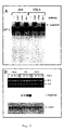
  • Panel A Selective induction and time course of MCP-1 expression. 5 x 10 5 cells were incubated in the presence of 250 U/ml of TNF- ⁇ for 5 and 18 hours, respectively. 5 ⁇ g RNA of each HeLa cells, non-tumorigenic HeLa-fibroblast hybrids ("444") and their tumorigenic segregants ("CGL3") was separated in a 1% agarose gel. After blotting, the filter was consecutively hybridized with cDNA probes encoding the MCP-1 and the ⁇ -actin gene. (-): untreated control cells. The positions of the 18 and 28S rRNAs are indicated.
  • Panel B cell morphology of HeLa-, "444"- and "CGL3" cells either untreated (A, C, E) or after simultaneous incubation with TNF- ⁇ (500 U/ml) and cycloheximide (10 ⁇ g/ml) for 4 hours at 37°C. Phase-contrast microscopy (Magnification: 100 x).
  • Panel C degradation and re-synthesis of I ⁇ B- ⁇ after treatment of HeLa-, "444"-, and "CGL3"-cells with 250 U/ml TNF- ⁇ for 30 minutes (min) or 5 hours (h). 30 ⁇ g total cellular protein was separated in a 12% SDS-PAGE gel for Western blot analysis. After electrotransfer, the filter was probed with a polyclonal antibody directed against I ⁇ B- ⁇ . To confirm equal loading, an identical filter was incubated with a ⁇ -actin specific antibody.
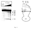
  • Panel A isotonic nuclei corresponding to an amount of 100 ⁇ g total cellular DNA from untreated (-) and TNF- ⁇ treated (250 U/ml, for 5 hours) "444"- and "CGL3"-Cells were digested with increasing amounts of Dra I (25 and 50 units for 15 minutes at 37°C). After restriction enzyme digestion, the DNAs were processed as described in Fig. 3. "Cont”: DNA from nuclei incubated in digestion buffer for 15 minutes at 37°C without any enzyme. The schematic presentation shows the length and the derivation of the subbands generated after Dra I/Kpn I cleavage (for details, see Fig. 3).
  • Panel B same as panel A, only after treatment of the nuclei with Apa I restriction endonuclease.
  • the length of the subbands generated after DNAse I/Hind III digestion is indicated in kb.
  • Const 1 nuclei immediately after preparation
  • Const 2 DNA from nuclei incubated in digestion buffer without adding enzyme.
  • the schematic presentation below shows the location (indicated by an arrow) of the major and minor DNAse I hypersensitive regions (DHSR) relative to the Hind III site (pos -428/-423, based on the transcriptional start site).
  • the length of the parental 4.7 kb Hind III/Hind III fragment, the position of hybridization probe ("probe"), the three exons (“Exon I-III”) and the location of the chemokine specific polyadenylation signal (“poly-A”) are indicated.
  • FIG. 6 Nucleotide sequence analysis of the 5'- and 3' regulatory region of the human MCP-I gene covered by DNAse I hypersensitive regions (DHSRs) in native chromatin
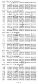
  • Nucleotide numbering was done relative to the transcriptional start site (Ueda et al., 1994, J Immunol. 153, p. 2052-2063). Some DHSRs coincide with a DNA fragment shown to harbor functional binding sites for the transcriptional factors, AP-1 and NF-KB (see nucleotide stretch ranging from pos. -2799 to -300). The positions of additional putative binding sequences for the transcription factors AP-1, NF-1 L-6, Sp1 and the restriction endonucleases Apa I and Dra I are indicated. The complete nucleotide sequence (11793 bp) of the whole MCP-1 gene locus is available at the EMBL nucleotide sequence data base (accession number: Y 18933).
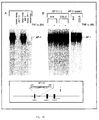
  • Panel A 2.5 ⁇ g "444" cell extract was incubated with a 32 P-Iabeled oligonucleotide from pos. +2587/+2607 (referred as AP-1 (3'), see schematic presentation depicted below).
  • Lane 1 without competition;
  • lane 2 addition of a 100-fold molar excess of unlabeled homologous oligonueleotides ("hom. comp. AP-1 3"');
  • lane 3 addition of a 100-fold molar excess of an oligonucleotide harbouring a recognition site for the transcription factor NF-1("hetero. comp.
  • NF-1 NF-1
  • lane 4 competition with a 100-fold excess of an oligonucleotide containing an AP-1 site of the human collagenase gene ("hom. comp. consensus”).
  • Panel B (lanes 1-4) band-shift analysis using the AP-1 3'-oligonucleotide ("AP-1 3'-end") after incubation with nuclear extracts derived from control (-) and TNF- ⁇ treated (+) "444"- and "CGL3" cells.
  • Lanes 5 and 6 band-shift pattern of an AP-1 oligonucleotide from the human collagenase gene ("AP-1 consens.") using either "444" or "CGL3" nuclear extracts.
  • Panel A Supershift-analysis of the AP-1 complex in "444" cells and "CGL3" cells.
  • 32 P-labeled AP-1 specific oligonucleotides derived from the 3'-DHSR (referred as "AP-1 3'-end") were incubated with nuclear extracts from "444"- and "CGL3" cells either in the absence (lanes 1 and 5) or in the presence of specific antibodies raised against c-jun (lanes 2 and 6), c-fos (lanes 3 and 7) or fra-1 (lanes 4 and 8).
  • the arrowhead marks the position of the retarded "super-shift" complexes.
  • Panel B selective induction of the human type I collagenase gene in "444" cells after TNF- ⁇ treatment (250 U/ml) for 30 minutes (30'), 4 and 8 hours (4h, 8h). RNA was extracted and monitored by Northern blot analysis. The same filter was consecutively hybridized with a cDNA probe homologous to the human collagenase and the ⁇ -actin. gene. The ethidium bromide stained gel shows the integrity and the position of the 28S and 1M rRNA. (-): untreated control cells.
  • the HindIII/HindIII-fragment (c.f. Fig. 5) was cloned into pBluescript, purified by CsCI centifugation and digested with increasing amounts of S1 nuclease.
  • the DNas were separated on 1% agarose gels and hybridized with a cloned PCR fragment (position -539 to +52 relative to the transcriptional start site) located adjacent to the HindIII site for indirect-end-labeling.
  • the length of the subbands generated after S1 nuclease digestion is indicated in kb.
  • Cont1 untreated plasmids
  • Cont.2 DNA incubated in digestion buffer without adding enzyme.
  • the schematic presentation below shows the location (indicated by an arrow) of the S1 hypersensitive sites (SHS) relative to the HindIII site (position -428/-423).
  • the length of the parental 4.7 kb HindIII/HindIII fragment, the position of the hybridization probe ("probe"), the three exons (Exons I-III) and the localisation of the chemokine specific polyadenylation site (polyA) are indicated.
  • HPV 18-positive cervical carcinoma cells HeLa
  • non-tumorigenic somatic cell hybrids made between HeLa cells and normal human fibroblasts (“444")
  • CGL3 tumorigenic segregants
  • CGL3 tumorigenic segregants
  • human glioblastoma cell line A172 Tumorbank DKFZ, Heidelberg
  • Dulbecco's modified Eagle's medium supplemented with 10% fetal calf serum and 1% penicillin/streptomycin
  • cytokine treatment the cells were incubated with TNF- ⁇ (specific activity: 1 x 10 8 units/mg) (Pharma Biotechnologie, Hannover, Germany) for different periods of time as described in the figure legends. Cycloheximide (Sigma, St. Louise, USA) was stored as a 10 mg/ml stock solution at -20°C.
  • Total cellular RNA was extracted according to the guanidine-thiocyanate procedure (Chomczynski and Sacchi, Anal. Biochem. 162 (1987), 156-159). Approximately 5 ⁇ g of total cellular RNA was separated on 1% agarose gels as described elsewere (Rösl et al., J. Virol. 68 (1994), 2142-2150).
  • the rate of apoptosis was determined by a cell death detection ELISA PLUS kit (Roche) as recommended by the manufacturer.
  • the enrichment factor was calculated by setting the absorbance of the untreated cells as 1.
  • a chromosome 17 specific cosmid library (Resourcentechnik Berlin; Zehetner, G. Lehrach, H. (1994), Nature 367, p. 489-491) was screened using the cDNA of MCP-1 as a probe.
  • the sequence of the flanking genomic regions was finally determined using two plasmid clones harboring overlapping inserts of about 7.7 kb (a Kpn I/Kpn I-fragment for the 5'-region) and a 4.7 kb (Hind III/Hind III-fragment for intron/exon and the 3'-region) in size.
  • DNA sequence analysis was done on both strands using chemically synthesized primers and fluorescent sequencing on an automatic sequencer (Model 377, Perkin-Elmer/Applied Biosystems (Foster City, USA) and the dye terminator method according to the manufacturers protocol ("ABI PRISM Big Dye Ready Reaction Terminator Cycle Sequencing Kit", Perkin-Elmer/Applied Biosystems). Putative binding sites for transcription factors were identified by the computer program "Transfac version 3.3" (Wingender et al., Nucl. Acids. Res. 25 (1997), 265-268).
  • HPV 18 represents the unit-length of HPV 18 DNA cloned in pBR322 (Boshart et al., EMBO J. 3 (1984), 1151-1157). pHFAI (Gunning et al., Mol. Cell. Biol. 3 (1983), 787-795) containing an approximately full-length insert of the fibroblast ⁇ -actin gene was a generous gift from L. Kedes (Medical Center, Palo Alto, USA). The full length cDNA encoding the monocyte-chemoattractant-protein-1 (MCP-1) (Rollins et al., Mol. Cell. Biol.
  • Nuclei from exponentially growing cells were prepared as described recently (Maehama et al., Int. J. Cancer 76 (1998), 639-646). Heat-denaturated linearized DNA fragments were fixed onto nitrocellulose filters using the "S+S minifold II slot apparatus" (Schleicher and Schuell, Dassel, Germany) and hybridized with equal amounts of 32 P-labelled RNA. After 48h, the filters were washed with 2 x SSC/0.1% SDS at 68°C.
  • Nuclei from HeLa-, "CGL3"- and “444"-cells were prepared (Rösl et al., Mol. Carcinog. 2 (1989), 72-80) and digested as described in the figure legends. Southem blot analyses were done using standard protocols.
  • oligonucleotides were used: the AP-1 binding site (5'-GGAAGGT TGAGTCA AGGGATT-3') located within the DNAse I hypersensitive site downstream of the MCP-1 gene (pos.
  • EXAMPLE 2 Selective MCP-1 inducibility by TNF- ⁇ : demonstration of the integrity of the TNF- ⁇ signal transduction pathway in malignant and non-malignant HPV 18-positive cells
  • MCP-1 was transcribed and TNF- ⁇ indicible only in non-malignant hybrids made between HeLa cervical carcinoma cells and normal human fibroblasts (Fig. 1A, designated as "444").
  • CGL3 tumorigenic segregants from the same hybrids
  • CGL3 parental tumorigenic HeLa cells
  • TNF- ⁇ pathway The integrity of the TNF- ⁇ pathway was further confirmed by the rapid proteolysis (after 30 minutes) and delayed resynthesis (after 5 hours) of the inhibitory component I ⁇ B- ⁇ (Fig. 1D), leading to the activation of the transcription factor NF- ⁇ B.
  • EXAMPLE 3 MCP-1 mRNA expression is regulated at the level of initiation of transcription
  • EXAMPLE 4 Mapping of DNAse I hypersenvitive regions (DHSRs) at the 5' - and 3'-end of the MCP-1 gene in native chromatin: evidence for a novel TNF- ⁇ inducible DHSR at the 3'-end
  • One is located at the 5'-end, being characterized by their higher complexity in terms of location and distribution of DHSRs; the other is situated approximately 600 bp downstream of the MCP-1 specific polyadenylation signal within a 3'-regulatory domain, whose chromatin structure only became accessible after cytokine treatment.
  • EXAMPLE 5 DNA sequencing and band-shift analysis led to the detection of a novel AP-1 site within the 3'-DNAse I hypersensitive region
  • a chromosome-specific library was screened to isolate the whole genomic locus of the human MCP-1 gene using the corresponding cDNA (Rollins et al., 1989, Mol. Cell. Biol. 9, p. 4687-4695) and a PCR amplified promoter fragment (Rösl et al., 1994, J. Virol. 68, p. 2142-2150) as hybridization probe.
  • DNA sequence analysis essentially confirmed the sequence data of the 3.8 kb upstream region published by Ueda and co-workers (Ueda et al., 1994) (see also Fig. 6).
  • Single nucleotide variations may be dependent on different origins of the clones used for scquencing. Inspecting the nucleotide sequence surrounding the minor 5'-DHSR 1.5 kb upstream of the Kpn I site, a putative NF-IL-6 (C/EBP- ⁇ ) consensus sequence was found (Fig. 6). Similar sequences could be detected within the minor 2.0 kb subfragment and the major 3'-end DHSR 2.7-3.4 bp subfragments (see Fig. 6). The nucleotide stretch covered by the major DHSR in native "444"-chromatin, where two contiguous NF- ⁇ B sites and one AP-1 site are present, could be detected (Fig. 6).
  • an additional downstream AP-1 site (5'-TGAGTCA-3'; pos. +2594/+2600) (Fig. 6) could be detected.
  • electrophoretic band-shift assays were performed. Using the oligonucleotide 5'-GGAAGGT TGAGTCA AGGGATT-3' (pos. +2587/+2607), a single band can be visualized after incubation with a nuclear extract obtained from "444"-cells (Fig. 7A, lane 1).
  • Antibodies which have been used recognized c-Fos (epitope corresponding to amino acids 3-16 mapping at the N-terminus of human c-Fos protein), Fra-1 (epitope corresponding to amino acids 3-22 mapping at the N-terminus of the human Fra-1 protein), c-Jun (epitope corresponding to amino acids 56-69 mapping within the carboxy-terminal domain of the mouse c-Jun protein; recognizes both the unphosphorylated and phosphorylated form of c-Jun). All antibodies were obtained from Santa Cruz Biotechnology (Santa Cruz, California, USA) as "TransCruz" supershift reagents (c.f. Soto et al. (1999), Oncogene 18, p.

Landscapes

  • Health & Medical Sciences (AREA)
  • Life Sciences & Earth Sciences (AREA)
  • Chemical & Material Sciences (AREA)
  • Medicinal Chemistry (AREA)
  • Organic Chemistry (AREA)
  • Zoology (AREA)
  • Gastroenterology & Hepatology (AREA)
  • General Health & Medical Sciences (AREA)
  • Proteomics, Peptides & Aminoacids (AREA)
  • Epidemiology (AREA)
  • Biochemistry (AREA)
  • Immunology (AREA)
  • Bioinformatics & Cheminformatics (AREA)
  • Animal Behavior & Ethology (AREA)
  • Engineering & Computer Science (AREA)
  • Public Health (AREA)
  • Veterinary Medicine (AREA)
  • Toxicology (AREA)
  • Pharmacology & Pharmacy (AREA)
  • Biophysics (AREA)
  • Genetics & Genomics (AREA)
  • Molecular Biology (AREA)
  • Marine Sciences & Fisheries (AREA)
  • Medicines That Contain Protein Lipid Enzymes And Other Medicines (AREA)
  • Pharmaceuticals Containing Other Organic And Inorganic Compounds (AREA)
  • Micro-Organisms Or Cultivation Processes Thereof (AREA)
  • Medicines Containing Material From Animals Or Micro-Organisms (AREA)

Abstract

Described is a nucleic acid molecule comprising (a) a nucleic acid sequence encoding the monocyte-chemoattractant-protein-1 (MCP-1) or a protein having the biological activity of the monocyte-chemoattractant-protein-1 (MCP-1) and (b) a hypersensitivity region. Also described are pharmaceutical compositions for the treatment of diseases associated with dysregulation of MCP-1 expression, e.g. atherosclerosis or cancer. <IMAGE>

Description

The present invention relates to a nucleic acid molecule comprising (a) a nucleic acid sequence encoding the monocyte-chemoattractant-protein-1 (MCP-1) or a protein having the biological activity of the monocyte-chemoattractant-protein-1 (MCP-1); and (b) a hypersensitive region. The present invention also relates to pharmaceutical compositions for the treatment of diseases associated with dysregulation of MCP-1 expression, e.g. atherosclerosis or cancer. These pharmaceutical compositions comprise either compounds directly or indirectly interacting with the hypersensensitive regions or are based on the replacement of the original hypersensitive sequences by mutated versions thereof.
The monocyte-chemoattractant-protein-1 (MCP-1) triggers the infiltration and activation of cells of the monocyte-macrophage lineage, representing known producers of growth-modulatory cytokines. Synthesis of cytokines can be considered an important regulatory loop in the immunological surveillance, since TNF-α or TGF-β can negatively interfere with the expression of high risk human papillomaviruses (HPV) types, the causative agents of cancer of the cervix uteri. Besides disturbance of a functional T-cell surveillance, dys-regulation of chemokine expression may represent another important counter-selection event during the multi-step progression to cervical cancer. Consistent with this anticipation was the recent finding that MCP-1 expression indeed gradually disappears in different stages of premalignant cervical intraepithelial neoplasias, a process, which may explain the severe reduction of intraepithelial macrophages and Langerhans' cells. It is also known that MCP-1 does not only play a critical role during tumor progression but also as regards atherosclerosis. However, due to the fact that so far the knowledge about the complex regulation of MCP-1 gene expression and regulatory regions involved was limited, a therapeutically useful modulation of MCP-1 gene expression was not possible.
Thus, the technical problem underlying the present invention is to provide means for searching active substances for the treatment of diseases associated with an abnormal MCP-1 expression, e.g. atherosclerosis and cancer.
The solution to said technical problem is achieved by providing the embodiments characterized in the claims.
It has been, surprisingly, found that the expression of MCP-1 is dependent on a variety of cis-acting sequences which were so far not known. In the experiments leading to the present invention it could be shown that the expression of MCP-1 is closely linked with a non-tumorigenic phenotype in somatic cell hybrids made between the human papilloma virus type 18 (HPV 18) positive cervical carcinoma cell line HeLa and normal human fibroblasts. In contrast, MCP-1 transcription is absent in tumorigenic segregants derived from the same hybrids or in parental HeLa cells. Selectivity of MCP-1 transcription, which is regulated at the level of initiation of transcription, is mainly based on differences in the location and extension of DNAse I-hypersensitive regions (DHSR) at both ends of the gene or S1-hypersensitive sites (SHS). DNA sequencing showed that the hypersensitive sites often coincide with binding sites for transcription factors, like an AP-1 site, SP1 site, NF-IL6 site or NF kappa B site. Analyses of AP-1 composition revealed that MCP-1 is only expressed in those cells where jun-family members were mainly heterodimerized with the fos-related protein fra-1. In contrast, in tumorigenic cells the 1:1 ratio between jun and fra-1 is disturbed and the MCP-1 gene is no longer expressed. Hence, alterations in the heterodimerization pattern of AP-1 and its selective accessibility to opened chromatin may represent a novel regulatory pathway in the regulation of chemokines in malignant and non-malignant HPV-positive cells.
In particular the present invention relates to a nucleic acid molecule comprising
  • (a) a nucleic acid sequence encoding the monocyte-chemoattractant-protein-1 (MCP-1) or a protein having the biological activity of the monocyte-chemoattractant-protein-1 (MCP-1); and
  • (b) a 3'-DHSR comprising a nucleic acid molecule which is located 2430 bp to 3019 bp downstream of the transcriptional start site of the MCP-1 gene, or a 3'-DHSR comprising a nucleic acid molecule which is located 1550 bp to 1749 downstream of the transcriptional start site of the MCP-1 gene, or a 3'-DHSR comprising a nucleic acid molecule which is located 750 bp to 899 bp downstream of the transcriptional start site of the MCP-1 gene, or a 5'-DHSR comprising a nucleic acid molecule which is located 500 bp to 251 bp upstream of the transcriptional start site of the MCP-1 gene, or a 5'-DHSR comprising a nucleic acid molecule which is located 1300 bp to 1001 bp upstream of the transcriptional start site of the MCP-1 gene, or a 5'-DHSR comprising a nucleic acid molecule which is located 5050 bp to 4751 bp upstream of the transcriptional start site of the MCP-1 gene, or a S1 hypersensitive site comprising a nucleic acid molecule which is located in the 1st intron (+180 to +350) of the MCP-1 gene
  • As used herein, the term "a nucleic acid molecule encoding the monocyte-chemoattractant-protein-1 (MCP-1) or a protein having the biological activity of the monocyte-chemoattractant-protein-1 (MCP-1)" relates to the published sequence of the MCP-1 gene, e.g. the EMBL database sequence (accession number: Y 18933), the sequence published by Rollins et al., Mol. Cell. Biol. 9 (1989), 4687-4695) or a related sequence which still encodes a protein having at least one of the acitivities of MCP-1.
    These related sequences comprise sequences which hybridize to the above published MCP-1 nucleic acid sequences. As used herein, the term "hybridize" has the meaning of hybridization under conventional hybridization conditions, preferably under stringent conditions as described, for example, in Sambrook et al., Molecular Cloning, A Laboratory Manual, 2nd edition (1989) Cold Spring Harbor Laboratory Press, Cold Spring Harbor, NY. Also contemplated are nucleic acid sequences that hybridize to the MCP-1 nucleic acid sequences at lower stringency hybridization conditions. Changes in the stringency of hybridization and signal detection are primarily accomplished through the manipulation of formamide concentration (lower percentages of formamide result in lowered stringency), salt conditions, or temperature. For example, lower stringency conditions include an overnight incubation at 37°C in a solution comprising 6X SSPE (20 x SSPE = 3M NaCI; 0.2M NaH2PO4; 0.02M EDTA, pH 7.4), 0.5% SDS, 30% formamide, 100 µg/ml salmon sperm blocking DNA, followed by washes at 50°C with 1 x SSPE, 0.1% SDS. In addition, to achieve even lower stringency, washes performed following stringent hybridization can be done at higher salt concentrations (e.g. 5 x SSC). Variations in the above conditions may be accomplished through the inclusion and/or substitution of alternate blocking reagents used to suppress background in hybridization experiments. The inclusion of specific blocking reagents may require modification of the hybridization conditions described above, due to problems with compatibility.
    These related nucleic acid sequences also include sequences that are degenerate as a result of the genetic code or fragments, derivatives and allelic variants of the nucleic acid molecules described above encoding a MCP-1 protein or a protein having MCP-1 activity. "Fragments" are understood to be parts of the nucleic acid sequences that are long enough to encode one of the described proteins. The term "derivative" in this context means that the sequences of these molecules differ from the sequences of the nucleic acid sequences described above at one or several positions but have a high level of homology to these sequences. Homology hereby means a sequence identity of at least 40%, in particular an identity of at least 60%, preferably of more than 80% and particularly preferred of more than 90%. These proteins encoded by the nucleic acid molecules have a sequence identity to the amino acid sequence encoded by the EMBL clone (Y 18933) of at least 80%, preferably of 85% and particularly preferred of more than 90%, 95%, 97% and 99%. The deviations to the above-described nucleic acid sequences may have been produced by deletion, substitution, insertion or recombination.
    The nucleic acid sequences that are homologous to the above-described sequences and that represent derivatives of these sequences usually are variations of these molecules that represent modifications having the same biological function. They can be naturally occurring variations, for example sequences from other organisms, or mutations that can either occur naturally or that have been introduced by specific mutagenesis. Furthermore, the variations can be synthetically produced sequences. The allelic variants can be either naturally occurring variants or synthetically produced variants or variants produced by recombinant DNA processes.
    The hypersensitivity regions as identified in the present invention are depicted in Figure 6. One of the preferred hypersensitive regions is the 3'-DHSR comprising the nucleic acid sequences from position + 2430 bp to + 3019 bp as shown in Fig. 6. In a more preferred embodiment of the present invention, the 3'-DHSR comprises the nucleic acid sequence GGAAGGTTGAGTCAAGGATT. In an even more preferred embodiment of the present invention, the 3'-DHSR comprises the nucleic acid molecule TGAGTCA. As used herein, the 3'-DHSR sequences also comprise sequences containing alterations compared to the particular sequences shown in the figures or to the published sequences which do not lead to a loss of the function as cis-acting element, e.g. as regards gene expression.
    The S1 hypersensitivity region (SHS) in the 1st intron (0.6 kb) of MCP-1 has been identified by S1 nuclease mapping (c.f. Figs. 9 and 10). This SHS contains an AP-1 site which has been verified by band-shift analysis and TNF induction.
    The present invention also relates to a nucleic acid molecule wherein the hypersensitive sites contain mutations resulting in a modified DNAse I hypersensitivity and S1 hypersensitivity, respectively, or altered interaction with transcription factors, in particular altered interaction with AP-1. Generally, by means of conventional molecular biological processes it is possible (see, e.g., Sambrook et al., 1989, Molecular Cloning, A Laboratory Manual, 2nd edition Cold Spring Harbor Laboratory Press, Cold Spring Harbor, NY) to introduce different mutations. One possibility is the production of deletion mutants in which nucleic acid molecules are produced by continuous deletions from the 5'- or 3'-terminus of the DNA sequence. Another possibility is the introduction of single-point mutation at positions where a modification influences the interaction with transcription factors, e.g. AP-1. The identity of the mutated sequence with the original sequence is at least 40%, in particular at least 60%, preferably more than 80% and particularly preferred more than 90%.
    For the manipulation in prokaryotic cells by means of genetic engineering the nucleic acid molecules of the invention or parts of these molecules can be introduced into plasmids allowing a mutagenesis or a modification of a sequence by recombination of DNA sequences. By means of conventional methods (cf. Sambrook et al., 1989, Molecular Cloning: A Laboratory Manual, 2nd edition, Cold Spring Harbor Laboratory Press, NY, USA) bases can be exchanged and natural or synthetic sequences can be added. In order to link the DNA fragments with each other adapters or linkers can be added to the fragments. Furthermore, manipulations can be performed that provide suitable cleavage sites or that remove superfluous DNA or cleavage sites. If insertions, deletions or substitutions are possible, in vitro mutagenesis, primer repair, restriction or ligation can be performed. As analysis method usually sequence analysis, restriction analysis and other biochemical or molecular biological methods are used.
    The invention furthermore relates to vectors containing the nucleic acid molecules of the invention. Preferably, they are plasmids, cosmids, viruses, bacteriophages and other vectors usually used in the field of genetic engineering. Vectors suitable for use in the present invention include, but are not limited to T7-based expression vectors for expression in bacteria, the pMSXND expression vector for expression in mammalian cells and baculovirus-derived vectors for expression in insect cells. Preferably, the nucleic acid molecule of the invention is operatively linked to the regulatory elements in the recombinant vector of the invention that guarantee the transcription and synthesis of an RNA in prokaryotic and/or eukaryotic cells that can be translated. The nucleotide sequence to be transcribed can be operably linked to a promotor like a T7, metallothionein I or polyhedrin promotor.
    In a further embodiment, the present invention relates to recombinant host cells transiently or stably containing the nucleic acid molecules or vectors of the invention.
    Preferably, these cells are prokaryotic or eukaryotic cells, for example mammalian cells, bacterial cells, insect cells or yeast cells.
    The present invention also relates to a pharmaceutical composition comprising a compound which is capable of regulating the expression of the MCP-1 gene by directly or indirectly interacting with the nucleic acid sequence corresponding to the hypersensitivity regions as defined above, e.g. the transcription factor itself (e.g. AP-1) or a protein associated with the transcription factor thereby modulating its activity, or the vector containing a nucleic sequence with a modified hypersensitivity region as described above. Depending on the desired medical use these compounds may be compounds interacting with the hypersensitivity region, thus, increasing MCP-1 expression, but also compounds which inhibit the activity of compounds naturally interacting with the hypersensitivity region, thus leading to a decreased expression of MCP-1. Such a compound comprises, e.g. inhibitors which can be, for instance, structural analogues of the transcription factor or a protein associated therewith that act as antagonist. Such an inhibitor is, e.g., a synthetic organic chemical, a natural fermentation product, a substance extracted from a microorganism, plant or animal, or a peptid. Such an inhibitor ca also be an antibody or a nucleic acid molecule. The term "antibody", preferably, relates to antibodies which consist essentially of pooled monoclonal antibodies with different epitopic specificities, as well as distinct monclonal antibody preparations. Monoclonal antibodies are made from an antigen containing fragments of the transcription factor or a protein associated therewith by methods well known to those skilled in the art (see, e.g., Köhler et al., Nature 256 (1975), 495). As used herein, the term "antibody" (Ab) or "monoclonal antibody" (Mab) is meant to include intact molecules as well as antibody fragments (such as, for example, Fab and F(ab')2 fragments) which are capable of specifically binding to protein. Fab and F(ab')2 fragments lack the Fc fragment of intact antibody, clear more rapidly from the circulation, and may have less non-specific tissue binding than an intact antibody. (Wahl et al., J. Nucl. Med. 24:316-325 (1983).) Thus, these fragments are preferred, as well as the products of a FAB or other immunoglobulin expression library. Moreover, antibodies of the present invention include chimeric, single chain, and humanized antibodies.
    In a preferred embodiment, the compound comprised by the pharmaceutical composition is (a) a protein capable of interacting with the transcription factor (e.g. AP-1, SP1, NF kappa B) or (b) a nucleic acid sequence encoding said protein. Preferrably said nucleic acid sequence is integrated into a vector as described above or below, preferably a vector useful for gene therapy.
    In a more preferred embodiment, the compound comprised by the pharmaceutical composition is jun (e.g. junD, junB), fra-1, ATF-2, jab-1, fra-2 or a mixture thereof. It is apparent from the Examples below that by the administration of a mixture containing a particular ratio of jun and fra-1 expression of MCP-1 can be manipulated. Probably, it will be sufficient to overexpress one of these factors in cells (e.g. tumor cells) to reactivate the MCP-1 gene.
    For administration these compounds are preferably combined with suitable pharmaceutical carriers. Examples of suitable pharmaceutical carriers are well known in the art and include phosphate buffered saline solutions, water, emulsions, such as oil/water emulsions, various types of wetting agents, sterile solutions etc.. Such carriers can be formulated by conventional methods and can be administered to the subject at a suitable dose. Administration of the suitable compositions may be effected by different ways, e.g. by intravenous, intraperetoneal, subcutaneous, intramuscular, topical or intradermal administration. The route of administration, of course, depends on the nature of the disease and the kind of compound contained in the pharmaceutical composition. The dosage regimen will be determined by the attending physian and other clinical factors. As is well known in the medical arts, dosages for any one patient depends on many factors, including the patient's size, body surface area, age, sex, the particular compound to be administered, time and route of administration, the kind of the disease, general health and other drugs being adminstered concurrently.
    The delivery of nucleic acid molecules encoding proteins useful for regulating MCP-1 expression or of nucleic acid molecules with mutated hypersensitivity region sequences can be achieved by direct application or, preferably, by using a recombinant expression vector such as a chimeric virus containing these compounds or a colloidal dispersion system. By using appropriate vectors the mutated sequences can be inserted into the genome and, e.g., replace the original sequences. By delivering these nucleic acids to the desired target, the intracellular expression of MCP-1 and, thus, the level of MCP-1 can, e.g., be decreased resulting in the inhibition of the negative effects of MCP-1, e.g. as regards atherosclerosis. On the orther hand, by using this approach an increased MCP-1 expression (or re-expression of MCP-1) can be achieved, e.g. for the treatment or prevention of tumors, e.g. cervical carcinoma.
    The above nucleic acid sequences can be administered directly to the target site, e.g., by ballistic delivery, as a colloidal dispersion system or by catheter to a site in artery. The colloidal dispersion systems which can be used for delivery of the above nucleic acids include macromolecule complexes, nanocapsules, microspheres, beads and lipid-based systems including oil-in-water emulsions, (mixed) micelles, liposomes and lipoplexes. The preferred colloidal system is a liposome. The composition of the liposome is usually a combination of phospholipids and steroids, especially cholesterol. The skilled person is in a position to select such liposomes which are suitable for the delivery of the desired nucleic acid molecule. Organ-specific or cell-specific liposomes can be used, e.g. in order to achieve delivery only to the desired tissue. The targeting of liposomes can be carried out by the person skilled in the art by applying commonly known methods. This targeting includes passive targeting (utilizing the natural tendency of the liposomes to distribute to cells of the RES in organs which contain sinusoidal capillaries) or active targeting (for example by coupling the liposome to a specific ligand, e.g., an antibody, a receptor, sugar, glycolipid, protein etc., by well known methods). In the present invention monoclonal antibodies are preferably used to target liposomes to specific tumors via specific cell-surface ligands.
    Preferred recombinant vectors useful for gene therapy are viral vectors, e.g. adenovirus, herpes virus, vaccinia, or, more preferably, an RNA virus such as a retrovirus. Even more preferably, the retroviral vector is a derivative of a murine or avian retrovirus. Examples of such retroviral vectors which can be used in the present invention are: Moloney murine leukemia virus (MoMuLV), Harvey murine sarcoma virus (HaMuSV), murine mammary tumor virus (MuMTV) and Rous sarcoma virus (RSV). Most preferably, a non-human primate retroviral vector is employed, such as the gibbon ape leukemia virus (GaLV), providing a broader host range compared to murine vectors. Since recombinant retroviruses are defective, assistance is required in order to produce infectious particles. Such assistance can be provided, e.g., by using helper cell lines that contain plasmids encoding all of the structural genes of the retrovirus under the control of regulatory sequences within the LTR. Suitable helper cell lines are well known to those skilled in the art. Said vectors can additionally contain a gene encoding a selectable marker so that the transduced cells can be identified. Moreover, the retroviral vectors can be modified in such a way that they become target specific. This can be achieved, e.g., by inserting a polynucleotid encoding a sugar, a glycolipid, or a protein, preferably an antibody. Those skilled in the art know additional methods for generating target specific vectors. Further suitable vectors and methods for in vitro- or in vivo-gene therapy are described in the literature and are known to the persons skilled in the art; see, e.g., WO 94/29469 or WO 97/00957.
    Brief description of the drawings Figure 1:
    Panel A: Selective induction and time course of MCP-1 expression. 5 x 10⁵ cells were incubated in the presence of 250 U/ml of TNF-α for 5 and 18 hours, respectively. 5 µg RNA of each HeLa cells, non-tumorigenic HeLa-fibroblast hybrids ("444") and their tumorigenic segregants ("CGL3") was separated in a 1% agarose gel. After blotting, the filter was consecutively hybridized with cDNA probes encoding the MCP-1 and the β-actin gene. (-): untreated control cells. The positions of the 18 and 28S rRNAs are indicated. Panel B: cell morphology of HeLa-, "444"- and "CGL3" cells either untreated (A, C, E) or after simultaneous incubation with TNF-α (500 U/ml) and cycloheximide (10 µg/ml) for 4 hours at 37°C. Phase-contrast microscopy (Magnification: 100 x). Panel C: degradation and re-synthesis of IκB-α after treatment of HeLa-, "444"-, and "CGL3"-cells with 250 U/ml TNF-α for 30 minutes (min) or 5 hours (h). 30 µg total cellular protein was separated in a 12% SDS-PAGE gel for Western blot analysis. After electrotransfer, the filter was probed with a polyclonal antibody directed against IκB-α. To confirm equal loading, an identical filter was incubated with a β-actin specific antibody.
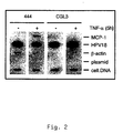
    Figure 2: Nuclear run-on analysis
    Ongoing transcription from TNF-α treated (250 U/ml, for 5 hours) and control nuclei was examined by hybridizing equal counts of 32P-UTP nascent RNA to nitrocellulose strips carrying the same amount of heat-denaturated DNA probes (2.5 µg of MCP-1, HPV 18, β-actin and p"Bluescript" plasmid DNA). To confirm identical hybridization conditions, 50 ng of total cellular DNA was loaded as an intemal reference. (-): untreated cells; (+) cells incubated with 250 U/ml of TNF-α.
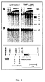
    Figure 3: DNAse I mapping analysis of the 5'-regulatory region
    "444" cells (panel A) and "CGL3" cells (panel B) were treated for 5 hours with TNF-α (250 U/ml). Isotonic nuclei equivalent to 100 µg of total DNA from either untreated or TNF-α treated cells were subsequently digested with increasing amounts of DNAse I (1.0; 2.0; 3.0; 4.0; and 5.0 units of DNAse I/100 µl volume) for 5 minutes at 37°C. After extraction, the DNAs were cleaved with Kpn I, separated on 1% agarose gels and hybridized with a cloned PCR fragment located adjacent to Kpn I (pos. -539 to +52 relative to the transcriptional start site defined by Ueda and coworkers (Ueda et al., J. Immunol. 153 (1994), 2052-2063) for indirect end-labeling. The length of the subbands generated after DNAse I/Kpn I digestion is indicated in kb. "Cont 1": nuclei lysed immediately after preparation: "Cont 2": DNA from nuclei incubated in digestion buffer without DNAse I. The schematic presentation below shows the location of the major and minor DNAse I hypersensitive regions (DHSR) relative to the Kpn I site (pos. +211/+216) in both cell lines (indicated by an arrow). The length of the parental 7.7 kb Kpn I/Kpn I fragment and the position of hybridization probe ("probe") are indicated.
    Figure 4: Accessibility of the DHSRs to restriction endonucleases in native chromatin
    Panel A: isotonic nuclei corresponding to an amount of 100 µg total cellular DNA from untreated (-) and TNF-α treated (250 U/ml, for 5 hours) "444"- and "CGL3"-Cells were digested with increasing amounts of Dra I (25 and 50 units for 15 minutes at 37°C). After restriction enzyme digestion, the DNAs were processed as described in Fig. 3. "Cont": DNA from nuclei incubated in digestion buffer for 15 minutes at 37°C without any enzyme. The schematic presentation shows the length and the derivation of the subbands generated after Dra I/Kpn I cleavage (for details, see Fig. 3). Panel B: same as panel A, only after treatment of the nuclei with Apa I restriction endonuclease.
    Figure 5: DNAse I mapping analysis of the 3'-regulatory region
    "444" cells (panel A) and "CGL3" cells (panel B) were treated with TNF-α as descibed in Fig. 3. Nuclei corresponding to 100 µg of total DNA obtained from untreated and cytokine treated cells were digested with increasing amounts of DNAse I (1.0; 2.0, 3.0, 4.0, and 5.0 units of DNAse I/100 µl volume) for 5 minutes at 37°C. After lysis, the purified DNAs were cleaved with Hind III, separated on 1% agarose gels and hybridized with a cloned PCR fragment (pos. -539 to +52 relative to the transcriptional start site) located adjacent to the Hind III site for indirect end-labeling. The length of the subbands generated after DNAse I/Hind III digestion is indicated in kb. "Cont 1": nuclei immediately after preparation; "Cont 2": DNA from nuclei incubated in digestion buffer without adding enzyme. The schematic presentation below shows the location (indicated by an arrow) of the major and minor DNAse I hypersensitive regions (DHSR) relative to the Hind III site (pos -428/-423, based on the transcriptional start site). The length of the parental 4.7 kb Hind III/Hind III fragment, the position of hybridization probe ("probe"), the three exons ("Exon I-III") and the location of the chemokine specific polyadenylation signal ("poly-A") are indicated.
    Figure 6: Nucleotide sequence analysis of the 5'- and 3' regulatory region of the human MCP-I gene covered by DNAse I hypersensitive regions (DHSRs) in native chromatin
    Nucleotide numbering was done relative to the transcriptional start site (Ueda et al., 1994, J Immunol. 153, p. 2052-2063). Some DHSRs coincide with a DNA fragment shown to harbor functional binding sites for the transcriptional factors, AP-1 and NF-KB (see nucleotide stretch ranging from pos. -2799 to -300). The positions of additional putative binding sequences for the transcription factors AP-1, NF-1 L-6, Sp1 and the restriction endonucleases Apa I and Dra I are indicated. The complete nucleotide sequence (11793 bp) of the whole MCP-1 gene locus is available at the EMBL nucleotide sequence data base (accession number: Y 18933).
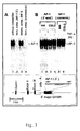
    Figure 7: Band shift analysis of the 3'AP-1 site within the major DHSR
    Panel A: 2.5 µg "444" cell extract was incubated with a 32P-Iabeled oligonucleotide from pos. +2587/+2607 (referred as AP-1 (3'), see schematic presentation depicted below). Lane 1: without competition; lane 2: addition of a 100-fold molar excess of unlabeled homologous oligonueleotides ("hom. comp. AP-1 3"'); lane 3: addition of a 100-fold molar excess of an oligonucleotide harbouring a recognition site for the transcription factor NF-1("hetero. comp. NF-1"); lane 4: competition with a 100-fold excess of an oligonucleotide containing an AP-1 site of the human collagenase gene ("hom. comp. consensus"). Panel B: (lanes 1-4) band-shift analysis using the AP-1 3'-oligonucleotide ("AP-1 3'-end") after incubation with nuclear extracts derived from control (-) and TNF-α treated (+) "444"- and "CGL3" cells. Lanes 5 and 6: band-shift pattern of an AP-1 oligonucleotide from the human collagenase gene ("AP-1 consens.") using either "444" or "CGL3" nuclear extracts. Cells were treated with TNF-α (250 U/ml) for 5 hours (5h). The position of the AP-1 specific DNA-protein complex is indicated. The schematic presentation below shows the location and sequence of the 3'-AP-1 site within the 3'-major DHSR relative to the genome organization of the MCP-1 gene.
    Figure 8:
    Panel A: Supershift-analysis of the AP-1 complex in "444" cells and "CGL3" cells. 32P-labeled AP-1 specific oligonucleotides derived from the 3'-DHSR (referred as "AP-1 3'-end") were incubated with nuclear extracts from "444"- and "CGL3" cells either in the absence (lanes 1 and 5) or in the presence of specific antibodies raised against c-jun (lanes 2 and 6), c-fos (lanes 3 and 7) or fra-1 (lanes 4 and 8). The arrowhead marks the position of the retarded "super-shift" complexes. Panel B: selective induction of the human type I collagenase gene in "444" cells after TNF-α treatment (250 U/ml) for 30 minutes (30'), 4 and 8 hours (4h, 8h). RNA was extracted and monitored by Northern blot analysis. The same filter was consecutively hybridized with a cDNA probe homologous to the human collagenase and the β-actin. gene. The ethidium bromide stained gel shows the integrity and the position of the 28S and 1M rRNA. (-): untreated control cells.
    Figure 9:
    The HindIII/HindIII-fragment (c.f. Fig. 5) was cloned into pBluescript, purified by CsCI centifugation and digested with increasing amounts of S1 nuclease. The DNas were separated on 1% agarose gels and hybridized with a cloned PCR fragment (position -539 to +52 relative to the transcriptional start site) located adjacent to the HindIII site for indirect-end-labeling. The length of the subbands generated after S1 nuclease digestion is indicated in kb. "Cont1": untreated plasmids; "Cont.2": DNA incubated in digestion buffer without adding enzyme. The schematic presentation below shows the location (indicated by an arrow) of the S1 hypersensitive sites (SHS) relative to the HindIII site (position -428/-423). The length of the parental 4.7 kb HindIII/HindIII fragment, the position of the hybridization probe ("probe"), the three exons (Exons I-III) and the localisation of the chemokine specific polyadenylation site (polyA) are indicated.
    Figure 10:
    Band-shift analysis of the AP-1 site located within the 1. intron of the MCP-1 gene. A: 2,5 µg "444" extract was incubated with a 32P-labeled oligonucleotide from position +247/+267 (GATAAGGTGACTCAGAAAAGG, refered as AP-1(I), see schematic presentation depicted below). Lane 1: without competition, lane 2: addition of a 100-fold molar excess of unlabeled homologous oligonucleotides ("homol. comp.(AP-1(I)); lane 3: addition of a 100-fold molar excess of an oligonucleotide haboring a recognition site for the transcription factor NF-1 ("heter. comp."); lane 4: competition with a 100-fold excess of an oligonucleotide containing an AP-1 site of the human collagenase gene ("comp. AP-1 consensus")
    B: First four lanes represent band-shift analysis using the AP-1 (I) oligonucleotide (AP-1 (I)) after incubation with nuclear extracts derived from control (-) and TNF-α treated (+) "444"- and "CGL3" cells. Right two lanes represent band-shift pattern of an AP-1 oligonucleotide from the human collagenase gene (AP-1 cons.) using either "444"- or "CGL3" nuclear extracts. Cells were treated with TNF-α (250 U/ml) for 5 h. The position of the AP-1 specific DNA protein complex is indicated. The schematic presentation below shows the location and the sequence of the AP-1 (I) site within the 1st intron relative to the genome organisation of the MCP-1 gene.
    The following examples illustrate the invention.
    EXAMPLE 1: GENERAL METHODS (A) Cell lines
    HPV 18-positive cervical carcinoma cells (HeLa), non-tumorigenic somatic cell hybrids made between HeLa cells and normal human fibroblasts ("444") (Stanbridge, Cancer Survey 3 (1984), 335-350), the corresponding tumorigenic segregants ("CGL3") (Stanbridge, Cancer Survey 3 (1984), 335-350) and the human glioblastoma cell line A172 (Tumorbank DKFZ, Heidelberg) were maintained in Dulbecco's modified Eagle's medium, supplemented with 10% fetal calf serum and 1% penicillin/streptomycin
    (B) Cytokines and reagents
    For cytokine treatment, the cells were incubated with TNF-α (specific activity: 1 x 10⁸ units/mg) (Pharma Biotechnologie, Hannover, Germany) for different periods of time as described in the figure legends. Cycloheximide (Sigma, St. Louise, USA) was stored as a 10 mg/ml stock solution at -20°C.
    (C) RNA analysis
    Total cellular RNA was extracted according to the guanidine-thiocyanate procedure (Chomczynski and Sacchi, Anal. Biochem. 162 (1987), 156-159). Approximately 5 µg of total cellular RNA was separated on 1% agarose gels as described elsewere (Rösl et al., J. Virol. 68 (1994), 2142-2150).
    (D) Quantification of apoptosis
    The rate of apoptosis was determined by a cell death detection ELISAPLUS kit (Roche) as recommended by the manufacturer. The enrichment factor was calculated by setting the absorbance of the untreated cells as 1.
    (E) Isolation and DNA sequence analysis of the human MCP-1 gene
    A chromosome 17 specific cosmid library (Resourcenzentrum Berlin; Zehetner, G. Lehrach, H. (1994), Nature 367, p. 489-491) was screened using the cDNA of MCP-1 as a probe. The sequence of the flanking genomic regions was finally determined using two plasmid clones harboring overlapping inserts of about 7.7 kb (a Kpn I/Kpn I-fragment for the 5'-region) and a 4.7 kb (Hind III/Hind III-fragment for intron/exon and the 3'-region) in size. DNA sequence analysis was done on both strands using chemically synthesized primers and fluorescent sequencing on an automatic sequencer (Model 377, Perkin-Elmer/Applied Biosystems (Foster City, USA) and the dye terminator method according to the manufacturers protocol ("ABI PRISM Big Dye Ready Reaction Terminator Cycle Sequencing Kit", Perkin-Elmer/Applied Biosystems). Putative binding sites for transcription factors were identified by the computer program "Transfac version 3.3" (Wingender et al., Nucl. Acids. Res. 25 (1997), 265-268). The complete 11793 bp sequence of the human MCP-1 gene locus encompassing the 5'- and 3'-regulatory regions as well as the introns is available at the EMBL nucleotide sequence data base (accession number: Y18933).
    (F) RNA and DNA probes
    HPV 18 represents the unit-length of HPV 18 DNA cloned in pBR322 (Boshart et al., EMBO J. 3 (1984), 1151-1157). pHFAI (Gunning et al., Mol. Cell. Biol. 3 (1983), 787-795) containing an approximately full-length insert of the fibroblast β-actin gene was a generous gift from L. Kedes (Medical Center, Palo Alto, USA). The full length cDNA encoding the monocyte-chemoattractant-protein-1 (MCP-1) (Rollins et al., Mol. Cell. Biol. 9 (1989), 4687-4695) was obtained from the American Type Culture Collection (Kockville, MD, USA; ATCC 61364/61365, plasmid clone pGEM-hJE34, HGLM probe ID: p11696). The cDNA of the human type 1 collagenase gene (Offringa et al., Cell 62 (1990), 527-538) was kindly provided by P. Angel (Deutsches Krebsforschungszentrum). The PCR product encompassing parts of the MCP-1 promotor ranges from -539 to +52 (according to Ueda et al., 1994) was amplified and subcloned as described previously (Rösl et al., 1994).
    (G) SDS-PAGE and Western blotting
    Cellular extracts (Dignam et al., Nucl. Acids Res. 11, (1983), 1475-1489) were separated in sodium dodecyl sulfate (SDS) 12% polyacrylamide gels (PAGE), electrotransferred to PVDF membranes (Immobilon-P, Millipore Corporation Bedford, MA, USA) and probed with polyclonal rabbit IκBα antibodies (epitope corresponding to the amino acids 297-317 mapping at the carboxy terminus, Santa Cruz Biotechnology, California, USA) as described recently (Soto et al., Oncogene 18 (1999), 3187-3198). Equal protein transfer and loading was routinously monitored by incubating identical filters with a monoclonal β-actin specific antibody (ICN Biomedicals, Ohio, USA).
    (H) Nuclear run-on assays
    Nuclei from exponentially growing cells were prepared as described recently (Maehama et al., Int. J. Cancer 76 (1998), 639-646). Heat-denaturated linearized DNA fragments were fixed onto nitrocellulose filters using the "S+S minifold II slot apparatus" (Schleicher and Schuell, Dassel, Germany) and hybridized with equal amounts of 32P-labelled RNA. After 48h, the filters were washed with 2 x SSC/0.1% SDS at 68°C.
    (I) Isolation of nuclei and nuclease digestion conditions for chromatin analysis
    Nuclei from HeLa-, "CGL3"- and "444"-cells were prepared (Rösl et al., Mol. Carcinog. 2 (1989), 72-80) and digested as described in the figure legends. Southem blot analyses were done using standard protocols.
    (J) Electrophoretic mobility shift assays
    To analyse protein/DNA interactions by electrophoretic mobility shift assays (EMSAs), the following oligonucleotides were used: the AP-1 binding site (5'-GGAAGGTTGAGTCAAGGGATT-3') located within the DNAse I hypersensitive site downstream of the MCP-1 gene (pos. +2587 / +2607), an AP-1 consensus sequence, 5'-CGCTTGATGACTCAGCCGGAA-3' (Lee et al., Cell 49 (1987), 741-752), an oligonuoleotide containing a recognition site for the nuclear factor 1 (NF-1) derived from the adenovirus origin, 5'-TTTTGGATTGAAGCCAATATGATAA-3' (Kenny and Hurwitz, J. Biol. Chem. 263 (1988), 9809-9817). The DNAs were synthesized using a phosphoramitide chemistry (Applied Biosystems synthesizer) and further purified by HPLC. Preparation of nuclear extracts, electrophoretic mobility shift and supershift assays were performed exactly as described (Soto et al., Oncogene 18 (1999), 3187-3198).
    EXAMPLE 2: Selective MCP-1 inducibility by TNF-α: demonstration of the integrity of the TNF-α signal transduction pathway in malignant and non-malignant HPV 18-positive cells
    By monitoring the expression of the MCP-1 gene in HPV 18 positive cells differing in their potential to form tumors in nude mice, it was found that MCP-1 was transcribed and TNF-α indicible only in non-malignant hybrids made between HeLa cervical carcinoma cells and normal human fibroblasts (Fig. 1A, designated as "444"). In contrast, examination of either the tumorigenic segregants from the same hybrids (designated as "CGL3") or the parental tumorigenic HeLa cells showed the complete absence of detectable transcripts under the same experimental conditions. The intensity of the MCP-1 mRNA reached a maximum 5 hours after TNF-α addition and declined to almost baseline level 18 hours after TNF-α treatment. The absence of MCP-1 inducibility in the tumorigenic cells cannot be attributed to disturbances in the cytokine signal transduction pathway, since the simultaneous application of TNF-α and a protein synthesis inhibitor (cycloheximide) for 4 hours induced blebbing of the cellular membrane (Fig. 1B), a morphological feature characteristic for TNF-α mediated apoptosis. The extend of apoptosis was quantified using a cell death detection ELISA kit which determines the amount of free histones within the cytoplasmic fraction after nuclear envelope breakdown and DNA fragmentation into discrete multiplicities of mononucleosomes (Fig. 1C). The integrity of the TNF-α pathway was further confirmed by the rapid proteolysis (after 30 minutes) and delayed resynthesis (after 5 hours) of the inhibitory component IκB-α (Fig. 1D), leading to the activation of the transcription factor NF-κB.
    EXAMPLE 3: MCP-1 mRNA expression is regulated at the level of initiation of transcription
    In order to address the question of whether the restricted expression of MCP-1 and its TNF-α inducibility in non-tumorigenic cells was due to a transcriptional block, nuclear run-on experiments were carried out. Hybridization of equal counts of 32P-UTP labeled total RNA to defined amounts of immobilized DNA showed the absence of MCP-1 expression in TNF-α treated tumorigenic "CGL3" cells, while the gene became strongly induced in "444" cells 5 hours after TNF-α addition (Fig. 2). The induction was selective, which was confirmed by a direct comparison with the degree of transcriptional activity of other reference genes such as HPV 18 or β-actin. Furthermore, loading a small amount of total cellular DNA on nitrocellulose strips demonstrated that the total 32P-UTP incorporation of the nascent RNA was the same. These data indicate that TNF-α mediated MCP-1 induction is regulated at the level of initiation of transcription rather than by post-transcriptional mechanisms.
    EXAMPLE 4: Mapping of DNAse I hypersenvitive regions (DHSRs) at the 5' - and 3'-end of the MCP-1 gene in native chromatin: evidence for a novel TNF-α inducible DHSR at the 3'-end
    In order to obtain more information on MCP-1 transcriptional regulation, the chromatin structure of the whole MCP-1 locus was examined. As shown in Figure 3A, there is one major DNAse I hypersensitive region (DHSR) in "444"-cells, whose maximum DNAse I cutting frequency was located approximately 2.4-2.9 kb upstream of the Kpn I site (Pos. +211/+216.relative to the transcriptional start site; see Ueda et al., 1994). In contrast, the major DHSR visible in "CGL3" cells has a smaller extension only ranging from 2.7-2.9 kb (Fig. 3B). In addition, the 5'-chromatin structure in "444"-cells revealed the presence of two minor DHSRs (see schematic presentation below): a distal and a proximal one, located 5.1 and 1.5 kb, respectively, upstream of the Kpn I site (for DNA sequence analysis, see Fig. 6).
    Taking advantage that the restriction endonuclease Dra I is located not only within the minor distal DHSR (pos. -1259/-1254) but also at the 3'-boundary of the major "444"-DHSR, (pos. -2186/-2191, see also Fig. 6), incubation of isolated nuclei with Dra I provides additional evidence for the larger size and higher TNF-α mediated susceptibility of this chromatin stretch in non-malignant cells. As shown in Fig. 4A, digestion of "444" derived nuclei led to a fast and strong appearance of two Dra I-derived subfragments (2.4 and 1.5 kb, respectively), whose intensity were even pronounced after TNF-α application. Quantifying the resulting subbands relative to the intensity of the undigested parental 7.7 kb fragment showed that already at 15 minutes incubation with 25 units of Dra I was sufficient to cleave almost 45% of the chromatin (corresponding to 100 µg DNA). In contrast, the same region was much more resistant in untreated "CGL3" control cells (Fig. 4A). Accessibilily to Dra I was only weakly elevated by TNF-α (accumulation of the cleaved subbands increased from 6 to 12% relative to the parental fragment), unequivocally demonstrating a different degree of chromatin decondensation in both cell lines. Treating the same nuclei with Apa I (pos. -2692/-2697), a restriction endonuclease which is located directly within the major 5'-DHSRs in both cell lines, no differences in the temporal appearance and the intensity of the generated subbands could be observed (Fig. 4B).
    Cytokine treatment led to a strong appearance of a prominent 3'-DHSR, which is located between 2.7-3.4 kb downstream of the Hind III recognition site (pos. -428/-423). Furthermore, several sensitive regions could be noticed (Fig. 5, see schematic overview). It should be stressed that major 3'-DHSR formation was not merely the consequence of MCP-1 transcription per se, because the chromatin of the untreated "444" control cells was resistant against DNAse I (Fig. 5, panel A, left), despite ongoing baseline MCP-1 expression was detectable (see Fig. 1, panel. A). Furthermore, TNF-α kinetics and subsequent DNAse I mapping analyses indicated that DHSR configuration even slightly preceded MCP-1 induction, suggesting that changes in the nucleosomal organization was a requisite rather than a consequence of TNF-α mediated MCP-1 transcription. Carrying out the same experimental approach for both the tumorigenic and segregants (Fig. 5B) or the parental HeLa cells, no 3'-DHSR could be detected. One can therefore conclude that the differences of MCP-1 transcription and TNF-α inducibility in tumorigenic and non-tumorigenic HPV-positive cells is obviously mediated by a concerted action of at least two different regulatory regions. One is located at the 5'-end, being characterized by their higher complexity in terms of location and distribution of DHSRs; the other is situated approximately 600 bp downstream of the MCP-1 specific polyadenylation signal within a 3'-regulatory domain, whose chromatin structure only became accessible after cytokine treatment.
    EXAMPLE 5: DNA sequencing and band-shift analysis led to the detection of a novel AP-1 site within the 3'-DNAse I hypersensitive region
    Since the MCP-1 gene is located at chromosome 17 a chromosome-specific library was screened to isolate the whole genomic locus of the human MCP-1 gene using the corresponding cDNA (Rollins et al., 1989, Mol. Cell. Biol. 9, p. 4687-4695) and a PCR amplified promoter fragment (Rösl et al., 1994, J. Virol. 68, p. 2142-2150) as hybridization probe. DNA sequence analysis essentially confirmed the sequence data of the 3.8 kb upstream region published by Ueda and co-workers (Ueda et al., 1994) (see also Fig. 6). Single nucleotide variations may be dependent on different origins of the clones used for scquencing. Inspecting the nucleotide sequence surrounding the minor 5'-DHSR 1.5 kb upstream of the Kpn I site, a putative NF-IL-6 (C/EBP-β) consensus sequence was found (Fig. 6). Similar sequences could be detected within the minor 2.0 kb subfragment and the major 3'-end DHSR 2.7-3.4 bp subfragments (see Fig. 6). The nucleotide stretch covered by the major DHSR in native "444"-chromatin, where two contiguous NF-κB sites and one AP-1 site are present, could be detected (Fig. 6). Since all three cis-regulatory sequences were shown to be functional in DNA binding assays, they were not further followed up in this experiment. Both the distal 5.1 kb and the proximal 1.5 kb minor 5'-DHSRs coincide with putative consensus sequences for the transcription factor Sp 1.
    Scanning the 3'-end for potential regulatory sequences, an additional downstream AP-1 site (5'-TGAGTCA-3'; pos. +2594/+2600) (Fig. 6) could be detected. To prove the functionality of this sequence in terms of DNA binding, electrophoretic band-shift assays were performed. Using the oligonucleotide 5'-GGAAGGTTGAGTCAAGGGATT-3' (pos. +2587/+2607), a single band can be visualized after incubation with a nuclear extract obtained from "444"-cells (Fig. 7A, lane 1). The specificity of AP-1 binding was confirmed in competition experiments by adding a 100-fold molar unlabeled excess of either the homologous oligonucleotide (lane 2; "hom. comp. AP-1 3"', 5'-GGAAGGTTGAGTCAAGGGATT-3') or a oligonucleotide containing the AP-1 site (lane 4; "comp. AP-1 consensus", 5'-CGCTTGATGACTCAGCCGGAA-3') of the collagenase TPA responsive element, leading in both cases to complete disappearance of the 3'-AP-1 retarded band. No competition, however, could be achieved, when a heterologous, nuclear factor 1 ("NF-1") specific oligonucleotide (lane 3; "hetero.comp. NF-1", 5'-TTTTGGATTGAAGCCAATATGATAA-3') was applied. In order to discern whether or nor the extent of AP-1 binding is different between "444"- and "CGL3"-cells and whether the affinity was modulated by TNF-α, the binding properties of different extracts were analysed in the same polyacrylamide gel (Fig. 7B). Although "CGL3"-cells lack MCP-1 expression (see Fig. 1 and 2), all nuclear extracts showed identical binding affinity independently of whether the cells were treated with TNF-α or not prior to cell harvesting (Fig. 7B, lanes 1-4). The absence of enhanced AP-1 binding was not an exceptional feature of the 3'-AP-1 site, because the affinity to the collagenase specific site was also significantly altered (Fig. 7B, lanes 5 and 6).
    Since it could previously be demonstrated that the composition of AP-1 differs considerably with respect to the jun/fos and the jun/fra-1 ratio in tumorigenic and non-tumorigenic cells, band-shift assays in combination with specific antibodies raised against AP- 1 family members were carried out. Antibodies which have been used recognized c-Fos (epitope corresponding to amino acids 3-16 mapping at the N-terminus of human c-Fos protein), Fra-1 (epitope corresponding to amino acids 3-22 mapping at the N-terminus of the human Fra-1 protein), c-Jun (epitope corresponding to amino acids 56-69 mapping within the carboxy-terminal domain of the mouse c-Jun protein; recognizes both the unphosphorylated and phosphorylated form of c-Jun). All antibodies were obtained from Santa Cruz Biotechnology (Santa Cruz, California, USA) as "TransCruz" supershift reagents (c.f. Soto et al. (1999), Oncogene 18, p. 3187-3198.) As illustrated in Fig. 8A, addition of a c-jun antibody resulted in a strong super-shift indicating that there exists substantial amounts of c-jun homo- and heterodimers in both cell lines (lanes 2 and 6). A completely different situation, however, was obtained when antibodies against fra-1 were added after the binding reaction. In "444"-cells, most (50%) of jun-family members (e. g. c-jun) were mainly heterodimerized with the fos-related protein fra-1 (Fig. 9A, lane 4), while in tumorigenic "CGL3"-cells only approximately 15% of the radioactivity (as determined by quantification of the radioactivity in a "Phospholmager") was retarded within the super-shift complex (lane 8). Conversely, AP-1 in "CGL3"-cells contain c-fos (between 5-7%) (lane 7), which is not detectable in non-malignant "444"-cells (lane 3), even after prolonged exposure to the autoradiography. EMSA supershift pattern was not significantly altered after TNF-α addition. To unequivocally demonstrate a different functionality of the transcription factor AP-1 upon cytokine addition in the experimental cell system, the expression of a marker gene which is known to be tightly regulated by AP-1 was studied. As already shown for MCP-1 (Fig. 1, panel A), treatment with TNF-α resulted in a strong induction of the type I collagenase exclusively in "444"-cells, while the gene was transcriptionally silent in the tumorigenic segregants of the same hybrids ("CGL3") or in the parentat HeLa cells (Fig. 8, panel B).
    Annex to the application documents - subsequently filed sequences listing
    Figure 00240001
    Figure 00250001
    Figure 00260001

    Claims (15)

    1. A nucleic acid molecule comprising
      (a) a nucleic acid sequence encoding the monocyte-chemoattractant-protein-1 (MCP-1) or a protein having the biological activity of the monocyte-chemoattractant-protein-1 (MCP-1); and
      (b) a 3'-DHSR comprising a nucleic acid molecule which is located 2430 bp to 3019 bp downstream of the transcriptional start site of the MCP-1 gene, or
      a 3'-DHSR comprising a nucleic acid molecule which is located 1550 bp to 1749 downstream of the transcriptional start site of the MCP-1 gene, or
      a 3'-DHSR comprising a nucleic acid molecule which is located 750 bp to 899 bp downstream of the transcriptional start site of the MCP-1 gene, or
      a 5'-DHSR comprising a nucleic acid molecule which is located 500 bp to 251 bp upstream of the transcriptional start site of the MCP-1 gene, or
      a 5'-DHSR comprising a nucleic acid molecule which is located 1300 bp to 1001 bp upstream of the transcriptional start site of the MCP-1 gene, or
      a 5'-DHSR comprising a nucleic acid molecule which is located 5050 bp to 4751 bp upstream of the transcriptional start site of the MCP-1 gene, or
      a S1 hypersensitive site comprising a nucleic acid molecule which is located in the 1st intron (+180 - +350) of the MCP-1 gene.
    2. The nucleic acid molecule of claim 1, wherein the 3'-DHSR comprises the nucleic acid sequence from pos. +2430 to +3019 as depicted in Figure 6.
    3. The nucleid molecule of claim 2, wherin the 3'-DHSR comprises the nucleic acid sequence GGAAGGTTGAGTCAAGGATT.
    4. The nucleic acid molecule of claim 3, wherein the 3'-DHSR comprises the nucleic acid sequence TGAGTCA.
    5. The nucleic acid molecule of any one of claims 1 to 4, wherein the hypersensitivity sequences (b) contain mutations resulting in a modified DNAse I hypersensitivity, S1 hypersensitivity and/or altered interaction with transcription factors.
    6. The nucleic acid molecule of claim 5, wherein the transcription factor is AP-1, SP1, NF-IL6 or NF-kappa B.
    7. A recombinant vector containing the nucleic acid molecule of any one of claim 1 to 6.
    8. The recombinant vector of claim 7 wherein the nucleic acid molecule is operatively linked to regulatory elements allowing transcription and synthesis of a translatable RNA in prokaryotic and/or eukaryotic host cells.
    9. A recombinant host cell which contains a nucleic acid molecule according to any one of claims 1 to 6 or the recombinant vector of claim 7 or 8.
    10. The recombinant host cell of claim 9, which is a mammalian cell, a bacterial cell, an insect cell or a yeast cell.
    11. A pharmaceutical composition comprising a compound which is capable of regulating the expression of the MCP-1 gene by directly or indirectly interacting with the nucleic acid sequence (b) of any one of claims 1 to 6 or the recombinant vector of claim 7 or 8.
    12. The pharmaceutical composition of claim 11, wherein the compound is a protein capable of interacting with a transcription factor, in particular AP-1, or a nucleic acid molecule encoding said protein.
    13. The pharmaceutical composition of claim 12, wherein the compound is jun, fra-1, ATF-2, jab-1, fra-2 or a mixture thereof.
    14. Use of the compounds as defined in any one of claims 11 to 13 for the preparation of a medicament for the treatment of atherosclerosis or cancer.
    15. Use according to claim 14, wherein the cancer is a cervical carcinoma.
    EP00114560A 2000-07-06 2000-07-06 Regulatory sequences of the human MCP-1 gene Expired - Lifetime EP1170372B1 (en)

    Priority Applications (4)

    Application Number Priority Date Filing Date Title
    EP00114560A EP1170372B1 (en) 2000-07-06 2000-07-06 Regulatory sequences of the human MCP-1 gene
    AT00114560T ATE323772T1 (en) 2000-07-06 2000-07-06 REGULATORY SEQUENCES OF THE HUMAN MCP-1 GENE
    DE60027419T DE60027419D1 (en) 2000-07-06 2000-07-06 Regulatory sequences of the human MCP-1 gene
    US09/899,276 US20020106355A1 (en) 2000-07-06 2001-07-06 Novel regulatory sequences of the MCP-1 gene

    Applications Claiming Priority (1)

    Application Number Priority Date Filing Date Title
    EP00114560A EP1170372B1 (en) 2000-07-06 2000-07-06 Regulatory sequences of the human MCP-1 gene

    Publications (2)

    Publication Number Publication Date
    EP1170372A1 true EP1170372A1 (en) 2002-01-09
    EP1170372B1 EP1170372B1 (en) 2006-04-19

    Family

    ID=8169184

    Family Applications (1)

    Application Number Title Priority Date Filing Date
    EP00114560A Expired - Lifetime EP1170372B1 (en) 2000-07-06 2000-07-06 Regulatory sequences of the human MCP-1 gene

    Country Status (4)

    Country Link
    US (1) US20020106355A1 (en)
    EP (1) EP1170372B1 (en)
    AT (1) ATE323772T1 (en)
    DE (1) DE60027419D1 (en)

    Families Citing this family (3)

    * Cited by examiner, † Cited by third party
    Publication number Priority date Publication date Assignee Title
    JP4458327B2 (en) * 2003-08-28 2010-04-28 キヤノン株式会社 Method for quantifying target substance and probe carrier used in the method
    ES2434039T3 (en) * 2006-07-24 2013-12-13 Yeda Research And Development Co. Ltd. Pharmaceutical compositions comprising ccl2 for use in the treatment of inflammation
    WO2024052734A2 (en) * 2022-07-13 2024-03-14 Intergalactic Therapeutics, Inc. Respiratory vectors and uses thereof

    Non-Patent Citations (7)

    * Cited by examiner, † Cited by third party
    Title
    DATABASE EMBL 1 January 1900 (1900-01-01), BIRREN B., ET AL.: "HOMO SAPIENS CHROMOSOME 17, CLONE HRPK.215 E 13, COMPLETE SEQUENCE", XP002161329, Database accession no. AC005549 *
    FINZER, P. ET AL.: "Differential transcriptional regulation of the monocyte-chemoattractant protein-1 (MCP-1) gene in tumorigenic and non-tumorigenic HPV 18 positive cells: The role of the chromatin structure and AP-1 composition", ONCOGENE, vol. 19, no. 29, 6 July 2000 (2000-07-06), pages 3235 - 3244, XP000926181 *
    SHYY, Y.J. ET AL.: "Fluid shear stress induces a biphasic response of human monocyte chemotactic protein 1 gene expression in vascular endothelium", PROCEEDINGS OF THE NATIONAL ACADEMY OF SCIENCES OF USA, vol. 91, no. 11, 24 May 1994 (1994-05-24), pages 4678 - 4682, XP002161316 *
    SOTO, U. ET AL.: "Conversion of HPV 18 positive non-tumorigenic HeLa-fibroblast hybrids to invasive growth involves loss of TNF-alpha mediated repression of viral transcription and modification of AP-1 transcription complex", ONCOGENE, vol. 18, no. 21, 27 May 1999 (1999-05-27), pages 3187 - 3198, XP000926178 *
    UEDA, A. ET AL.: "NF-kappaB and Sp1 Regulate Transcription of the Human Monocyte Chemoattractant Protein-1 Gene", JOURNAL OF IMMUNOLOGY, vol. 153, no. 5, 1 September 1994 (1994-09-01), pages 2052 - 2063, XP000926179 *
    UEDA, A. ET AL.: "Transcriptional Regulation of The Human Monocyte Chemoattractant Protein-1 Gene", JOURNAL OF BIOLOGICAL CHEMISTRY, vol. 272, no. 49, 5 December 1997 (1997-12-05), pages 31092 - 31099, XP002161315 *
    WAGNER, K. ET AL.: "Mapping and functional characterization of DNase I hypersensitive sites in the mouse MCP-1 locus", KIDNEY INTERNATIONAL, vol. 50, no. 5, November 1996 (1996-11-01), pages 1815, XP000926180 *

    Also Published As

    Publication number Publication date
    DE60027419D1 (en) 2006-05-24
    EP1170372B1 (en) 2006-04-19
    ATE323772T1 (en) 2006-05-15
    US20020106355A1 (en) 2002-08-08

    Similar Documents

    Publication Publication Date Title
    Friend et al. A human DNA segment with properties of the gene that predisposes to retinoblastoma and osteosarcoma
    AU744755B2 (en) The translation enhancer element of the human amyloid precursor protein gene
    Guy et al. Expression of the neu protooncogene in the mammary epithelium of transgenic mice induces metastatic disease.
    AU760224B2 (en) Isolated nucleic acid molecules which encode T cell inducible factors (TIFs), the proteins encoded, and uses thereof
    US6436700B1 (en) Anti-sense p21 k-ras
    US5874415A (en) Enhancer sequence for modulating expression in epithelial cells
    EP1009753B1 (en) Hypoxia-regulated genes
    Ehrlich et al. Identification of negative and positive regulatory elements associated with a class I major histocompatibility complex gene
    RU2198683C2 (en) Self-regulating apoptosis of inflammatory cells using genetic therapy
    US6395548B1 (en) Methods of modulating of angiogenesis
    US6524851B1 (en) Hybrid nucleic acid molecules and vectors including β-globin regulatory elements
    US5888765A (en) Endothelial-cell specific promoter
    JP2002525081A (en) Sequences characteristic of hypoxia-regulated gene transcription
    WO2002062848A2 (en) Regulation of cc chemokine receptor 3 (ccr3) expression
    EP1170372A1 (en) Regulatory sequences of the human MCP-1 gene
    KR100267837B1 (en) Inhibitors with sensitization to chemotherapeutic agents
    Pankow et al. The HTLV-I tax protein transcriptionally modulates OX40 antigen expression
    Zimmermann et al. Heterogenic mRNAs with an identical protein-coding region of the human embryonic myosin alkali light chain in skeletal muscle cells
    JP2003514528A (en) Pentraxin I and Pentraxin Receptors, Inhibitors of the Proteins and Pharmaceutical Compositions Containing the Compounds - Patent application
    US5910442A (en) Tumor suppressor gene
    US7973156B2 (en) Hypoxia-regulated genes
    US5534631A (en) Cellular factor ILF
    US6037121A (en) DNA sequence encoding a tumor suppressor gene
    US7041497B2 (en) Nuclear factor κB inducing factor
    US20040038247A1 (en) Nucleic acid constructs including a novel t-cell active promoters, and pharmaceutical compositions and methods utilizing same for regulating t-cell mediated immune response

    Legal Events

    Date Code Title Description
    PUAI Public reference made under article 153(3) epc to a published international application that has entered the european phase

    Free format text: ORIGINAL CODE: 0009012

    AK Designated contracting states

    Kind code of ref document: A1

    Designated state(s): AT BE CH CY DE DK ES FI FR GB GR IE IT LI LU MC NL PT SE

    AX Request for extension of the european patent

    Free format text: AL;LT;LV;MK;RO;SI

    17P Request for examination filed

    Effective date: 20020703

    AKX Designation fees paid

    Free format text: AT BE CH CY DE DK ES FI FR GB GR IE IT LI LU MC NL PT SE

    17Q First examination report despatched

    Effective date: 20030530

    GRAP Despatch of communication of intention to grant a patent

    Free format text: ORIGINAL CODE: EPIDOSNIGR1

    GRAS Grant fee paid

    Free format text: ORIGINAL CODE: EPIDOSNIGR3

    GRAA (expected) grant

    Free format text: ORIGINAL CODE: 0009210

    AK Designated contracting states

    Kind code of ref document: B1

    Designated state(s): AT BE CH CY DE DK ES FI FR GB GR IE IT LI LU MC NL PT SE

    PG25 Lapsed in a contracting state [announced via postgrant information from national office to epo]

    Ref country code: IT

    Free format text: LAPSE BECAUSE OF FAILURE TO SUBMIT A TRANSLATION OF THE DESCRIPTION OR TO PAY THE FEE WITHIN THE PRESCRIBED TIME-LIMIT;WARNING: LAPSES OF ITALIAN PATENTS WITH EFFECTIVE DATE BEFORE 2007 MAY HAVE OCCURRED AT ANY TIME BEFORE 2007. THE CORRECT EFFECTIVE DATE MAY BE DIFFERENT FROM THE ONE RECORDED.

    Effective date: 20060419

    Ref country code: FI

    Free format text: LAPSE BECAUSE OF FAILURE TO SUBMIT A TRANSLATION OF THE DESCRIPTION OR TO PAY THE FEE WITHIN THE PRESCRIBED TIME-LIMIT

    Effective date: 20060419

    Ref country code: BE

    Free format text: LAPSE BECAUSE OF FAILURE TO SUBMIT A TRANSLATION OF THE DESCRIPTION OR TO PAY THE FEE WITHIN THE PRESCRIBED TIME-LIMIT

    Effective date: 20060419

    Ref country code: CH

    Free format text: LAPSE BECAUSE OF FAILURE TO SUBMIT A TRANSLATION OF THE DESCRIPTION OR TO PAY THE FEE WITHIN THE PRESCRIBED TIME-LIMIT

    Effective date: 20060419

    Ref country code: AT

    Free format text: LAPSE BECAUSE OF FAILURE TO SUBMIT A TRANSLATION OF THE DESCRIPTION OR TO PAY THE FEE WITHIN THE PRESCRIBED TIME-LIMIT

    Effective date: 20060419

    Ref country code: LI

    Free format text: LAPSE BECAUSE OF FAILURE TO SUBMIT A TRANSLATION OF THE DESCRIPTION OR TO PAY THE FEE WITHIN THE PRESCRIBED TIME-LIMIT

    Effective date: 20060419

    Ref country code: NL

    Free format text: LAPSE BECAUSE OF FAILURE TO SUBMIT A TRANSLATION OF THE DESCRIPTION OR TO PAY THE FEE WITHIN THE PRESCRIBED TIME-LIMIT

    Effective date: 20060419

    REG Reference to a national code

    Ref country code: GB

    Ref legal event code: FG4D

    REF Corresponds to:

    Ref document number: 60027419

    Country of ref document: DE

    Date of ref document: 20060524

    Kind code of ref document: P

    REG Reference to a national code

    Ref country code: IE

    Ref legal event code: FG4D

    PG25 Lapsed in a contracting state [announced via postgrant information from national office to epo]

    Ref country code: IE

    Free format text: LAPSE BECAUSE OF NON-PAYMENT OF DUE FEES

    Effective date: 20060706

    PG25 Lapsed in a contracting state [announced via postgrant information from national office to epo]

    Ref country code: GB

    Free format text: LAPSE BECAUSE OF NON-PAYMENT OF DUE FEES

    Effective date: 20060719

    Ref country code: DK

    Free format text: LAPSE BECAUSE OF FAILURE TO SUBMIT A TRANSLATION OF THE DESCRIPTION OR TO PAY THE FEE WITHIN THE PRESCRIBED TIME-LIMIT

    Effective date: 20060719

    Ref country code: SE

    Free format text: LAPSE BECAUSE OF FAILURE TO SUBMIT A TRANSLATION OF THE DESCRIPTION OR TO PAY THE FEE WITHIN THE PRESCRIBED TIME-LIMIT

    Effective date: 20060719

    PG25 Lapsed in a contracting state [announced via postgrant information from national office to epo]

    Ref country code: DE

    Free format text: LAPSE BECAUSE OF FAILURE TO SUBMIT A TRANSLATION OF THE DESCRIPTION OR TO PAY THE FEE WITHIN THE PRESCRIBED TIME-LIMIT

    Effective date: 20060720

    PG25 Lapsed in a contracting state [announced via postgrant information from national office to epo]

    Ref country code: ES

    Free format text: LAPSE BECAUSE OF FAILURE TO SUBMIT A TRANSLATION OF THE DESCRIPTION OR TO PAY THE FEE WITHIN THE PRESCRIBED TIME-LIMIT

    Effective date: 20060730

    PG25 Lapsed in a contracting state [announced via postgrant information from national office to epo]

    Ref country code: MC

    Free format text: LAPSE BECAUSE OF NON-PAYMENT OF DUE FEES

    Effective date: 20060731

    PG25 Lapsed in a contracting state [announced via postgrant information from national office to epo]

    Ref country code: PT

    Free format text: LAPSE BECAUSE OF FAILURE TO SUBMIT A TRANSLATION OF THE DESCRIPTION OR TO PAY THE FEE WITHIN THE PRESCRIBED TIME-LIMIT

    Effective date: 20060919

    NLV1 Nl: lapsed or annulled due to failure to fulfill the requirements of art. 29p and 29m of the patents act
    REG Reference to a national code

    Ref country code: CH

    Ref legal event code: PL

    PLBE No opposition filed within time limit

    Free format text: ORIGINAL CODE: 0009261

    STAA Information on the status of an ep patent application or granted ep patent

    Free format text: STATUS: NO OPPOSITION FILED WITHIN TIME LIMIT

    26N No opposition filed

    Effective date: 20070122

    GBPC Gb: european patent ceased through non-payment of renewal fee

    Effective date: 20060719

    EN Fr: translation not filed
    PG25 Lapsed in a contracting state [announced via postgrant information from national office to epo]

    Ref country code: GR

    Free format text: LAPSE BECAUSE OF FAILURE TO SUBMIT A TRANSLATION OF THE DESCRIPTION OR TO PAY THE FEE WITHIN THE PRESCRIBED TIME-LIMIT

    Effective date: 20060720

    Ref country code: FR

    Free format text: LAPSE BECAUSE OF FAILURE TO SUBMIT A TRANSLATION OF THE DESCRIPTION OR TO PAY THE FEE WITHIN THE PRESCRIBED TIME-LIMIT

    Effective date: 20070309

    PG25 Lapsed in a contracting state [announced via postgrant information from national office to epo]

    Ref country code: LU

    Free format text: LAPSE BECAUSE OF NON-PAYMENT OF DUE FEES

    Effective date: 20060706

    PG25 Lapsed in a contracting state [announced via postgrant information from national office to epo]

    Ref country code: FR

    Free format text: LAPSE BECAUSE OF FAILURE TO SUBMIT A TRANSLATION OF THE DESCRIPTION OR TO PAY THE FEE WITHIN THE PRESCRIBED TIME-LIMIT

    Effective date: 20060419

    Ref country code: CY

    Free format text: LAPSE BECAUSE OF FAILURE TO SUBMIT A TRANSLATION OF THE DESCRIPTION OR TO PAY THE FEE WITHIN THE PRESCRIBED TIME-LIMIT

    Effective date: 20060419