EP1159620B1 - Detection of mtoc disrupting substances - Google Patents
Detection of mtoc disrupting substances Download PDFInfo
- Publication number
- EP1159620B1 EP1159620B1 EP00907799A EP00907799A EP1159620B1 EP 1159620 B1 EP1159620 B1 EP 1159620B1 EP 00907799 A EP00907799 A EP 00907799A EP 00907799 A EP00907799 A EP 00907799A EP 1159620 B1 EP1159620 B1 EP 1159620B1
- Authority
- EP
- European Patent Office
- Prior art keywords
- asp
- substance
- mtoc
- microtubule
- cell
- Prior art date
- Legal status (The legal status is an assumption and is not a legal conclusion. Google has not performed a legal analysis and makes no representation as to the accuracy of the status listed.)
- Expired - Lifetime
Links
Images


Classifications
-
- C—CHEMISTRY; METALLURGY
- C07—ORGANIC CHEMISTRY
- C07K—PEPTIDES
- C07K14/00—Peptides having more than 20 amino acids; Gastrins; Somatostatins; Melanotropins; Derivatives thereof
- C07K14/435—Peptides having more than 20 amino acids; Gastrins; Somatostatins; Melanotropins; Derivatives thereof from animals; from humans
- C07K14/43504—Peptides having more than 20 amino acids; Gastrins; Somatostatins; Melanotropins; Derivatives thereof from animals; from humans from invertebrates
- C07K14/43563—Peptides having more than 20 amino acids; Gastrins; Somatostatins; Melanotropins; Derivatives thereof from animals; from humans from invertebrates from insects
- C07K14/43577—Peptides having more than 20 amino acids; Gastrins; Somatostatins; Melanotropins; Derivatives thereof from animals; from humans from invertebrates from insects from flies
- C07K14/43581—Peptides having more than 20 amino acids; Gastrins; Somatostatins; Melanotropins; Derivatives thereof from animals; from humans from invertebrates from insects from flies from Drosophila
-
- C—CHEMISTRY; METALLURGY
- C07—ORGANIC CHEMISTRY
- C07K—PEPTIDES
- C07K16/00—Immunoglobulins [IG], e.g. monoclonal or polyclonal antibodies
- C07K16/18—Immunoglobulins [IG], e.g. monoclonal or polyclonal antibodies against material from animals or humans
Definitions
- the present invention relates to the use of the Asp protein in a method of identifying substances capable of arresting cells in mitosis by disrupting microtubule organising centre formation and/or maintenance. Such substances may be used in therapeutic methods to inhibit cell growth.
- the aim of many anti-cancer therapies is to inhibit cell division, i.e. mitosis, in tumour cells.
- Assembly of the spindle apparatus is a key event in cell mitosis since the spindle apparatus is required for chromosome segregation between daughter cells.
- Spindle apparatus assembly is thus a potential target for inhibition of mitosis and tumour therapy.
- the spindle apparatus comprises microtubules which are organised at its poles by a microtubule organising centre (MTOC) that in most animal cells is termed the centrosome.
- the centrosome itself usually comprises a pair of centrioles associated with a cloud of poorly defined pericentriolar material (PCM).
- PCM pericentriolar material
- the centrosome nucleates a cytoplasmic array of microtubules that project outward toward the cell perimeter.
- Duplication of the centrosome occurs during interphase.
- the two centrosomes remain together as a single complex prior to mitosis but as mitosis begins, this complex splits in two.
- Each centrosome then serves as a separate MTOC that nucleates a radial array of microtubules called an aster.
- the two asters move to opposite sides of the nucleus to form the two poles of the mitotic spindle.
- the Asp protein was present in the cytoplasm during interphase, and on microtubules in the polar regions of the spindle in mitosis until telophase, when it became associated with the central region of the spindle (Saunders et al ., 1997).
- substances that bind to Asp and interfere with the interaction of Asp with the centrosome may be used to disrupt MTOC integrity and consequently normal spindle formation, resulting in mitotic arrest. These substances may therefore also be used to inhibit mitosis and cellular proliferation.
- the present invention provides a method for identifying a substance capable of disrupting microtubule organising centre (MTOC) integrity, which method comprises contacting an Asp polypeptide as shown in SEQ ID NO: 1 or fragment thereof capable of forming and/or maintaining MTOCs in the absence of the substance, with a candidate substance in the presence of components required for MTOC formation and microtubule nucleation therefrom, and determining whether the substance disrupts MTOC integrity.
- MTOC microtubule organising centre
- disrupting MTOC integrity means reducing or preventing the formation and/or maintenance of MTOCs, particularly centrosomes, capable of nucleating microtubules assembly from an organised centre to produce aster-like polymerised microtubule structures, such as asters themselves.
- said components comprise KI-extracted centrosomes and an Asp-depleted soluble cellular extract. In another preferred embodiment, said components comprise a partially purified centrosome preparation and tubulin.
- Candidate substances identified by the assay method of the invention may be further tested in mitosis inhibition assays comprising administering substance, which has been determined to disrupt MTOC integrity by the assay method described above, to a cell and determining whether the substance inhibits mitosis in the cell.
- the present invention also provides the use of an Asp polypeptide as shown in SEQ ID NO: 1 or fragment thereof capable of stimulating formation of and/or maintaining MTOCs, in an assay for identifying a substance capable of disrupting MTOC integrity.
- Asp polypeptides for use in the assay methods of the present invention include the Drosophila Asp polypeptide, described in Saunders et al ., 1997, J. Cell Biol., Vol. 137, 4, p.881-890, and homologues, variants, derivatives and fragments thereof.
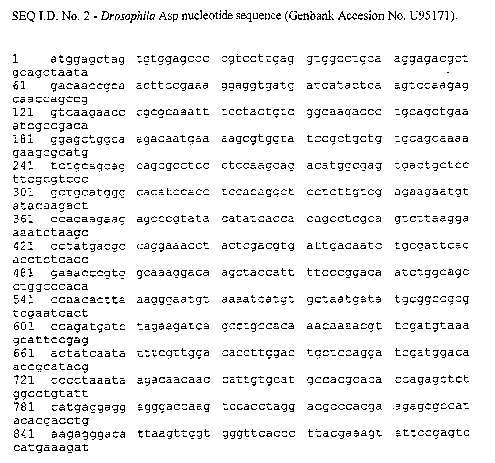
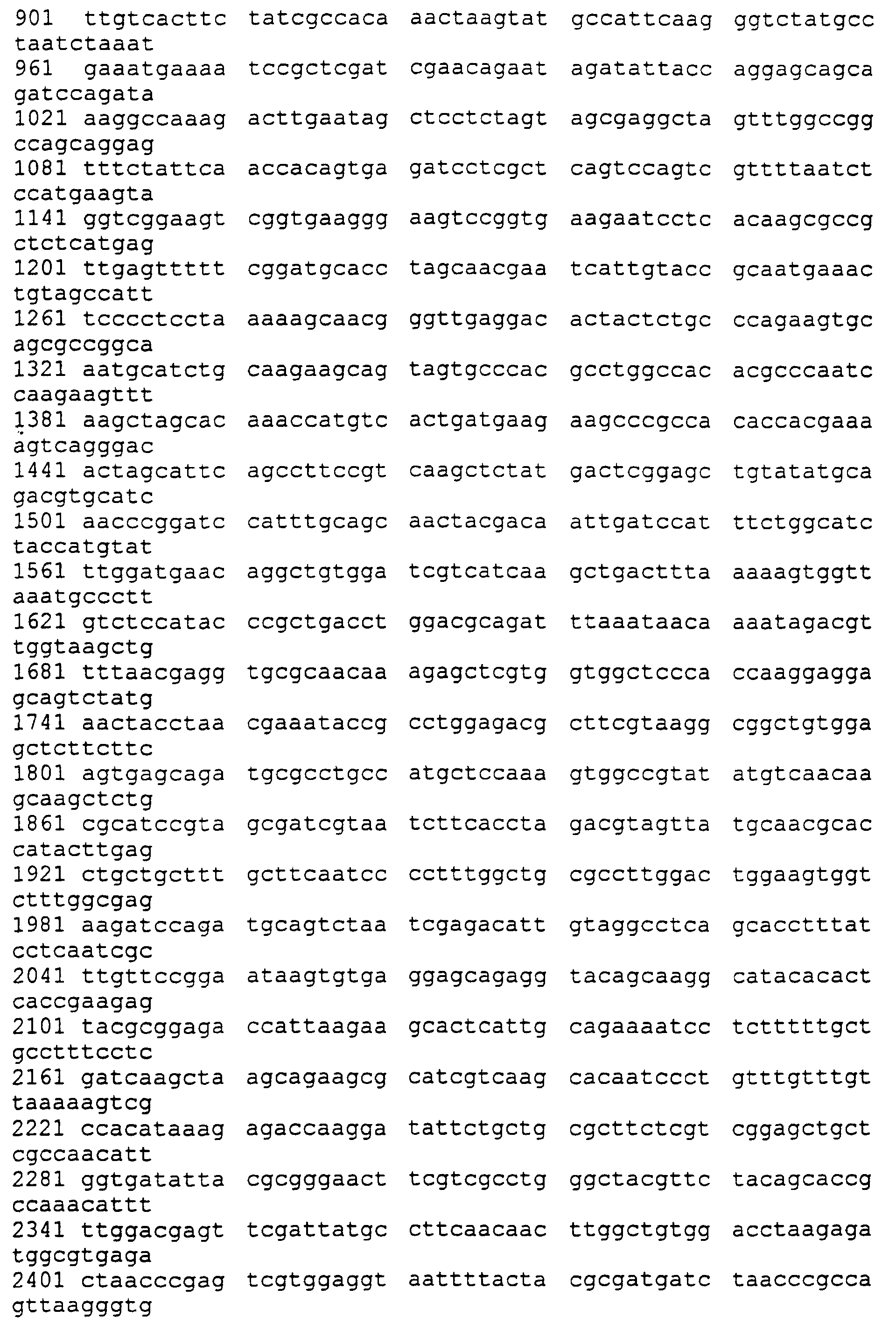
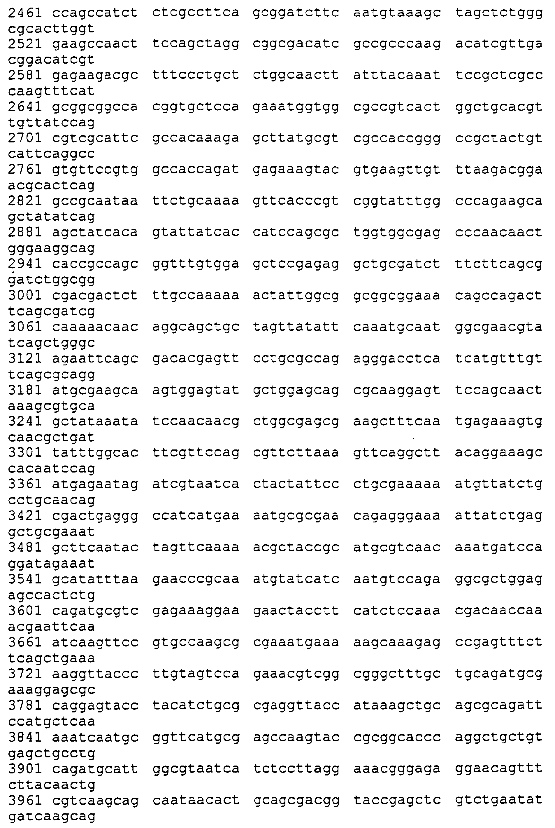
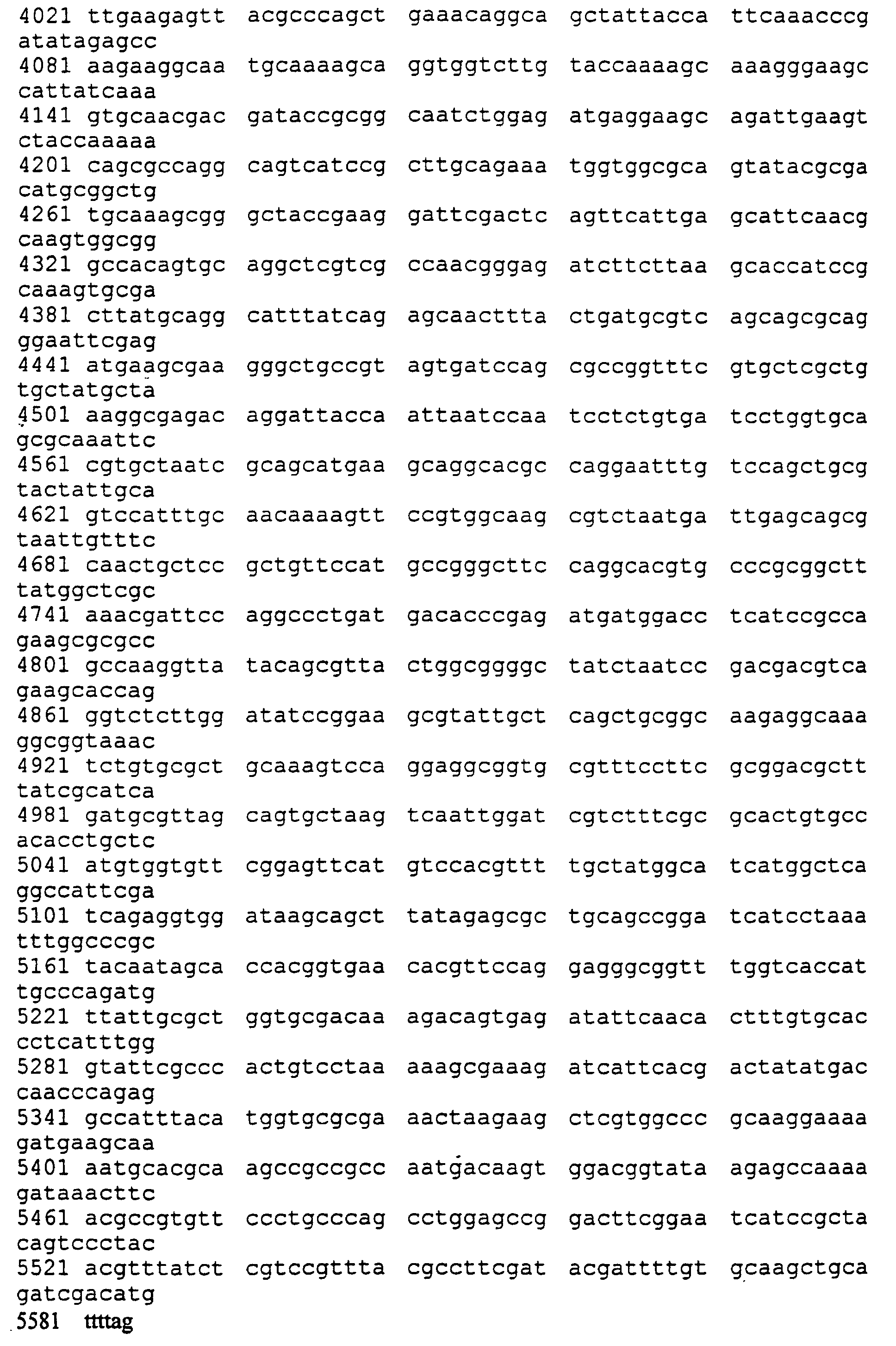
- the amino acid and nucleotide sequence of Drosophila Asp described in Saunders et al ., 1997 is shown as SEQ I.D. No 1.
- fragments With respect to fragments, truncated versions of the above may be used in the assays of the present invention provided that the fragments are capable of stimulating MTOC formation and/or maintenance as determined by the ability of the MTOCs to nucleate asters.
- microtubule nucleation centre means a centrosome that is capable of acting as a centre for microtubule nucleation.
- the centrosome is capable of acting as a centre for microtubule nucleation during mitosis.
- microtubule nucleation centre refers to an intact MTOC (centrosome) that is capable of nucleating asters of microtubules.
- microtubule organising centre integrity used throughout includes both the formation of MTOCs and their maintenance in a state capable of nucleating asters.
- Asp polypeptides for use in the invention may be made by recombinant means, for example as described below. However they may also be made by synthetic means using techniques well known to skilled persons such as solid phase synthesis. Polypeptides for use in the invention may also be produced as fusion proteins, for example to aid in extraction and purification. Examples of fusion protein partners include glutathione-S-transferase (GST), 6xHis, GAL4 (DNA binding and/or transcriptional activation domains) and ⁇ -galactosidase. It may also be convenient to include a proteolytic cleavage site between the fusion protein partner and the protein sequence of interest to allow removal of fusion protein sequences. Preferably the fusion protein will not hinder the activity of the protein of interest in maintaining microtubule organising centre integrity.
- Asp polypeptides may be purified from vertebrate cells, such as insect or mammalian cells. A detailed protocol is described in section E.
- Proteins for use in the invention may be in a substantially isolated form. It will be understood that the protein may be mixed with carriers or diluents which will not interfere with the intended purpose of the protein and still be regarded as substantially isolated.
- a protein of the invention may also be in a substantially purified form, in which case it will generally comprise the protein in a preparation in which more than 90%, e.g. 95%, 98% or 99% of the protein in the preparation is an Asp polypeptide.
- Polynucleotides for use according to the invention comprise nucleic acid sequences encoding the amino acid sequences of the invention. They also include the Drosophila Asp cDNA sequence shown as SEQ I.D. No. 2, and fragments. It will be understood by a skilled person that numerous different polynucleotides can encode the same polypeptide as a result of the degeneracy of the genetic code. In addition, it is to be understood that skilled persons may, using routine techniques, make nucleotide substitutions that do not affect the polypeptide sequence encoded by the polynucleotides for use according to the invention to reflect the codon usage of any particular host organism in which the polypeptides of the invention are to be expressed.
- Polynucleotides for use according to the present invention may comprise DNA or RNA. They may be single-stranded or double-stranded. They may also be polynucleotides which include within them synthetic or modified nucleotides. A number of different types of modification to oligonucleotides are known in the art. These include methylphosphonate and phosphorothioate backbones, addition of acridine or polylysine chains at the 3' and/or 5' ends of the molecule. For the purposes of the present invention, it is to be understood that the polynucleotides described herein may be modified by any method available in the art. Such modifications may be carried out in order to enhance the in vivo activity or life span of polynucleotides of the invention.
- Polynucleotides for use according to the invention may be used to produce a primer, e.g. a PCR primer, a primer for an alternative amplification reaction, a probe e.g. labelled with a revealing label by conventional means using radioactive or non-radioactive labels, or the polynucleotides may be cloned into vectors.
- a primer e.g. a PCR primer, a primer for an alternative amplification reaction, a probe e.g. labelled with a revealing label by conventional means using radioactive or non-radioactive labels, or the polynucleotides may be cloned into vectors.
- Such primers, probes and other fragments will be at least 15, preferably at least 20, for example at least 25, 30 or 40 nucleotides in length, and are also encompassed by the term polynucleotides for use in the invention as used herein.
- Polynucleotides such as a DNA polynucleotides and probes for use according to the invention may be produced recombinantly, synthetically, or by any means available to those of skill in the art. They may also be cloned by standard techniques.
- primers will be produced by synthetic means, involving a step wise manufacture of the desired nucleic acid sequence one nucleotide at a time. Techniques for accomplishing this using automated techniques are readily available in the art.
- Longer polynucleotides will generally be produced using recombinant means, for example using a PCR (polymerase chain reaction) cloning techniques. This will involve making a pair of primers (e.g. of about 15 to 30 nucleotides) flanking a region of the Asp sequence which it is desired to clone, bringing the primers into contact with mRNA or cDNA obtained from an animal or human cell, performing a polymerase chain reaction under conditions which bring about amplification of the desired region, isolating the amplified fragment (e.g. by purifying the reaction mixture on an agarose gel) and recovering the amplified DNA.
- the primers may be designed to contain suitable restriction enzyme recognition sites so that the amplified DNA can be cloned into a suitable cloning vector
- Polynucleotides for use in the present invention can be incorporated into a recombinant replicable vector.
- the vector may be used to replicate the nucleic acid in a compatible host cell.
- the invention provides a method of making polynucleotides for use in the invention by introducing a polynucleotide of the invention into a replicable vector, introducing the vector into a compatible host cell, and growing the host cell under conditions which bring about replication of the vector.
- the vector may be recovered from the host cell.
- Suitable host cells include bacteria such as E. coli , yeast, mammalian cell lines and other eukaryotic cell lines, for example insect Sf9 cells.
- a polynucleotide for use according to the invention in a vector is operably linked to a control sequence that is capable of providing for the expression of the coding sequence by the host cell, i.e. the vector is an expression vector.
- operably linked means that the components described are in a relationship permitting them to function in their intended manner.
- a regulatory sequence "operably linked" to a coding sequence is ligated in such a way that expression of the coding sequence is achieved under condition compatible with the control sequences.
- control sequences may be modified, for example by the addition of further transcriptional regulatory elements to make the level of transcription directed by the control sequences more responsive to transcriptional modulators.
- Vectors for use according to the invention may be transformed or transfected into a suitable host cell as described below to provide for expression of a protein of the invention.
- This process may comprise culturing a host cell transformed with an expression vector as described above under conditions to provide for expression by the vector of a coding sequence encoding the protein, and optionally recovering the expressed protein.
- the vectors may be for example, plasmid or virus vectors provided with an origin of replication, optionally a promoter for the expression of the said polynucleotide and optionally a regulator of the promoter.
- the vectors may contain one or more selectable marker genes, for example an ampicillin resistance gene in the case of a bacterial plasmid or a neomycin resistance gene for a mammalian vector. Vectors may be used, for example, to transfect or transform a host cell.
- Control sequences operably linked to sequences encoding Asp polypeptides include promoters/enhancers and other expression regulation signals. These control sequences may be selected to be compatible with the host cell for which the expression vector is designed to be used in.
- promoter is well-known in the art and encompasses nucleic acid regions ranging in size and complexity from minimal promoters to promoters including upstream elements and enhancers.
- the promoter is typically selected from promoters which are functional in mammalian cells, although prokaryotic promoters and promoters functional in other eukaryotic cells, in particular insect cells, may be used.
- the promoter is typically derived from promoter sequences of viral or eukaryotic genes. For example, it may be a promoter derived from the genome of a cell in which expression is to occur. With respect to eukaryotic promoters, they may be promoters that function in a ubiquitous manner (such as promoters of ⁇ -actin, ⁇ -actin, tubulin) or, alternatively, a tissue-specific manner (such as promoters of the genes for pyruvate kinase).
- Viral promoters may also be used, for example the Moloney murine leukaemia virus long terminal repeat (MMLV LTR) promoter, the rous sarcoma virus (RSV) LTR promoter or the human cytomegalovirus (CMV) IE promoter.
- MMLV LTR Moloney murine leukaemia virus long terminal repeat
- RSV rous sarcoma virus
- CMV human cytomegalovirus
- the promoters may also be advantageous for the promoters to be inducible so that the levels of expression of the heterologous gene can be regulated during the life-time of the cell. Inducible means that the levels of expression obtained using the promoter can be regulated.
- any of these promoters may be modified by the addition of further regulatory sequences, for example enhancer sequences.
- Chimeric promoters may also be used comprising sequence elements from two or more different promoters described above.
- Vectors and polynucleotides for use according to the invention may be introduced into host cells for the purpose of replicating the vectors/polynucleotides and/or expressing Asp polypeptides.
- Asp polypeptides may be produced using prokaryotic cells as host cells, it is preferred to use eukaryotic cells, for example yeast, insect or mammalian cells, in particular mammalian cells.
- Vectors/polynucleotides for use in the invention may introduced into suitable host cells using a variety of techniques known in the art, such as transfection, transformation and electroporation. Where vectors/polynucleotides of the invention are to be administered to animals, several techniques are known in the art, for example infection with recombinant viral vectors such as retroviruses, herpes simplex viruses and adenoviruses, direct injection of nucleic acids and biolistic transformation.
- retroviruses such as retroviruses, herpes simplex viruses and adenoviruses
- Host cells comprising polynucleotides for use according to the invention may be used to express proteins for use according to the invention.
- Host cells may be cultured under suitable conditions which allow expression of the Asp polypeptides.
- Expression of the Asp polypeptides may be constitutive such that they are continually produced, or inducible, requiring a stimulus to initiate expression.
- protein production can be initiated when required by, for example, addition of an inducer substance to the culture medium, for example dexamethasone or IPTG.
- Proteins for use according to the invention can be extracted from host cells by a variety of techniques known in the art, including enzymatic, chemical and/or osmotic lysis and physical disruption.
- Polypeptides for use in the present invention may also be purified from extracts of cells in which they naturally occur.
- Drosophila Asp may be purified from Drosophila embryos.
- Source cells may be, for example, obtained from whole organisms or from cells in tissue culture.
- a particular purification protocol for Drosophila Asp from embryos is as follows.
- Drosophila embryos are collected over a three hour period and dechorionated by incubation in 50% sodium hypochloride for 3 min. The bleach is then removed by extensive washes with water. Embryos are then washed in lysis buffer (0.1 M Pipes/NaOH, pH 6.6, 5 mM EGTA, 1 mM MgSO 4 , 0.9 M glycerol, 1 mM DTT, 1 mM PMSF, 1 mg/ml aprotinin, 1 mg/ml leupeptin and 1 mg/ml pepstatin). About 3 ml of embryos are homogenised in 2 volumes of ice-cold lysis buffer with a Dounce homogeniser or a pestle and mortar.
- microtubules are depolymerised by incubation on ice for 15 min and the extract centrifuged at 16,000 g for 30 min at 4°C. The supernatant is recentrifuged at 135,000 g for 90 min at 4°C. Microtubules in the resulting supernatant are polymerised by addition of GTP to a final concentration of 1 mM, addition of taxol to a final concentration of 20 ⁇ M and incubation at room temperature for 30 min. 3 ml aliquots of extract are layered on top of 3 ml 15% sucrose cushions prepared in lysis buffer supplemented with 20 ⁇ M taxol and 1 mM GTP.
- the pellet After centrifuging at 54,000 g for 30 min at 20°C using a swing out rotor, the pellet is suspended in lysis buffer containing taxol and GTP. This also provides a suitable cell extract for use in microtubule nucleation assays described below.
- the pellet is extracted with 400-500 mM NaCl. After centrifugation, the pellet is them extracted with 1 M NaCl, centrifuged and finally extracted with 1-2 M KI. Purified Asp protein is present in the soluble KI fraction which is then typically concentrated ultrafiltration using Millipore Ultrafree systems. Fractions may also be dialysed against a suitable buffer prior to storage and use.
- the protein retains biological activity and when added to an asp-mutant embryo extract it can correct the defect in organisation of the microtubule nucleating activity of centrosomes.
- Modified protocols will be applicable to other types of organisms. For example, protocols are known to the skilled person for the purification of mammalian microtubules and their associated proteins (MAPs). Once the MAPs have been extracted according to these protocols using NaCl, the Asp protein may typically still be present in the pellet. The Asp protein may then be extracted with 1-2 M KI. Exact conditions may vary but may easily be determined by a person skilled in fractionation of cellular extracts.
- the assays of the present invention are suitable for identifying substances that inhibit microtubule nucleation centre organising activity mediated by an Asp polypeptide (reference to which includes homologues, variants, derivatives and fragments as described above). Such assays are typically in vitro. Assays are also provided that test the effects of candidate substances identified in preliminary in vitro assays on intact cells in whole cell assays.
- a substance that inhibits microtubule nucleation centre organising activity (i.e. maintenance of MTOC integrity) mediated by an Asp polypeptide may do so in several ways. It may directly disrupt the binding Asp to a component of the centrosome of the two components by, for example, binding to Asp and masking or altering the site of interaction with the other component.
- Candidate substances of this type may conveniently be preliminarily screened by in vitro binding assays as, for example, described below and then tested in a microtubule nucleation assay as described below. Examples of candidate substances include antibodies which recognise Asp.
- a substance which can bind directly to Asp may also inhibit its microtubule nucleation centre organising activity by altering its subcellular localisation thus preventing Asp and components of the centrosome from coming into contact within the cell. This can be tested using, for example the whole cells assays described below.
- Non-functional homologues of Asp may also be tested for inhibition of microtubule nucleation centre organising activity since they may compete with Asp for binding to components of the centrosome whilst being incapable of stimulating microtubule organising centre nucleation activity or block the function of Asp bound to the centrosome.
- Such non-functional homologues may include naturally occuring Asp mutants and modified Asp sequences or fragments thereof.
- the substance may suppress the biologically available amount of Asp. This may be by inhibiting expression of the component, for example at the level of transcription, transcript stability, translation or post-translational stability.
- An example of such a substance would be antisense RNA or double-stranded interfering RNA sequences which suppresses the amount of Asp mRNA biosynthesis.
- Suitable candidate substances include peptides, especially of from about 5 to 30 or 10 to 25 amino acids in size, based on the sequence of the various domains of Drosophila Asp described in section A, or variants of such peptides in which one or more residues have been substituted. Peptides from panels of peptides comprising random sequences or sequences which have been varied consistently to provide a maximally diverse panel of peptides may be used.
- Particularly preferred peptides include peptides containing, for example, one or more p34 cdc2 consensus phosphorylation sites (such as residues 267-271, 272-276, 291-295, 387-391, 421-425 or 473-477 of SEQ I.D. No. 1), one or more MAP kinase consensus phosphorylation sites (such as residues 140-144, 237-241, 269-273 or 471-475 of SEQ I.D. No. 1), one or more monoclonal antibody MPM2 epitope phosphorylation sites (such as residues 155-160, or 237-243 of SEQ I.D. No.
- a putative actin binding site such as residues 743-875 of SEQ I.D. No. 1
- one or more IQ motifs sites of interaction with members of the calmodulin family of proteins (Cheny and Mooseker, 1992)) (such as residues 911-940, 1293-1322, 1374-1403, 1563-1591 or 1597-1624 of SEQ I.D. No. 1).
- These peptides may be modified by insertion, deletion or substitution (including the use of non-naturally occuring amino acids and analogues).
- preferred peptides include peptides consisting essentially of from 5 to 35 amino acids comprising a amino acid sequence selected from residues x-y of SEQ I.D. No. 1 wherein x-y is 267-271, 272-276, 291-295, 387-391, 421-425, 473-477, 140-144, 237-241, 269-273, 471-475, 155-160, 237-243, 743-875, 911-940, 1293-1322, 1374-1403, 1563-1591 and 1597-1624.
- Suitable candidate substances also include antibody products (for example, monoclonal and polyclonal antibodies, single chain antibodies, chimeric antibodies and CDR-grafted antibodies) which are specific for Asp.
- antibody products for example, monoclonal and polyclonal antibodies, single chain antibodies, chimeric antibodies and CDR-grafted antibodies
- combinatorial libraries, peptide and peptide mimetics, defined chemical entities, oligonucleotides, and natural product libraries may be screened for activity as inhibitors of Asp-mediated microtubule nucleation centre organising activity in assays such as those described below.
- the candidate substances may be used in an initial screen in batches of, for example 10 substances per reaction, and the substances of those batches which show inhibition tested individually.
- Candidate substances which show activity in in vitro screens such as those described below can then be tested in whole cell systems, such as mammalian cells which will be exposed to the inhibitor and tested for inhibition of mitosis.
- One type of preliminary assay for identifying substances that bind to Asp involves contacting an Asp polypeptide, which is immobilised on a solid support, with a non-immobilised candidate substance determining whether and/or to what extent the Asp polypeptide and candidate substance bind to each other.
- the candidate substance may be immobilised and the Asp polypeptide non-immobilised.
- the Asp polypeptide is immobilised on beads such as agarose beads.
- beads such as agarose beads.
- binding of the candidate substance, which is not a GST-fusion protein, to the immobilised Asp polypeptide is determined in the absence of the Asp polypeptide.
- the binding of the candidate substance to the immobilised Asp polypeptide is then determined.
- This type of assay is known in the art as a GST pulldown assay. Again, the candidate substance may be immobilised and the Asp polypeptide non-immobilised.
- Binding of the Asp polypeptide to the candidate substance may be determined by a variety of methods well-known in the art.
- the non-immobilised component may be labelled (with for example, a radioactive label, an epitope tag or an enzyme-antibody conjugate).
- binding may be determined by immunological detection techniques.
- the reaction mixture can be Western blotted and the blot probed with an antibody that detects the non-immobilised component. ELISA techniques may also be used.
- Candidate substances may be screening using a microtubule organising centre nucleation activity assay to determine if they are capable of disrupting MTOCs as measured by, for example, aster formation.
- This assay in its simplest form comprises adding the candidate substance to a cellular extract which in the absence of the candidate substance has microtubule organising centre nucleation activity resulting in formation of asters.
- the assay system comprises (i) an Asp polypeptide or homologue, variant, derivative, fragment thereof and (ii) components required for microtubule organising centre nucleation activity except for functional Asp, which is typically removed by immunodepletion (or by the use of extracts from Asp mutants).
- the components themselves are typically in two parts such that microtubule nucleation does not occur until the two parts are mixed (microtubule nucleation can take place in the absence of Asp but the result is a mass of disorganised microtubules rather than normal aster - see Figure 4A vs Figure 4C).
- the Asp may be present in one of the two parts intially or added subsequently prior to mixing of the two parts.
- Asp polypeptide and candidate substance are added to the component mix and microtubule nucleation from centrosomes measured, for example by immunostaining for Asp and visualising aster formation by immuno-fluorescence microscopy.
- Asp polypeptide may be preincubated with the candidate substance before addition to the component mix.
- both Asp polypeptide and the candidate substance may be added directly to the component mix, simultaneously or sequentially in either order.
- the components required for microtubule organising centre formation typically include salt-stripped centrosomes prepared as described in Moritz et al ., 1998. Stripping centrosome preparations with 2 M KI removes the centrosome proteins CP60, CP190, CNN and ⁇ -tubulin. Of these, neither CP60 nor CP190 appear to be required for microtubule nucleation.
- the other minimal components are typically provided as an Asp-depleted cellular extract, or conveniently, as a cellular extract from cells with non-functional Asp, for example Drosophila embryo extracts from asp-mutant embryos.
- labelled tubulin (usually ⁇ -tubulin) is also added to assist in visualising aster formation.
- partially purified centrosomes that have not been salt-stripped may be used as part of the components.
- only tubulin, preferably labelled tubulin is required to complete the component mix.
- Candidate substances are typically added to a final concentration of from 1 to 1000 nmol/ml, more preferably from 1 to 100 nmol/ml.
- the final concentration used is typically from 100 to 500 ⁇ g/ml, more preferably from 200 to 300 ⁇ g/ml.
- the degree of inhibition of aster formation by the candidate substance may be determined by measuring the number of normal asters per unit area for control untreated cell preperation and measuring the number of normal asters per unit area for cells treated with the candidate substance and comparing the result.
- a candidate substance is considered to be capable of disrupting MTOC integrity if the treated cell preparations have less than 50%, preferably less than 40, 30, 20 or 10% of the number of asters found in untreated cells preparations. It may also be desirable to stain cells for ⁇ -tubulin to determine the maximum number of possible MTOCs present to allow normalisation between samples.
- Candidate substances may also be tested on whole cells for their affect microtubule organising centre integrity and downstream affects on mitosis.
- the candidate substances Preferably have been identified by the above-described in vitro methods.
- rapid throughout screens for substances capable of inhibiting mitosis may be used as a preliminary screen and then used in the in vitro assay described above to confirm that the affect is on MTOC intregrity.
- the candidate substance i.e. the test compound
- the cell may be transfected with a nucleic acid construct which directs expression of the polypeptide in the cell.
- the expression of the polypeptide is under the control of a regulatable promoter.
- an assay to determine the effect of a candidate substance identified by the method of the invention on cell mitosis comprises administering the candidate substance to a cell and determining whether the substance inhibits mitosis.
- Techniques for measuring mitosis in a cell population are well known in the art. The extent of mitosis in treated cells is compared with the extent of mitosis in an untreated control cell population to determine the degree of inhibition, if any. Cells may also be examined by fluorescence microscopy using suitable antibodies to microtubule/aster components to determine any effects on microtubule nucleation in treated cells.
- the concentration of candidate substances used will typically be such that the final concentration in the cells is similar to that described above for the in vitro assays.
- a candidate substance is typically considered to be an inhibitor of mitosis if mitosis is reduced to below 50%, preferably below 40, 30, 20 or 10% of that observed in untreated control cell populations.
- substances capable of inhibiting Asp-mediated maintenance or MTOCs may be used to inhibit mitosis, for example in rapidly dividing cells such as are found in tumour tissue.
- substances capable of inhibiting Asp-mediated maintenance or MTOCs may be used in a method of inhibiting mitosis in a cell such as a mammalian cell, preferably a human cell.
- the cell is a tumour cell.
- compositions of the invention may preferably be combined with various components to produce compositions of the invention.
- the compositions are combined with a pharmaceutically acceptable carrier or diluent to produce a pharmaceutical composition (which may be for human or animal use).
- Suitable carriers and diluents include isotonic saline solutions, for example phosphate-buffered saline.
- the composition of the invention may be administered by direct injection.
- the composition may be formulated for parenteral, intramuscular, intravenous, subcutaneous, intraocular or transdermal administration.
- each protein may be administered at a dose of from 0.01 to 30 mg/kg body weight, preferably from 0.1 to 10 mg/kg, more preferably from 0.1 to 1 mg/kg body weight.
- Polynucleotides/vectors encoding polypeptide components for use in inhibiting mitosis may be administered directly as a naked nucleic acid construct, preferably further comprising flanking sequences homologous to the host cell genome.
- the amount of nucleic acid administered may typically be in the range of from 1 ⁇ g to 10 mg, preferably from 100 ⁇ g to 1 mg. It is particularly preferred to use polynucleotides/vectors that target specifically tumour cells, for example by virtue of suitable regulatory constructs or by the use of targeted viral vectors.
- Uptake of naked nucleic acid constructs by mammalian cells is enhanced by several known transfection techniques for example those including the use of transfection agents.
- transfection agents include cationic agents (for example calcium phosphate and DEAE-dextran) and lipofectants (for example lipofectamTM and transfectamTM).
- cationic agents for example calcium phosphate and DEAE-dextran
- lipofectants for example lipofectamTM and transfectamTM.
- nucleic acid constructs are mixed with the transfection agent to produce a composition.
- the polynucleotide or vector according to the invention is combined with a pharmaceutically acceptable carrier or diluent to produce a pharmaceutical composition.
- Suitable carriers and diluents include isotonic saline solutions, for example phosphate-buffered saline.
- the composition may be formulated for parenteral, intramuscular, intravenous, subcutaneous, intraocular or transdermal administration.
- Centrosomes were purified from Drosophila embryos according to published protocols for the preparation of centrosomes from chinese hamster ovary cells (Barton et al ., 1995) The final centrifugation was through a 20-62.5% (w/w) sucrose gradient over a 70% sucrose cushion in a SW27 (Beckman) rotor for 90 min at 65,000 g at 4°C.
- Microtubule nucleation assays extraction with 1 M KI and complementation assays were carried out as previously described (Moritz et al ., 1998). Microtubules were polymerised from Drosophila embryo extracts by addition of guanosine triphosphate (GTP) and taxol (Saunders et al ., 1997). The microtubule pellet was sequentially extracted with 500 mM NaCl, 750 mM NaCl and 1 M KI. Soluble fractions were concentrated by ultrafiltration using Millipore Ultrafree systems. Tubulin and rhodamin-labelled tubulin used in the centrosome nucleation assays were purchased from Molecular Probes.
- Asp was immunodepleted from 100 ⁇ l of Drosophila embryo extracts by incubation with affinity-purified anti-Asp (20-50 ⁇ g) coupled to proteinG-Sepharose beads (Sigma).
- SDS-polyacrylamide gel electrophoresis (SDS-PAGE) gels were blotted to polyvinylidene difluoride membranes (Millipore).
- the following antibodies were used: anti-Asp, Rb3133 (Saunders et al ., 1997); anti-KLP61F (Barton et al ., 1995); anti-Polo, MA294; anti- ⁇ -tubulin GTU88 (Sigma); anti- ⁇ -tubulin, BX69. All primary antibodies were diluted 1:500, with the exception of MA294 and BX69 which were diluted 1:4.
- Peroxidase-conjugated secondary antibodies were purchased from Jackson Immunochemicals and used according to the suppliers instructions. Bound antibodies were detected by chemi-luminescence using chemicals from Amersham (ECL) or Pierce (Supersignal).
- Asp is a 220 kDa microtubule-associated protein (MAP) found at the poles of mitotic spindles in the syncytial embryos of Drosophila melanogaster . It has consensus phosphorylation sites for p34 cdc2 and mitogen-activated protein kinases, and putative binding domains for actin and calmodulin. Mutations in asp result in abnormal spindle morphology leading to mitotic arrest, or to a high frequency of meiotic non-disjunction. Because the mitotic defects of asp mutants are best studied in the larval central nervous system, we sought to examine its distribution more carefully in cells of whole-mount preparations of this tissue.
- MAP microtubule-associated protein
- Asp did not immunostain at the spindle poles.
- the mutant alleles that we examined are asp 1 , asp dd1 , asp dd 4 , asp dd 7 and asp dd8 (White-Cooper et al ., 1996).
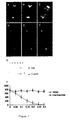
- the majority of mitotically arrested asp cells had bipolar spindles with broad unfocused poles of micotubules (Fig. 2, A and B). In a small proportion of cells, one pole could be sufficiently disorganised so that the spindle appeared monopolar.
- the ⁇ -tubulin was not present within a well-organized centrosome in these cells but was found in dispersed clumps at the spindle poles (Fig. 2, C and D). This suggested that Asp might be required to maintain the structure of the centrosomal microtubule-organizing center (MTOC) during mitosis.
- MTOC centrosomal microtubule-organizing center
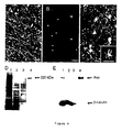
- a number of control antibodies have been used, including one against the centrosomal component CP190. None of these would prevent microtubule nucleation by the centrosome preparation. The number of asters of microtubules formed decreased in proportion to the concentration of antibody (Fig.1H). However, the number of ⁇ -tubulin positive bodies remained constant, suggesting that the antibody was blocking microtubule nucleation rather than disrupting the centrosomes.
- ⁇ -tubulin and its ring-complex are required to rescue the aster-forming ability of KI-extracted centrosomes, they are not sufficient and have to be supplemented by a high molecular weight microtubule-associated factor postulated to be perientric (Moritz et al ., 1998).
- Asp protein might be required to reconstitute MTOCs from KI-extracted centrosomes, we immunodepleted Asp from a soluble embryonic extract under conditions where several other centrosomally associated proteins, including ⁇ -tubulin, CP190, KLP61F and Polo, were not removed (Fig. 3G).
- a spindle can still form, most likely reflecting the known ability of mitotic chromatin and motor proteins to organize a bipolar spindle in the absence of centrosomes.
- one consequence of the disorganized centrosomes and spindle poles is that the cells arrest in a metaphase-like state, suggesting that the spindle integrity checkpoint has been activated.
- Asp protein function is likely to be modified later in the mitotic cycle, since it was observed to associate with the microtubules of the telophase spindle, a property consistent with its purification as a MAP.
Landscapes
- Life Sciences & Earth Sciences (AREA)
- Health & Medical Sciences (AREA)
- Chemical & Material Sciences (AREA)
- Organic Chemistry (AREA)
- Insects & Arthropods (AREA)
- Zoology (AREA)
- Proteomics, Peptides & Aminoacids (AREA)
- Molecular Biology (AREA)
- Biochemistry (AREA)
- Biophysics (AREA)
- General Health & Medical Sciences (AREA)
- Genetics & Genomics (AREA)
- Medicinal Chemistry (AREA)
- Gastroenterology & Hepatology (AREA)
- Toxicology (AREA)
- Tropical Medicine & Parasitology (AREA)
- Immunology (AREA)
- Medicines That Contain Protein Lipid Enzymes And Other Medicines (AREA)
- Peptides Or Proteins (AREA)
- Investigating Or Analysing Materials By The Use Of Chemical Reactions (AREA)
- Investigating Or Analysing Biological Materials (AREA)
- Micro-Organisms Or Cultivation Processes Thereof (AREA)
- Devices For Conveying Motion By Means Of Endless Flexible Members (AREA)
- Mechanical Treatment Of Semiconductor (AREA)
- Measuring And Recording Apparatus For Diagnosis (AREA)
- Dental Preparations (AREA)
- Preparation Of Compounds By Using Micro-Organisms (AREA)
Abstract
Description


Claims (5)
- A method for identifying a substance capable of disrupting microtubule organising centre (MTOC) integrity, which method comprises
contacting an Asp polypeptide as shown in SEQ ID NO:1 or fragment thereof capable of forming and/or maintaining MTOCs in the absence of the substance, with a candidate substance in the presence of components required for MTOC formation and microtubule nucleation therefrom, and measuring microtubule nucleation from centrosomes in the presence of the candidate substance compared to in the absence of the candidate substance to determine whether the substance disrupts MTOC integrity. - A method according to claim 1 wherein said components comprise KI-extracted centrosomes and an Asp-depleted soluble cellular extract.
- A method according to claim 1 wherein said components comprise a centrosome preparation and tubulin.
- A method according to any one of claims 1 to 3 further comprising administering a substance, which has been determined to disrupt MTOC integrity, to a cell and determining whether said substance inhibits mitosis in the cell.
- Use of an Asp polypeptide as shown in SEQ ID NO:1 or fragment thereof capable of stimulating formation of and/or maintaining MTOCs, in an assay for identifying a substance capable of disrupting MTOC integrity comprising measuring microtubule nucleation from centrosomes in the presence of the candidate substance compared to in the absence of the candidate substance.
Applications Claiming Priority (3)
| Application Number | Priority Date | Filing Date | Title |
|---|---|---|---|
| GBGB9905007.2A GB9905007D0 (en) | 1999-03-04 | 1999-03-04 | Assay |
| GB9905007 | 1999-03-04 | ||
| PCT/GB2000/000785 WO2000052478A1 (en) | 1999-03-04 | 2000-03-03 | Detection of mtoc disrupting substances |
Publications (3)
| Publication Number | Publication Date |
|---|---|
| EP1159620A1 EP1159620A1 (en) | 2001-12-05 |
| EP1159620B1 true EP1159620B1 (en) | 2005-05-11 |
| EP1159620B8 EP1159620B8 (en) | 2005-07-06 |
Family
ID=10848975
Family Applications (1)
| Application Number | Title | Priority Date | Filing Date |
|---|---|---|---|
| EP00907799A Expired - Lifetime EP1159620B8 (en) | 1999-03-04 | 2000-03-03 | Detection of mtoc disrupting substances |
Country Status (6)
| Country | Link |
|---|---|
| EP (1) | EP1159620B8 (en) |
| AT (1) | ATE295543T1 (en) |
| AU (1) | AU2927100A (en) |
| DE (1) | DE60020087T2 (en) |
| GB (1) | GB9905007D0 (en) |
| WO (1) | WO2000052478A1 (en) |
Family Cites Families (1)
| Publication number | Priority date | Publication date | Assignee | Title |
|---|---|---|---|---|
| KR0182853B1 (en) * | 1995-08-07 | 1999-05-15 | 김신규 | Diagnostic technique of autoimmune disease |
-
1999
- 1999-03-04 GB GBGB9905007.2A patent/GB9905007D0/en not_active Ceased
-
2000
- 2000-03-03 EP EP00907799A patent/EP1159620B8/en not_active Expired - Lifetime
- 2000-03-03 AT AT00907799T patent/ATE295543T1/en not_active IP Right Cessation
- 2000-03-03 WO PCT/GB2000/000785 patent/WO2000052478A1/en not_active Ceased
- 2000-03-03 AU AU29271/00A patent/AU2927100A/en not_active Abandoned
- 2000-03-03 DE DE60020087T patent/DE60020087T2/en not_active Expired - Fee Related
Also Published As
| Publication number | Publication date |
|---|---|
| AU2927100A (en) | 2000-09-21 |
| GB9905007D0 (en) | 1999-04-28 |
| WO2000052478A1 (en) | 2000-09-08 |
| DE60020087D1 (en) | 2005-06-16 |
| EP1159620B8 (en) | 2005-07-06 |
| EP1159620A1 (en) | 2001-12-05 |
| ATE295543T1 (en) | 2005-05-15 |
| DE60020087T2 (en) | 2006-01-19 |
Similar Documents
| Publication | Publication Date | Title |
|---|---|---|
| Neufeld et al. | The Drosophila peanut gene is required for cytokinesis and encodes a protein similar to yeast putative bud neck filament proteins | |
| Karpova et al. | Jupiter, a new Drosophila protein associated with microtubules | |
| US20090099028A1 (en) | Protein binding miniature proteins and uses thereof | |
| CA2432111A1 (en) | Jfy1 protein induces rapid apoptosis | |
| CN101812127B (en) | Microtubule-associated protein and coding genes and application thereof | |
| ES2306672T3 (en) | METHOD OF SCREENING FOR CANDIDATE DRUGS. | |
| JP3839103B2 (en) | Apoptosis-inducing protein and gene encoding the same | |
| Eberl et al. | A new enhancer of position-effect variegation in Drosophila melanogaster encodes a putative RNA helicase that binds chromosomes and is regulated by the cell cycle | |
| US7338799B2 (en) | Polynucleotide encoding a mouse choline transporter | |
| CN115607675B (en) | Nav1.9 interaction protein PRMT7 and application of down regulator thereof in preparing analgesic drugs | |
| WO2000052478A1 (en) | Detection of mtoc disrupting substances | |
| US10273487B2 (en) | Transfection vector for pathogenic amoebae and uses thereof | |
| Berrard et al. | Molecular genetic approach to the study of mammalian choline acetyltransferase | |
| US6987003B1 (en) | Mammalian stauffen and use thereof | |
| EP1541173A1 (en) | Method of degrading transcriptional factors of saccharometabolism-associated gene, method of inhibiting the degradation and degradation inhibitor | |
| EP1637540A1 (en) | Hyperactive Stat molecules and their use in assays employing gene activation | |
| US20050228169A1 (en) | Polypeptide binding to human syntaxin 1a | |
| Hadad et al. | Drosophila spoonbill encodes a dual-specificity A-kinase anchor protein essential for oogenesis | |
| EP1259617B1 (en) | Bunyaviridal reaper proteins and uses therefor | |
| Dow | The cloning and characterization of the mel-26 gene of Caenorhabditis elegans. | |
| US20050084916A1 (en) | Identification of the IkappaBNS protein and its products | |
| EP1373500A2 (en) | Gene involved in v(d)j recombination and/or dna repair | |
| Zhen | Cloning, characterization and functional analysis of UBC-2, a gene encoding a ubiguitin-conjugating enzyme in the nematode Caenorhabditis elegans | |
| JP2002516096A (en) | CREB binding proteins and related p300-derived polypeptides for use in transcriptional regulation | |
| McCormick | Investigations into Xpat, a novel gene expressed in the germ plasm and primordial germ cells of Xenopus laevis |
Legal Events
| Date | Code | Title | Description |
|---|---|---|---|
| PUAI | Public reference made under article 153(3) epc to a published international application that has entered the european phase |
Free format text: ORIGINAL CODE: 0009012 |
|
| 17P | Request for examination filed |
Effective date: 20010927 |
|
| AK | Designated contracting states |
Kind code of ref document: A1 Designated state(s): AT BE CH CY DE DK ES FI FR GB GR IE IT LI LU MC NL PT SE |
|
| AX | Request for extension of the european patent |
Free format text: AL;LT;LV;MK;RO;SI |
|
| 17Q | First examination report despatched |
Effective date: 20031028 |
|
| GRAP | Despatch of communication of intention to grant a patent |
Free format text: ORIGINAL CODE: EPIDOSNIGR1 |
|
| GRAS | Grant fee paid |
Free format text: ORIGINAL CODE: EPIDOSNIGR3 |
|
| GRAA | (expected) grant |
Free format text: ORIGINAL CODE: 0009210 |
|
| AK | Designated contracting states |
Kind code of ref document: B1 Designated state(s): AT BE CH CY DE DK ES FI FR GB GR IE IT LI LU MC NL PT SE |
|
| PG25 | Lapsed in a contracting state [announced via postgrant information from national office to epo] |
Ref country code: IT Free format text: LAPSE BECAUSE OF FAILURE TO SUBMIT A TRANSLATION OF THE DESCRIPTION OR TO PAY THE FEE WITHIN THE PRESCRIBED TIME-LIMIT;WARNING: LAPSES OF ITALIAN PATENTS WITH EFFECTIVE DATE BEFORE 2007 MAY HAVE OCCURRED AT ANY TIME BEFORE 2007. THE CORRECT EFFECTIVE DATE MAY BE DIFFERENT FROM THE ONE RECORDED. Effective date: 20050511 Ref country code: FI Free format text: LAPSE BECAUSE OF FAILURE TO SUBMIT A TRANSLATION OF THE DESCRIPTION OR TO PAY THE FEE WITHIN THE PRESCRIBED TIME-LIMIT Effective date: 20050511 Ref country code: NL Free format text: LAPSE BECAUSE OF FAILURE TO SUBMIT A TRANSLATION OF THE DESCRIPTION OR TO PAY THE FEE WITHIN THE PRESCRIBED TIME-LIMIT Effective date: 20050511 |
|
| REG | Reference to a national code |
Ref country code: GB Ref legal event code: FG4D |
|
| REG | Reference to a national code |
Ref country code: CH Ref legal event code: EP Ref country code: CH Ref legal event code: NV Representative=s name: MICHELI & CIE INGENIEURS-CONSEILS |
|
| RAP2 | Party data changed (patent owner data changed or rights of a patent transferred) |
Owner name: CYCLACEL LIMITED |
|
| REG | Reference to a national code |
Ref country code: IE Ref legal event code: FG4D |
|
| REF | Corresponds to: |
Ref document number: 60020087 Country of ref document: DE Date of ref document: 20050616 Kind code of ref document: P |
|
| NLT2 | Nl: modifications (of names), taken from the european patent patent bulletin |
Owner name: CYCLACEL LIMITED |
|
| PG25 | Lapsed in a contracting state [announced via postgrant information from national office to epo] |
Ref country code: GR Free format text: LAPSE BECAUSE OF FAILURE TO SUBMIT A TRANSLATION OF THE DESCRIPTION OR TO PAY THE FEE WITHIN THE PRESCRIBED TIME-LIMIT Effective date: 20050811 Ref country code: DK Free format text: LAPSE BECAUSE OF FAILURE TO SUBMIT A TRANSLATION OF THE DESCRIPTION OR TO PAY THE FEE WITHIN THE PRESCRIBED TIME-LIMIT Effective date: 20050811 Ref country code: SE Free format text: LAPSE BECAUSE OF FAILURE TO SUBMIT A TRANSLATION OF THE DESCRIPTION OR TO PAY THE FEE WITHIN THE PRESCRIBED TIME-LIMIT Effective date: 20050811 |
|
| PG25 | Lapsed in a contracting state [announced via postgrant information from national office to epo] |
Ref country code: PT Free format text: LAPSE BECAUSE OF FAILURE TO SUBMIT A TRANSLATION OF THE DESCRIPTION OR TO PAY THE FEE WITHIN THE PRESCRIBED TIME-LIMIT Effective date: 20051019 |
|
| NLV1 | Nl: lapsed or annulled due to failure to fulfill the requirements of art. 29p and 29m of the patents act | ||
| PLBE | No opposition filed within time limit |
Free format text: ORIGINAL CODE: 0009261 |
|
| STAA | Information on the status of an ep patent application or granted ep patent |
Free format text: STATUS: NO OPPOSITION FILED WITHIN TIME LIMIT |
|
| ET | Fr: translation filed | ||
| PG25 | Lapsed in a contracting state [announced via postgrant information from national office to epo] |
Ref country code: MC Free format text: LAPSE BECAUSE OF NON-PAYMENT OF DUE FEES Effective date: 20060331 Ref country code: LU Free format text: LAPSE BECAUSE OF NON-PAYMENT OF DUE FEES Effective date: 20060331 |
|
| 26N | No opposition filed |
Effective date: 20060214 |
|
| PGFP | Annual fee paid to national office [announced via postgrant information from national office to epo] |
Ref country code: IE Payment date: 20061219 Year of fee payment: 8 |
|
| PGFP | Annual fee paid to national office [announced via postgrant information from national office to epo] |
Ref country code: BE Payment date: 20070115 Year of fee payment: 8 |
|
| PGFP | Annual fee paid to national office [announced via postgrant information from national office to epo] |
Ref country code: CH Payment date: 20070214 Year of fee payment: 8 |
|
| PGFP | Annual fee paid to national office [announced via postgrant information from national office to epo] |
Ref country code: GB Payment date: 20070313 Year of fee payment: 8 |
|
| PGFP | Annual fee paid to national office [announced via postgrant information from national office to epo] |
Ref country code: AT Payment date: 20070314 Year of fee payment: 8 |
|
| PGFP | Annual fee paid to national office [announced via postgrant information from national office to epo] |
Ref country code: DE Payment date: 20070525 Year of fee payment: 8 |
|
| PGFP | Annual fee paid to national office [announced via postgrant information from national office to epo] |
Ref country code: FR Payment date: 20061226 Year of fee payment: 8 |
|
| BERE | Be: lapsed |
Owner name: CYCLACEL LTD Effective date: 20080331 |
|
| REG | Reference to a national code |
Ref country code: CH Ref legal event code: PL |
|
| GBPC | Gb: european patent ceased through non-payment of renewal fee |
Effective date: 20080303 |
|
| PG25 | Lapsed in a contracting state [announced via postgrant information from national office to epo] |
Ref country code: AT Free format text: LAPSE BECAUSE OF NON-PAYMENT OF DUE FEES Effective date: 20080303 Ref country code: CY Free format text: LAPSE BECAUSE OF FAILURE TO SUBMIT A TRANSLATION OF THE DESCRIPTION OR TO PAY THE FEE WITHIN THE PRESCRIBED TIME-LIMIT Effective date: 20050511 |
|
| REG | Reference to a national code |
Ref country code: FR Ref legal event code: ST Effective date: 20081125 |
|
| REG | Reference to a national code |
Ref country code: IE Ref legal event code: MM4A |
|
| PG25 | Lapsed in a contracting state [announced via postgrant information from national office to epo] |
Ref country code: IE Free format text: LAPSE BECAUSE OF NON-PAYMENT OF DUE FEES Effective date: 20080303 Ref country code: CH Free format text: LAPSE BECAUSE OF NON-PAYMENT OF DUE FEES Effective date: 20080331 Ref country code: LI Free format text: LAPSE BECAUSE OF NON-PAYMENT OF DUE FEES Effective date: 20080331 Ref country code: DE Free format text: LAPSE BECAUSE OF NON-PAYMENT OF DUE FEES Effective date: 20081001 |
|
| PG25 | Lapsed in a contracting state [announced via postgrant information from national office to epo] |
Ref country code: BE Free format text: LAPSE BECAUSE OF NON-PAYMENT OF DUE FEES Effective date: 20080331 |
|
| PG25 | Lapsed in a contracting state [announced via postgrant information from national office to epo] |
Ref country code: FR Free format text: LAPSE BECAUSE OF NON-PAYMENT OF DUE FEES Effective date: 20080331 |
|
| PG25 | Lapsed in a contracting state [announced via postgrant information from national office to epo] |
Ref country code: GB Free format text: LAPSE BECAUSE OF NON-PAYMENT OF DUE FEES Effective date: 20080303 Ref country code: ES Free format text: LAPSE BECAUSE OF NON-PAYMENT OF DUE FEES Effective date: 20060331 |