EP1156103A2 - Oplophorus luciferase - Google Patents
Oplophorus luciferase Download PDFInfo
- Publication number
- EP1156103A2 EP1156103A2 EP01109479A EP01109479A EP1156103A2 EP 1156103 A2 EP1156103 A2 EP 1156103A2 EP 01109479 A EP01109479 A EP 01109479A EP 01109479 A EP01109479 A EP 01109479A EP 1156103 A2 EP1156103 A2 EP 1156103A2
- Authority
- EP
- European Patent Office
- Prior art keywords
- protein
- sequence
- luciferase
- kda
- polynucleotide
- Prior art date
- Legal status (The legal status is an assumption and is not a legal conclusion. Google has not performed a legal analysis and makes no representation as to the accuracy of the status listed.)
- Granted
Links
Images


Classifications
-
- C—CHEMISTRY; METALLURGY
- C12—BIOCHEMISTRY; BEER; SPIRITS; WINE; VINEGAR; MICROBIOLOGY; ENZYMOLOGY; MUTATION OR GENETIC ENGINEERING
- C12N—MICROORGANISMS OR ENZYMES; COMPOSITIONS THEREOF; PROPAGATING, PRESERVING, OR MAINTAINING MICROORGANISMS; MUTATION OR GENETIC ENGINEERING; CULTURE MEDIA
- C12N9/00—Enzymes; Proenzymes; Compositions thereof; Processes for preparing, activating, inhibiting, separating or purifying enzymes
- C12N9/0004—Oxidoreductases (1.)
- C12N9/0069—Oxidoreductases (1.) acting on single donors with incorporation of molecular oxygen, i.e. oxygenases (1.13)
-
- C—CHEMISTRY; METALLURGY
- C07—ORGANIC CHEMISTRY
- C07K—PEPTIDES
- C07K16/00—Immunoglobulins [IG], e.g. monoclonal or polyclonal antibodies
- C07K16/40—Immunoglobulins [IG], e.g. monoclonal or polyclonal antibodies against enzymes
Definitions
- the present invention relates to a novel luciferase derived from Decapoda. More specifically, the invention relates to a secretional luciferase from Oplophorus gracilirostris , which is composed of 19 kDa and 35 kDa proteins. The invention also relates to polynucleotides encoding at least one of these proteins, a recombinant expression vector comprising at least one of these polynucleotides, a host cell transformed with said vector, and process for producing said photoprotein.
- photoproteins and luciferases are an industrially important protein and have been utilized, for example, as a reporter enzyme.
- Various methods for detecting an analyte using luminescent reactions of these enzymes have been developed, and also some apparatuses to be used in these methods have been improved and widespread.
- these known photoproteins and luciferases there is no enzyme applicable to extensive purposes. Consequently, one has to choose a proper enzyme for individual purpose.
- the luminescent substrates include species specific and species non-specific substrates.
- the minimum unit in the enzymatic bioluminescent reaction consists of a luminescent enzyme (luciferase), a luminescent substrate (luciferin) and molecular oxygen.
- luciferase a luminescent enzyme
- luciferin a luminescent substrate
- molecular oxygen a luminescent oxygen
- Examples of the luciferase with luminescence in minimum unit include those derived from Renilla , Cypridina and Gonyaulax.
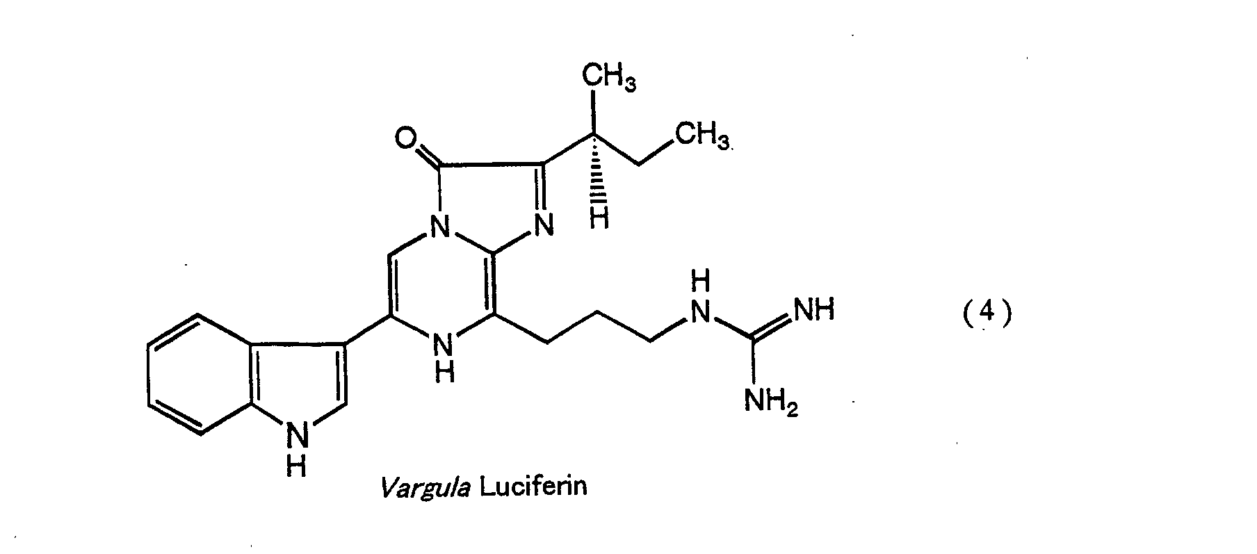
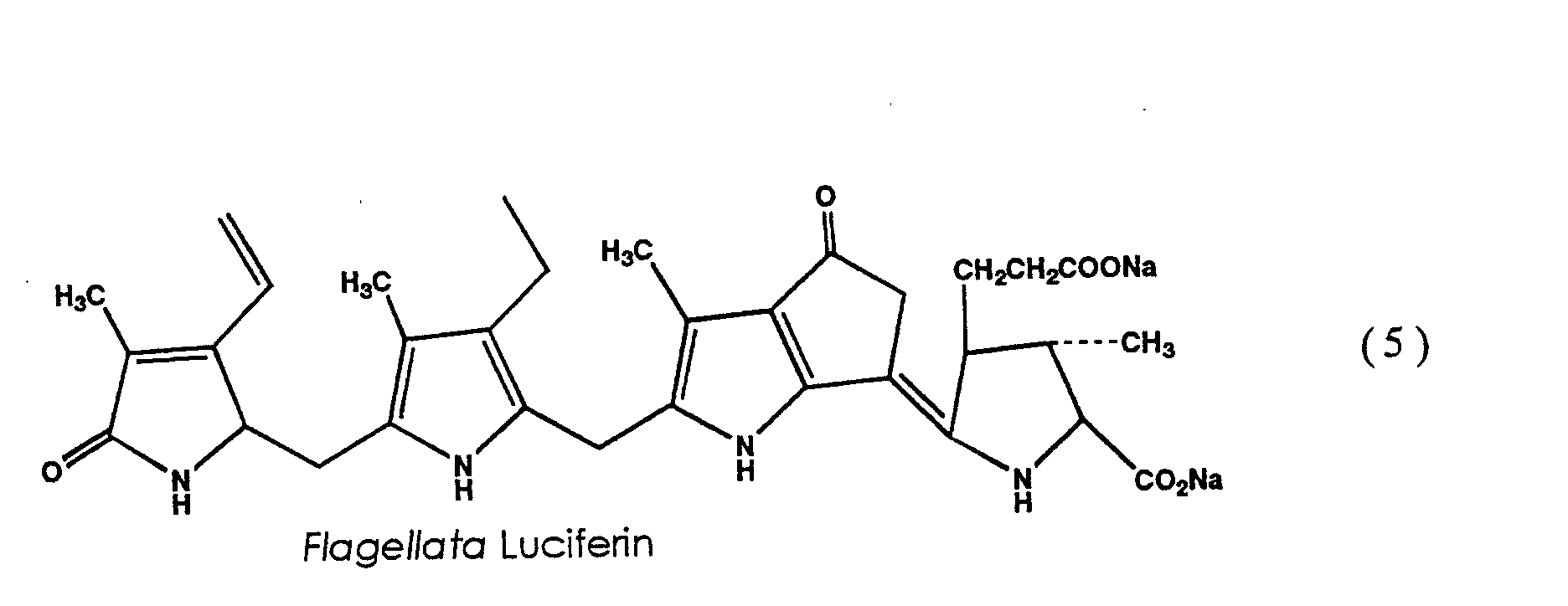
- the luciferins corresponding to these luciferases have very complicated structures as shown in the above formulas (4) and (5).
- the methods for synthesizing Cypridina and Gonyaulax luciferins are already known, but yield is remarkably low due to their complicated synthesizing process. Though the luciferins extracted from natural products are also used, they are very expensive with little industrial utility.
- Renilla luciferin known as coelenterazine and derivatives thereof are commercially available and inexpensive, because various methods for the production thereof have been established.
- a secretional luciferase is only Cypridina luciferase.
- the structure of the gene is reported in Thompson, E. M., et al., Proc. Natl. Acad. Sci. USA, 86, 6567-6571 (1989) and the application of the gene is reported in Inouye, S., et al., Proc. Natl. Acad. Sci. USA, 89, 9584-9587 (1992).
- the secretional luciferase In the construction of a bioassay system such as a drug screening system, the secretional luciferase has an industrial advantage in that the luminescence activity can be detected in living cells without cell disruption using the luciferase extracellularly secreted as a reporter.
- a secretional protein is not particularly difficult to produce if a suitable host-vector system is selected.
- the purification of a recombinant protein from cultured media is easier in comparison with the purification from a cell extract.
- mass production of the secretional luciferase may advantageously hold down purification costs involved.
- a particularly useful luciferase involves a luminescent system wherein the luminescent reaction occurs only among the minimal unit, i.e. a luciferase, a luciferin and molecular oxygen, the luciferin being coelenterazine or a derivative thereof which is readily available, and the luciferase itself being a secretional protein.
- a protein and a gene of such luciferase are advantageous not only scientifically but also industrially. However, isolation of gene encoding such luciferase and expression thereof in a living cell has not been reported yet.
- a luciferase derived from a luminous shrimp belonging to Decapoda has been reported as a secretional luciferase, the luminescent substrate of which is coelenterazine. It is known that a luminous shrimp involves a secretional luciferase (enzyme) and that a blue luminescence is emitted by the reaction of the luciferase, a luminescent substrate luciferin and molecular oxygen.
- the literature also reports that the luciferase has a quantum yield of 0.32 at 22°C, a high specific activity of 1.75 ⁇ 10 15 Photons/s. mg, an optimum light emission at 40°C and an excellent heat stability. It also describes that the luminescent reaction proceeds in a wide range of pH.
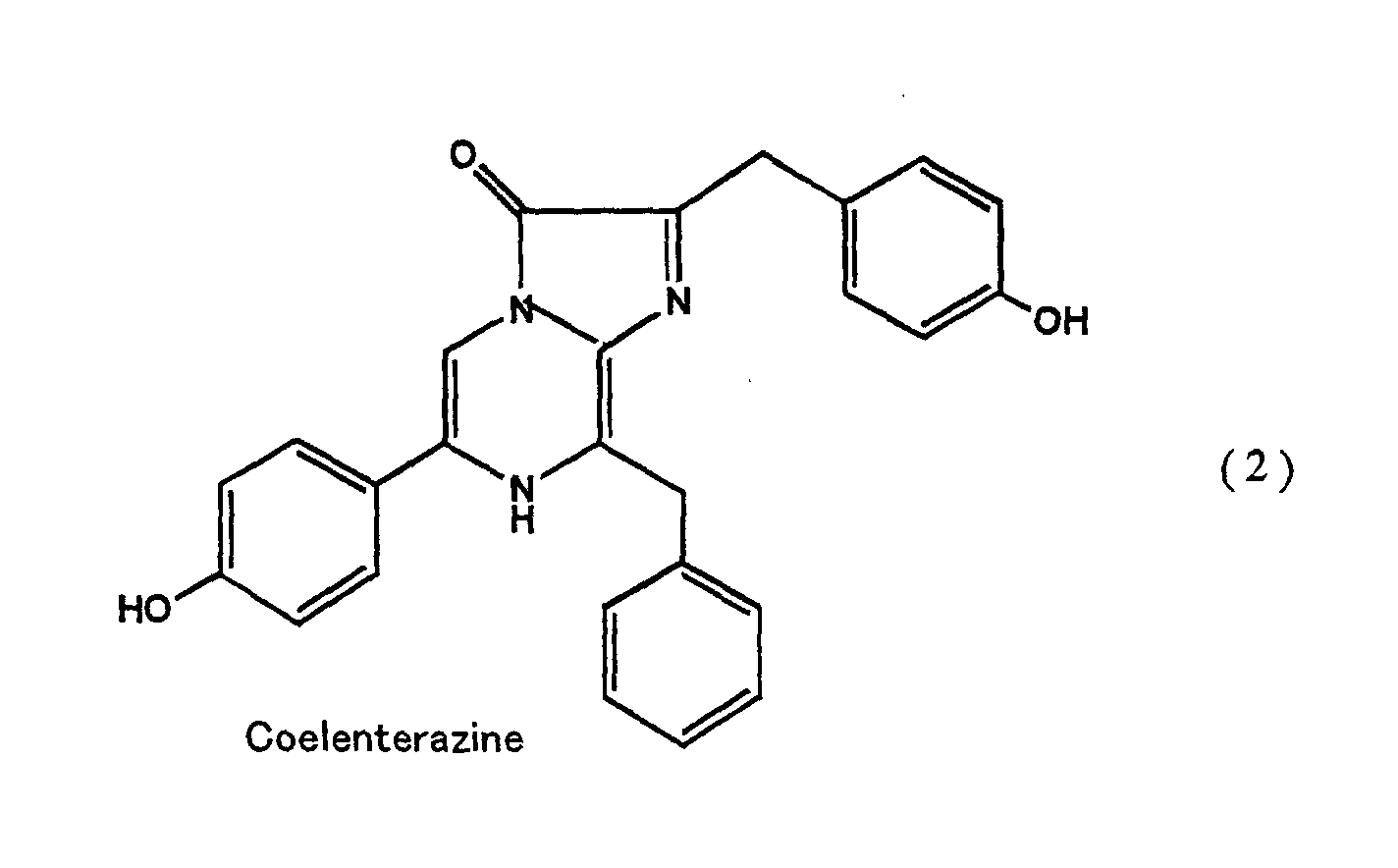
- the luciferin in the luminescent reaction of Oplophorus luciferase is coelenterazine represented by the above formula (2), which is also a luminescent substrate in the luminescent reactions of Renilla luciferase and a photoprotein, aequorin.
- the most important difference between these luminescent enzymes and Oplophorus luciferase is that Oplophorus luciferase has very broad substrate specificity in comparison with those of Renilla luciferase and aequorin.
- Oplophorus luciferase is more preferable than other luciferases, because it can utilize as a substrate bisdeoxycoelenterazine which is an analogue of coelenterazine and is available at a low cost.
- An object of the present invention is to provide polynucleotides encoding Oplophorus luciferase and a luminescent subunit thereof.
- Another object of the invention is to provide the recombinant secretional Oplophorus luciferase and the subunit encoded by the polynucleotides.
- Another object of the invention is to provide an expression vector containing the polynucleotide(s) and a host transformed with the vector.
- Further object of the invention is to provide a method for producing the recombinant Oplophorus luciferase or the recombinant photoprotein.
- Oplophorus luciferase is composed of 19 kDa and 35 kDa proteins. Then, the partial amino acid sequence of each protein was determined and the cloning of the proteins was carried out based on the information of their partial amino acid sequences. The genes coding for the two proteins was successfully cloned and the nucleotide sequences of the genes and the amino acid sequence of these proteins encoded by the gene could also be determined.
- the present inventor has succeeded in preparing the expression vector containing the polynucleotide coding for each gene and a host such as a microorganism or a cultured animal cell which had been transformed with the vector.
- These proteins could be recombinantly produced by culturing the host cell or by in vitro translation system using the recombinant expression vector.
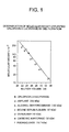
- Figure 1 illustrates the determination of molecular weight of purified Oplophorus luciferase by gel filtration.
- Figure 2 shows the alignment of the leucine-rich repeats structure of the 35 kDa Protein.
- Figure 3 shows the Western blot analysis of Oplophorus luciferase using the anti -Oplophorus luciferase polyclonal antibody according to the invention.
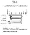
- Figure 4 schematically illustrates the restriction map of the 19 Kda protein and the construction of the expression vector.
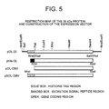
- Figure 5 schematically illustrates the restriction map of the 35 Kda protein and the construction of the expression vector.
- a photoprotein having a molecular weight of 19 kDa is one component of Oplophorus luciferase.
- the 19 kDa protein having a luminous activity contains an amino acid sequence selected from the group consisting of:
- the photoprotein further comprises an amino acid sequence for purification and/or a signal peptide sequence for extracellular secretion or intracellular transport, for example, a signal sequence contained in positions 1-27 of the amino acid sequence shown in SEQ ID NO: 2 or a signal peptide sequence known in the art.
- Examples of the signal peptide for the extracellular secretion include eucaryotic secretional signal peptides known in the art (see, e.g., von Heijne, G. Eur. J. Biochem 133 (1983), pp. 17-21) and procaryotic secretional signal peptides known in the art (see, e.g., von Heijne, G. & Abrahmsen, L. FEBS Lett. 244 (1989), pp. 439-446).
- Examples of the signal peptide for the intracellular transport include the signal peptides for the transport to mitochondria (Gavel, Y. & von Heijne, G., Protein Engineering 4 (1990), pp. 33-37), for the transport to chloroplast (see, e.g., Gavel, Y. & von Heijne, G., FEBS Lett. 261 (1990), pp. 455-458) and for the transport to nuclear (see, e.g., Dingwall, C. & Laskey, R. A., Trends Biochem. Sci. 16 (1991), pp. 478-481).
- a protein having a molecular weight of 35 kDa is another component of Oplophorus luciferase.
- the 35 kDa protein contains an amino acid sequence selected from the group consisting of:
- the present invention provides a polynucleotide encoding the photoprotein having a molecular weight of 19 kDa.
- polynucleotides include a deoxyribonucleic acid molecule such as cDNA or a genome DNA, a ribonucleic acid molecule such as mRNA and a derivative thereof.
- the polynucleotide encoding the 19 kDa protein which contains the luminous activity comprises:
- the invention also provides a polynucleotide encoding a protein having a molecular weight of 35 kDa which is one component of Oplophorus luciferase.
- the polynucleotide comprises:
- the foregoing polynucleotide encoding the 19 kDa and/or 35 kDa protein may comprise an additional poly- or oligo-nucletodie encoding a signal peptide known in the art, e.g., the peptide as described in the afore-mentioned literatures.
- a method for generating the luminescence comprises reacting the luciferase composed of the 19 kDa and 35 kDa proteins, or the 19 kDa protein with coelenterazine or derivatives thereof as a substrate.
- the luminescent reaction can occur by the luciferase or the 19 kDa protein as an enzyme, coelenterazine or derivatives thereof as a substrate luciferin and molecular oxygen.
- the luminescent reaction can be performed at pH of 5.5 to 11, preferably 7.0 to 11.
- the reaction temperature is in the range of 10°C to 50°C, preferably 20°C to 35°C.
- a recombinant vector comprises the polynucleotide encoding the 19 kDa protein or the polynucleotide encoding the 35 kDa protein as an insert.
- the recombinant vector is a recombinant expression vector capable of transcribing the polynucleotide of the invention.
- Such a vector can be prepared by any techniques known in the art.
- a vehicle used for the construction of the recombinant vector of the protein of the invention may be any vehicle known in the art that is suitable for in vitro translation system or the expression system using a host cell, for example, microorganism such as E. coli and yeast or a cultured animal cell. Such vehicles are commercially available.
- Examples of the vehicle for the in vitro translation and the expression in an animal cell include pTargetT vector incorporating the immediate-early enhancer/promoter from Human cytomegalovirus (CMV) containing T7 promoter sequence and multi-cloning site downstream thereto, pSI vector (Promega, Madison, WI, USA) incorporating SV40 enhancer and SV40 early promoter, pBK-CMV, CMV-Script, pCMV-Tag and pBK-RSV (Stratagene, USA) and the like.
- CMV Human cytomegalovirus
- pSI vector Promega, Madison, WI, USA
- Examples of the vehicle for the expression in E . coli include pET expression vector series incorporating T7 promoter (e.g., pET3a, pET27b(+) and pET28a(+); Novagen, Madison, WI, USA) and the like.
- Examples of the vehicle for the expression in yeast include pIC expression vector series incorporating the promoter from alcoholoxydase (e.g., pPIC9K, PIC3.5K; Invitorgen, La Jolla, CA, USA) and the like.
- alcoholoxydase e.g., pPIC9K, PIC3.5K; Invitorgen, La Jolla, CA, USA
- the present invention further provides a host cell transformed with the recombinant vector.
- a host cell include a microorganism, e.g., E. Coli and yeast and a cultured animal cell known in the art, e.g., COS7 cell and CHO cell.
- a method for producing the protein of the invention comprises culturing the host cell and then isolating the recombinant protein from cultured media and/or cells, for example, cell extract thereof, and optionally purifying the protein to give a substantially purified form.
- the isolation of the recombinant protein can be carried out in accordance with the standard technique in the art.
- the protein can be also isolated from a water-insoluble fraction of the cells by treating one or more solubilizing agents known in the art. Purification of the recombinant protein can be conducted by any procedure known in the art.
- a method for producing the protein comprises subjecting the recombinant expression vector to in vitro translation, isolating the recombinant protein expressed, and optionally purifying the protein to give a substantially purified form.
- the in vitro translation for producing the protein can be performed by a method known in the art (see, e.g., Spirin, A. S., et. al., Science 242 (1988), pp. 1162-1164; and Patnaik, R. & Swartz, J. M. Biotechniques 24 (1998), pp. 862-868).
- Commercially available in vitro translation kits e.g., TNT in vitro Transcription-translation kit; Promega may be used for the in vitro translation.
- the method for producing the protein of the invention further comprises renaturing the protein in the presence of a solvent such as one or more polyhydric alcohols for the reactivation of its enzyme activity, and optionally preserving the protein in the solvent.
- a solvent such as one or more polyhydric alcohols for the reactivation of its enzyme activity
- the luciferase and/or the photoprotein can be preserved over a long period of time without decreasing the luminescence activity.
- polyhydric alcohols as a solvent may include, but not limited to, glycerol, polyethylene glycol, polypropylene glycol, dextran, mannitol, sorbitol, inositol, xylitol, sucrose, fructose and glucose.
- glycerol polyethylene glycol, or polypropylene glycol
- glycerol is more preferable for preventing the protein from decreasing its enzyme activity.
- the concentration of one or more polyhydric alcohols is in the range of from 10 to 90% (v/w), preferably from 30 to 90% (v/w), more preferably from 50 to 70% (v/w).
- a protein constituting the luciferase and capable of stabilizing the luciferase contains one or more units of leucine-rich repeating sequence consisting of: (Leu/Ile)-Xaa-Xaa-Leu-Xaa-(Leu/Ile)-Xaa-Xaa-Asn-Xaa- (Leu/Ile)-Xaa-Xaa-Xaa-Pro wherein each Xaa may be any one of essential amino acids.
- This leucine-rich repeating structure is found in the amino acid sequence of the 35 kDa protein as shown in Fig. 2.
- a protein containing the above repeated sequence may be capable of stabilizing the luciferase as well as the 35 kDa protein.
- the invention provides a polyclonal or monoclonal antibody which is arisen against the luciferase of the invention, the 19 kDa or 35 kDa protein, or an immunogenic fragment thereof, and which can specifically bind to the luciferase and/or the protein.
- an antigen which may be used for the preparation of the antibody according to the present invention, include a protein from a natural source, a recombinant protein, a partially degraded product thereof and a synthetic peptide produced on the basis of the amino acid sequences of the proteins according to the invention.
- Such synthetic peptides comprise at least 5 contiguous amino acid residues, preferably 10-15 contiguous amino acid residues selected from the sequence consisting of amino acids at positions 28-196 of the sequence shown in SEQ ID NO: 2 and the sequence consisting of amino acids at positions 40-359 of the sequence shown in SEQ ID NO: 4.
- the antibody of the present invention can be prepared according to the standard technique in the art (see, e.g., Harlow, E. & Lane, D, in Antibodies-Laboratory manual Cold Spring Harbor Laboratory Press, pp. 53-138 (1988)).
- the antibody according to the present invention can be used to detect Oplophorus luciferase or a protein as a component of the luciferase.
- the antibody can also be used for screening other luciferases or photoproteins which are homologous with the luciferase or the protein of the present invention.
- the invention also provides a method for detecting or screening a luciferase or photoprotein using the antibody.
- the method according to the invention can serve to easily identify a luciferase or a photoprotein derived from systematically related species.
- a novel luciferase or photoprotein identified by the present method may be included within a scope of the present invention.
- crude extract of other luminous shrimp or other organisms or their tissues containing a photoprotein is used as a sample and detection and/or screening is carried out in the presence of a protein bound to the antibody of the present invention.
- the detection techniques may include the immunoblotting and the immuno-chromatography.
- the expression cloning can also be carried out with a gene library (e.g., a cDNA or genomic library) derived from other luminous shrimp or other organisms or their tissues containing a photoprotein using the antibody of the invention to obtain a novel luciferase or photoprotein or a gene thereof.
- a gene library e.g., a cDNA or genomic library
- the invention also provides an oligonucleotide comprising at least 10 contiguous nucleotides selected from a polynucleotide sequence encoding the 19 kDa protein shown in SEQ ID NO: 2 or a complementary sequence thereto.
- the oligonucleotide may be selected from a polynucleotide sequence shown in SEQ ID NO: 1 or a complementary sequence thereto.
- the invention also provides an oligonucleotide comprising at least 10 contiguous nucleotides selected from a polynucleotide sequence encoding the 35 kDa protein shown in SEQ ID NO: 4 or a complementary sequence thereto.
- this oligonucleotide may be selected from a polynucleotide sequence shown in SEQ ID NO: 3 or a complementary sequence thereto.
- the length of the oligonucleotide of the invention is preferably at least 14 nucleotides encoding 5 amino acids, more preferably at least 20 nucleotides encoding 7 amino acids.
- the oligonucleotide may include a suitable restriction site at its 5' end.
- the oligonucleotide can be used to detect a polynucleotide molecule such as DNA or RNA encoding the protein which is one component of the luciferase, by the standard technique known in the art, preferably by a polymerase-chain-reaction (PCR) method.
- PCR polymerase-chain-reaction
- a gene library (cDNA or genomic library) derived from other luminous shrimp or other organisms or their tissues containing a photoprotein (enzyme) may be subjected to a suitable cloning, preferably by the PCR method using the oligonucleotide of the invention to isolate a polynucleotide encoding a novel luciferase or a photoprotein.
- the invention also provides a method for detecting and/or screening a gene coding for a luciferase or photoprotein using the oligonucleotide of the invention.
- a novel gene coding for a novel luciferase or its subunit protein or a novel photoprotein identified by the method of the present invention may be included within a scope of the invention.
- the method can serve to easily identify a polynucleotide encoding a luciferase or a photoprotein derived from systematically related species, for example, a luciferase having more than 50% homology.
- the deep-sea luminous shrimp ( Oplophorus gracilirostris ) obtained in the Suruga Bay, Shizuoka, Japan were used as a starting material.
- a crude extract containing Oplophorus luciferase was prepared in the same manner as disclosed by Shimomura, et al., in Biochemistry 17 (1978) and further purified by chromatography in two steps. The first step was by hydrophobic interaction chromatography on a column of butyl Sepharose 4 Fast Flow (Pharmacia; 0.7 cm ⁇ 3.5 cm) using 20 mM Tris-HCl, pH 8.5, eluting with decreasing concentrations of ammonium sulfate starting at 1.5 M.
- the second step was by gel filtration on a column of Superdex 200 Prep (Pharmacia; 1 cm ⁇ 48 cm) in 20 mM Tris-HCl, pH 8.5, containing 50 mM NaCl.
- the molecular weight of Oplophorus luciferase was estimated to be about 106 kDa by gel filtration on the same column when compared with the molecular weight markers as follows: (a) amylase (200kDa); (b) alcohol dehydrogenase (150 kDa); (c) bovine serum albumin (67 kDa); (d) ovalbumin (45 kDa); (e) carbonic anhydrase (29 kDa); and (f) ribonuclease (13.7 kDa). The results are shown in Fig. 1.
- amino acid sequence analysis was carried out using Applied Biosystems model 470A gas phase sequencer according to protocols of the manufacturer. Sample proteins for the sequence analysis were prepared as described below.
- RNA Live specimens of O . gracilirostris obtained in the Suruga Bay were frozen on dry ice and stored at -80°C until used.
- Total RNA was prepared by the guanidine isothiocyanate method (see, e.g. Inouye, S. & Tsuji, F. I., FEBS Lett., 315 (1993), pp. 343-346), followed by precipitation with 2M LiCl.
- the yield of total RNA from two whole specimens (body size: 40 mm length, 2.8 g wet-weight) was approximately 0.9 mg.
- poly(A) + RNA (2 ⁇ g) was isolated by oligo(dT)-cellulose spun-column (Pharmacia, Piscataway, NJ, USA) and subjected to the synthesis of cDNA with dT 12-18 primers using a cDNA synthesis kit (Time saver cDNA synthesis kit; Pharmacia) according to Kakizuka, et al. (Essential Developmental Biology, Stern, C. D. ed., IRL Press, Oxford, U.K., pp. 223-232 (1993)).
- the synthesized cDNAs (20 ng) were ligated with EcoRI/NotI linker.
- cDNAs were ligated with 1 ⁇ g of EcoRI digested/calf intestinal alkaline phosphatase-treated ⁇ ZapII vector (Stratagene, La Jolla, CA, USA) in a total volume of 5 ⁇ l at 4°C for 16 hours and then packaged in vitro using Gigapack Gold III packaging kit (Stratagene).
- the titer of the cDNA library was 1.1 ⁇ 10 6 plaque-forming units.
- the sequences of the oligonucleotides were designed based on the information of the amino acid sequences determined in Example 2.
- the oligonucleotides chemically synthesized according to the standard technique were used for the screening or PCR method.
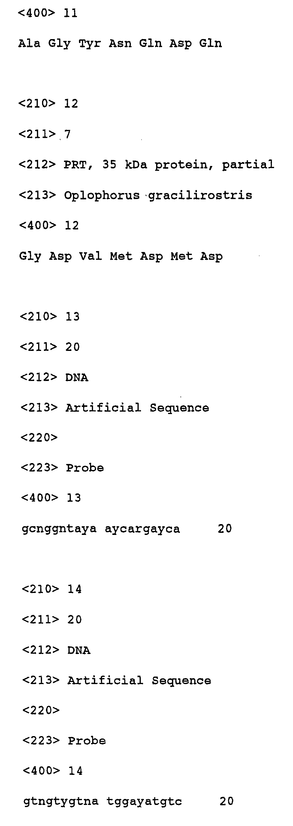
- oligonucleotide probe SOL-2 5'-GCN-GGN-TA-(T/C)-AA(T/C)-CA(A/G)-GA(T/C)-CA-3' (SEQ ID NO: 13) was synthesized.
- the oligonucleotide probe OL-3 5'-GTN-GT(T/C)-GTN-ATG-GA(T/C)-ATG-TC-3'(SEQ ID NO: 14) was synthesized.
- the Oplophorus cDNA library obtained in Example 3 was screened by the plaque hybridization technique according to Wallace, R. B., et al. (Nucl. Acids Res., 9 (1981), pp. 879-897) using synthetic oligonucleotide probes, SOL-2 for the 19 kDa protein or OL-3 for the 35 kDa protein. SOL-2 and OL-3 were labeled with [ ⁇ - 32 P]ATP (3000 Ci/mmol) at their 5'-end for use as a probe.
- the filter was washed three times in SSC containing 300 mM NaCl/30 mM sodium citrate at room temperature and subjected to the autoradiography. Resultant positive plaques were picked up and subjected to the second screening, which was carried as described above, and then each single phage clone was isolated.
- the cDNA inserts were excised as pBluescript phagemids (Stratagene). Resultantly, one positive clone was isolated from 300,000 independent plaques using SOL-2 probe and 9 positive clones were isolated from 70,000 independent plaques using OL-3 probe.
- the recombinant plasmid DNA for each clone obtained in Example 5 was prepared from Escherichia coli by the alkaline lysis method.
- One positive clone from SOL-2 for the 19 kDa protein was designated as pKAZ-412.
- restriction enzyme analysis of nine positive clones from OL-3 for the 35 kDa provided identical restriction maps and thus the longest clone was designated as pOL-23.
- the nucleotide sequence of each clone was determined by the dye-terminator cycle sequencing method using Applied Biosystems DNA sequencers.
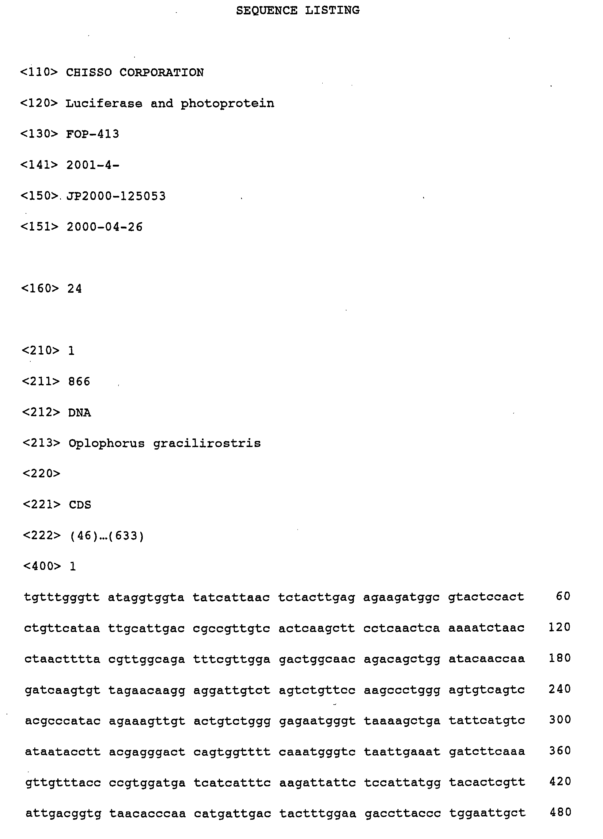
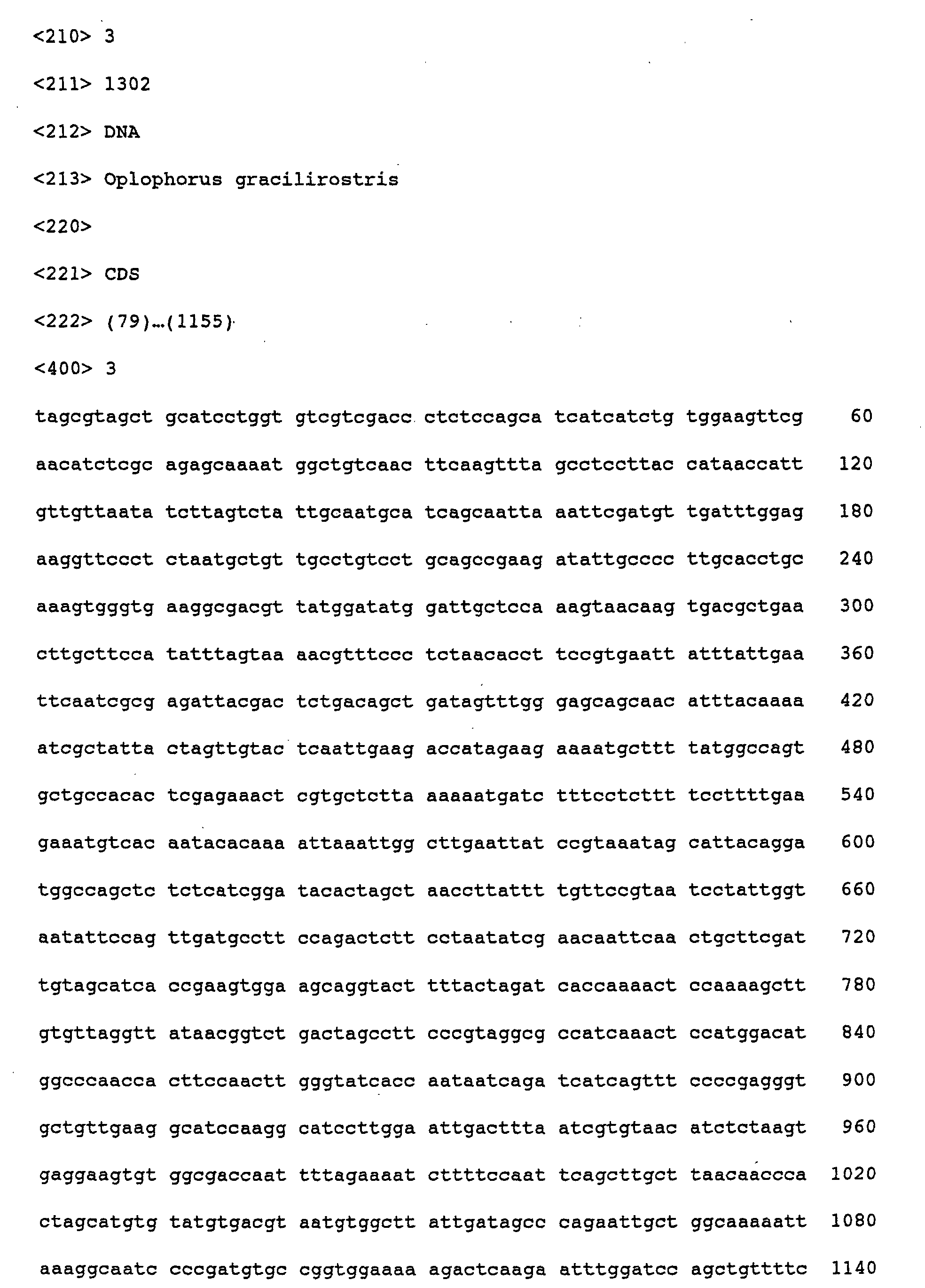
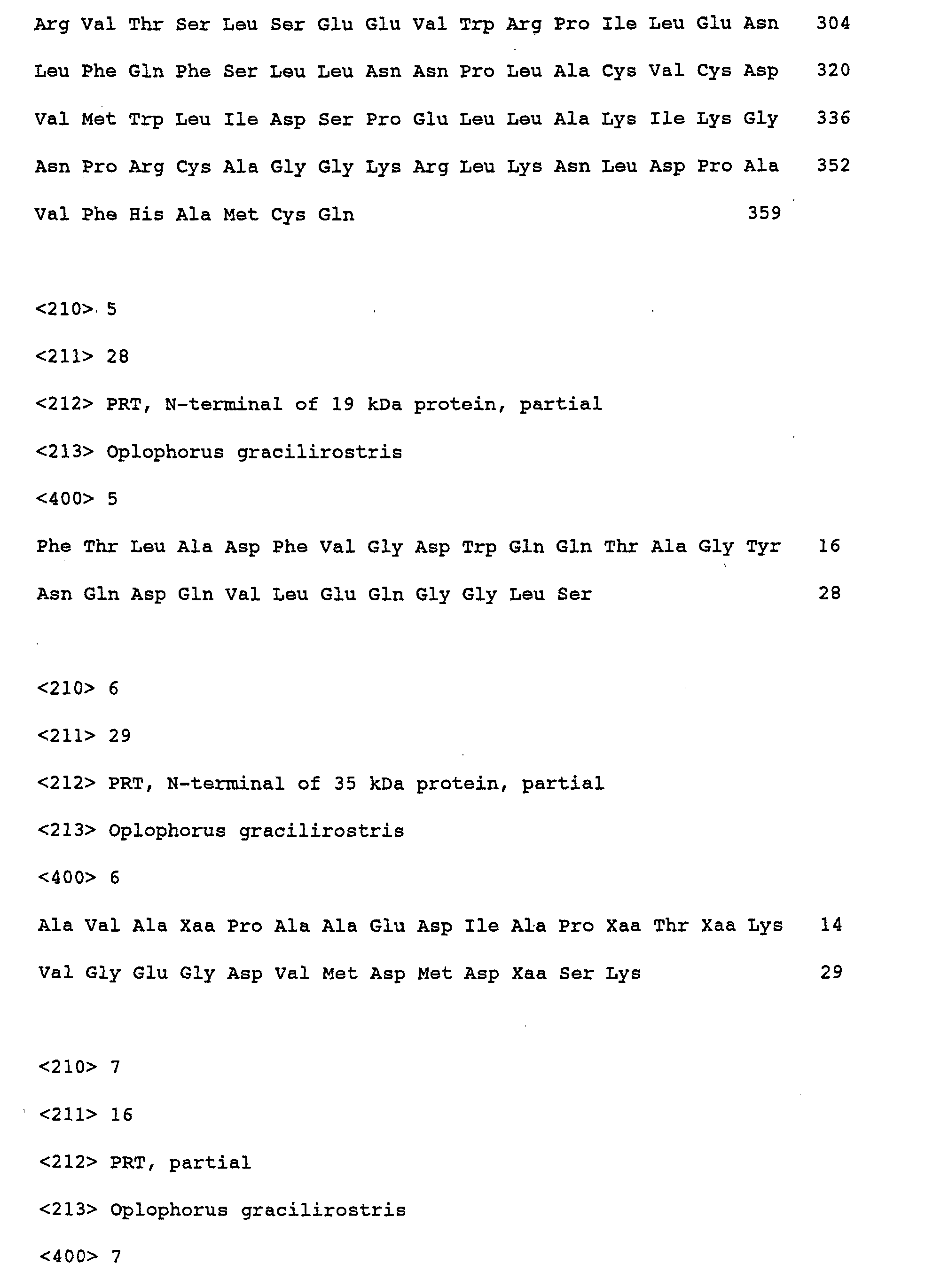
- the nucleotide sequences of the clones are shown in SEQ ID NOS: 1 and 3, and their deduced amino acid sequences are shown in SEQ ID NOS: 2 and 4.
- the 19 kDa protein consists of 196 amino acid residues including a putative signal peptide sequence for secretion, which correspond to the nucleotide sequence of positions 46-633 of the sequence shown in SEQ ID NO: 1. From the results of the N-terminal sequence analysis in Example 2, the mature protein is expected to consist of 169 amino acid residues corresponding to an amino acid sequence of positions 28-196 of the sequence shown in SEQ ID NO: 2 and to have a calculated molecular mass of 18,689.50 and an estimated pI value of 4.70.
- the 35 kDa protein consists of 359 amino acid residues including a putative signal peptide sequence for secretion, which correspond to the nucleotide sequence of positions 79-1155 of the sequence shown in SEQ ID NO: 3. From the results of the N-terminal sequence analysis in Example 2, the mature protein is expected to consist of 328 amino acid residues corresponding to an amino acid sequence of positions 40-359 of the sequence shown in SEQ ID NO: 4 and to have a calculated molecular mass of 34,837.08 and an estimated pI value of 4.61.
- nucleotide sequences shown in SEQ ID NOS: 1 and 3 and the amino acid sequences shown in SEQ ID NOS: 2 and 4 their sequence homology was studied by a gene database search with all the database registered for the National Center of Biotechnology Information (NCBI) using computer programs such as FASTA and BLAST.
- the nucleotide sequences shown in SEQ ID NOS: 1 and 3 were searched for all nucleotide sequences deposited.
- the amino acid sequences shown in SEQ ID NOS: 2 and 4 were searched for all amino acid sequences deposited and amino acid sequences deduced from the nucleotide sequences deposited.
- the nucleotide sequences shown in SEQ ID NOS: 1 and 3 have no significant homology with any sequence deposited.
- the amino acid sequences shown in SEQ ID NO: 2 and 4 also have no significant homology with any deposited sequence. Particularly, they have no significant homology with Renilla luciferase (36 kDa; Genebank, M63501), aequorin (21.5 kDa; Genebank, L29571), Renilla luciferin binding protein (20.5 kDa; SWISS-PRO, P05938), Cypridina luciferase (58.5 kDa; Genebank, M25666), firefly luciferase and bacterial luciferase.
- the amino acid sequence of the 19 kDa protein shown in SEQ ID NO: 2 has low homology [26% identity (44/169) and 49% similarity (83/169)] to D3-S1 domain (residues 217-392) of E . coli amine oxidase (Accession No. pir 140924).
- the sequence also has low homology [28% identity (13/47): 51% similarity (24/47)] with the amino-terminal region of a fatty acid binding protein (GenBank, L23322), whereas no functional relationship between the 19 kDa protein and these proteins was detected.
- the amino acid sequence of the 35 kDa protein shown in SEQ ID NO: 4 contains leucine-rich repeating structures consisting of: (Leu/Ile)-Xaa-Xaa-Leu-Xaa-(Leu/Ile)-Xaa-Xaa-Asn-Xaa-(Leu/Ile)-Xa-a-Xaa-Xaa-Pro wherein each Xaa represents any amino acid residue.
- Example 2 Purified native Oplophorus luciferase (80 ⁇ g) obtained in Example 1 was used to immunize a female New Zealand White rabbit according to the standard technique in the art.
- the resultant anti -Oplophorus luciferase serum (dilution: 500) was used for Western blot analysis as previously reported (Inouye, S., et al., Anal. Biochem., 201: 114-118 (1992)).
- the antibody specifically recognized the 19 kDa and 35 kDa proteins (Fig. 3).
- the recombinant proteins were expressed in E. coli or a cultured cell line by inserting the recombinant vector pKAZ-412 or pOL-23 obtained in Example 6 into the expression vector system.
- the restriction map of the expression vectors used in this example is shown in Figs. 4 and 5.
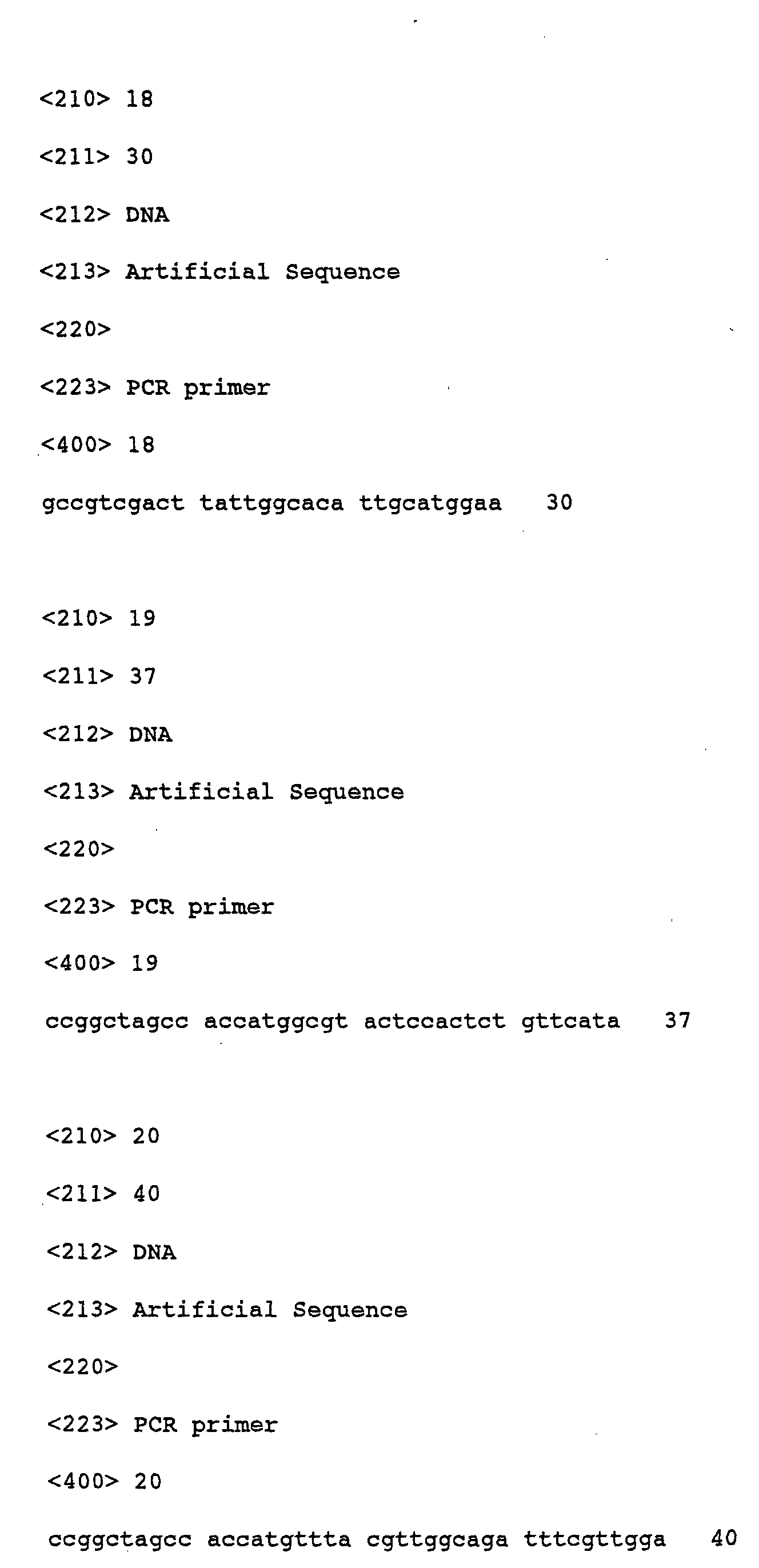
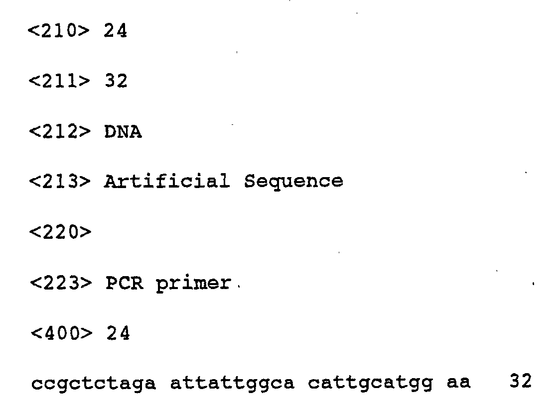
- the expression vectors for the 19 kDa protein, pSKAZ-CMV containing a putative signal sequence for secretion and pKAZ-CMV not containing the signal sequence were constructed in the same manner as described in (1) using the following primers;
- KAZ-1 and KAZ-5 were used.
- KAZ-2 and KAZ-5 were used.
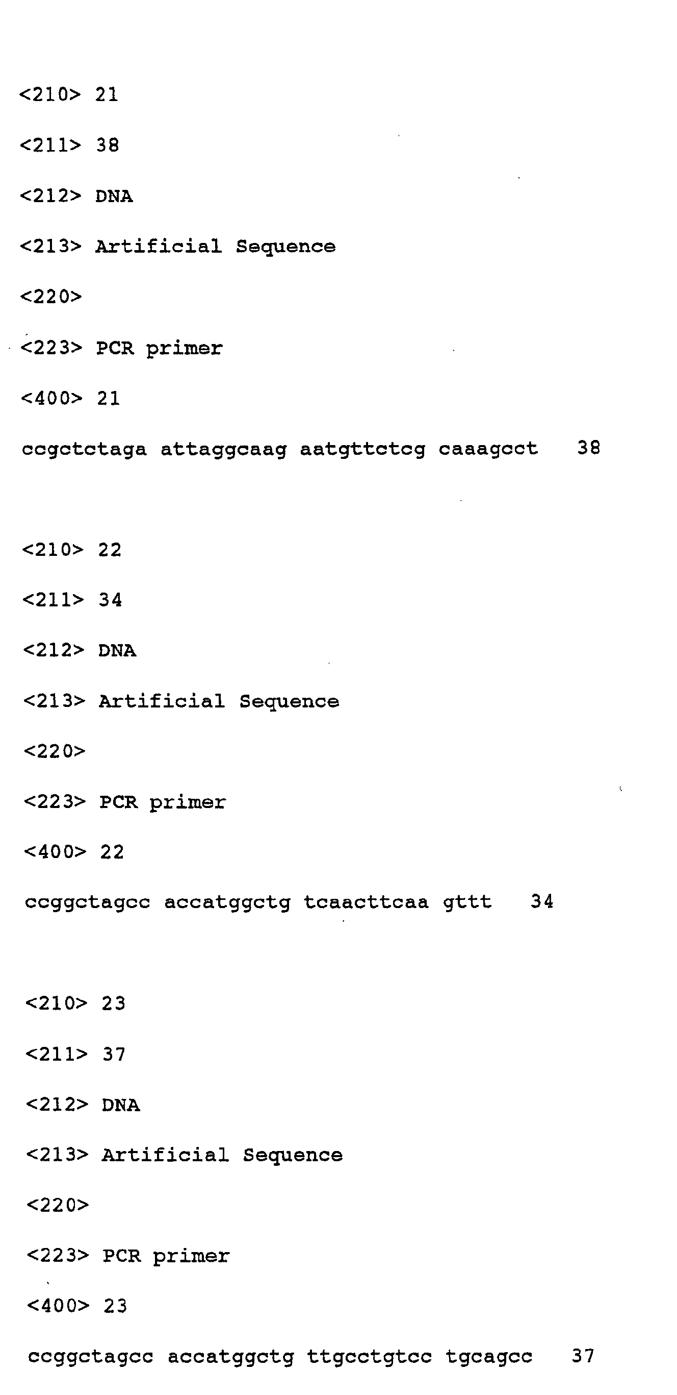
- the expression vectors for the 35 kDa protein, pSOL-CMV containing a putative signal sequence for secretion and pOL-CMV not containing the signal sequence were constructed in the same manner as described in (1) using the following primers;
- OL-4 and OL-6 were used.
- OL-5 and OL-6 were used.
- the 19 kDa or 35 kDa protein constituting Oplophorus luciferase was expressed by means of the in vitro transcription-translation system.
- pRL-CMV expressing Renilla luciferase was used as a positive control.
- the in vitro transcription-translation system is capable of producing a protein from a recombinant plasmid or mRNA prepared from the plasmid and is particularly useful for the luminescence system with high sensitivity for the detection.
- a commercially available in vitro translation kit (TNT in vitro transcription/translation kit; Promega) was used. A fraction of microsomal membranes capable of cleaving the signal sequence for secretion was also added in order to confirm the existence of the signal sequence in the expressed proteins.
- the in vitro translation mixture (25 ⁇ l in total) containing 0.5 ⁇ g of the plasmid DNA, 20 ⁇ l of rabbit reticulocyte lysate, 1 ⁇ l of 1 mM methionine and 2.5 ⁇ l of microsomal membranes was incubated at 30°C for 90 min and then 1 ⁇ l of the mixture was subjected to the measurement of its luminescent activity.
- a luminescent reaction mixture (100 ⁇ l in total) contains 1 ⁇ g of coelenterazine in 50 mM Tris-HCl/10 mM EDTA (pH 7.6). The reaction was started by the addition of a test sample and the intensity of the luminescence was measured by the luminometer. Results are shown in Table 2.
- Example 5 shows the in vitro expression of the 19 kDa or 35 kDa protein.
- E. coli host strain, BL21 was transformed with the expression vectors, pHis-KAZ and pHis-OL constructed in Example 10 and pTrcHis-B as a control plasmid in the standard technique in the art.
- One hundred microliter of overnight culture was transfered to Luria-Bertani (LB) broth containing 50 ⁇ g/ml of ampicillin and cultured for 2 hr at 37°C.
- Protein production was induced by the addition of isopropyl- ⁇ -thio-galactopyranoside (the final concentration: 0.2 mM) at 37°C in LB broth. After incubation for 3 hours, cells were harvested and then subjected to SDS-PAGE analysis to detect the protein products.
- the protein production was induced by IPTG as described above.
- the cells were harvested from 1 ml of the culture by centrifugation at 10,000 rpm and then disrupted by sonication in 1 ml of a sonication buffer (30 mM Tris-HCl/10 mM EDTA, pH 7.6). After centrifugation at 10,000 rpm at 4°C, the supernatant was collected and used as the cell extract for the luminescence assay.
- One ⁇ g of coelenterazine or bisdeoxycoelenterazine was added as a substrate into the cell extract and the intensity of the luminescence was measured by the luminometer. The results are shown in Table 3. Expression of the protein(s) of Oplophorus Luciferase in E.
- one relative luminescence unit corresponds to about 1.25 ⁇ 10 7 photons/second.
- Table 3 shows that the luminescence activity of the strain transformed with pHis-KAZ is approximately 10,000-fold higher than those of pTrcHis-B as a negative control and pHis-OL. Therefore, the above results demonstrate that only the 19 kDa protein out of the 19 kDa and 35 kDa proteins constituting Oplophorus luciferase has the luminescence activity and that the 19 kDa protein can independently generate the luminescence and can utilize both of coelenterazine and bisdeoxy-coelenterazine as a substrate. These facts suggest that the 35 kDa protein in the luciferase is functionally involved not in the substrate specificity but in the stability of the luciferase such as heat-resistance property.
- COS7 cells (2 x 10 5 cells) were grown in a 35 mm well-plate containing 3 ml of Dulbecco's modified Eagle's media (Gibco BRL, Rockville, MD, USA) supplemented with 10% (v/v) heat-inactivated fetal calf serum (Gibco BRL), 100 U/ml penicillin and 100 ⁇ g/ml of streptomycin.
- the cells were cultured at 37°C for 24 hours and then transfected with 2 ⁇ g of each plasmid DNA using FuGENE6 transfection reagent (Rosche Diagnostics, Mannheim, Germany). After further incubation for 36 hours, cells were separated from cultured media by centrifugation.
- the separated cells were suspended in 0.5 ml of phosphate-buffered saline, and subjected to repeated freeze-thawing at a temperature between 37°C and -80°C to obtain a cell extract.
- One ⁇ g of coelenterazine or bisdeoxy-coelenterazine was added as a substrate into the cell extract and the intensity of the luminescence was measured by the luminometer. The results are shown in Table 4.
- E. coli cells constructed in Example 12 which contain the expression vector pHis-KAZ and express the 19 kDa protein were disrupted by the sonication in 20 ml of a buffer comprising 20 mM Tris-HCl, pH 7.5. After centrifugation (12,000 ⁇ g, 20 minutes), the precipitated fraction was subjected to the solubilization by treating with 20 ml of 20 mM Tris-HCl, pH 7.5 containing 8 M urea and then centrifuged (12,000 ⁇ g, 20 minutes) to afford a soluble fraction. SDS-PAGE analysis of this fraction shows approximately 95% yield of the 19 kDa protein (data not shown). Subsequently, the soluble fraction was subjected to the nickel-chelated column chromatography and the desired protein was eluted by the linear gradient of imidazole from 0 to 0.3 M to give approximately 95% purity.
- the resultant protein has no luminescence activity at this state. Consequently, the protein was subjected to the renaturation step by treating with glycerol (the final concentration: from 0 to 90% (v/w)) as a solvent at 25°C for 30 minutes to afford the 19 kDa protein renatured. As described above, the intensity of the luminescence was measured by the luminometer. Results are shown in Table 5. Conc.
- the 19 kDa protein solubilized by the buffer containing a high concentration of urea could be renatured by treating with glycerol.
- glycerol concentration at 50% to 70% could significantly renature the luminescence activity of the protein.
- the 19 kDa protein renatured was subjected to the preservation test, which comprises preserving the protein in the absence or presence of 50% (v/w) or 70% (v/w) glycerol at 4°C for 30 days and then measuring its luminescence activity as mentioned above.
- the activity of the protein significantly decreased in the absence of glycerol, whereas in the presence of glycerol, the luminescence activity was maintained without decreasing.
- the present invention elucidates that the luciferase derived from the deep-sea shrimp, Oplophorus gracilirostris, is composed of the 19 kDa and 35 kDa proteins. Isolation of the 19 kDa and 35 kDa proteins from the Oplophorus gracilirostris and cloning of the genes encoding them can be achieved according to the invention.
- the recombinant vectors, the host cell such as a cultured animal cell or a microorganism transformed with the recombinant vector provided by the invention are used to produce the luciferase or the proteins of the invention in a large amount.
- the luciferase or the 19 kDa protein can be utilized for various methods for the measurement or analysis as a reporter.
- the antibody and oligonucleotide of the invention can be utilized for detecting the presence of a luciferase or the proteins constituting the enzyme and for cloning a gene encoding other luciferases. More specifically, they may be used for identification of a novel luciferase or photoprotein from systematically related species.
Landscapes
- Chemical & Material Sciences (AREA)
- Health & Medical Sciences (AREA)
- Life Sciences & Earth Sciences (AREA)
- Organic Chemistry (AREA)
- Genetics & Genomics (AREA)
- General Health & Medical Sciences (AREA)
- Biochemistry (AREA)
- Medicinal Chemistry (AREA)
- Molecular Biology (AREA)
- Engineering & Computer Science (AREA)
- Bioinformatics & Cheminformatics (AREA)
- Wood Science & Technology (AREA)
- Zoology (AREA)
- General Engineering & Computer Science (AREA)
- Microbiology (AREA)
- Biotechnology (AREA)
- Biomedical Technology (AREA)
- Immunology (AREA)
- Biophysics (AREA)
- Proteomics, Peptides & Aminoacids (AREA)
- Enzymes And Modification Thereof (AREA)
- Micro-Organisms Or Cultivation Processes Thereof (AREA)
Abstract
Description
| Reported genes of photoproteins and luciferases | ||||
| Protein | Origin | Discoverer (publication year) | Gene Accession No. | |
| 1. Photoprotein | ||||
| Aequorin | Aequorea victoria | Inouye et al. (1985) | AEVAQ440X: L29571 | |
| Aequorea victoria | Prasher et al. (1987) | AEVAEQA: M16103 | ||
| Clytin | Clytia gregarium | Inouye & Tsuji (1993) | CY1APOCLYT: L13247 | |
| Mitrocomin | Mitrocoma cellularia | Fagan et al. (1993) | MITMI17: L31623 | |
| Obelin | Obelia longissima | Illarionov et al. (1995) | OLU07128: U07128 | |
| 2. Luciferase | ||||
| Firefly | Photinus pyralis | de Wet et al. (1987) | PPYLUC: M15077 | |
| Luciola cruciata | Matuda et al. (1989) | FFLLUC: M26194 | ||
| Luciola lateralis | Tatsumi et al. (1992) | LLLUCI: X66919 | ||
| Luciola lateralis | Cho et al. (1995) | LLLUCIFMJ: Z49891 | ||
| Luciola mingrelica | Devine et al. (1993) | |||
| Photuris pennsylvanica | Zenno et al. (1993) | D25415: D25415 | ||
| Photuris pennsylvanica | Ye et al. (1997) | PPU31240: U31240 | ||
| Pyrocoelia miyako | Ohmiya et al. (1995) | PIBLUCA: L39928 | ||
| Hotaria parvula | Ohmiya et al. (1995) | HOTLUCI: L39929 | ||
| Glow worm | Lampyris noctiluca | Sala-Newby et al. (1996) | LNLUCPROT: X89479 | |
| Click beetle | Pyrophorus plagiophthalamus | Wood et al. (1989) | ||
| Railroad-worm | Phrixothrix vivianii | Viviani et al. (1999) | AF139644: AF139644 | |
| Phrixothrix hirtus | Viviani et al. (1999) | AF139645: AF139645 | ||
| Vargula | Vargula hilgendorfii | Kazami et al. (1988) | Pat. Appln. No. JP63-199295 | |
| Thompson et al. (1989) | VAHLUC: M25666 | |||
| Renilla | Renilla reniformis | Lorenz et al. (1991) | RELLUC: M63501 | |
| Gonyaulax | Gonyaulax polyedra | Bae & Hastings (1994) | GONLUCA: L04648 | |
| Bacteria | Vibrio fischeri | Foran & Brown (1988) | VFLUXAB: X06758 | |
| Vibrio harveyi | Cohn et al. (1985) | VIBHALUXA: M10961 | ||
| Johnston et al. (1986) | VIBHALUXA: M10961 | |||
| Photobacterium leiogathis | Illarionov et al. (1988) | PLLUXABG: X08036 | ||
| Lee et al. (1991) | PHRLUX: M63594 | |||
| Photobacterium phosphoreum | Ferri et al. (1991) | PHRLUXABDF: M65067 | ||
| Xenorhabdus luminescence | Johnston et al. (1990) | XENLUXABB: M55977 | ||
| Szittner & Meighen (1990) | XENLUXAB: M57416 | |||
| Kryptophanaron alfredi | Haygood (1986, 1990) | KRYLUC: M36597 | ||
| Alteromonas hanedai | Zenno et al. (1994) | Pat. Appln. No. JP06-035450 |


(Leu/Ile)-Xaa-Xaa-Leu-Xaa-(Leu/Ile)-Xaa-Xaa-Asn-Xaa- (Leu/Ile)-Xaa-Xaa-Xaa-Pro
wherein each Xaa may be any one of essential amino acids. This leucine-rich repeating structure is found in the amino acid sequence of the 35 kDa protein as shown in Fig. 2. Thus, a protein containing the above repeated sequence may be capable of stabilizing the luciferase as well as the 35 kDa protein.
The amino acid sequence of the 19 kDa proteins:
Phe-Thr-Leu-Ala-Asp-Phe-Val-Gly-Asp-Trp-Gln-Gln-Thr-Ala-Gly-Tyr-Asn-Gln-Asp-Gln-Val-Leu-Glu-Gln-Gly-Gly-Leu-Ser
The amino acid sequence of the 35 kDa proteins:
Ala-Val-Ala-Xaa-Pro-Ala-Ala-Glu-Asp-Ile-Ala-Pro-Xaa-Thr-Xaa-Lys-Val-Gly-Glu-Gly-Asp-Val-Met-Asp-Met-Asp-Xaa-Ser-Lys wherein Xaa represents an undetermined amino acid.
- SEQ ID NO: 7: Val-Thr-Ser-Asp-Ala-Glu-Leu-Ala-Ser-Ile-Phe-Ser-Lys-Thr-Phe-Pro
- SEQ ID NO: 8: Asn-Asp-Leu-Ser-Ser-Phe-Pro-Phe-Glu-Glu-Met-Ser-Gln-Tyr-Thr-Lys
- SEQ ID NO: 9: Leu-Val-Leu-Gly-Tyr-Asn-Gly-Leu-Thr-Ser-Leu-Pro-Val-Gly-Ala-Ile
- SEQ ID NO: 10: Asn-Leu-Asp-Pro-Ala-Val-Phe-His-Ala-Met-Xaa-Gln
| Expression of the protein(s) of Oplophorus Luciferase in Cell-free system | |||
| Expression Plasmid | Expressed protein | Addition of Microsomal membrane | Luminescence activity (rlu) |
| pRL-CMV | Renilla Luciferase | - | 3253.3 |
| pKAZ-CMV | 19kDa Protein | - | 1242.0 |
| pSKAZ-CMV | 19kDa Protein + Signal sequence | - | 3.0 |
| pSKAZ-CMV | 19kDa Protein + Signal sequence | + | 65.0 |
| pOL-CMV +pKAZ-CMV | 35kDa + 19kDa Proteins | - | 581.2 |
| pOL-CMV | 35kDa Protein | - | <0.001 |
| pSOL-CMV | 35kDa Protein + Signal sequence | - | <0.001 |
| pSOL-CMV | 35kDa Protein + Signal sequence | + | <0.001 |
| None | No addition | - | <0.001 |
| Expression of the protein(s) of Oplophorus Luciferase in E. coli | |||
| Strain/Host | Add. of IPTG | Luminescence activity (rlu) | |
| Coelenterazine | Bisdeoxycoelenterazine | ||
| pHis-KAZ/BL21 | - | 220 | 170 |
| pHis-KAZ/BL21 | + | 14,700 | 12,800 |
| pHis-OL/BL21 | - | 13 | 13 |
| pHis-OL/BL21 | + | 15 | 13 |
| pTrcHis-B/BL21 | - | 10 | 10 |
| pTrcHis-B/BL21 | + | 13 | 12 |
| Expression of the protein(s) of Oplophorus Luciferase in COS7 cell | |||
| Expression vector | Luminescence activity (rlu) | ||
| in Medium | in Cell extracts | ||
| Coelenterazine | Coelenterazine | Bisdeoxycoelenterazine | |
| pRL-CMV | 2.86 | 2,059.5 | 2.61 |
| pKAZ-CMV | 3.62 | 2,273.0 | 2,124.0 |
| pSKAZ-CMV | 1.89 | 297.5 | 217.0 |
| pOL-CMV | 2.45 | <0.001 | <0.001 |
| pSOL-CMV | 2.14 | <0.001 | <0.001 |
| None | 1.92 | <0.001 | <0.001 |
| Conc. of glycerol (%, v/w) | Luminescence Activity (rlu) |
| 0 | 83 (1.0) |
| 10 | 147 (1.8) |
| 20 | 375 (4.5) |
| 30 | 1,258 (15.2) |
| 40 | 1,818 (24.9) |
| 50 | 3,822 (46.0) |
| 60 | 4,860 (58.6) |
| 70 | 4,462 (53.8) |
| 80 | 2,842 (34.2) |
| 90 | 2,536 (30.6) |
| Concentration of glycerol (%, v/w) | Preserving temperature (°C) | Luminescence activity after preservation (rlu) | |
| 0 | 30 days | ||
| 0 | 4 | 102 | 23 |
| 50 | 4 | 3,840 | 3,734 |




Claims (29)
- A luciferase derived from Decapoda, which is composed of 19 kDa and 35 kDa proteins.
- The luciferase according to claim 1, which is a recombinant luciferase derived from Oplophorus gracilirostris.
- A photoprotein having a molecular weight of 19 kDa, which is one component of the luciferase of claim 1.
- The photoprotein according to claim 3, which contains an amino acid sequence selected from the group consisting of:(a) an amino acid sequence consisting of amino acids at positions 28-196 of the amino acid sequence shown in SEQ ID NO: 2; and(b) an amino acid sequence of (a) in which one or several amino acids are deleted, substituted or added.
- The photoprotein according to claim 4, which further contains a peptide sequence for purification and/or a signal peptide sequence for extracellular secretion or intracellular transport.
- A polynucleotide encoding the protein of claim 3.
- The polynucleotide according to claim 6, wherein its sequence is selected from the group consisting of:(a) a polynucleotide sequence consisting of positions 46-633 of the sequence shown in SEQ ID NO: 1;(b) a polynucleotide sequence hybridizing to the polynucleotide of (a) under the stringent hybridization condition and encoding a photoprotein; and(c) a polynucleotide sequence complementary to the sequence of (a) or (b).
- A protein having a molecular weight of 35 kDa which is one component of the luciferase of claim 1.
- The protein according to claim 8, which contains an amino acid sequence selected from the group consisting of:(a) an amino acid sequence consisting of amino acids at positions 40-359 of the amino acid sequence shown in SEQ ID NO: 4; and(b) an amino acid sequence of (a) in which one or several amino acids are deleted, substituted or added.
- The protein according to claim 9, which further contains a peptide sequence for purification and/or a signal peptide sequence for extracellular secretion or intracellular transport.
- A polynucleotide encoding the protein according to claim 8.
- The polynucleotide according to claim 11, wherein its sequence is selected from the group consisting of:(a) a polynucleotide sequence consisting of positions 79-1155 of the sequence shown in SEQ ID NO: 3;(b) a polynucleotide sequence hybridizing to the sequence of (a) under the stringent hybridization condition; and(c) a polynucleotide sequence complementary to the sequence of (a) or (b).
- A protein constituting the luciferase of claim 1 and being capable of stabilizing the luciferase, wherein its amino acid sequence contains one or more units of repeated sequence consisting of:
(Leu/Ile)-Xaa-Xaa-Leu-Xaa-(Leu/Ile)-Xaa-Xaa-Asn-Xaa-
(Leu/Ile)-Xaa-Xaa-Xaa-Pro
wherein Xaa may be any one of essential amino acids. - An antibody arisen against the luciferase of claim 1, the protein of claim 3 or 8, or an immunogenic fragment thereof, wherein the antibody can specifically bind to the luciferase and/or the protein.
- An antigen which is specifically recognized and bound to the antibody of claim 14.
- A recombinant expression vector which comprises at least one of the polynucleotide according to claim 6 or 11 operably linked to a promoter.
- A host cell transformed with the recombinant expression vector of claim 16.
- The host cell according to claim 17, which is selected form a cultured animal cell or a microorganic cell.
- A method for producing at least one protein according to claim 3 or 8, which comprises subjecting the recombinant expression vector of claim 17 to in vitro transcription-translation and isolating the recombinant protein expressed.
- A method for producing at least one protein according to claim 3 or 8, which comprises culturing the host cell of claim 17 and isolating the recombinant protein expressed.
- The method according to claim 19 or 20, which further comprises renaturing the protein in the presence of one or more polyhydric alcohols as a solvent for the reactivation of its enzyme activity, and optionally preserving in the solvent.
- The method according to claim 21, wherein the polyhydric alcohol is selected from the group comprising: glycerol, polyethylene glycol, polypropylene glycol, dextran, mannitol, sorbitol, inositol, xylitol, sucrose, fructose and glucose.
- An oligonucleotide comprising at least 10 contiguous nucleotides selected from a polynucleotide sequence shown in SEQ ID NO: 1 or a complementary sequence thereto.
- An oligonucleotide comprising at least 10 contiguous nucleotides selected from a polynucleotide sequence shown in SEQ ID NO: 3 or a complementary sequence thereto.
- A method for detecting an analyte in a test sample for the diagnosis, characterized in that the luciferase of claim 1 or the photoprotein of claim 3 is used as a reporter to generate a detectable signal.
- The method according to claim 25, wherein coelenterazine or derivatives thereof is used as a substrate for a luminescent reaction.
- A bioassay method for detecting an analyte in a test sample, wherein the polynucleotide of claim 3 or the expression vector of claim 16 is used as a reporter or marker to generate a detectable signal.
- The method according to claim 27, wherein the sample to be assayed is a living cell, or a living tissue, an organ or a part thereof.
- The method according claim 26, wherein coelenterazine or derivatives thereof is used as a substrate for the luminescent reaction.
Applications Claiming Priority (2)
| Application Number | Priority Date | Filing Date | Title |
|---|---|---|---|
| JP2000125053 | 2000-04-26 | ||
| JP2000125053 | 2000-04-26 |
Publications (3)
| Publication Number | Publication Date |
|---|---|
| EP1156103A2 true EP1156103A2 (en) | 2001-11-21 |
| EP1156103A3 EP1156103A3 (en) | 2002-09-11 |
| EP1156103B1 EP1156103B1 (en) | 2010-08-11 |
Family
ID=18635075
Family Applications (1)
| Application Number | Title | Priority Date | Filing Date |
|---|---|---|---|
| EP01109479A Expired - Lifetime EP1156103B1 (en) | 2000-04-26 | 2001-04-25 | Oplophorus luciferase |
Country Status (3)
| Country | Link |
|---|---|
| US (2) | US6544754B2 (en) |
| EP (1) | EP1156103B1 (en) |
| DE (1) | DE60142761D1 (en) |
Cited By (3)
| Publication number | Priority date | Publication date | Assignee | Title |
|---|---|---|---|---|
| WO2014151736A1 (en) | 2013-03-15 | 2014-09-25 | Promega Corporation | Activation of bioluminescence by structural complementation |
| GB2574745A (en) * | 2013-02-28 | 2019-12-18 | Jnc Corp | Codon-optimized gene for mutated shrimp luciferase and method for use thereof |
| CN111198272A (en) * | 2018-11-20 | 2020-05-26 | 香港理工大学深圳研究院 | Method for detecting interaction between proteins in vitro, detection kit and application thereof |
Families Citing this family (12)
| Publication number | Priority date | Publication date | Assignee | Title |
|---|---|---|---|---|
| GB2442118B (en) * | 2006-09-19 | 2011-08-17 | Chisso Corp | Process for production of recombinant proteins as a soluble form |
| ES2795287T3 (en) | 2009-05-01 | 2020-11-23 | Promega Corp | Synthetic Oplophorus Luciferases with Higher Light Emission |
| CA2815381C (en) | 2010-11-02 | 2019-03-19 | Promega Corporation | Coelenterazine derivatives and methods of using same |
| CN103443121B (en) | 2010-11-02 | 2016-04-20 | 普罗梅加公司 | Novel coelenterazine substrates and methods of use thereof |
| EP2783218B1 (en) * | 2011-11-23 | 2017-01-04 | Promega Corporation | Compositions and methods for monitoring transmembrane trafficking |
| JP6040846B2 (en) | 2013-04-08 | 2016-12-07 | Jnc株式会社 | Coelenterazine analog |
| JP6484987B2 (en) | 2013-10-21 | 2019-03-20 | Jnc株式会社 | Evil luciferase catalytic protein mutant gene and its usage |
| JP6442825B2 (en) | 2013-12-26 | 2018-12-26 | Jnc株式会社 | Evil luciferase catalytic protein mutant gene and its usage |
| US9790537B2 (en) | 2014-01-29 | 2017-10-17 | Promega Corporation | Quinone-masked probes as labeling reagents for cell uptake measurements |
| EP3099691B1 (en) | 2014-01-29 | 2019-11-20 | Promega Corporation | Pro-substrates for live cell applications |
| JP6256118B2 (en) | 2014-03-11 | 2018-01-10 | Jnc株式会社 | Chimeric gene of Evil luciferase catalytic protein and use thereof |
| JP6265020B2 (en) * | 2014-04-16 | 2018-01-24 | Jnc株式会社 | Evil luciferase catalytic protein mutant gene and its usage |
-
2001
- 2001-04-25 EP EP01109479A patent/EP1156103B1/en not_active Expired - Lifetime
- 2001-04-25 DE DE60142761T patent/DE60142761D1/en not_active Expired - Lifetime
- 2001-04-26 US US09/842,164 patent/US6544754B2/en not_active Expired - Lifetime
-
2003
- 2003-03-04 US US10/377,714 patent/US7125697B2/en not_active Expired - Lifetime
Non-Patent Citations (3)
| Title |
|---|
| DINGWALL, C.; LASKEY, R. A., TRENDS BIOCHEM. SCI., vol. 16, 1991, pages 478 - 481 |
| GAVEL, Y.; VON HEIJNE, G., FEBS LETT., vol. 261, 1990, pages 455 - 458 |
| PROTEIN ENGINEERING, vol. 4, 1990, pages 33 - 37 |
Cited By (19)
| Publication number | Priority date | Publication date | Assignee | Title |
|---|---|---|---|---|
| GB2574745A (en) * | 2013-02-28 | 2019-12-18 | Jnc Corp | Codon-optimized gene for mutated shrimp luciferase and method for use thereof |
| GB2574745B (en) * | 2013-02-28 | 2020-09-16 | Jnc Corp | Codon-optimized gene for mutated shrimp luciferase and method for use thereof |
| GB2516326B (en) * | 2013-02-28 | 2020-06-03 | Jnc Corp | Codon-optimized gene for mutated shrimp luciferase and method for use thereof |
| US10648971B2 (en) | 2013-03-15 | 2020-05-12 | Promega Corporation | Activation of bioluminescence by structural complementation |
| EP2970412A4 (en) * | 2013-03-15 | 2016-07-27 | Promega Corp | ACTIVATION OF BIOLUMINESCENCE BY STRUCTURAL COMPLEMENTATION |
| US10184936B2 (en) | 2013-03-15 | 2019-01-22 | Promega Corporation | Activation of bioluminescence by structural complementation |
| US10288605B2 (en) | 2013-03-15 | 2019-05-14 | Promega Corporation | Activation of bioluminescence by structural complementation |
| US9869670B2 (en) | 2013-03-15 | 2018-01-16 | Promega Corporation | Activation of bioluminescence by structural complementation |
| WO2014151736A1 (en) | 2013-03-15 | 2014-09-25 | Promega Corporation | Activation of bioluminescence by structural complementation |
| US12366572B2 (en) | 2013-03-15 | 2025-07-22 | Promega Corporation | Activation of bioluminescence by structural complementation |
| US10107800B2 (en) | 2013-03-15 | 2018-10-23 | Promega Corporation | Activation of bioluminescence by structural complementation |
| CN105143249A (en) * | 2013-03-15 | 2015-12-09 | 普洛麦格公司 | Activation of bioluminescence by structural complementation |
| EP3783011A1 (en) * | 2013-03-15 | 2021-02-24 | Promega Corporation | Activation of bioluminescence by structural complementation |
| US11493504B2 (en) | 2013-03-15 | 2022-11-08 | Promega Corporation | Activation of bioluminescene by structural complementation |
| EP4169935A1 (en) * | 2013-03-15 | 2023-04-26 | Promega Corporation | Activation of bioluminescence by structural complementation |
| EP4177261A1 (en) * | 2013-03-15 | 2023-05-10 | Promega Corporation | Activation of bioluminescence by structural complementation |
| US12367513B2 (en) | 2013-03-15 | 2025-07-22 | Promega Corporation | Activation of bioluminescence by structural complementation |
| CN111198272B (en) * | 2018-11-20 | 2023-09-08 | 香港理工大学深圳研究院 | Method for in vitro detection of interaction between proteins, detection kit and application thereof |
| CN111198272A (en) * | 2018-11-20 | 2020-05-26 | 香港理工大学深圳研究院 | Method for detecting interaction between proteins in vitro, detection kit and application thereof |
Also Published As
| Publication number | Publication date |
|---|---|
| US6544754B2 (en) | 2003-04-08 |
| US20020102687A1 (en) | 2002-08-01 |
| DE60142761D1 (en) | 2010-09-23 |
| US7125697B2 (en) | 2006-10-24 |
| US20040002127A1 (en) | 2004-01-01 |
| EP1156103B1 (en) | 2010-08-11 |
| EP1156103A3 (en) | 2002-09-11 |
Similar Documents
| Publication | Publication Date | Title |
|---|---|---|
| Inouye et al. | Secretional luciferase of the luminous shrimp Oplophorus gracilirostris: cDNA cloning of a novel imidazopyrazinone luciferase | |
| US7125697B2 (en) | Luciferase and photoprotein | |
| KR102594240B1 (en) | Novel luciferase and methods of using the same | |
| JP4613441B2 (en) | Novel luciferase and photoprotein | |
| US20100105090A1 (en) | Secreted luciferase mluc7 and use thereof | |
| US8563270B2 (en) | Calcium-binding photoprotein, gene encoding the same, and use thereof | |
| JP5582476B2 (en) | Marine plankton-derived photoprotein | |
| GB2441208A (en) | Fusion proteins | |
| KR20030051865A (en) | Isolated Luciferases and The Use Thereof | |
| EP1306435B1 (en) | Genes encoding proteins capable of regenerating luciferin, recombinant dna and process for producing protein capable of regenerating luciferin | |
| US6759524B2 (en) | Photoprotein derived from okinawan squid and gene encoding the photoprotein | |
| AU767483B2 (en) | Pholasin | |
| EP1229114B1 (en) | Gene encoding protein capable of regenerating luciferin, novel recombinant dna and process for producing protein capable of regenerating luciferin | |
| CA2464942A1 (en) | Signal system and elements used therein | |
| JP2002335961A (en) | Dinoflagellate luciferase and the gene encoding it | |
| JP2002034573A (en) | Gene encoding a protein capable of regenerating luciferin, recombinant DNA, and method for producing protein capable of regenerating luciferin | |
| ZA200403508B (en) | Signal system and elements used therein | |
| KR20080021018A (en) | Isolated photoprotein Adadecike and uses thereof |
Legal Events
| Date | Code | Title | Description |
|---|---|---|---|
| PUAI | Public reference made under article 153(3) epc to a published international application that has entered the european phase |
Free format text: ORIGINAL CODE: 0009012 |
|
| AK | Designated contracting states |
Kind code of ref document: A2 Designated state(s): AT BE CH CY DE DK ES FI FR GB GR IE IT LI LU MC NL PT SE TR |
|
| AX | Request for extension of the european patent |
Free format text: AL;LT;LV;MK;RO;SI |
|
| PUAL | Search report despatched |
Free format text: ORIGINAL CODE: 0009013 |
|
| AK | Designated contracting states |
Kind code of ref document: A3 Designated state(s): AT BE CH CY DE DK ES FI FR GB GR IE IT LI LU MC NL PT SE TR |
|
| AX | Request for extension of the european patent |
Free format text: AL;LT;LV;MK;RO;SI |
|
| 17P | Request for examination filed |
Effective date: 20030215 |
|
| AKX | Designation fees paid |
Designated state(s): DE GB |
|
| GRAP | Despatch of communication of intention to grant a patent |
Free format text: ORIGINAL CODE: EPIDOSNIGR1 |
|
| GRAS | Grant fee paid |
Free format text: ORIGINAL CODE: EPIDOSNIGR3 |
|
| GRAA | (expected) grant |
Free format text: ORIGINAL CODE: 0009210 |
|
| AK | Designated contracting states |
Kind code of ref document: B1 Designated state(s): DE GB |
|
| REG | Reference to a national code |
Ref country code: GB Ref legal event code: FG4D |
|
| REF | Corresponds to: |
Ref document number: 60142761 Country of ref document: DE Date of ref document: 20100923 Kind code of ref document: P |
|
| PLBE | No opposition filed within time limit |
Free format text: ORIGINAL CODE: 0009261 |
|
| REG | Reference to a national code |
Ref country code: GB Ref legal event code: 732E Free format text: REGISTERED BETWEEN 20110616 AND 20110622 |
|
| 26N | No opposition filed |
Effective date: 20110512 |
|
| REG | Reference to a national code |
Ref country code: DE Ref legal event code: R097 Ref document number: 60142761 Country of ref document: DE Effective date: 20110512 |
|
| REG | Reference to a national code |
Ref country code: DE Ref legal event code: R082 Ref document number: 60142761 Country of ref document: DE Representative=s name: GILLE HRABAL STRUCK NEIDLEIN PROP ROOS, DE |
|
| REG | Reference to a national code |
Ref country code: DE Ref legal event code: R081 Ref document number: 60142761 Country of ref document: DE Owner name: JNC CORPORATION, JP Free format text: FORMER OWNER: CHISSO CORP., OSAKA, JP Effective date: 20120117 Ref country code: DE Ref legal event code: R082 Ref document number: 60142761 Country of ref document: DE Representative=s name: GILLE HRABAL, DE Effective date: 20120117 |
|
| PGFP | Annual fee paid to national office [announced via postgrant information from national office to epo] |
Ref country code: DE Payment date: 20200415 Year of fee payment: 20 |
|
| PGFP | Annual fee paid to national office [announced via postgrant information from national office to epo] |
Ref country code: GB Payment date: 20200416 Year of fee payment: 20 |
|
| REG | Reference to a national code |
Ref country code: DE Ref legal event code: R071 Ref document number: 60142761 Country of ref document: DE |
|
| REG | Reference to a national code |
Ref country code: GB Ref legal event code: PE20 Expiry date: 20210424 |
|
| PG25 | Lapsed in a contracting state [announced via postgrant information from national office to epo] |
Ref country code: GB Free format text: LAPSE BECAUSE OF EXPIRATION OF PROTECTION Effective date: 20210424 |