EP1154443A1 - Magnetic glass particles, method for their preparation and uses thereof - Google Patents
Magnetic glass particles, method for their preparation and uses thereof Download PDFInfo
- Publication number
- EP1154443A1 EP1154443A1 EP00110165A EP00110165A EP1154443A1 EP 1154443 A1 EP1154443 A1 EP 1154443A1 EP 00110165 A EP00110165 A EP 00110165A EP 00110165 A EP00110165 A EP 00110165A EP 1154443 A1 EP1154443 A1 EP 1154443A1
- Authority
- EP
- European Patent Office
- Prior art keywords
- suspension
- magnetic
- procedure
- particles
- composition according
- Prior art date
- Legal status (The legal status is an assumption and is not a legal conclusion. Google has not performed a legal analysis and makes no representation as to the accuracy of the status listed.)
- Withdrawn
Links
Images


Classifications
-
- B—PERFORMING OPERATIONS; TRANSPORTING
- B82—NANOTECHNOLOGY
- B82Y—SPECIFIC USES OR APPLICATIONS OF NANOSTRUCTURES; MEASUREMENT OR ANALYSIS OF NANOSTRUCTURES; MANUFACTURE OR TREATMENT OF NANOSTRUCTURES
- B82Y25/00—Nanomagnetism, e.g. magnetoimpedance, anisotropic magnetoresistance, giant magnetoresistance or tunneling magnetoresistance
-
- B—PERFORMING OPERATIONS; TRANSPORTING
- B03—SEPARATION OF SOLID MATERIALS USING LIQUIDS OR USING PNEUMATIC TABLES OR JIGS; MAGNETIC OR ELECTROSTATIC SEPARATION OF SOLID MATERIALS FROM SOLID MATERIALS OR FLUIDS; SEPARATION BY HIGH-VOLTAGE ELECTRIC FIELDS
- B03C—MAGNETIC OR ELECTROSTATIC SEPARATION OF SOLID MATERIALS FROM SOLID MATERIALS OR FLUIDS; SEPARATION BY HIGH-VOLTAGE ELECTRIC FIELDS
- B03C1/00—Magnetic separation
- B03C1/005—Pretreatment specially adapted for magnetic separation
- B03C1/01—Pretreatment specially adapted for magnetic separation by addition of magnetic adjuvants
-
- C—CHEMISTRY; METALLURGY
- C12—BIOCHEMISTRY; BEER; SPIRITS; WINE; VINEGAR; MICROBIOLOGY; ENZYMOLOGY; MUTATION OR GENETIC ENGINEERING
- C12N—MICROORGANISMS OR ENZYMES; COMPOSITIONS THEREOF; PROPAGATING, PRESERVING, OR MAINTAINING MICROORGANISMS; MUTATION OR GENETIC ENGINEERING; CULTURE MEDIA
- C12N15/00—Mutation or genetic engineering; DNA or RNA concerning genetic engineering, vectors, e.g. plasmids, or their isolation, preparation or purification; Use of hosts therefor
- C12N15/09—Recombinant DNA-technology
- C12N15/10—Processes for the isolation, preparation or purification of DNA or RNA
- C12N15/1003—Extracting or separating nucleic acids from biological samples, e.g. pure separation or isolation methods; Conditions, buffers or apparatuses therefor
- C12N15/1006—Extracting or separating nucleic acids from biological samples, e.g. pure separation or isolation methods; Conditions, buffers or apparatuses therefor by means of a solid support carrier, e.g. particles, polymers
- C12N15/1013—Extracting or separating nucleic acids from biological samples, e.g. pure separation or isolation methods; Conditions, buffers or apparatuses therefor by means of a solid support carrier, e.g. particles, polymers by using magnetic beads
-
- C—CHEMISTRY; METALLURGY
- C12—BIOCHEMISTRY; BEER; SPIRITS; WINE; VINEGAR; MICROBIOLOGY; ENZYMOLOGY; MUTATION OR GENETIC ENGINEERING
- C12Q—MEASURING OR TESTING PROCESSES INVOLVING ENZYMES, NUCLEIC ACIDS OR MICROORGANISMS; COMPOSITIONS OR TEST PAPERS THEREFOR; PROCESSES OF PREPARING SUCH COMPOSITIONS; CONDITION-RESPONSIVE CONTROL IN MICROBIOLOGICAL OR ENZYMOLOGICAL PROCESSES
- C12Q1/00—Measuring or testing processes involving enzymes, nucleic acids or microorganisms; Compositions therefor; Processes of preparing such compositions
- C12Q1/68—Measuring or testing processes involving enzymes, nucleic acids or microorganisms; Compositions therefor; Processes of preparing such compositions involving nucleic acids
- C12Q1/6806—Preparing nucleic acids for analysis, e.g. for polymerase chain reaction [PCR] assay
-
- C—CHEMISTRY; METALLURGY
- C12—BIOCHEMISTRY; BEER; SPIRITS; WINE; VINEGAR; MICROBIOLOGY; ENZYMOLOGY; MUTATION OR GENETIC ENGINEERING
- C12Q—MEASURING OR TESTING PROCESSES INVOLVING ENZYMES, NUCLEIC ACIDS OR MICROORGANISMS; COMPOSITIONS OR TEST PAPERS THEREFOR; PROCESSES OF PREPARING SUCH COMPOSITIONS; CONDITION-RESPONSIVE CONTROL IN MICROBIOLOGICAL OR ENZYMOLOGICAL PROCESSES
- C12Q1/00—Measuring or testing processes involving enzymes, nucleic acids or microorganisms; Compositions therefor; Processes of preparing such compositions
- C12Q1/68—Measuring or testing processes involving enzymes, nucleic acids or microorganisms; Compositions therefor; Processes of preparing such compositions involving nucleic acids
- C12Q1/6844—Nucleic acid amplification reactions
- C12Q1/6848—Nucleic acid amplification reactions characterised by the means for preventing contamination or increasing the specificity or sensitivity of an amplification reaction
-
- H—ELECTRICITY
- H01—ELECTRIC ELEMENTS
- H01F—MAGNETS; INDUCTANCES; TRANSFORMERS; SELECTION OF MATERIALS FOR THEIR MAGNETIC PROPERTIES
- H01F1/00—Magnets or magnetic bodies characterised by the magnetic materials therefor; Selection of materials for their magnetic properties
- H01F1/0036—Magnets or magnetic bodies characterised by the magnetic materials therefor; Selection of materials for their magnetic properties showing low dimensional magnetism, i.e. spin rearrangements due to a restriction of dimensions, e.g. showing giant magnetoresistivity
- H01F1/0045—Zero dimensional, e.g. nanoparticles, soft nanoparticles for medical/biological use
- H01F1/0054—Coated nanoparticles, e.g. nanoparticles coated with organic surfactant
-
- H—ELECTRICITY
- H01—ELECTRIC ELEMENTS
- H01F—MAGNETS; INDUCTANCES; TRANSFORMERS; SELECTION OF MATERIALS FOR THEIR MAGNETIC PROPERTIES
- H01F1/00—Magnets or magnetic bodies characterised by the magnetic materials therefor; Selection of materials for their magnetic properties
- H01F1/0036—Magnets or magnetic bodies characterised by the magnetic materials therefor; Selection of materials for their magnetic properties showing low dimensional magnetism, i.e. spin rearrangements due to a restriction of dimensions, e.g. showing giant magnetoresistivity
- H01F1/0045—Zero dimensional, e.g. nanoparticles, soft nanoparticles for medical/biological use
- H01F1/0063—Zero dimensional, e.g. nanoparticles, soft nanoparticles for medical/biological use in a non-magnetic matrix, e.g. granular solids
-
- H—ELECTRICITY
- H01—ELECTRIC ELEMENTS
- H01F—MAGNETS; INDUCTANCES; TRANSFORMERS; SELECTION OF MATERIALS FOR THEIR MAGNETIC PROPERTIES
- H01F1/00—Magnets or magnetic bodies characterised by the magnetic materials therefor; Selection of materials for their magnetic properties
- H01F1/44—Magnets or magnetic bodies characterised by the magnetic materials therefor; Selection of materials for their magnetic properties of magnetic liquids, e.g. ferrofluids
-
- C—CHEMISTRY; METALLURGY
- C12—BIOCHEMISTRY; BEER; SPIRITS; WINE; VINEGAR; MICROBIOLOGY; ENZYMOLOGY; MUTATION OR GENETIC ENGINEERING
- C12Q—MEASURING OR TESTING PROCESSES INVOLVING ENZYMES, NUCLEIC ACIDS OR MICROORGANISMS; COMPOSITIONS OR TEST PAPERS THEREFOR; PROCESSES OF PREPARING SUCH COMPOSITIONS; CONDITION-RESPONSIVE CONTROL IN MICROBIOLOGICAL OR ENZYMOLOGICAL PROCESSES
- C12Q1/00—Measuring or testing processes involving enzymes, nucleic acids or microorganisms; Compositions therefor; Processes of preparing such compositions
- C12Q1/68—Measuring or testing processes involving enzymes, nucleic acids or microorganisms; Compositions therefor; Processes of preparing such compositions involving nucleic acids
- C12Q1/6813—Hybridisation assays
- C12Q1/6816—Hybridisation assays characterised by the detection means
-
- C—CHEMISTRY; METALLURGY
- C12—BIOCHEMISTRY; BEER; SPIRITS; WINE; VINEGAR; MICROBIOLOGY; ENZYMOLOGY; MUTATION OR GENETIC ENGINEERING
- C12Q—MEASURING OR TESTING PROCESSES INVOLVING ENZYMES, NUCLEIC ACIDS OR MICROORGANISMS; COMPOSITIONS OR TEST PAPERS THEREFOR; PROCESSES OF PREPARING SUCH COMPOSITIONS; CONDITION-RESPONSIVE CONTROL IN MICROBIOLOGICAL OR ENZYMOLOGICAL PROCESSES
- C12Q2561/00—Nucleic acid detection characterised by assay method
- C12Q2561/101—Taqman
Definitions
- This invention relates to magnetic particles having a glass surface and are substantially spherical. This invention also relates to methods for making them, as well as to suspensions thereof and their uses for the purification of biological material in particular in automated processes.
- Biospecific binding assays allow of the detection of specific analytes, e.g. nucleic acids, or specific analyte properties and play a major role in the field of diagnostics and bioanalytics. Examples therefor are hybridisation assays, immuno assays and receptorligand assays.
- Nucleic acids are comparatively complex analytes which have normally to be extracted from complex mixtures before they can be used in a probe-based assay.
- the procedure known from the prior art entails the selective binding of nucleic acids to glass surfaces in chaotropic salt solutions and separating the nucleic acids from contaminants such as agarose, proteins or cell residue.
- the particles are either centrifuged or fluids are drawn through glass fiber filters. This is a limiting step, however, that prevents the procedure from being used to process large quantities of samples.
- magnetic particles covered with a glass surface offer considerable advantages for isolating biological materials. If the magnetic particles have not been brought in contact with a magnetic field, gravity is the only force that can cause them to sediment out. They can be resuspended by shaking the solution. The sedimentation procedure that does not utilize a magnetic field proceeds more slowly than the immobilization of biological materials on the surface of the particles. This is especially true for nucleic acids.
- the magnetic particles can be easily collected at a specific location in the sample fluid by means of a magnet. The fluid is then separated from the particles and, therefore, from the immobilized biological materials.
- Magnetic, porous glass is also available on the market that contains magnetic particles in a porous, particular glass matrix and is covered with a layer containing streptavidin.
- This product can be used to isolate biological materials, e.g., proteins or nucleic acids, if they are modified in a complex preparation step so that they bind covalently to biotin.
- Magnetizable particular adsorbents proved to be very efficient and suitable for automatic sample preparation.
- Ferrimagnetic and ferromagnetic as well as superparamagnetic pigments may be used for this purpose.
- Particles are solid materials having a small diameter. Particles like these are often also referred to as pigments.
- Those materials are referred to as magnetic that are drawn to a magnet, i.e., ferromagnetic or superparamagnetic materials, for instance.
- Superparamagnetism is seen as advantageous and preferable in the state of the art (e.g. US 5,928,958; US 5,925,573; EP 757 106).
- the glass or organosilane surfaces are often functionalised in order to be used for biospecific capture reactions, e.g. US 5,928,958, US 5,898,071, US 5,925,573, EP 937 497, US 4,554,088 or US 4,910,148.
- glass or organosilane surfaces may be treated with various solvents or salts to modify their hydrophilicity and/ or electropositivity, e.g. US 5,438,127.
- the underivatized silanol groups of the glass or the silane surface may be used for the adsorption with pure physico-chemical forces under suitable reaction conditions as described in DE 195 20 964, DE 195 37 985, WO 96/41840, WO 96/41811, EP 757 106 or US 5,520,899.
- magnetic cores or magnetic core aggregates are covered with a glass surface which is formed by an acid- or base-catalyzed sol-gel-process. These particles are called core-shell particles.
- the glass shell then has a typical layer thickness (see e.g. DE 195 20 964) wherein the size and shape of the pigment, which may contain a non-magnetic support as e.g.
- mica in addition to the magnetic metal oxide, determines size and form of the produced particle (see e.g. DE 195 37 985 and corresponding WO 96/41811).
- glass material with a high porosity is used (see e.g. EP 757 106; WO 99/26605).
- composite magnetic particles are described, e.g. silicate-covered ferric oxide covered with an inorganic silica matrix from silica particles (EP 757 106) or mixtures of glass and silica gel (WO95/ 06652).
- the problem to be solved by the present invention can be seen as providing magnetic glass particles with improved properties for sample preparation and for biological assays, in particular for automated processes.
- the magnetic glass particles (MGPs) according to the present invention are a solid dispersion of small magnetic cores in glass.
- the MGPs are comparatively small and are substantially spherical.
- the non-magnetic fine content of a composition of the MPGs is very low because of the method of their preparation. This has the effect that suspensions of the MGPs sediment slowly and can therefore be advantageously used for processes in molecular biology which can be automatized.
- compositions and suspensions of the MGPs according to the present invention are provided.
- a method for the composition of the MGPs is provided.
- a method for the purification of DNA or RNA is provided in which the MGPs according to the present invention are used.
- This has surprising consequences on the sedimentation kinetics, quantified by the half time values t 1/2 , which is the time span until 50 % of the particles have sedimented from a specific volume element (see Example 6).
- the half-life period for the sedimentation of a 3 mg/ ml weight-per-volume suspension of the composition in isopropanol is more than 3 min, preferably 4 min, more preferably 6 min. However the most preferred values for the half-life period is more than 10 min or even more than 20 min.
- the MGPs according to the present invention are glass droplets in which very small non-aggregating magnetic objects are dispersed.
- Those objects that are referred to as magnetic are drawn to a magnet, i.e., ferromagnetic or superparamagnetic materials, for instance.
- Preferred are ferromagnetic materials, in particular if they have not yet been premagnetized. Premagnetization in this context is understood to mean bringing in contact with a magnet, which increases the remanence.
- Preferred magnetic materials are iron or iron oxide as e.g. magnetite (Fe 3 O 4 ) or Fe 2 O 3 , preferably ⁇ -Fe 2 O 3 .
- the magnetic objects may be e.g. a magnetic pigment.
- the size of the magnetic objects is in the nanoscale range, i.e. according to the present invention the diameter is between 5 to 500 nm, preferably between 10 to 200 nm, most preferably between 15 to 50 nm .
- Suitable magnetic pigments are manufactured by the company CERAC which have a mean diameter of 23 nm and consist of ⁇ -Fe 2 O 3 (BET-surface 50 m 2 /g, CERAC: P.O. Box 1178, Milwaukee, Wisconsin 53201-1178 USA; Article-No. I-2012)..
- the magnetic glass particles according to the present invention are further characterized by the fact that the MGPs have a particle diameter between 0.5 ⁇ m and 5 ⁇ m, preferably between 1 ⁇ m to 2 ⁇ m as determined by high resolution scanning electron microscopy, whereas the magnetic objects have a diameter between 5 to 500 nm, preferably between 10 to 200 nm, most preferably in the range of 15 to 50 nm as said above.
- the MGPs of the present invention are further characterized by a diameter ratio of magnetic pigment core to magnetic glass particle of less than 1 to 10 as determined by high resolution scanning electron microscopy .
- the MGPs according to the present invention are microporous but have highly-structured and therefore relatively large surface with more than 6 m 2 /g.
- the magnetic glass particles according to the present invention have a surface area in the range of 5 to 100 m 2 / g, preferably 5 to 90 m 2 / g, more preferably in the range of 10 to 50 m 2 / g, most preferably in the range of 15 to 30 m 2 / g. This surface is approximately double the size of the particles described in DE 195 37 985.
- the sample EJ0096.5R-01 which is of preferential interest (see Example 1 and Table 1 to Table 3 for a summary of the production parameters) has a BET-surface of 26.8525 m 2 /g, a micropore area of 2.3058 m 2 / g and an average pore diameter of 24.9132 nm. This means that the pore surface is less than 10 % of the total surface and that the magnetic glass particle is microporous.
- a pore is understood to be a recess in the outer surface of the particle.
- the surface reaches so far into the particle that a perpendicular line drawn in the recess on the surface cuts the particle at least once in the direction of the adjacent environment of the particle.
- pores reach into the particle to a depth that is greater than one radius of the pore.
- Radiotracing experiments showed that the binding behavior with regard to DNA and RNA was the same when compared to reference material known in the state of the art. Surprisingly, the production parameters had an influence on the performance in the radiotracing experiments.
- a further advantage of the MGP-type of the present invention is that no tensions in the glass layer can lead to fissure during the drying process and corresponding damages in the glass shell because of the inner structure (solid dispersion of small magnetic cores in a glass drop). This can be investigated by image-producing methods (see Example 3).
- Another embodiment of the present invention is a suspension of magnetic particles. It is obvious for the person skilled in the art to produce a suspension by adding a liquid to a composition of the MGPs and mix the suspension to homogeneity.
- a liquid according to the present invention may comprise any liquid which does not affect the stability of the magnetic particles and may be used to produce a homogenous suspension.
- liquids are used which are suitable for processes in molecular biology, in particular desoxyribonucleic acid (DNA) or ribonucleic acid (RNA) purification processes which make use of the binding of these substances to glass particles under certain conditions.
- Preferred liquids comprise alcohols or any mixtures thereof with water or ketones.
- other alcohols can also be used if they are suitable for molecular biology purposes as e.g. glycerol.
- the alcohols isopropanol, ethanol or mixtures thereof with water preferably a mixture of 80 volume parts of isopropanol with 20 volume parts of water (.
- the liquid comprises ketones as e.g. acetone.
- these suspensions contain between 5 to 60 mg/ ml MGPs.
- the MGPs are suspended in aqueous buffered solutions which may optionally contain a chaotropic agent in a concentration of between 2 and 8 mol/l, and preferably between 4 and 6 mol/l.
- Chaotropic salts can be sodium iodide, sodium perchlorate, guanidinium thiocyanate, guanidinium isothiocyanate or guanidinium hydrochlorite. Other compounds are also possible.
- a chaotropic agent according to the present invention will be any chemical substance which will disturb the ordered structure of liquid water and will have the effect that DNA or RNA will bind to the MGPs according to the present invention if this agent is present in the DNA or RNA containing solution. It is obvious for the artisan to produce suitable aqueous buffered solutions. Buffer systems which may be used for molecular biology purposes may be found e.g. in Sambrook et al.
- buffer substances are Tris-hydroxymethylamine (TRIS), phosphate, N-(2-Hydroxyethyl)piperazine-N'-(2-ethanesulfonic acid) (HEPES), salts thereof or other suitable substances.
- substances may be present which modify the ionic strength of the solution as e.g. NaCl, KCl or CaCl 2 or which are metal cation complexing agents as e.g. ethylene-diamine-tetra-acetic acid (EDTA) or the salts thereof.
- EDTA ethylene-diamine-tetra-acetic acid
- Biological material known to the expert in the field may also be present.
- the suspension of MGPs may additionally contain DNA or RNA optionally in a mixture with proteins, fatty acids, carbohydrates and other material from biological origin.
- the liquid may contain a mixture of one or more constituents selected from the group of alcohols, ketones, aqueous buffered solutions, chaotropic agents, substances which modify the ionic strength of the solution, complexing agents, biological material, DNA or RNA all with the features as described above.
- a tube or reaction vessel containing the suspension according to the invention is provided.
- the tube can be made of plastics but it may also be part of a larger structure, e.g. part of a microtitreplate in 96- or 384-wellformat.
- a storage container is provided which contains a composition of magnetic glass particles or suspensions thereof.
- kits of parts which comprises a storage container containing the magnetic glass particles or a suspension thereof according to the present invention.
- the kit may be used for the purification of DNA or RNA.
- kits known in the art further comprise plastics ware which may be used during the purification procedure as e.g. microtitreplates in the 96 or 384 well format or just ordinary reaction tubes manufactured e.g. by Eppendorf, Hamburg, Germany.
- the kit may further comprise a washing solution which is suitable for the washing step of the magnetic glass particles when DNA or RNA is bound thereto. Often the washing solution is provided as a stock solution which has to be diluted before the use.
- the kit may further comprise an eluent, i.e.
- a solution or a buffer e.g. TE, 10 mM Tris, 1 mM EDTA, pH 8.0
- additional reagents may be present which can be used for the purification process of a nucleic acid, i.e.DNA or RNA.
- the kit of parts according to the present invention is used for the purification of a nucleic acid.
- composition of MGPs may be used to produce a suspension as already described.
- the suspensions according to the present invention may be used for the purification of nucleic acids, i.e. RNA or DNA, from complex mixtures with other biological substances containing them.
- nucleic acids i.e. RNA or DNA
- mixtures of different nucleic acids may be purified, even mixtures containing a nucleic acid of interest in low abundance.
- the purification effect results from the behavior of DNA or RNA to bind to magnetic glass particles under certain conditions e.g. in the presence of certain concentration of a chaotropic agent.
- the MGPs with the bound DNA or RNA are washed afterwards at least once, preferably with a mixture of 70 volume parts ethanol with 30 volume parts water ("70 % Ethanol"). Afterwards, the conditions are reversed, e.g.
- the concentration of the chaotropic agent is decreased, to elute the DNA or RNA bound to the MGP the particle.
- this is done by the pelleting of the magnetic glass particles, e.g. by gravity force or by the use of a magnet, and resuspending in a solution with no or only a low amount of chaotropic agent.
- the solution can be diluted with a solution with no or only a low amount of chaotropic agent.
- the purified DNA or RNA can now be used for other reactions.
- a glass according to the present invention is understood to be an amorphous material that contains silicium. Glass can contain other materials such as B 2 O 3 (0 - 30%), Al 2 O 3 (0 - 20%), CaO (0 - 20%), BaO (0 - 10%), K 2 O (0 - 20%), Na 2 O (0 - 20%), MgO (0 - 18%), Pb 2 O 3 (0 - 15%). Glass can also contain a smaller percentage (0 - 5%) of a number of other oxides such as Mn 2 O 3 , TiO 2 , As 2 O 3 , Fe 2 O 3 , CuO, CoO, etc.
- glasses that are formed using the gel sol process described in WO 96/41811 and then dried and compressed.
- the basic principles of this process are known and were described, for instance, in C.J. Brinker, G.W. Scherer "Sol Gel Science - The Physics and Chemistry of Sol Gel Processing", Academic Press Inc. 1990, Sol-Gel Optics, Processing and Applications, Lisa C. Klein, Ed., Kluwer Academic Publishers 1994, p. 450 ff., and in DE-A-1941191, DE-A-3719339, DE-A-4117041, DE-A-4217432 and WO96/41811.
- alkoxides of network-forming components e.g., SiO 2 , B 2 O 3 , Al 2 O 3 , TiO 2 , ZrO 2 , GeO 2
- oxides and salts of other components e.g., in an alcohol solution
- the equation below describes the procedure for making sodium boroaluminium silicate glass: Water is added to begin the hydrolysis process of the starting components. The reaction proceeds relatively quickly because the alkali ions have a catalytic effect on the speed of hydrolysis of the silicic acid ester.
- the gel Once the gel is formed it can be dried and densified (or condensed) by means of a thermal process to form glass.
- the composition of the glass (code RN) was 74 Mol% SiO 2 , 15 Mol% B 2 O 3 , 5,00 Mol% Al 2 O 3 , 4,00 Mol% K 2 O, 2,00 Mol% CaO.
- the composition of the glass (code EP) was 73,61 Mol% SiO 2 , 14,93 Mol% B 2 O 3 , 5,21 Mol% Al 2 O 3 , 4,17 Mol% K 2 O, 2,08 Mol% CaO.
- the glass may be produced with methods known in the state of the art by the melting of the raw material SiO 2 and of carbonates of the alkali or alkaline earth metals Na 2 CO 3 , K 2 CO 3 or CaCO 3 .
- the sol:pigment ratio has a considerable effect on the yield of magnetic particles provided by this invention. It is essential for the process that the sol can still be pumped and sprayed which is in the skill of artisan.
- the slurry is preferably sprayed through a two-fluid nozzle as described in Figure 1 and in Example 1.3.
- Suitable spray-drying systems are produced by Nubilosa Molekularzerstäubung, Ladisch GmbH & Co. KG, Konstanz, Germany, e.g. the "Labor-Zerstäubungstrockner (Typ LTK)" or by Büchi AG, Uster, Switzerland, e.g. the Mini Spray Dryer (Type B-191).
- the geometry and the number of incorporated magnetic cores or of their inert carriers do not determine shape and size of the particles but the conditions of manufacturing, in particular the conditions during spray drying.
- the choice of pressure, inlet temperature, outlet temperature and flow rate during the spray drying procedure are the degrees of freedom which will determine the size distribution, the shape of the glass drops and thereby will modify the MGPs.
- the size distribution will shift into the sub- ⁇ range.
- the decreased temperature of the spray drying process will lead to slower evaporation of the solvent and thereby the form of the MGPs will come closer to an ideal sphere, i.e. the ratio of the radii in xy- and xz-plane will become approximately 1.
- the ratio of the radii will vary between 0,8 and 1,2, preferably between 0,9 and 1,1.
- the nozzles are heated.
- the inlet temperature is between 120 °C and 500 °C, preferably between 170 °C and 230 °C or 150 °C and 230 °C, most preferably between 150 °C and 200 °C or 190 °C and 210 °C or at 200 °C or slightly less.
- the outlet temperature depends on the boiling point of the sol and thereby on the solvent and may be above, equal or slightly under, i.e. less than 10°C, the boiling point of the solvent.
- ethanol is used as solvent, it is between 50 °C and 300 °C, preferably 70 °C and 150 °C, most preferably between 80 °C and 110 °C.
- the densification or sinter temperature should be as high as possible, i.e. slightly below the melting range. If it is too high, however, the particles will stick together and form agglomerates that must be sieved out. If too low, the MGPs will not be optimally densified. Additional treatment of the particles at too high temperature will result in a loss of magnetic properties. Too high temperatures should therefore be omitted. The exact temperatures depend on the glass composition but may be between 400°C to 1200°C. In the case of the EJ glass composition the sinter temperature is between 720°C and 770°C, preferably around 750°C. It is in the skill of the artisan to find out the temperatures for each glass composition when taking the teachings of the present invention into account.
- the spray-dried MGP powder may be further processed as depicted in Figure 2 and described in Example 1.4.
- the powder is heated for 1 hour to 200 °C, optionally cooled to room temperature and heated to 750 °C (densification or sinter temperature) in a nitrogen atmosphere with a heating rate of 1 K/min and is held at that temperature for 1 hour. Then the furnace is cooled to 150 °C and heated again to 200 °C for one hour in air. After the cooling to room temperature, the powder is transferred to a sieve (50 ⁇ m) and sieved for 30 min. The sieved sample is bottled and sterilized at 200 °C for 4 h and then cooled to 80 °C. Then the glass vessels are taken from the oven, covered with sterile foil and closed.
- the magnetic particles provided by the invention are especially suited for isolating biological materials from samples.
- the core material is a natural resource and therefore causes little ecological concern.
- the particles according to the invention are inexpensive and easy to manufacture.
- Another object of the invention is a procedure for isolating a biological material by bringing a sample containing the biological material in a liquid in contact with the magnetic particles according to the invention under conditions in which the biological material binds to the particle surface, and separating the biological material from the liquid.
- the term "in a liquid” means that liquid may be added to the sample before the magnetic particles are added.
- the order of reagent addition may be varied according to process requirements.
- Biological materials are understood to mean materials with a particular or molecular basis. They include, in particular, cells such as viruses or bacteria, as well as isolated cells from multicellular organisms as e.g. human and animal cells such as leucocytes, and immunologically active low and high molecular chemical compounds such as haptens, antigens, antibodies and nucleic acids. Nucleic acids such as DNA or RNA are especially preferred. In one embodiment of the invention mixtures of specific nucleic acids are purified, in which the target nucleic acid(s) may be a minor component in terms of concentration (or may be present in low abundance). According to the present invention, a target nucleic acid shall be the nucleic acid of interest, i.e.
- a nucleic acid which shall be investigated as its presence is indicative of a certain condition or disease of a human or animal.
- a viral sequence e.g. from Hepatitis B Virus, Hepatitis C Virus or Human immunodeficiency virus
- this viral sequence would be the target sequence.
- Other target sequences are sequences which are indicative of a predisposition of an individual to a certain disease as e.g. an inherited disease as sickle cell anemia or to certain types of cancer. This examples should be illustrative of the invention but should not ne delimiting.
- Native biological material is understood to be material, the structure of which was not irreversibly changed compared with the naturally-occurring biological materials. This does not mean that other components of the sample can not be modified, however.
- Modified biological materials include materials that do not occur in nature, e.g., nucleic acids that are modified by attaching to them groups that are reactive, detectable or capable of immobilization. An example of this are biotinylated nucleic acids.
- the sample can also contain other components in a liquid such as cell residue, proteins, salts and other substances that are not to be isolated.
- This sample which preferably contains the biological material in native form, is brought in contact with the particles under conditions in which the target biological material binds to the particle surface.
- the conditions for this depend on the type of biological material involved, but are basically known. They also depend on the method by which the biological material is bound to the surface. If immunological interactions are utilized for the binding, for instance, conditions must be selected that are suitable for the formation of immunocomplexes.
- the binding can take place via the groups of nucleic acids that represent the modification, e.g., biotin via binding with streptavidin-coated surfaces.
- nucleic acids in particular, however, a direct binding of nucleic acids to glass is preferred because among other reasons the nucleic acids do not have to be modified and even native nucleic acids can be bound.
- the procedure for binding native nucleic acids to glass particles can be analogous to the procedure described in the prior art. It is preferably performed in the presence of chaotropic salts with a concentration of between 2 and 8 mol/l, and preferably between 4 and 6 mol/l.
- Chaotropic salts can be sodium iodide, sodium perchlorate, guanidinium thiocyanate, guanidinium isothiocyanate or guanidinium hydrochlorite. Other compounds are also possible.
- the sample is mixed with the particles and incubated for a period of time sufficient for the binding to occur.
- Experts are usually familiar with the duration of the incubation step from procedures for performing treatment with non-magnetic particles. This step can be optimized by determining the quantity of immobilized biological material on the surface at different points in time. Incubation times of between 10 seconds and 30 minutes can be appropriate for nucleic acids.
- the particles either separate out of the fluid during the incubation period itself or the suspension remains intact for a longer period of time. If the particles are very small and unmagnetized, the suspension remains intact for a longer period of time. If the particles are of larger size, the particles slowly separate out of the fluid during the incubation period.
- Immobilization is preferably not performed via precipitation by lowering the solubility of the materials to be immobilized. Rather, immobilization is based on biospecific interactions (capture molecules) or adsorption. This largely prevents contaminants from being non-specifically included.
- the biological material is separated from the liquid. This is achieved in general by separating the material bound to the magnetic particles by applying a magnetic field. For instance, the magnetic particles can be pulled to the wall of the vessel in which incubation was performed. The liquid containing the sample contents that were not bound to the magnetic particles can then be removed. The removal procedure used depends on the type of vessel in which incubation was performed. Suitable steps include removing the liquid via pipetting or aspiration.
- the magnetic particles can then be purified one or more times using a wash solution, if desired.
- a wash solution is used that does not cause the biological material to be released from the particle surface but that washes away the undesired contaminants as thoroughly as possible.
- This wash step preferably takes place by incubating the wash solution with the particles.
- the particles are preferable resuspended during this step, e.g., by means of shaking or applying a magnetic field that is not identical to the first magnetic field.
- the contaminated wash solution is preferably separated just like the sample in the step described above for binding the biological material.
- the magnetic particles can be dried briefly in a vacuum, or the fluid can be allowed to evaporate. A pretreatment step using acetone may also be performed. If desired, the biological material purified in this manner can be separated from the magnetic particles. This step also depends on the manner in which the biological material was bound to the magnetic particles. If the biological material is native nucleic acids and the magnetic particles are glass-coated particles, the nucleic acids can be removed from the particles according to the invention using an elution buffer having a low salt content. Buffers of this nature are known from DE 3724442 and Analytical Biochemistry 175, 196-201 (1988). The elution buffers with a low salt content are in particular buffers with a content of less than 0.2 mol/l. In an especially preferred embodiment, the elution buffer contains Tris. In another special embodiment, the elution buffer is demineralized water.
- the purification and isolation procedure described is performed after the cells (e.g., viral particles or prokaryotic or eukaryotic cells) are separated immunomagnetically from a bodily fluid or tissue.
- the sample is incubated, e.g., while shaking, with magnetic particles to which an antibody against an antigen on the cell is immobilized.
- These particles can be particles according to the invention or commercially available particles (e.g., MACS Microbeads from Miltenyi Biotec GmbH, Bergisch Gladbach, Germany).
- a magnetic field is applied, one or more wash steps are performed using a saline solution. Particles are obtained to which the desired cells are bound. The bound cells are then resuspended in a saline buffer.
- this saline buffer is a chaotropic saline solution so that the nucleic acids contained in the cell are released from the cells.
- An especially advantageous procedure for isolating nucleic acids from samples containing cells is achieved by combining the isolation of cells described above with the isolation of nucleic acids also described above, on the magnetic particles according to the invention.
- the advantage of this embodiment is its potential simplicity (single-tube method), high sensitivity (especially important in medical microbiology and oncology), and the ease with which it can be automated.
- the biological materials isolated using the procedure according to the invention can now be used further as necessary. For instance, they can be used as a substrate for various enzymatic reactions.
- nucleic acids When nucleic acids are involved, they can be used for sequencing, radioactive or non-radioactive labelling, amplification of one or more of the sequences they contain, transcription, hybridization with labelled probe nucleic acids, translation or ligation.
- An advantage of the procedure according to the invention is that it is very easy to separate the biological material from the fluid.
- a centrifugation step was used to separate the glass particles from contaminants, or, when the biological material is bound to glass fiber filters, the fluid is drawn through the filters. This is a limiting step that makes it difficult to process large quantities of sample.
- the biological materials can be separated from contaminants more effectively using the particles according to the invention. In particular, inhibitors for certain enzymatic reactions can be removed to a large extent according to the invention.
- the particles according to the invention are added to the lysis mixture. After a suitable period of time for adsorption to take place ⁇ which can be optimized by mechanical agitation ⁇ the particles are separated from the surrounding fluid that contains additional cell components that are not to be detected. This is performed preferably by applying a magnetic field by placing a magnet against the vessel wall.
- a preferred embodiment of the present invention is to use the MGPs of the present invention in automatable methods as e.g. described in WO 99/16781.
- Automatable method means that the steps of the method are suitable to be carried out with an apparatus or machine capable of operating with little or no external control or influence by a human being.
- Automatized method means that the steps of the method are carried out with an apparatus or machine capable of operating with little or no external control or influence by a human being. Only the preparation steps for the method may have to be done by hand, e.g. the storage containers have to filled up and put into place, the choice of the samples has to be done by a human being and further steps known to the expert in the field, e.g. the operation of the controlling computer.
- the apparatus or machine may e.g.
- the MGPs according to the present invention are used in semiautomated process which means that some reaction steps may have to be done manually.
- a suspension containing MGPs according to the present invention is taken from a storage container and partial volumes are added to different reaction vessels.
- Reaction vessels may be reaction tubes made from plastics eventually in mictrotitreplate format contain 96 or 384 or more wells where a reaction can be carried out. However, these vessels may be made from other material e.g. from steel.
- a procedure for the purification of biological material followed by a detection step wherein the amplification and/ or detection reaction is a homogeneous solution-phase multiplex assay for the simultaneous detection of multiple targets (see examples 7.2).
- the described purification procedure is combined with a amplification procedure using one of the methods described in the following, preferably the use of blocking oligonucleotides.
- a problem often associated with amplification especially of low amounts of target nucleic acid is the activity of thermostable polymerases at lower temperatures (room temperature up to 40°C). At this temperature primer oligonucleotides often bind unspecifically to each other or to background nucleic acid and may be extended by the polymerase. This entails a decrease of reaction components as well as to a higher level of background signal and consequently to a decreased sensitivity. It may also lead to false-positive results.
- aptamers in another embodiment of the invention can be used in an amplification reaction and the detection methods connected thereto.
- the 3'-terminal nucleobase preferably an adenin is modified with a p-(t-butyl)-benzyl-residue. Further modifications, including those at the 3'-1-position are described in EP 866 071 A2 which is incorporated herein by reference.
- alcohols are easily evaporated during spray-drying; ideally no recristallisation of salts on the surface
- alcoholates are turned into hydroxides, which yield by way of elimination of water the corresponding oxides.
- These then , form a 3-dimensional, amorphous glass matrix consisting of SiO 2 / B 2 O 3 / Al 2 O 3 , into which certain metal oxide ingredients are embedded as matrix bond separators, e.g.
- TEOS Tetraethoxysilane
- TEB Triethylborate
- K-methanolate 25% W/V
- Methanol 1601 ml
- Ethanol 11292 ml
- Aluminiumisopropanolate 1385 g
- the vessel is closed thereafter.
- the sol is heated to 70 °C and stirred overnight (15 h).
- the temperature is regulated via a thermosensor which dips into the liquid.
- the lid is opened and 10249 g magnetic pigment (CERAC) is added under vigorous stirring.
- the prepared sol is transferred into the spray dryer via a hose.
- the inlet temperature of the spray dryer is regulated to 200 °C.
- the nozzle pressure is regulated to 6 bar and the nozzle is cooled for 3 min with ethanol.
- the hose is connected to the outlet of the glass container and the pigment-containing sol is pumped at a rate of 110 ml/min to the two fluid nozzle (diameter of the opening: 2 mm; supplier: Nubilosa, Type 1B1VVS1) of the spray dryer via a ultrasound device (200 W).
- the spray drying system is shown schematically in Figure 3. The particles which are formed in the first two minutes are discarded. After the complete spraying of the sol the container under the cyclone (AVO, see Figure 3) with the particles will be taken and the particles will be further treated as described in the next steps.
- the pigment containing sol is stirred in the stirring vessel to prevent sedimentation of the suspended particles.
- the sol is transferred from the vessel to the nozzle using a pump (SP).Nitrogen heated by an electric heater (EWT) is used as drying gas.
- EWT electric heater
- the coated pigment is transferred from the drying chamber (T) to the cyclone (ZY).
- the dried powder an be removed from the container (AVO) under the cyclone.
- Very fine particles are removed by a filter (SF) from the nitrogen.
- the spray dryer is used with overpressure to prevent an air intake into the dryer.
- the gas flow is produced by gas blower (AV).
- the powder is transferred into a ceramics bowl and heated in an furnace to 200 °C at a heating rate of 1 K/min. Then temperature is held at 200 °C for one hour and the oven is cooled to room temperature afterwards.
- the bowl is transferred to an atmosphere furnace with a volume of 27 l which is rinsed with 60 1 nitrogen per hour.
- the furnace is heated to 750 °C with a heating rate of 1 K/min and is held at that temperature for 1 hour.
- the furnace is cooled to 150°C and rinsed with 60 l air per hour.
- the furnace is heated to 200 °C with a heating rate of 2 K/min and is held at that temperature for 1 hour and is cooled to temperature afterwards.
- the powder is transferred to a sieve (50 ⁇ m) and sieved for 30 min. Afterwards, the sieved sample is bottled in glass containers, which are sterilized unclosed in an oven which is heated to 200 °C with a heating rate of 1 K/min, held at that temperature for 4 h and then cooled to 80 °C. Then the glass vessels are taken from the furnace, covered with sterile foil and closed with a lid.
- the composition of the glass (code RN) was 74 Mol% SiO 2 , 15 Mol% B 2 O 3 , 5.00 Mol% Al 2 O 3 , 4.00 Mol% K 2 O, 2.00 Mol% CaO.
- the composition of the glass (code EP) was 73.61 Mol% SiO 2 , 14.93 Mol% B 2 O 3 , 5.21 Mol% Al 2 O 3 , 4.17 Mol% K 2 O, 2.08 Mol% CaO.
- the Figure 4 shows MGPs with mica covered with ferric oxide as magnet pigment (BM).
- BM magnet pigment
- the particles with CERAC pigment do not have fissure, as the magnet pigment (23 nm average particle size) does not cause tension forces in the layer so that no fissures are created during the spray drying process. Furthermore, the small CERAC-particles are also incorporated in the fine content so that there are no non-magnetic particles contained therein. The particles are also substantially smaller than the particles with mica which can be observed in these figures with the same magnification. MGPs with the other magnetic pigments are shown at the same magnification in Figure 6 to Figure 10.
- the extinction of the supernatant is measured in an UV-VISNIR spectrometer (Hitachi U-3000) against a corresponding cuvette filled with deionized water.
- the measuring range was 200 to 1100 nm in steps of 1 nm to investigate this wavelength range for the absorption bands of eventual impurities.
- the extinction at a wavelength of 280 nm and 400 nm was taken as a reference.
- a gas pyknometer (AccuPyc 1330, Fa. Micromeritics) is used. Helium 6.0 is used as gas. The device is calibrated with the supplied standard (steel beads with known volume). Further, the sample is dried for at least 1 hour at 150 °C and then filled into the measurement container and weighed. After the measuring container is put into the gas pyknometer, 10 washing cycles and 5 measuring cycles are used to determine the average value and the standard deviation.
- the iron solubility of the coated and temperature-treated samples is determined with Inductive Coupled Plasma - Atomic Emission Spectroscopy (JY 24, company ISA). Therefor, 1 g of the sample is transferred into a 50 ml polypropylene-tube, filled up with distilled water and kept at a temperature of 60 °C for 20 hours. Then, the samples are filtrated with a 0.2 ⁇ m syringe filter and the filtrate is measured. Four single determinations are carried out at a wavelength of 259,940 nm and an average value is computed therefrom.
- JY 24, company ISA Inductive Coupled Plasma - Atomic Emission Spectroscopy
- a PP-weighing tube (Company Licefa, Art. Nr. V2-3) completely filled with MGPs is weighed.
- this sample container is placed in the middle of LDPE tube (Company Kartell, TS 735) burdened with brass so that the lid of the tube can be still closed.
- the vessel is positioned in the middle of a balance (detection accuracy 0.1 g) with the help of another stencil.
- a plastics cap which contains an cylinder-shaped magnet (diameter: 30 mm; height: 113.5 mm; material: samarium-cobalt 2/17) is positioned above the scale.
- the surfaces were determined using a device from Micromeritics (Type ASAP 2400). The measurement was performed at liquid nitrogen temperature. Nitrogen 5.0 was used as measuring gas and Helium 4.6 was used as inert gas. Typically, 5 g of sample was used for one measurement.
- the surface of the MGPs plays an obvious role for the isolation of DNA and RNA.
- Table 4 The data for the measured BET-surfaces for some samples are summarized in Table 4.
- BASF CE-SU particles have a size distribution in between the small (BASF FA, STREM and CERAC) and the big Merck particles. In contrast to the Merck particles, they do not possess a structured surface so that these particles have a very smooth glass surface. This results into a smaller surface. A higher pressure was used with the smaller spray dryer and the nozzle of this spray dryer is remarkably smaller than that of the bigger spray dryer. This causes smaller particles whereby the surface increases remarkably. This is in good agreement with the results of Example 5.2.1
- radioactively labelled DNA or RNA is synthesized enzymatically in a PCR or in vitro transcription process in the presence of 32 P-markied desoxynucleoside or nucleoside-triphosphates. Then, the marked DNA or RNA is separated from the free nucleoside-triphosphates, the content determined and a defined dilution is prepared.
- a small amount of marked DNA or RNA is added to each sample before the examination.
- all nucleic acids bind to the MGPs in the presence of chaotropic agents.
- the MGPs are pelleted by the exertion of magnetic forces and the supernatants can be discarded.
- the pellet is washed and the bound nucleic acids are eluted at elevated temperature by the reversal of the reaction conditions, i.e. by the addition of a low salt elution buffer.
- an aliquot of the particle supernatant is spotted onto a filter.
- the eluate as well as the in water redispersed MGP are spotted onto a filter as well and dried thereafter.
- the filters are measured in a Beta.Counter and for each sample preparation a distribution is calculated.
- RNA parameter proved to be the most sensitive in the course of the studies and was therefore chosen as parameter.
- Figure 11 shows BASF-CE is not suitable as core material because only small amounts of the bound RNA can be found in the eluate.
- the EJ/ CERAC particles which were sprayed in the Büchi system at 6 bar showed the performance of the reference EJ/ MMB which were sprayed in the Nubilosa system.
- the reference particles are EJ /MMB MGP.
- the DNA and RNA binding properties are investigated. Whereas the DNA parameter shows no dependence from the spraying pressure, the RNA parameter shows significant performance differences.
- the performance of the EJ/ CERAC particles produced with 1.5 bar spraying pressure with the Nubilosa system is lower than that of the reference. There is a lower performance in the series in dependence of the spraying pressure which varied from 1.5 to 3.4 bar ( ⁇ 30%). Particles which are sprayed with 4.3 bar reached 90 % of the performance of the particles which are sprayed at 1.5 bar (see Figure 12).
- MGPs are produced on the Nubilosa system at a pressure of 1.5 bar, 4.3 bar and 6 bar using nitrogen as spraying gas.
- An Uvikon Spectrophotometer Model 930 produced by Kontron Instruments is used for the evaluation of the sedimentation behaviour of the magnetic glass particles.
- This spectrophotometer is modified to allow variable adjustment of the cuvette along a scale bar.
- the cuvette is set at a position in which the measuring beam with a wavelength of 650 nm traverses the cuvette at a position which is at 2/3 of the filling height (scale position 7.5).
- Macro cuvettes made from polystyrene are used which have a volume of 4 ml and a path length of 1 cm. Prior to measurements, calibration is made in a one beam procedure against pure suspension medium.
- the MGP samples are dispersed to homogeneity in suspension medium, typically at a mass/volume ratio of 3 mg/ml, and measured immediately. The change of the extinction with time is monitored continuously at the said wavelength of 650 nm.
- the above-described device further allows the installation of a magnet beneath the cuvette. Thereby the velocity of the magnetic separation can be determined.
- MGP-types with different cores in the range of nano- or micrometers and produced with EJ glass chemistry are investigated spectrophotometrically for their sedimentation and separation behaviour, i.e. with and without magnet under the cuvette. Mass/volume is 6 mg/ml in this case.
- the suspension medium is a 1:1 mixture of isopropanol and lysis buffer. The results are summarized in .
- the particles from the CERAC type show a clear advantage in the stability of the suspension but no disadvantages for the magnetic separation. (See also Figure 14).
- biotin-tagged amplificate After PCR with at least one biotinylated primer, an aliquot of biotin-tagged amplificate is mixed with hybridization buffer and detection probe. After incubation, streptavidin-coated beads are added, followed by another incubation. Finally the beads are washed, signal buffer is added and the chemiluminescent signal intensity measured, which is correlated with the mass of amplified nucleic acid bound and, thus, the viral load of the plasma sample.
- the expert in the field may also carry out the 1 ml protocol manually.
- Proteinase K liquid in glycerol/Ca-acetate, 20 mg/ml
- Poly-A-RNA, 1 mg/ml, use level is a 1:1000 dilution in lysis buffer lysis buffer, consisting of:
- Amplification/ Mastermix - PCR-buffer, consisting of buffer medium
- Stop-reagent for UNG after the amplification - N-lauroylsarcosine e.g. Roche Id.Nr. 133895, 1% w/v
- RNA internal control (IC, see below) is spiked into each and every sample, the extraction process is thermostated at ca. 40°C, and volumes are adjusted to 2 ml total volume of lysis mix, i.e.
- UNG is blocked through the addition of laurylsarcosin concentration 1% use level.
- Thermocycling profiles are as follows: HBV UNG-step 1x 10 min 37°C PCR 35x 30 sec 30 sec 40 sec 92°C 55°C 72°C HCV UNG-step 1x 10 min 37°C reverse transcriptase step denaturation 1x 30 min 1 min 60°C 95°C PCR 2x 10 sec 20 sec 95°C 60°C 33x 15 sec 20 sec 90°C 60°C follow-up incubation 7 min 72°C HIV UNG-step 1x 10 min 37°C reverse transcriptase step 1x 30 min 60°C PCR 4x 10 sec 10 sec 10 sec 95°C 55°C 72°C 31x 10 sec 10 sec 10 sec 95°C 60°C 72°C
- detection probes with 2 particular chemical modification are added to the PCR mastermix.
- a fluorogenic reporter group for instance a derivative of 6-carboxy-fluorescein
- the other one is a dye (for instance a polymethine-cyanine derivative) capable of absorbing the fluorescent light of the reporter and to quench it (quencher).
- the quencher is typically attached to the probe backbone at the 5'-end, whereas the reporter is located within the oligo sequence, spaced from the quencher by a number of nucleotide building blocks.
- These probes bind to the target nucleic acid (sense or anti-sense strand) close to the 3'-end of a primer (reverse or forward).
- primer reverse or forward
- elongation starts. Due to the 5'-nuclease activity of the enzyme, simultaneously with copy strand synthesis the probe is cleaved as soon as the polymerase reaches the probe binding site, reporter and quencher get separated and the fluorescent signal becomes measurable.
- ct is a measure of assay sensitivity: the smaller the ct-value, the more sensitive is the assay.
- ct values can be calculated by means of different mathematical operations, for example cut-off approaches (average background signal intensity multiplied by a constant factor yields a cut-off signal intensity to discern negative from positive) or approaches where the location of the maximum of the first differentiation of the signal-over-time curve (i.e. the steepness profile), or the location of the maximum of the second differentiation on the time axis, are computed and defined as ct.
- This amplification / detection technology allows for real-time monitoring of PCR and for closed-tube processing as well, i.e. in contrast to standard procedures, the PCR tubes remains closed after pipetting eluate and mastermix which effectively reduces contamination risks.
- nucleic acids present in the sample are extracted by way of physico-chemical adsorption to the particle surface, independent of sequence characteristics.
- This will also include human DNA (hDNA), released from disrupted blood cells , e.g. leucocytes, the titer of which may differ widely according to the physiological or pathological status of the individual blood sample donor. For instance, in cases of autoimmune diseases like SLE, hDNA levels may be elevated substantially.
- hDNA and one or more pathogenic nucleic acids present in a given sample constitute a mixture of various nucleic acids of different sequence specificities which are co-extracted. They constitute a matrix of polynucleotides that are extracted without discrimination, followed by sequence-specific amplification and detection of target nucleic acids via interaction with specific primers and probes under appropriate reaction conditions.
- an artificial nucleic acid construct may be added to the samples, preferably packaged (armored) in a modified virus particle, which is co-extracted and co-amplified with the natural target.
- This internal control (IC) features a unique probe binding region for an IC detection probe, which differs from target-specific probes by a different reporter group with distinguishable emission characteristics. Thus, IC signal can be discerned from target signal, and as the IC is known to be present in the sample, it functions as a monitoring agent. So, multi labeling expands the utility of multiplex assay technology.
- the internal control (IC) is described in WO98/00547.
- the common multiplex thermocycling profile for all test parameters is as follows: UNG-step 45°C - 10 min denaturation 94°C - 30 sec reverse transcription 58°C - 30 min PCR 95°C - 20 sec / 59°C - 50 sec 5x 91°C - 15 sec / 52°C - 50 sec 55x
- FAM 6-carboxy-fluorescein coupled to a 2-(amino-cyclohexyl-)propane-1,3-diol-linker
- ⁇ EX 485 nm
- ⁇ EM 515 nm.
- Some of the oligonucleotides are derivatised with which is hexachloro-6-carboxy-fluorescein coupled to a 2-(amino-cyclohexyl-)propane-1,3-diol-linker (Biogenex CX-HEX
- the data are summarized in Table 7.
- EJ0047.2R (MMB) and EJ0100.5R (CERAC) are sprayed under standard conditions, i.e. 1.5 bar, 230°C inlet temperature and approximately 110 °C outlet temperature.
- EJ0102.2R (MMB) and EJ0108.5R (CERAC) are development variants with regard to the spraying conditions (inlet temperature decreased to 200°C [MMB]), and spraying pressure increased to 4.3 bar [CERAC]).
- the results are summarized in Table 8 and show the potential of the CERAC nano-core particles to obtain higher sensitivity as exemplified by the earlier threshold cycle and/or the larger signal differences (saturation signal minus noise level, S-N).
- MGP Different types of MGP were used to extract HCV in vitro transcripts which were diluted to various titer levels in diluent containing 10 mmol/l Tris, pH 80, 1 mM EDTA, 20 ⁇ g/ml poly-A-RNA and 0.05% NaN 3 , and spiked into pooled plasma.
- hBG-DNA Human background DNA
- MGP preparation EJ0096.5R which represents the MGP quality according to the present invention the most, shows very clearcut advantages both in terms of minimal shift of ct values, and reducing loss of specific signal generation (S-N), as indicated by the data presented in Table 13.
Landscapes
- Chemical & Material Sciences (AREA)
- Engineering & Computer Science (AREA)
- Life Sciences & Earth Sciences (AREA)
- Health & Medical Sciences (AREA)
- Organic Chemistry (AREA)
- Zoology (AREA)
- Wood Science & Technology (AREA)
- Biomedical Technology (AREA)
- General Health & Medical Sciences (AREA)
- Molecular Biology (AREA)
- Nanotechnology (AREA)
- Genetics & Genomics (AREA)
- Proteomics, Peptides & Aminoacids (AREA)
- Bioinformatics & Cheminformatics (AREA)
- General Engineering & Computer Science (AREA)
- Biotechnology (AREA)
- Analytical Chemistry (AREA)
- Microbiology (AREA)
- Chemical Kinetics & Catalysis (AREA)
- Biochemistry (AREA)
- Biophysics (AREA)
- Physics & Mathematics (AREA)
- Power Engineering (AREA)
- Immunology (AREA)
- Crystallography & Structural Chemistry (AREA)
- Plant Pathology (AREA)
- Inorganic Chemistry (AREA)
- Glass Compositions (AREA)
Abstract
This invention relates to magnetic nanosized particles having a glass surface. This invention also relates to methods for making them, as well as to suspensions thereof and their uses for the purification of DNA or RNA in particular in automated processes.
Description
This invention relates to magnetic particles having a glass surface and are substantially
spherical. This invention also relates to methods for making them, as well as to
suspensions thereof and their uses for the purification of biological material in particular
in automated processes.
Many biological materials, especially nucleic acids, present special challenges in terms of
isolating them from their natural environment. On the one hand, they are often present
in very small concentrations and, on the other hand, they are often found in the
presence of many other solid and dissolved substances that make them difficult to isolate
or to measure, in particular in biospecific assays.
Biospecific binding assays allow of the detection of specific analytes, e.g. nucleic acids, or
specific analyte properties and play a major role in the field of diagnostics and
bioanalytics. Examples therefor are hybridisation assays, immuno assays and receptorligand
assays.
Hybridisation assays make use of the specific base-pairing for the molecular detection of
nucleic acid analytes e.g. RNA and DNA. Hence, oligonucleotide probes with a length of
18 to 20 nucleotides may enable the specific recognition of a selected sequence in the
human genome. Another assay which makes use of the selective binding of two
oligonucleotide primers is the polymerase chain reaction (PCR) described in US
4,683,195. This method makes use of the selective amplification of a specific nucleic acid
region to detectable levels by a thermostable polymerase in the presence of
desoxynucleotide triphosphates in several cycles.
Nucleic acids are comparatively complex analytes which have normally to be extracted
from complex mixtures before they can be used in a probe-based assay.
There are several methods for the extraction of nucleic acids:
- sequence-dependent or biospecific methods as e.g.:
- affinity chromatography
- hybridisation to immobilised probes on beads
- sequence-independent or physico-chemical methods as e.g.:
- liquid-liquid extraction with e.g. phenol-chloroform
- precipitation with e.g. pure ethanol
- extraction with filter paper
- extraction with micelle-forming agents as cetyl-trimethyl-ammonium-bromide
- binding to immobilised, intercalating dyes, e.g. acridine derivatives
- adsorption to silica gel or diatomic earths
- adsorption to magnetic glass particles (MGP) or organo silane particles under chaotropic conditions
Many procedures and materials for isolating nucleic acids from their natural
environment have been proposed in recent years by the use of their binding behavior to
glass surfaces. In Proc. Natl. Acad. USA 76, 615-691 (1979), for instance, a procedure for
binding nucleic acids in agarose gels in the presence of sodium iodide in ground flint
glass is proposed.
The purification of plasmid DNA from bacteria on glass dust in the presence of sodium
perchlorate is described in Anal. Biochem. 121, 382-387 (1982).
In DE-A 37 34 442, the isolation of single-stranded M13 phage DNA on glass fiber filters
by precipitating phage particles using acetic acid and lysis of the phage particles with
perchlorate is described. The nucleic acids bound to the glass fiber filters are washed and
then eluted with a menthol-containing buffer in Tris/EDTA buffer.
A similar procedure for purifying DNA from lambda phages is described in Anal.
Biochem. 175, 196-201 (1988).
The procedure known from the prior art entails the selective binding of nucleic acids to
glass surfaces in chaotropic salt solutions and separating the nucleic acids from
contaminants such as agarose, proteins or cell residue. To separate the glass particles
from the contaminants according to the prior art, the particles are either centrifuged or
fluids are drawn through glass fiber filters. This is a limiting step, however, that prevents
the procedure from being used to process large quantities of samples.
It has been demonstrated that magnetic particles covered with a glass surface offer
considerable advantages for isolating biological materials. If the magnetic particles have
not been brought in contact with a magnetic field, gravity is the only force that can cause
them to sediment out. They can be resuspended by shaking the solution. The
sedimentation procedure that does not utilize a magnetic field proceeds more slowly
than the immobilization of biological materials on the surface of the particles. This is
especially true for nucleic acids. The magnetic particles can be easily collected at a
specific location in the sample fluid by means of a magnet. The fluid is then separated
from the particles and, therefore, from the immobilized biological materials.
The use of magnetic particles to immobilize nucleic acids after precipitation by adding
salt and ethanol is described in Anal. Biochem. 201, 166-169 (1992) and PCT GB
91/00212. In this procedure, the nucleic acids are agglutinated along with the magnetic
particles. The agglutinate is separated from the original solvent by applying a magnetic
field and performing a wash step. After one wash step, the nucleic acids are dissolved in
a Tris buffer. This procedure has a disadvantage, however, in that the precipitation is not
selective for nucleic acids. Rather, a variety of solid and dissolved substances are
agglutinated as well. As a result, this procedure can not be used to remove significant
quantities of any inhibitors of specific enzymatic reactions that may be present.
Magnetic, porous glass is also available on the market that contains magnetic particles in
a porous, particular glass matrix and is covered with a layer containing streptavidin. This
product can be used to isolate biological materials, e.g., proteins or nucleic acids, if they
are modified in a complex preparation step so that they bind covalently to biotin.
Magnetizable particular adsorbents proved to be very efficient and suitable for
automatic sample preparation. Ferrimagnetic and ferromagnetic as well as
superparamagnetic pigments may be used for this purpose.
Particles, according to the expert, are solid materials having a small diameter. Particles
like these are often also referred to as pigments.
Those materials are referred to as magnetic that are drawn to a magnet, i.e.,
ferromagnetic or superparamagnetic materials, for instance. Superparamagnetism is
seen as advantageous and preferable in the state of the art (e.g. US 5,928,958; US
5,925,573; EP 757 106). The glass or organosilane surfaces are often functionalised in
order to be used for biospecific capture reactions, e.g. US 5,928,958, US 5,898,071, US
5,925,573, EP 937 497, US 4,554,088 or US 4,910,148. Alternatively, glass or
organosilane surfaces may be treated with various solvents or salts to modify their
hydrophilicity and/ or electropositivity, e.g. US 5,438,127.
However, the underivatized silanol groups of the glass or the silane surface may be used
for the adsorption with pure physico-chemical forces under suitable reaction conditions
as described in DE 195 20 964, DE 195 37 985, WO 96/41840, WO 96/41811, EP 757 106
or US 5,520,899. Typically, magnetic cores or magnetic core aggregates are covered with
a glass surface which is formed by an acid- or base-catalyzed sol-gel-process. These
particles are called core-shell particles. The glass shell then has a typical layer thickness
(see e.g. DE 195 20 964) wherein the size and shape of the pigment, which may contain a
non-magnetic support as e.g. mica in addition to the magnetic metal oxide, determines
size and form of the produced particle (see e.g. DE 195 37 985 and corresponding WO
96/41811). To obtain a high surface activity, glass material with a high porosity is used
(see e.g. EP 757 106; WO 99/26605). Further, composite magnetic particles are
described, e.g. silicate-covered ferric oxide covered with an inorganic silica matrix from
silica particles (EP 757 106) or mixtures of glass and silica gel (WO95/ 06652).
The problem to be solved by the present invention can be seen as providing magnetic
glass particles with improved properties for sample preparation and for biological assays,
in particular for automated processes.
The deficiencies of the magnetic glass particles in the state of the art are overcome by the
findings of the present invention.
It is an object of the invention to provide a composition of magnetic glass particles. The
magnetic glass particles (MGPs) according to the present invention are a solid
dispersion of small magnetic cores in glass. The MGPs are comparatively small and are
substantially spherical. The non-magnetic fine content of a composition of the MPGs is
very low because of the method of their preparation. This has the effect that suspensions
of the MGPs sediment slowly and can therefore be advantageously used for processes in
molecular biology which can be automatized. In one embodiment of the invention
compositions and suspensions of the MGPs according to the present invention are
provided. In another embodiment of the invention a method for the composition of the
MGPs is provided. In still another embodiment of the invention a method for the
purification of DNA or RNA is provided in which the MGPs according to the present
invention are used.
It is a object of the present invention to provide a composition of magnetic glass
particles which are substantially spherical and have a small diameter and contain at least
one magnetic object with a diameter between 5 and 500 nm. This has surprising
consequences on the sedimentation kinetics, quantified by the half time values t1/2,
which is the time span until 50 % of the particles have sedimented from a specific
volume element (see Example 6). The half-life period for the sedimentation of a 3 mg/
ml weight-per-volume suspension of the composition in isopropanol is more than 3
min, preferably 4 min, more preferably 6 min. However the most preferred values for
the half-life period is more than 10 min or even more than 20 min. The smaller and
closer to the ideal sphere, the longer the MGPs will be suspended. This may be explained
by the fact that the closer the form will resemble an ideal sphere, the lower the possibility
that two or more particles will stick together and built up aggregates which may
sediment more rapidly. These data are shown in Example 6 and high resolution
scanning electron microscopical images can be seen in Figure 4 to Figure 10. This has
the advantage that for automated processes the required mixing intensity and mixing
frequency of the storage containers containing the MGP suspension is reduced as the
repetitive dosage of a specific MGP suspension volume from a surplus volume sucked
into a syringe is easier (more precise delivery with regard to massMGP / volume).
The MGPs according to the present invention are glass droplets in which very small
non-aggregating magnetic objects are dispersed. Those objects that are referred to as
magnetic are drawn to a magnet, i.e., ferromagnetic or superparamagnetic materials, for
instance. Preferred are ferromagnetic materials, in particular if they have not yet been
premagnetized. Premagnetization in this context is understood to mean bringing in
contact with a magnet, which increases the remanence. Preferred magnetic materials are
iron or iron oxide as e.g. magnetite (Fe3O4) or Fe2O3, preferably γ-Fe2O3. In principle,
barium ferrite, nickel, cobalt, Al-Ni-Fe-Co alloys or other ferri- or ferromagnetic could
be used. The magnetic objects may be e.g. a magnetic pigment. The size of the magnetic
objects is in the nanoscale range, i.e. according to the present invention the diameter is
between 5 to 500 nm, preferably between 10 to 200 nm, most preferably between 15 to
50 nm . Suitable magnetic pigments are manufactured by the company CERAC which
have a mean diameter of 23 nm and consist of γ-Fe2O3 (BET-surface 50 m2/g, CERAC:
P.O. Box 1178, Milwaukee, Wisconsin 53201-1178 USA; Article-No. I-2012).. The
magnetic glass particles according to the present invention are further characterized by
the fact that the MGPs have a particle diameter between 0.5 µm and 5 µm, preferably
between 1 µm to 2 µm as determined by high resolution scanning electron microscopy,
whereas the magnetic objects have a diameter between 5 to 500 nm, preferably between
10 to 200 nm, most preferably in the range of 15 to 50 nm as said above. Hence, the
MGPs of the present invention are further characterized by a diameter ratio of magnetic
pigment core to magnetic glass particle of less than 1 to 10 as determined by high
resolution scanning electron microscopy . Because of these diameter ratios as well as the
absence of any inert carrier that would determine shape and size of the particles, the
geometry of the MGPs and the number of incorporated magnetic objects , are
determined by the conditions of manufacturing. The MGPs according to the present
invention are microporous but have highly-structured and therefore relatively large
surface with more than 6 m2/g. Preferably, the magnetic glass particles according to the
present invention have a surface area in the range of 5 to 100 m2/ g, preferably 5 to 90
m2/ g, more preferably in the range of 10 to 50 m2/ g, most preferably in the range of 15
to 30 m2/ g. This surface is approximately double the size of the particles described in
DE 195 37 985. This can be determined by the Braunauer-Emett-Teller-method using an
automated commercial apparatus (see Example 4). For a discussion of this method,
familiarly called the BET method, see S. Braunauer. The Adsorption of Gases and
Vapors, Vol. 1, Princeton University Press, 1943. For example, the sample EJ0096.5R-01
which is of preferential interest (see Example 1 and Table 1 to Table 3 for a summary of
the production parameters) has a BET-surface of 26.8525 m2/g, a micropore area of
2.3058 m2/ g and an average pore diameter of 24.9132 nm. This means that the pore
surface is less than 10 % of the total surface and that the magnetic glass particle is
microporous.
A pore is understood to be a recess in the outer surface of the particle. The surface
reaches so far into the particle that a perpendicular line drawn in the recess on the
surface cuts the particle at least once in the direction of the adjacent environment of the
particle. In addition, pores reach into the particle to a depth that is greater than one
radius of the pore.
The slower sedimentation kinetics, larger surface and the aggregation-inhibiting
spherical form manifest themselves in the better functional performance as adsorbent in
the nucleic acid diagnosis (see Example 3, 5 and 7) when compared to the German
patent applications DE 198 54 973.3 or DE 198 55 259.9. This criterion can be quantified
by a shift of the threshold cycles in so-called TaqMan® assays, the signal-to-noise ratio
and of the statistically validated lower detection limit. The methods for this assay are
disclosed in WO92/02638 and the corresponding US patents US 5,210,015, US
5,804,375, US 5,487,972). Radiotracing experiments (see Example 5.2) showed that the
binding behavior with regard to DNA and RNA was the same when compared to
reference material known in the state of the art. Surprisingly, the production parameters
had an influence on the performance in the radiotracing experiments. A further
advantage of the MGP-type of the present invention is that no tensions in the glass layer
can lead to fissure during the drying process and corresponding damages in the glass
shell because of the inner structure (solid dispersion of small magnetic cores in a glass
drop). This can be investigated by image-producing methods (see Example 3).
Another embodiment of the present invention is a suspension of magnetic particles. It is
obvious for the person skilled in the art to produce a suspension by adding a liquid to a
composition of the MGPs and mix the suspension to homogeneity. A liquid according
to the present invention may comprise any liquid which does not affect the stability of
the magnetic particles and may be used to produce a homogenous suspension.
Preferably liquids are used which are suitable for processes in molecular biology, in
particular desoxyribonucleic acid (DNA) or ribonucleic acid (RNA) purification
processes which make use of the binding of these substances to glass particles under
certain conditions. Preferred liquids comprise alcohols or any mixtures thereof with
water or ketones. Alcohols shall include according to the invention preferably primary,
secondary or tertiary alcohols of the general formula R-OH where the R stands for the
general formula -(-CH2)n-CH3 with n >= 0. However, other alcohols can also be used if
they are suitable for molecular biology purposes as e.g. glycerol. Particularly suitable are
the alcohols isopropanol, ethanol or mixtures thereof with water, preferably a mixture of
80 volume parts of isopropanol with 20 volume parts of water (. In another embodiment
of the invention the liquid comprises ketones as e.g. acetone. In a preferred embodiment
of the invention these suspensions contain between 5 to 60 mg/ ml MGPs. In another
embodiment of the invention the MGPs are suspended in aqueous buffered solutions
which may optionally contain a chaotropic agent in a concentration of between 2 and 8
mol/l, and preferably between 4 and 6 mol/l. Chaotropic salts can be sodium iodide,
sodium perchlorate, guanidinium thiocyanate, guanidinium isothiocyanate or
guanidinium hydrochlorite. Other compounds are also possible. A chaotropic agent
according to the present invention will be any chemical substance which will disturb the
ordered structure of liquid water and will have the effect that DNA or RNA will bind to
the MGPs according to the present invention if this agent is present in the DNA or RNA
containing solution. It is obvious for the artisan to produce suitable aqueous buffered
solutions. Buffer systems which may be used for molecular biology purposes may be
found e.g. in Sambrook et al. (1989), Molecular Cloning, Cold Spring Harbor University
Press, New York, NY, USA. Preferred buffer substances are Tris-hydroxymethylamine
(TRIS), phosphate, N-(2-Hydroxyethyl)piperazine-N'-(2-ethanesulfonic acid) (HEPES),
salts thereof or other suitable substances. Additionally, substances may be present which
modify the ionic strength of the solution as e.g. NaCl, KCl or CaCl2 or which are metal
cation complexing agents as e.g. ethylene-diamine-tetra-acetic acid (EDTA) or the salts
thereof. Biological material known to the expert in the field may also be present. In
another embodiment of the invention the suspension of MGPs may additionally contain
DNA or RNA optionally in a mixture with proteins, fatty acids, carbohydrates and other
material from biological origin. In another embodiment of the invention the liquid may
contain a mixture of one or more constituents selected from the group of alcohols,
ketones, aqueous buffered solutions, chaotropic agents, substances which modify the
ionic strength of the solution, complexing agents, biological material, DNA or RNA all
with the features as described above.
In another embodiment of the invention a tube or reaction vessel containing the
suspension according to the invention is provided. The tube can be made of plastics but
it may also be part of a larger structure, e.g. part of a microtitreplate in 96- or 384-wellformat.
In still another embodiment of the invention a storage container is provided
which contains a composition of magnetic glass particles or suspensions thereof.
In another embodiment of the invention a kit of parts is provided which comprises a
storage container containing the magnetic glass particles or a suspension thereof
according to the present invention. The kit may be used for the purification of DNA or
RNA. Such kits known in the art further comprise plastics ware which may be used
during the purification procedure as e.g. microtitreplates in the 96 or 384 well format or
just ordinary reaction tubes manufactured e.g. by Eppendorf, Hamburg, Germany. The
kit may further comprise a washing solution which is suitable for the washing step of the
magnetic glass particles when DNA or RNA is bound thereto. Often the washing
solution is provided as a stock solution which has to be diluted before the use. The kit
may further comprise an eluent, i.e. a solution or a buffer (e.g. TE, 10 mM Tris, 1 mM
EDTA, pH 8.0) or pure water to elute the DNA or RNA bound to the magnetic glass
particles. Further, additional reagents may be present which can be used for the
purification process of a nucleic acid, i.e.DNA or RNA. In one embodiment of the
invention the kit of parts according to the present invention is used for the purification
of a nucleic acid.
In one embodiment of the invention the composition of MGPs may be used to produce
a suspension as already described.
In another embodiment of the invention the suspensions according to the present
invention may be used for the purification of nucleic acids, i.e. RNA or DNA, from
complex mixtures with other biological substances containing them. Thereby also
mixtures of different nucleic acids may be purified, even mixtures containing a nucleic
acid of interest in low abundance. The purification effect results from the behavior of
DNA or RNA to bind to magnetic glass particles under certain conditions e.g. in the
presence of certain concentration of a chaotropic agent. Preferably, the MGPs with the
bound DNA or RNA are washed afterwards at least once, preferably with a mixture of 70
volume parts ethanol with 30 volume parts water ("70 % Ethanol"). Afterwards, the
conditions are reversed, e.g. the concentration of the chaotropic agent is decreased, to
elute the DNA or RNA bound to the MGP the particle. Preferably, this is done by the
pelleting of the magnetic glass particles, e.g. by gravity force or by the use of a magnet,
and resuspending in a solution with no or only a low amount of chaotropic agent.
Alternatively, the solution can be diluted with a solution with no or only a low amount
of chaotropic agent. The purified DNA or RNA can now be used for other reactions.
It is one object of the invention to provide a production method for the MGPs
according to the present invention. A glass according to the present invention is
understood to be an amorphous material that contains silicium. Glass can contain other
materials such as B2O3 (0 - 30%), Al2O3 (0 - 20%), CaO (0 - 20%), BaO (0 - 10%), K2O
(0 - 20%), Na2O (0 - 20%), MgO (0 - 18%), Pb2O3 (0 - 15%). Glass can also contain a
smaller percentage (0 - 5%) of a number of other oxides such as Mn2O3, TiO2, As2O3,
Fe2O3, CuO, CoO, etc.
Especially preferred according to the present invention are glasses that are formed using
the gel sol process described in WO 96/41811 and then dried and compressed. The basic
principles of this process are known and were described, for instance, in C.J. Brinker,
G.W. Scherer "Sol Gel Science - The Physics and Chemistry of Sol Gel Processing",
Academic Press Inc. 1990, Sol-Gel Optics, Processing and Applications, Lisa C. Klein,
Ed., Kluwer Academic Publishers 1994, p. 450 ff., and in DE-A-1941191, DE-A-3719339,
DE-A-4117041, DE-A-4217432 and WO96/41811. Principally, in the gel-sol process,
alkoxides of network-forming components, e.g., SiO2, B2O3, Al2O3, TiO2, ZrO2, GeO2,
are combined with oxides and salts of other components, e.g., in an alcohol solution,
and then hydrolized. The equation below describes the procedure for making sodium
boroaluminium silicate glass:
 Water is added to begin the hydrolysis process of the starting components. The reaction
proceeds relatively quickly because the alkali ions have a catalytic effect on the speed of
hydrolysis of the silicic acid ester. Once the gel is formed it can be dried and densified
(or condensed) by means of a thermal process to form glass.
Water is added to begin the hydrolysis process of the starting components. The reaction
proceeds relatively quickly because the alkali ions have a catalytic effect on the speed of
hydrolysis of the silicic acid ester. Once the gel is formed it can be dried and densified
(or condensed) by means of a thermal process to form glass.
In one embodiment of the invention the glass matrix will be produced by acid or base
catalyzed sol-gel synthesis as shown schematically in Figure 1 and Figure 2 and described
in detail in Example 1. Here, use is made of colloidal systems wherein at first the solid
constituents are dispersed in the liquid phase (=sol) and after the processing they are
interconnected like a honeycomb pattern (=gel). The composition of the glass (code EJ)
as calculated from the quantity of the educts was 70,67 Mol% SiO2, 14,33 Mol% B2O3,
5,00 Mol% Al2O3, 4,00 Mol% K2O, 2,00 Mol% CaO, 4,00 Mol% ZnO. The composition
of the glass (code RN) was 74 Mol% SiO2, 15 Mol% B2O3, 5,00 Mol% Al2O3, 4,00 Mol%
K2O, 2,00 Mol% CaO. The composition of the glass (code EP) was 73,61 Mol% SiO2,
14,93 Mol% B2O3, 5,21 Mol% Al2O3, 4,17 Mol% K2O, 2,08 Mol% CaO.
The reaction may be described as follows:
either acid-catalyzed, e.g.:BCl3 + 3 H2O → B(OH)3 + 3 H(+)Cl(-)
AlCl3 + 3 H2O → Al(OH)3 + 3 H(+)Cl(-)
SiCl4 + 4 H2O → Si(OH)4 + 4 H(+)Cl(-)
Na(+)NO3 (-) + H2O → Na(+)OH(-) + H(+) NO3 (-)
K(+) (-)OOCCH3+ H2O → K(+)OH(-) + H(+)(-)OOCCH3
or alkaline-catalyzed, e.g.
K(+) (-)OCH2CH3 + H2O → K(+)OH(-) + HOCH2CH3
Na(+) (-)OCH3 + H2O → Na(+)OH(-) + HOCH3
Al(3+) [(-)OCH2 CH2CH3]3 + 3 H2O → Al(OH)3 + 3 HOCH2CH2CH3
B(OCH2CH3)3 + 3 H2O → B(OH)3 + 3 HOCH2CH3
either acid-catalyzed, e.g.:
The diverse hydroxides condensate to the corresponding oxides which form a threedimensional
network, the amorphous glass matrix of SiO2 / B2O3 / Al2O3 with metal ions
occupying interstitial sites. In addition to the above-captioned alkaline and alkaline
earth metal ions, transition metal ions as e.g. Zn2+ and Zr2+ may be incorporated in the
matrix as network modifying agents.

In another embodiment of the invention, the glass may be produced with methods
known in the state of the art by the melting of the raw material SiO2 and of carbonates of
the alkali or alkaline earth metals Na2CO3, K2CO3 or CaCO3.
The reaction may be described as follows:
Na2CO3 + SiO2 → Na2SiO3 (= Na2O. SiO2) + CO2 ↑
K2CO3 + SiO2 → K2SiO3 (= K2O. SiO2) + CO2 ↑
CaCO3 + SiO2 → CaSiO3 (= CaO. SiO2) + CO2 ↑
However in most cases, there is no pure silicate matrix but a borate-aluminate-silicatematrix
i.e. regarding the network building constituent a part of SiO2 is substituted by
B2O3 and Al2O3).
The sol:pigment ratio has a considerable effect on the yield of magnetic particles
provided by this invention. It is essential for the process that the sol can still be pumped
and sprayed which is in the skill of artisan.
To create a powder, the slurry is preferably sprayed through a two-fluid nozzle as
described in Figure 1 and in Example 1.3. Suitable spray-drying systems are produced by
Nubilosa Molekularzerstäubung, Ladisch GmbH & Co. KG, Konstanz, Germany, e.g.
the "Labor-Zerstäubungstrockner (Typ LTK)" or by Büchi AG, Uster, Switzerland, e.g.
the Mini Spray Dryer (Type B-191).
Because of the diameter ratios of magnetic cores to the glass shell are less than 1 to 10,
preferably between 1:10 and 1:1000, the geometry and the number of incorporated
magnetic cores or of their inert carriers do not determine shape and size of the particles
but the conditions of manufacturing, in particular the conditions during spray drying.
In other words, the choice of pressure, inlet temperature, outlet temperature and flow
rate during the spray drying procedure are the degrees of freedom which will determine
the size distribution, the shape of the glass drops and thereby will modify the MGPs.
When the spray pressure is increased, the size distribution will shift into the sub-µrange.
The decreased temperature of the spray drying process will lead to slower
evaporation of the solvent and thereby the form of the MGPs will come closer to an
ideal sphere, i.e. the ratio of the radii in xy- and xz-plane will become approximately 1.
The ratio of the radii will vary between 0,8 and 1,2, preferably between 0,9 and 1,1.
In a preferred embodiment of the invention the nozzles are heated. The inlet
temperature is between 120 °C and 500 °C, preferably between 170 °C and 230 °C or 150
°C and 230 °C, most preferably between 150 °C and 200 °C or 190 °C and 210 °C or at
200 °C or slightly less. The outlet temperature depends on the boiling point of the sol
and thereby on the solvent and may be above, equal or slightly under, i.e. less than 10°C,
the boiling point of the solvent. When ethanol is used as solvent, it is between 50 °C and
300 °C, preferably 70 °C and 150 °C, most preferably between 80 °C and 110 °C. The
optimal temperature is between 90 °C to 100 °C. The nozzle pressure is more than 3 bar,
preferably it is regulated to 4 to 6 bar. The artisan will appreciate the fact that the exact
parameters will depend on the spray-drying system used. However, he can transfer the
teachings of the present invention to any other spray-drying and find out the parameters
by taking the disclosures of this invention into account. Formula as described in
Masters: Spray Drying Handbook, Fifth Edition, John Wiley & Sons, 1991, New York
can lead him the way to find out which parameters have to be chosen for another
setting. Preferably, he will question the manuals of his spray-drying system or contact
the technical service of the spray-drying system manufacturer.
To optimize the yield, the densification or sinter temperature should be as high as
possible, i.e. slightly below the melting range. If it is too high, however, the particles will
stick together and form agglomerates that must be sieved out. If too low, the MGPs will
not be optimally densified. Additional treatment of the particles at too high temperature
will result in a loss of magnetic properties. Too high temperatures should therefore be
omitted. The exact temperatures depend on the glass composition but may be between
400°C to 1200°C. In the case of the EJ glass composition the sinter temperature is
between 720°C and 770°C, preferably around 750°C. It is in the skill of the artisan to
find out the temperatures for each glass composition when taking the teachings of the
present invention into account. According to the present invention, the spray-dried
MGP powder may be further processed as depicted in Figure 2 and described in
Example 1.4. Preferably, the powder is heated for 1 hour to 200 °C, optionally cooled to
room temperature and heated to 750 °C (densification or sinter temperature) in a
nitrogen atmosphere with a heating rate of 1 K/min and is held at that temperature for 1
hour. Then the furnace is cooled to 150 °C and heated again to 200 °C for one hour in
air. After the cooling to room temperature, the powder is transferred to a sieve (50 µm)
and sieved for 30 min. The sieved sample is bottled and sterilized at 200 °C for 4 h and
then cooled to 80 °C. Then the glass vessels are taken from the oven, covered with sterile
foil and closed.
Surprisingly, the magnetic particles provided by the invention are especially suited for
isolating biological materials from samples. In addition, the core material is a natural
resource and therefore causes little ecological concern. Moreover, the particles according
to the invention are inexpensive and easy to manufacture.
Another object of the invention is a procedure for isolating a biological material by
bringing a sample containing the biological material in a liquid in contact with the
magnetic particles according to the invention under conditions in which the biological
material binds to the particle surface, and separating the biological material from the
liquid. According to the invention the term "in a liquid" means that liquid may be
added to the sample before the magnetic particles are added. However, it shall also
comprise the situation when the sample itself has a low viscosity and is itself a liquid so
that no additional liquid or buffer may have to be added to the sample for the sample
preparation which may be done by adding solid agents before the magnetic glass
particles are added. The order of reagent addition may be varied according to process
requirements.
Biological materials are understood to mean materials with a particular or molecular
basis. They include, in particular, cells such as viruses or bacteria, as well as isolated cells
from multicellular organisms as e.g. human and animal cells such as leucocytes, and
immunologically active low and high molecular chemical compounds such as haptens,
antigens, antibodies and nucleic acids. Nucleic acids such as DNA or RNA are especially
preferred. In one embodiment of the invention mixtures of specific nucleic acids are
purified, in which the target nucleic acid(s) may be a minor component in terms of
concentration (or may be present in low abundance). According to the present
invention, a target nucleic acid shall be the nucleic acid of interest, i.e. a nucleic acid
which shall be investigated as its presence is indicative of a certain condition or disease
of a human or animal. For example, the presence of a viral sequence (e.g. from Hepatitis
B Virus, Hepatitis C Virus or Human immunodeficiency virus) indicates that the
respective individual is infected by the specific virus. Then, this viral sequence would be
the target sequence. Other target sequences are sequences which are indicative of a
predisposition of an individual to a certain disease as e.g. an inherited disease as sickle
cell anemia or to certain types of cancer. This examples should be illustrative of the
invention but should not ne delimiting.
Samples according to the invention include clinical samples such as blood, serum, oral
rinses, urine, cerebral fluid, sputum, stool, biopsy specimens and bone marrow samples.
The sample can also be of a type used for environmental analysis, food analysis or
molecular biology research, e.g., from bacterial cultures, phage lysates and products of
amplification procedures such as the PCR.
The procedure described can be used to isolate native or modified biological material.
Native biological material is understood to be material, the structure of which was not
irreversibly changed compared with the naturally-occurring biological materials. This
does not mean that other components of the sample can not be modified, however.
Modified biological materials include materials that do not occur in nature, e.g., nucleic
acids that are modified by attaching to them groups that are reactive, detectable or
capable of immobilization. An example of this are biotinylated nucleic acids.
In certain cases the sample can be used without pretreatment in the isolation procedure
according to the invention. In many cases, however, the sample should be lysed using an
appropriate method, releasing the biological material contained in the sample.
Procedures for lysing samples are known by the expert and can be chemical, enzymatic
or physical in nature. A combination of these procedures is applicable as well. For
instance, lysis can be performed using ultrasound, high pressure, by shear forces, using
alkali, detergents or chaotropic saline solutions, or by means of proteinases or lipases.
With regard for the lysis procedure to obtain nucleic acids, special reference is made to
Sambrook et al.: Molecular Cloning, A Laboratory Manual, 2nd Addition, Cold Spring
Harbour Laboratory Press, Cold Spring Harbour, NY and Ausubel et al.: Current
Protocols in Molecular Biology 1987, J. Wiley and Sons, NY.
In addition to the biological material to be isolated, the sample can also contain other
components in a liquid such as cell residue, proteins, salts and other substances that are
not to be isolated. This sample, which preferably contains the biological material in
native form, is brought in contact with the particles under conditions in which the target
biological material binds to the particle surface. The conditions for this depend on the
type of biological material involved, but are basically known. They also depend on the
method by which the biological material is bound to the surface. If immunological
interactions are utilized for the binding, for instance, conditions must be selected that
are suitable for the formation of immunocomplexes. If modified nucleic acids are used,
the binding can take place via the groups of nucleic acids that represent the
modification, e.g., biotin via binding with streptavidin-coated surfaces. With nucleic
acids in particular, however, a direct binding of nucleic acids to glass is preferred
because among other reasons the nucleic acids do not have to be modified and even
native nucleic acids can be bound. The procedure for binding native nucleic acids to
glass particles can be analogous to the procedure described in the prior art. It is
preferably performed in the presence of chaotropic salts with a concentration of between
2 and 8 mol/l, and preferably between 4 and 6 mol/l. Chaotropic salts can be sodium
iodide, sodium perchlorate, guanidinium thiocyanate, guanidinium isothiocyanate or
guanidinium hydrochlorite. Other compounds are also possible.
To bring the sample in contact with the particles, the sample is mixed with the particles
and incubated for a period of time sufficient for the binding to occur. Experts are
usually familiar with the duration of the incubation step from procedures for
performing treatment with non-magnetic particles. This step can be optimized by
determining the quantity of immobilized biological material on the surface at different
points in time. Incubation times of between 10 seconds and 30 minutes can be
appropriate for nucleic acids.
Depending on the size and type of magnetic particles, the particles either separate out of
the fluid during the incubation period itself or the suspension remains intact for a
longer period of time. If the particles are very small and unmagnetized, the suspension
remains intact for a longer period of time. If the particles are of larger size, the particles
slowly separate out of the fluid during the incubation period.
Immobilization is preferably not performed via precipitation by lowering the solubility
of the materials to be immobilized. Rather, immobilization is based on biospecific
interactions (capture molecules) or adsorption. This largely prevents contaminants from
being non-specifically included.
After incubation, the biological material is separated from the liquid. This is achieved in
general by separating the material bound to the magnetic particles by applying a
magnetic field. For instance, the magnetic particles can be pulled to the wall of the vessel
in which incubation was performed. The liquid containing the sample contents that
were not bound to the magnetic particles can then be removed. The removal procedure
used depends on the type of vessel in which incubation was performed. Suitable steps
include removing the liquid via pipetting or aspiration.
The magnetic particles can then be purified one or more times using a wash solution, if
desired. A wash solution is used that does not cause the biological material to be released
from the particle surface but that washes away the undesired contaminants as
thoroughly as possible. This wash step preferably takes place by incubating the wash
solution with the particles. The particles are preferable resuspended during this step,
e.g., by means of shaking or applying a magnetic field that is not identical to the first
magnetic field. The contaminated wash solution is preferably separated just like the
sample in the step described above for binding the biological material.
After the last wash step, the magnetic particles can be dried briefly in a vacuum, or the
fluid can be allowed to evaporate. A pretreatment step using acetone may also be
performed. If desired, the biological material purified in this manner can be separated
from the magnetic particles. This step also depends on the manner in which the
biological material was bound to the magnetic particles. If the biological material is
native nucleic acids and the magnetic particles are glass-coated particles, the nucleic
acids can be removed from the particles according to the invention using an elution
buffer having a low salt content. Buffers of this nature are known from DE 3724442 and
Analytical Biochemistry 175, 196-201 (1988). The elution buffers with a low salt content
are in particular buffers with a content of less than 0.2 mol/l. In an especially preferred
embodiment, the elution buffer contains Tris. In another special embodiment, the
elution buffer is demineralized water.
In yet another embodiment, the purification and isolation procedure described is
performed after the cells (e.g., viral particles or prokaryotic or eukaryotic cells) are
separated immunomagnetically from a bodily fluid or tissue. In this step, the sample is
incubated, e.g., while shaking, with magnetic particles to which an antibody against an
antigen on the cell is immobilized. These particles can be particles according to the
invention or commercially available particles (e.g., MACS Microbeads from Miltenyi
Biotec GmbH, Bergisch Gladbach, Germany). After a magnetic field is applied, one or
more wash steps are performed using a saline solution. Particles are obtained to which
the desired cells are bound. The bound cells are then resuspended in a saline buffer. In a
preferred embodiment, this saline buffer is a chaotropic saline solution so that the
nucleic acids contained in the cell are released from the cells.
An especially advantageous procedure for isolating nucleic acids from samples
containing cells is achieved by combining the isolation of cells described above with the
isolation of nucleic acids also described above, on the magnetic particles according to
the invention. The advantage of this embodiment is its potential simplicity (single-tube
method), high sensitivity (especially important in medical microbiology and oncology),
and the ease with which it can be automated.
The biological materials isolated using the procedure according to the invention can
now be used further as necessary. For instance, they can be used as a substrate for
various enzymatic reactions. When nucleic acids are involved, they can be used for
sequencing, radioactive or non-radioactive labelling, amplification of one or more of the
sequences they contain, transcription, hybridization with labelled probe nucleic acids,
translation or ligation. An advantage of the procedure according to the invention is that
it is very easy to separate the biological material from the fluid. In the prior art, a
centrifugation step was used to separate the glass particles from contaminants, or, when
the biological material is bound to glass fiber filters, the fluid is drawn through the
filters. This is a limiting step that makes it difficult to process large quantities of sample.
The biological materials can be separated from contaminants more effectively using the
particles according to the invention. In particular, inhibitors for certain enzymatic
reactions can be removed to a large extent according to the invention.
In the preferred embodiment, the particles according to the invention are added to the
lysis mixture. After a suitable period of time for adsorption to take place ― which can
be optimized by mechanical agitation ― the particles are separated from the
surrounding fluid that contains additional cell components that are not to be detected.
This is performed preferably by applying a magnetic field by placing a magnet against
the vessel wall.
To remove any contaminants that may still be present, a wash step is preferably
performed with a fluid that does not cause the nucleic acids to be determined to be
released from the glass surface. An elution buffer having reagent conditions under which
the nucleic acids separate from the glass surface is added to remove the nucleic acids
from the glass surface. These conditions are low salt conditions in particular. Depending
on the intended further use of the nucleic acids, the fluid can now be separated from the
particles and processed further. This separation step is preferably performed via
application of a magnetic field so that the particles are separated from the eluate.
A preferred embodiment of the present invention is to use the MGPs of the present
invention in automatable methods as e.g. described in WO 99/16781. Automatable
method means that the steps of the method are suitable to be carried out with an
apparatus or machine capable of operating with little or no external control or influence
by a human being. Automatized method means that the steps of the method are carried
out with an apparatus or machine capable of operating with little or no external control
or influence by a human being. Only the preparation steps for the method may have to
be done by hand, e.g. the storage containers have to filled up and put into place, the
choice of the samples has to be done by a human being and further steps known to the
expert in the field, e.g. the operation of the controlling computer. The apparatus or
machine may e.g. add automatically liquids, mix the samples or carry out incubation
steps at specific temperatures. Typically, such a machine or apparatus is a robot
controlled by a computer which carries out a program in which the single steps and
commands are specified. Preferred automatic methods are those which are carried out
in a high-throughput format which means that the methods and the used machine or
apparatus are optimized for a high-throughput of samples in a short time.
In another embodiment the MGPs according to the present invention are used in semiautomated
process which means that some reaction steps may have to be done
manually. In a preferred embodiment of the invention, a suspension containing MGPs
according to the present invention is taken from a storage container and partial volumes
are added to different reaction vessels. Reaction vessels may be reaction tubes made
from plastics eventually in mictrotitreplate format contain 96 or 384 or more wells
where a reaction can be carried out. However, these vessels may be made from other
material e.g. from steel.
A preferred embodiment of the invention are purification methods followed by a
detection step or purification methods followed by an amplification and detection step.
The target nucleic or nucleic acid or nucleic acids of interest may be contained in a
matrix of non-target nucleic acids, and may even be a minor component in said mixture
of specific nucleic acids. Suitable DNA detection methods are known to the expert in the
field and are described in standard textbooks as Sambrook et al.: Molecular Cloning, A
Laboratory Manual, 2nd Addition, Cold Spring Harbour Laboratory Press, Cold Spring
Harbour, NY and Ausubel et al.: Current Protocols in Molecular Biology 1987, J. Wiley
and Sons, NY. There may be also further purification steps before the DNA detection
step is carried out as e.g. a precipitation step. The detection methods may include but
are not limited to the binding or intercalating of specific dyes as ethidiumbromide
which intercalates into the double-stranded DNA and changes its fluorescence
thereafter. The purified DNA may also be separated by electrophoretic methods
optionally after a restriction digest and visualized thereafter. There are also probe-based
assays which make use of a oligonucleotide hybridisation to specific sequences and
following detection of the hybrid. It is also possible to sequence the DNA after further
steps known to the expert in the field. Newer methods apply a diversity of DNA
sequences to a silicon chip to which specific probes are bound and yield a signal when a
complementary sequences bind.
Preferred methods according to the invention are amplification methods as the ligase
chain reaction and the polymerase chain reaction which specifically amplify target
sequences to detectable amounts. Particularly preferred detection methods are the
TaqMan® method disclosed in WO92/02638 and the corresponding US patents US
5,210,015, US 5,804,375, US 5,487,972. This method makes use of the exonuclease
activity of a polymerase to generate a signal. In detail, the nucleic acid is detected by a
process comprising contacting the sample with an oligonucleotide containing a
sequence complementary to a region of the target nucleic acid and a labeled
oligonucleotide containing a sequence complementary to a second region of the same
target nucleic acid sequence strand, but not including the nucleic acid sequence defined
by the first oligonucleotide, to create a mixture of duplexes during hybridization
conditions, wherein the duplexes comprise the target nucleic acid annealed to the first
oligonucleotide and to the labeled oligonucleotide such that the 3'-end of the first
oligonucleotide is adjacent to the 5'-end of the labeled oligonucleotide. Then this
mixture is treated with a template-dependent nucleic acid polymerase having a 5' to 3'
nuclease activity under conditions sufficient to permit the 5' to 3' nuclease activity of the
polymerase to cleave the annealed, labeled oligonucleotide and release labeled
fragments; and the signal generated by the hydrolysis of the labeled oligonucleotide is
detected and/ or measured. TaqMan® technology eliminates the need for a solid phase
bound reaction complex to be formed and made detectable.
In more general terms, a procedure for the purification of biological material followed
by a detection step is disclosed wherein the amplification and/ or detection reaction is a
homogeneous solution-phase multiplex assay for the simultaneous detection of multiple
targets (see examples 7.2).
In another embodiment of the invention, the described purification procedure is
combined with a amplification procedure using one of the methods described in the
following, preferably the use of blocking oligonucleotides. A problem often associated
with amplification especially of low amounts of target nucleic acid is the activity of
thermostable polymerases at lower temperatures (room temperature up to 40°C). At this
temperature primer oligonucleotides often bind unspecifically to each other or to
background nucleic acid and may be extended by the polymerase. This entails a decrease
of reaction components as well as to a higher level of background signal and
consequently to a decreased sensitivity. It may also lead to false-positive results. In order
to avoid unspecific activity of polymerases several approaches have been described, like
"hot start"-PCR (Chou et al., 1992, Nucl. Acid Res 20, 1717-1723), covalently modified
polymerases (e.g. AmpliTaq Gold, Perkin Elmer) or antibodies (Scalice et al., J
Immunol. Methods, 172, 147 - 163, 1994) and oligonucleotides (Dang and Jayasena,
JMB 264, 268-278, 1996; US 5,763,173; US 5,693,502). According to the present
invention blocking oligonucleotides shall be oligonucleotides which are able to block the
active center of the polymerases up to the said temperature. These oligonucleotides may
be e.g. an aptamer as described in Example 7.2.2.1.3.
In another embodiment of the invention aptamers (see e.g. in Example 7.2.2.1.3) and/
or modified primers alone (see e.g. in Example 7.2.2.1.3) can be used in an amplification
reaction and the detection methods connected thereto. This has advantageous effects
and can be considered to be an invention on its own which provides superior results. In
a preferred embodiment of the invention the 3'-terminal nucleobase, preferably an
adenin is modified with a p-(t-butyl)-benzyl-residue. Further modifications, including
those at the 3'-1-position are described in EP 866 071 A2 which is incorporated herein
by reference.
In still another embodiment of the invention for the purification of biological material
followed by a detection step is disclosed wherein for at least 5 cycles of the polymerase
chain reaction, the annealing temperature is less than 8°C, preferrably less than 3°C
above the dissociation temperature of the polymerase-aptamer complex.
The following examples serve to illustrate the embodiments of the invention.
Classically, raw materials like SiO2 and alkali- or alkaline earth carbonates (Na2CO3,
K2CO3 CaCO3) are molten together
type of reaction:Na2CO3 + SiO2 → Na2SiO3 (= Na2O. SiO2) + CO2 ↑
K2CO3 + SiO2 → K2SiO3 (= K2O. SiO2) + CO2 ↑
CaCO3 + SiO2 → CaSiO3 (= CaO. SiO2) + CO2 ↑
type of reaction:
In most cases, however, a composite matrix of silicate, borate and aluminate is used, i.e.
pertinent to the matrix constituents, SiO2 is partially replaced by B2O3 and Al2O3.
Alternatively, glass can be synthesized via sol-gel-reaction.
type of reaction:
either acid-catalyzed sol-gel reaction, e.g.BCl3+ 3 H2O → B(OH)3 + 3 H(+)Cl(-)
AlCl3 + 3 H2O → Al(OH)3 + 3 H(+)Cl(-)
SiCl4 + 4 H2O → Si(OH)4 + 4 H(+)Cl(-)
Na(+)NO3 (-) + H2O → Na(+)OH(-) + H(+) NO3 (-)
K(+)(-)OOCCH3+ H2O → K(+)OH(-) + H(+)(-)OOCCH3
or base-catalyzed, as in our case, e.g.
K(+) (-)OCH2CH3 + H2O → K(+)OH(-) + HOCH2CH3
Na(+) (-)OCH3 + H2O → Na(+)OH(-) + HOCH3
Al(3+) [(-)OCH2 CH2CH3]3 + 3 H2O → Al(OH)3 + 3 HOCH2CH2CH3
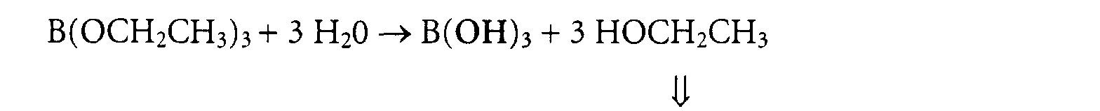
type of reaction:
either acid-catalyzed sol-gel reaction, e.g.
Advantage: alcohols are easily evaporated during spray-drying; ideally no
recristallisation of salts on the surface
Thus, alcoholates are turned into hydroxides, which yield by way of elimination of water
the corresponding oxides. These, then , form a 3-dimensional, amorphous glass matrix
consisting of SiO2 / B2O3 / Al2O3, into which certain metal oxide ingredients are
embedded as matrix bond separators, e.g.

For the experiments described here, the glass component was produced via base-catalyzed
sol-gel synthesis. The glass composition used in all experiments was (unless
stated otherwise) as follows:
70.67 Mol% SiO2, 14.33 Mol% B2O3, 5.00 Mol% Al2O3, 4.00 Mol% K2O, 2.00 Mol% CaO, 4.00 Mol% ZnO
(calculated from the mass of particular educts brought to reaction)
70.67 Mol% SiO2, 14.33 Mol% B2O3, 5.00 Mol% Al2O3, 4.00 Mol% K2O, 2.00 Mol% CaO, 4.00 Mol% ZnO
(calculated from the mass of particular educts brought to reaction)
Different types of magnetic pigments were investigated and are summarized in Table 1.
The educts are added in the following order and amount into the heatable stirring vessel:
| Tetraethoxysilane (TEOS) | 10700 ml |
| Triethylborate (TEB) | 3305 ml |
| K-methanolate 25% (W/V) in Methanol | 1601 ml |
| Ethanol | 11292 ml |
| Aluminiumisopropanolate | 1385 g |
| Calcium | 54.4 g |
| Zincacetate | 498 g |
The vessel is closed thereafter. The sol is heated to 70 °C and stirred overnight (15 h).
The temperature is regulated via a thermosensor which dips into the liquid.
Then the sol is heated to 90 °C and the alcohol/ water mixture (Ethanol: 3781 ml, H2O:
1512 ml) is added at a rate of 5000 ml/h. The vessel is cooled to 20 °C after the complete
addition of the said mixture.
The lid is opened and 10249 g magnetic pigment (CERAC) is added under vigorous
stirring. The prepared sol is transferred into the spray dryer via a hose.
The inlet temperature of the spray dryer is regulated to 200 °C. The nozzle pressure is
regulated to 6 bar and the nozzle is cooled for 3 min with ethanol. Then the hose is
connected to the outlet of the glass container and the pigment-containing sol is pumped
at a rate of 110 ml/min to the two fluid nozzle (diameter of the opening: 2 mm; supplier:
Nubilosa, Type 1B1VVS1) of the spray dryer via a ultrasound device (200 W). The spray
drying system is shown schematically in Figure 3. The particles which are formed in the
first two minutes are discarded. After the complete spraying of the sol the container
under the cyclone (AVO, see Figure 3) with the particles will be taken and the particles
will be further treated as described in the next steps.
The pigment containing sol is stirred in the stirring vessel to prevent sedimentation of
the suspended particles. The sol is transferred from the vessel to the nozzle using a pump
(SP).Nitrogen heated by an electric heater (EWT) is used as drying gas. The coated
pigment is transferred from the drying chamber (T) to the cyclone (ZY). The dried
powder an be removed from the container (AVO) under the cyclone. Very fine particles
are removed by a filter (SF) from the nitrogen. The spray dryer is used with overpressure
to prevent an air intake into the dryer. The gas flow is produced by gas blower (AV).
The powder is transferred into a ceramics bowl and heated in an furnace to 200 °C at a
heating rate of 1 K/min. Then temperature is held at 200 °C for one hour and the oven is
cooled to room temperature afterwards. The bowl is transferred to an atmosphere
furnace with a volume of 27 l which is rinsed with 60 1 nitrogen per hour. The furnace is
heated to 750 °C with a heating rate of 1 K/min and is held at that temperature for 1
hour. Then the furnace is cooled to 150°C and rinsed with 60 l air per hour. The furnace
is heated to 200 °C with a heating rate of 2 K/min and is held at that temperature for 1
hour and is cooled to temperature afterwards. The powder is transferred to a sieve (50
µm) and sieved for 30 min. Afterwards, the sieved sample is bottled in glass containers,
which are sterilized unclosed in an oven which is heated to 200 °C with a heating rate of
1 K/min, held at that temperature for 4 h and then cooled to 80 °C. Then the glass
vessels are taken from the furnace, covered with sterile foil and closed with a lid.
A summary of the important process parameters is presented in Table 2 and Table 3, the
produced individual samples with some data are provided in Table 3. Each sample has a
specific code. The first two letters describe the used glass chemistry (see below) and the
next four numbers encode the production process (see Table 2). The number after the
describes the used pigment (see Table 1). The letter R means that the fine content has
not been removed, whereas E means that the fine content has been removed with
ethanol. The last two numbers describe the number of the lot. The composition of the
glass (code EJ) as calculated from the quantity of the educts was 70.67 Mol% SiO2, 14.33
Mol% B2O3, 5.00 Mol% Al2O3, 4.00 Mol% K2O, 2.00 Mol% CaO, 4.00 Mol% ZnO. The
composition of the glass (code RN) was 74 Mol% SiO2, 15 Mol% B2O3, 5.00 Mol%
Al2O3, 4.00 Mol% K2O, 2.00 Mol% CaO. The composition of the glass (code EP) was
73.61 Mol% SiO2, 14.93 Mol% B2O3, 5.21 Mol% Al2O3, 4.17 Mol% K2O, 2.08 Mol%
CaO.
To obtain information about the surface, size and form of the MGP, we carried out
investigations with a high resolution scanning electron microscope manufactured by the
company JEOL (JSM). The samples were spread onto the sample holder with an
electrically conducting double-sided adhesive and sputtered with gold for 36 s with a
current of 30 mA. For the imagining of the surface, the emitted secondary electrons
(topography) as well as the back-scattered electrons (order number contrast) were
viewed. The used primary electron voltage was 10 kV (secondary electrons) or 25 kV
(back-scattered electrons).
The Figure 4 shows MGPs with mica covered with ferric oxide as magnet pigment (BM).
The fissure in the glass shell can be clearly seen which stem from the spray drying of the
particles when the layer starts to shrink on a non-shrinking substrate (drying fissures).
Further more a greater number of spheric particles can be seen which do not contain
one of the bigger magnet particles (10 - 60 µm). These spheres without magnetic core
(non-magnetic fine content) cannot be removed in a magnetic field, may be transferred
into the PCR reaction and will interfere with this reaction.
The particles with CERAC pigment (see Figure 5) do not have fissure, as the magnet
pigment (23 nm average particle size) does not cause tension forces in the layer so that
no fissures are created during the spray drying process. Furthermore, the small CERAC-particles
are also incorporated in the fine content so that there are no non-magnetic
particles contained therein. The particles are also substantially smaller than the particles
with mica which can be observed in these figures with the same magnification. MGPs
with the other magnetic pigments are shown at the same magnification in Figure 6 to
Figure 10.
To allow an evaluation of the MGPs before the actual functional tests, in addition the
HREM investigations further physical measurements were done with the MGPs. It was
interesting to learn about the iron solubility in water, the magnetic force, the fine
content and the density. In the following the experiments are described and the results
presented.
To test whether the fines have been removed, 1000 mg of the temperature-treated and
sieved powder, are weighed into a 50 ml centrifugation tube (Sarstedt), 40 ml water
added and dispersed by shaking. Then, the tube is dipped into a ultrasonic bath
(Sonorex (RK 102H; 120/240W) with the total filling height and is put into a magnetic
separator (Roche Diagnostics GmbH RD Art.Nr. 1858 025). After 3.5 min magnetic
separation, liquid is taken from the vessel with a pasteur glass pipette in the height of the
15 ml mark at the opposite side of the magnet and filled into a 5 mm quartz cuvette
(Type 110-QS, Hellma). The extinction of the supernatant is measured in an UV-VISNIR
spectrometer (Hitachi U-3000) against a corresponding cuvette filled with
deionized water. The measuring range was 200 to 1100 nm in steps of 1 nm to
investigate this wavelength range for the absorption bands of eventual impurities. The
extinction at a wavelength of 280 nm and 400 nm was taken as a reference.
For the density measurements a gas pyknometer (AccuPyc 1330, Fa. Micromeritics) is
used. Helium 6.0 is used as gas. The device is calibrated with the supplied standard (steel
beads with known volume). Further, the sample is dried for at least 1 hour at 150 °C and
then filled into the measurement container and weighed. After the measuring container
is put into the gas pyknometer, 10 washing cycles and 5 measuring cycles are used to
determine the average value and the standard deviation.
The iron solubility of the coated and temperature-treated samples is determined with
Inductive Coupled Plasma - Atomic Emission Spectroscopy (JY 24, company ISA).
Therefor, 1 g of the sample is transferred into a 50 ml polypropylene-tube, filled up with
distilled water and kept at a temperature of 60 °C for 20 hours. Then, the samples are
filtrated with a 0.2 µm syringe filter and the filtrate is measured. Four single
determinations are carried out at a wavelength of 259,940 nm and an average value is
computed therefrom.
For the measurement of the magnetic force a PP-weighing tube (Company Licefa, Art.
Nr. V2-3) completely filled with MGPs is weighed. With the help of a stencil, this sample
container is placed in the middle of LDPE tube (Company Kartell, TS 735) burdened
with brass so that the lid of the tube can be still closed. The vessel is positioned in the
middle of a balance (detection accuracy 0.1 g) with the help of another stencil. After the
equalization of the balance, a plastics cap which contains an cylinder-shaped magnet
(diameter: 30 mm; height: 113.5 mm; material: samarium-cobalt 2/17) is positioned
above the scale.
Thereby, the MGPs in the scale are attracted against to the gravity thereby decreasing
their weight. After the adjustment of the equilibrium (1.5 min) the weight loss of the
sample is determined and the value is normalized for 250 mg MGP.
The results of the physical characterization of the MGPs with EJ composition and
different pigments are summarized in Table 3. It is noticeable that the extinction values
are very low when CERAC pigment is used. This can be explained by the lack of non-magnetic
fine content because the small CERAC pigment (23 nm) can be incorporated
into each magnetic glass particle. The density of the CERAC-MGPs is not very high. This
has a positive influence on the sedimentation velocity. The magnetic force is subjected to
high fluctuations, however, it is positively influenced by the use of CERAC-pigments.
The CERAC-MGPs have a comparatively high leak of iron to the water. However, even
ten-fold higher values did not show any effect on the PCR process.
In summary, it can be said that the physical properties do not have any significant
differences between the different pigments but the CERAC-MGPs have a very low fine
content.
The surfaces were determined using a device from Micromeritics (Type ASAP 2400).
The measurement was performed at liquid nitrogen temperature. Nitrogen 5.0 was used
as measuring gas and Helium 4.6 was used as inert gas. Typically, 5 g of sample was used
for one measurement.
The surface of the MGPs plays an obvious role for the isolation of DNA and RNA. The
higher the surface the more DNA can be bound by the same mass of MGPs. It is also
possible to use less MGPs and obtain the same performance. This has the effect that the
intergrain volume is less which means that less alcohol may be introduced as
contaminant into the PCR reaction. The data for the measured BET-surfaces for some
samples are summarized in Table 4. When the surfaces of the samples produced on the
big spray dryer are compared, it should be noted that the samples with small pigments
(BASF FA, STREM and CERAC) have relatively high surfaces, whereas the big Merck
pigments have small surfaces. This may be caused by the fact that big magnetic particles
cause big MGPs whereas the small magnetic particles are incorporated into the spherical
droplets during the spray drying process and have therefore similar surfaces under
similar spraying conditions. The BASF CE-SU particles have a size distribution in
between the small (BASF FA, STREM and CERAC) and the big Merck particles. In
contrast to the Merck particles, they do not possess a structured surface so that these
particles have a very smooth glass surface. This results into a smaller surface. A higher
pressure was used with the smaller spray dryer and the nozzle of this spray dryer is
remarkably smaller than that of the bigger spray dryer. This causes smaller particles
whereby the surface increases remarkably. This is in good agreement with the results of
Example 5.2.1
Any influence of the magnetic pigment or the spraying pressure on the results of further
physical investigations cannot be recognized except the above-mentioned influence on
the fissure formation and the fine content. However, only the measurements of the
surface show a direct connection to the results of the functional investigations. Other
physical investigation results do not show this direct correlation.
There are several methods for the evaluation of MGPs with regard to their suitability for
the extraction of nucleic acids. The determination of the extinction at 260 nm before
and after the purification is not very sensitive and does not resemble the situation when
small amounts of target nucleic acid are extracted from clinical material. The results of
functional evaluation methods which rely upon a method for the amplification of
nucleic acids, as e.g. by PCR, RT-PCR, NASBA or anything similar, are often not enough
convincing. Furthermore, these methods, which are suitable for the determination of a
small number of copies of the target genome, are susceptible to disturbances by
substances from the sample material or from the sample preparation process.
Radioactive binding studies are a suitable analytical method which makes it possible to
analyze the sample preparation process step by step. The performance data are no
absolute data but are always relative to the performance of a reference particle
At first, radioactively labelled DNA or RNA is synthesized enzymatically in a PCR or in
vitro transcription process in the presence of 32P-markied desoxynucleoside or
nucleoside-triphosphates. Then, the marked DNA or RNA is separated from the free
nucleoside-triphosphates, the content determined and a defined dilution is prepared.
A small amount of marked DNA or RNA is added to each sample before the
examination. During sample preparation, all nucleic acids bind to the MGPs in the
presence of chaotropic agents. The MGPs are pelleted by the exertion of magnetic forces
and the supernatants can be discarded. The pellet is washed and the bound nucleic acids
are eluted at elevated temperature by the reversal of the reaction conditions, i.e. by the
addition of a low salt elution buffer. After the binding and an optionally after the
washing step, an aliquot of the particle supernatant is spotted onto a filter. The eluate as
well as the in water redispersed MGP are spotted onto a filter as well and dried
thereafter. At last, the filters are measured in a Beta.Counter and for each sample
preparation a distribution is calculated.
- Expand High Fidelity PCR System (Roche Molecular Biochemicals Cat. No. 732641)
- dNTP Mix (Roche Molecular Biochemicals Cat. No. 1277049)
- Deoxycytidine 5'-alpha-P32 Triphosphate dCTP 3000 Ci/mmol (Amersham Cat. No. PB 10205)
- lambda-DNA (Roche Cat. No. 1029053) concentration 1ng/ml
- Radiotracer primer 1 (SEQ ID NO 1)
concentration 5,3 OD260 / ml Radiotracer primer 2 (SEQ ID NO 2)concentration - QIA Quick PCR Purification Kit (Qiagen Cat. No. 28104)
- 29.5µl double distilled water
- 5µl Expand High Fidelity Buffer
- 2.5µl dNTP Mix (1:10) diluted with double distilled water
- 1µl Radiotracer primer 1 (SEQ ID NO 1)
- 1µl Radiotracer primer 2 (SEQ ID NO 2)
- 0.3µl 32P-dCTP
- 10µl lambda-DNA
- 0.75µl Expand High Fidelity -Enzyme Mix
- 2 min. 94°C
- 10 cycles (10 sec. 94°C / 30 sec. 60°C / 60 sec. 72°C)
- 20 cycles (10 sec. 94°C / 30 sec. 60°C / 60 sec. 72°C + 10 sec. 72°C extension per cycle)
- 7 min 72°C
- 4°C
- according to the QIA Quick PCR Purification Protocol (Qiagen)
Dilution of DNA 1:10 in double distilled water and measurement in the Beta-Counter
- SP6 / T7 Transcription Kit (Roche Molecular Biochemicals Cat. No. 999644)
- Uridine 5'-alpha-P32 Triphosphate UTP 3000 Ci/mmol(Amersham Cat. No. PB 10203)
- Plasmid pBKBH10S, linearized with EcoRI at 100µg/ml
- High Pure RNA Isolation Kit (Roche Molecular Biochemicals Cat. No. 1828665)
- 2µl 10x buffer
- 3µl NTP Mix (AGC)
- 1µl UTP (1:50 with double distilled water)
- 5µl 32P-UTP
- 7µl linearized plasmid
- 1µl RNAse Inhibitor (Roche Molecular Biochemicals Cat. No. 802808)
- 1µl T7 RNA-Polymerase
- 20 min incubation at 37°C
- Addition of 2 µl DNAse, RNAse free
- 10 min incubation at 37°C
- Addition of 178 µl double distilled water
- according to the High Pure RNA Isolation Protocol (Roche Molecular Biochemicals)
Dilution of RNA 1:30 in double distilled water and measurement at the Beta-Counter
- negative plasma
- Proteinase K concentration 20mg/ml (e.g. Roche Id.Nr. 1942387)
- Poly-A-RNA (e.g. Roche Id.Nr. 108626) concentration 1mg/ml; diluted 1:1000 (volume/ volume ratio) with lysis buffer
- lysis buffer (50mM Tris pH 7.0, 15% (v/v) Polydocanol, 5M Guanidiniumisothiocyanate, 1mM DTT)
- MGP (with glass composition EJ and different core pigments (BM, MMB, CERAC, STREM, BASF-FA, BASF-CE)) suspended in isopropanol at 60mg/ml or at 6mg/ml
- Washing buffer (20mM Tris pH 7.5, 20mM NaCl, 70% (v/v) Ethanol)
- Elution solution: double distilled water
- Auxiliary material: Whatman GF/D Filter
- 80µl Proteinase K
- add 410µl negative plasma and mix
- add 500µl lysis buffer and mix
- add 10µl radioactive labelled DNA or RNA and mix
- 10 min incubation at room temperature under shaking
- add 500µl MGP suspension (concentration 6mg/ml) and mix
- 20 min. incubation at room temperature under shaking
- separation for 2 min in Magnetic separator (Dynal)
- discard supernatant and spot 300µl onto a filter
- add 750µl wash buffer, vortex, 2min separation
- discard supernatant, eventually spot 375µl supernatant onto a filter
- repeat washing procedure twice
- add 100µl elution solution, incubate 5 min. at 80°C in a thermomixer
- separate 2 min in a magnet holder, spot supernatant onto a filter
- add 100µl elution solution, resuspend and spot MGPs onto a filter
- dry filters for 60 min at 75°C in a drying oven
- transfer filter into scintillation tubes, add 5ml scintillation solution and measure in the beta-counter
- 25µl Proteinase K
- add 415µl negative plasma and mix
- add 500µl lysis buffer and mix
- add 10µl radioactive labelled DNA or RNA and mix
- 5 min incubation at room temperature under shaking
- add 50µl MGP suspension (concentration 60 mg/ml) and mix
- 20 min. incubation at room temperature under shaking
- separation for 2 min in the magnetic separator (Dynal)
- discard supernatant and spot 300µl onto a filter
- add 700µl wash buffer, vortex, 2min separation
- discard supernatant, eventually spot 350µl supernatant onto a filter
- repeat washing procedure twice
- add 120µl elution solution, incubate 10 min. at 80°C in a thermomixer
- separate 2 min in a magnet holder, spot supernatant onto a filter
- add 100µl elution solution, resuspend and spot MGPs onto a filter
- dry filters for 60 min at 75°C in a drying oven
- transfer filter into scintillation tubes, add 5ml scintillation solution and measure in the beta-counter
Different MGP-types, which were produced using EJ glass chemistry and various cores
in the nano- or mikrorange (MMB, CERAC, etc.) on different spray dryers (Büchi or
Nubilosa), were characterized with regard to their behaviour at the nucleic acid
extraction from virus-negative pool material with the radiotracing method according to
Example 5.1.3.2. The RNA parameter proved to be the most sensitive in the course of
the studies and was therefore chosen as parameter. Figure 11 shows BASF-CE is not
suitable as core material because only small amounts of the bound RNA can be found in
the eluate. The EJ/ CERAC particles which were sprayed in the Büchi system at 6 bar
showed the performance of the reference EJ/ MMB which were sprayed in the Nubilosa
system.
Different MGP types produced with the Nubilosa system with different spraying
pressures are compared according to 5.1.3.3. The reference particles are EJ /MMB MGP.
The DNA and RNA binding properties are investigated. Whereas the DNA parameter
shows no dependence from the spraying pressure, the RNA parameter shows significant
performance differences. The performance of the EJ/ CERAC particles produced with
1.5 bar spraying pressure with the Nubilosa system is lower than that of the reference.
There is a lower performance in the series in dependence of the spraying pressure which
varied from 1.5 to 3.4 bar (~30%). Particles which are sprayed with 4.3 bar reached 90
% of the performance of the particles which are sprayed at 1.5 bar (see Figure 12).
The experiments prove the direct connection between the process parameters for the
production of the MGPs and of the performance in the test. It should be noted that an
increase in the spraying pressure leads to a lower test performance but, beginning from a
certain spraying pressure, the particle properties are improved leading to a better
performance.
These experiments should lead to the result whether a variation of the spraying pressure
leads to MPGs with even better performance.
Therefor, MGPs are produced on the Nubilosa system at a pressure of 1.5 bar, 4.3 bar
and 6 bar using nitrogen as spraying gas.
In order to obtain a careful drying process, inlet and outlet temperature of the spray
dryer are additionally decreased. The influence of these factors on the RNA isolation is
investigated according to 5.1.3.3. using EJ / MMB MGP as reference.
The results (see Figure 13) show the surprising effect of a dramatic performance
reduction which is caused by the decrease of the spraying temperature and could not be
foreseen. It was possible to reach the performance of the reference quality by the
increase of the spraying pressure. Concomitant with the superior suspension stability,
these particles are therefore superior to the reference.
An Uvikon Spectrophotometer Model 930 produced by Kontron Instruments is used for
the evaluation of the sedimentation behaviour of the magnetic glass particles. This
spectrophotometer is modified to allow variable adjustment of the cuvette along a scale
bar. For the measurements the cuvette is set at a position in which the measuring beam
with a wavelength of 650 nm traverses the cuvette at a position which is at 2/3 of the
filling height (scale position 7.5). Macro cuvettes made from polystyrene are used which
have a volume of 4 ml and a path length of 1 cm. Prior to measurements, calibration is
made in a one beam procedure against pure suspension medium. The MGP samples are
dispersed to homogeneity in suspension medium, typically at a mass/volume ratio of 3
mg/ml, and measured immediately. The change of the extinction with time is monitored
continuously at the said wavelength of 650 nm.
The physical quantity used for the comparison of the sedimentaton velocity of different
samples is the half life (t1/2) in a particular suspension medium. This is the peroid of
time until the extinction of the suspension in the upper third of the cuvette is half the
value at the beginning of the measurement. The decline of extinction is caused by the
sedimentation of the particles and the concomitant clearance of the upper third of test
volume.
The above-described device further allows the installation of a magnet beneath the
cuvette. Thereby the velocity of the magnetic separation can be determined.
Several MGP-types with different cores in the range of nano- or micrometers and
produced with EJ glass chemistry are investigated spectrophotometrically for their
sedimentation and separation behaviour, i.e. with and without magnet under the
cuvette. Mass/volume is 6 mg/ml in this case. The suspension medium is a 1:1 mixture
of isopropanol and lysis buffer. The results are summarized in . The particles from the
CERAC type show a clear advantage in the stability of the suspension but no
disadvantages for the magnetic separation. (See also Figure 14).
The sedimentation behaviour of different MGPs from the type CERAC produced with
EJ glass chemistry are investigated in pure isopropanol.
Important production parameters
Virus particles in the sample (e.g. plasma) are lysed in the presence of protease and high
concentrations of chaotropic salt as well as detergent. Then, the particulate adsorbent (=
MGP) is added for physico-chemical adsorption of released nucleic acids onto the glass
surface, followed by magnetic separation and washing of the loaded beads, i.e.
bound/free separation. Finally, dissociation of bound nucleic acids from the beads (=
elution) is conducted under reversed reaction conditions relative to adsorption, i.e. with
low salt buffer or even distilled water.An aliquot of eluate is then mixed with an aliquot
of PCR mastermix to start amplificatin of the nucleic acids recovered in the eluate. This
reaction is characterized by the equation
After PCR with at least one biotinylated primer, an aliquot of biotin-tagged amplificate
is mixed with hybridization buffer and detection probe. After incubation, streptavidin-coated
beads are added, followed by another incubation. Finally the beads are washed,
signal buffer is added and the chemiluminescent signal intensity measured, which is
correlated with the mass of amplified nucleic acid bound and, thus, the viral load of the
plasma sample.
The expert in the field may also carry out the 1 ml protocol manually.
Proteinase K, liquid in glycerol/Ca-acetate, 20 mg/ml
Poly-A-RNA, 1 mg/ml, use level is a 1:1000 dilution in lysis buffer
lysis buffer, consisting of:
- Tris-buffer, 50 mmol/l, pH 7.0 (1.0 & extractor protocol, respectively) or 4.0 (1.5 ml protocol)
- Polydocanol, 15% (v/v) (1.0 & extractor protocol, respectively) or Triton X-100 20% (v/v) (1.5 ml protocol)
- Guanidinium-isothiocyanate, 5 mol/l
- 1 mmol/l DTT
- magnetic glass particles (MGP) either of the type EJ/BM, EJ/MMB oder EJ/CERAC suspended in isopropanol (99,8% purity) at 60 mg/ml
- wash buffer consisting of
- Tris-Puffer, 20 mmol/l, pH 7.5
- 60% Ethanol / aq (1.0 & extractor protocol, respectively) or 70 % (1.5 ml protocol)
- NaCl, 20 mmol/l
Amplification/ Mastermix:
- PCR-buffer, consisting of buffer medium
- PCR-buffer, consisting of buffer medium
Stop-reagent for UNG after the amplification
- N-lauroylsarcosine (e.g. Roche Id.Nr. 133895), 1% w/v); 5 µl per 100 µl amplificate
- N-lauroylsarcosine (e.g. Roche Id.Nr. 133895), 1% w/v); 5 µl per 100 µl amplificate
Primer:


Detection probes:

Basically the same procedure and reagent set as above. Additionally, an amoured RNA
internal control (IC, see below) is spiked into each and every sample, the extraction
process is thermostated at ca. 40°C, and volumes are adjusted to 2 ml total volume of
lysis mix, i.e.
| stock solution | end concentration in PCR / µl | |
| Water ( PCR-Grade ) | 4 µl | |
| DNA-Mastermix | 2-x | 1-x / 50 µl |
| MgCl2 | 25 mM | 3.0mM / 12µl |
| Primer SEQ ID NO 7 | 5 µM | 0.2 µM / 4µl |
| Primer |
5 µM | 0.4 µM / 8 µl |
| UNG | 1U/µl | 2 U / 2 µl |
| stock solution | end concentration in PCR / µl | |
| Water ( PCR-Grade) | 18 µl | |
| Bicine-Buffer RT 5x | 5-x | 1-x/ 20 µl |
| MnOAc | 25 mM | 2.5mM / 10µl |
| DNTP-Mix (dUTP/ | 30 mM | 0.6mM |
| DATP/dCTP/dGTP) | 10 mM | 0.2mM/ 2 ul |
| Primer |
10 µM | 300 nM/ 3 µl |
| Primer |
10 µM | 300 nM/ 3 µl |
| UNG | 1 U/µl | 2 U / 2 µl |
| Tth-Polymerase | 5.5 U/µl | 10 U / 2 µl |
| stock solution | end concentration in PCR / µl | ||
| Water ( PCR-Grade ) | 20 µl | ||
| Bicine-Buffer RT 5x | 5-x | 1-x/ 20 µl | |
| MnOAc | 25 mM | 1.25mM / 5µl | |
| dNTP-Mix (dUTP/ | |||
| dATP/dCTP/dGTP) | 10 mM | 0.2mM/ 2 µl | |
| Primer SEQ ID NO 3 | 5 µM | 0.2 µM / 4 µl | |
| Primer |
5 µM | 0.2 µM / 4 µl | |
| UNG | 1 U/µl | 2 U / 2 µl | |
| Tth-Polymerase | 5.5 U/µl | 15 U / 3 µl |
After PCR, UNG is blocked through the addition of laurylsarcosin concentration 1% use
level.
Thermocycling profiles are as follows:
| HBV | UNG- | 1x | 10 min | 37°C | |
| PCR | 35x | 30 sec 30 sec 40 sec | 92°C 55°C 72°C | ||
| HCV | UNG- | 1x | 10 min | 37°C | |
| reverse transcriptase step denaturation | 1x | 30 min 1 min | 60°C 95° | ||
| PCR | 2x | ||||
| 10 sec 20 sec | 95°C 60°C | ||||
| 33x | 15 sec 20 sec | 90°C 60°C | |||
| follow-up incubation | 7 min | 72°C | |||
| HIV | UNG- | 1x | 10 min | 37°C | |
| reverse transcriptase step | 1x | 30 min | 60° | ||
| PCR | 4x | ||||
| 10 sec 10 sec 10 sec | 95°C 55°C 72° | ||||
| 31x | |||||
| 10 sec 10 sec 10 sec | 95°C 60°C 72°C |
| reagent | volume (HBV, HCV, HIV) | incubation time (HBV, HCV, HIV) |
| |
10 µl | |
| denaturation solution | 35 |
5 min |
| detection probe | 120 µl | 30 min |
| SA-beads | 35 |
10 min |
Lysis of viral particles as well as adsorption, purification and elution of nucleic acids
extracted from the sample are conducted as described above (see Example 7.1.2). Again,
amplification of target molecules is described by the equation Ni = N0 x (1+E)n, with
With 5'-nuclease technology, however, amplification and detection reactions,
respectively, are closely interwoven, and occur in solution phase, i.e. without solid phase
immbilisation and corresponding washing steps(= homogeneous PCR) . To this end,
detection probes with 2 particular chemical modification are added to the PCR
mastermix. One of these modifications is a fluorogenic reporter group (R, for instance a
derivative of 6-carboxy-fluorescein) covalently atached to the backbone of the probe, the
other one is a dye (for instance a polymethine-cyanine derivative) capable of absorbing
the fluorescent light of the reporter and to quench it (quencher). The quencher is
typically attached to the probe backbone at the 5'-end, whereas the reporter is located
within the oligo sequence, spaced from the quencher by a number of nucleotide
building blocks. These probes bind to the target nucleic acid (sense or anti-sense strand)
close to the 3'-end of a primer (reverse or forward). As soon as primer has annealed to
the target and DNA-polymerse gets bound to the primer:target hybrid, elongation starts.
Due to the 5'-nuclease activity of the enzyme, simultaneously with copy strand synthesis
the probe is cleaved as soon as the polymerase reaches the probe binding site, reporter
and quencher get separated and the fluorescent signal becomes measurable. This process
is repeated with every cycle, and more and more fluorescent reporter is accumulated in
solution until reagent depletion at the end of the reaction. So, in a signal-over-time plot,
sigmoidal growth curves are generated. The bigger N0, the earlier the signal curve rises
above noise level.
The point on the time axis, where the fluorescent signal can be significantly
distinguished from background singal is called threshold cycle (ct). ct is a measure of
assay sensitivity: the smaller the ct-value, the more sensitive is the assay. ct values can be
calculated by means of different mathematical operations, for example cut-off
approaches (average background signal intensity multiplied by a constant factor yields a
cut-off signal intensity to discern negative from positive) or approaches where the
location of the maximum of the first differentiation of the signal-over-time curve (i.e.
the steepness profile), or the location of the maximum of the second differentiation on
the time axis, are computed and defined as ct.
This amplification / detection technology allows for real-time monitoring of PCR and
for closed-tube processing as well, i.e. in contrast to standard procedures, the PCR tubes
remains closed after pipetting eluate and mastermix which effectively reduces
contamination risks.
Moreover, if one uses primer and probe sets for different viral species (and, thus,
different analyte molecules and target sequences) combined in one mastermix, it is
possible to perform multiple amplification / detection reactions simultaneously,
depending on the viral load of the individual under inspection. This is the basis for so-called
multiplex assays.
What is more, due to the generic nature of MGP based sample preparation, all nucleic
acids present in the sample are extracted by way of physico-chemical adsorption to the
particle surface, independent of sequence characteristics. This will also include human
DNA (hDNA), released from disrupted blood cells , e.g. leucocytes, the titer of which
may differ widely according to the physiological or pathological status of the individual
blood sample donor. For instance, in cases of autoimmune diseases like SLE, hDNA
levels may be elevated substantially. So, hDNA and one or more pathogenic nucleic
acids present in a given sample constitute a mixture of various nucleic acids of different
sequence specificities which are co-extracted. They constitute a matrix of polynucleotides
that are extracted without discrimination, followed by sequence-specific
amplification and detection of target nucleic acids via interaction with specific primers
and probes under appropriate reaction conditions.
Given a pathological level of 4000 ng/ml hDNA in plasma in case of SLE, and a low viral
load of 50 copies of viral genomic RNA per ml of plasma., and 10000 nucleotides per
genomic RNA. With ca. 325 dalton ( 1 dalton = 1,66 x 10⁻²⁴ g) per nucleotide, 50 copies
or 500000 nucleotides of target RNA make ca. 2,7 x 10⁻⁷ ng per ml of plasma, resulting
in a relative abundance of target: non-target nucleic acid of approximately 1 : 10+12 !
hat is more, in order to monitor the whole process, an artificial nucleic acid construct
may be added to the samples, preferably packaged (armored) in a modified virus
particle, which is co-extracted and co-amplified with the natural target. This internal
control (IC) features a unique probe binding region for an IC detection probe, which
differs from target-specific probes by a different reporter group with distinguishable
emission characteristics. Thus, IC signal can be discerned from target signal, and as the
IC is known to be present in the sample, it functions as a monitoring agent. So, multi
labeling expands the utility of multiplex assay technology. The internal control (IC) is
described in WO98/00547.
See above (Example 7.1.2 => automated protocol on in-house-built extractor)
| Reagent | Concentration of stock solution | Final concentration in the test | Addition (µl / reaction) |
| Mn(OAc)2 pH 6.5 | 50 mM | 3 mM | 5.75 |
| dNTPs | 2.50 | ||
| (dG, dA, dC) | 100 mM | 300 µM | |
| (dT) | 50 µM | ||
| (dU) | 500 µM | ||
| DEPC-H2O | - | 9.50 | |
| Glycerol | 80% | 5,64% in total 2.8% added here |
3.50 |
| K Oac pH 7.5 | 2 M | 100 mM | 5.00 |
| Tricine pH 8.3 | 1 M | 50 mM | 5.00 |
| DMSO | 80% | 5.0% | 6.25 |
| Primer |
50 µM | 150 nM | 0.30 |
| Primer SEQ ID NO 13 | 50 µM | 150 nM | 0.30 |
| Primer |
50 µM | 400 nM | 0.80 |
| Primer SEQ ID NO 15 | 50 µM | 150 nM | 0.30 |
| Primer |
50 µM | 400 nM | 0.80 |
| Primer SEQ ID NO 17 | 50 µM | 150 nM | 0.30 |
| Primer |
50 µM | 150 nM | 0.30 |
| Probe SEQ ID NO 19 | 50 µM | 100 nM | 0.20 |
| Probe |
50 µM | 100 nM | 0.20 |
| Probe SEQ ID NO 21 | 50 µM | 100 nM | 0.20 |
| Probe SEQ ID NO 22 | 50 µM | 100 nM | 0.20 |
| UNG | 2.4 U / µl | 10 U | 4.20 |
| ZO5 | 10 U / µl | 40 U | 4.00 |
| Aptamer SEQ ID NO 23 | 50.4 µM | 200 nM | 0.40 |
The common multiplex thermocycling profile for all test parameters is as follows:
| UNG-step | 45°C - 10 min |
| denaturation | 94°C - 30 sec |
| reverse transcription | 58°C - 30 min |
| PCR | 95°C - 20 sec / 59°C - 50 sec 5x |
| 91°C - 15 sec / 52°C - 50 sec 55x |
Primer:
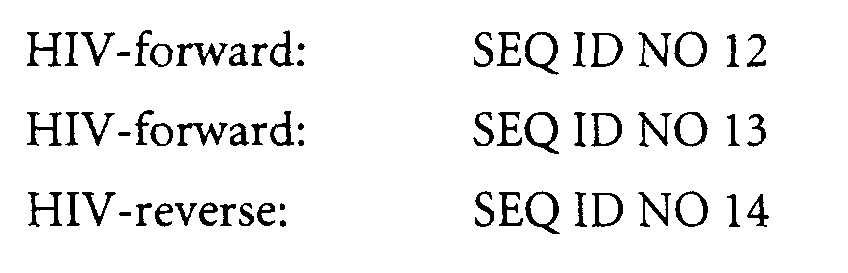


Probes:
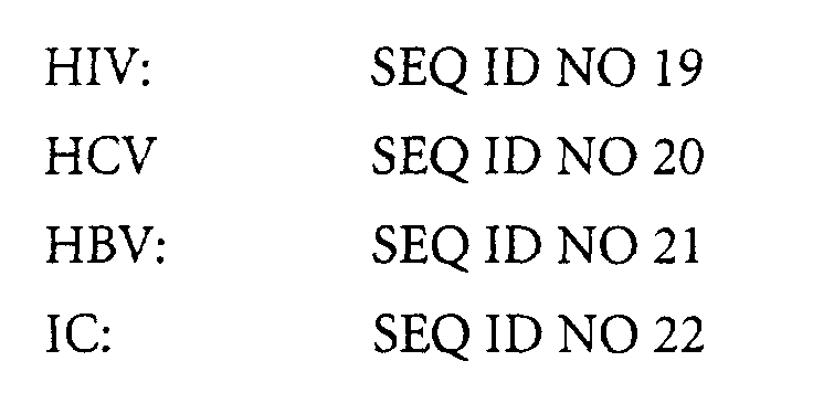
Melting temperature of the DNA polymerase-aptamer complex = 51,7 °C (=50 %
dissociation)
Some of the oligonucleotides are derivatised with Cy5 which is a Pentamethine-diindocarbocyanine
coupled to alkylphosphatidyl-linker (Pharmacia Biotech Cy5-N-ethyl-phosphoramidite)and
which functions as quencher (Q); λEX = 630 nm, λEM = 665
nm. Some of the oligonucleotides are derivatised with FAM which is 6-carboxy-fluorescein
coupled to a 2-(amino-cyclohexyl-)propane-1,3-diol-linker (Biogenex CX-FAM-phosphoramidite)and
which functions as s reporter for targets (R); λEX = 485 nm,
λEM = 515 nm.Some of the oligonucleotides are derivatised with which is hexachloro-6-carboxy-fluorescein
coupled to a 2-(amino-cyclohexyl-)propane-1,3-diol-linker
(Biogenex CX-HEX-phosphoramidite)and which functions as reporter for the internal
control (R); λEX = 530 nm, λEM = 585 nm
The reaction conditions are described in the results of the experiments (see Example
7.3).
- virus-negative plasma (0-Matrix, single plasma or Plasma-Pool), e.g. citrate-plasma or EDTA-plasma
- virus-positive plasma or virus-containing culture supernatants, which can be used to adjust certain virus titers by mixing with 0-matrix in appropriate ratios.
- alternatively: in vitro-transcripts with the target-sequences to be investigated
- human placental DNA (a. genomic, Sigma cat. no. D4642; b. fragmented, Sigma cat.no. D3287), added to the samples to simulate pathological conditions which lead to the enhanced release of intracellular substances and thus to enhanced levels of DNA/ RNA in the blood, e.g. auto immune disease as SLE or hemolysis.
Different MGP-samples of the type EJ/CERAC are investigated with the methods
described in 7.1. The test variable was the spraying pressure:
It could be demonstrated that with increasing spraying pressure, i.e. shift towards
smaller average bead diameter, the unspecific binding (USB) decreases whereas the
highly specific signal generation is unchanged which results in elevated signal-to-noise
ratios
The virus titer adjustment 0.5xGG is approximately 100 sgu/ml (sgu = signal generating
units) in the case of HBV, and approximately 150 sgu/ml in the case of HIV. The data
are summarized in Table 7.
Two variants each of the type EJ/MMB and EJ/CERAC, respectively, are functionally
compared using the method as described in Example 7.2 in conjunction with multipool
(MP) and individual (PL) plasma samples. EJ0047.2R (MMB) and EJ0100.5R (CERAC)
are sprayed under standard conditions, i.e. 1.5 bar, 230°C inlet temperature and
approximately 110 °C outlet temperature. EJ0102.2R (MMB) and EJ0108.5R (CERAC)
are development variants with regard to the spraying conditions (inlet temperature
decreased to 200°C [MMB]), and spraying pressure increased to 4.3 bar [CERAC]). The
results are summarized in Table 8 and show the potential of the CERAC nano-core
particles to obtain higher sensitivity as exemplified by the earlier threshold cycle and/or
the larger signal differences (saturation signal minus noise level, S-N).
Several MGP preparations of the type EJ/MMB and EJ/CERAC (see Table 9) are
functionally compared using the method as described in Example 7.2. The results are
summarized in Table 10 and show the potential of the of the EJ0096.5R-01-type
particles to obtain higher sensitivity as exemplified by the earlier threshold cycle or
higher hit rate irrespective of the particular assay.
Several MGP preparations of the type EJ/MMB and EJ/CERAC are functionally
compared using the method as described in Example 7.1 wherein the adsorption
incubation is conducted with or without shaking. Thereby, the sedimentation velocity
becomes decisive, i.e. the higher the sedimentation velocity the higher the number of
particles which are removed from the interaction with the analyte in the forming
precipitate. In contrast, if the sedimentation velocity is smaller then time span is longer
in which analyte molecules from the liquid may adsorb to the surface of the adsorbent.
The results are summarized in Table 11 (EJ0047.2R was used as a reference) show that
without mechanical agitation the loss of performance is highest (>40 %) with the MMB-type
(differs considerably from the characteristics of this invention), and least, or
practically absent, with the CERAC-type (most close to the characteristics of the
invention within this series).
To be able to assess the functionality of the different types of MGP thoroughly, a
performance index was calculated for each type in the following way:
Different types of MGP were used to extract HCV in vitro transcripts which were diluted
to various titer levels in diluent containing 10 mmol/l Tris, pH 80, 1 mM EDTA, 20
µg/ml poly-A-RNA and 0.05% NaN3, and spiked into pooled plasma.
Human background DNA (hBG-DNA) was spiked at 0 or 4000 ng/ml into the sample,
respectively. Surprisingly, it is possible to reduce interference by hBG-DNA by way of
bead selection, as demonstrated by the data presented in Table 12.
Various types of MGP were used to process HCV positive plasma samples. Human
background DNA (hBG-DNA) was spiked at 0 or 4000 ng/ml into the sample,
respectively. Again, a significant impact of bead size and bead geometry is being
observed. MGP preparation EJ0096.5R, which represents the MGP quality according to
the present invention the most, shows very clearcut advantages both in terms of minimal
shift of ct values, and reducing loss of specific signal generation (S-N), as indicated by
the data presented in Table 13.
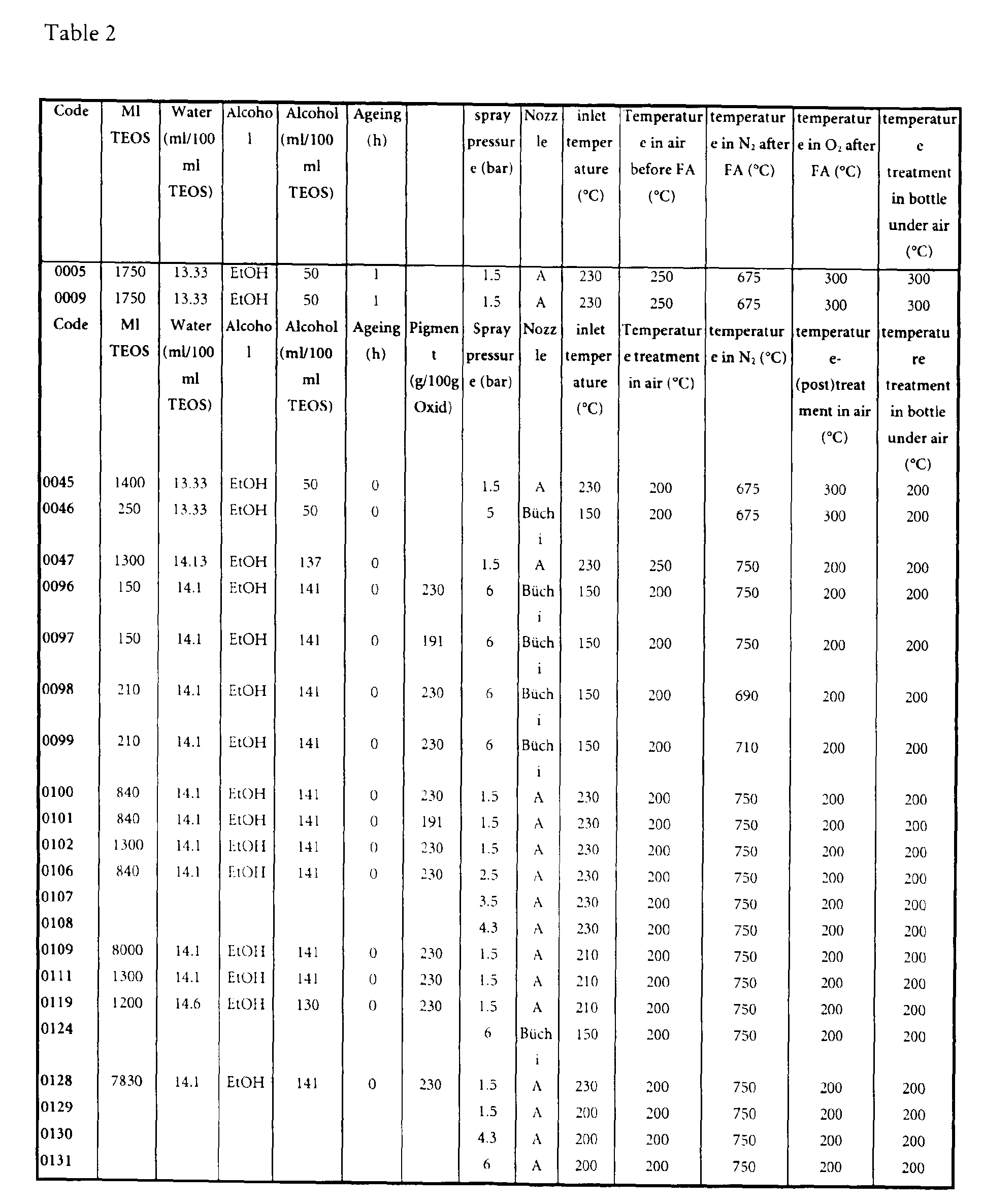
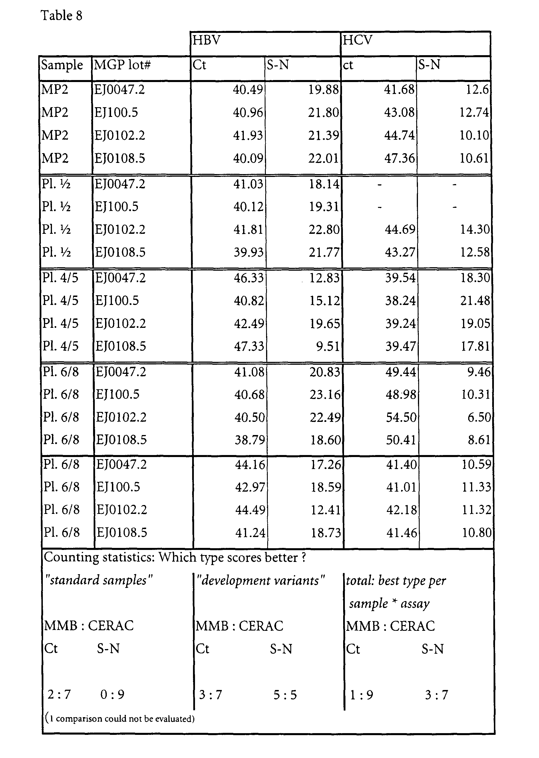
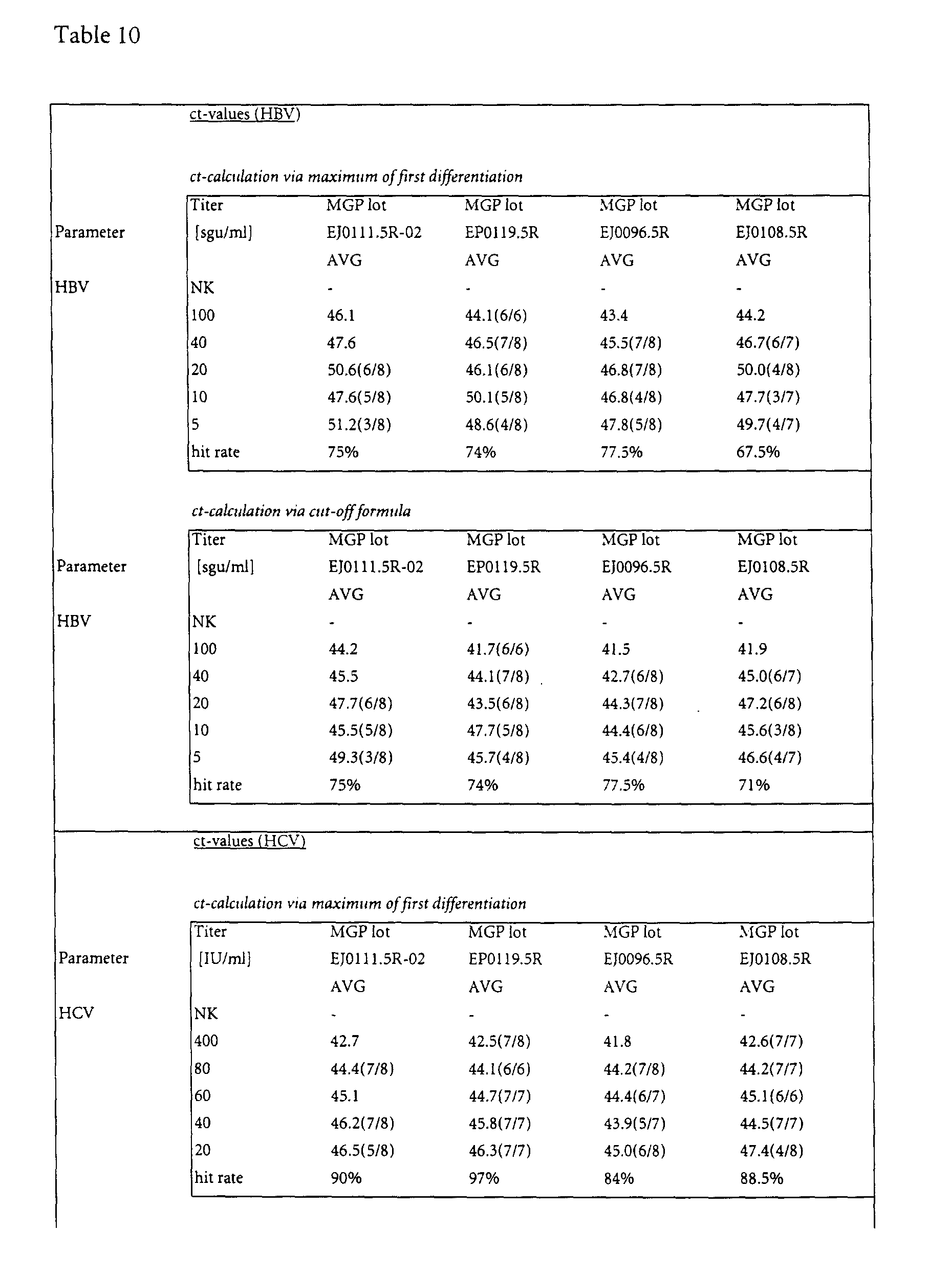

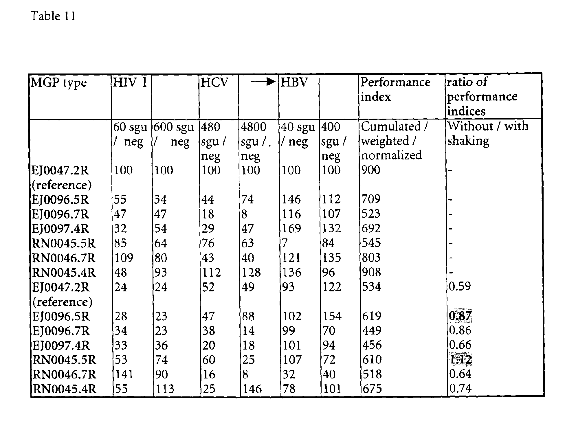
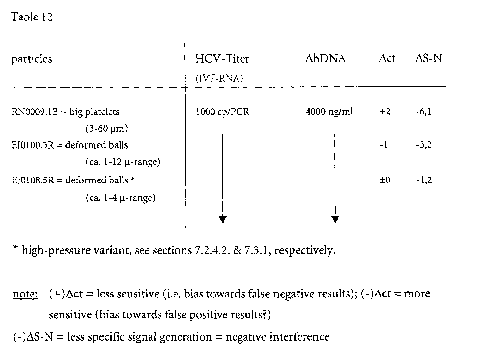
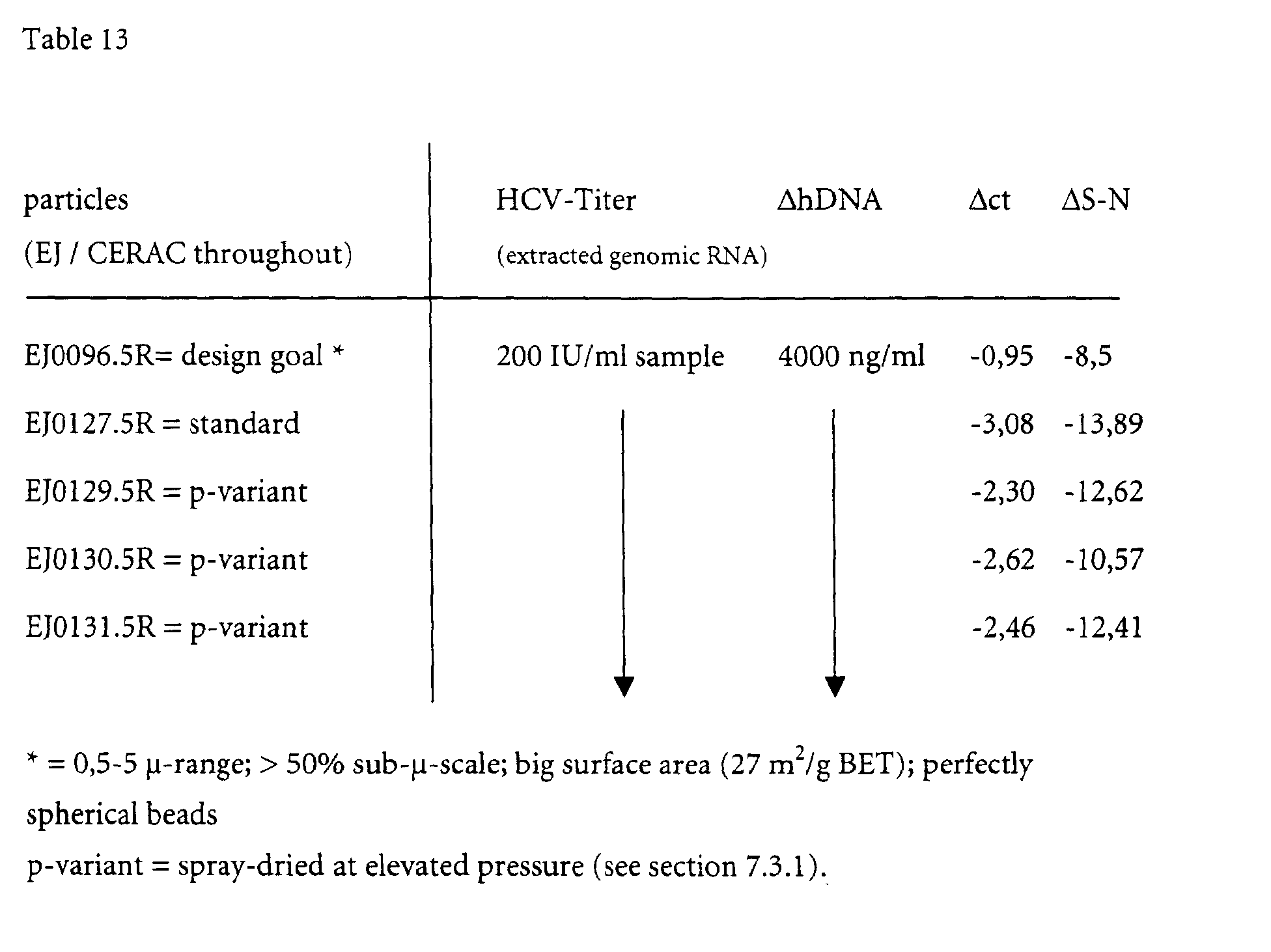
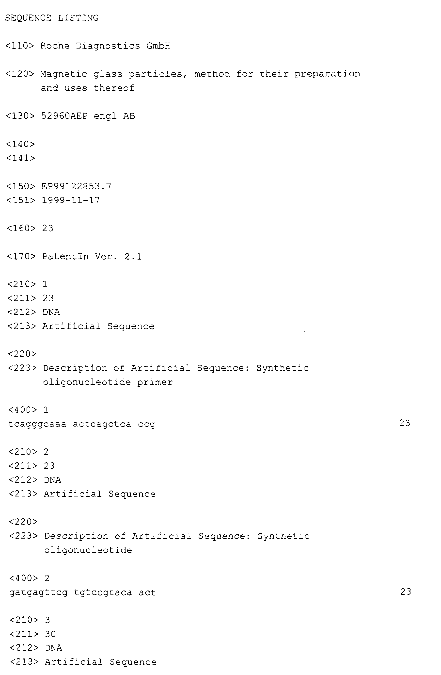
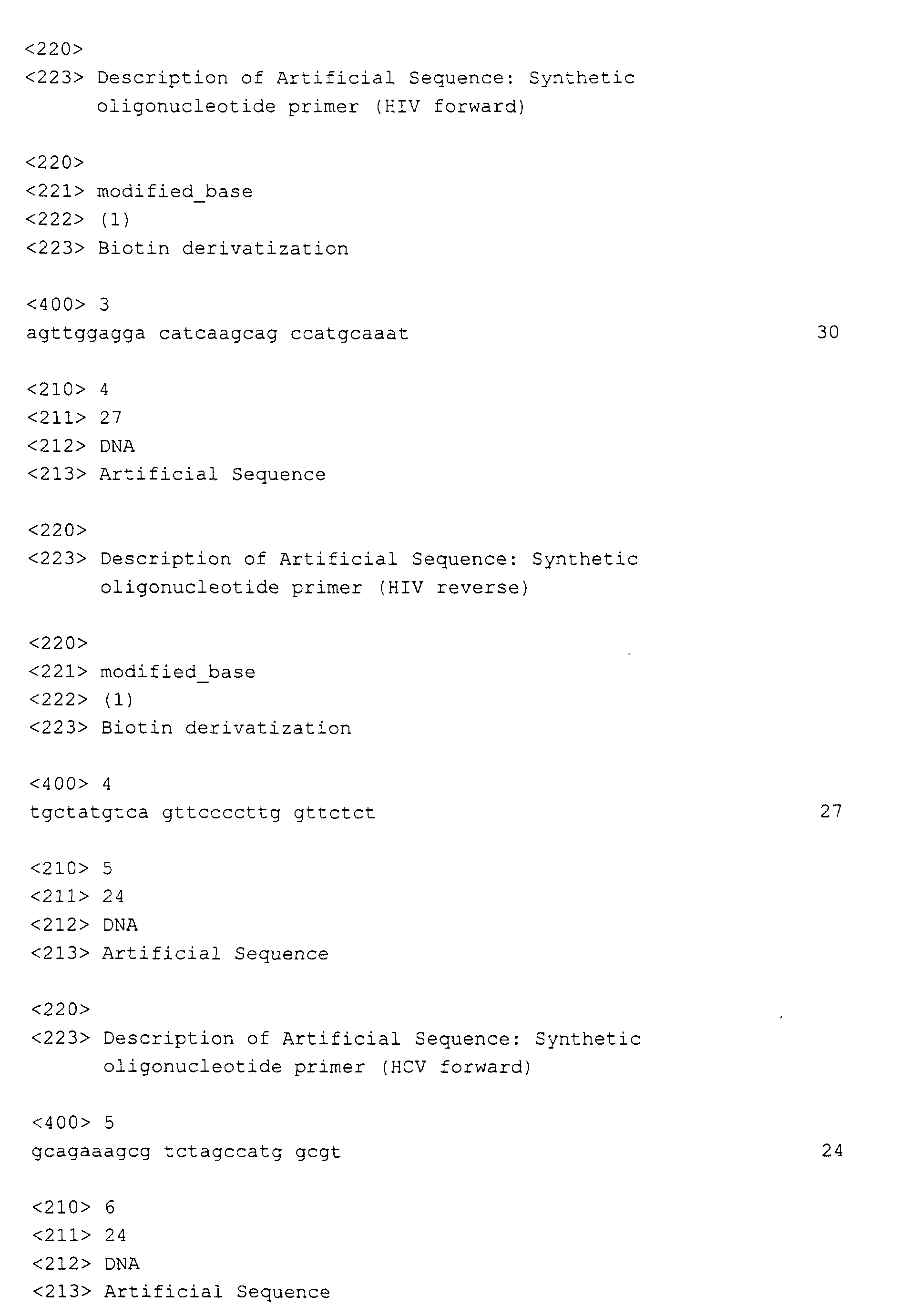


| Company | Abbreviation | Description | Code (see Example 1.4) |
| --- | --- | No pigment | |
| Merck Darmstadt, Germany | BM | Merck Black Mica: Small plates of mica (support), diameter = 10-60 µm, thickness approximately 1-2 µm, Fe3O4-ashlars and TiO2-grains are attached; typically, several pigment units are aggregated (Merck Iriodin®) | 0 1 |
| Merck Darmstadt, Germany | MMB | Merck Microna Matte Black Small plates of mica (support), diameter = 3-15 µm, thickness approximately 1-2 µm, Fe3O4- ashlars are attached; typically, several pigment units are aggregated | 2 |
| BASF Ludwigshafen, Germany | BASF-FA | BASF iron oxide FA28-41: Needles in the nanoscale range made from γ-Fe2O3, length approximately 200-400 nm, very high tendency to aggregate to particles in the µ-scale because of the form and large highly structured surface | 4 |
| Cerac, Milwaukee, WI, USA | CERAC | CERAC γ-Fe2O3 (Catalogue number I-2012, distributed by Chemco Chemieprodukte GmbH in Germany) γ-Fe2O3-microbeads in the nanoscale (pure ferric oxide), diameter = 23 nm, non-aggregating | 5 |
| Strem Chemicals, Newburyport, MA, USA | STREM | STREM Fe3O4 >95% (Catalogue-No.: 93-2616) Microbeads in the nanoscale range made from precipitated Fe3O4, diameter = 200-500 nm, strong tendency to aggregate to particles in the µ-range | 7 |
| BASF, Ludwigshafen, Germany | BASF-CE | Beads in the µ-scale made from metallic iron - BASF Carbonyl iron powder HQ: 10% < 1.04 µm, 50% < 1.91 µm, 90% < 3.71 µm - BASF Carbonyl iron powder SU: 10% < 0,43 µm, 50% < 0.93 µm, 90% < 1.81 µm | 8 9 |
| Sample | Outlet temperature (°C) | Density (g/cm3) | Extinction (280 nm) | Extinction (400 nm) | Iron solubility (mg/l) | Magnetic force (g / 250 mg) |
| RN0005.2E-01 | 116 | 3.420 | 0.037 | 0.027 | < determination limit | 16.04 |
| RN0009.1E-01 | 119 | 3.155 | 0.106 | 0.093 | < determination limit | 9.50 |
| EI0028.2R-01 | 118 | 3.602 | 0.019 | 0.024 | < determination limit | 12.25 |
| RN0045.4R-01 | 130 | 3.647 | 0.032 | 0.034 | < determination limit | 9.23 |
| RN0046.7R-01 | 98 | 3.866 | 0.013 | 0.013 | < determination limit | 13.12 |
| RN0045.5R-01 | 120 | 3.802 | 0.019 | 0.017 | 0.047 | 9.52 |
| EJ0047.2R-01 | 118 | 3.659 | 0.022 | 0.018 | < determination limit | 12.00 |
| EJ0096.5R-01 | 96 | 4.203 | 0.001 | 0.000 | < determination limit | 13.53 |
| EJ0096.7R-01 | 96 | 4.328 | 0.000 | 0.001 | < determination limit | 18.36 |
| EJ0097.4R-01 | 96 | 3.859 | 0.001 | 0.000 | < determination limit | 15.17 |
| EJ0098.8R-01 | 95 | 5.588 | 0.065 | 0.055 | < determination limit | 15.76 |
| EJ0099.9R-01 | 95 | 5.538 | 0.103 | 0.101 | 0.016 | 14.13 |
| EJ0100.7R-01 | 122 | 4.196 | 0.027 | 0.003 | < determination limit | 16.29 |
| EJ0100.5R-01 | 126 | 4.069 | 0.012 | 0.012 | < determination limit | 12.42 |
| EJ0101.4R-01 | 119 | 4.079 | 0.083 | 0.084 | 0.015 | 11.67 |
| EJ0102.2R-01 | 128 | 3.668 | 0.009 | 0.009 | < determination limit | 13.36 |
| EJ0047.2R-07 | 118 | 3.612 | 0.024 | 0.022 | 0.159 | 11.26 |
| EJ0100.5R-02 | 127 | 4.107 | 0.029 | 0.023 | 0.054 | 14.51 |
| EJ0100.5R-03 | 127 | 4.173 | 0.063 | 0.045 | 0.126 | 10.44 |
| EJ0106.5R-01 | 127 | 4.191 | 0.004 | 0.004 | 0.025 | 10.09 |
| EJ0107.5R-01 | 127 | 4.235 | 0.016 | 0.009 | 0.037 | 10.25 |
| EJ0108.5R-01 | 127 | 4.233 | 0.026 | 0.026 | 0.039 | 11.56 |
| EJ0109.2R-01 | 110 | 3.700 | 0.046 | 0.043 | 0.028 | 11.23 |
| EJ0111.5R-01 | 117 | 3.612 | 0.071 | 0.036 | 0.166 | 16.48 |
| EJ0111.5R-02 | 121 | 4.082 | 0.065 | 0.040 | 0.161 | 15.95 |
| EJ0124.5R-01 | 3.992 | 0.001 | 0.001 | 0.051 | 17.57 | |
| EJ0128.5R-01 | 125 | 4.078 | 0.023 | 0.028 | 8.97 | |
| EJ0129.5R-01 | 92 | 4.013 | 0.044 | 0.043 | 9.64 | |
| EJ0130.5R-01 | 87 | 4.022 | 0.031 | 0.036 | 11.70 | |
| EJ0131.5R-01 | 89 | 3.908 | 0.033 | 0.037 | 11.73 | |
| EP0119.5R-01 | 127 | 4.110 | 0.003 | 0.003 | 0.079 | 17.15 |
| Lot | BET surface [m2/g] | Remarks |
| EJ0096.5R-01 | 26.85 | Cerac, small spray dryer, 6 bar |
| EJ0100.5R-01 | 8.95 | Cerac, big spray dryer, 1,5 bar |
| EJ0101.4R-01 | 8.47 | BASF FA, big spray dryer, 1,5 bar |
| RN0005.2E-01 | 3.54 | MERCK MMB, big spray dryer, 1,5 bar |
| RN0009.1E-01 | 3.25 | MERCK BM, big spray dryer, 1,5 bar |
| EJ0099.9R | 0.74 | BASF CE-SU, big spray dryer, 6 bar |
| EJ0100.7R | 8.53 | STREM, big spray dryer, 1,5 bar |
| MGP-type | Size of the isolated cores | Constitution | Form | t1/2 sedimentation [min] | t1/2 separation [sec] |
| MMB | 3-15 µm | Aggregated | Plates | 3.9 | 14 |
| CERAC | 23 nm | Singular | Beads | >> 10 | 16 |
| STREM | 300-500 nm | Aggregated | Beads | 4.8 | 10 |
| BASF-FA | 200-300 nm | Aggregated | Needles | 4.6 | 12 |
| MGP-sample | t0,9 (90%-Wert) sedimentation [min] | t1/2 (50%-Wert) sedimentation [min] |
| EJ0100.5R-01 | 0.35 | 2.78 |
| EJ0108.5R-01 | 0.62 | 7.58 |
| EJ0096.5R-01 | 1.39 | 19.85 |
| MGP | USB [counts] - 0-matrix | SB [counts] - 0.5xGG | ||
| HIV | HBV | HIV | HBV | |
| EJ0100.5R-01 | 4997 | 2277 | 20380 | 80292 |
| EJ0100.5R-02 | 6100 | 3590 | 22036 | 53417 |
| EJ0100.5R-03 | 4856 | 2270 | 19791 | 65109 |
| EJ0106.5R-01 | 4427 | 2263 | 19030 | 75128 |
| EJ0107.5R-01 | 3940 | 2261 | 19255 | 68285 |
| EJ0108.5R-01 | 3429 | 2217 | 20048 | 80426 |
| MGP | Description |
| EJ0096.5R | 6.0 bar spraying pressure, 150 °C inlet temperature, Büchi-laboratory equipment. This preparation approximates the characteristics according to the invention very well, see Figure 15. |
| EJ0111.5R | 1.5 bar spraying pressure, 230°C inlet temperature, Nubilosa- laboratory equipment |
| EP0119.5R | 1.5 bar spraying pressure, 230°C inlet temperature, Nubilosa- laboratory equipment, changed glass composition without Zn-acetate, K-methanolate substituted by K-acetate, with identical stoichometry |
| EJ108,5R | 4.3 bar spraying pressure, 230°C inlet temperature, Nubilosa- laboratory equipment |
Claims (64)
- Composition of magnetic glass particles
characterized in that
the magnetic glass particles contain at least one magnetic object with a mean diameter between 5 to 500 nm. - Composition according to claim 1
characterized in that
the magnetic object has a mean diameter between 10 to 200 nm, preferably between 15 to 50 nm. - Composition according to any of the claims 1 to 2
characterized in that
the half-life period for the sedimentation of a 3 mg/ ml weight-per-volume suspension of the composition in isopropanol is more than 3 min. - Composition according to claim 3
characterized in that the half-life period is more than 6 min. - Composition according to any of the claims 1 to 4
characterized in that the magnetic object has a mean diameter of 23 nm. - Composition according to claim 1 or 5
characterized in that the diameter ratio of the magnetic body to the glass shell is less than 1 to 10. - Composition according to any of the claims 1 to 6
characterized in that the magnetic glass particles have a mean diameter between 0,5 µm and 5 µm. - Composition according to any of the claims 1 to 7
characterized in that
the magnetic glass particles have a mean diameter between 1 µm to 2 µm. - Composition according to any of the claims 1 to 8
characterized in that the magnetic glass particles are microporous. - Composition according to any of the claims 1 to 9
characterized in that
the pore surface of the magnetic glass particles is less than 10 % of the total surface. - Composition according to any of the claims 1 to 10
characterized in that the magnetic object is superparamagnetic. - Composition according to any of the claims 1 to 10
characterized in that the magnetic object is ferrimagnetic or ferromagnetic. - Composition according to any of the claims 1 to 12
characterized in that the magnetic object comprises iron or iron oxide. - Composition according to claim 13
characterized in that the iron oxide is Fe3O4 or γ-Fe2O3. - Composition according to any of the claims 1 to 14
characterized in that the magnetic glass particles have a surface area of more than 4 m2/ g. - Composition according to claim 15
characterized in that
the magnetic glass particles have a surface area between 5 to 100 m2/ g, preferably in the range of 10 to 50 m2/ g, most preferably in the range of 15 to 30 m2/ g. - Composition according to any of the claims 1 to 16
characterized in that
the magnetic glass particles are substantially spherical. - Suspension containing a composition of magnetic glass particles in a liquid according to any of the claims 1 to 17.
- Suspension according to claim 18
characterized in that
the liquid comprises an alcohol. - Suspension according to claim 19
characterized in that
the alcohol is isopropanol or ethanol. - Suspension according to claim 19
characterized in that
the liquid is 70 % (V/V) ethanol. - Suspension according to claim 18 characterized in that the liquid is a buffered aqueous solution.
- Suspension according to claim 22 characterized in that the solution is buffered by Tris-hydroxymethylamine, phosphate, N- (2-Hydroxyethyl)piperazine-N'- (2-ethanesulfonic acid) or salts thereof.
- Suspension according to any of the claims 22 to 23
characterized in that
the solution further contains substances modifying the ionic strength or metalcomplexing agents. - Suspension according to any of the claims 22 to 24
characterized in that the suspension additionally contains DNA or RNA. - Suspension according to any of the claims 22 to 24
characterized in that
the suspension contains additionally DNA or RNA or both in a mixture with one or more compounds selected from the group of proteins, fatty acids, carbohydrates or any other biological material. - Suspension according to any of the claims 18 to 26
characterized in that the suspension additionally contains a chaotropic agent. - Suspension according to claim 27
characterized in that
the chaotropic agent is selected from the group consisting of sodium iodite, sodium perchlorate, guanidinium thiocyanate, guanidinium isothiocyanate or guanidinium hydrochloride. - Suspension according to any of the claims 28 to 29
characterized in that
the chaotropic agent is present in a concentration between 2 and 8 mol/l, and preferably between 4 and 6 mol/l. - Suspension according to any of the claims 18 to 29
characterized in that
the suspension contains 5 to 60 mg/ ml of a composition according to any one of the claims 1 to 17. - A tube containing a composition according to any of the claims 1 to 17 or a suspension according to any of the claims 18 to 30.
- A storage container containing a composition according to any of the claims 1 to 17 or a suspension according to any of the claims 18 to 30.
- A kit of parts containing a tube according to claim 31 or a storage container according to claim 32.
- A kit of parts according to claim 33 further containing washing solution or eluent.
- A kit of parts according to any of the claims 33 to 34 further comprising reagents suitable for the purification of a nucleic acid.
- Use of a composition according to any of the claims 1 to 17 for the preparation of an suspension.
- Use of a suspension according to any of the claims 18 to 30 for the purification of nucleic acids.
- Use of a kit of parts according to any of the claims 33 to 35 for the purification of nucleic acids.
- Method for the production of a composition of magnetic glass particles according to any of the claims 1 to 17
comprising the steps ofa) suspending magnetic objects with a diameter between 5 and 500 nm in a sol,b) spray-drying the suspension in a two-nozzle spray-drier, andc) sintering the spray-dried powder. - Method according to claim 39
characterized in that
the inlet temperature of the two-nozzle spray-drier is between 120 °C and 500 °C, the outlet temperature is chosen according to the boiling point of the sol and the spray pressure is at least 3 bar. - Method according to any one of the claims 39 to 40
characterized in that
the inlet temperature of the two-nozzle spray-drier is between 170 °C and 230 °C, preferably between 190 °C and 210 °C. - Method according to any one of the claims 39 to 41
characterized in that
the inlet temperature of the two-nozzle spray-drier is 200 °C. - Method according to any one of the claims 39 to 42
characterized in that
the spray pressure is between 4 and 6 bar. - Method according to any one of the claims 39 to 43
characterized in that
the sol contains ethanol as solvent. - Method according to claim 44
characterized in that
the outlet temperature is between 50°C and 300°C. - Method according to any one of the claims 44 to 45
characterized in that
the outlet temperature is between 90°C and 100°C. - Method according to any one of the claims 39 to 46
characterized in that
the sinter temperature is between 400°C to 1200°C, preferably between 720°C and 770°C. - Magnetic glass particles obtainable by a process as claimed in any of the claims 39 to 47.
- Procedure for isolating a biological material comprisinga) bringing a sample that contains the biological material in a liquid in contact with a composition according to any one of the claims 1 to 17, magnetic glass particles according to claim 48 or a suspension according to any of the claims 18 to 30 under conditions in which the biological material binds directly to the glass surface, andb) separating the biological material from the liquid.
- Procedure according to claim 49, characterized in that the biological material is a nucleic acid.
- Procedure according to claim 49, characterized in that the biological material contains a mixture of nucleic acids, wherein the target nucleic acid or target nucleic acids is/ are present in low abundance.
- Procedure according to any one of the claims 49 to51,
characterized in that
the separation is accomplished with the aid of a magnet. - Procedure according to any of the claims 49 to 52,
characterized in that
the magnetic particles are not premagnetized when brought into contact with the sample. - Procedure according to any of the claims 49 to 53
characterized in that
the procedure is automatized. - Procedure according to any of the claims 49 to 54
characterized in that
the procedure is in a high-throughput format. - Procedure according to any of the claims 49 to 55
characterized in that
during the procedure a suspension according to any of the claims 18 to 26 is taken from a storage container and partial volumes of the suspension are added to different reaction vessels. - Procedure according to any of the claims 49 to 56
characterized in that
the nucleic acid is detected after the purification. - Procedure according to any of the claims 49 to 57
characterized in that
the nucleic acid is detected after an amplification step. - Procedure according to any of the claims 57 to 58
characterized in that
the nucleic acid is detected by a process comprising:a) contacting the sample with an oligonucleotide containing a sequence complementary to a region of the target nucleic acid and a labeled oligonucleotide containing a sequence complementary to a second region of the same target nucleic acid sequence strand, but not including the nucleic acid sequence defined by the first oligonucleotide, to create a mixture of duplexes during hybridization conditions, wherein the duplexes comprise the target nucleic acid annealed to the first oligonucleotide and to the labeled oligonucleotide such that the 3'-end of the first oligonucleotide is adjacent to the 5'-end of the labeled oligonucleotide;b) maintaining the mixture of step a) with a template-dependent nucleic acid polymerase having a 5' to 3' nuclease activity under conditions sufficient to permit the 5' to 3' nuclease activity of the polymerase to cleave the annealed, labeled oligonucleotide and release labeled fragments; andc) detecting and/ or measuring the signal generated by the hydrolysis of the labeled oligonucleotide. - Procedure according to any of the claims 58 to 59
characterized in that
the detection is carried out in the presence of a blocking oligonucleotide. - Procedure according to claim 60
characterized in that
the blocking oligonucleotide is an aptamer. - Procedure according to claim 61
characterized in that
the aptamer has the sequence SEQ ID NO: 23. - Procedure according to any of the claims 60 to 62, characterized in that the amplification and detection reaction is carried out in a homogeneous solution-phase multiplex assay format for the simultaneous detection of multiple targets.
- Procedure according to any of the claims 61 to 63, characterized in that for at least 5 cycles of the polymerase chain reaction, the annealing temperature is less than 8°C, preferrably less than 3°C above the dissociation temperature of the polymerase-aptamer complex.
Priority Applications (19)
| Application Number | Priority Date | Filing Date | Title |
|---|---|---|---|
| EP00110165A EP1154443A1 (en) | 2000-05-12 | 2000-05-12 | Magnetic glass particles, method for their preparation and uses thereof |
| PCT/EP2000/011459 WO2001037291A1 (en) | 1999-11-17 | 2000-11-17 | Magnetic glass particles, method for their preparation and uses thereof |
| PL356771A PL202358B1 (en) | 1999-11-17 | 2000-11-17 | Magnetic glass particles, method for their preparation and uses thereof |
| CA002392009A CA2392009C (en) | 1999-11-17 | 2000-11-17 | Magnetic glass particles, method for their preparation and uses thereof |
| SK664-2002A SK287932B6 (en) | 1999-11-17 | 2000-11-17 | Magnetic glass particles, method for their preparation and uses thereof |
| EP00981267A EP1232502B1 (en) | 1999-11-17 | 2000-11-17 | Magnetic glass particles, method for their preparation and uses thereof |
| HU0203386A HUP0203386A3 (en) | 1999-11-17 | 2000-11-17 | Magnetic glass particles, method for their preparation and uses thereof |
| DE60025529T DE60025529T2 (en) | 1999-11-17 | 2000-11-17 | MAGNETIC GLASS PARTICLES, MANUFACTURING METHOD AND USE |
| AT00981267T ATE315826T1 (en) | 1999-11-17 | 2000-11-17 | MAGNETIC GLASS PARTICLES, PROCESS OF PRODUCTION AND USE |
| JP2001537749A JP2003514383A (en) | 1999-11-17 | 2000-11-17 | Magnetic glass particles, their production method and their use |
| DK00981267T DK1232502T3 (en) | 1999-11-17 | 2000-11-17 | Magnetic glass particles, processes for their manufacture and their applications |
| ES00981267T ES2256068T3 (en) | 1999-11-17 | 2000-11-17 | MAGNETIC GLASS PARTICLES, METHOD FOR THEIR PREPARATION AND USES OF THE SAME. |
| PT00981267T PT1232502E (en) | 1999-11-17 | 2000-11-17 | MAGNETIC GLASS PARTICLES, METHOD FOR THEIR PREPARATION AND THEIR USES |
| CZ20021608A CZ20021608A3 (en) | 1999-11-17 | 2000-11-17 | Magnetic glass particles, processes of their preparation and use |
| US10/147,679 US20030224366A1 (en) | 1999-11-17 | 2002-05-16 | Magnetic glass particles, method for their preparation and uses thereof |
| US11/128,024 US8129118B2 (en) | 1995-06-08 | 2005-05-11 | Magnetic glass particles, method for their preparation and uses thereof |
| JP2006019516A JP4361905B2 (en) | 1999-11-17 | 2006-01-27 | Magnetic glass particles, methods for their production, and their use |
| CY20061100461T CY1105229T1 (en) | 1999-11-17 | 2006-04-03 | MAGNETIC GLASS PARTICLES, METHOD FOR THEIR PRODUCTION AND USE THEREOF |
| US13/372,218 US20120247150A1 (en) | 1995-06-08 | 2012-02-13 | Magnetic Glass Particles, Method for Their Preparation and Uses Thereof |
Applications Claiming Priority (2)
| Application Number | Priority Date | Filing Date | Title |
|---|---|---|---|
| EP00110165A EP1154443A1 (en) | 2000-05-12 | 2000-05-12 | Magnetic glass particles, method for their preparation and uses thereof |
| US10/147,679 US20030224366A1 (en) | 1999-11-17 | 2002-05-16 | Magnetic glass particles, method for their preparation and uses thereof |
Publications (1)
| Publication Number | Publication Date |
|---|---|
| EP1154443A1 true EP1154443A1 (en) | 2001-11-14 |
Family
ID=32094939
Family Applications (1)
| Application Number | Title | Priority Date | Filing Date |
|---|---|---|---|
| EP00110165A Withdrawn EP1154443A1 (en) | 1995-06-08 | 2000-05-12 | Magnetic glass particles, method for their preparation and uses thereof |
Country Status (1)
| Country | Link |
|---|---|
| EP (1) | EP1154443A1 (en) |
Cited By (10)
| Publication number | Priority date | Publication date | Assignee | Title |
|---|---|---|---|---|
| WO2003057910A2 (en) | 2002-01-08 | 2003-07-17 | Roche Diagnostics Gmbh | Use of silica material in an amplification reaction |
| EP1376129A3 (en) * | 2002-06-27 | 2004-02-04 | Toyo Boseki Kabushiki Kaisha | Magnetic carrier for biological substance, production method thereof and isolation method of biological substance using the same |
| EP2108700A1 (en) | 2008-04-08 | 2009-10-14 | Roche Diagnostics GmbH | Analytical processing and detection device |
| EP2138234A1 (en) | 2008-06-24 | 2009-12-30 | F. Hoffmann-Roche AG | Flexible disposable tip interface |
| WO2011083076A1 (en) | 2010-01-08 | 2011-07-14 | Roche Diagnostics Gmbh | Improved recovery of nucleic acids from magnetic glass particles |
| CN104319055A (en) * | 2014-11-18 | 2015-01-28 | 成都理想财富投资咨询有限公司 | Preparation method of magnetic fluid for plasmid extraction |
| WO2021198289A1 (en) * | 2020-03-30 | 2021-10-07 | Université de Liège | Preparation of magnetic core-shell particles |
| EP4015074A1 (en) * | 2020-12-17 | 2022-06-22 | Université de Liège | Preparation of magnetic core-shell particles |
| CN117504806A (en) * | 2017-10-31 | 2024-02-06 | 豪夫迈·罗氏有限公司 | Improved magnetic particles and their uses |
| WO2026003005A1 (en) | 2024-06-25 | 2026-01-02 | Qiagen Gmbh | Method for removing inhibitory contaminants from nucleic acid-containing samples |
Citations (3)
| Publication number | Priority date | Publication date | Assignee | Title |
|---|---|---|---|---|
| WO1995006652A1 (en) * | 1993-08-30 | 1995-03-09 | Promega Corporation | Nucleic acid purification compositions and methods |
| DE19520964A1 (en) * | 1995-06-08 | 1996-12-12 | Inst Neue Mat Gemein Gmbh | Coated inorganic pigments, process for their preparation and their use |
| EP0757106A2 (en) * | 1995-07-07 | 1997-02-05 | Toyo Boseki Kabushiki Kaisha | Nucleic acid-bondable magnetic carrier and method for isolating nucleic acid using the same |
-
2000
- 2000-05-12 EP EP00110165A patent/EP1154443A1/en not_active Withdrawn
Patent Citations (3)
| Publication number | Priority date | Publication date | Assignee | Title |
|---|---|---|---|---|
| WO1995006652A1 (en) * | 1993-08-30 | 1995-03-09 | Promega Corporation | Nucleic acid purification compositions and methods |
| DE19520964A1 (en) * | 1995-06-08 | 1996-12-12 | Inst Neue Mat Gemein Gmbh | Coated inorganic pigments, process for their preparation and their use |
| EP0757106A2 (en) * | 1995-07-07 | 1997-02-05 | Toyo Boseki Kabushiki Kaisha | Nucleic acid-bondable magnetic carrier and method for isolating nucleic acid using the same |
Non-Patent Citations (2)
| Title |
|---|
| CHAKRAVORTY D ET AL: "Nanocrystalline nickel-zinc ferrite prepared by the glass-ceramic route", JOURNAL OF MAGNETISM AND MAGNETIC MATERIALS,NL,ELSEVIER SCIENCE PUBLISHERS, AMSTERDAM, vol. 164, no. 1, 1 November 1996 (1996-11-01), pages 256 - 260, XP004055624, ISSN: 0304-8853 * |
| PAL M ET AL: "Preparation of nanocrystalline barium hexaferrite in a glass medium", NANOSTRUCTURED MATERIALS,US,PERGAMON PRESS, TARRYWOOD, NY, vol. 8, no. 6, 1 September 1997 (1997-09-01), pages 731 - 738, XP004097109, ISSN: 0965-9773 * |
Cited By (14)
| Publication number | Priority date | Publication date | Assignee | Title |
|---|---|---|---|---|
| WO2003057910A2 (en) | 2002-01-08 | 2003-07-17 | Roche Diagnostics Gmbh | Use of silica material in an amplification reaction |
| WO2003057910A3 (en) * | 2002-01-08 | 2004-03-25 | Roche Diagnostics Gmbh | Use of silica material in an amplification reaction |
| US8026068B2 (en) | 2002-01-08 | 2011-09-27 | Roche Molecular Systems, Inc. | Use of silica material in an amplification reaction |
| EP1376129A3 (en) * | 2002-06-27 | 2004-02-04 | Toyo Boseki Kabushiki Kaisha | Magnetic carrier for biological substance, production method thereof and isolation method of biological substance using the same |
| EP2108700A1 (en) | 2008-04-08 | 2009-10-14 | Roche Diagnostics GmbH | Analytical processing and detection device |
| EP2108699A1 (en) | 2008-04-08 | 2009-10-14 | F.Hoffmann-La Roche Ag | Analytical processing and detection device |
| EP2140941A1 (en) | 2008-06-24 | 2010-01-06 | Roche Diagnostics GmbH | Flexible disposable tip interface |
| EP2138234A1 (en) | 2008-06-24 | 2009-12-30 | F. Hoffmann-Roche AG | Flexible disposable tip interface |
| WO2011083076A1 (en) | 2010-01-08 | 2011-07-14 | Roche Diagnostics Gmbh | Improved recovery of nucleic acids from magnetic glass particles |
| CN104319055A (en) * | 2014-11-18 | 2015-01-28 | 成都理想财富投资咨询有限公司 | Preparation method of magnetic fluid for plasmid extraction |
| CN117504806A (en) * | 2017-10-31 | 2024-02-06 | 豪夫迈·罗氏有限公司 | Improved magnetic particles and their uses |
| WO2021198289A1 (en) * | 2020-03-30 | 2021-10-07 | Université de Liège | Preparation of magnetic core-shell particles |
| EP4015074A1 (en) * | 2020-12-17 | 2022-06-22 | Université de Liège | Preparation of magnetic core-shell particles |
| WO2026003005A1 (en) | 2024-06-25 | 2026-01-02 | Qiagen Gmbh | Method for removing inhibitory contaminants from nucleic acid-containing samples |
Similar Documents
| Publication | Publication Date | Title |
|---|---|---|
| EP1232502B1 (en) | Magnetic glass particles, method for their preparation and uses thereof | |
| US8026068B2 (en) | Use of silica material in an amplification reaction | |
| US6255477B1 (en) | Particles having a magnetic core and outer glass layer for separating biological material | |
| US7371830B2 (en) | Method for separating biological material from a fluid using magnetic particles | |
| JP4048022B2 (en) | Universal sample preparation method that can be automated | |
| EP1154443A1 (en) | Magnetic glass particles, method for their preparation and uses thereof | |
| HK1051048B (en) | Magnetic pigment |
Legal Events
| Date | Code | Title | Description |
|---|---|---|---|
| PUAI | Public reference made under article 153(3) epc to a published international application that has entered the european phase |
Free format text: ORIGINAL CODE: 0009012 |
|
| AK | Designated contracting states |
Kind code of ref document: A1 Designated state(s): AT BE CH CY DE DK ES FI FR GB GR IE IT LI LU MC NL PT SE |
|
| AX | Request for extension of the european patent |
Free format text: AL;LT;LV;MK;RO;SI |
|
| STAA | Information on the status of an ep patent application or granted ep patent |
Free format text: STATUS: THE APPLICATION IS DEEMED TO BE WITHDRAWN |
|
| 18D | Application deemed to be withdrawn |
Effective date: 20010913 |