EP1148128A1 - Gene locus involved in regulating hair pigmentation, vestibular function and fertility - Google Patents
Gene locus involved in regulating hair pigmentation, vestibular function and fertility Download PDFInfo
- Publication number
- EP1148128A1 EP1148128A1 EP01201037A EP01201037A EP1148128A1 EP 1148128 A1 EP1148128 A1 EP 1148128A1 EP 01201037 A EP01201037 A EP 01201037A EP 01201037 A EP01201037 A EP 01201037A EP 1148128 A1 EP1148128 A1 EP 1148128A1
- Authority
- EP
- European Patent Office
- Prior art keywords
- nucleic acid
- subject
- ysb
- locus
- protein
- Prior art date
- Legal status (The legal status is an assumption and is not a legal conclusion. Google has not performed a legal analysis and makes no representation as to the accuracy of the status listed.)
- Granted
Links
- 108090000623 proteins and genes Proteins 0.000 title claims description 111
- 230000001720 vestibular Effects 0.000 title claims description 36
- 230000001105 regulatory effect Effects 0.000 title claims description 12
- 230000003793 hair pigmentation Effects 0.000 title description 3
- 230000035558 fertility Effects 0.000 title 1
- 108020004707 nucleic acids Proteins 0.000 claims abstract description 175
- 102000039446 nucleic acids Human genes 0.000 claims abstract description 175
- 150000007523 nucleic acids Chemical class 0.000 claims abstract description 175
- 108700019146 Transgenes Proteins 0.000 claims abstract description 52
- 230000010354 integration Effects 0.000 claims abstract description 41
- 210000000349 chromosome Anatomy 0.000 claims abstract description 37
- 210000003027 ear inner Anatomy 0.000 claims description 54
- 238000000034 method Methods 0.000 claims description 53
- 230000004064 dysfunction Effects 0.000 claims description 51
- 230000019612 pigmentation Effects 0.000 claims description 32
- 102000004169 proteins and genes Human genes 0.000 claims description 31
- 210000004027 cell Anatomy 0.000 claims description 27
- 239000013598 vector Substances 0.000 claims description 25
- 229920001184 polypeptide Polymers 0.000 claims description 20
- 102000004196 processed proteins & peptides Human genes 0.000 claims description 20
- 108090000765 processed proteins & peptides Proteins 0.000 claims description 20
- 108020004414 DNA Proteins 0.000 claims description 19
- 230000002159 abnormal effect Effects 0.000 claims description 19
- 238000012217 deletion Methods 0.000 claims description 19
- 230000037430 deletion Effects 0.000 claims description 19
- 230000010261 cell growth Effects 0.000 claims description 12
- 150000001875 compounds Chemical class 0.000 claims description 11
- 208000016354 hearing loss disease Diseases 0.000 claims description 11
- 239000003550 marker Substances 0.000 claims description 11
- 230000002779 inactivation Effects 0.000 claims description 10
- 230000001537 neural effect Effects 0.000 claims description 10
- 210000000130 stem cell Anatomy 0.000 claims description 10
- 206010011878 Deafness Diseases 0.000 claims description 9
- 230000002759 chromosomal effect Effects 0.000 claims description 7
- 239000002773 nucleotide Substances 0.000 claims description 7
- 125000003729 nucleotide group Chemical group 0.000 claims description 7
- 230000008707 rearrangement Effects 0.000 claims description 7
- 238000011830 transgenic mouse model Methods 0.000 claims description 7
- 230000012292 cell migration Effects 0.000 claims description 6
- 231100000219 mutagenic Toxicity 0.000 claims description 6
- 230000003505 mutagenic effect Effects 0.000 claims description 6
- 210000000944 nerve tissue Anatomy 0.000 claims description 6
- 230000004913 activation Effects 0.000 claims description 5
- 239000003102 growth factor Substances 0.000 claims description 5
- 230000011664 signaling Effects 0.000 claims description 5
- 241000124008 Mammalia Species 0.000 claims description 4
- 108090000301 Membrane transport proteins Proteins 0.000 claims description 4
- 102000003939 Membrane transport proteins Human genes 0.000 claims description 4
- RJKFOVLPORLFTN-LEKSSAKUSA-N Progesterone Chemical compound C1CC2=CC(=O)CC[C@]2(C)[C@@H]2[C@@H]1[C@@H]1CC[C@H](C(=O)C)[C@@]1(C)CC2 RJKFOVLPORLFTN-LEKSSAKUSA-N 0.000 claims description 4
- 241000251539 Vertebrata <Metazoa> Species 0.000 claims description 4
- 230000030833 cell death Effects 0.000 claims description 4
- 230000008034 disappearance Effects 0.000 claims description 4
- 239000005556 hormone Substances 0.000 claims description 4
- 229940088597 hormone Drugs 0.000 claims description 4
- 239000013612 plasmid Substances 0.000 claims description 4
- 230000035755 proliferation Effects 0.000 claims description 4
- 238000013518 transcription Methods 0.000 claims description 4
- 230000035897 transcription Effects 0.000 claims description 4
- 108010078791 Carrier Proteins Proteins 0.000 claims description 3
- 230000006907 apoptotic process Effects 0.000 claims description 3
- 230000004663 cell proliferation Effects 0.000 claims description 3
- 239000002299 complementary DNA Substances 0.000 claims description 3
- 231100000888 hearing loss Toxicity 0.000 claims description 3
- 230000010370 hearing loss Effects 0.000 claims description 3
- 238000004519 manufacturing process Methods 0.000 claims description 3
- 239000000203 mixture Substances 0.000 claims description 3
- 239000002243 precursor Substances 0.000 claims description 3
- 230000001172 regenerating effect Effects 0.000 claims description 3
- 230000008439 repair process Effects 0.000 claims description 3
- 241000271566 Aves Species 0.000 claims description 2
- 241000283690 Bos taurus Species 0.000 claims description 2
- 102000005701 Calcium-Binding Proteins Human genes 0.000 claims description 2
- 108010045403 Calcium-Binding Proteins Proteins 0.000 claims description 2
- 241000282465 Canis Species 0.000 claims description 2
- 102000014914 Carrier Proteins Human genes 0.000 claims description 2
- 241000283073 Equus caballus Species 0.000 claims description 2
- 241000701959 Escherichia virus Lambda Species 0.000 claims description 2
- 241000282324 Felis Species 0.000 claims description 2
- 102000003676 Glucocorticoid Receptors Human genes 0.000 claims description 2
- 108090000079 Glucocorticoid Receptors Proteins 0.000 claims description 2
- 108010009202 Growth Factor Receptors Proteins 0.000 claims description 2
- 102000009465 Growth Factor Receptors Human genes 0.000 claims description 2
- 241001529936 Murinae Species 0.000 claims description 2
- 102000016978 Orphan receptors Human genes 0.000 claims description 2
- 108070000031 Orphan receptors Proteins 0.000 claims description 2
- 241000288906 Primates Species 0.000 claims description 2
- 239000012190 activator Substances 0.000 claims description 2
- 230000001580 bacterial effect Effects 0.000 claims description 2
- 230000022131 cell cycle Effects 0.000 claims description 2
- 229940127089 cytotoxic agent Drugs 0.000 claims description 2
- 239000002254 cytotoxic agent Substances 0.000 claims description 2
- 231100000599 cytotoxic agent Toxicity 0.000 claims description 2
- 230000001419 dependent effect Effects 0.000 claims description 2
- 210000003527 eukaryotic cell Anatomy 0.000 claims description 2
- 239000007850 fluorescent dye Substances 0.000 claims description 2
- 108020001507 fusion proteins Proteins 0.000 claims description 2
- 102000037865 fusion proteins Human genes 0.000 claims description 2
- 230000010189 intracellular transport Effects 0.000 claims description 2
- 239000000186 progesterone Substances 0.000 claims description 2
- 229960003387 progesterone Drugs 0.000 claims description 2
- 102000003998 progesterone receptors Human genes 0.000 claims description 2
- 108090000468 progesterone receptors Proteins 0.000 claims description 2
- 230000004844 protein turnover Effects 0.000 claims description 2
- 230000002285 radioactive effect Effects 0.000 claims description 2
- 230000002103 transcriptional effect Effects 0.000 claims description 2
- 102100031918 E3 ubiquitin-protein ligase NEDD4 Human genes 0.000 claims 1
- 101000636713 Homo sapiens E3 ubiquitin-protein ligase NEDD4 Proteins 0.000 claims 1
- 241000699670 Mus sp. Species 0.000 description 85
- 239000000523 sample Substances 0.000 description 34
- 230000007547 defect Effects 0.000 description 33
- 230000035772 mutation Effects 0.000 description 33
- 241000699666 Mus <mouse, genus> Species 0.000 description 30
- 238000004458 analytical method Methods 0.000 description 19
- 210000002257 embryonic structure Anatomy 0.000 description 18
- 230000013707 sensory perception of sound Effects 0.000 description 17
- 230000009261 transgenic effect Effects 0.000 description 16
- 108010072151 Agouti Signaling Protein Proteins 0.000 description 14
- 102000006822 Agouti Signaling Protein Human genes 0.000 description 14
- 241000484025 Cuniculus Species 0.000 description 13
- 101000979182 Mus musculus Transcription factor MafB Proteins 0.000 description 13
- 210000003754 fetus Anatomy 0.000 description 13
- 238000012360 testing method Methods 0.000 description 13
- 230000005856 abnormality Effects 0.000 description 12
- 230000006870 function Effects 0.000 description 12
- 230000001953 sensory effect Effects 0.000 description 12
- 230000018109 developmental process Effects 0.000 description 11
- 238000002474 experimental method Methods 0.000 description 11
- 238000003780 insertion Methods 0.000 description 11
- 230000037431 insertion Effects 0.000 description 11
- 210000001202 rhombencephalon Anatomy 0.000 description 11
- 238000012512 characterization method Methods 0.000 description 10
- 210000002569 neuron Anatomy 0.000 description 10
- 238000011161 development Methods 0.000 description 9
- 102000005962 receptors Human genes 0.000 description 9
- 108020003175 receptors Proteins 0.000 description 9
- 238000013459 approach Methods 0.000 description 8
- 230000033228 biological regulation Effects 0.000 description 8
- 230000015572 biosynthetic process Effects 0.000 description 8
- 210000001161 mammalian embryo Anatomy 0.000 description 8
- 210000002480 semicircular canal Anatomy 0.000 description 8
- 210000000981 epithelium Anatomy 0.000 description 7
- 238000007901 in situ hybridization Methods 0.000 description 7
- 239000000049 pigment Substances 0.000 description 7
- 230000004044 response Effects 0.000 description 7
- 101150059401 EGR2 gene Proteins 0.000 description 6
- 101150074546 Hmx3 gene Proteins 0.000 description 6
- 241000699660 Mus musculus Species 0.000 description 6
- 238000002105 Southern blotting Methods 0.000 description 6
- 230000003542 behavioural effect Effects 0.000 description 6
- 239000012634 fragment Substances 0.000 description 6
- 101150066555 lacZ gene Proteins 0.000 description 6
- 230000011218 segmentation Effects 0.000 description 6
- 230000000638 stimulation Effects 0.000 description 6
- 230000013985 hindbrain development Effects 0.000 description 5
- 210000005036 nerve Anatomy 0.000 description 5
- 210000000278 spinal cord Anatomy 0.000 description 5
- 238000010186 staining Methods 0.000 description 5
- 108091028043 Nucleic acid sequence Proteins 0.000 description 4
- 108091023040 Transcription factor Proteins 0.000 description 4
- 102000040945 Transcription factor Human genes 0.000 description 4
- 208000037516 chromosome inversion disease Diseases 0.000 description 4
- 210000004209 hair Anatomy 0.000 description 4
- 238000009396 hybridization Methods 0.000 description 4
- 230000032561 inner ear development Effects 0.000 description 4
- 230000033001 locomotion Effects 0.000 description 4
- 210000002985 organ of corti Anatomy 0.000 description 4
- 238000003786 synthesis reaction Methods 0.000 description 4
- OPIFSICVWOWJMJ-AEOCFKNESA-N 5-bromo-4-chloro-3-indolyl beta-D-galactoside Chemical compound O[C@@H]1[C@@H](O)[C@@H](O)[C@@H](CO)O[C@H]1OC1=CNC2=CC=C(Br)C(Cl)=C12 OPIFSICVWOWJMJ-AEOCFKNESA-N 0.000 description 3
- 208000016718 Chromosome Inversion Diseases 0.000 description 3
- 108700005087 Homeobox Genes Proteins 0.000 description 3
- 206010025421 Macule Diseases 0.000 description 3
- 102000008763 Neurofilament Proteins Human genes 0.000 description 3
- 108010088373 Neurofilament Proteins Proteins 0.000 description 3
- 102000007568 Proto-Oncogene Proteins c-fos Human genes 0.000 description 3
- 108010071563 Proto-Oncogene Proteins c-fos Proteins 0.000 description 3
- 150000001413 amino acids Chemical class 0.000 description 3
- 210000004556 brain Anatomy 0.000 description 3
- 230000008859 change Effects 0.000 description 3
- 210000000860 cochlear nerve Anatomy 0.000 description 3
- 230000000295 complement effect Effects 0.000 description 3
- 230000001276 controlling effect Effects 0.000 description 3
- 231100000895 deafness Toxicity 0.000 description 3
- 210000005069 ears Anatomy 0.000 description 3
- 230000002068 genetic effect Effects 0.000 description 3
- 210000002768 hair cell Anatomy 0.000 description 3
- 210000003780 hair follicle Anatomy 0.000 description 3
- 210000003128 head Anatomy 0.000 description 3
- 238000012744 immunostaining Methods 0.000 description 3
- 238000002955 isolation Methods 0.000 description 3
- 238000013507 mapping Methods 0.000 description 3
- 230000007246 mechanism Effects 0.000 description 3
- 230000001002 morphogenetic effect Effects 0.000 description 3
- 210000005044 neurofilament Anatomy 0.000 description 3
- 230000009467 reduction Effects 0.000 description 3
- 210000003491 skin Anatomy 0.000 description 3
- 102100022455 Adrenocorticotropic hormone receptor Human genes 0.000 description 2
- 108700028369 Alleles Proteins 0.000 description 2
- 208000012639 Balance disease Diseases 0.000 description 2
- 102100026189 Beta-galactosidase Human genes 0.000 description 2
- 102000004219 Brain-derived neurotrophic factor Human genes 0.000 description 2
- 108090000715 Brain-derived neurotrophic factor Proteins 0.000 description 2
- 101150082216 COL2A1 gene Proteins 0.000 description 2
- 102100029136 Collagen alpha-1(II) chain Human genes 0.000 description 2
- 102000004190 Enzymes Human genes 0.000 description 2
- 108090000790 Enzymes Proteins 0.000 description 2
- 101150098270 FGF3 gene Proteins 0.000 description 2
- 101000771163 Homo sapiens Collagen alpha-1(II) chain Proteins 0.000 description 2
- 101100350576 Mus musculus Otx1 gene Proteins 0.000 description 2
- 101150117329 NTRK3 gene Proteins 0.000 description 2
- 108090000742 Neurotrophin 3 Proteins 0.000 description 2
- 101150056950 Ntrk2 gene Proteins 0.000 description 2
- 108700026244 Open Reading Frames Proteins 0.000 description 2
- 210000001015 abdomen Anatomy 0.000 description 2
- 230000036982 action potential Effects 0.000 description 2
- 230000001154 acute effect Effects 0.000 description 2
- 230000004075 alteration Effects 0.000 description 2
- 239000005557 antagonist Substances 0.000 description 2
- 210000003984 auditory pathway Anatomy 0.000 description 2
- 108010005774 beta-Galactosidase Proteins 0.000 description 2
- 210000002459 blastocyst Anatomy 0.000 description 2
- 101150067309 bmp4 gene Proteins 0.000 description 2
- 210000000133 brain stem Anatomy 0.000 description 2
- 210000000984 branchial region Anatomy 0.000 description 2
- 230000014107 chromosome localization Effects 0.000 description 2
- 238000010367 cloning Methods 0.000 description 2
- 210000003477 cochlea Anatomy 0.000 description 2
- 210000003792 cranial nerve Anatomy 0.000 description 2
- 230000009547 development abnormality Effects 0.000 description 2
- 230000004069 differentiation Effects 0.000 description 2
- 238000003167 genetic complementation Methods 0.000 description 2
- 108091008039 hormone receptors Proteins 0.000 description 2
- 238000011065 in-situ storage Methods 0.000 description 2
- 238000002347 injection Methods 0.000 description 2
- 239000007924 injection Substances 0.000 description 2
- 238000007852 inverse PCR Methods 0.000 description 2
- 231100000518 lethal Toxicity 0.000 description 2
- 230000001665 lethal effect Effects 0.000 description 2
- 230000036244 malformation Effects 0.000 description 2
- 230000001404 mediated effect Effects 0.000 description 2
- 210000002752 melanocyte Anatomy 0.000 description 2
- 108020004999 messenger RNA Proteins 0.000 description 2
- 230000031864 metaphase Effects 0.000 description 2
- 238000010369 molecular cloning Methods 0.000 description 2
- -1 msx1 Proteins 0.000 description 2
- 210000000276 neural tube Anatomy 0.000 description 2
- 210000003458 notochord Anatomy 0.000 description 2
- 210000004940 nucleus Anatomy 0.000 description 2
- 238000013116 obese mouse model Methods 0.000 description 2
- 210000000056 organ Anatomy 0.000 description 2
- 210000001265 otolithic membrane Anatomy 0.000 description 2
- 238000004806 packaging method and process Methods 0.000 description 2
- 239000003973 paint Substances 0.000 description 2
- 230000003076 paracrine Effects 0.000 description 2
- 230000002093 peripheral effect Effects 0.000 description 2
- 230000001242 postsynaptic effect Effects 0.000 description 2
- 230000011514 reflex Effects 0.000 description 2
- 238000012216 screening Methods 0.000 description 2
- 230000036421 sense of balance Effects 0.000 description 2
- 238000002864 sequence alignment Methods 0.000 description 2
- 210000004894 snout Anatomy 0.000 description 2
- 210000003594 spinal ganglia Anatomy 0.000 description 2
- 230000008719 thickening Effects 0.000 description 2
- 210000001519 tissue Anatomy 0.000 description 2
- 208000027491 vestibular disease Diseases 0.000 description 2
- 210000001213 vestibule labyrinth Anatomy 0.000 description 2
- XLYOFNOQVPJJNP-UHFFFAOYSA-N water Substances O XLYOFNOQVPJJNP-UHFFFAOYSA-N 0.000 description 2
- 108091032973 (ribonucleotides)n+m Proteins 0.000 description 1
- 238000011714 129 mouse Methods 0.000 description 1
- 108020005065 3' Flanking Region Proteins 0.000 description 1
- FWBHETKCLVMNFS-UHFFFAOYSA-N 4',6-Diamino-2-phenylindol Chemical compound C1=CC(C(=N)N)=CC=C1C1=CC2=CC=C(C(N)=N)C=C2N1 FWBHETKCLVMNFS-UHFFFAOYSA-N 0.000 description 1
- 108020005029 5' Flanking Region Proteins 0.000 description 1
- 241000251468 Actinopterygii Species 0.000 description 1
- 101710115814 Adrenocorticotropic hormone receptor Proteins 0.000 description 1
- 101150035467 BDNF gene Proteins 0.000 description 1
- 101100222854 Bacillus subtilis (strain 168) czcO gene Proteins 0.000 description 1
- 102100024505 Bone morphogenetic protein 4 Human genes 0.000 description 1
- 102000010831 Cytoskeletal Proteins Human genes 0.000 description 1
- 108010037414 Cytoskeletal Proteins Proteins 0.000 description 1
- 239000003298 DNA probe Substances 0.000 description 1
- 238000001712 DNA sequencing Methods 0.000 description 1
- 101100281017 Danio rerio fgf3 gene Proteins 0.000 description 1
- 101100350574 Danio rerio otx1b gene Proteins 0.000 description 1
- 101150001833 EDNRB gene Proteins 0.000 description 1
- 206010014016 Ear malformation Diseases 0.000 description 1
- 108010055179 EphA4 Receptor Proteins 0.000 description 1
- 102000021727 EphA4 Receptor Human genes 0.000 description 1
- 101100127166 Escherichia coli (strain K12) kefB gene Proteins 0.000 description 1
- 108091029865 Exogenous DNA Proteins 0.000 description 1
- 108700024394 Exon Proteins 0.000 description 1
- 102000010834 Extracellular Matrix Proteins Human genes 0.000 description 1
- 108010037362 Extracellular Matrix Proteins Proteins 0.000 description 1
- 101150019331 FGF2 gene Proteins 0.000 description 1
- 101150025764 FGFR3 gene Proteins 0.000 description 1
- 108091006027 G proteins Proteins 0.000 description 1
- 102000030782 GTP binding Human genes 0.000 description 1
- 108091000058 GTP-Binding Proteins 0.000 description 1
- 208000003098 Ganglion Cysts Diseases 0.000 description 1
- 208000016621 Hearing disease Diseases 0.000 description 1
- 208000032041 Hearing impaired Diseases 0.000 description 1
- 101000678419 Homo sapiens Adrenocorticotropic hormone receptor Proteins 0.000 description 1
- 101000762379 Homo sapiens Bone morphogenetic protein 4 Proteins 0.000 description 1
- 208000027601 Inner ear disease Diseases 0.000 description 1
- 108050002021 Integrator complex subunit 2 Proteins 0.000 description 1
- 102100033265 Integrator complex subunit 2 Human genes 0.000 description 1
- 108090000862 Ion Channels Proteins 0.000 description 1
- 102000004310 Ion Channels Human genes 0.000 description 1
- 241001465754 Metazoa Species 0.000 description 1
- 101100537961 Methanosarcina mazei (strain ATCC BAA-159 / DSM 3647 / Goe1 / Go1 / JCM 11833 / OCM 88) trkA2 gene Proteins 0.000 description 1
- 101100446506 Mus musculus Fgf3 gene Proteins 0.000 description 1
- 101100395372 Mus musculus Hmx2 gene Proteins 0.000 description 1
- 101100125257 Mus musculus Hoxa3 gene Proteins 0.000 description 1
- 101100071843 Mus musculus Hoxb1 gene Proteins 0.000 description 1
- 101100518992 Mus musculus Pax2 gene Proteins 0.000 description 1
- 102000003505 Myosin Human genes 0.000 description 1
- 108060008487 Myosin Proteins 0.000 description 1
- 108010025020 Nerve Growth Factor Proteins 0.000 description 1
- 102000007072 Nerve Growth Factors Human genes 0.000 description 1
- XDMCWZFLLGVIID-SXPRBRBTSA-N O-(3-O-D-galactosyl-N-acetyl-beta-D-galactosaminyl)-L-serine Chemical compound CC(=O)N[C@H]1[C@H](OC[C@H]([NH3+])C([O-])=O)O[C@H](CO)[C@H](O)[C@@H]1OC1[C@H](O)[C@@H](O)[C@@H](O)[C@@H](CO)O1 XDMCWZFLLGVIID-SXPRBRBTSA-N 0.000 description 1
- 241000356847 Otolithes Species 0.000 description 1
- 208000016012 Phenotypic abnormality Diseases 0.000 description 1
- 108700008625 Reporter Genes Proteins 0.000 description 1
- 240000004808 Saccharomyces cerevisiae Species 0.000 description 1
- 238000012300 Sequence Analysis Methods 0.000 description 1
- 208000005400 Synovial Cyst Diseases 0.000 description 1
- 102000003425 Tyrosinase Human genes 0.000 description 1
- 108060008724 Tyrosinase Proteins 0.000 description 1
- 239000003708 ampul Substances 0.000 description 1
- 230000019552 anatomical structure morphogenesis Effects 0.000 description 1
- 229940035674 anesthetics Drugs 0.000 description 1
- 210000004436 artificial bacterial chromosome Anatomy 0.000 description 1
- 238000000211 autoradiogram Methods 0.000 description 1
- 239000011324 bead Substances 0.000 description 1
- 230000008901 benefit Effects 0.000 description 1
- 238000009395 breeding Methods 0.000 description 1
- 230000001488 breeding effect Effects 0.000 description 1
- 239000002775 capsule Substances 0.000 description 1
- 230000000453 cell autonomous effect Effects 0.000 description 1
- 210000003169 central nervous system Anatomy 0.000 description 1
- 210000000262 cochlear duct Anatomy 0.000 description 1
- 239000003086 colorant Substances 0.000 description 1
- 238000010276 construction Methods 0.000 description 1
- 230000003436 cytoskeletal effect Effects 0.000 description 1
- 238000001514 detection method Methods 0.000 description 1
- 208000037265 diseases, disorders, signs and symptoms Diseases 0.000 description 1
- 208000035475 disorder Diseases 0.000 description 1
- 238000009826 distribution Methods 0.000 description 1
- 230000004577 ear development Effects 0.000 description 1
- 230000000694 effects Effects 0.000 description 1
- 230000013020 embryo development Effects 0.000 description 1
- 210000002744 extracellular matrix Anatomy 0.000 description 1
- YSFTYXKQUONNFY-NQXPEFQPSA-N fgf2 Chemical compound C([C@@H](C(=O)N[C@@H](CCCCN)C(=O)N[C@@H](CC(O)=O)C(=O)N1CCC[C@H]1C(=O)N[C@@H](CCCCN)C(=O)N[C@@H](CCCNC(N)=N)C(=O)N[C@@H](CC(C)C)C(=O)N[C@@H](CC=1C=CC(O)=CC=1)C(O)=O)NC(=O)[C@H](CC=1N=CNC=1)NC(=O)CNC(=O)[C@H]1N(CCC1)C(=O)[C@H]1N(CCC1)C(=O)[C@H](CC=1C=CC=CC=1)NC(=O)[C@H](C)NC(=O)CNC(=O)[C@H](CO)NC(=O)CNC(=O)CNC(=O)[C@H](CC(O)=O)NC(=O)[C@H](CCC(O)=O)NC(=O)[C@H]1N(CCC1)C(=O)[C@H](CC(C)C)NC(=O)[C@H](C)NC(=O)[C@H]1NCCC1)C1=CC=CC=C1 YSFTYXKQUONNFY-NQXPEFQPSA-N 0.000 description 1
- 210000002950 fibroblast Anatomy 0.000 description 1
- 210000000609 ganglia Anatomy 0.000 description 1
- 239000003193 general anesthetic agent Substances 0.000 description 1
- PCHJSUWPFVWCPO-UHFFFAOYSA-N gold Chemical compound [Au] PCHJSUWPFVWCPO-UHFFFAOYSA-N 0.000 description 1
- 239000010931 gold Substances 0.000 description 1
- 229910052737 gold Inorganic materials 0.000 description 1
- 230000004886 head movement Effects 0.000 description 1
- 230000036541 health Effects 0.000 description 1
- 208000013403 hyperactivity Diseases 0.000 description 1
- 230000002055 immunohistochemical effect Effects 0.000 description 1
- 238000000338 in vitro Methods 0.000 description 1
- 230000001939 inductive effect Effects 0.000 description 1
- 230000003834 intracellular effect Effects 0.000 description 1
- 230000031146 intracellular signal transduction Effects 0.000 description 1
- 238000011835 investigation Methods 0.000 description 1
- 239000004816 latex Substances 0.000 description 1
- 229920000126 latex Polymers 0.000 description 1
- 230000004807 localization Effects 0.000 description 1
- 239000011159 matrix material Substances 0.000 description 1
- 230000035800 maturation Effects 0.000 description 1
- 210000001259 mesencephalon Anatomy 0.000 description 1
- 208000024191 minimally invasive lung adenocarcinoma Diseases 0.000 description 1
- 230000000877 morphologic effect Effects 0.000 description 1
- 108700039855 mouse a Proteins 0.000 description 1
- 230000008035 nerve activity Effects 0.000 description 1
- 210000000118 neural pathway Anatomy 0.000 description 1
- 230000010004 neural pathway Effects 0.000 description 1
- 230000036403 neuro physiology Effects 0.000 description 1
- 239000003900 neurotrophic factor Substances 0.000 description 1
- 238000012346 open field test Methods 0.000 description 1
- 230000002842 otolith Effects 0.000 description 1
- 238000010422 painting Methods 0.000 description 1
- 230000037361 pathway Effects 0.000 description 1
- 238000002360 preparation method Methods 0.000 description 1
- 230000037452 priming Effects 0.000 description 1
- 230000008569 process Effects 0.000 description 1
- 210000004129 prosencephalon Anatomy 0.000 description 1
- 230000007261 regionalization Effects 0.000 description 1
- 230000003252 repetitive effect Effects 0.000 description 1
- 238000011160 research Methods 0.000 description 1
- 108091008146 restriction endonucleases Proteins 0.000 description 1
- PYWVYCXTNDRMGF-UHFFFAOYSA-N rhodamine B Chemical compound [Cl-].C=12C=CC(=[N+](CC)CC)C=C2OC2=CC(N(CC)CC)=CC=C2C=1C1=CC=CC=C1C(O)=O PYWVYCXTNDRMGF-UHFFFAOYSA-N 0.000 description 1
- 230000028527 righting reflex Effects 0.000 description 1
- 238000004626 scanning electron microscopy Methods 0.000 description 1
- 210000003900 secondary neuron Anatomy 0.000 description 1
- 230000003248 secreting effect Effects 0.000 description 1
- 238000005204 segregation Methods 0.000 description 1
- 210000000413 sensory ganglia Anatomy 0.000 description 1
- 210000000697 sensory organ Anatomy 0.000 description 1
- 210000002265 sensory receptor cell Anatomy 0.000 description 1
- 102000027509 sensory receptors Human genes 0.000 description 1
- 108091008691 sensory receptors Proteins 0.000 description 1
- 230000012488 skeletal system development Effects 0.000 description 1
- 238000010561 standard procedure Methods 0.000 description 1
- 210000000645 stria vascularis Anatomy 0.000 description 1
- 230000004083 survival effect Effects 0.000 description 1
- 230000009182 swimming Effects 0.000 description 1
- 229960002197 temoporfin Drugs 0.000 description 1
- 101150025395 trkA gene Proteins 0.000 description 1
- 101150113435 trkA1 gene Proteins 0.000 description 1
- 108010064892 trkC Receptor Proteins 0.000 description 1
- 102000047459 trkC Receptor Human genes 0.000 description 1
- 238000011144 upstream manufacturing Methods 0.000 description 1
- 210000003273 vestibular nerve Anatomy 0.000 description 1
- 210000004440 vestibular nuclei Anatomy 0.000 description 1
- 210000000752 vestibulocochlear nerve Anatomy 0.000 description 1
- 238000012800 visualization Methods 0.000 description 1
- 238000012745 whole-mount immunostaining Methods 0.000 description 1
Images










Classifications
-
- C—CHEMISTRY; METALLURGY
- C07—ORGANIC CHEMISTRY
- C07K—PEPTIDES
- C07K14/00—Peptides having more than 20 amino acids; Gastrins; Somatostatins; Melanotropins; Derivatives thereof
- C07K14/435—Peptides having more than 20 amino acids; Gastrins; Somatostatins; Melanotropins; Derivatives thereof from animals; from humans
- C07K14/46—Peptides having more than 20 amino acids; Gastrins; Somatostatins; Melanotropins; Derivatives thereof from animals; from humans from vertebrates
- C07K14/47—Peptides having more than 20 amino acids; Gastrins; Somatostatins; Melanotropins; Derivatives thereof from animals; from humans from vertebrates from mammals
-
- A—HUMAN NECESSITIES
- A61—MEDICAL OR VETERINARY SCIENCE; HYGIENE
- A61P—SPECIFIC THERAPEUTIC ACTIVITY OF CHEMICAL COMPOUNDS OR MEDICINAL PREPARATIONS
- A61P27/00—Drugs for disorders of the senses
-
- A—HUMAN NECESSITIES
- A01—AGRICULTURE; FORESTRY; ANIMAL HUSBANDRY; HUNTING; TRAPPING; FISHING
- A01K—ANIMAL HUSBANDRY; AVICULTURE; APICULTURE; PISCICULTURE; FISHING; REARING OR BREEDING ANIMALS, NOT OTHERWISE PROVIDED FOR; NEW BREEDS OF ANIMALS
- A01K2217/00—Genetically modified animals
- A01K2217/05—Animals comprising random inserted nucleic acids (transgenic)
-
- A—HUMAN NECESSITIES
- A61—MEDICAL OR VETERINARY SCIENCE; HYGIENE
- A61K—PREPARATIONS FOR MEDICAL, DENTAL OR TOILETRY PURPOSES
- A61K2123/00—Preparations for testing in vivo
-
- A—HUMAN NECESSITIES
- A61—MEDICAL OR VETERINARY SCIENCE; HYGIENE
- A61K—PREPARATIONS FOR MEDICAL, DENTAL OR TOILETRY PURPOSES
- A61K38/00—Medicinal preparations containing peptides
-
- C—CHEMISTRY; METALLURGY
- C07—ORGANIC CHEMISTRY
- C07K—PEPTIDES
- C07K2319/00—Fusion polypeptide
Definitions
- mice bearing a recessive mutation, caused by insertion of a transgene, were created. These mice are characterized by a changed pigmentation of hair to give yellow coat color, circling behaviour and an inability to swim. It has been shown that the transgene has inserted into two sites on mouse chromosome 3. A search of the mouse genome database ascertained that the mutation is novel. The mutant locus has been named yellow submarine (ysb). DNA flanking the transgene insertion has been isolated and used to screen for the corresponding normal (unmutated) sequences.
- ysb yellow submarine
- Molecular genetics, bioinformatics, developmental biology, transgenic and physiological approaches are used to 1) identify and characterize the gene(s) at the wild-type ysb (+ ysb ) locus; 2) determine the nature of the ysb mutation; 3) study the molecular and developmental bases underlying the defect(s) in ysb mice; and 4) charactersize the neurophysiological changes in balance and hearing in ysb mice.
- Approximately 1/1000 infants are affected by hearing defects at birth, two thirds of which have a genetic basis.
- Ysb mice are a valuable model to identify molecules involved in controlling balance. These studies provide fundamental information on the development of the inner ear and the mechanisms by which hearing and balance defects may arise. Identification and characterization of the ysb gene(s) and the molecular defect in ysb mice also yield new insight into the complex regulatory pathways controlling agouti coat color in mice.
- This invention provides an isolated nucleic acid which defines a yellow submarine locus.
- This invention provides an isolated nucleic acid which defines a mutant yellow submarine locus, wherein the mutant yellow submarine locus is identical to a wildtype yellow submarine locus except for an integration of a pAA2 transgene into at least one region on a chromosome.
- This invention provides the above isolated nucleic acid further comprising a rearrangement of chromosomal sequences of a region of the chromosome designated A3 region.
- This invention provides a replicable vector comprising the above nucleic acid.
- This invention provides a host cell comprising the vector.
- This invention provides a nucleic acid of at least 14 nucleotides capable of specifically hybridizing with the nucleic acids of the subject invention.
- This invention provides an isolated nucleic acid which defines a human locus which corresponds to a yellow submarine locus.
- This invention provides an isolated nucleic acid which defines a human locus which corresponds to a mutant yellow submarine locus containing a pAA2 transgene integrated into at least one region the genome.
- This invention provides a method of diagnosing inner ear dysfunction in a subject comprising: a) obtaining a suitable sample from a subject; b) extracting nucleic acid from the sample; c) contacting the nucleic acid with the above nucleic acid which binds specifically to the mutated portion of the ysb locus; and d) detecting the labeled nucleic acid, thereby detecting mutant ysb and thereby diagnosing inner ear dysfunction in the subject.
- This invention provides a method of diagnosing pigmentation dysfunction in a subject comprising: a) obtaining a suitable sample from a subject; b) extracting nucleic acid from the sample; c) contacting the nucleic acid with the above nucleic acid which binds specifically to the mutated portion of the ysb locus; and d) detecting the labeled nucleic acid, thereby detecting mutant ysb and thereby diagnosing pigmentation dysfunction in the subject.
- This invention provides a method of diagnosing cell growth dysfunction, cell proliferation dysfunction, or cell death dysfunction in a subject comprising: a) obtaining a suitable sample from a subject; b) extracting nucleic acid from the sample; c) contacting the nucleic acid with the above nucleic acid which binds specifically to the mutated portion of the ysb locus; and d) detecting the labeled nucleic acid, thereby detecting mutant ysb and thereby diagnosing cell growth dysfunction or proliferation dysfunction in the subject.
- the invention provides a polypeptide encodes by the nucleic acid of the subject invention.
- the invention provides a method of repairing and regenerating nerve tissue in a subject comprising administering an effective amount of the above protein to the subject, wherein the protein is a wildtype protein, so as to thereby repair and regenerate nerve tissue in the subject.
- the invention provides a method of regulating cell migration in a subject comprising administering an effective amount of the above protein to the subject, wherein the protein is a wildtype protein, so as to thereby regulate cell migration in the subject.
- the invention provides a method of regulating cell growth in a subject comprising administering an effective amount of the above protein to the subject, wherein the protein is a wildtype protein, so as to thereby regulate cell growth in the subject.
- the invention provides an antibody which binds to the polypeptide of the subject invention.
- the invention provides a composition comprising the antibody of the subject invention.
- the invention provides a method of producing a protein encoded by a nucleic acid in a wildtype ysb locus which comprises growing a host vector system under suitable conditions permitting production of the polypeptide and recovering the polypeptide so produced.
- the invention provides a method determining vestibular dysfunction in a subject comprising: a) obtaining a suitable sample from a subject; b) extracting nucleic acid from the sample; c) contacting the nucleic acid with the above nucleic acid which binds specifically to a mutated portion of the ysb locus; d) detecting the labeled nucleic acid, thereby detecting gene responsible for yellow coat color, thereby indicating the presence of vestibular dysfunction in the subject.
- the invention provides a method determining vestibular dysfunction using embryonal stem cells: comprising a) obtaining a suitable sample from the embryonal stem cells; b)extracting nucleic acid from the sample; c) contacting the nucleic acid with the above nucleic acid which binds specifically to a mutated portion of the ysb locus; d) detecting the labeled nucleic acid, thereby detecting gene responsible for yellow coat color, thereby indicating the presence of vestibular dysfunction in the embryonal stem cells.
- the invention provides a method of determining successful deletion or inactivation of a gene which comprises examining coat color or pigmentation of a subject, wherein yellow coat color or pigmentation indicates that a normal gene has ben inactivated, thereby resulting in yellow coat color or pigmentation, thereby indicating the successful deletion or inactivation of the gene.
- the invention provides a method of determining successful deletion or inactivation of a gene comprising determining whether repairing the abnormal gene results in the disappearance of the yellow coat color or pigmentation.
- the invention provides a method of treating vestibular dysfunction in a subject comprising introducing a nucleic acid comprising a gene or genes from the wildtype locus encoding a ysb into a suitable cell under conditions such that the nucleic acid expresses ysb so as to thereby treat vestibular dysfunction.
- the invention provides a method of treating hearing impairment in a subject which comprises introducing a nucleic acid comprising a gene or genes from the wild-type locus into a suitable cell under conditionssuch that the nucleic acid expresses ysb so as to thereby treat hearing loss.
- the invention provides a transgenic mouse line designated KM12.
- the invention provides a method of determining whether a compound is mutagenic comprising:(a) examining the coat color or pigmentation of a subject; b) administering the compound to the subject; (c) examining the coat color or pigmentation of a subject; (d) comparing the result obtained in step (c) with the result obtained in step (a); and (e)determining if the coat color or pigmentation of the subject is yellow so as to thereby determine whether the compound is mutagenic.
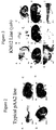
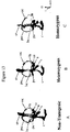
- Figure 1 A homozygous ysb mouse showing yellow coat colour compared with (B) a heterozygous littermate, agouti and (C) a black C57BL.
- Figure 2 Whole-mount X-gal staining (blue) of different pAA2 mouse embryos showing the typical LacZ expression pattern at different embryonic stages.
- A 9.0 dpc embryo;
- B 12.5 dpc embryo;
- C 13.5 dpc,
- D 13.5 dpc embryo.
- Expression sites include branchial arch (ba), notochord (no), prevertebrae (pv), snout (sn) and digits (dg).
- FIG. 3 LacZ expression as seen by X-gal staining (blue) of KM12 transgenic embryos at different embryonic stages. LacZ is expressed in sites typical for pAA2 (Fig 2) and in extra sites including rhombomeres (r) 2,3 & 5 in hindbrain (hb), neural tube (nt), spinal cord (sc) and hair follicles (hf) which are not the normal expression sites of the Col2a1 transgene.
- A whole-mount X-gal stained 9.5 dpc KM12 embryo shows LacZ expression in branchial arch (ba); heart (he); notochord (no); rhombomeres (r) 2.3 and 5; otic vesicle (ov); neural tube (nt).
- B shows sagittal section of 10.5 dpc KM12 transgenic embryo.
- C At 10.5 dpc., KM12 transgenic embryo has additional expression sites at prevertebrae (pv), forebrain (fb) and dorsal root ganglia (drg) (not shown in this view) but with the disappearance of r5 expression.
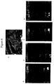
- FIG. 4 swimming test: A), a wild-type mouse - floats and swims in water; B),C),D), and E) a homozygous yellow submarine (ysb) mouse - circles and submerges in water
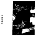
- Figure 5 Reaching response test. Mouse on left, a ysb homozygote with vestibular dyfunction, showing a tendency to curl up towards the belly when held by tail. Mouse on right, a normal reaching response, with outstretched body.
- FIG. 6 Southern blot hybridization of genomic DNA from mice heterozygous (he) and homozygous (ho) for the pAA2 transgene in the KM12 line and non-transgenic (nt) control littermates using (A) nikler, (B) agouti, and (C) ⁇ -MSHR cDNA probes.
- the structural genes of nikler, agouti and ⁇ -MSHR have not been disrupted by COL2A1-lacZ transgene in ysb mice.
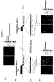
- Figure 7 The ysb locus and chromosomal localisation of the pAA2 transgene.
- A Localisation of pAA2 (cheah et al, 1995) in KM12/ysb transgenic mice to chromosome 3 by fluorescence in situ hybridization (FISH) using pAA2 as probe. The transgene maps to A2 and B-C region on metaphase ysb chromosomes.
- B Chromosomal map of transgenic integration.
- C Genomic clones isolated by PCR probes for integration sites 1 and 2.
- D Chromosomal localisation of genomic clones for integration sites 1 and 2 by FISH to wild-type metaphase chromosomes.
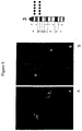
- FIG. 8 Fluorescence in situ hybridisation (FISH) of ysb chromosomes using transgene (pAA2) as probe.
- the transgene has integrated into two insertion sites in the chromosome. Arrow shows position of fluorescence signals.
- DAPI banding pattern shows that pAA2 insertion sites correspond to chromosome 3.
- C Corresponding bands that show fluorescence signal is shown by dots.
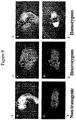
- Figure 9 Whole-mount in situ hybridization of 9.5 dpc embryos using Krox-20 riboprobe, double-stained for ⁇ -galactosidase activity (reddish). Homozygous ysb embryos (E,F) and heterozygous ysb embryos (C,D) have normal Krox-20 expression pattern in rhombomeres (r) 3 and 5 at 9.5 dpc. Sagittal view (A), and dorsal view (B) of non-transgenic littermate showing normal pattern.
- FIG. 10 Whole-mount neurofilament immunostaining of 10.5 dpc non-transgenic control (A,B), heterozygous ysb (C,D), and homozygous ysb (E,F) embryos, using monoclonal antibody (2h3), double-stained for ⁇ -galactosidase activing. A reduction of the eighth nerve (VIIIn) is observed in homozygous ysb at 10.5 dpc.
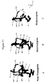
- FIG 11 3-D reconstructed images of inner ears of a)non-transgenic, b) heterozygous ysb, and c) homozygous ysb 16.5 dpc fetuses showing structural malformations in developing homozygous ysb inner ears. Note superior semicircular canal is obliterated (*) in homozygous ysb.
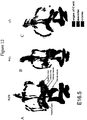
- FIG. 12 3-D reconstructed images of inner ears of 16.5 dpc. fetuses a) wild-type non-transgenic (+/+), b)heterozygous ysb (+/-) and c) homozygous ysb (-/-). Note that neuroepithelial structures are missing in developing inner ear of homozygous ysb fetuses. In 16.5 dpc homozygous ysb (-/-), superior semicircular canal is partially obliterated* and ends in a blind sac (c); the utricular macula, superior and lateral cristae are missing.
- Figure 13 3-D reconstructed images of inner ears at 13.75 dpc showing structural malformations in developing homozygous ysb inner ears. Superior semicircular canal is obliterated* in homozygous ysb.
- FIG. 15 Southern analyses of ysb genomic DNA using flanking probes at integration sites 1 and 2. Different size bands are observed in the mutant alleles when probed with flanking probes from both integration sites (shown by corresponding arrows). No segregation of the two integration sites are observed in the ysb mice studied (6 th generation). Wt, wild-type; he, heteozygous ysb; ho, homozygous ysb
- FIG. 16 Deletion in ysb at integration site 2.
- A 20 kb deletion in ysb. B-E; Autoradiograms of Southern blots of genomic DNA digested with EcoRI (B,D), SacI (C) and Xbal (E) from ysb heteozygote, homozygote and non-transgenic mice. Probes were: B), pSD14 (1.4kb EcoRI); C), pSD11 (1.9kb SacI); D), pSD9 (3.2kb BamHI) ; E) pSD8 (2.0kb Sac I) (B) Note rearrangement band is observed when pSD14 (1.4kb EcoRI) is used to probe homozygous ysb genomic DNA. *signifies homozygous ysb Note deleted sequences revealed by probes pSD11(C); pSD9 (D); and pSD8 (E)
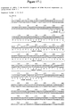
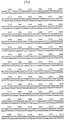
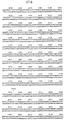
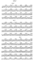
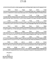
- Figure 17 Sequences information for the 8.1 kb within ysb locus deletion.
- Figure 18 Summary map of pPL5 showing location of EST-IMAGE sequences
- This invention provides an isolated nucleic acid which defines a yellow submarine locus.
- This invention provides an isolated nucleic acid which defines a mutant yellow submarine locus, wherein the mutant yellow submarine locus is identical to a wildtype yellow submarine locus except for an integration of a pAA2 transgene into at least one region on a chromosome.
- the chromosome is mouse chromosome 3.
- the pAA2 transgene is inserted into a region of mouse chromosome 3 designated A2.
- the pAA2 transgene is inserted into a region of mouse chromosome 3 designated B-C.
- the yellow submarine locus comprises a deletion of a nucleic acid segment having a sequence set forth in SEQ ID NO:1. See Figure 17.
- the yellow submarine locus comprises a deletion of a nucleic acid segment having a sequence set forth in SEQ ID NO:2. See Figure 17.
- the yellow submarine locus comprises a deletion of a nucleic acid segment having a sequence set forth in SEQ ID NO:3. See Figure 17.
- This invention provides the above isolated nucleic acid further comprising a rearrangement of chromosomal sequences of a region of the chromosome designated A3 region.
- the rearrangement comprises an inversion of a nucleotide segment.
- the isolated nucleic acid of the subject invention includes genomic DNA, RNA, cDNA.
- the nucleic acid may be labeled with a detectable marker.
- the detectable marker includes but is not limited to a radioactive, a colorimetric, a luminescent, or a fluorescent label.
- This invention provides a replicable vector comprising the above nucleic acid.
- This invention provides a host cell comprising the vector.
- the cell is a eukaryotic cell.
- the cell is a bacterial cell.
- the vectors of the subject invention include but are not limited to a plasmid, cosmid, ⁇ phage, YAC, BAC, or PAC.
- This invention provides a nucleic acid of at least 14 nucleotides capable of specifically hybridizing with the nucleic acids of the subject invention.
- the nucleic acid encodes a growth factor. In another embodiment, the nucleic acid encodes a growth factor receptor. In another embodiment, the nucleic acid encodes an orphan receptor. In another embodiment, the nucleic acid encodes a signaling molecule. In another embodiment, the nucleic acid encodes a transcriptional regulator. In another embodiment, the nucleic acid encodes an intracellular transport protein. In another embodiment, the nucleic acid encodes a neural precursor cell which is an expressed and developmentally down-regulated 4 (NEDD4) family molecule. In another embodiment, the nucleic acid encodes a SOX protein. In another embodiment, the nucleic acid encodes a regulator of apoptosis.
- NEDD4 expressed and developmentally down-regulated 4
- the nucleic acid encodes a regulator of protein turnover. In another embodiment, the nucleic acid encodes a cell cycle regulator. In another embodiment, the nucleic acid encodes a calcium binding protein. In another embodiment, the nucleic acid encodes a potentiator of hormone dependent activation of transcription by progesterone or glucocorticoid receptors. In another embodiment, the nucleic acid encodes a membrane transport protein. In another embodiment, the nucleic acid encodes a co-activator of transcription.
- This invention provides a nucleic acid, wherein the nucleic acid is isolated from a mouse.
- This invention provides an isolated nucleic acid which defines a human locus which corresponds to a yellow submarine locus.
- This invention provides an isolated nucleic acid which defines a human locus which corresponds to a mutant yellow submarine locus containing a pAA2 transgene integrated into at least one region the genome.
- This invention provides a method of diagnosing inner ear dysfunction in a subject comprising: a) obtaining a suitable sample from a subject; b) extracting nucleic acid from the sample; c) contacting the nucleic acid with the above nucleic acid which binds specifically to the mutated portion of the ysb locus; and d) detecting the labeled nucleic acid, thereby detecting mutant ysb and thereby diagnosing inner ear dysfunction in the subject.
- This invention provides a method of diagnosing pigmentation dysfunction in a subject comprising: a) obtaining a suitable sample from a subject; b) extracting nucleic acid from the sample; c) contacting the nucleic acid with the above nucleic acid which binds specifically to the mutated portion of the ysb locus; and d) detecting the labeled nucleic acid, thereby detecting mutant ysb and thereby diagnosing pigmentation dysfunction in the subject.
- This invention provides a method of diagnosing cell growth dysfunction, cell proliferation dysfunction, or cell death dysfunction in a subject comprising: a) obtaining a suitable sample from a subject; b) extracting nucleic acid from the sample; c) contacting the nucleic acid with the above nucleic acid which binds specifically to the mutated portion of the ysb locus; and d) detecting the labeled nucleic acid, thereby detecting mutant ysb and thereby diagnosing cell growth dysfunction or proliferation dysfunction in the subject.
- the subject is mammal. In another embodiment, the subject is a non-mammal.
- the subject mat be a human, a primate, an equine, an opine, an avian, a bovine, a porcine, a canine, a feline or a murine.
- the subject may be a vertebrate.
- the invention provides a polypeptide encodes by the nucleic acid of the subject invention.
- This invention provides a fusion protein comprising the above polypeptide.
- the polypeptide is labeled with a detectable marker.
- the invention provides a method of repairing and regenerating nerve tissue in a subject comprising administering an effective amount of the above protein to the subject, wherein the protein is a wildtype protein, so as to thereby repair and regenerate nerve tissue in the subject.
- the invention provides a method of regulating cell migration in a subject comprising administering an effective amount of the above protein to the subject, wherein the protein is a wildtype protein, so as to thereby regulate cell migration in the subject.
- the invention provides a method of regulating cell growth in a subject comprising administering an effective amount of the above protein to the subject, wherein the protein is a wildtype protein, so as to thereby regulate cell growth in the subject.
- the invention provides an antibody which binds to the polypeptide of the subject invention.
- the antibody is a monoclonal antibody.
- the antibody is a polyclonal antibody.
- the antibody is conjugated to a cytotoxic agent.
- the antibody is labeled with a detectable marker.
- the invention provides a composition comprising the antibody of the subject invention.
- the invention provides a method of producing a protein encoded by a nucleic acid in a wildtype ysb locus which comprises growing a host vector system under suitable conditions permitting production of the polypeptide and recovering the polypeptide so produced.
- the invention provides a method determining vestibular dysfunction in a subject comprising: a) obtaining a suitable sample from a subject; b) extracting nucleic acid from the sample; c) contacting the nucleic acid with the above nucleic acid which binds specifically to a mutated portion of the ysb locus; d) detecting the labeled nucleic acid, thereby detecting gene responsible for yellow coat color, thereby indicating the presence of vestibular dysfunction in the subject.
- the invention provides a method determining vestibular dysfunction using embryonal stem cells: comprising a) obtaining a suitable sample from the embryonal stem cells; b)extracting nucleic acid from the sample; c) contacting the nucleic acid with the above nucleic acid which binds specifically to a mutated portion of the ysb locus; d) detecting the labeled nucleic acid, thereby detecting gene responsible for yellow coat color, thereby indicating the presence of vestibular dysfunction in the embryonal stem cells.
- the invention provides a method of determining successful deletion or inactivation of a gene which comprises examining coat color or pigmentation of a subject, wherein yellow coat color or pigmentation indicates that a normal gene has ben inactivated, thereby resulting in yellow coat color or pigmentation, thereby indicating the successful deletion or inactivation of the gene.
- the invention provides a method of determining successful deletion or inactivation of a gene comprising determining whether repairing the abnormal gene results in the disappearance of the yellow coat color or pigmentation.
- the invention provides a method of treating vestibular dysfunction in a subject comprising introducing a nucleic acid comprising a gene or genes from the wildtype locus encoding a ysb into a suitable cell under conditions such that the nucleic acid expresses ysb so as to thereby treat vestibular dysfunction.
- the invention provides a method of treating hearing impairment in a subject which comprises introducing a nucleic acid comprising a gene or genes from the wild-type locus into a suitable cell under conditionssuch that the nucleic acid expresses ysb so as to thereby treat hearing loss.
- the invention provides a transgenic mouse line designated KM12.
- the invention provides a method of determining whether a compound is mutagenic comprising: (a) examining the coat color or pigmentation of a subject; b) administering the compound to the subject; (c) examining the coat color or pigmentation of a subject; (d) comparing the result obtained in step (c) with the result obtained in step (a); and (e) determining if the coat color or pigmentation of the subject is yellow so as to thereby determine whether the compound is mutagenic.
- mice A mouse line (KM12) bearing a recessive mutation, caused by insertion of a transgene was created. These mice are characterized by a change in coat colour to yellow and circular behaviour. (Figure 1)
- mice The yellow coat of KM12 mice suggests abnormal regulation of pigment synthesis. Hair pigmentation in mice is mediated by many developmental and signalling processes involving enzymes, transcription factors, a growth factor and its receptor, a membrane transport protein, a hormone receptor and an antagonist of hormone binding.
- wild-type colour hair is agouti and contains two pigments in which the bases and tips contain the black pigment eumelanin and an intermediate band containing phaeomelanin.
- Two genes in mice, agouti ( A ) on chromosome 2 and extension ( E ) on chromosome 8 are involved in the regulation of the relative amounts of eumelanin and phaeomelanin in the mouse. Mutations in these two loci result in a yellow coat colour in mice.
- a gene encodes a 131 amino acid polypeptide with a structure consistent with its proposed paracrine function.
- E encodes the (-MSH receptor (MSH-R). It has been established that neither A nor E have been mutated in KM12 mice.
- the transgene has been mapped to two integration sites on mouse chromosome 3 ( Figure 7), consistent with a mutation outside the A and E loci.
- the circular behaviour of KM12 mice suggests improper inner ear function which affects balance.
- the mature inner ear consists of the auditory apparatus, which is responsible for the perception of sound (organ of Corti) and the vestibular apparatus which is responsible for the sense of balance.
- Many genes have been found to regulate the development and functions of the inner ear and mutations which cause hearing and balance defects have been found in both human and mouse. These genes encode diverse classes of proteins such as transcription factors, secreted growth factors, signalling molecules, receptors, cytoskeletal components, intracellular transporters, and ion channel proteins.
- pigmentation defects associated with deafness have been found (such as micropthalmia, dilute) to date no mutations causing both yellow coat and inner ear defects have been identified in mice.
- mutant locus has been named yellow submarine (ysb).
- Molecular genetics, bioinformatics, developmental biology, transgenic and physiological approaches are used to identify the gene(s) at the + ysb locus and study the molecular and developmental bases underlying the defect(s) in ysb mice.
- the following methods are used : 1) isolation and characterization of the + ysb gene(s); 2) determination of the nature of the ysb mutation; 3) performing genetic complementation tests between ysb and lcc mice, 4) performing transgenic rescue experiments, 5) characterization of the developmental defects in the inner ears of ysb mice using 3-D reconstruction analyses, molecular markers and chimera studies; 6) characterization of the neurophysiological changes in balance and hearing in ysb mice.
- transgenic mice In making transgenic mice sometimes integration of the exogenous DNA disrupts the function of one or more genes. While studying the regulation of the COL2A1 gene, using a recombinant plasmid (pAA2) which contained regulatory DNA sequences from COL2A1 linked to the lacZ reporter gene (Cheah et al. 1995), a recessive mutation, caused by insertion of the transgene, was created. These mice are characterized by a change in coat colour to yellow and circular behaviour, only seen in offspring homozygous for the transgene (Fig. 1, Appendix).
- pAA2A1 recombinant plasmid
- the transgene is expressed not only in the sites expected for COL2A1 but also in additional expression domains such as specific rhombomeres of the hindbrain (r2,3,5), the spinal cord, dorsal root ganglia, and in the hair follicles of the skin.
- additional expression domains such as specific rhombomeres of the hindbrain (r2,3,5), the spinal cord, dorsal root ganglia, and in the hair follicles of the skin.
- These neural and skin sites of expression are not typical of the endogenous Co12a-1 gene but are consistent with the coat colour and behavioural phenotype of KM12 mice ( Figures 2,3).
- mice The yellow coat of KM12 mice suggests abnormal regulation of pigment synthesis.
- wild-type color hair is agouti and contains two pigments in which the bases and tips contain the black pigment eumelanin and an intermediate band containing phaeomelanin.
- the regulation of hair pigmentation in mice is mediated by many developmental and signalling processes involving several enzymes, transcription factors, a growth factor and its receptor, a membrane transport protein, a G protein-coupled hormone receptor and an antagonist of hormone binding. These pigments are synthesized in melanocytes by tyrosinase (reviewed in ) .
- mice agouti ( A ) on chromosome 2 and extension ( E ) on chromosome 8 are involved in the regulation of the relative amounts of eumelanin and phaeomelanin in the mouse. Mutations in these two loci result in a yellow coat colour in mice. Dominant mutations in the A gene, such as lethal yellow and viable yellow, result in an obese mouse which is completely or almost all yellow because only phaeomelanin is made. In contrast mutations in E which result in yellow coat colour ( e ) are recessive. Both the A and E loci have been cloned. The A gene encodes a 131 amino acid polypeptide with a structure consistent with its proposed paracrine function.
- E encodes the (-MSH receptor (MSH-R), a 35kD polypeptide with seven transmembrane domains and is expressed in melanocytes. Activation of MSH-R promotes eumelanin synthesis while agouti protein enhances phaeomelanin synthesis .
- KM12 mice Many deaf mouse mutants are characterized by the classic shaker/waltzer behaviour of circling, head tossing and hyperactivity.
- the circular behavior of KM12 mice suggests improper inner ear function which affects balance and hearing.
- KM12 mice also show other characteristics of inner ear defects such as head-tossing, and an inability to swim (Figure 4).
- KM12 mice show an abnormal reaching response. When picked up by the tail, KM12 mice do not stretch out their limbs as normal mice do, but rather curl up towards their bellies (Fig.5, Appendix). Although KM12 mice show some response to sharp sounds (Preyer reflex), this does not appear as strong as for wild-type mice. Therefore it is possible that KM12 mice are partially hearing impaired.
- the inner ear is a complex sensory organ which develops from a single-cell-layered epithelium (otic placode) which invaginates by a series of cell and tissue movements and closes to form the otic vesicle. Further morphogenetic movements and multi-step inductive events, which regulate differentiation and proliferation, lead to the formation of the organ for hearing and balance.
- the mature inner ear consists of the auditory apparatus, which is responsible for the perception of sound (organ of Corti) and the vestibular apparatus which is responsible for the sense of balance. Many genes have been shown to be expressed in the developing inner ear. These include genes encoding transcription factors (e.g.
- secreted factors e.g. Bmp4, fgf3, fgf2, bdnf
- receptors e.g. EphA4, trkA, trkB, trkC, Ednrb, PTHrpR
- cytoskeletal proteins such as unconventional myosins (e.g. Myo7a, myo15 ).
- Inner ear defects have been traced to one or more of three types of abnormalities: morphogenetic, cochleo-saccular, and neuroepithelial.
- Morphogenetic defects are caused by developmental abnormalities in structure of the inner ear (labyrinth).
- Cochleo-saccular abnormalities result from defects in the secretory epithelium of the cochlear duct.
- Neuroepithelial defects arise from failure of the sensory epithelia to complete normal maturation.
- abnormal hindbrain development may result in vestibular dysfunction.
- nikler mice are characterized by deafness and circular behaviour in adults, and in the embryo, by abnormalities in the positioning of the otic vesicles and segmentation of the hindbrain.
- mice are good models for the studying auditory defects because of the similarity in structure and development between mouse and human inner ears. Mutations in several genes expressed in the inner ear have been shown to cause hearing and/or balance disorders in human and mouse. These include Myo7a, Myo15, Vietnameseler, hmx3 (nkx5.1), fgfr3 and others.
- the following methods are performed: a) characterize abnormalities in hindbrain and ear development in KM12 mice and b) isolate the DNA sequences at the site(s) of integration of the transgene in KM12 mice.
- the recessive nature of the mutant phenotype suggests that the transgene has not integrated into the A gene.
- the chromosomal assignment of the KM12 transgene is an essential and informative first step in defining the nature of the mutation.
- the transgene has been mapped by FISH to two integration sites on mouse chromosome 3, consistent with the Southern analyses ( Figure 7,8).
- the data are also consistent with the results excluding the A, E, Kreisler loci.
- the mutant locus has been named yellow submarine (ysb) (and homozygous mutants hereafter referred to as ysb, wild-type as + ysb ; Leung, K.K, S. Dong, A. Tang, H. Heng, L.C. Tsui, P.P.L. Tam & K.S.E. Cheah Yellow submarine (ysb) a newly discovered locus regulating hair colour and inner ear function. Manuscript in preparation)
- Ysb mice are a valuable model to identify molecules involved in controlling pigmentation and balance.
- the structural abnormalities in ysb mice have been analyzed.
- the phenotype and pattern of expression of the transgene in ysb mice indicate that integration of the transgene has caused a recessive mutation which affects the regulation of pigmentation and also causes abnormal inner ear development.
- the initial studies have focused on the inner ear abnomalities in ysb mice.
- Krox-20 is a useful marker for hindbrain segmentation being normally expressed in r3 and 5 at 9.0-9.5 days, and lacZ expression was found in r3 of ysb embryos.
- lacZ expression was found in r3 of ysb embryos.
- the pattern of expression of Krox-20 was studied in heterozygote and homozygote 9.0-9.5 day embryos by in situ hybrization in whole mount and on sections and compared to that for wild-type.
- Whole-mount in situ hybridization showed no alterations in the rhombomere expression of Krox-20 mRNA in homozygotes and heterozygotes at 9.5 dpc ( Figure 9). This is unlike the Kreisler mutant suggesting that the mutation has not caused gross alterations in rhomomeres 3 and 5.
- 3-D reconstructions of the inner ear of the mice were generated using histological serial sections. 3D reconstructions of images of sections of the ears of wild-type, heterozygous and homozygous ysb fetuses at 16.5 dpc (days post coitum) were carried out. This was followed by painting in the lumen to give an impression of the whole structure of the labyrinth. The data show superior and lateral canal and ampulla defects in two ysb fetuses, with one more severely affected than the other (Appendix, Fig.11).
- the different types of sensory areas including the organ of Corti (detector of sound), the maculae (detectors of linear motion), and the cristae (detectors of rotational motion), are represented in different colours to demonstrate more easily which sensory areas were affected.
- Fig. 7 summarizes the progress in isolating the wild-type locus at the site of transgene integration.
- inverse-PCR was used to isolate DNA sequences flanking the integration sites (Fig 7).
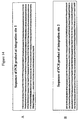
- Sequence analysis ( Figure 14) showed the presence of a possible polyA attachment signal in SpeI-1 but a BLAST search of the genomic and EST databases did not reveal significant matches. Longer 5' flanking sequence have been obtained for integration site 1 (1.8kb) and 3' flanking sequence for site 2 (500bp).
- the ysb gene(s) are being identified and characterized, and the molecular and developmental bases underlying the defect(s) in ysb mice are being studied.
- the current results are being built on, and molecular genetics, bioinformatics, developmental biology, transgenic and physiological approaches are used to a) identify and characterise the + ysb gene(s) and the encoded transcripts; b) determine the nature of the ysb mutation; c) perform genetic complementation tests and transgenic rescue experiments, d) further characterise the developmental defects in the inner ears of ysb mice using 3-D reconstruction analyses, molecular markers and chimera studies; e) characterise neurophysiological changes in balance and hearing.
- a.Bioinformatics The isolated genomic clones are sequenced and the data analysed for the presence of potential exons using bioinormatics tools. These predictions are very important for identifying regions to follow up.
- b.Expressed gene sequences are identified using a combination of approaches. First DNA fragments containing exon sequences are screened for by Northern analyses. Once such fragments are identified, exon trapping approaches are used to identify regions with transcribed sequences. Potential exon containing fragments are used in in situ hybridization studies and the pattern of expression compared with that of the transgene in ysb mice .
- bioinformatic tools to screen for homologous sequences in other model genome databases e.g. yeast, fly, worm, fish and human.
- cosmid genomic library will be constructed from DNA isolated from homozygous ysb mice.
- the cosmid, pCos8 (gift of Dr. A-M Frischauf, Imperial Cancer Research Fund, London), is used as a vector in constructing the library. This vector does not contain lacZ sequences and its use helps minimize isolating false positives during library screening.
- Primary embryo fibroblast cultures from KM12 homozygote mice are established and used as a source of high molecular weight genomic DNA.
- the cosmid library is constructed by standard methods. To ensure high efficiency of cosmid packaging, tested packaging mixes (e.g. Stratagene's Gigapack Gold) are obtained from commercial sources. DNA probes covering the 5 and 3' ends of the transgene and lacZ sequences are used to screen the library by colony hybridization. Cosmid libraries and isolated genes have been successfully constructed in the past.
- Cosmids are further characterized for the presence of the transgene by restriction enzyme mapping and Southern blot analyses using the appropriate probes. Once cosmid(s) containing the transgene are identified, subclones are made and the nature of the sequences flanking both 5' and 3' ends of the insertion site are determined by DNA sequencing. Sequences obtained are then analyzed using bioinformatic tools to find potential open reading frames (ORFs) and also are used to scan the nucleotide and amino acid sequence databases for homologous sequences. The ysb clones are used to analyze genomic DNA from the Harwell mutants. To determine whether the ysb and Harwell mutants are allelic, a complementation test is carried out by crossing the two mutants.
- ORFs open reading frames
- both mutants involve two possible mutation sites and non-complementation will immediately cut down the number of sites to be investigated at a molecular level, as there is only one chromosomal region of overlap between the two mutations.
- BACs in transgenic mice have been successfully used to correct deafness in shaker-2 mice.
- sequences characterized are those which have been mutated in ysb
- bacterial artificial chromosome clones will be screened for, using the genomic clones for integration site 2 as probe.
- BAC clones spanning the ysb locus will rescue the phenotype of ysb mice by transgenesis. It would also be important to compare the ysb locus with that of the Harwell mutant.
- the aim is to be able to test if any human vestibular disorder is caused by mutations in the human homologue of of ysb. Therefore it is important to isolate human BAC clones which cover the equivalent ysb locus.
- the mouse genomic clones are used to screen a human BAC library. Once these are isolated they are characterized as for the mouse clones in terms of sequence etc. Comparison is made between the human and mouse clones. Once candidate human clones are isolated, their chromosomal locations are mapped and the mutant databases (e.g. OMIM- Online Mendelian Inheritance in Man) scanned for possible associated human disorders.
- the availability of human clones is a resource for future studies on human patients with hearing and balance problems.
- the morphology of the ysb embryonic ears at 11, 13 (a critical time point in canal formation) and 16.5 dpc are carefully dissected out, cleared and the lumen filled with white paint and then examined for abnormality.
- This technique provides a quick and accurate visualization of the 3-dimensional structure of the ear to assess variability in the homozygote phenotype and detect subtle defects in heterozygotes.
- the otoconia in the extracellular matrix, which lies atop the hair cells of the maculae are easily observed after clearing, before paint injection. Whether this matrix forms normally in the mutant is studied, since the maculae appear abnormal at 16.5dpc.
- the inner ears at 18dpc are also examined by scanning electron microscopy to determine if there are any abnormalities in the distribution and structure of the sensory cells.
- Nkx5.1 is expressed in the dorso-lateral portion of the otocyst (and later in the semi-circular canals) and inactivation of the gene affects the semicircular canals.
- Otx-1 is expressed postereo-laterally in the otocyst and the lateral semicircular canal is missing when the gene is "knocked out" in mice.
- neurotrophic factors e.g. BDNF and NT-3
- Trkb, Trkc neurotrophic factors
- BDNF and NT-3 neurotrophic factors
- Trkb, Trkc their receptors
- the neurogeninl gene has recently been shown to be essential for the determination of neuronal precursors for proximal cranial sensory ganglia and will be a good marker for studying the prospective ganglion cells in ysb mice. The same markers are used to compare the Harwell and ysb mutants should the complementation tests prove them to be allelic (see above).
- Embryonal stem cells are derived from ysb blastocysts and used to generate chimeras by blastocyst injection. Chimeric embryos analyzed (5-10 per stage) are collected at stages of development between 9.5days and birth and the relative contribution of ES cells in the developing inner ear as judged by lacZ expression in the chimeras is studied. The preferential loss or under-representation of ysb ES cell contribution in particular sites in the developing inner ear is indicative of specific changes in lineage potency. The lineages are characterised by in situ hybridizations using the appropriate molecular markers described above.
- the neurons that innervate the inner ear are derived from the otic placode. These neurons arise from the placode early in development of the inner ear to form the acoustic and vestibular ganglia.
- the neurofilament staining experiments had shown a reduction of the eighth cranial nerve in 10.5 days ysb embryos, suggesting failure to innervate the inner ear could be a cause of the balance and/or hearing problem in ysb mice. It is important to assess whether or not the ysb mice can effectively convey (a) head movement signals and (b) auditory signals to the central nervous system.
- the expression of c-fos is used as an indicator of functionally activated neuronal activity in the brainstem.
- Fos expression as an indicator of postsynaptic stimulation is an established method for identifying functional connections between peripheral sensory receptors and central neurons.
- Fos immunostaining of brain sections is therefore used to map the central neurons involved in the functional neural pathway.
- Natural vestibular stimulations are used, viz. sinusoidal rotations on the yaw or pitch plane and constant velocity off-vertical axis rotations.
- the former stimulates the respective pairs of semicircular canals while the latter selectively activates the utricular hair cells in a sequential manner.
- secondary neurons in the vestibular nucleus that have functional connection with hair cells on the respective canal pairs or the utricualr maculae should be excited and should express Fos.
- Immunohistochemical techniques involving c-fos are used to map the pattern of postsynaptic vestibular stimulation within the vestibular nuclei and other parts of the brainstem.
- brain sections are examined for neurons where Fos immunostaining and retrogradely transported rhodamine-labeled latex beads (previously injected in the spinal cord) are co-localized.
- the auditory pathway is activated by repetitive stimulation paradigms using tone bursts.
- the sound stimulation is conducted in a darkened soundproof chamber.
- Each freely moving mouse will be presented with pure tone bursts delivered from the ceiling of the chamber.
- Functional connection between central neurons and the peripheral auditory receptors is indicated by Fos expression.
- the following studies complement the neurophysiological studies and greatly enhance the scope of the investigation, facilitating the definition of the underlying defect(s) in ysb mice.
- ysb mice Using the ysb mice, a breeding colony is established to provide mutants for study.
- the behavioural consequences of the vestibular defects are described using a standard battery of simple tests of balance, including, contact righting response, elevated platform test, and open field test, reaching response, Preyer's reflex, and quantify the extent and type of the behavioural abnormality.
- the function of the cochlea in young adult ysb and littermate controls is by measuring thresholds for detection of a compound action potential from the cochlea in response to tonebursts at various frequencies and intensities. This approach gives an indication of any hearing impairment in the mutant.
- the molecular cloning and characterization of the potential ysb clones is performed. 3-D reconstructions on ysb, heterozygous and wild-type 11day fetuses and newborns is performed. In situ hybridization experiments are performed. The detailed restriction map of the genomic clones isolated for the ysb locus is made and then clones are tested for the presence of expressed sequences by Northern analyses. Bioinformatics is used to analyse the DNA sequences that are obtained.
- Robertson, E.J. in Teratocarcinomas and embryonic stem cells ed Robertson, E.J. 71-112 ( Oxford IRL press, Oxford, 1987).
Landscapes
- Health & Medical Sciences (AREA)
- Chemical & Material Sciences (AREA)
- Organic Chemistry (AREA)
- Life Sciences & Earth Sciences (AREA)
- General Health & Medical Sciences (AREA)
- Medicinal Chemistry (AREA)
- Toxicology (AREA)
- Zoology (AREA)
- Chemical Kinetics & Catalysis (AREA)
- General Chemical & Material Sciences (AREA)
- Bioinformatics & Cheminformatics (AREA)
- Animal Behavior & Ethology (AREA)
- Engineering & Computer Science (AREA)
- Public Health (AREA)
- Nuclear Medicine, Radiotherapy & Molecular Imaging (AREA)
- Veterinary Medicine (AREA)
- Pharmacology & Pharmacy (AREA)
- Gastroenterology & Hepatology (AREA)
- Biochemistry (AREA)
- Biophysics (AREA)
- Genetics & Genomics (AREA)
- Molecular Biology (AREA)
- Proteomics, Peptides & Aminoacids (AREA)
- Measuring Or Testing Involving Enzymes Or Micro-Organisms (AREA)
- Micro-Organisms Or Cultivation Processes Thereof (AREA)
Abstract
Description
- This application is a continuation-in-part application of U.S. Serial No. 09/274,634, filed March 23, 1999, claiming the benefit of U.S. Provisional Application No. 60/079,020, filed March 23, 1998, the contents of which are hereby incorporated by reference into this application.
- A mouse line bearing a recessive mutation, caused by insertion of a transgene, was created. These mice are characterized by a changed pigmentation of hair to give yellow coat color, circling behaviour and an inability to swim. It has been shown that the transgene has inserted into two sites on
mouse chromosome 3. A search of the mouse genome database ascertained that the mutation is novel. The mutant locus has been named yellow submarine (ysb). DNA flanking the transgene insertion has been isolated and used to screen for the corresponding normal (unmutated) sequences. Genomic clones spanning 20kb of the unmutated ysb locus (+ ysb ) have been isolated and mapping experiments indicate that transgene integration has caused a deletion and chromosomal inversion resulting in two transgene integration sites. Circular behaviour can reflect abnormality in the inner ear or be due to an abnormality in hindbrain development. Studies show abnormal structure of the inner ears of ysb mice and a stunted acoustic nerve. Ysb mice are therefore a novel mutant showing both abnormal regulation of pigmentation and inner ear dysfunction. Molecular genetics, bioinformatics, developmental biology, transgenic and physiological approaches are used to 1) identify and characterize the gene(s) at the wild-type ysb (+ ysb ) locus; 2) determine the nature of the ysb mutation; 3) study the molecular and developmental bases underlying the defect(s) in ysb mice; and 4) charactersize the neurophysiological changes in balance and hearing in ysb mice. Approximately 1/1000 infants are affected by hearing defects at birth, two thirds of which have a genetic basis. Ysb mice are a valuable model to identify molecules involved in controlling balance. These studies provide fundamental information on the development of the inner ear and the mechanisms by which hearing and balance defects may arise. Identification and characterization of the ysb gene(s) and the molecular defect in ysb mice also yield new insight into the complex regulatory pathways controlling agouti coat color in mice. - This invention provides an isolated nucleic acid which defines a yellow submarine locus.
- This invention provides an isolated nucleic acid which defines a mutant yellow submarine locus, wherein the mutant yellow submarine locus is identical to a wildtype yellow submarine locus except for an integration of a pAA2 transgene into at least one region on a chromosome.
- This invention provides the above isolated nucleic acid further comprising a rearrangement of chromosomal sequences of a region of the chromosome designated A3 region.
- This invention provides a replicable vector comprising the above nucleic acid. This invention provides a host cell comprising the vector.
- This invention provides a nucleic acid of at least 14 nucleotides capable of specifically hybridizing with the nucleic acids of the subject invention.
- This invention provides an isolated nucleic acid which defines a human locus which corresponds to a yellow submarine locus. This invention provides an isolated nucleic acid which defines a human locus which corresponds to a mutant yellow submarine locus containing a pAA2 transgene integrated into at least one region the genome.
- This invention provides a method of diagnosing inner ear dysfunction in a subject comprising: a) obtaining a suitable sample from a subject; b) extracting nucleic acid from the sample; c) contacting the nucleic acid with the above nucleic acid which binds specifically to the mutated portion of the ysb locus; and d) detecting the labeled nucleic acid, thereby detecting mutant ysb and thereby diagnosing inner ear dysfunction in the subject.
- This invention provides a method of diagnosing pigmentation dysfunction in a subject comprising: a) obtaining a suitable sample from a subject; b) extracting nucleic acid from the sample; c) contacting the nucleic acid with the above nucleic acid which binds specifically to the mutated portion of the ysb locus; and d) detecting the labeled nucleic acid, thereby detecting mutant ysb and thereby diagnosing pigmentation dysfunction in the subject.
- This invention provides a method of diagnosing cell growth dysfunction, cell proliferation dysfunction, or cell death dysfunction in a subject comprising: a) obtaining a suitable sample from a subject; b) extracting nucleic acid from the sample; c) contacting the nucleic acid with the above nucleic acid which binds specifically to the mutated portion of the ysb locus; and d) detecting the labeled nucleic acid, thereby detecting mutant ysb and thereby diagnosing cell growth dysfunction or proliferation dysfunction in the subject.
- The invention provides a polypeptide encodes by the nucleic acid of the subject invention.
- The invention provides a method of repairing and regenerating nerve tissue in a subject comprising administering an effective amount of the above protein to the subject, wherein the protein is a wildtype protein, so as to thereby repair and regenerate nerve tissue in the subject.
- The invention provides a method of regulating cell migration in a subject comprising administering an effective amount of the above protein to the subject, wherein the protein is a wildtype protein, so as to thereby regulate cell migration in the subject.
- The invention provides a method of regulating cell growth in a subject comprising administering an effective amount of the above protein to the subject, wherein the protein is a wildtype protein, so as to thereby regulate cell growth in the subject.
- The invention provides an antibody which binds to the polypeptide of the subject invention. The invention provides a composition comprising the antibody of the subject invention.
- The invention provides a method of producing a protein encoded by a nucleic acid in a wildtype ysb locus which comprises growing a host vector system under suitable conditions permitting production of the polypeptide and recovering the polypeptide so produced.
- The invention provides a method determining vestibular dysfunction in a subject comprising: a) obtaining a suitable sample from a subject; b) extracting nucleic acid from the sample; c) contacting the nucleic acid with the above nucleic acid which binds specifically to a mutated portion of the ysb locus; d) detecting the labeled nucleic acid, thereby detecting gene responsible for yellow coat color, thereby indicating the presence of vestibular dysfunction in the subject.
- The invention provides a method determining vestibular dysfunction using embryonal stem cells: comprising a) obtaining a suitable sample from the embryonal stem cells; b)extracting nucleic acid from the sample; c) contacting the nucleic acid with the above nucleic acid which binds specifically to a mutated portion of the ysb locus; d) detecting the labeled nucleic acid, thereby detecting gene responsible for yellow coat color, thereby indicating the presence of vestibular dysfunction in the embryonal stem cells.
- The invention provides a method of determining successful deletion or inactivation of a gene which comprises examining coat color or pigmentation of a subject, wherein yellow coat color or pigmentation indicates that a normal gene has ben inactivated, thereby resulting in yellow coat color or pigmentation, thereby indicating the successful deletion or inactivation of the gene.
- The invention provides a method of determining successful deletion or inactivation of a gene comprising determining whether repairing the abnormal gene results in the disappearance of the yellow coat color or pigmentation.
- The invention provides a method of treating vestibular dysfunction in a subject comprising introducing a nucleic acid comprising a gene or genes from the wildtype locus encoding a ysb into a suitable cell under conditions such that the nucleic acid expresses ysb so as to thereby treat vestibular dysfunction.
- The invention provides a method of treating hearing impairment in a subject which comprises introducing a nucleic acid comprising a gene or genes from the wild-type locus into a suitable cell under conditionssuch that the nucleic acid expresses ysb so as to thereby treat hearing loss.
- The invention provides a transgenic mouse line designated KM12.
- The invention provides a method of determining whether a compound is mutagenic comprising:(a) examining the coat color or pigmentation of a subject; b) administering the compound to the subject; (c) examining the coat color or pigmentation of a subject; (d) comparing the result obtained in step (c) with the result obtained in step (a); and (e)determining if the coat color or pigmentation of the subject is yellow so as to thereby determine whether the compound is mutagenic.
- Figure 1
(A) A homozygous ysb mouse showing yellow coat colour
compared with (B) a heterozygous littermate, agouti and (C) a black C57BL. - Figure 2
Whole-mount X-gal staining (blue) of different pAA2 mouse embryos showing the typical LacZ expression pattern at different embryonic stages.
(A) 9.0 dpc embryo; (B) 12.5 dpc embryo; (C)13.5 dpc, (D), 13.5 dpc embryo. Expression sites include branchial arch (ba), notochord (no), prevertebrae (pv), snout (sn) and digits (dg). - Figure 3
LacZ expression as seen by X-gal staining (blue) of KM12 transgenic embryos at different embryonic stages. LacZ is expressed in sites typical for pAA2 (Fig 2) and in extra sites including rhombomeres (r) 2,3 & 5 in hindbrain (hb), neural tube (nt), spinal cord (sc) and hair follicles (hf) which are not the normal expression sites of the Col2a1 transgene. (A), whole-mount X-gal stained 9.5 dpc KM12 embryo shows LacZ expression in branchial arch (ba); heart (he); notochord (no); rhombomeres (r) 2.3 and 5; otic vesicle (ov); neural tube (nt). (B) shows sagittal section of 10.5 dpc KM12 transgenic embryo. (C), At 10.5 dpc., KM12 transgenic embryo has additional expression sites at prevertebrae (pv), forebrain (fb) and dorsal root ganglia (drg) (not shown in this view) but with the disappearance of r5 expression. (D) expression pattern of 12.5 dpc fetus, at this stage staining is also in midbrain (mb), hindbrain (hb), spinal cord (sc). In (E), LacZ expression in 13.5 dpc embryos is also seen in snout (sn), digits (dg) and ribs (rb). (F) At 16.5 dpc, expression in the brain and vertebrate continue to be seen in sagittally-halved fetus. Expression is seen in hair follicles in whole-mount (G), and sectioned (H) 16.5 embryos, this continues to be present in 3.5 days postnatal skin which is whole-mount x-gal stained and cleared (I). - Figure 4
Swimming test: A), a wild-type mouse - floats and swims in water; B),C),D), and E) a homozygous yellow submarine (ysb) mouse - circles and submerges in water - Figure 5
Reaching response test. Mouse on left, a ysb homozygote with vestibular dyfunction, showing a tendency to curl up towards the belly when held by tail. Mouse on right, a normal reaching response, with outstretched body. - Figure 6
Southern blot hybridization of genomic DNA from mice heterozygous (he) and homozygous (ho) for the pAA2 transgene in the KM12 line and non-transgenic (nt) control littermates using (A) kreisler, (B) agouti, and (C) α-MSHR cDNA probes. The structural genes of kreisler, agouti and α-MSHR have not been disrupted by COL2A1-lacZ transgene in ysb mice. - Figure 7
The ysb locus and chromosomal localisation of the pAA2 transgene. (A) Localisation of pAA2 (cheah et al, 1995) in KM12/ysb transgenic mice tochromosome 3 by fluorescence in situ hybridization (FISH) using pAA2 as probe. The transgene maps to A2 and B-C region on metaphase ysb chromosomes.(B) Chromosomal map of transgenic integration.(C) Genomic clones isolated by PCR probes forintegration sites integration sites chromosomes 3. Reference: K.S.E. Cheah, A Levy, P.A. Trainor, A.W.K. Wai, T. Kuffner, C.L. So, K.K.H. Leung, R.H. Lovell-Badge and P.P.L. Tam. (1995) Human COL2A1-directed SV40 T-antigen expression in transgenic and chimeric mice results in abnormal skeletal development. J.Cell Biol. 128 223-237. - Figure 8
Fluorescence in situ hybridisation (FISH) of ysb chromosomes using transgene (pAA2) as probe. (A) The transgene has integrated into two insertion sites in the chromosome. Arrow shows position of fluorescence signals.(B) DAPI banding pattern shows that pAA2 insertion sites correspond tochromosome 3. (C) Corresponding bands that show fluorescence signal is shown by dots. - Figure 9
Whole-mount in situ hybridization of 9.5 dpc embryos using Krox-20 riboprobe, double-stained for β-galactosidase activity (reddish). Homozygous ysb embryos (E,F) and heterozygous ysb embryos (C,D) have normal Krox-20 expression pattern in rhombomeres (r) 3 and 5 at 9.5 dpc. Sagittal view (A), and dorsal view (B) of non-transgenic littermate showing normal pattern. - Figure 10
Whole-mount neurofilament immunostaining of 10.5 dpc non-transgenic control (A,B), heterozygous ysb (C,D), and homozygous ysb (E,F) embryos, using monoclonal antibody (2h3), double-stained for β-galactosidase activing. A reduction of the eighth nerve (VIIIn) is observed in homozygous ysb at 10.5 dpc. - Figure 11
3-D reconstructed images of inner ears of a)non-transgenic, b) heterozygous ysb, and c) homozygous ysb 16.5 dpc fetuses showing structural malformations in developing homozygous ysb inner ears. Note superior semicircular canal is obliterated (*) in homozygous ysb. - Figure 12
3-D reconstructed images of inner ears of 16.5 dpc. fetuses a) wild-type non-transgenic (+/+), b)heterozygous ysb (+/-) and c) homozygous ysb (-/-). Note that neuroepithelial structures are missing in developing inner ear of homozygous ysb fetuses. In 16.5 dpc homozygous ysb (-/-), superior semicircular canal is partially obliterated* and ends in a blind sac (c); the utricular macula, superior and lateral cristae are missing. - Figure 13
3-D reconstructed images of inner ears at 13.75 dpc showing structural malformations in developing homozygous ysb inner ears. Superior semicircular canal is obliterated* in homozygous ysb. - Figure 14
- A) Sequence of PCR product at
integration site 1 - B) Sequence of PCR product at
integration site 2 -
- Figure 15
Southern analyses of ysb genomic DNA using flanking probes atintegration sites - Figure 16
Deletion in ysb atintegration site 2.
(A) 20 kb deletion in ysb. B-E; Autoradiograms of Southern blots of genomic DNA digested with EcoRI (B,D), SacI (C) and Xbal (E) from ysb heteozygote, homozygote and non-transgenic mice. Probes were: B), pSD14 (1.4kb EcoRI); C), pSD11 (1.9kb SacI); D), pSD9 (3.2kb BamHI) ; E) pSD8 (2.0kb Sac I)
(B) Note rearrangement band is observed when pSD14 (1.4kb EcoRI) is used to probe homozygous ysb genomic DNA.
*signifies homozygous ysb
Note deleted sequences revealed by probes pSD11(C); pSD9 (D); and pSD8 (E) - Figure 17
Sequences information for the 8.1 kb within ysb locus deletion.
Locus:chromosome 3
Definition: pPL5
Keywords: genomic DNA
Source: house mouse
Organism: Mus Musculus
Vector: pBluescript
Inset: 8114 bp
Insertion sites: Hind III
Information: - A. Sequence of pPL5 containing 8114 bp DNA fragment (Hind
III cut) of 20 kb deleted sequences in
integration site 2 - B. Sequence alignment showing pPL5 (1743 bp-1906 bp)
contains
sequences 90% identity to IMAGE 1196866 - C. Sequence alignment showing pPL5 (106-395 bp) contains sequences with 100% identity to IMAGE 636095
-
- Figure 18
Summary map of pPL5 showing location of EST-IMAGE sequences - This invention provides an isolated nucleic acid which defines a yellow submarine locus.
- This invention provides an isolated nucleic acid which defines a mutant yellow submarine locus, wherein the mutant yellow submarine locus is identical to a wildtype yellow submarine locus except for an integration of a pAA2 transgene into at least one region on a chromosome.
- In one embodiment, the chromosome is
mouse chromosome 3. In one embodiment, the pAA2 transgene is inserted into a region ofmouse chromosome 3 designated A2. In one embodiment, the pAA2 transgene is inserted into a region ofmouse chromosome 3 designated B-C. - In one embodiment, the yellow submarine locus comprises a deletion of a nucleic acid segment having a sequence set forth in SEQ ID NO:1. See Figure 17.
- In one embodiment, the yellow submarine locus comprises a deletion of a nucleic acid segment having a sequence set forth in SEQ ID NO:2. See Figure 17.
- In one embodiment, the yellow submarine locus comprises a deletion of a nucleic acid segment having a sequence set forth in SEQ ID NO:3. See Figure 17.
- This invention provides the above isolated nucleic acid further comprising a rearrangement of chromosomal sequences of a region of the chromosome designated A3 region. In one embodiment, the rearrangement comprises an inversion of a nucleotide segment.
- The isolated nucleic acid of the subject invention includes genomic DNA, RNA, cDNA. The nucleic acid may be labeled with a detectable marker. The detectable marker includes but is not limited to a radioactive, a colorimetric, a luminescent, or a fluorescent label.
- This invention provides a replicable vector comprising the above nucleic acid. This invention provides a host cell comprising the vector. In one embodiment, the cell is a eukaryotic cell. In one embodiment, the cell is a bacterial cell. The vectors of the subject invention include but are not limited to a plasmid, cosmid, λ phage, YAC, BAC, or PAC.
- This invention provides a nucleic acid of at least 14 nucleotides capable of specifically hybridizing with the nucleic acids of the subject invention.
- In one embodiment of the invention, the nucleic acid encodes a growth factor. In another embodiment, the nucleic acid encodes a growth factor receptor. In another embodiment, the nucleic acid encodes an orphan receptor. In another embodiment, the nucleic acid encodes a signaling molecule. In another embodiment, the nucleic acid encodes a transcriptional regulator. In another embodiment, the nucleic acid encodes an intracellular transport protein. In another embodiment, the nucleic acid encodes a neural precursor cell which is an expressed and developmentally down-regulated 4 (NEDD4) family molecule. In another embodiment, the nucleic acid encodes a SOX protein. In another embodiment, the nucleic acid encodes a regulator of apoptosis. In another embodiment, the nucleic acid encodes a regulator of protein turnover. In another embodiment, the nucleic acid encodes a cell cycle regulator. In another embodiment, the nucleic acid encodes a calcium binding protein. In another embodiment, the nucleic acid encodes a potentiator of hormone dependent activation of transcription by progesterone or glucocorticoid receptors. In another embodiment, the nucleic acid encodes a membrane transport protein. In another embodiment, the nucleic acid encodes a co-activator of transcription.
- This invention provides a nucleic acid, wherein the nucleic acid is isolated from a mouse. This invention provides an isolated nucleic acid which defines a human locus which corresponds to a yellow submarine locus. This invention provides an isolated nucleic acid which defines a human locus which corresponds to a mutant yellow submarine locus containing a pAA2 transgene integrated into at least one region the genome.
- This invention provides a method of diagnosing inner ear dysfunction in a subject comprising: a) obtaining a suitable sample from a subject; b) extracting nucleic acid from the sample; c) contacting the nucleic acid with the above nucleic acid which binds specifically to the mutated portion of the ysb locus; and d) detecting the labeled nucleic acid, thereby detecting mutant ysb and thereby diagnosing inner ear dysfunction in the subject.
- This invention provides a method of diagnosing pigmentation dysfunction in a subject comprising: a) obtaining a suitable sample from a subject; b) extracting nucleic acid from the sample; c) contacting the nucleic acid with the above nucleic acid which binds specifically to the mutated portion of the ysb locus; and d) detecting the labeled nucleic acid, thereby detecting mutant ysb and thereby diagnosing pigmentation dysfunction in the subject.
- This invention provides a method of diagnosing cell growth dysfunction, cell proliferation dysfunction, or cell death dysfunction in a subject comprising: a) obtaining a suitable sample from a subject; b) extracting nucleic acid from the sample; c) contacting the nucleic acid with the above nucleic acid which binds specifically to the mutated portion of the ysb locus; and d) detecting the labeled nucleic acid, thereby detecting mutant ysb and thereby diagnosing cell growth dysfunction or proliferation dysfunction in the subject.
- In one embodiment of the above method, the subject is mammal. In another embodiment, the subject is a non-mammal. The subject mat be a human, a primate, an equine, an opine, an avian, a bovine, a porcine, a canine, a feline or a murine. The subject may be a vertebrate.
- The invention provides a polypeptide encodes by the nucleic acid of the subject invention. This invention provides a fusion protein comprising the above polypeptide. In one embodiment, the polypeptide is labeled with a detectable marker.
- The invention provides a method of repairing and regenerating nerve tissue in a subject comprising administering an effective amount of the above protein to the subject, wherein the protein is a wildtype protein, so as to thereby repair and regenerate nerve tissue in the subject.
- The invention provides a method of regulating cell migration in a subject comprising administering an effective amount of the above protein to the subject, wherein the protein is a wildtype protein, so as to thereby regulate cell migration in the subject.
- The invention provides a method of regulating cell growth in a subject comprising administering an effective amount of the above protein to the subject, wherein the protein is a wildtype protein, so as to thereby regulate cell growth in the subject.
- The invention provides an antibody which binds to the polypeptide of the subject invention. In one embodiment, the antibody is a monoclonal antibody. In one embodiment, the antibody is a polyclonal antibody. In one embodiment, the antibody is conjugated to a cytotoxic agent. In one embodiment, the antibody is labeled with a detectable marker.
- The invention provides a composition comprising the antibody of the subject invention.
- The invention provides a method of producing a protein encoded by a nucleic acid in a wildtype ysb locus which comprises growing a host vector system under suitable conditions permitting production of the polypeptide and recovering the polypeptide so produced.
- The invention provides a method determining vestibular dysfunction in a subject comprising: a) obtaining a suitable sample from a subject; b) extracting nucleic acid from the sample; c) contacting the nucleic acid with the above nucleic acid which binds specifically to a mutated portion of the ysb locus; d) detecting the labeled nucleic acid, thereby detecting gene responsible for yellow coat color, thereby indicating the presence of vestibular dysfunction in the subject.
- The invention provides a method determining vestibular dysfunction using embryonal stem cells: comprising a) obtaining a suitable sample from the embryonal stem cells; b)extracting nucleic acid from the sample; c) contacting the nucleic acid with the above nucleic acid which binds specifically to a mutated portion of the ysb locus; d) detecting the labeled nucleic acid, thereby detecting gene responsible for yellow coat color, thereby indicating the presence of vestibular dysfunction in the embryonal stem cells.
- The invention provides a method of determining successful deletion or inactivation of a gene which comprises examining coat color or pigmentation of a subject, wherein yellow coat color or pigmentation indicates that a normal gene has ben inactivated, thereby resulting in yellow coat color or pigmentation, thereby indicating the successful deletion or inactivation of the gene.
- The invention provides a method of determining successful deletion or inactivation of a gene comprising determining whether repairing the abnormal gene results in the disappearance of the yellow coat color or pigmentation.
- The invention provides a method of treating vestibular dysfunction in a subject comprising introducing a nucleic acid comprising a gene or genes from the wildtype locus encoding a ysb into a suitable cell under conditions such that the nucleic acid expresses ysb so as to thereby treat vestibular dysfunction.
- The invention provides a method of treating hearing impairment in a subject which comprises introducing a nucleic acid comprising a gene or genes from the wild-type locus into a suitable cell under conditionssuch that the nucleic acid expresses ysb so as to thereby treat hearing loss.
- The invention provides a transgenic mouse line designated KM12.
- The invention provides a method of determining whether a compound is mutagenic comprising: (a) examining the coat color or pigmentation of a subject; b) administering the compound to the subject; (c) examining the coat color or pigmentation of a subject; (d) comparing the result obtained in step (c) with the result obtained in step (a); and (e) determining if the coat color or pigmentation of the subject is yellow so as to thereby determine whether the compound is mutagenic.
- A mouse line (KM12) bearing a recessive mutation, caused by insertion of a transgene was created. These mice are characterized by a change in coat colour to yellow and circular behaviour. (Figure 1)
- The yellow coat of KM12 mice suggests abnormal regulation of pigment synthesis. Hair pigmentation in mice is mediated by many developmental and signalling processes involving enzymes, transcription factors, a growth factor and its receptor, a membrane transport protein, a hormone receptor and an antagonist of hormone binding. In mice, wild-type colour hair is agouti and contains two pigments in which the bases and tips contain the black pigment eumelanin and an intermediate band containing phaeomelanin. Two genes in mice, agouti (A) on
chromosome 2 and extension (E) on chromosome 8 are involved in the regulation of the relative amounts of eumelanin and phaeomelanin in the mouse. Mutations in these two loci result in a yellow coat colour in mice. Dominant mutations in the A gene, such as lethal yellow and viable yellow, result in an obese mouse which is completely or almost all yellow because only phaeomelanin is made. In contrast mutations in E which result in yellow coat colour (e) are recessive. Both the agouti and extension loci have been cloned. The A gene encodes a 131 amino acid polypeptide with a structure consistent with its proposed paracrine function. E encodes the (-MSH receptor (MSH-R). It has been established that neither A nor E have been mutated in KM12 mice. In addition, the transgene has been mapped to two integration sites on mouse chromosome 3 (Figure 7), consistent with a mutation outside the A and E loci. - The circular behaviour of KM12 mice suggests improper inner ear function which affects balance. The mature inner ear consists of the auditory apparatus, which is responsible for the perception of sound (organ of Corti) and the vestibular apparatus which is responsible for the sense of balance. Many genes have been found to regulate the development and functions of the inner ear and mutations which cause hearing and balance defects have been found in both human and mouse. These genes encode diverse classes of proteins such as transcription factors, secreted growth factors, signalling molecules, receptors, cytoskeletal components, intracellular transporters, and ion channel proteins. Although pigmentation defects associated with deafness have been found (such as micropthalmia, dilute) to date no mutations causing both yellow coat and inner ear defects have been identified in mice.
- The databases for candidate genes and known mutant loci on
chromosome 3 which may account for the yellow coat and vestibular dysfunction were searched and none were found none. The mutation is therefore a novel one. Based on the phenotypic features of KM12 homozygous mice, the mutant locus has been named yellow submarine (ysb). - To define the molecular defect(s) in ysb mice, a necessary prerequisite is to identify the mutated gene(s) and characterise the phenotypic abnormalities. In the current RGC project Analysis of the vestibular abnormalities in ysb mice has been started. Abnormalities in the acoustic nerve and inner ear have been found in ysb embryos. Inverse PCR was used to isolate flanking DNA from both the transgene insertion sites. These flanking DNA have been used to isolate genomic clones spanning a total of 49kb DNA. Southern analyses using these clones show that approximately 20kb DNA has been deleted at
integration site 2. Clones for both integration sites have been mapped to the same site onchromosome 3. The data suggest that the transgene integration has caused a deletion and chromosomal inversion. It has also been determined that achromosome 3 recessive mutant (named lcc: light coat and circling) with features similar to ysb, which arose as a result of X-ray irradiation and which may be allelic to ysb (Dr. C. Tease (MRC Mammalian Genetics Unit, Harwell) and a comparison of the two mutants is being performed. Intercrosses between ysb and lcc mice show that the ysb mutants cannot complement the lcc mutation strongly suggesting the mutations are allelic. - Molecular genetics, bioinformatics, developmental biology, transgenic and physiological approaches are used to identify the gene(s) at the + ysb locus and study the molecular and developmental bases underlying the defect(s) in ysb mice. The following methods are used : 1) isolation and characterization of the +ysb gene(s); 2) determination of the nature of the ysb mutation; 3) performing genetic complementation tests between ysb and lcc mice, 4) performing transgenic rescue experiments, 5) characterization of the developmental defects in the inner ears of ysb mice using 3-D reconstruction analyses, molecular markers and chimera studies; 6) characterization of the neurophysiological changes in balance and hearing in ysb mice. Although some emphasis was placed on the inner ear defects of ysb, by cloning and characterizing the ysb gene(s) insight is gained into the biochemical and possible intracellular signal transduction defect(s) underlying the coat colour change. Approximately 1/1000 infants are affected by hearing defects at birth, two thirds of which have a genetic basis. About half of children with hearing impairment also have vestibular dysfunction. Mice are good models for studying auditory defects because of the similarity in structure and development between mouse and human inner ears. These studies provide fundamental information on the mechanisms of inner ear development and the molecular basis by which inner ear defects may arise in balance disorders of mice and human.
- In making transgenic mice sometimes integration of the exogenous DNA disrupts the function of one or more genes. While studying the regulation of the COL2A1 gene, using a recombinant plasmid (pAA2) which contained regulatory DNA sequences from COL2A1 linked to the lacZ reporter gene (Cheah et al. 1995), a recessive mutation, caused by insertion of the transgene, was created. These mice are characterized by a change in coat colour to yellow and circular behaviour, only seen in offspring homozygous for the transgene (Fig. 1, Appendix). In developing KM12 embryos the transgene is expressed not only in the sites expected for COL2A1 but also in additional expression domains such as specific rhombomeres of the hindbrain (r2,3,5), the spinal cord, dorsal root ganglia, and in the hair follicles of the skin. These neural and skin sites of expression are not typical of the endogenous Co12a-1 gene but are consistent with the coat colour and behavioural phenotype of KM12 mice (Figures 2,3).
- The yellow coat of KM12 mice suggests abnormal regulation of pigment synthesis. In mice, wild-type color hair is agouti and contains two pigments in which the bases and tips contain the black pigment eumelanin and an intermediate band containing phaeomelanin. The regulation of hair pigmentation in mice is mediated by many developmental and signalling processes involving several enzymes, transcription factors, a growth factor and its receptor, a membrane transport protein, a G protein-coupled hormone receptor and an antagonist of hormone binding. These pigments are synthesized in melanocytes by tyrosinase (reviewed in ) . Two genes in mice, agouti (A) on
chromosome 2 and extension (E) on chromosome 8 are involved in the regulation of the relative amounts of eumelanin and phaeomelanin in the mouse. Mutations in these two loci result in a yellow coat colour in mice. Dominant mutations in the A gene, such as lethal yellow and viable yellow, result in an obese mouse which is completely or almost all yellow because only phaeomelanin is made. In contrast mutations in E which result in yellow coat colour (e) are recessive. Both the A and E loci have been cloned. The A gene encodes a 131 amino acid polypeptide with a structure consistent with its proposed paracrine function. E encodes the (-MSH receptor (MSH-R), a 35kD polypeptide with seven transmembrane domains and is expressed in melanocytes. Activation of MSH-R promotes eumelanin synthesis while agouti protein enhances phaeomelanin synthesis . - Many deaf mouse mutants are characterized by the classic shaker/waltzer behaviour of circling, head tossing and hyperactivity. The circular behavior of KM12 mice suggests improper inner ear function which affects balance and hearing. KM12 mice also show other characteristics of inner ear defects such as head-tossing, and an inability to swim (Figure 4). In addition KM12 mice show an abnormal reaching response. When picked up by the tail, KM12 mice do not stretch out their limbs as normal mice do, but rather curl up towards their bellies (Fig.5, Appendix). Although KM12 mice show some response to sharp sounds (Preyer reflex), this does not appear as strong as for wild-type mice. Therefore it is possible that KM12 mice are partially hearing impaired. Physiological tests which measure endocochlear potential (EP) and compound action potential (CAP) show that ysb mice are deaf and that ysb/lcc compound heterozygotes are profoundly deaf. These results suggest that the stria vascularis is not functioning properly and cochlear nerve activity in response to a sound is also abnormal.
- The inner ear is a complex sensory organ which develops from a single-cell-layered epithelium (otic placode) which invaginates by a series of cell and tissue movements and closes to form the otic vesicle. Further morphogenetic movements and multi-step inductive events, which regulate differentiation and proliferation, lead to the formation of the organ for hearing and balance. The mature inner ear consists of the auditory apparatus, which is responsible for the perception of sound (organ of Corti) and the vestibular apparatus which is responsible for the sense of balance. Many genes have been shown to be expressed in the developing inner ear. These include genes encoding transcription factors (e.g. Nkx5.1/ hmx3, Nkx5.2/hmx2, otx-1, msx1, pax2, kreisler, etc); secreted factors (e.g. Bmp4, fgf3, fgf2, bdnf), receptors (e.g. EphA4, trkA, trkB, trkC, Ednrb, PTHrpR), cytoskeletal proteins such as unconventional myosins (e.g. Myo7a, myo15).
- Inner ear defects have been traced to one or more of three types of abnormalities: morphogenetic, cochleo-saccular, and neuroepithelial. Morphogenetic defects are caused by developmental abnormalities in structure of the inner ear (labyrinth). Cochleo-saccular abnormalities result from defects in the secretory epithelium of the cochlear duct. Neuroepithelial defects arise from failure of the sensory epithelia to complete normal maturation. In addition abnormal hindbrain development may result in vestibular dysfunction. For example, kreisler mice are characterized by deafness and circular behaviour in adults, and in the embryo, by abnormalities in the positioning of the otic vesicles and segmentation of the hindbrain.
- Mice are good models for the studying auditory defects because of the similarity in structure and development between mouse and human inner ears. Mutations in several genes expressed in the inner ear have been shown to cause hearing and/or balance disorders in human and mouse. These include Myo7a, Myo15, kreisler, hmx3 (nkx5.1), fgfr3 and others.
- The following methods are performed: a) characterize abnormalities in hindbrain and ear development in KM12 mice and b) isolate the DNA sequences at the site(s) of integration of the transgene in KM12 mice.
- The recessive nature of the mutant phenotype suggests that the transgene has not integrated into the A gene.
- Insertion of the transgene into the coding sequences of MSH-R, ACTH-R, agouti and Kreisler genes has been tested because they have been shown to be important for agouti coat colour or inner ear development. Southern blot analyses of DNA of KM12 mice, using probes for the Kreisler, agouti, E and ACTHR (Mc2r) genes, [gifts of Dr. G. Barsh (UCSF), Dr. R. Woychik (Oak Ridge National Laboratory, Oak Ridge) and Dr. R. Cone (Oregon Health Sciences University, Portland )], show that the coding sequences of these genes have not been interrupted by the transgene (Figure 6). These results therefore exclude chromosomes 2 (agouti, kreisler), 8 (E), and 18 (Mc2r).
- Using the whole transgene and its end fragments as probes in Southern blot analyses, we have determined that 3 copies of the transgene have integrated into 2 sites in the genome. There are two copies of the transgene, head to tail, at one integration site (Int.1) and one copy at the other (Int2) (Summarized in Fig 7, Appendix). However, since the coat colour and behavioral phenotypes have not segregated over approximately 176 meioses (6 generations), the transgene may have caused a rearrangement and/or deletion of part of the chromosome. Therefore the coat coloor and behavioral phenotypes of KM12 mice could be caused by mutation of one or more genes.
- The chromosomal assignment of the KM12 transgene is an essential and informative first step in defining the nature of the mutation. The transgene has been mapped by FISH to two integration sites on
mouse chromosome 3, consistent with the Southern analyses (Figure 7,8). The data are also consistent with the results excluding the A, E, Kreisler loci. - To date no mutations causing both yellow coat and inner ear defects have been identified in mice. The databases have been searched for candidate genes and known mutant loci on
chromosome 3 which may account for the yellow coat and vestibular dysfunction and found none. The mutation is therefore a novel one. Based on the phenotypic features of KM12 homozygous mice, the mutant locus has been named yellow submarine (ysb) (and homozygous mutants hereafter referred to as ysb, wild-type as +ysb ; Leung, K.K, S. Dong, A. Tang, H. Heng, L.C. Tsui, P.P.L. Tam & K.S.E. Cheah Yellow submarine (ysb) a newly discovered locus regulating hair colour and inner ear function. Manuscript in preparation) - Ysb mice are a valuable model to identify molecules involved in controlling pigmentation and balance. The structural abnormalities in ysb mice have been analyzed. The phenotype and pattern of expression of the transgene in ysb mice indicate that integration of the transgene has caused a recessive mutation which affects the regulation of pigmentation and also causes abnormal inner ear development. The initial studies have focused on the inner ear abnomalities in ysb mice.
- In vertebrate embryogenesis, the process of segmentation in which reiterating blocks of tissue form along the anterior-posterior body axis, are fundamental to pattern formation and differentiation. In the development of the hindbrain, segmentation occurs with rhombomere formation in the neural epithelium. A detailed study of kreisler embryos using markers for hindbrain segementation such as Krox-20, and Hox genes have shown that the kreisler mutation is probably due to the consequence of abnormal segmentation of the hindbrain resulting in the loss of rhombomeres (r) 5 and 6. Krox-20 is a useful marker for hindbrain segmentation being normally expressed in r3 and 5 at 9.0-9.5 days, and lacZ expression was found in r3 of ysb embryos. In order to assess if the phenotype of ysb mice is associated with a problem in hindbrain development, initially the pattern of expression of Krox-20 was studied in heterozygote and homozygote 9.0-9.5 day embryos by in situ hybrization in whole mount and on sections and compared to that for wild-type. Whole-mount in situ hybridization showed no alterations in the rhombomere expression of Krox-20 mRNA in homozygotes and heterozygotes at 9.5 dpc (Figure 9). This is unlike the Kreisler mutant suggesting that the mutation has not caused gross alterations in
rhomomeres - Whole-mount immunostaining studies on 10.5 day ysb embryos using an antibody (2H3) against neurofilament, showed a reduction of the VIIIth nerve (which gives rise to the vestibular nerve) in homozygote mutants, while the other cranial nerves appeared normal (Fig.10, Appendix) . Heterozygotes appeared normal, although the VIIIth nerve seemed to be thinner compared to wild-type. This result is consistent with the vestibular dysfunction in the ysb mice. The mutation could therefore have a) affected the ability of the VIIIth nerve to grow towards the otic capsule; b) caused a failure of the full number of progenitors to migrate from the otocyst initially; or c) caused abnormal cell death (apoptosis).
- To determine whether there are any inner ear malformations in homozygous ysb mice, 3-D reconstructions of the inner ear of the mice were generated using histological serial sections. 3D reconstructions of images of sections of the ears of wild-type, heterozygous and homozygous ysb fetuses at 16.5 dpc (days post coitum) were carried out. This was followed by painting in the lumen to give an impression of the whole structure of the labyrinth. The data show superior and lateral canal and ampulla defects in two ysb fetuses, with one more severely affected than the other (Appendix, Fig.11).
- The reconstructions of the lumen in two 16.5 dpc homozygote ysb fetuses show the superior and lateral canal were truncated and their ampullae were absent. At 16.5dpc the sensory regions are distinguishable from non-sensory regions because the cells have differentiated into a pseudostratified epithelium. This epithelium appears thicker when compared to the surrounding epithelium because of the two or more layers of nuclei. Because this difference could be observed easily with the light microscope, these thickened areas were traced and painted onto the lumen of ears that had previously been reconstructed (Fig.12, Appendix). The different types of sensory areas including the organ of Corti (detector of sound), the maculae (detectors of linear motion), and the cristae (detectors of rotational motion), are represented in different colours to demonstrate more easily which sensory areas were affected.
- Addition of the sensory information to these reconstructions showed that several different types of sensory areas were abnormal in the homozygote. The superior and lateral cristae which reside in the ampullae were absent (Fig 4, Appendix). This observation is consistent with the previous observation that the ampullae were absent. The detailed analysis of sensory epithelia also showed that there was no ectopic formation of these cristae. Both maculae also appeared to be affected by the mutation: the utricular macula showed almost no thickening in the expected region and while the saccular macula did demonstrate some thickening, it appeared abnormal. The only vestibular patch that appeared normal was the posterior crista. The hearing organ, the organ of Corti, also appeared normal at this time of development. No abnormalities were observed in the heterozygote using this method of analysis, and the wild-type (+/+) littermate was used as a control. Reconstruction of sections from 13.75 dpc fetuses showed a similar defect in the semicircular canals (Figure 13).
- Fig. 7 summarizes the progress in isolating the wild-type locus at the site of transgene integration. By priming within the transgene, inverse-PCR was used to isolate DNA sequences flanking the integration sites (Fig 7). Two PCR products, 350bp (SpeI-1) and 600bp (SpeI-2), were obtained for
integration sites - Southern blot hybridization using the flanking sequences for both integration sites show that a deletion has occurred at
integration site 2 but not at site 1 (Figure 15). These PCR fragments were then used to screen a normal 129 mouse genomic 1 phage library. Four overlapping genomic clones were obtained forintegration site 1. Two genomic clones were obtained forintegration site 2. Another two clones were isolated upon further screening. Altogether genomic sequences spanning 19kb and 30kb atintegration sites integration site 2 hybridized to 15.5 dpc mouse fetus mRNA in Northen analyses. The genomic clones forintegration sites chromosome 3, suggesting that a chromosomal inversion may also have occurred (Figure 7). These results would explain why the two transgene insertions have not segregated over so many meioses. The sequence of 8114bp (cloned into a plasmid pPL5) within the 20kb region deleted atintegration site 2 in ysb has been determined (Figure 17), This region contains sequences with 90% and 100% homology respectively to IMAGEclones 1196866 and 636096 in the EST database (Figure 17). The positions of these IMAGE clone sequences within pPL5 is summarized in Figure 18. - The ysb gene(s) are being identified and characterized, and the molecular and developmental bases underlying the defect(s) in ysb mice are being studied. Towards these aims, the current results are being built on, and molecular genetics, bioinformatics, developmental biology, transgenic and physiological approaches are used to a) identify and characterise the +ysb gene(s) and the encoded transcripts; b) determine the nature of the ysb mutation; c) perform genetic complementation tests and transgenic rescue experiments, d) further characterise the developmental defects in the inner ears of ysb mice using 3-D reconstruction analyses, molecular markers and chimera studies; e) characterise neurophysiological changes in balance and hearing.
- These studies provide fundamental information on the mechanisms by which inner ear defects may arise.
- a.Bioinformatics: The isolated genomic clones are sequenced and the data analysed for the presence of potential exons using bioinormatics tools. These predictions are very important for identifying regions to follow up.
- b.Expressed gene sequences are identified using a combination of approaches. First DNA fragments containing exon sequences are screened for by Northern analyses. Once such fragments are identified, exon trapping approaches are used to identify regions with transcribed sequences. Potential exon containing fragments are used in in situ hybridization studies and the pattern of expression compared with that of the transgene in ysb mice .
- In addition, to gain insight into the function of the gene(s), we use bioinformatic tools to screen for homologous sequences in other model genome databases e.g. yeast, fly, worm, fish and human.
- In order to isolate the site of transgene integrations a cosmid genomic library will be constructed from DNA isolated from homozygous ysb mice. The cosmid, pCos8 (gift of Dr. A-M Frischauf, Imperial Cancer Research Fund, London), is used as a vector in constructing the library. This vector does not contain lacZ sequences and its use helps minimize isolating false positives during library screening. Primary embryo fibroblast cultures from KM12 homozygote mice are established and used as a source of high molecular weight genomic DNA. The cosmid library is constructed by standard methods. To ensure high efficiency of cosmid packaging, tested packaging mixes (e.g. Stratagene's Gigapack Gold) are obtained from commercial sources. DNA probes covering the 5 and 3' ends of the transgene and lacZ sequences are used to screen the library by colony hybridization. Cosmid libraries and isolated genes have been successfully constructed in the past.
- The Cosmids are further characterized for the presence of the transgene by restriction enzyme mapping and Southern blot analyses using the appropriate probes. Once cosmid(s) containing the transgene are identified, subclones are made and the nature of the sequences flanking both 5' and 3' ends of the insertion site are determined by DNA sequencing. Sequences obtained are then analyzed using bioinformatic tools to find potential open reading frames (ORFs) and also are used to scan the nucleotide and amino acid sequence databases for homologous sequences. The ysb clones are used to analyze genomic DNA from the Harwell mutants. To determine whether the ysb and Harwell mutants are allelic, a complementation test is carried out by crossing the two mutants. If the two mutations do not complement, this information is useful for the identification of the responsible gene, because two different alleles would be available. Furthermore, both mutants involve two possible mutation sites and non-complementation will immediately cut down the number of sites to be investigated at a molecular level, as there is only one chromosomal region of overlap between the two mutations.
- Expression of BACs in transgenic mice have been successfully used to correct deafness in shaker-2 mice. To establish if the sequences characterized are those which have been mutated in ysb, bacterial artificial chromosome clones (BAC) will be screened for, using the genomic clones for
integration site 2 as probe. We will test the ability of BAC clones spanning the ysb locus to rescue the phenotype of ysb mice by transgenesis. It would also be important to compare the ysb locus with that of the Harwell mutant. - The aim is to be able to test if any human vestibular disorder is caused by mutations in the human homologue of of ysb. Therefore it is important to isolate human BAC clones which cover the equivalent ysb locus. The mouse genomic clones are used to screen a human BAC library. Once these are isolated they are characterized as for the mouse clones in terms of sequence etc. Comparison is made between the human and mouse clones. Once candidate human clones are isolated, their chromosomal locations are mapped and the mutant databases (e.g. OMIM- Online Mendelian Inheritance in Man) scanned for possible associated human disorders. The availability of human clones is a resource for future studies on human patients with hearing and balance problems.
- From the reconstructions of the lumen of 13.75 and 16.5 day fetuses, it was clear that in the homozygote mutant the superior and lateral canal were truncated and their ampullae were absent. To trace the timing of the developmental abnormally, the heads of 11, 12.5 day fetuses and newborns from non-transgenic control and ysb mice are fixed and processed for 3-D reconstruction. The structure of the inner ears are reconstructed in 3-D using software.
- To determine when the abnormal canal morphogenesis first becomes apparent, the morphology of the ysb embryonic ears at 11, 13 (a critical time point in canal formation) and 16.5 dpc are carefully dissected out, cleared and the lumen filled with white paint and then examined for abnormality. This technique provides a quick and accurate visualization of the 3-dimensional structure of the ear to assess variability in the homozygote phenotype and detect subtle defects in heterozygotes. The otoconia in the extracellular matrix, which lies atop the hair cells of the maculae, are easily observed after clearing, before paint injection. Whether this matrix forms normally in the mutant is studied, since the maculae appear abnormal at 16.5dpc. The inner ears at 18dpc are also examined by scanning electron microscopy to determine if there are any abnormalities in the distribution and structure of the sensory cells.
- In addition, a careful in situ hybridization analysis is made to compare, in ysb and wild-type embryos, the expression of genes known to have a role in inner ear development, using molecular markers such as Hox genes (Hoxa3, Hoxb1 and Hoxb2), Fgf3, nkx 5.1, otx1, Bmp4, Trkb, Trkc, NT-3, BDNF, neurogeninl for the following reasons. In kreisler mice the defect was shown to be the consequence of abnormal segmentation of the hindbrain. The hox gene and fgf3 probes are used to examine hindbrain development in ysb mice. In vitro fate mapping has shown the lateral half of the otocyst gives rise to the canals and crisae. Nkx5.1 is expressed in the dorso-lateral portion of the otocyst (and later in the semi-circular canals) and inactivation of the gene affects the semicircular canals. Otx-1 is expressed postereo-laterally in the otocyst and the lateral semicircular canal is missing when the gene is "knocked out" in mice. Since Xgal staining was found in the ventro-lateral part of the otic epithelium in 9.5day ysb embryos, expression of these genes is studied to determine if the ysb gene(s) are acting upstream or downstream of these two genes. BMP4 will be used as marker to study the cristae, sensory areas of the ear.
- Several neurotrophic factors (e.g. BDNF and NT-3) and their receptors (Trkb, Trkc) are important for the early development of the inner ear. They regulate survival of vestibular and cochlear neurons and the neurons which innervate the inner ear. Since the VIIIth nerve in ysb mice is stunted, the expression of these genes will also be studied. The neurogeninl gene has recently been shown to be essential for the determination of neuronal precursors for proximal cranial sensory ganglia and will be a good marker for studying the prospective ganglion cells in ysb mice. The same markers are used to compare the Harwell and ysb mutants should the complementation tests prove them to be allelic (see above).
- Developmental analysis of marked ES cells in mouse chimeras is a powerful approach to studying cell-fate and lineage-specific gene function. The fact that the ysb mutant locus is marked by lacZ is exploited to dissect lineage-specific gene function as well as to test whether the gene defect is cell-autonomous or non-autonomous.
- Embryonal stem cells are derived from ysb blastocysts and used to generate chimeras by blastocyst injection. Chimeric embryos analyzed (5-10 per stage) are collected at stages of development between 9.5days and birth and the relative contribution of ES cells in the developing inner ear as judged by lacZ expression in the chimeras is studied. The preferential loss or under-representation of ysb ES cell contribution in particular sites in the developing inner ear is indicative of specific changes in lineage potency. The lineages are characterised by in situ hybridizations using the appropriate molecular markers described above.
- The neurons that innervate the inner ear are derived from the otic placode. These neurons arise from the placode early in development of the inner ear to form the acoustic and vestibular ganglia. The neurofilament staining experiments had shown a reduction of the eighth cranial nerve in 10.5 days ysb embryos, suggesting failure to innervate the inner ear could be a cause of the balance and/or hearing problem in ysb mice. It is important to assess whether or not the ysb mice can effectively convey (a) head movement signals and (b) auditory signals to the central nervous system. The expression of c-fos is used as an indicator of functionally activated neuronal activity in the brainstem. Fos expression as an indicator of postsynaptic stimulation is an established method for identifying functional connections between peripheral sensory receptors and central neurons. Fos immunostaining of brain sections is therefore used to map the central neurons involved in the functional neural pathway.
- Natural vestibular stimulations are used, viz. sinusoidal rotations on the yaw or pitch plane and constant velocity off-vertical axis rotations. The former stimulates the respective pairs of semicircular canals while the latter selectively activates the utricular hair cells in a sequential manner. With these modes of stimulation, secondary neurons in the vestibular nucleus that have functional connection with hair cells on the respective canal pairs or the utricualr maculae should be excited and should express Fos. Immunohistochemical techniques involving c-fos are used to map the pattern of postsynaptic vestibular stimulation within the vestibular nuclei and other parts of the brainstem. To determine the spinal projection pattern of Fos-expressing central vestibular neurons, brain sections are examined for neurons where Fos immunostaining and retrogradely transported rhodamine-labeled latex beads (previously injected in the spinal cord) are co-localized.
- As anesthetics have been known to influence the levels of Fos expression, conscious animals will be used. Control experiments are performed to ensure that the observed results are specifically due to the activation of canal and otolith receptors: (a) intact mice mounted but without rotation, (b) acute labyrinthectomized mice mounted but without rotation, and (c) acute labyrinthectomized mice mounted and then subjected to rotation. As the expression of c-fos can be induced by sensory stimuli other than that intentionally delivered in this study, care is taken to minimize uncontrolled variables and other sensory inputs. Control mice with sham operation on the spinal cord are also prepared.
- These studies are carried out in conjunction with the vestibular experiments. The auditory pathway is activated by repetitive stimulation paradigms using tone bursts. The sound stimulation is conducted in a darkened soundproof chamber. Each freely moving mouse will be presented with pure tone bursts delivered from the ceiling of the chamber. Functional connection between central neurons and the peripheral auditory receptors is indicated by Fos expression. These experiments provide information on the activation pattern of neurons in the central auditory pathway.
- The following studies complement the neurophysiological studies and greatly enhance the scope of the investigation, facilitating the definition of the underlying defect(s) in ysb mice. Using the ysb mice, a breeding colony is established to provide mutants for study. The behavioural consequences of the vestibular defects are described using a standard battery of simple tests of balance, including, contact righting response, elevated platform test, and open field test, reaching response, Preyer's reflex, and quantify the extent and type of the behavioural abnormality. The function of the cochlea in young adult ysb and littermate controls is by measuring thresholds for detection of a compound action potential from the cochlea in response to tonebursts at various frequencies and intensities. This approach gives an indication of any hearing impairment in the mutant.
- The molecular cloning and characterization of the potential ysb clones is performed. 3-D reconstructions on ysb, heterozygous and wild-type 11day fetuses and newborns is performed. In situ hybridization experiments are performed. The detailed restriction map of the genomic clones isolated for the ysb locus is made and then clones are tested for the presence of expressed sequences by Northern analyses. Bioinformatics is used to analyse the DNA sequences that are obtained.
- Leung, K.K., Ng, L.J., Ho, K.K.Y., Tam, P.P.L. & Cheah, K.S.E. J.Cell Biol. 141, 1291-1300 (1998).
- Jackson, I.J. Curr.Biol. 3, 518-521 (1993).
- Conklin, B.R. & Bourne, H.R. Nature 364, 110-110 (1993).
- Bultman, S.J., Michaud, E.J. & Woychik, R.P. Cell 71, 1195-1204 (1992).
- Miller, M.W., Duhl, D.M.J., Vrieling, H., et al. Genes Dev. 7, 454-467 (1993).
- Mountjoy, K.G., Robbins, L.S., Mortrud, M.T. & Cone, R.D. Science 257, 1248-1251 (1992).
- Robbins, L.S., Nadeau, J.H., Johnson, K.R., et al. Cell 72, 827-834 (1993).
- Hughes, D.C.
Audiol Neurootol 2, 3-11 (1997). - Steel, K.P. & Brown, S.D.M. Trends Genet. 10, 428-435 (1994).
- Steel, K.P. Annu.Rev.Genet. 29, 675-701 (1995). Steel, K.P. & Hardisty, R. Neuroscience
Short Course Syllabus 1, 26-38 (1996). - Torres, M. & Giraldez, F. Mech.Dev. 71, 5-21 (1998).
- Hertwig, P. Z.KonstLehnre 28, 327-354 (1994).
- Deol, M.S.
JEEM 12, 475-490 (1964). - Frohman, M.A., Martin, G.R., Cordes, S.P., Halamek, L.P. & Barsh, G.S. Development 117, 925-936 (1993).
- Kalatzis, V. & Petit, C. Hum.Mol.Genet. 7, 1589-1597 (1998).
- Petit, C. Nat.Genet. 14, 385-391 (1996).
- Probst, F.J., Fridell, R.A., Raphael, Y., et al.
Science 280, 1447 (1998). - Wang, A., Liang, Y., Fridell, R.A., et al.
Science 280, 1447-1451 (1998). - Hadrys, T., Braun, T., Rinkwitz-Brandt, S., Arnold, H.-H. & Bober, E. Development 125, 33-39 (1998).
- Wang, W., Van de Water, T.R. & Lufkin, T. Development 125, 621-634 (1998).
- McKay, I.J., Muchamore, I., Krumlauf, R., Maden, M., Lumsden, A. & Lewis,
J. Development 120, 2199-2211 (1994). - Birren, B. Green, E.D. Klapholz, S. Myers, R.M. & Roskams, J. Genome Analysis: A laboratory manual. Volume 2: Detecting Genes ( Cold Spring Harbor Laboratory Press, Cold Spring Harbor NY, 1998).
- Krizman, D.B. (eds Birren, B., Green, E.D., Klapholz, S., Myers, R.M. & Roskams, J.) Exon trapping. Cold Spring Harbor, NY: Cold Spring Harbor Laboratory Press. (1998). 191 p. (2): Genome analysis: a
laboratory manual Volume 2 Detecting genes. - Martin, P. & Swanson, G.J. Dev.Biol. 159, 549-558 (1993).
- Kiernan, A.E., Nunes, F., Wu, D.K. & Fekete, D.M. Dev.Biol. 191, 215-229 (1997).
- Rinkwitz-Brandt, S., Arnold, H.-H. & Bober, E. Hear.Res. 99, 129-138 (1996).
- Acampora, D., Mazan, S., Avantaggiato, V., et al. Nat.Genet. 14, 218-222 (1996).
- Simeone, A., Acampora, D., Mallamaci, A., et al. EMBO J. 12, 2735-2747 (1993).
- Morsli, H., Choo, D., Ryan, A., Johnson, R. & Wu, D.K. J.Neurosci. 18, 3327-3335 (1998).
- Fritzsch, B., Farinas, I. & Reichardt, L.F. J.Neurosci. 17, 6213-6225 (1997).
- Fritzsch, B., Silos-Santiago, I., Bianchi, L.M. & Farinas, I. Trends Neurosci. 20, 159-164 (1997).
- Fritzsch, B., Silos-Santiago, I., Bianchi, L.M. & Farinas, I. Seminars in Cell Dev.Biol 8, 277-284 (1997).
- Ma, Q., Chen, Z., del Barco Barrantes, I., De la Pompa, J.L. & Anderson, D.J.
Neuron 20, 469-482 (1998). - Rossant, J. & Spencer, A. Trends Genet. 14, 358-363 (1998).
- Robertson, E.J. in Teratocarcinomas and embryonic stem cells (ed Robertson, E.J.) 71-112 ( Oxford IRL press, Oxford, 1987).
- Morgan, J.I. & Curran, T. Annu.Rev.Neurosci. 14 , 421-451 (1991).
- Kaufman, G.D., Anderson, J.H. & Beitz, A.J. J.Neurosci. 12, 4489-4500 (1992).
- Steel, K.P. & Smith, R.J. Nat.Genet. 2, 75-79 (1992).
-
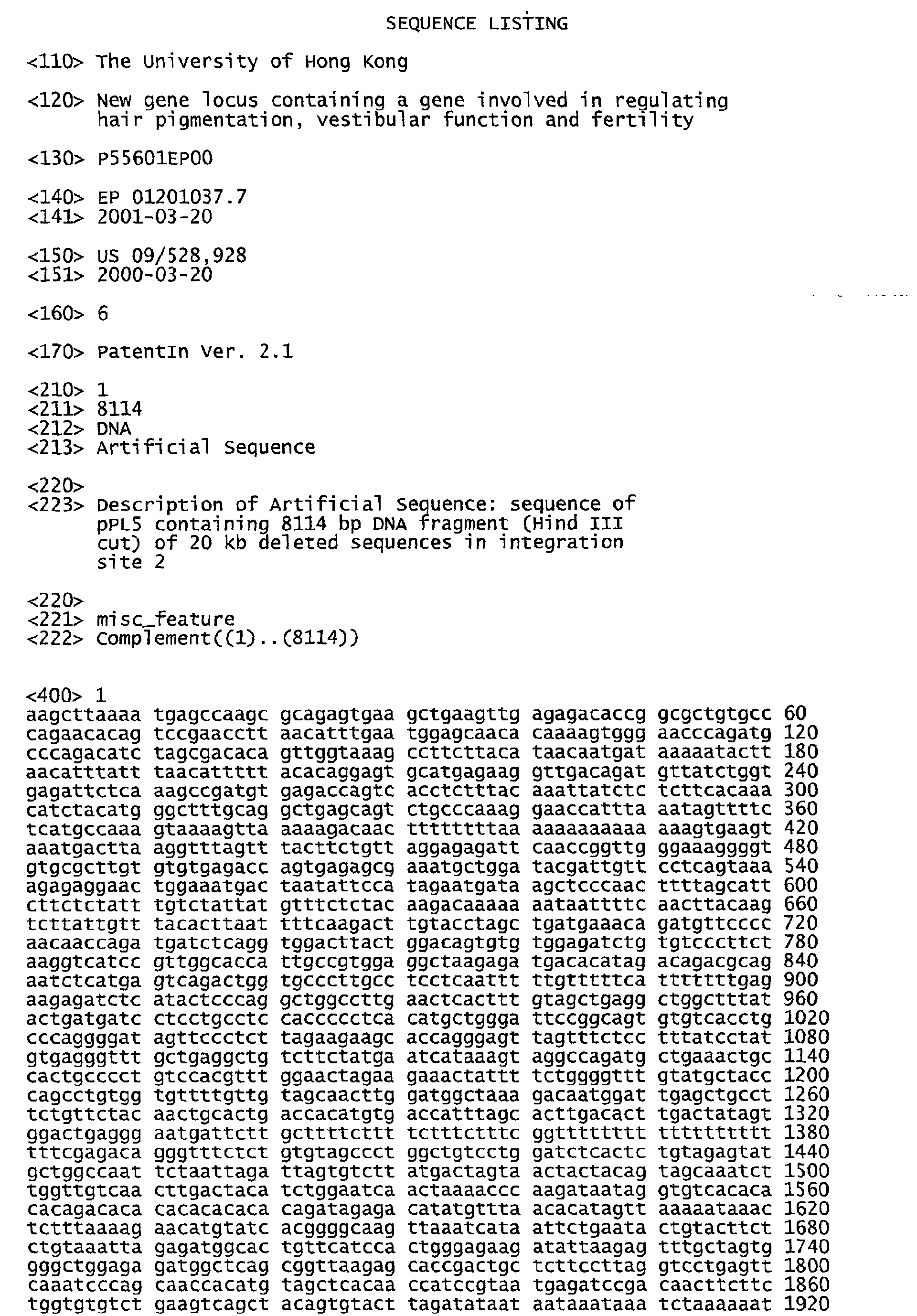
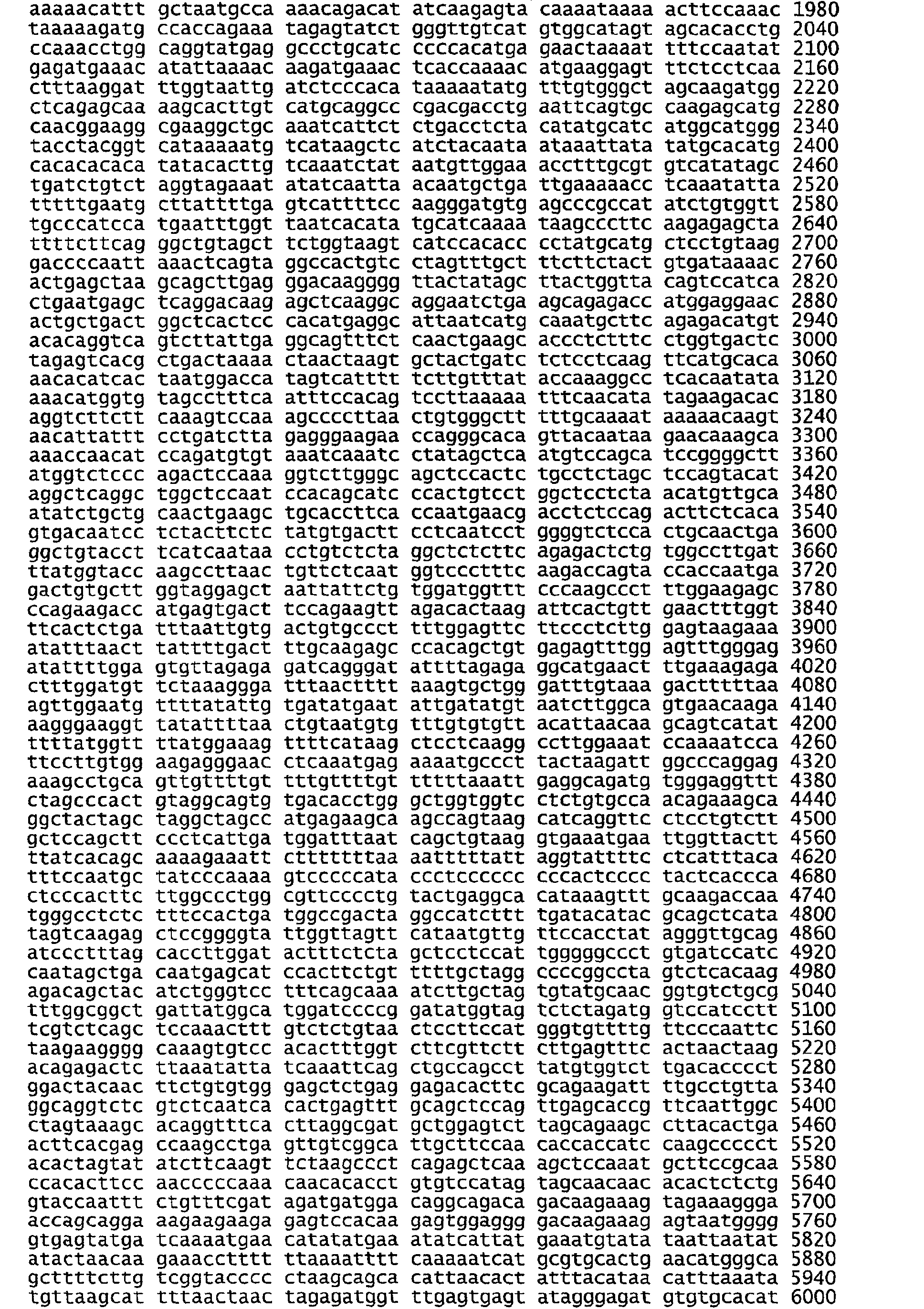
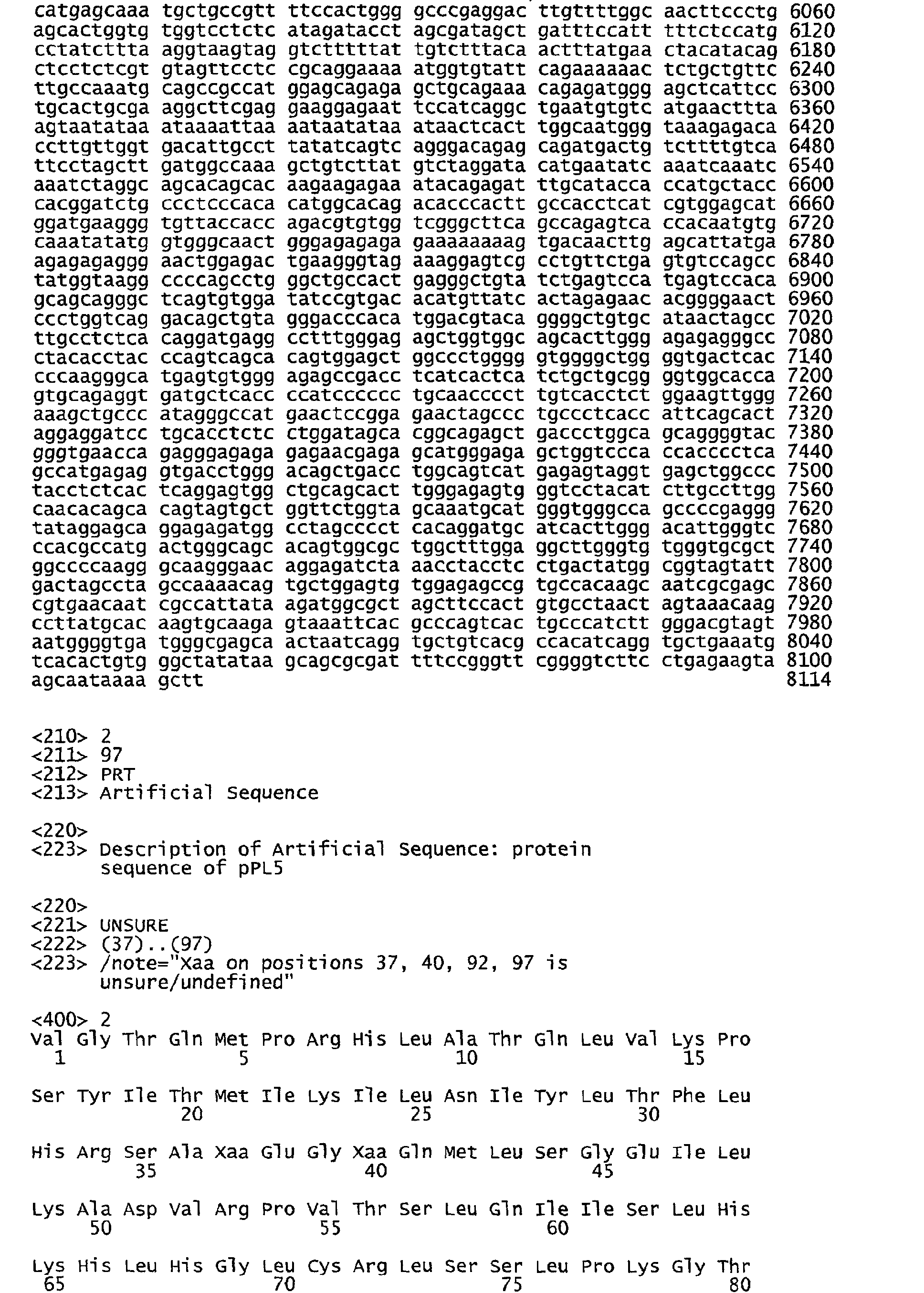
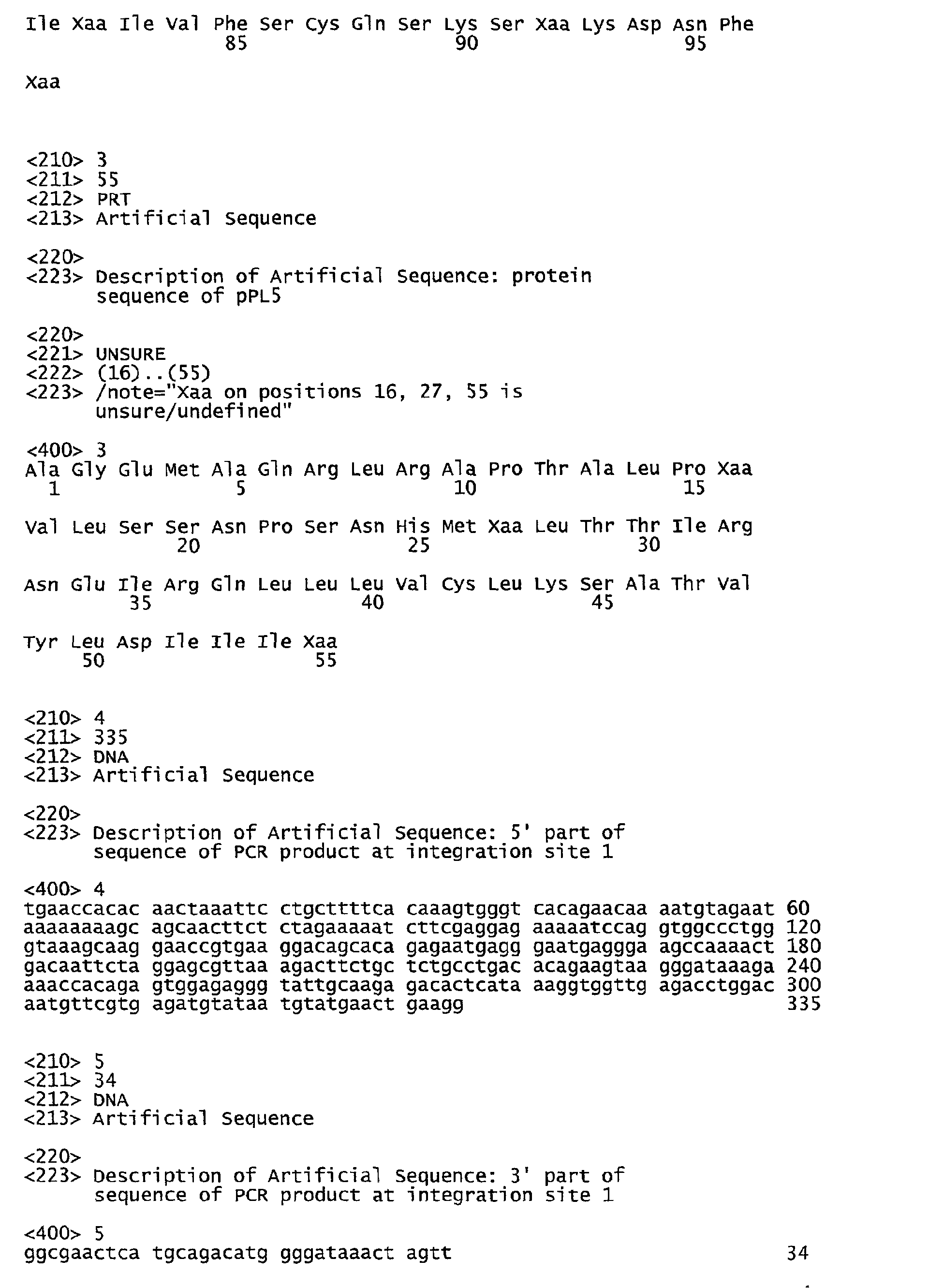
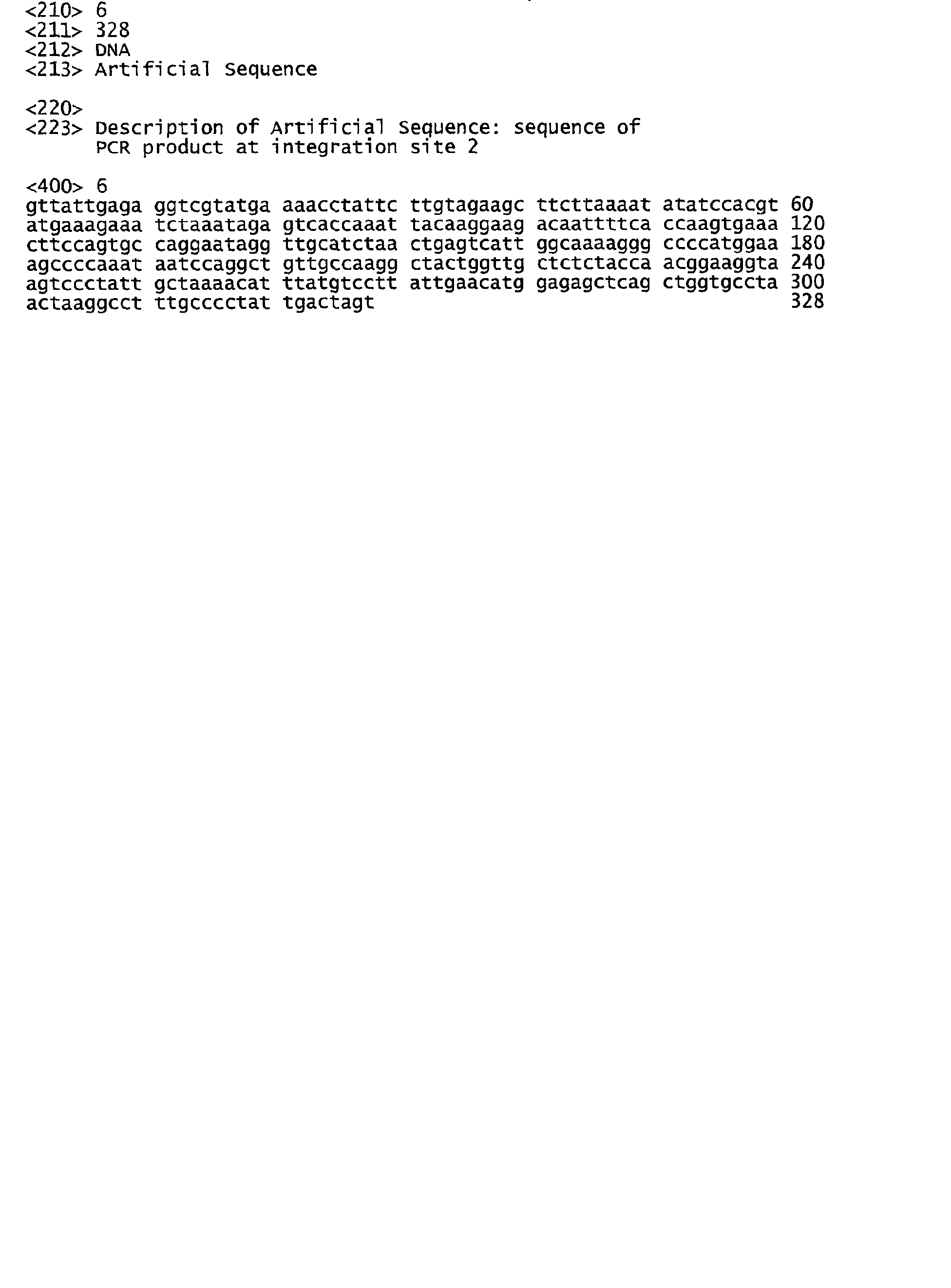
Claims (68)
- An isolated nucleic acid which defines a yellow submarine locus.
- An isolated nucleic acid which defines a mutant yellow submarine locus, wherein the mutant yellow submarine locus is identical to a wildtype yellow submarine locus except for an integration of a pAA2 transgene into at least one region on a chromosome.
- The isolated nucleic acid of claim 2, wherein the chromosome is mouse chromosome 3.
- The isolated nucleic acid of claim 3, wherein the pAA2 transgene is inserted into a region of mouse chromosome 3 designated A2.
- The isolated nucleic acid of claim 3, wherein the pAA2 transgene is inserted into a region of mouse chromosome 3 designated B-C.
- The isolated nucleic acid of claim 2, further comprising a rearrangement of chromosomal sequences of a region of the chromosome designated A3 region.
- The isolated nucleic acid of claim 6, wherein the rearrangement comprises an inversion of a nucleotide segment.
- The nucleic acid of claim 1 or 2 wherein the nucleic acid is genomic DNA.
- The nucleic acid of claim 1 or 2 wherein the nucleic acid is RNA.
- The nucleic acid of claim 1 or 2 wherein the nucleic acid is cDNA.
- The nucleic acid of claim 1 or 2, wherein the nucleic acid is labeled with a detectable marker.
- The nucleic acid of claim 6, wherein the marker is a radioactive, a colorimetric, a luminescent, or a fluorescent label.
- A replicable vector comprising the nucleic acid of claim 1 or 2.
- A host cell comprising the vector of claim 8.
- The host cell of claim 9 wherein the cell is a eukaryotic cell.
- The host cell of claim 9 wherein the cell is a bacterial cell.
- The vector of claim 8 wherein the vector is a plasmid.
- The vector of claim 8 wherein the vector is a cosmid.
- The vector of claim 8 wherein the vector is a λ phage.
- The vector of claim 8 wherein the vector is a YAC.
- The vector of claim 8, wherein the vector is a BAC.
- The vector of claim 8, wherein the vector is a PAC.
- A nucleic acid of at least 14 nucleotides capable of specifically hybridizing with the nucleic acid of claim 1.
- A nucleic acid of at least 14 nucleotides capable of specifically hybridizing with the nucleic acid of claim 2.
- The nucleic acid of claim 1 or 2, wherein the nucleic acid encodes a growth factor.
- The nucleic acid of claim 1 or 2, wherein the nucleic acid encodes a growth factor receptor.
- The nucleic acid of claim 1 or 2, wherein the nucleic acid encodes an orphan receptor.
- The nucleic acid of claim 1 or 2, wherein the nucleic acid encodes a signaling molecule.
- The nucleic acid of claim 1 or 2, wherein the nucleic acid encodes a transcriptional regulator.
- The nucleic acid of claim 1 or 2, wherein the nucleic acid encodes an intracellular transport protein.
- The nucleic acid of claim 1 or 2, wherein the nucleic acid encodes a neural precursor cell which is an expressed and developmentally down-regulated 4 (NEDD4) family molecule.
- The nucleic acid of claim 1 or 2, wherein the nucleic acid encodes a SOX protein.
- The nucleic acid of claim 1 or 2, wherein the nucleic acid encodes a regulator of apoptosis.
- The nucleic acid of claim 1 or 2, wherein the nucleic acid encodes a regulator of protein turnover.
- The nucleic acid of claim 1 or 2, wherein the nucleic acid encodes a cell cycle regulator.
- The nucleic acid of claim 1 or 2, wherein the nucleic acid encodes a calcium binding protein.
- The nucleic acid of claim 1 or 2, wherein the nucleic acid encodes a potentiator of hormone dependent activation of transcription by progesterone or glucocorticoid receptors.
- The nucleic acid of claim 1 or 2, wherein the nucleic acid encodes a membrane transport protein.
- The nucleic acid of claim 1 or 2, wherein the nucleic acid encodes a co-activator of transcription.
- The nucleic acid of claim 1 or 2, wherein the nucleic acid is isolated from a mouse.
- An isolated nucleic acid which defines a human locus which corresponds to a yellow submarine locus.
- An isolated nucleic acid which defines a human locus which corresponds to a mutant yellow submarine locus containing a pAA2 transgene integrated into at least one region the genome.
- A method of diagnosing inner ear dysfunction in a subject comprising:a) obtaining a suitable sample from a subject;b) extracting nucleic acid from the sample;c) contacting the nucleic acid with the nucleic acid of claim 24 which binds specifically to the mutated portion of the ysb locus; andd) detecting the labeled nucleic acid, thereby detecting mutant ysb and thereby diagnosing inner ear dysfunction in the subject.
- A method of diagnosing pigmentation dysfunction in a subject comprising:a) obtaining a suitable sample from a subject;b) extracting nucleic acid from the sample;c) contacting the nucleic acid with the nucleic acid of claim 24 which binds specifically to the mutated portion of the ysb locus;d) detecting the labeled nucleic acid, thereby detecting mutant ysb and thereby diagnosing pigmentation dysfunction in the subject.
- A method of diagnosing cell growth dysfunction, cell proliferation dysfunction, or cell death dysfunction in a subject comprising:a) obtaining a suitable sample from a subject;b) extracting nucleic acid from the sample;c) contacting the nucleic acid with the nucleic acid of claim 24 which binds specifically to the mutated portion of the ysb locus; andd) detecting the labeled nucleic acid, thereby detecting mutant ysb and thereby diagnosing cell growth dysfunction or proliferation dysfunction in the subject.
- The method of any one of claims 43-45, wherein the subject is mammal or non-mammal.
- The method of any one of claims 43-45, wherein the subject is a human, a primate, an equine, an opine, an avian, a bovine, a porcine, a canine, a feline or a murine.
- The method of any one of claims 43-45, wherein the subject is a vertebrate.
- A polypeptide encoded by the nucleic acid of claim 1, 2, 41 or 42.
- A fusion protein comprising the polypeptide of claim 49.
- The polypeptide of claim 49, wherein the polypeptide is labeled with a detectable marker.
- A method of repairing and regenerating nerve tissue in a subject comprising administering an effective amount of the protein of claim 49 to the subject, wherein the protein is a wildtype protein, so as to thereby repair and regenerate nerve tissue in the subject.
- A method of regulating cell migration in a subject comprising administering an effective amount of the protein of claim 49 to the subject, wherein the protein is a wildtype protein, so as to thereby regulate cell migration in the subject.
- A method of regulating cell growth in a subject comprising administering an effective amount of the protein of claim 49 to the subject, wherein the protein is a wildtype protein, so as to thereby regulate cell growth in the subject.
- An antibody which binds to the polypeptide of claim 49.
- The antibody of claim 55, wherein the antibody is a monoclonal antibody.
- The antibody of claim 55, wherein the antibody is conjugated to a cytotoxic agent.
- The antibody of claim 55, wherein the antibody is labeled with a detectable marker.
- A composition comprising the antibody of claim 55.
- A method of producing a protein encoded by a nucleic acid in a wildtype ysb locus which comprises growing a host vector system under suitable conditions permitting production of the polypeptide and recovering the polypeptide so produced.
- A method determining vestibular dysfunction in a subject comprising:a) obtaining a suitable sample from a subject;b) extracting nucleic acid from the sample;c) contacting the nucleic acid with the nucleic acid of claim 24 which binds specifically to a mutated portion of the ysb locus;d) detecting the labeled nucleic acid, thereby detecting gene responsible for yellow coat color, thereby indicating the presence of vestibular dysfunction in the subject.
- A method determining vestibular dysfunction using embryonal stem cells:a) obtaining a suitable sample from the embryonal stem cells;b) extracting nucleic acid from the sample;c) contacting the nucleic acid with the nucleic acid of claim 24 which binds specifically to a mutated portion of the ysb locus;d) detecting the labeled nucleic acid, thereby detecting gene responsible for yellow coat color, thereby indicating the presence of vestibular dysfunction in the embryonal stem cells.
- A method of determining successful deletion or inactivation of a gene which comprises examining coat color or pigmentation of a subject, wherein yellow coat color or pigmentation indicates that a normal gene has ben inactivated, thereby resulting in yellow coat color or pigmentation, thereby indicating the successful deletion or inactivation of the gene.
- A method of determining successful deletion or inactivation of a gene comprising determining whether repairing the abnormal gene results in the disappearance of the yellow coat color or pigmentation.
- A method of treating vestibular dysfunction in a subject comprising introducing a nucleic acid comprising a gene or genes from the wildtype locus encoding a ysb into a suitable cell under conditions such that the nucleic acid expresses ysb so as to thereby treat vestibular dysfunction.
- A method of treating hearing impairment in a subject which comprises introducing a nucleic acid comprising a gene or genes from the wild-type locus into a suitable cell under conditionssuch that the nucleic acid expresses ysb so as to thereby treat hearing loss.
- A transgenic mouse line designated KM12.
- A method of determining whether a compound is mutagenic comprising:(a) examining the coat color or pigmentation of a subject;(b) administering the compound to the subject;(c) examining the coat color or pigmentation of a subject;(d) comparing the result obtained in step (c) with the result obtained in step (a); and(e) determining if the coat color or pigmentation of the subject is yellow so as to thereby determine whether the compound is mutagenic.
Applications Claiming Priority (2)
| Application Number | Priority Date | Filing Date | Title |
|---|---|---|---|
| US52892800A | 2000-03-20 | 2000-03-20 | |
| US528928 | 2000-03-20 |
Publications (2)
| Publication Number | Publication Date |
|---|---|
| EP1148128A1 true EP1148128A1 (en) | 2001-10-24 |
| EP1148128B1 EP1148128B1 (en) | 2006-09-06 |
Family
ID=24107794
Family Applications (1)
| Application Number | Title | Priority Date | Filing Date |
|---|---|---|---|
| EP01201037A Expired - Lifetime EP1148128B1 (en) | 2000-03-20 | 2001-03-20 | Gene locus involved in regulating hair pigmentation, vestibular function and fertility |
Country Status (2)
| Country | Link |
|---|---|
| EP (1) | EP1148128B1 (en) |
| DE (1) | DE60122816T2 (en) |
Citations (1)
| Publication number | Priority date | Publication date | Assignee | Title |
|---|---|---|---|---|
| US5723719A (en) * | 1991-08-08 | 1998-03-03 | Health Research Inc. | Transgenic mouse as model for diseases involving dopaminergic dysfunction |
-
2001
- 2001-03-20 DE DE60122816T patent/DE60122816T2/en not_active Expired - Lifetime
- 2001-03-20 EP EP01201037A patent/EP1148128B1/en not_active Expired - Lifetime
Patent Citations (1)
| Publication number | Priority date | Publication date | Assignee | Title |
|---|---|---|---|---|
| US5723719A (en) * | 1991-08-08 | 1998-03-03 | Health Research Inc. | Transgenic mouse as model for diseases involving dopaminergic dysfunction |
Non-Patent Citations (4)
| Title |
|---|
| CHEAH KATHRYN S E ET AL: "Human COL2A1-directed SV40 T Antigen Expression in Transgenic and Chimeric Mice Results in Abnormal Skeletal Development.", JOURNAL OF CELL BIOLOGY, vol. 128, no. 1-2, 1995, NEW YORK US, pages 223 - 237, XP000926594, ISSN: 0021-9525 * |
| DATABASE SWALL [online] 1 November 1999 (1999-11-01), TAKADA, S. ET AL: "Mouse cDNA similar to 5' region of human EXML1 cDNA", XP002174568, retrieved from EMBL-EBI accession no. Q9WTN9 Database accession no. Q9WTN9 * |
| LANG R: "THE MAMMALIAN SPOT TEST AND ITS USE FOR TESTING OF MUTAGENIC AND CARCINOGENIC POTENTIAL EXPERIENCE WITH THE PESTICIDE CHLORDIMEFORM ITS PRINCIPAL METABOLITES AND THE DRUG LISURIDE HYDROGEN MALEATE", MUTATION RESEARCH, vol. 135, no. 3, 1984, AMSTERDAM NL, pages 219 - 224, XP000926604, ISSN: 0027-5107 * |
| MICHAUD EDWARD J ET AL: "The embryonic lethality of homozygous lethal yellow mice (A-y/A-y) is associated with the disruption of a novel RNA-binding protein.", GENES & DEVELOPMENT, vol. 7, no. 7A, 1993, pages 1203 - 1213, XP002076781, ISSN: 0890-9369 * |
Also Published As
| Publication number | Publication date |
|---|---|
| EP1148128B1 (en) | 2006-09-06 |
| DE60122816D1 (en) | 2006-10-19 |
| DE60122816T2 (en) | 2007-09-13 |
Similar Documents
| Publication | Publication Date | Title |
|---|---|---|
| van Genderen et al. | Development of several organs that require inductive epithelial-mesenchymal interactions is impaired in LEF-1-deficient mice. | |
| Zhang et al. | Neural tube, skeletal and body wall defects in mice lacking transcription factor AP-2 | |
| Ramfrez-Solis et al. | Hoxb-4 (Hox-2.6) mutant mice show homeotic transformation of a cervical vertebra and defects in the closure of the sternal rudiments | |
| Vogl et al. | Tryptophan‐rich basic protein (WRB) mediates insertion of the tail‐anchored protein otoferlin and is required for hair cell exocytosis and hearing | |
| Zheng et al. | β-Amyloid precursor protein-deficient mice show reactive gliosis and decreased locomotor activity | |
| Balschun et al. | Vesicular glutamate transporter VGLUT1 has a role in hippocampal long-term potentiation and spatial reversal learning | |
| Rhee et al. | Pbx3 deficiency results in central hypoventilation | |
| CN104450602B (en) | Non-human mammal animal model of neuropsychiatric disease and preparation method and application thereof | |
| EP3420811A1 (en) | Non-human model for neurofibromatosis type 1 | |
| Tsou et al. | Important roles of ring finger protein 112 in embryonic vascular development and brain functions | |
| Wang et al. | CIB2 and CIB3 regulate stereocilia maintenance and mechanoelectrical transduction in mouse vestibular hair cells | |
| Roux et al. | Assessment of the expression and role of the α1-nAChR subunit in efferent cholinergic function during the development of the mammalian cochlea | |
| Peeters et al. | Sensory deficits in mice hypomorphic for a mammalian homologue of unc-53 | |
| KR100227240B1 (en) | The ciliary neurotrophic factor receptor | |
| WO2009042723A1 (en) | Transgenic mice expressing hypersensitive nicotinic receptors | |
| EP1148128B1 (en) | Gene locus involved in regulating hair pigmentation, vestibular function and fertility | |
| Balu et al. | Behavioral and physiological characterization of PKC-dependent phosphorylation in the Grin2a∆ PKC mouse | |
| Terzi et al. | In vivo optical interrogation of neuronal responses to genetic, cell type-specific silencing | |
| US20130174285A1 (en) | Model Animal for Studying Hair Growth Cycle | |
| CA2457317A1 (en) | Transgenic rodents as animal models for modulation of b1 bradykinin receptor protein | |
| Jung | The putative transcription factor Prdm8 is required for the differentiation and maintenance of rod and cone bipolar cells in the mammalian retina | |
| Popko | Mouse models in the study of genetic neurological disorders | |
| Li | Functional analysis of murine homeobox genes | |
| WO2006096648A2 (en) | Mouse model | |
| WO1997017982A1 (en) | Methods and compositions for hair growth promotion |
Legal Events
| Date | Code | Title | Description |
|---|---|---|---|
| PUAI | Public reference made under article 153(3) epc to a published international application that has entered the european phase |
Free format text: ORIGINAL CODE: 0009012 |
|
| AK | Designated contracting states |
Kind code of ref document: A1 Designated state(s): DE FI FR GB SE Kind code of ref document: A1 Designated state(s): AT BE CH CY DE DK ES FI FR GB GR IE IT LI LU MC NL PT SE TR |
|
| AX | Request for extension of the european patent |
Free format text: AL;LT;LV;MK;RO;SI |
|
| 17P | Request for examination filed |
Effective date: 20020424 |
|
| AKX | Designation fees paid |
Free format text: AT BE CH CY DE LI |
|
| RBV | Designated contracting states (corrected) |
Designated state(s): DE FI FR GB SE |
|
| RBV | Designated contracting states (corrected) |
Designated state(s): DE FI FR GB SE |
|
| 17Q | First examination report despatched |
Effective date: 20030425 |
|
| GRAP | Despatch of communication of intention to grant a patent |
Free format text: ORIGINAL CODE: EPIDOSNIGR1 |
|
| GRAS | Grant fee paid |
Free format text: ORIGINAL CODE: EPIDOSNIGR3 |
|
| GRAA | (expected) grant |
Free format text: ORIGINAL CODE: 0009210 |
|
| AK | Designated contracting states |
Kind code of ref document: B1 Designated state(s): DE FI FR GB SE |
|
| PG25 | Lapsed in a contracting state [announced via postgrant information from national office to epo] |
Ref country code: FI Free format text: LAPSE BECAUSE OF FAILURE TO SUBMIT A TRANSLATION OF THE DESCRIPTION OR TO PAY THE FEE WITHIN THE PRESCRIBED TIME-LIMIT Effective date: 20060906 |
|
| REG | Reference to a national code |
Ref country code: GB Ref legal event code: FG4D |
|
| REF | Corresponds to: |
Ref document number: 60122816 Country of ref document: DE Date of ref document: 20061019 Kind code of ref document: P |
|
| PG25 | Lapsed in a contracting state [announced via postgrant information from national office to epo] |
Ref country code: SE Free format text: LAPSE BECAUSE OF FAILURE TO SUBMIT A TRANSLATION OF THE DESCRIPTION OR TO PAY THE FEE WITHIN THE PRESCRIBED TIME-LIMIT Effective date: 20061206 |
|
| ET | Fr: translation filed | ||
| PLBE | No opposition filed within time limit |
Free format text: ORIGINAL CODE: 0009261 |
|
| STAA | Information on the status of an ep patent application or granted ep patent |
Free format text: STATUS: NO OPPOSITION FILED WITHIN TIME LIMIT |
|
| 26N | No opposition filed |
Effective date: 20070607 |
|
| PGFP | Annual fee paid to national office [announced via postgrant information from national office to epo] |
Ref country code: FR Payment date: 20110404 Year of fee payment: 11 |
|
| PGFP | Annual fee paid to national office [announced via postgrant information from national office to epo] |
Ref country code: DE Payment date: 20110325 Year of fee payment: 11 Ref country code: GB Payment date: 20110321 Year of fee payment: 11 |
|
| GBPC | Gb: european patent ceased through non-payment of renewal fee |
Effective date: 20120320 |
|
| REG | Reference to a national code |
Ref country code: FR Ref legal event code: ST Effective date: 20121130 |
|
| REG | Reference to a national code |
Ref country code: DE Ref legal event code: R119 Ref document number: 60122816 Country of ref document: DE Effective date: 20121002 |
|
| PG25 | Lapsed in a contracting state [announced via postgrant information from national office to epo] |
Ref country code: FR Free format text: LAPSE BECAUSE OF NON-PAYMENT OF DUE FEES Effective date: 20120402 Ref country code: GB Free format text: LAPSE BECAUSE OF NON-PAYMENT OF DUE FEES Effective date: 20120320 |
|
| PG25 | Lapsed in a contracting state [announced via postgrant information from national office to epo] |
Ref country code: DE Free format text: LAPSE BECAUSE OF NON-PAYMENT OF DUE FEES Effective date: 20121002 |