EP1146118A1 - Organs for transplantation induced in vitro - Google Patents
Organs for transplantation induced in vitro Download PDFInfo
- Publication number
- EP1146118A1 EP1146118A1 EP00900388A EP00900388A EP1146118A1 EP 1146118 A1 EP1146118 A1 EP 1146118A1 EP 00900388 A EP00900388 A EP 00900388A EP 00900388 A EP00900388 A EP 00900388A EP 1146118 A1 EP1146118 A1 EP 1146118A1
- Authority
- EP
- European Patent Office
- Prior art keywords
- organ
- vitro
- vitro induced
- transplantation
- induced organ
- Prior art date
- Legal status (The legal status is an assumption and is not a legal conclusion. Google has not performed a legal analysis and makes no representation as to the accuracy of the status listed.)
- Withdrawn
Links
Images



Classifications
-
- C—CHEMISTRY; METALLURGY
- C12—BIOCHEMISTRY; BEER; SPIRITS; WINE; VINEGAR; MICROBIOLOGY; ENZYMOLOGY; MUTATION OR GENETIC ENGINEERING
- C12N—MICROORGANISMS OR ENZYMES; COMPOSITIONS THEREOF; PROPAGATING, PRESERVING, OR MAINTAINING MICROORGANISMS; MUTATION OR GENETIC ENGINEERING; CULTURE MEDIA
- C12N5/00—Undifferentiated human, animal or plant cells, e.g. cell lines; Tissues; Cultivation or maintenance thereof; Culture media therefor
- C12N5/06—Animal cells or tissues; Human cells or tissues
- C12N5/0602—Vertebrate cells
-
- A—HUMAN NECESSITIES
- A61—MEDICAL OR VETERINARY SCIENCE; HYGIENE
- A61K—PREPARATIONS FOR MEDICAL, DENTAL OR TOILETRY PURPOSES
- A61K35/00—Medicinal preparations containing materials or reaction products thereof with undetermined constitution
- A61K35/12—Materials from mammals; Compositions comprising non-specified tissues or cells; Compositions comprising non-embryonic stem cells; Genetically modified cells
-
- C—CHEMISTRY; METALLURGY
- C12—BIOCHEMISTRY; BEER; SPIRITS; WINE; VINEGAR; MICROBIOLOGY; ENZYMOLOGY; MUTATION OR GENETIC ENGINEERING
- C12N—MICROORGANISMS OR ENZYMES; COMPOSITIONS THEREOF; PROPAGATING, PRESERVING, OR MAINTAINING MICROORGANISMS; MUTATION OR GENETIC ENGINEERING; CULTURE MEDIA
- C12N2501/00—Active agents used in cell culture processes, e.g. differentation
- C12N2501/10—Growth factors
- C12N2501/16—Activin; Inhibin; Mullerian inhibiting substance
-
- C—CHEMISTRY; METALLURGY
- C12—BIOCHEMISTRY; BEER; SPIRITS; WINE; VINEGAR; MICROBIOLOGY; ENZYMOLOGY; MUTATION OR GENETIC ENGINEERING
- C12N—MICROORGANISMS OR ENZYMES; COMPOSITIONS THEREOF; PROPAGATING, PRESERVING, OR MAINTAINING MICROORGANISMS; MUTATION OR GENETIC ENGINEERING; CULTURE MEDIA
- C12N2501/00—Active agents used in cell culture processes, e.g. differentation
- C12N2501/30—Hormones
- C12N2501/38—Hormones with nuclear receptors
- C12N2501/385—Hormones with nuclear receptors of the family of the retinoic acid recptor, e.g. RAR, RXR; Peroxisome proliferator-activated receptor [PPAR]
-
- C—CHEMISTRY; METALLURGY
- C12—BIOCHEMISTRY; BEER; SPIRITS; WINE; VINEGAR; MICROBIOLOGY; ENZYMOLOGY; MUTATION OR GENETIC ENGINEERING
- C12N—MICROORGANISMS OR ENZYMES; COMPOSITIONS THEREOF; PROPAGATING, PRESERVING, OR MAINTAINING MICROORGANISMS; MUTATION OR GENETIC ENGINEERING; CULTURE MEDIA
- C12N2506/00—Differentiation of animal cells from one lineage to another; Differentiation of pluripotent cells
- C12N2506/02—Differentiation of animal cells from one lineage to another; Differentiation of pluripotent cells from embryonic cells
Definitions
- the present invention relates to development engineering or organ engineering, more particularly in vitro induced organs for transplantation such as kidney, heart, pancreas, liver, enteric canal, notochord, skeletal muscle, leukocyte, erythrocyte, lymphocyte and the like, which can function in vivo when they are in vivo transplanted, and their preparation method and evaluation method.
- Amphibian embryo has conventionally been regarded as a valuable material in the field of experimental embryology with which many studies have been made. This is because Amphibian egg fertilizes and develops externally, its large egg makes embryo operation possible, and its time course changes can easily be observed.
- Amphibian dorsal blastopore lip of gastrula is a special region and when it is transplanted into ventral side of other embryo, a secondary embryo including head or body-tail part is induced. This is why dorsal lip of blastopore is named "organizer" as a central region of morphogenesis which determines the embryo system. It is well known that organizer induces central nervous system by functioning to presumptive ectoderm during invagination of the primitive gut, meanwhile the organizer itself differentiates into dorsal mesoderm.
- a non-fertilized egg of Xenopus originally contains vertical axis which combines animal pole and vegetal pole, whereas its dorsoventral axis and mediolateral axis are not yet determined. It is known that, when fertilized, rotation occurs between cortex and inner cytoplasm of the egg resulting in that the sperm intruding side becomes the future ventral side and the opposite side becomes the future dorsal side, that sperm always intrude into the animal hemisphere and 180 degree opposite of the intrusion point becomes future blastopore, and that cortical rotation deviates distribution of body axis determinant factor (determinant) included in cytoplasma of fertilized egg, which results in the establishment of dorsoventral axis.
- a schematic view of the process of Xenopus development is shown in Fig.1.
- mesoderm induction is the primary induction which starts when cleavage progresses and the embryo reaches at morula stage.
- the presence of the induction is experimentally proved as a phenomenon in which blastomere of the vegetal hemisphere functions to blastomere of the animal hemisphere to induce mesoderm in the zone.
- the vegetal hemisphere blastomere has the inducing gradient along dorsoventral axis, and the ventral blastomere induces ventral mesoderm and the dorsal blastomere induces dorsal mesoderm including organizer.
- the mesoderm induction is known to localize mesoderm and specify the organizer region.
- the mesoderm induction mentioned is not specific to amphibians but is involved in the formation of mesoderm and organizer in early embryo of all the vertebrates.
- mesoderm induction the one secreted from the cell in the vegetal hemisphere to the side of the animal hemisphere is called mesoderm inducing factor.
- the fixation of mesoderm inducing factor existing in the embryo is one of the most critical theme in embryology. A variety of embryo operations have long been exercised using amphibian embryos and assay systems for inducing factor has already been established.
- animal cap assay as a method of direct investigation of mesoderm inducing factor activity
- injection assay as a method of investigating influence on normal development by injecting mRNA of major applicant factor, receptor molecules or the like, into 2-cell to 8-cell stage fertilized eggs.
- the present inventors reported for the first time by the above mentioned animal cap assay (Roux' Arch. Dev. Biol. 198:330-335, 1990), that activin, a cell growth factor of TGF- ⁇ family, contains mesoderm inducing activation.
- kidneys On the other hand, three kidneys, pronephros, mesonephros, and metanephros, are developed in succession in the course of development process of vertebrates, and function as the excretory system. These kidneys are derived from intermediate mesoderm and are formed temporally and spatially well-defined. Especially, the pronephros is the excretory organ established the earliest with quite a simple structure. The basic unit of the kidneys, the nephron, varies in number in three types of kidneys, yet its cytological features are common to them all. Thus, the pronephros is known to provide an attractive model system for studying the mechanism of kidney organogenesis.
- the present inventors have established a relatively simple experimental system for inducing pronephros (pronephric tubule), and the pronephric tubule has been shown to be induced when Xenopus blastula-stage ectoderm is treated with activin and retinoic acid.
- the pronephric tubule is shown to express several molecular markers that are expressed in a tadpole pronephros. But whether the pronephros (pronephric tubule) can function as an organ when in vivo transplanted has been unknown.
- the subject of the present invention is to provide an organ for transplantation to open a way to the transplantation treatment for such as innate kidney diseases or the like in higher organisms by establishing in vitro induction of organs and the method of in vivo transplantation, by taking advantage of development engineering or organ engineering in order to evaluate whether in vitro induced organs actually function in vivo.
- the present inventors have demonstrated the following fact for the first time in the world that Xenopus embryo ectoderm (the cell population with pluripotency) treated with activin and retinoic acid induced differentiation of pronephros, the most basic excretory organ established the earliest in ontogeny, and that when ectoderm oriented to differentiate into pronephros was transplanted into embryo which had been removed pronephros primordium (a region where pronephros is formed originally) following the culture of up to a certain stage corresponding to the pronephros primordium stage, the in vitro induced pronephros functioned in vivo. They have confirmed that in vitro induced organ can be transplanted in vivo and the present invention has accomplished.
- the present invention relates to a preparation method of organs functioning in vivo when in vitro induced organ is transplanted into a recipient of the same species, wherein the preparation method of in vitro induced organ for transplantation is characterized in that the organ is cultured to a certain stage corresponding to the stage of the recipient.
- the present invention also relates to: the preparation method of in vitro induced organ for transplantation according to claim 1 characterized in that a certain stage corresponding to the recipient is the same stage as the recipient: the preparation method of in vitro induced organ for transplantation according to any one of claims 1 or 2 characterized in that the certain stage is examined by using genome DNA which expresses corresponding to the stage of the in vitro induced organ as a molecular marker; the preparation method of in vitro induced organ for transplantation according to any one of claims 1 to 3 characterized in that the in vitro induced organ is an organ induced from ectoderm region which has been cut off from the blastula; the preparation method of in vitro induced organ for transplantation according to any one of claims 1 to 4 characterized in that in vitro induction takes place in the presence of TGF (Transforming Growth Factor)- ⁇ family.
- TGF Transforming Growth Factor
- the present invention further relates to the preparation method of in vitro induced organ for transplantation according to claim 5 characterized in that TGF (Transforming Growth Factor)- ⁇ family is activin; the preparation method of in vitro induced organ for transplantation according to any one of claims 1 to 4 characterized in that in vitro induction takes place in the presence of activin and retinoic acid; the preparation method of in vitro induced organ for transplantation according to any one of claims 1 to 7 characterized in that the organ is selected from kidney, heart, pancreas, liver, enteric canal, notochord, skeletal muscle, leukocyte, erythrocyte and lymphocyte; the preparation method of in vitro induced organ for transplantation according to any one of claims 1 to 8 characterized in that a living organism of the recipient is embryo; and the preparation method of in vitro induced organ for transplantation according to any one of claims 1 to 9 characterized in that the recipient belongs to vertebrate.
- TGF Transforming Growth Factor
- the present invention relates to an in vitro induced organ for transplantation characterized in that the organ is prepared by the preparation method of in vitro induced organ for transplantation according to any one of claims 1 to 10; an evaluation method of in vitro induced organ of non-human animal characterized in that in vitro induced organ of non-human animal is transplanted into a living organism of non-human recipient of the same species; an evaluation method of in vitro induced organ of non-human animal characterized in that the in vitro induced organ of non-human animal is cultured up to a certain stage corresponding to the stage of non-human recipient of the same species, wherein the cultured organ is in vivo transplanted into the recipient.
- in vitro induced organ of non-human animal characterized in that the certain stage is examined by using genome DNA which expresses corresponding to the stage of in vitro induced organ as a molecular marker; the evaluation method of in vitro induced organ of non-human animal according to either one of claims 13 or 14 characterized in that the in vitro induced organ is an organ induced from ectoderm region which has been cut off from the blastula; the evaluation method of in vitro induced organ of non-human animal according to any one of claims 13 to 15 characterized in that in vitro induction takes place in the presence of TGF (Transforming Growth Factor)- ⁇ family; and the evaluation method of in vitro induced organ of non-human animal according to any one of claims 13 to 16 characterized in that the organ is selected from kidney, heart, pancreas, liver, enteric canal, notochord, skeletal muscle, leukocyte, erythrocyte and lymphocyte.
- TGF Transforming Growth Factor
- the preparation method of in vitro induced organ for transplantation of the present invention is a preparation method of an organ which functions in vivo when in vitro induced organ is transplanted into a living organism of a recipient of the same species, characterized in that the organ is cultured up to a certain stage corresponding to the stage of the recipient.
- Organs induced in vitro of the present invention include, for convenience, tissues such as notochord, skeletal muscle, leukocyte, erythrocyte, lymphocyte and the like besides organs that compose internal organs or the like of animals such as kidney, heart, pancreas, liver, enteric canal and the like.
- the recipients of the present invention include human besides organisms of the same species as in vitro induced organs, namely vertebrate of the same "species" taxonomically, for example, non-human animals like Amphibia such as newts or frogs or the like, Pisces, Aves, or Mammalia such as mouse or the like.
- ectoderm region (animal cap) removed from the blastula is preferable for in vitro induction of organs.
- presumptive ectoderm region of the blastula turns into epidermis-like cell lump (formless epidermis) without differentiating the tissues when there is no mesoderm inducing activity agent in the culture solution.
- mesoderm inducing activity exists in the culture solution, it becomes a tissue lump including mesoderm such as hemocyte, muscle, notochord or the like.
- mesoderm inducing activity exists in the culture solution, it becomes a tissue lump including mesoderm such as hemocyte, muscle, notochord or the like.
- in vitro induced organs can function in vivo when transplanted to a living organism of the recipients of the same species depends largely on the stage at which transplantation is performed. Therefore in vitro induced organs for transplantation need to be cultured up to a certain stage corresponding to the recipients of the same species, that is up to the stage where in vivo transplanted organ can function in the living organism. In many cases, when an organ is transplanted to in vivo of a recipient after being cultured up to the same stage as the recipient, the capability of functioning in vivo when transplanted into a living organism increases.
- a stage means each stage to which the progression process of ontogeny, where one living organism finishes as an adult starting from a fertilized egg or a parthenogenetic egg, is divided into and numbered.
- the progression process to metamorphosis is divided into about 60 stages.
- testing stages of the in vitro induced organs can be performed by observing the tissues and by the method using antibodies as above mentioned. But more defined testing can be performed by using genome DNA, which expresses corresponding to the stage of in vitro induced organ, as a molecular marker. For example, in case of normal development of Amphibia embryo, expression of genome DNA rapidly increases from the middle blastula stage where cleavage took place 12 times. After that, it is known that the quantity and variety of genes that express corresponding to embryo stages show changes in strict tiers. In case of in vitro induced organs treated with activin, these tiers are also entirely observed as shown in Table 1.
- Xlim-1 expressed at stages 11 to 31 Xpax-2 expressed at and after stage 20, XCIRP or XeWT1 expressed at and after stage 22, Na + K + -ATPase expressed at and after stage 27, and Cap-1 or X1caax-1 expressed at and after stage 31.
- TGF- ⁇ family TGF is also called transforming growth factor and functions to transform normal growing property of the cell to transforming growth property, alone or in cooperation with other factors when added to the culture environment.
- TGF- ⁇ having common receptor with epidermal growth factor (EGF) and TGF- ⁇ showing TGF activity in cooperation with TGF- ⁇ are known as the kinds of TGF.
- EGF- ⁇ family TGF- ⁇ , activin, inhibin, Mullerian duct inhibiting factor are exemplified. Among these, activin is most preferable from the view point of mesoderm inducing activity.
- Mammalia activin which is secreted from ovary membrane granulocyte and which is a cell growth factor with activity promoting follicle-stimulating hormone secretion (FSH) from anterior hypophysis and which exists as a dimer showing S-S binding to inhibin ⁇ chain subunit having FSH secretion inhibiting factor activity, induces mesoderm tissues in presumptive ectoderm with the concentration of 0.1 ng/ml or more.
- Mammlia activin can cause expression of mesodermal marker genes such as muscle actin or homeobox gene or the like, and has property that it can induce all the mesodermal tissues from ventral side to dorsal side in every assay system.
- activin Function of activin is obviously concentration dependent, so that the higher the concentration is, differentiation occurs the more in dorsal mesoderm.
- Hemocyte, coelomic epithelium and mesenchyme differentiate in the animal cap treated with about 0.3 to 1.0 ng/ml of activin, muscle differentiate at the concentration of 5 to 10 ng/ml, and notochord the most dorsal mesoderm differentiate at the concentration of 50 to 100 ng/ml respectively.
- FGF Fibroblast Growth Factor
- TGF- ⁇ family such as activin or the like.
- neural tissues are often observed inside activin-treated grafts, which is thought to be a result of neural induction to non-differentiated cell by organizer induced by activin of high concentration. These show that activin not only merely induces tissue differentiation but is deeply involved in dorsoventral axis determination. Organogenesis induction by activin to animal cap is shown in Fig.2.
- kidney differentiation is induced by activin/retinoic acid treatment of Xenopus animal cap at an adequate concentration and that hemocyte differentiation can be controlled by activin/BMP (Bone Morphogenesis Protein) or by IL-11, SCF (Stem Cell Factor).
- Retinoic acid shown as RA in Fig.2 as mentioned above is also called Vitamin A acid and is an important metabolic product of Vitamin A (retinol) which is indispensable for normal development of animals and for differentiation and growth of epithelium tissues or cartilage. It has morphogen-like function in the process of vertebrate development, which function is demonstrated by controlling gene expression via retinoic acid receptor.
- the in vivo differential pattern of the activin and/or retinoic acid treated graft was examined by transplantation method.
- the graft (0.3 mm X 0.3 mm) prepared in a same way as in Embodiment 1 was transplanted into left side of Xenopus embryo (stage 20), region where pronephros primordium had been removed (cut off), then in vivo differential and inducing pattern of the grafts was examined after 5 days of transplantation.
- the results are shown in Table 2 as Embodiment 1.
- ectoderm treated with mixed solution including 10ng/ml activin and 10 -4 M retinoic acid displayed pronephric tubules differentiation at a high frequency at day 4: all the treated ectoderm formed pronephric tubules in the host embryos: in case of treatment with activin alone induced muscle, coelomic epithelium, and epidermis in ectoderm, with very few of grafts differentiated into pronephric tubules: and grafts without any treatment and with retinoic acid treatment alone displayed little differentiation and the ectoderm forms a part of epidermis in the host embryo.
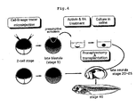
- Ectoderm to be differentiated into pronephric tubule, which was differentiated and induced in vitro by using mixed solution of activin and retinoic acid, and which was then examined as being at stage 20 by being detected with genome DNA, Xpax-2 as a molecule marker, which expresses corresponding to stage 20, was transplanted into one of Xenopus embryo side where bilateral pronephric primordia (located under somites 3 to 5) had been removed at stages 20, 23, and 25. Whether the grafts can function in vivo was investigated. A schema of the transplantation is shown in Fig.4. Bilateral pronephrectomy Xenopus which had not been transplanted was used as control. The results are shown in Table 3.
- Stage 20 25 Control Graft Control Graft Control Graft The number of test samples 96 141 36 48 10 19 Normal embryo 7(7) 29(21) 1(3) 4(8) 0 1(5) Edema embryo 89(93) 112(79) 35(97) 44(92) 10(100) 18(95)
- Fig.6 The relationship between post-operation days of the transplantation or pronephrectomy and the survival rate at stage 20 is shown in Fig.6.
- the following facts are understood from Fig.6.
- Fig.6 When only pronephrectomy was carried out and not the transplantation, the test samples showed edema-formation and died within 9 days.
- 29 (21%) of the 141 transplants developed normally without edema-formation as above mentioned, with some of them even survived for more than 1 month.
- the present invention enables in vitro induction of an organ by a relatively simple experimental system using such as activin alone or activin/RA or the like as mesoderm inducing factor, it also enables to prepare an organ which can function in vivo when transplanted in vivo of the recipient of the same species following culture of the in vitro induced organ up to a certain stage corresponding to the stage of the recipient. Further the present invention enables to evaluate whether the in vitro induced organ is suitable for transplantation by performing in vivo transplantation following culture of the in vitro induced organ up to a certain stage corresponding to the stage of the recipient of the same species.
- the present invention makes it possible to confirm whether the in vitro induced organ actually functions in vivo. Thereby not only many significant views to clarify structure of formation of kidney or other such organs are demonstrated but also the present invention contributes to establish a new field in developing engineering or organ engineering that involves in vitro organ formation and its in vivo transplantation. Further, the present invention enables organ transplantation fitting immunologically and it will open a way to treat diseases of higher organisms including human such as kidney diseases or the like.
Landscapes
- Engineering & Computer Science (AREA)
- Health & Medical Sciences (AREA)
- Biomedical Technology (AREA)
- Life Sciences & Earth Sciences (AREA)
- Chemical & Material Sciences (AREA)
- Biotechnology (AREA)
- Organic Chemistry (AREA)
- Bioinformatics & Cheminformatics (AREA)
- Genetics & Genomics (AREA)
- Wood Science & Technology (AREA)
- Zoology (AREA)
- Microbiology (AREA)
- Biochemistry (AREA)
- General Engineering & Computer Science (AREA)
- General Health & Medical Sciences (AREA)
- Cell Biology (AREA)
- Measuring Or Testing Involving Enzymes Or Micro-Organisms (AREA)
- Micro-Organisms Or Cultivation Processes Thereof (AREA)
Abstract
Description
| Genes | The starting time of expression | Properties |
| Primary response genes | ||
| Mix.1 | about 30 min. | homeobox gene, similar to paired of Drosophila |
| goosecoid | about 30 min. | homeobox gene, similar to gooseberry and bicoid of Drosophila |
| Xlim-1 | about 2 hours | homeobox gene with LIM domain |
| Xbra | 90-250 min. | similar to mouse Brachyury (T) |
| Secondary | ||
| XIHbox | ||
| 1 | about 11 hours | homeobox gene, similar to Drosophila antennapedia |
| XIHbox 6 | about 13 hours | homeobox gene, similar to Drosophila abdominal-B |
| Xwnt-8 | about 16 hours | similar to oncogene Wnt-1 (int-1) |
| XMyoD | about 16 hours | similar to mouse MyoD |
| a-actin | about 19 hours | actin gene of myocardium and skelton |
| N-CAM | about 19 hours | gene of neural cell adhesion molecule |
| In vitro | In vivo | |||||||
| Conc.of activin A(ng/ml) | 0 | 10 | 0 | 10 | ||||
| Conc.of retinoic acid (M) | 0 | 10-4 | 0 | 10-4 | 0 | 10-4 | 0 | 10-4 |
| The number of test samples | 37 | 32 | 35 | 37 | 14 | 15 | 23 | 23 |
| Formless epidermis | 37 | 32 | 0 | 1 | 14 | 15 | 0 | 0 |
| (100) | (100) | (3) | (100) | (100) | ||||
| | 0 | 0 | 34 | 35 | 0 | 0 | 6 | 8 |
| (97) | (95) | (26) | (35) | |||||
| | 0 | 0 | 28 | 2 | 0 | 0 | 1 | 0 |
| (80) | (5) | (4) | ||||||
| | 0 | 0 | 32 | 2 | 0 | 0 | 16 | 0 |
| (91) | (5) | (70) | ||||||
| | 0 | 0 | 0 | 32 | 0 | 0 | 4 | 23 |
| (86) | (17) | (100) | ||||||
| | 0 | 0 | 27 | 7 | 0 | 0 | 0 | 0 |
| (77) | (19) | |||||||
| | 0 | 0 | 3 | 7 | 0 | 0 | 10 | 8 |
| (86) | (19) | (43) | (35) | |||||
| | 0 | 0 | 0 | 0 | 1 | 1 | 8 | 4 |
| (7) | (7) | (35) | (17) |
| | 20 | 23 | 25 | |||
| Control | Graft | Control | Graft | Control | Graft | |
| The number of test samples | 96 | 141 | 36 | 48 | 10 | 19 |
| Normal embryo | 7(7) | 29(21) | 1(3) | 4(8) | 0 | 1(5) |
| Edema embryo | 89(93) | 112(79) | 35(97) | 44(92) | 10(100) | 18(95) |
Claims (17)
- A preparation method of organs functioning in vivo when in vitro induced organ is transplanted into a recipient of the same species, wherein the preparation method of in vitro induced organ for transplantation is characterized in that the organ is cultured to a certain stage corresponding to the stage of the recipient.
- The preparation method of in vitro induced organ for transplantation according to claim 1 characterized in that a certain stage corresponding to the recipient is the same stage as the recipient.
- The preparation method of in vitro induced organ for transplantation according to any one of claims 1 or 2 characterized in that the certain stage is examined by using genome DNA which expresses corresponding to the stage of the in vitro induced organ as a molecular marker.
- The preparation method of in vitro induced organ for transplantation according to any one of claims 1 to 3 characterized in that the in vitro induced organ is an organ induced from ectoderm region which has been cut off from the blastula.
- The preparation method of in vitro induced organ for transplantation according to any one of claims 1 to 4 characterized in that in vitro induction takes place in the presence of TGF (Transforming Growth Factor)-β family.
- The preparation method of in vitro induced organ for transplantation according to claim 5 characterized in that TGF (Transforming Growth Factor)-β family is activin.
- The preparation method of in vitro induced organ for transplantation according to any one of claims 1 to 4 characterized in that in vitro induction takes place in the presence of activin and retinoic acid.
- The preparation method of in vitro induced organ for transplantation according to any one of claims 1 to 7 characterized in that the organ is selected from kidney, heart, pancreas, liver, enteric canal, notochord, skeletal muscle, leukocyte, erythrocyte and lymphocyte.
- The preparation method of in vitro induced organ for transplantation according to any one of claims 1 to 8 characterized in that a living organism of the recipient is embryo.
- The preparation method of in vitro induced organ for transplantation according to any one of claims 1 to 9 characterized in that the recipient belongs to vertebrate.
- An in vitro induced organ for transplantation characterized in that the organ is prepared by the preparation method of in vitro induced organ for transplantation according to any one of claims 1 to 10.
- An evaluation method of in vitro induced organ of non-human animal characterized in that in vitro induced organ of non-human animal is transplanted into a living organism of non-human recipient of the same species.
- An evaluation method of in vitro induced organ of non-human animal characterized in that the in vitro induced organ of non-human animal is cultured up to a certain stage corresponding to the stage of non-human recipient of the same species, wherein the cultured organ is in vivo transplanted into the recipient.
- The evaluation method of in vitro induced organ of non-human animal according to claim 13 characterized in that the certain stage is examined by using genome DNA which expresses corresponding to the stage of in vitro induced organ as a molecular marker.
- The evaluation method of in vitro induced organ of non-human animal according to either one of claims 13 or 14 characterized in that the in vitro induced organ is an organ induced from ectoderm region which has been cut off from the blastula.
- The evaluation method of in vitro induced organ of non-human animal according to any one of claims 13 to 15 characterized in that in vitro induction takes place in the presence of TGF (Transforming Growth Factor)-β family.
- The evaluation method of in vitro induced organ of non-human animal according to any one of claims 13 to 16 characterized in that the organ is selected from kidney, heart, pancreas, liver, enteric canal, notochord, skeletal muscle, leukocyte, erythrocyte and lymphocyte.
Applications Claiming Priority (3)
| Application Number | Priority Date | Filing Date | Title |
|---|---|---|---|
| JP2107799 | 1999-01-29 | ||
| JP11021077A JP2000217571A (en) | 1999-01-29 | 1999-01-29 | In vitro induced organs for transplantation |
| PCT/JP2000/000148 WO2000044881A1 (en) | 1999-01-29 | 2000-01-14 | ORGANS FOR TRANSPLANTATION INDUCED $i(IN VITRO) |
Publications (2)
| Publication Number | Publication Date |
|---|---|
| EP1146118A1 true EP1146118A1 (en) | 2001-10-17 |
| EP1146118A4 EP1146118A4 (en) | 2002-08-14 |
Family
ID=12044836
Family Applications (1)
| Application Number | Title | Priority Date | Filing Date |
|---|---|---|---|
| EP00900388A Withdrawn EP1146118A4 (en) | 1999-01-29 | 2000-01-14 | IN VITRO PRODUCTS TRANSPLANTANTS |
Country Status (3)
| Country | Link |
|---|---|
| EP (1) | EP1146118A4 (en) |
| JP (1) | JP2000217571A (en) |
| WO (1) | WO2000044881A1 (en) |
Families Citing this family (3)
| Publication number | Priority date | Publication date | Assignee | Title |
|---|---|---|---|---|
| JP4494586B2 (en) * | 2000-04-27 | 2010-06-30 | 独立行政法人科学技術振興機構 | Method for forming vertebrate pancreas in vitro |
| EP1773986B1 (en) * | 2004-07-09 | 2019-01-16 | Viacyte, Inc. | Preprimitive streak and mesendoderm cells |
| EP2322604A4 (en) | 2008-08-04 | 2014-01-08 | Univ Showa | TECHNIQUE FOR REGULATING THE REGENERATION OF TISSUE OR A FAILURE OR ABNORMAL PART IN AN ORGAN WITH NELL-1 |
Family Cites Families (3)
| Publication number | Priority date | Publication date | Assignee | Title |
|---|---|---|---|---|
| JPH03147782A (en) * | 1989-10-31 | 1991-06-24 | Bio Material Kenkyusho:Kk | Method for transplanting cell |
| US5851832A (en) * | 1991-07-08 | 1998-12-22 | Neurospheres, Ltd. | In vitro growth and proliferation of multipotent neural stem cells and their progeny |
| JPH06277050A (en) * | 1993-03-26 | 1994-10-04 | Kanegafuchi Chem Ind Co Ltd | Immobilization of animal cells and culture method |
-
1999
- 1999-01-29 JP JP11021077A patent/JP2000217571A/en active Pending
-
2000
- 2000-01-14 WO PCT/JP2000/000148 patent/WO2000044881A1/en not_active Ceased
- 2000-01-14 EP EP00900388A patent/EP1146118A4/en not_active Withdrawn
Also Published As
| Publication number | Publication date |
|---|---|
| WO2000044881A1 (en) | 2000-08-03 |
| JP2000217571A (en) | 2000-08-08 |
| EP1146118A4 (en) | 2002-08-14 |
Similar Documents
| Publication | Publication Date | Title |
|---|---|---|
| US9115342B2 (en) | Method for purifying cardiomyocytes or programmed cardiomyocytes derived from stem cells or fetuses | |
| JP4383896B2 (en) | Novel method for identifying, isolating or differentiating angiogenic progenitor cells in vitro | |
| JP2020124219A (en) | Methods for reprogramming cells and uses thereof | |
| US20030219866A1 (en) | Stem cell-like cells | |
| CA2937882A1 (en) | Method for generating cell condensate for self-organization | |
| KR20060010847A (en) | Isolated pluripotent adult stem cells and methods for isolation and culture thereof | |
| EA022736B1 (en) | Method for producing endothelial cells (variants) | |
| CN109689858B (en) | Methods for producing mesodermal and/or endothelial colony forming cell-like cells having in vivo angiogenic capacity | |
| Lim et al. | Generation of dopamine neurons from rodent fibroblasts through the expandable neural precursor cell stage | |
| KR101562366B1 (en) | Scaffold for inducing myocardiocyte differentiation compring methacrylated gelatin | |
| WO2004104184A1 (en) | Preparation of endodermal stem cells | |
| Asashima et al. | In vitro control of organogenesis and body patterning by activin during early amphibian development | |
| Galli et al. | Adult neural stem cells | |
| Asashima et al. | Elucidation of the role of activin in organogenesis using a multiple organ induction system with amphibian and mouse undifferentiated cells in vitro | |
| JP7021828B2 (en) | How to induce meiosis of ovarian-derived stem cells | |
| WO2000044881A1 (en) | ORGANS FOR TRANSPLANTATION INDUCED $i(IN VITRO) | |
| AU2012227337B2 (en) | A method for purifying cardiomyocytes or programmed cardiomyocytes derived from stem cells or fetuses | |
| Grunz | Developmental biology of amphibians after Hans Spemann in Germany | |
| KR20210135428A (en) | A cell composition comprising retinal cell having characteristics of photoreceptor cells and retinal pigment epithelial cell simultaneously and use thereof | |
| AU2021315931A1 (en) | Methods for spermatogonial culture | |
| Bianco et al. | Rapid serum-free isolation of oligodendrocyte progenitor cells from adult rat spinal cord | |
| Myoishi et al. | Induction of tooth and eye by transplantation of activin A-treated, undifferentiated presumptive ectodermal Xenopus cells into the abdomen | |
| Armstrong et al. | Neural stem cell technology as a novel treatment for Parkinson's disease | |
| Okabayashi et al. | In vitro organogenesis using amphibian pluripotent cells | |
| WO2025166963A1 (en) | Composition and method for culturing signal organizer and micro-organ using same |
Legal Events
| Date | Code | Title | Description |
|---|---|---|---|
| PUAI | Public reference made under article 153(3) epc to a published international application that has entered the european phase |
Free format text: ORIGINAL CODE: 0009012 |
|
| 17P | Request for examination filed |
Effective date: 20010801 |
|
| AK | Designated contracting states |
Kind code of ref document: A1 Designated state(s): AT BE CH CY DE DK ES FI FR GB GR IE IT LI LU MC NL PT SE |
|
| A4 | Supplementary search report drawn up and despatched |
Effective date: 20020627 |
|
| AK | Designated contracting states |
Kind code of ref document: A4 Designated state(s): DE FR GB IT SE |
|
| RBV | Designated contracting states (corrected) |
Designated state(s): DE FR GB IT SE |
|
| RAP1 | Party data changed (applicant data changed or rights of an application transferred) |
Owner name: JAPAN SCIENCE AND TECHNOLOGY AGENCY |
|
| STAA | Information on the status of an ep patent application or granted ep patent |
Free format text: STATUS: THE APPLICATION IS DEEMED TO BE WITHDRAWN |
|
| 18D | Application deemed to be withdrawn |
Effective date: 20130801 |
|
| REG | Reference to a national code |
Ref country code: DE Ref legal event code: R079 Free format text: PREVIOUS MAIN CLASS: C12N0005060000 Ipc: C12N0005070000 |
|
| REG | Reference to a national code |
Ref country code: DE Ref legal event code: R079 Free format text: PREVIOUS MAIN CLASS: C12N0005060000 Ipc: C12N0005070000 Effective date: 20140602 |