EP1135533B1 - Method and system for detecting oligonucleotides in a sample - Google Patents
Method and system for detecting oligonucleotides in a sample Download PDFInfo
- Publication number
- EP1135533B1 EP1135533B1 EP99973075A EP99973075A EP1135533B1 EP 1135533 B1 EP1135533 B1 EP 1135533B1 EP 99973075 A EP99973075 A EP 99973075A EP 99973075 A EP99973075 A EP 99973075A EP 1135533 B1 EP1135533 B1 EP 1135533B1
- Authority
- EP
- European Patent Office
- Prior art keywords
- oligonucleotide
- recognition
- agent
- signal
- verification
- Prior art date
- Legal status (The legal status is an assumption and is not a legal conclusion. Google has not performed a legal analysis and makes no representation as to the accuracy of the status listed.)
- Expired - Lifetime
Links
- 108091034117 Oligonucleotide Proteins 0.000 title claims abstract description 238
- JLCPHMBAVCMARE-UHFFFAOYSA-N [3-[[3-[[3-[[3-[[3-[[3-[[3-[[3-[[3-[[3-[[3-[[5-(2-amino-6-oxo-1H-purin-9-yl)-3-[[3-[[3-[[3-[[3-[[3-[[5-(2-amino-6-oxo-1H-purin-9-yl)-3-[[5-(2-amino-6-oxo-1H-purin-9-yl)-3-hydroxyoxolan-2-yl]methoxy-hydroxyphosphoryl]oxyoxolan-2-yl]methoxy-hydroxyphosphoryl]oxy-5-(5-methyl-2,4-dioxopyrimidin-1-yl)oxolan-2-yl]methoxy-hydroxyphosphoryl]oxy-5-(6-aminopurin-9-yl)oxolan-2-yl]methoxy-hydroxyphosphoryl]oxy-5-(6-aminopurin-9-yl)oxolan-2-yl]methoxy-hydroxyphosphoryl]oxy-5-(6-aminopurin-9-yl)oxolan-2-yl]methoxy-hydroxyphosphoryl]oxy-5-(6-aminopurin-9-yl)oxolan-2-yl]methoxy-hydroxyphosphoryl]oxyoxolan-2-yl]methoxy-hydroxyphosphoryl]oxy-5-(5-methyl-2,4-dioxopyrimidin-1-yl)oxolan-2-yl]methoxy-hydroxyphosphoryl]oxy-5-(4-amino-2-oxopyrimidin-1-yl)oxolan-2-yl]methoxy-hydroxyphosphoryl]oxy-5-(5-methyl-2,4-dioxopyrimidin-1-yl)oxolan-2-yl]methoxy-hydroxyphosphoryl]oxy-5-(5-methyl-2,4-dioxopyrimidin-1-yl)oxolan-2-yl]methoxy-hydroxyphosphoryl]oxy-5-(6-aminopurin-9-yl)oxolan-2-yl]methoxy-hydroxyphosphoryl]oxy-5-(6-aminopurin-9-yl)oxolan-2-yl]methoxy-hydroxyphosphoryl]oxy-5-(4-amino-2-oxopyrimidin-1-yl)oxolan-2-yl]methoxy-hydroxyphosphoryl]oxy-5-(4-amino-2-oxopyrimidin-1-yl)oxolan-2-yl]methoxy-hydroxyphosphoryl]oxy-5-(4-amino-2-oxopyrimidin-1-yl)oxolan-2-yl]methoxy-hydroxyphosphoryl]oxy-5-(6-aminopurin-9-yl)oxolan-2-yl]methoxy-hydroxyphosphoryl]oxy-5-(4-amino-2-oxopyrimidin-1-yl)oxolan-2-yl]methyl [5-(6-aminopurin-9-yl)-2-(hydroxymethyl)oxolan-3-yl] hydrogen phosphate Polymers Cc1cn(C2CC(OP(O)(=O)OCC3OC(CC3OP(O)(=O)OCC3OC(CC3O)n3cnc4c3nc(N)[nH]c4=O)n3cnc4c3nc(N)[nH]c4=O)C(COP(O)(=O)OC3CC(OC3COP(O)(=O)OC3CC(OC3COP(O)(=O)OC3CC(OC3COP(O)(=O)OC3CC(OC3COP(O)(=O)OC3CC(OC3COP(O)(=O)OC3CC(OC3COP(O)(=O)OC3CC(OC3COP(O)(=O)OC3CC(OC3COP(O)(=O)OC3CC(OC3COP(O)(=O)OC3CC(OC3COP(O)(=O)OC3CC(OC3COP(O)(=O)OC3CC(OC3COP(O)(=O)OC3CC(OC3COP(O)(=O)OC3CC(OC3COP(O)(=O)OC3CC(OC3COP(O)(=O)OC3CC(OC3COP(O)(=O)OC3CC(OC3CO)n3cnc4c(N)ncnc34)n3ccc(N)nc3=O)n3cnc4c(N)ncnc34)n3ccc(N)nc3=O)n3ccc(N)nc3=O)n3ccc(N)nc3=O)n3cnc4c(N)ncnc34)n3cnc4c(N)ncnc34)n3cc(C)c(=O)[nH]c3=O)n3cc(C)c(=O)[nH]c3=O)n3ccc(N)nc3=O)n3cc(C)c(=O)[nH]c3=O)n3cnc4c3nc(N)[nH]c4=O)n3cnc4c(N)ncnc34)n3cnc4c(N)ncnc34)n3cnc4c(N)ncnc34)n3cnc4c(N)ncnc34)O2)c(=O)[nH]c1=O JLCPHMBAVCMARE-UHFFFAOYSA-N 0.000 title claims abstract description 59
- 238000000034 method Methods 0.000 title claims abstract description 44
- 238000012795 verification Methods 0.000 claims abstract description 60
- 230000000295 complement effect Effects 0.000 claims abstract description 30
- 239000002773 nucleotide Substances 0.000 claims abstract description 26
- 238000001514 detection method Methods 0.000 claims abstract description 25
- 125000003729 nucleotide group Chemical group 0.000 claims abstract description 25
- 239000003795 chemical substances by application Substances 0.000 claims description 83
- 239000002502 liposome Substances 0.000 claims description 71
- 239000000523 sample Substances 0.000 claims description 58
- YBJHBAHKTGYVGT-ZKWXMUAHSA-N (+)-Biotin Chemical compound N1C(=O)N[C@@H]2[C@H](CCCCC(=O)O)SC[C@@H]21 YBJHBAHKTGYVGT-ZKWXMUAHSA-N 0.000 claims description 44
- 230000027455 binding Effects 0.000 claims description 28
- 108090001008 Avidin Proteins 0.000 claims description 25
- 229960002685 biotin Drugs 0.000 claims description 23
- 239000011616 biotin Substances 0.000 claims description 23
- 235000020958 biotin Nutrition 0.000 claims description 22
- 239000013078 crystal Substances 0.000 claims description 17
- 230000015572 biosynthetic process Effects 0.000 claims description 16
- 108090000790 Enzymes Proteins 0.000 claims description 13
- 102000004190 Enzymes Human genes 0.000 claims description 13
- 238000003380 quartz crystal microbalance Methods 0.000 claims description 12
- 238000006243 chemical reaction Methods 0.000 claims description 10
- 238000005259 measurement Methods 0.000 claims description 7
- 239000002245 particle Substances 0.000 claims description 7
- 238000004458 analytical method Methods 0.000 claims description 6
- 238000001566 impedance spectroscopy Methods 0.000 claims description 6
- 239000007795 chemical reaction product Substances 0.000 claims description 5
- 239000003153 chemical reaction reagent Substances 0.000 claims description 5
- 238000002848 electrochemical method Methods 0.000 claims description 4
- 108010090804 Streptavidin Proteins 0.000 claims description 3
- 230000003197 catalytic effect Effects 0.000 claims 1
- 230000027756 respiratory electron transport chain Effects 0.000 description 33
- 108020004414 DNA Proteins 0.000 description 32
- 230000003993 interaction Effects 0.000 description 19
- 239000000047 product Substances 0.000 description 18
- 239000012491 analyte Substances 0.000 description 16
- 230000008859 change Effects 0.000 description 16
- 238000003199 nucleic acid amplification method Methods 0.000 description 12
- 230000003321 amplification Effects 0.000 description 11
- 238000002474 experimental method Methods 0.000 description 11
- 229940088598 enzyme Drugs 0.000 description 10
- 239000010931 gold Substances 0.000 description 9
- 238000001556 precipitation Methods 0.000 description 9
- 238000003556 assay Methods 0.000 description 8
- 108010030694 avidin-horseradish peroxidase complex Proteins 0.000 description 7
- 230000001588 bifunctional effect Effects 0.000 description 7
- 238000009396 hybridization Methods 0.000 description 7
- 238000001453 impedance spectrum Methods 0.000 description 7
- 230000035945 sensitivity Effects 0.000 description 7
- 108010001336 Horseradish Peroxidase Proteins 0.000 description 6
- HVYWMOMLDIMFJA-DPAQBDIFSA-N cholesterol Chemical compound C1C=C2C[C@@H](O)CC[C@]2(C)[C@@H]2[C@@H]1[C@@H]1CC[C@H]([C@H](C)CCCC(C)C)[C@@]1(C)CC2 HVYWMOMLDIMFJA-DPAQBDIFSA-N 0.000 description 6
- 108090000623 proteins and genes Proteins 0.000 description 6
- 230000002210 biocatalytic effect Effects 0.000 description 5
- 230000007423 decrease Effects 0.000 description 5
- 238000011534 incubation Methods 0.000 description 5
- 239000010410 layer Substances 0.000 description 5
- 239000002244 precipitate Substances 0.000 description 5
- 230000008569 process Effects 0.000 description 5
- 239000000758 substrate Substances 0.000 description 5
- PCHJSUWPFVWCPO-UHFFFAOYSA-N gold Chemical compound [Au] PCHJSUWPFVWCPO-UHFFFAOYSA-N 0.000 description 4
- 229910052737 gold Inorganic materials 0.000 description 4
- -1 mercaptohexyl Chemical group 0.000 description 4
- 230000004044 response Effects 0.000 description 4
- 239000000243 solution Substances 0.000 description 4
- 108020004635 Complementary DNA Proteins 0.000 description 3
- MHAJPDPJQMAIIY-UHFFFAOYSA-N Hydrogen peroxide Chemical compound OO MHAJPDPJQMAIIY-UHFFFAOYSA-N 0.000 description 3
- 238000010804 cDNA synthesis Methods 0.000 description 3
- 239000002299 complementary DNA Substances 0.000 description 3
- 238000011161 development Methods 0.000 description 3
- 230000003287 optical effect Effects 0.000 description 3
- 230000008520 organization Effects 0.000 description 3
- 230000037361 pathway Effects 0.000 description 3
- 102000004169 proteins and genes Human genes 0.000 description 3
- 230000009870 specific binding Effects 0.000 description 3
- 238000010361 transduction Methods 0.000 description 3
- LVSPDZAGCBEQAV-UHFFFAOYSA-N 4-chloronaphthalen-1-ol Chemical compound C1=CC=C2C(O)=CC=C(Cl)C2=C1 LVSPDZAGCBEQAV-UHFFFAOYSA-N 0.000 description 2
- JZNWSCPGTDBMEW-UHFFFAOYSA-N Glycerophosphorylethanolamin Natural products NCCOP(O)(=O)OCC(O)CO JZNWSCPGTDBMEW-UHFFFAOYSA-N 0.000 description 2
- 208000026350 Inborn Genetic disease Diseases 0.000 description 2
- 229920005654 Sephadex Polymers 0.000 description 2
- 239000012507 Sephadex™ Substances 0.000 description 2
- IQFYYKKMVGJFEH-XLPZGREQSA-N Thymidine Chemical compound O=C1NC(=O)C(C)=CN1[C@@H]1O[C@H](CO)[C@@H](O)C1 IQFYYKKMVGJFEH-XLPZGREQSA-N 0.000 description 2
- 230000004888 barrier function Effects 0.000 description 2
- DZBUGLKDJFMEHC-UHFFFAOYSA-N benzoquinolinylidene Natural products C1=CC=CC2=CC3=CC=CC=C3N=C21 DZBUGLKDJFMEHC-UHFFFAOYSA-N 0.000 description 2
- 235000012000 cholesterol Nutrition 0.000 description 2
- 238000002484 cyclic voltammetry Methods 0.000 description 2
- 238000011156 evaluation Methods 0.000 description 2
- 238000005194 fractionation Methods 0.000 description 2
- 239000012634 fragment Substances 0.000 description 2
- 208000016361 genetic disease Diseases 0.000 description 2
- 150000002632 lipids Chemical class 0.000 description 2
- 239000011159 matrix material Substances 0.000 description 2
- 238000012986 modification Methods 0.000 description 2
- 230000004048 modification Effects 0.000 description 2
- 230000035772 mutation Effects 0.000 description 2
- WTJKGGKOPKCXLL-RRHRGVEJSA-N phosphatidylcholine Chemical compound CCCCCCCCCCCCCCCC(=O)OC[C@H](COP([O-])(=O)OCC[N+](C)(C)C)OC(=O)CCCCCCCC=CCCCCCCCC WTJKGGKOPKCXLL-RRHRGVEJSA-N 0.000 description 2
- 150000008104 phosphatidylethanolamines Chemical class 0.000 description 2
- BASFCYQUMIYNBI-UHFFFAOYSA-N platinum Chemical compound [Pt] BASFCYQUMIYNBI-UHFFFAOYSA-N 0.000 description 2
- 238000002360 preparation method Methods 0.000 description 2
- 230000009467 reduction Effects 0.000 description 2
- 238000011160 research Methods 0.000 description 2
- 239000002356 single layer Substances 0.000 description 2
- 238000001179 sorption measurement Methods 0.000 description 2
- 241000894007 species Species 0.000 description 2
- 238000001228 spectrum Methods 0.000 description 2
- 230000036962 time dependent Effects 0.000 description 2
- 230000026683 transduction Effects 0.000 description 2
- PORPENFLTBBHSG-MGBGTMOVSA-N 1,2-dihexadecanoyl-sn-glycerol-3-phosphate Chemical compound CCCCCCCCCCCCCCCC(=O)OC[C@H](COP(O)(O)=O)OC(=O)CCCCCCCCCCCCCCC PORPENFLTBBHSG-MGBGTMOVSA-N 0.000 description 1
- BFSVOASYOCHEOV-UHFFFAOYSA-N 2-diethylaminoethanol Chemical compound CCN(CC)CCO BFSVOASYOCHEOV-UHFFFAOYSA-N 0.000 description 1
- FWMNVWWHGCHHJJ-SKKKGAJSSA-N 4-amino-1-[(2r)-6-amino-2-[[(2r)-2-[[(2r)-2-[[(2r)-2-amino-3-phenylpropanoyl]amino]-3-phenylpropanoyl]amino]-4-methylpentanoyl]amino]hexanoyl]piperidine-4-carboxylic acid Chemical compound C([C@H](C(=O)N[C@H](CC(C)C)C(=O)N[C@H](CCCCN)C(=O)N1CCC(N)(CC1)C(O)=O)NC(=O)[C@H](N)CC=1C=CC=CC=1)C1=CC=CC=C1 FWMNVWWHGCHHJJ-SKKKGAJSSA-N 0.000 description 1
- CFXQRZKTOIVDMH-UHFFFAOYSA-N 5-methyl-1h-pyrimidine-2,4-dione;trihydroxy(sulfanylidene)-$l^{5}-phosphane Chemical compound OP(O)(O)=S.CC1=CNC(=O)NC1=O CFXQRZKTOIVDMH-UHFFFAOYSA-N 0.000 description 1
- 241000251468 Actinopterygii Species 0.000 description 1
- 102000002260 Alkaline Phosphatase Human genes 0.000 description 1
- 108020004774 Alkaline Phosphatase Proteins 0.000 description 1
- DWRXFEITVBNRMK-UHFFFAOYSA-N Beta-D-1-Arabinofuranosylthymine Natural products O=C1NC(=O)C(C)=CN1C1C(O)C(O)C(CO)O1 DWRXFEITVBNRMK-UHFFFAOYSA-N 0.000 description 1
- RYGMFSIKBFXOCR-UHFFFAOYSA-N Copper Chemical compound [Cu] RYGMFSIKBFXOCR-UHFFFAOYSA-N 0.000 description 1
- 102000053602 DNA Human genes 0.000 description 1
- 238000007702 DNA assembly Methods 0.000 description 1
- 108010015776 Glucose oxidase Proteins 0.000 description 1
- 239000004366 Glucose oxidase Substances 0.000 description 1
- BQCADISMDOOEFD-UHFFFAOYSA-N Silver Chemical compound [Ag] BQCADISMDOOEFD-UHFFFAOYSA-N 0.000 description 1
- 230000003466 anti-cipated effect Effects 0.000 description 1
- 239000000427 antigen Substances 0.000 description 1
- 238000000149 argon plasma sintering Methods 0.000 description 1
- 238000000429 assembly Methods 0.000 description 1
- 230000000712 assembly Effects 0.000 description 1
- IQFYYKKMVGJFEH-UHFFFAOYSA-N beta-L-thymidine Natural products O=C1NC(=O)C(C)=CN1C1OC(CO)C(O)C1 IQFYYKKMVGJFEH-UHFFFAOYSA-N 0.000 description 1
- 239000006227 byproduct Substances 0.000 description 1
- 238000011088 calibration curve Methods 0.000 description 1
- 238000006555 catalytic reaction Methods 0.000 description 1
- 238000004587 chromatography analysis Methods 0.000 description 1
- 239000000084 colloidal system Substances 0.000 description 1
- 229910052802 copper Inorganic materials 0.000 description 1
- 239000010949 copper Substances 0.000 description 1
- 238000013461 design Methods 0.000 description 1
- 239000000975 dye Substances 0.000 description 1
- 238000002296 dynamic light scattering Methods 0.000 description 1
- 239000008151 electrolyte solution Substances 0.000 description 1
- 238000007306 functionalization reaction Methods 0.000 description 1
- 238000005227 gel permeation chromatography Methods 0.000 description 1
- 238000010448 genetic screening Methods 0.000 description 1
- 229940116332 glucose oxidase Drugs 0.000 description 1
- 235000019420 glucose oxidase Nutrition 0.000 description 1
- 230000002209 hydrophobic effect Effects 0.000 description 1
- 238000009413 insulation Methods 0.000 description 1
- 238000002372 labelling Methods 0.000 description 1
- 239000002523 lectin Substances 0.000 description 1
- 239000003446 ligand Substances 0.000 description 1
- 229920002521 macromolecule Polymers 0.000 description 1
- 239000002184 metal Substances 0.000 description 1
- 229910052751 metal Inorganic materials 0.000 description 1
- 239000003068 molecular probe Substances 0.000 description 1
- 230000009871 nonspecific binding Effects 0.000 description 1
- 230000003647 oxidation Effects 0.000 description 1
- 238000007254 oxidation reaction Methods 0.000 description 1
- 244000052769 pathogen Species 0.000 description 1
- 150000003013 phosphoric acid derivatives Chemical class 0.000 description 1
- INAAIJLSXJJHOZ-UHFFFAOYSA-N pibenzimol Chemical compound C1CN(C)CCN1C1=CC=C(N=C(N2)C=3C=C4NC(=NC4=CC=3)C=3C=CC(O)=CC=3)C2=C1 INAAIJLSXJJHOZ-UHFFFAOYSA-N 0.000 description 1
- 229910052697 platinum Inorganic materials 0.000 description 1
- 229920000642 polymer Polymers 0.000 description 1
- 238000012207 quantitative assay Methods 0.000 description 1
- 238000002494 quartz crystal microgravimetry Methods 0.000 description 1
- 238000012216 screening Methods 0.000 description 1
- 238000000926 separation method Methods 0.000 description 1
- 229910052709 silver Inorganic materials 0.000 description 1
- 239000004332 silver Substances 0.000 description 1
- 239000007787 solid Substances 0.000 description 1
- 238000002198 surface plasmon resonance spectroscopy Methods 0.000 description 1
- 239000000725 suspension Substances 0.000 description 1
- 238000012360 testing method Methods 0.000 description 1
- 125000003831 tetrazolyl group Chemical group 0.000 description 1
- 125000003396 thiol group Chemical class [H]S* 0.000 description 1
- RYYWUUFWQRZTIU-UHFFFAOYSA-K thiophosphate Chemical compound [O-]P([O-])([O-])=S RYYWUUFWQRZTIU-UHFFFAOYSA-K 0.000 description 1
- 229940104230 thymidine Drugs 0.000 description 1
- 229910052723 transition metal Inorganic materials 0.000 description 1
- 150000003624 transition metals Chemical class 0.000 description 1
Images



Classifications
-
- C—CHEMISTRY; METALLURGY
- C12—BIOCHEMISTRY; BEER; SPIRITS; WINE; VINEGAR; MICROBIOLOGY; ENZYMOLOGY; MUTATION OR GENETIC ENGINEERING
- C12Q—MEASURING OR TESTING PROCESSES INVOLVING ENZYMES, NUCLEIC ACIDS OR MICROORGANISMS; COMPOSITIONS OR TEST PAPERS THEREFOR; PROCESSES OF PREPARING SUCH COMPOSITIONS; CONDITION-RESPONSIVE CONTROL IN MICROBIOLOGICAL OR ENZYMOLOGICAL PROCESSES
- C12Q1/00—Measuring or testing processes involving enzymes, nucleic acids or microorganisms; Compositions therefor; Processes of preparing such compositions
- C12Q1/68—Measuring or testing processes involving enzymes, nucleic acids or microorganisms; Compositions therefor; Processes of preparing such compositions involving nucleic acids
- C12Q1/6813—Hybridisation assays
- C12Q1/6816—Hybridisation assays characterised by the detection means
- C12Q1/682—Signal amplification
-
- C—CHEMISTRY; METALLURGY
- C12—BIOCHEMISTRY; BEER; SPIRITS; WINE; VINEGAR; MICROBIOLOGY; ENZYMOLOGY; MUTATION OR GENETIC ENGINEERING
- C12Q—MEASURING OR TESTING PROCESSES INVOLVING ENZYMES, NUCLEIC ACIDS OR MICROORGANISMS; COMPOSITIONS OR TEST PAPERS THEREFOR; PROCESSES OF PREPARING SUCH COMPOSITIONS; CONDITION-RESPONSIVE CONTROL IN MICROBIOLOGICAL OR ENZYMOLOGICAL PROCESSES
- C12Q1/00—Measuring or testing processes involving enzymes, nucleic acids or microorganisms; Compositions therefor; Processes of preparing such compositions
- C12Q1/68—Measuring or testing processes involving enzymes, nucleic acids or microorganisms; Compositions therefor; Processes of preparing such compositions involving nucleic acids
- C12Q1/6813—Hybridisation assays
- C12Q1/6816—Hybridisation assays characterised by the detection means
- C12Q1/6825—Nucleic acid detection involving sensors
-
- C—CHEMISTRY; METALLURGY
- C12—BIOCHEMISTRY; BEER; SPIRITS; WINE; VINEGAR; MICROBIOLOGY; ENZYMOLOGY; MUTATION OR GENETIC ENGINEERING
- C12Q—MEASURING OR TESTING PROCESSES INVOLVING ENZYMES, NUCLEIC ACIDS OR MICROORGANISMS; COMPOSITIONS OR TEST PAPERS THEREFOR; PROCESSES OF PREPARING SUCH COMPOSITIONS; CONDITION-RESPONSIVE CONTROL IN MICROBIOLOGICAL OR ENZYMOLOGICAL PROCESSES
- C12Q1/00—Measuring or testing processes involving enzymes, nucleic acids or microorganisms; Compositions therefor; Processes of preparing such compositions
- C12Q1/68—Measuring or testing processes involving enzymes, nucleic acids or microorganisms; Compositions therefor; Processes of preparing such compositions involving nucleic acids
- C12Q1/6813—Hybridisation assays
- C12Q1/6834—Enzymatic or biochemical coupling of nucleic acids to a solid phase
- C12Q1/6837—Enzymatic or biochemical coupling of nucleic acids to a solid phase using probe arrays or probe chips
Definitions
- the present invention relates to a method and system for the detection of oligonucleotides in a sample.
- DNA-sensor devices attracts substantial recent research efforts directed to gene analysis, detection of genetic disorders, tissue matching and forensic applications.
- Optical detection of DNA was accomplished by the application of fluorescence labeled oligonucleotides (1,2) or by the use of surface plasmon resonance (3,4) .
- Electronic transduction of the formation of oligonucleotide complexes with a target DNA, and, particularly, in the quantitative assay of DNA is a major challenge of bioelectronics (5) .
- the organization of DNA-sensors requires the assembly of the sensing interface on a transducer, and the design of the appropriate electronic output that signals the formation of the recognition complex with the target DNA-analyte on the transducer element.
- Electrochemical DNA sensors based on the electrostatic attraction of electroactive transition metal complexes or organic dyes to oligonucleotide-DNA ds-complexes, e.g. Co(bpy) 3 3+ , acridin or Hoechst 33258 were reported (6-10) .
- Microgravimetric quartz-crystal- microbalance, QCM (11) analyses were also applied to sense the formation of complementary oligonucleotide-DNA complexes.
- detect or “ detection” refers collectively to both a qualitative determination of the presence of the target oligonucleotide in the sample as well as at times for evaluation of the level of the target oligonucleotide in the sample.
- a method for detecting a target oligonucleotide in a sample comprising:
- the sample may be a biological specimen or a fractionation product thereof containing the oligonucleotides; a biological specimen treated to free and solubilized oligonucleotides; a specimen treated in a manner so as to digest nucleotide sequence into smaller oligonucleotides; a sample of oligonucleotides obtained by a PCR (Polymer Chain Reaction) process or any other oligonucleotide amplification process; etc.
- PCR Polymer Chain Reaction
- the present invention may be applied for a variety of genetic screening assays, such as, for example, screening intended to locate mutant genes.
- the invention may be applied for identifying pathogens in a sample.
- assaying techniques available for detecting oligonucleotides which are based on hybridizing a probe oligonucleotide to the target oligonucleotide.
- assay techniques wherein a probe oligonucleotide is bound to a solid support which hybridize and " fish out " the target oligonucleotide from a tested sample.
- the invention is however unique in that it makes use of a verification oligonucleotide which increases both specificity and sensitivity of the assay.
- the verification oligonucleotide serves as an indicator for the presence of the target oligonucleotide in the sample.
- detection of an immobilized verification oligonucleotide on the surface is an indication that the target oligonucleotide is bound to the sensing surface and hence that it existed in the sample.
- the detection of the verification oligonucleotide on the sensing surface may be achieved by a number of means.
- the sensor device comprises an electrochemical probe for electrical/ electrochemical measurements, e.g. for Faradaic impedance spectroscopy measurement or amperometric detection of the oligonucleotide.
- detection may also be carried out by a number of other electrochemical techniques known per se based on the control of interfacial electron transfer rates between the sensing interface and the surrounding medium.
- the sensing surface is formed on a conductive matrix on which the capturing oligonucleotides are bound.
- Such an electrically conducting matrix may for example be made or coated by a metal such as gold, platinum, silver or copper.
- the sensing device is a quartz crystal microbalance (QCM) probe in which case the presence of the verification oligonucleotide on the sensing surface is based on measurement of changes in resonance frequency of the probe.
- QCM quartz crystal microbalance
- Microgravimetric QCM techniques are known per se , and are described, for example, in PCT Application WO 97/04314 (12) .
- the verification oligonucleotide is conjugated to a recognition agent which specifically binds to a signal-amplifying agent.
- the signal-amplifying agent comprises a recognition partner capable of specific binding to the recognition agent.
- the recognition agent and the recognition partner constitute together a recognition couple.
- the verification oligonucleotide is bound to or complexed directly with a signal-amplifying agent.
- the recognition couple may, for example, be one of the couples selected from the group of biotin-avidin or biotin-streptavidin, receptor-ligand, sugar-lectin, antibody-antigen (the term " antibody " should be understood as referring to a polyclonal or a polyclonal antibody, to a fraction of an antibody comprising the variable, antigen-biotin binding portion, etc.).
- the recognition agent may be one member of the aforementioned couples, while the recognition partner may then be the other member of the recognition couple.
- the verification oligonucleotide comprises a first recognition agent and the signal-amplifying agent comprises a second recognition agent, with the first and the second recognition agents being the same or different, and both being capable of specific binding to a recognition partner to form a recognition couple.
- the recognition partner is thus capable of specific binding to both the first and the second recognition agents and thus its introduction to a sensing surface to which the verification oligonucleotide has bound, will yield binding of the signal-amplifying agent to the sensing interface.
- An example of a recognition partner is avidin or streptavidin, with both the first and second recognition agents being biotin.
- the signal-amplifying agent comprises a plurality of said second recognition agents and thereby, by a sequence of exposures of the sensing interface to said recognition partner and said signal-amplifying agent, a complex comprising two or more signal amplifying agents bound to each verification oligonucleotide on the sensing interface, may thereby be obtained to yield an increased signal amplification.
- the signal-amplifying agent is a moiety or particle which directly increases the mass immobilized on the sensing surface.
- the signal-amplifying agent may, for example, comprise molecules, a super molecular structure, or particle, e.g. colloid particles, macromolecules, clusters or molecules, liposomes, etc.
- the signal-amplifying agent may also be conjugated to or complexed with a label including, but not limited to an enzyme label.
- an enzyme label the enzyme is of a kind that can catalyze a reaction giving rise to an insoluble product.
- the enzyme after the signal-amplifying agent binds to the recognition agent, is allowed to catalyze a reaction which gives rise to the insoluble product, and the product then precipitates onto the sensing surface.
- This product may then be detected by a variety of electric-electronic or optical detection means.
- a precipitate is preferably detected by the large change in electrode impedance resulting therefrom or, alternatively, it may be detected by the mass change on a piezaelectric crystal resulting in a frequency change of the crystal.
- a particle serving as a single-amplifying agent by its own right, may also carry an enzyme for further amplification of the binding-associated signal.
- an enzyme for further amplification of the binding-associated signal.
- a liposome used as a signal-amplification agent may be bound to or complexed with said enzyme to allow further increase in mass as a result of precipitation of the enzyme-catalyzed insoluble product on the sensing surface, and thus a further amplified binding- related signal.
- the invention also provides, for use in the above method and system, one or more reagents, selected from the group consisting of:
- Oligonucleotide 100 serving as the capturing oligonucleotide, includes a first portion 102 , typically a 12-base sequence, that is complementary to a first portion of the target oligonucleotide, and a second portion 106 for binding to the electrode, e.g. a gold (Au) electrode 108 .
- the binding portion 106 may, for example, be a several base (e.g 5) thiosphosphate thymidine (TS) sequence, illustrated in Fig. 2C. Occasionally the two portions 102 , 106 may be separated by one or more separator base-nucleotides.
- the electrode 108 and oligonucleotide 100 are reacted such that portion 106 binds to the surface of the Au electrode. As a result, functionalized electrode with a sensing surface 110 is formed.
- a verification oligonucleotide 112 is contacted with a sample which contains the target oligonucleotide 104 whereby a partial double-stranded structure 114 is formed. This structure is then contacted with the sensing surface (step B) yielding a bifunctional double-stranded oligonucleotide assembly 116 . It should be noted that it is possible in accordance with another embodiment of the invention to first contact the sensing surface 110 with the sample and only then bring a reagent solution which comprises the verification oligonucleotide 112 into contact with the sensing surface.
- the presence of the verification oligonucleotide 116 on the sensing surface serves as an indication of the presence of the target oligonucleotide 104 in the sample.
- the detection of the verification oligonucleotide on the sensing surface may be achieved by a number of means some of which were explained above.
- the verification oligonucleotide may carry a label which may be detected electrically, e.g. by determining change in impedance, or electron transport between the electrode 108 and the surrounding medium.
- the label by one embodiment, is an enzyme which can catalyze a reaction yielding an insoluble reaction product which precipitates on the surface's electrode thus increasing impedance. This is illustrated in step C of Fig. 1.
- verification oligonucleotide 112 is bound to a biotin moiety 117 .
- a label complex 118 which comprises an avidin 119 bound to an enzyme 120 is contacted with the sensing surface (step C) resulting in binding of complex 118 to the sensing surface.
- Enzyme 120 can catalyze a reaction converting a substrate ( S ) into an insoluble product ( P ) which is thus deposited on the sensing surface.
- Both the binding of the labeling complex 118 to the sensing surface as well as the precipitation of product ( P ) onto the sensing surface can be monitored similarly as above (i.e. change of impedance or a change of mass in the case of QCM-type measurement).
- a verification oligonucleotide-modified liposome 134 that is bound to the double-stranded immobilized assembly 132 , to form an immobilized double-stranded oligonucleotide-liposome assembly 138 .
- the binding of the labeled verification liposome onto the sensing surface can be monitored as described above.
- the sensing of the target oligonucleotide 104 in accordance with a further embodiment, can be further amplified by using a double-step avidin/biotin-labeled-liposome amplification pathway as shown schematically in Fig. 6.
- Functionalized electrode 110 is first hybridized with target DNA 104 pre-treated with biotin-labeled oligonucleotide 112 having a portion sequence complementary with oligonucleotide 100 , immobilized on said electrode 108 , to form bifunctional double-stranded biotinylated assembly 116 .
- the formed assembly is then reacted with biotinylated liposomes 142 to form a liposome containing assembly 144 .
- This assembly can further be reacted with avidin and additional biotinylated liposomes, to yield a multi-liposome assembly 146 .
- Fig. 1 can be employed for various different assays than that specifically exemplified herein.
- a similar scheme mutatis mutandis , may also be used for assaying a target oligonucleotide in other assay techniques, e.g. microgravimetric QCM.
- the measurement is of change in resonance frequency of the piezoelectric crystal as a result of mass change.
- Example 1 Enzyme-amplified detection of a target oligonucleotide in a sample
- Fig. 1 The sensor preparation sequence as used in the Example can be seen in Fig. 1, while the sequences of the oligonucleotides used can be seen in Fig. 2D.
- Fig. 2D each oligonucleotide is identified by the reference numerals used in the example.
- oligonucleotide 100 which included a 12-base sequence 102 that is complementary to a part of the analyte, the Tay-Sachs (TS) mutant 104 (SEQ ID NO: 2) was used.
- oligonucleotide 100 included a 5-base thiophosphate thymine-TS tag 106 for its assembly on the gold (Au) electrode 108 , and a single T-base separating the tag from the sensing oligonucleotide sequence.
- a disc Au-electrode 108 was interacted with oligonucleotide 100 (20 ⁇ M, 10 hours) resulting in the assembly of the sensing interface on the gold support (step A in Fig. 1).
- the resulting functionalized electrode 110 was interacted with a solution that included the target analyte, the TS-mutant sequence 104 (5.8 x 10 -7 g/mL -1 , 4 hours), and a biotinylated verification oligonucleotide 112 (SEQ ID NO: 3, bound via the 5' end to biotin, Fig. 2D), 2 x 10 -5 g/mL -1 (step B in Fig. 1).
- Verification oligonucleotide 112 is complementary to one portion of an oligonucleotide 104 and consequently these two oligonucleotides hybridize to form a partial double-stranded structure 114 .
- Target oligonucleotide 104 has another sequence complementary to portion 102 of capturing oligonucleotide 100 and thus step B results in the formation of a bifunctional double-stranded DNA-oligonucleotide assembly 116 .
- Sensing surface with bifunctional double-stranded DNA- oligonucleotide assembly 116 is then treated with an avidin labeled with horseradish peroxidase (HRP) (1 x 10 -8 g/mL -1 , 3 hours) (step C in Fig. 1).
- HRP horseradish peroxidase
- S 4-chloro-1-naphthol
- P insoluble product
- enzyme-substrate couples yielding an insoluble product include: alkaline phosphatase and indoyl phosphate derivatives as substrates; glucose oxidase and tetrazolium salts as substrates; etc.
- the electrostatic repulsion of a negatively-charged redox-probe, e.g. Fe(CN) 6 3-/4- , from the electrode support is anticipated to perturb the interfacial electron transfer. This is expected to introduce an electron transfer resistance that can be detected by Faradaic impedance spectroscopy or other electrochemical means such as reduction of the amperometric response of the electrode.
- the biocatalytic precipitation of the product ( P ) on the electrode is expected to further insulate the conductive support and to lead to a high interfacial electron transfer resistance or a reduction of the amperometric response of an electroactive species solubilized in the medium surrounding the electrode.
- Fig. 3A shows the impedance features, using Fe(CN) 6 3-/4- as redox- probe, presented as Nyquist plots (Z im vs. Z re ), of the bare electrode 108 (curve a), of the functionalized electrode with the sensing surface 110 (curve b) and of the layered bifunctional double-stranded oligonucleotide-target DNA and biotinylated oligonucleotide assembly (curve c).
- the respective semicircle diameters correspond to the interfacial electron transfer resistnaces, R et . It can be seen that the electron transfer resistance increases upon the build-up of the biotinylated oligonucleotide-DNA assembly.
- Fig. 3B shows the impedance spectra of the bifunctional double-stranded assembly consisting of the target DNA linked to the sensing interface and the biotinylated oligonucleotide, before (curve c) and after (curve d) interaction with the avidin-HRP conjugate.
- the avidin-HRP biocatalytic conjugate Upon the association of the avidin-HRP biocatalytic conjugate to the layer, a considerable increase in the electron transfer resistance is observed due to the partial insulation of the electrode by the proteins.
- the two parameters controlling the sensitivity of the DNA-sensing devices are the time of incubation of the functionalized-monolayer-electrode 110 with the complex 114 and more important, the time-interval used to precipitate the product by the avidin-HRP biocatalyic conjugate.
- the same assay was performed with a DNA fragment 104' (SEQ ID NO: 4) that corresponds to the normal gene sequence in which the 7-based mutation leads to the TS-genetic disorder.
- the system was subjected to the biocatalytic precipitation process using the avidin-HRP conjugate, using the same protocol as illustrated in Fig. 1.
- Cyclic voltammetry experiments (see insert Fig. 3B) further confirm the stepwise organization of the bifunctional double-stranded complex 116 , and that the precipitation of the insulating layer formed by product P on the electrode, gradually perturb the electron-transfer kinetics of Fe(CN) 6 3- .
- Fig. 3B inset shows the cyclic voltammograms of Fe(CN) 6 3- at a bare Au- electrode (curve a), upon formation of the sensing assembly 110 (curve b), and upon the formation of the double-stranded assembly 110 (curve c).
- the stepwise assembly of the layers is accompanied by a decrease in the amperometric response of the electrode and an increase in the peak-to-peak separation between the cathodic and anodic waves of the redox-probe. This is consistent with the enhanced electron transfer barriers introduced upon the assembly of the negatively-charged oligonucleotide assembly.
- Association of the avidin-HRP conjugate onto the layer (curve d), further separates the redox waves of Fe(CN) 6 3- implying that binding of the protein insulates the electrode and perturbs the interfacial electron transfer.
- Biocatalytic precipitation of P onto the electrode insulates the conductive support, and the electrical response of the redox-probe is almost entirely blocked, (curve e).
- the result shown in the inset of Fig. 3B demonstrates that amperometric transduction of the formation of the complex 116, binding of avidin-linked HRP 118, and further precipitation of the product P is possible.
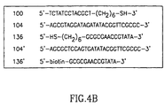
- Fig. 4A The sensor preparation sequence as used in the example can be seen in Fig. 4A, while the sequence of the oligonucleotides used can be seen in Fig. 4B.
- Fig. 4B each oligonucleotide is identified by the reference numeral as used in the examples.
- a mercaptohexyl oligonucleotide 100 (SEQ ID NO: 5 bound to the mercaptohexyl via the 3' end) including a portion 102 that is complementary to a part of the analyte 104 (SEQ ID NO: 6) and the mercapto-derived portion 106 for its assembly as a monolayer on an Au-electrode 108 was used as a capturing agent.
- the mercaptohexyl oligonucleotide 100 was assembled on the Au-electrode 108 as a monolayer, to obtain the sensing interface 110 (step A in Fig. 4A).
- a surface coverage of the electrode of 1.1x10 -11 mole/cm 2 was determined by Tarlov's electrochemical method [Tarlov M.J. et al . Anal. Chem. 70 :4670 (1998)], and comparable results were obtained by QCM analyses.
- the resulting monolayer-functionalized electrode 110 was then brought into contact with a sample containing the target analyte, oligonucleotide 104 (5x10 -6 M, 15 hours incubation, 25°C), to yield a double-stranded assembly 132 (step B in Fig. 4A) wherein at least part ( 130 ) of the assembled analyte is left free for further hybridization.
- oligonucleotide-labeled liposome 134 (lipid concentration 0.2mM, 15 min. 25°C).
- the oligonucleotide moiety 136 (SEQ ID NO: 7, bound to a mercaptohexyl group via the 3' end, Fig. 4B) within the labeled liposome 134 is complementary to the residual base-sequence 130 of the analyte.
- a liposome-linked three-component double-stranded assembly 138 consisting of the capturing agent 100 , the analyte 104 , and the liposome tagged with oligonucleotide 136 , is generated on the electrode support.
- the oligonucleotide-labeled-liposome was prepared by the assembly of liposomes that are composed of phosphatidic acid, phosphatidyl choline, maleimide-phosphatidylethanolamine, cholesterol (marked with 3H-cholesterol, 45 Ci/mole) at a ratio of 79:20:1:0.1, that were modified with oligonucleotide 136 by incubation therewith for 20 hours at 4°C and purified by chromatography (Sephadex G-75).
- Oligreen Molecular probe
- the size of the liposomes was determined by dynamics light-scattering and corresponded to 220 ⁇ 20 nm.
- the oligonucleotide-labeled liposomes 134 are negatively charged in order to eliminate non-specific adsorption of the liposomes onto the sensing interface.
- the liposomes associated with the electrode support represent "giant" negatively charged amplifying agents that electrostatically repel a negatively charged redox-probe stabilized in the electrolyte solution. That is, the biorecognition event between the capturing oligonucleotide 100 and the target oligonucleotide 104 is amplified by the generation of a highly-charged microenvironment that repels the electroactive probe, Fe(CN) 6 3-/4- , in solution. The electron transfer resistance produced by the assembly 138 was then assayed by Faradaic impedance spectroscopy.
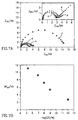
- Fig. 5A shows the impedance spectra (in the form of Nyquist plots, Z im vs. Z re ) of oligonucleotide-functionalized electrode 110 (curve a) after hybridization with the target oligonucleotide 100 to form the layered double-stranded oligonucleotide assembly 132 (curve b), and after interaction with the probing oligonucleotide-labled liposome 136 to form the amplified assembly 138 (curve c). While a bare Au-electrode exhibits an electron transfer resistance of 0.5 k ⁇ , the associated of the capturing oligonucleotide 100 onto the conducting support increased the electron transfer resistance to 3 k ⁇ .
- Fig. 5A further shows the impedance spectrum of the functionalized-electrode 110 after its treatment with the mutant 104' (curve e) and the impedance spectrum of the resulting electrode after further treatment with the oligonucleotide-labeled liposome 134 (curve e).
- the extent of increase in the electron transfer resistance upon the binding of the analyte-oligonucleotide, and the secondary association of the modified liposome is controlled by the bulk concentration of the analyte, as shown in Fig. 5B.
- the sensing system may be further amplified as schematically illustrated in Fig. 6, wherein the presence of target oligonucleotide 104 (SEQ ID NO:1) was detected using the negatively-charged liposomes 142 carrying the biotinylated oligonucleotide 136' (SEQ ID NO:9 bound via the 5' end to biotin). Accordingly, oligonucleotide-functianalized electrode 110 is reacted with the target oligonucleotide (5x10 -6 M, 15 min. of hybridization, at 25°C), pre-treated with biotinylated verification oligonucleotide 112 (SEQ ID NO:5) 1x10 -5 M, interaction time 2 hr.
- target oligonucleotide 104 SEQ ID NO:1
- biotinylated oligonucleotide 136' SEQ ID NO:9 bound via the 5' end to biotin
- step A in Fig. 6
- step A in Fig. 6
- This process results in a three-component double-stranded-assembly on the electrode, consisting of the capturing oligonucleotide 100 , the analyte oligonucleotide 104 and the biotin-labeled oligonucleotide 112 .
- Association of avidin 118 (8 min. of incubation, step B in Fig. 6) and then the biotin-tagged-liposome 142 (8 min. of incubation, step C in Fig.
- the biotin-labeled liposomes were composed of phosphatidyl choline, phosphatidylethanolamine, cholesterol (marked with 3H-cholesterol, 45Ci/mole) and biotinylated phosphatidylethanolamine with a ratio corresponding to 80:20:0.1:0.1.
- the average coverage of the liposomes with biotin corresponded to 550, which were purified by gel chromatography (DEAE Sephadex A-25).
- the size of the liposomes was determined by dynamic light scattering to be 180 ⁇ 40 nm.
- Fig. 7A shows the impedance spectra of the array in the different steps of modification.
- the sensing of the target-DNA was further amplified by the application of a second step of association of the avidin-biotinylated liposomes under the same conditions (step D in Fig. 6), that enhanced the electron transfer resistance, respectively, to 17 k ⁇ and 20 k ⁇ (curves e and f, in Fig. 7).
- the sensing interface was interacted with mutant, non-complementary DNA 104' (SEQ ID NO:4, 5x10 -6 M), pre-treated with biotinylated oligonucleotide 112 and subsequently treated with avidin and the biotinylated liposome, under the same conditions.
- a minute increase in the electron-transfer resistance corresponding to R et 3.4 k ⁇ . was observed, attributed to non-specific adsorption of avidin to the sensing interface.
- oligonucleotide capturing agent was assembled on an Au/quarts crystal.
- the functionalized interface was then hybridized with a target DNA (concentration 5x10 -6 M) followed by interaction thereof with the oligonucleotide-labeled liposome.
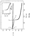
- Fig. 8A shows also the results of a control experiment in which the sensing interface was interacted with the mutated, non-complementary oligonucleotide (5x10 -6 M, point c) followed by treatment with the tagged liposome (point d).
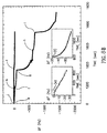
- Fig. 8B shows the results of sensing a target DNA in a sample the manner described in connection with Fig. 6, however also in this case, wherein the a capturing oligonucleotide is assembled on a Au/quarts crystal. Accordingly, first an analyte-double-stranded biotinylated system was associated wit the sensing interface which resulted in a frequency decrease of 25 Hz (curve e). Binding of avidin to the biotinylated assembly yielded a frequency change of ⁇ f ⁇ -50 Hz (point f).
Landscapes
- Chemical & Material Sciences (AREA)
- Organic Chemistry (AREA)
- Life Sciences & Earth Sciences (AREA)
- Zoology (AREA)
- Proteomics, Peptides & Aminoacids (AREA)
- Health & Medical Sciences (AREA)
- Engineering & Computer Science (AREA)
- Wood Science & Technology (AREA)
- Analytical Chemistry (AREA)
- Microbiology (AREA)
- Physics & Mathematics (AREA)
- Molecular Biology (AREA)
- Immunology (AREA)
- Biotechnology (AREA)
- Biophysics (AREA)
- Biochemistry (AREA)
- Bioinformatics & Cheminformatics (AREA)
- General Engineering & Computer Science (AREA)
- General Health & Medical Sciences (AREA)
- Genetics & Genomics (AREA)
- Measuring Or Testing Involving Enzymes Or Micro-Organisms (AREA)
- Investigating Or Analyzing Materials By The Use Of Electric Means (AREA)
Abstract
Description
- The present invention relates to a method and system for the detection of oligonucleotides in a sample.
- The following is a list of prior art references which are relevant for a better understanding of the background of the invention:
- 1. Piunno, P.A.E., Krull, V.J., Hudson, R.H.E., Damha, M.J., Cohen, H., Anal. Chim. Acta, 288 :205-209, (1994).
- 2. Mandenius, C.F., Chollet, A., Lenburg, M.M., Lundström, I., Anal. Lett., 22 :2961-2964, (1989).
- 3. Lidberg, B., Nylander, C., Lundström, I., Sensors and Actuators, 4 :299-302, (1993).
- 4. Jonsson, V., Biotechniques, 11 :620-624, (1991).
- 5. Mikkelsen, S.R., Electroanalysis, 8 :15-23, (1996).
- 6. Millan, K.M.; Sanauloo, A., Mikkelsen, S.R., Anal. Chem., 66 :3830-3833, (1994).
- 7. Hashimoto, K., Ito, K., Ishimori, Y., Anal. Chem., 66 :1236-1241, (1994).
- 8. Hashimoto, K., Ito, K., Ishimori, Y., Anal. Chim. Acta, 286 :219-224, (1994).
- 9. Wang, J., Palecek, E., Nielson, P.E., J. Am. Chem. Soc., 118 :7667-7670, (1996).
- 10. Ihara, T., Nakayama, M., Murata, K., Maeda, M., Chem. Commun., 1069-1070, (1997).
- 11. Bardea, A., Dagan, A., Ben-Dov, I., Amit, B., Willner, I., Chem. Commun., 839-840, (1998).
- 12. PCT Application No. WO 97/04314.
-
- Acknowledgement of these references in the description below will be made by indicating the number from the above list.
- The development of DNA-sensor devices attracts substantial recent research efforts directed to gene analysis, detection of genetic disorders, tissue matching and forensic applications. Optical detection of DNA was accomplished by the application of fluorescence labeled oligonucleotides(1,2) or by the use of surface plasmon resonance(3,4). Electronic transduction of the formation of oligonucleotide complexes with a target DNA, and, particularly, in the quantitative assay of DNA is a major challenge of bioelectronics(5). The organization of DNA-sensors requires the assembly of the sensing interface on a transducer, and the design of the appropriate electronic output that signals the formation of the recognition complex with the target DNA-analyte on the transducer element. Electrochemical DNA sensors based on the electrostatic attraction of electroactive transition metal complexes or organic dyes to oligonucleotide-DNA ds-complexes, e.g. Co(bpy)3 3+, acridin or Hoechst 33258 were reported(6-10). Microgravimetric quartz-crystal- microbalance, QCM(11) analyses were also applied to sense the formation of complementary oligonucleotide-DNA complexes.
- Two major difficulties are still encountered in the development of DNA sensors and relate to the sensitivity and specificity of the resulting sensing systems.
- It is an object of the invention to provide a method and system for detecting target oligonucleotides in a sample.
- The term "detect" or "detection" refers collectively to both a qualitative determination of the presence of the target oligonucleotide in the sample as well as at times for evaluation of the level of the target oligonucleotide in the sample.
- In accordance with the first aspect of the invention there is provided a method for detecting a target oligonucleotide in a sample, comprising:
- (a) providing a sensor device having a sensing interface carrying capturing oligonucleotides having each a nucleotide sequence complementary in at least a portion thereof to a first portion of the target oligonucleotide;
- (b) providing verification oligonucleotides having each a nucleotide sequence complementary in at least a portion thereof to a second portion of the target oligonucleotide, other than said first portion;
- (c) contacting the sample with the sensing interface under conditions which allow the target oligonucleotides, if present in the sample, to hybridize to the capturing oligonucleotides;
- (d) prior to (c) or thereafter, allowing the verification oligonucleotides to hybridize to the target oligonucleotides if present in the sample; and
- (e) detecting the presence of said verification oligonucleotides on the sensing interface. In accordance with another aspect, the present invention provides a system for detecting a target oligonucleotide in a sample, comprising:
- (i) a sensor device having a sensing interface carrying capturing oligonucleotides having each a nucleotide sequence complementary in at least a portion thereof to a first portion of the target oligonucleotides;
- (ii) verification oligonucleotides having each a nucleotide sequence complementary in at least a portion thereof to a second portion of the target oligonucleotide, other than said first portion; and
- (iii) a combination comprising one or both of apparatus and reagents for detecting a verification oligonucleotide bound to the sensing interface.
-
- The sample may be a biological specimen or a fractionation product thereof containing the oligonucleotides; a biological specimen treated to free and solubilized oligonucleotides; a specimen treated in a manner so as to digest nucleotide sequence into smaller oligonucleotides; a sample of oligonucleotides obtained by a PCR (Polymer Chain Reaction) process or any other oligonucleotide amplification process; etc.
- In accordance with one embodiment, the present invention may be applied for a variety of genetic screening assays, such as, for example, screening intended to locate mutant genes.
- In accordance with another embodiment, the invention may be applied for identifying pathogens in a sample.
- There is a wide variety of assaying techniques available for detecting oligonucleotides which are based on hybridizing a probe oligonucleotide to the target oligonucleotide. Also known are assay techniques wherein a probe oligonucleotide is bound to a solid support which hybridize and "fish out" the target oligonucleotide from a tested sample. The invention is however unique in that it makes use of a verification oligonucleotide which increases both specificity and sensitivity of the assay.
- In accordance with the invention, the verification oligonucleotide serves as an indicator for the presence of the target oligonucleotide in the sample. In other words, detection of an immobilized verification oligonucleotide on the surface is an indication that the target oligonucleotide is bound to the sensing surface and hence that it existed in the sample. In accordance with the invention there are thus two discrimination means to ensure specificity and sensitivity:
- 1. Hybridization of the target oligonucleotide to the capturing oligonucleotide on the sensing surface. The complementary sequence of the capturing oligonucleotide will typically, but not exclusively, comprise a number of oligonucleotides completing about one helix of the nucleotide strand, i.e. about twelve nucleotides. A complementary sequence of twelve oligonucleotides ensure on the one hand stable hybridization. On the other hand, a 12-mer oligonucleotide decreases the chance of binding to an incorrect oligonucleotide than a longer sequence. In the case the sample is a digested specimen of genomic DNA, or a fractionation product thereof comprising the oligonucleotides, there is some probability, which increases with the length of the capturing oligonucleotide, of binding to an incorrect oligonucleotide, namely an oligonucleotide other than the target oligonucleotide. This probability is lower, as aforesaid in the case of a shorter oligonucleotide. A sequence of about 12 nucleotides is preferred as it is optimal as far as ensuring binding stability, on the one hand, and reducing incorrect binding on the other hand. The invention is, however, not limited to such a capturing oligonucleotide.
- 2. Hybridization of the verification oligonucleotide to the target oligonucleotide.
-
- These two independent binding events thus reduce the chance of false positive or false negative results.
- The detection of the verification oligonucleotide on the sensing surface may be achieved by a number of means. In accordance with one embodiment of the invention, the sensor device comprises an electrochemical probe for electrical/ electrochemical measurements, e.g. for Faradaic impedance spectroscopy measurement or amperometric detection of the oligonucleotide. In addition, detection may also be carried out by a number of other electrochemical techniques known per se based on the control of interfacial electron transfer rates between the sensing interface and the surrounding medium. For this electrochemical embodiment of the invention, the sensing surface is formed on a conductive matrix on which the capturing oligonucleotides are bound. Such an electrically conducting matrix may for example be made or coated by a metal such as gold, platinum, silver or copper.
- In accordance with another embodiment of the invention, the sensing device is a quartz crystal microbalance (QCM) probe in which case the presence of the verification oligonucleotide on the sensing surface is based on measurement of changes in resonance frequency of the probe. Microgravimetric QCM techniques are known per se, and are described, for example, in PCT Application WO 97/04314(12).
- In accordance with one preferred embodiment of the invention, the verification oligonucleotide is conjugated to a recognition agent which specifically binds to a signal-amplifying agent. The signal-amplifying agent, according to this embodiment, comprises a recognition partner capable of specific binding to the recognition agent. The recognition agent and the recognition partner constitute together a recognition couple. In accordance with another preferred embodiment of the invention the verification oligonucleotide is bound to or complexed directly with a signal-amplifying agent.
- The recognition couple may, for example, be one of the couples selected from the group of biotin-avidin or biotin-streptavidin, receptor-ligand, sugar-lectin, antibody-antigen (the term "antibody" should be understood as referring to a polyclonal or a polyclonal antibody, to a fraction of an antibody comprising the variable, antigen-biotin binding portion, etc.). The recognition agent may be one member of the aforementioned couples, while the recognition partner may then be the other member of the recognition couple.
- In accordance with one embodiment of the invention, the verification oligonucleotide comprises a first recognition agent and the signal-amplifying agent comprises a second recognition agent, with the first and the second recognition agents being the same or different, and both being capable of specific binding to a recognition partner to form a recognition couple. The recognition partner is thus capable of specific binding to both the first and the second recognition agents and thus its introduction to a sensing surface to which the verification oligonucleotide has bound, will yield binding of the signal-amplifying agent to the sensing interface. An example of a recognition partner is avidin or streptavidin, with both the first and second recognition agents being biotin. In accordance with an embodiment of the invention, the signal-amplifying agent comprises a plurality of said second recognition agents and thereby, by a sequence of exposures of the sensing interface to said recognition partner and said signal-amplifying agent, a complex comprising two or more signal amplifying agents bound to each verification oligonucleotide on the sensing interface, may thereby be obtained to yield an increased signal amplification.
- The signal-amplifying agent, according to one embodiment of the invention is a moiety or particle which directly increases the mass immobilized on the sensing surface. The signal-amplifying agent may, for example, comprise molecules, a super molecular structure, or particle, e.g. colloid particles, macromolecules, clusters or molecules, liposomes, etc. In addition, the signal-amplifying agent may also be conjugated to or complexed with a label including, but not limited to an enzyme label. In case of an enzyme label, the enzyme is of a kind that can catalyze a reaction giving rise to an insoluble product. In accordance with this embodiment, the enzyme, after the signal-amplifying agent binds to the recognition agent, is allowed to catalyze a reaction which gives rise to the insoluble product, and the product then precipitates onto the sensing surface. This product may then be detected by a variety of electric-electronic or optical detection means. In an assay carried out in accordance with the electrochemical embodiment, such a precipitate is preferably detected by the large change in electrode impedance resulting therefrom or, alternatively, it may be detected by the mass change on a piezaelectric crystal resulting in a frequency change of the crystal.
- In accordance with an embodiment of the invention, a particle, serving as a single-amplifying agent by its own right, may also carry an enzyme for further amplification of the binding-associated signal. For example, a liposome used as a signal-amplification agent may be bound to or complexed with said enzyme to allow further increase in mass as a result of precipitation of the enzyme-catalyzed insoluble product on the sensing surface, and thus a further amplified binding- related signal.
- The invention also provides, for use in the above method and system, one or more reagents, selected from the group consisting of:
- (i) said verification oligonucleotide;
- (ii) an amplifying agent for amplifying the signal resulting from binding of said verification oligonucleotide to said sensing interface.
-
- The invention will now be described with reference to a non-limiting specific embodiment. As will no doubt be appreciated, this description is a mere illustrative example of the wider scope of the invention as defined in the appended claims.
-
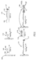
- Fig. 1 illustrates a bioelectronic system in accordance with an embodiment of the invention including assembly of the sensor and its use in detection of an target oligonucleotide in a sample.
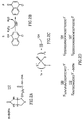
- Figs. 2A-2D illustrate in somewhat more details some of the components of the system of Fig. 1 as used in the exemplary experiments: Fig. 2A shows a DNA strand covalently bound to biotin; Fig. 2B illustrates the chemical reaction, catalyzed by horseradish peroxidase (HRP), in which 4-chloro- naphthol is reacted to form an insoluble product; Fig. 2C shows the structure of the thiophosphate thymine; and Fig. 2D shows the sequence of some oligonucleotides used in the exemplary experiments.
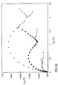
- Figs. 3A and 3B show the impedance features, presented as Nyquist plots, of a bare electrode (curve a), after functionalization of the electrode with the capturing oligonucleotide (curve b), after binding the target DNA and the biotinylated oligonucleotide hybrid (curve c), after interaction with the avidin-HRP conjugate (curve d) and after some period of catalysis of the enzyme resulting in deposit of insoluble product on the sensing surface (curve e). It should be noted that Figs. 3A and 3B are of the same experiment but drawn to different scales.
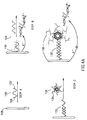
- Fig. 4A illustrates a bioelectronic system in accordance with a further embodiment of the invention including assembly of the sensor and its use in detection of a target oligonucleotide in a sample.
- Fig. 4B shows the sequence of the different oligonucleotides used in a system of the kind illustrated in Fig. 4B, in the accompanying experiments.
- Figs. 5A shows the impedance spectra presented as Nyquist plots of the feature illustrated in Fig. 4, the spectra including: a functionalized electrode carrying the capturing oligonucleotides (curve a); after interacting the functionalized electrode with a sample carrying the target oligonucleotide (curve b); after interaction with the oligonucleotide-functionalized liposome (curve c); after interacting an electrode functionalized with a mutated capturing oligonucleotide (curve d); and after treatment of the mutated capturing oligonucleotide bearing electrode with funcionalized liposome (curve e).
- Fig. 5B shows the changes in electron transfer resistance of a functionalized electrode upon treatment with different concentrations of the target oligonucleotide and amplification with labeled liposomes.
- Fig. 6 illustrates a bioelectronic system in accordance with a yet further embodiment of the invention including assembly of the sensor and its use in detection of a target oligonucleotide in a sample.
- Fig. 7A shows the impedance spectra presented as Nyquist plots of the features illustrated in Fig. 6, the spectra including: a functionalized electrode (curve a); after interaction with a sample carrying the target oligonucleotide pre-treated with a biotin-labeled verification oligonucleotide (curve b); after subsequent interaction with avidin (curve c); after subsequent interaction with biotinylated liposomes (curve d); after subsequent interaction for a second time with avidin (curve e); and after a subsequent interaction for a second time with the biotinylated liposomes (curve f).
- Fig. 7B shows a calibration curve which corresponds to the changes in the electron transfer resistances of the functionalized electrode upon interaction with different concentrations of the target oligonucleotide and enhancement of the detecting processes by a double-step avidin/biotinylated liposome amplification path.
- Fig. 8A shows time-dependent frequency changes of oligonucleotide-bound crystal after interaction with a sample containing the target oligonucleotide (curve a); after interaction of the resulting electrode with an oligonucleotide-labeled liposome (curve b); after functionalizing a crystal with a mutated capturing oligonucleotide (curve c); after bringing into contact the mutated oligonucleotide-labeled crystal with oligonucleotide-labeled liposome (curve d); after treating an oligonucleotide-labeled crystal with oligonucleotide-labeled liposomes (curve e).
- Fig. 8B shows time-dependent frequency changes of oligonucleotide-labeled crystal after interacting with a sample containing the target oligonucleotide (Curve e); after interacting the resulting interface with avidin (curve f); after interacting the resulting assembly with biotinylated liposome (curve h); after further interacting with avidin (curve g); after further interacting with biotinylated liposome (curve i); after treating an oligonucleotide labeled crystal with a mutated oligonucleotide pre-treated with biotinylated liposomes (curve j); after treating the resulting interface (of curve j) with avidin (curve k); and after treating with biotinylated liposomes.
-
- The manner of assembly of the DNA sensor in accordance with an embodiment of the invention and its use, are illustrated in Fig. 1.
Oligonucleotide 100, serving as the capturing oligonucleotide, includes afirst portion 102, typically a 12-base sequence, that is complementary to a first portion of the target oligonucleotide, and asecond portion 106 for binding to the electrode, e.g. a gold (Au)electrode 108. The bindingportion 106 may, for example, be a several base (e.g 5) thiosphosphate thymidine (TS) sequence, illustrated in Fig. 2C. Occasionally the twoportions electrode 108 andoligonucleotide 100 are reacted such thatportion 106 binds to the surface of the Au electrode. As a result, functionalized electrode with asensing surface 110 is formed. - A
verification oligonucleotide 112 is contacted with a sample which contains thetarget oligonucleotide 104 whereby a partial double-strandedstructure 114 is formed. This structure is then contacted with the sensing surface (step B) yielding a bifunctional double-strandedoligonucleotide assembly 116. It should be noted that it is possible in accordance with another embodiment of the invention to first contact thesensing surface 110 with the sample and only then bring a reagent solution which comprises theverification oligonucleotide 112 into contact with the sensing surface. This will first yield binding of the target oligonucleotide 104 (if present in the sample) to the sensing surface and then binding ofverification oligonucleotide 112 to yieldassembly 116. In both cases, the presence of theverification oligonucleotide 116 on the sensing surface serves as an indication of the presence of thetarget oligonucleotide 104 in the sample. - The detection of the verification oligonucleotide on the sensing surface may be achieved by a number of means some of which were explained above. For example, the verification oligonucleotide may carry a label which may be detected electrically, e.g. by determining change in impedance, or electron transport between the
electrode 108 and the surrounding medium. The label, by one embodiment, is an enzyme which can catalyze a reaction yielding an insoluble reaction product which precipitates on the surface's electrode thus increasing impedance. This is illustrated in step C of Fig. 1. - In accordance with one embodiment,
verification oligonucleotide 112 is bound to abiotin moiety 117. Alabel complex 118 which comprises anavidin 119 bound to anenzyme 120 is contacted with the sensing surface (step C) resulting in binding of complex 118 to the sensing surface.Enzyme 120 can catalyze a reaction converting a substrate (S) into an insoluble product (P) which is thus deposited on the sensing surface. Both the binding of thelabeling complex 118 to the sensing surface as well as the precipitation of product (P) onto the sensing surface can be monitored similarly as above (i.e. change of impedance or a change of mass in the case of QCM-type measurement). - In Figs. 4A, 4B and 6, the same reference numerals as used in Fig. 1 are used for like components.
- According to another embodiment illustrated in Fig. 4A, use is made with a verification oligonucleotide-modified
liposome 134 that is bound to the double-strandedimmobilized assembly 132, to form an immobilized double-stranded oligonucleotide-liposome assembly 138. The binding of the labeled verification liposome onto the sensing surface can be monitored as described above. - The sensing of the
target oligonucleotide 104 in accordance with a further embodiment, can be further amplified by using a double-step avidin/biotin-labeled-liposome amplification pathway as shown schematically in Fig. 6.Functionalized electrode 110 is first hybridized withtarget DNA 104 pre-treated with biotin-labeledoligonucleotide 112 having a portion sequence complementary witholigonucleotide 100, immobilized on saidelectrode 108, to form bifunctional double-strandedbiotinylated assembly 116. The formed assembly is then reacted withbiotinylated liposomes 142 to form aliposome containing assembly 144. This assembly can further be reacted with avidin and additional biotinylated liposomes, to yield amulti-liposome assembly 146. - The invention will now be further illustrated by the following example:
- For clarity, in the description below the same reference numeral to those used above will be used. However, by doing so, it should not, in any way, limit the scope of the invention to the specific examples below.
- It should be noted that the scheme shown in Fig. 1 can be employed for various different assays than that specifically exemplified herein. Furthermore, a similar scheme, mutatis mutandis, may also be used for assaying a target oligonucleotide in other assay techniques, e.g. microgravimetric QCM. In this latter case rather than electric/electronic measurements, the measurement is of change in resonance frequency of the piezoelectric crystal as a result of mass change.
- The sensor preparation sequence as used in the Example can be seen in Fig. 1, while the sequences of the oligonucleotides used can be seen in Fig. 2D. In Fig. 2D each oligonucleotide is identified by the reference numerals used in the example.
- An 18-mer oligonucleotide 100 (SEQ ID NO: 1) which included a 12-
base sequence 102 that is complementary to a part of the analyte, the Tay-Sachs (TS) mutant 104 (SEQ ID NO: 2) was used. In addition,oligonucleotide 100 included a 5-base thiophosphate thymine-TS tag 106 for its assembly on the gold (Au)electrode 108, and a single T-base separating the tag from the sensing oligonucleotide sequence. A disc Au-electrode 108, 0.05 cm2, was interacted with oligonucleotide 100 (20 µM, 10 hours) resulting in the assembly of the sensing interface on the gold support (step A in Fig. 1). The resultingfunctionalized electrode 110 was interacted with a solution that included the target analyte, the TS-mutant sequence 104 (5.8 x 10⁻⁷ g/mL-1, 4 hours), and a biotinylated verification oligonucleotide 112 (SEQ ID NO: 3, bound via the 5' end to biotin, Fig. 2D), 2 x 10⁻⁵ g/mL-1 (step B in Fig. 1). -
Verification oligonucleotide 112 is complementary to one portion of anoligonucleotide 104 and consequently these two oligonucleotides hybridize to form a partial double-strandedstructure 114.Target oligonucleotide 104 has another sequence complementary toportion 102 of capturingoligonucleotide 100 and thus step B results in the formation of a bifunctional double-stranded DNA-oligonucleotide assembly 116. - Sensing surface with bifunctional double-stranded DNA-
oligonucleotide assembly 116 is then treated with an avidin labeled with horseradish peroxidase (HRP) (1 x 10⁻⁸ g/mL-1, 3 hours) (step C in Fig. 1). HRP can catalyze the oxidation of 4-chloro-1-naphthol (S) by hydrogen peroxide (H2O2) giving rise to the formation of an insoluble product (P) which precipitates on the electrode. Other enzyme-substrate couples yielding an insoluble product which may be used include: alkaline phosphatase and indoyl phosphate derivatives as substrates; glucose oxidase and tetrazolium salts as substrates; etc. - As the oligonucleotide and oligonucleotide-DNA layered assemblies are negatively charged, the electrostatic repulsion of a negatively-charged redox-probe, e.g. Fe(CN)6 3-/4-, from the electrode support is anticipated to perturb the interfacial electron transfer. This is expected to introduce an electron transfer resistance that can be detected by Faradaic impedance spectroscopy or other electrochemical means such as reduction of the amperometric response of the electrode. The biocatalytic precipitation of the product (P) on the electrode is expected to further insulate the conductive support and to lead to a high interfacial electron transfer resistance or a reduction of the amperometric response of an electroactive species solubilized in the medium surrounding the electrode.
- Fig. 3A shows the impedance features, using Fe(CN)6 3-/4- as redox- probe, presented as Nyquist plots (Zim vs. Zre), of the bare electrode 108 (curve a), of the functionalized electrode with the sensing surface 110 (curve b) and of the layered bifunctional double-stranded oligonucleotide-target DNA and biotinylated oligonucleotide assembly (curve c). The respective semicircle diameters correspond to the interfacial electron transfer resistnaces, Ret. It can be seen that the electron transfer resistance increases upon the build-up of the biotinylated oligonucleotide-DNA assembly. For example, for the functionalized electrode Ret = 1.1 kΩ whereas Ret is increased to about 2.2 kΩ upon the association of the complex 114. These results are consistent with the fact that the negative charge increases upon the two-step organization of the assembly. This results in the enhanced electrostatic repulsion of the redox-probe, and introduces higher interfacial electron transfer resistance.
- Fig. 3B shows the impedance spectra of the bifunctional double-stranded assembly consisting of the target DNA linked to the sensing interface and the biotinylated oligonucleotide, before (curve c) and after (curve d) interaction with the avidin-HRP conjugate. Upon the association of the avidin-HRP biocatalytic conjugate to the layer, a considerable increase in the electron transfer resistance is observed due to the partial insulation of the electrode by the proteins. In the presence of H2O2 and the substrate (S), the biocatalytic precipitation of the product onto the electrode occurs. This insulates the conductive support, resulting in a very high increase in the electron transfer resistance, curve (e), Ret = 17 kΩ. It should be noted that the two parameters controlling the sensitivity of the DNA-sensing devices are the time of incubation of the functionalized-monolayer-
electrode 110 with the complex 114 and more important, the time-interval used to precipitate the product by the avidin-HRP biocatalyic conjugate. Using this configuration, and upon precipitation of P for 40 min. it was possible to sense thetarget DNA 104 at a concentration of 20 x 10-9 g/mL-1, Ret = 7.9 kΩ. - Control experiments show that the oligonucleotide sensing assembly has a high specificity and selectivity. Treatment of the
functinalized electrode 110 with thebiotinylated oligonucleotide 112 and then with the avidin-HRP conjugate 118, but without the interaction with thetarget DNA 104, yielded only a minute change in the electron transfer resistance. - In order to test the specificity of the system, the same assay was performed with a DNA fragment 104' (SEQ ID NO: 4) that corresponds to the normal gene sequence in which the 7-based mutation leads to the TS-genetic disorder. After contact of the sensing interface with a complex between fragment 104' and the
verification oligonucleotide 112, the system was subjected to the biocatalytic precipitation process using the avidin-HRP conjugate, using the same protocol as illustrated in Fig. 1. However, no noticeable changes in the electron's transfer resistance at the electrodes were observed, implying that the lack of formation of a complex between the capturing oligonucleotide on the sensing surface and the complex formed between thetarget oligonucleotide 104 and theverification oligonucleotide 112 which prevented the subsequent formation of the precipitant layer on the electrode. - Cyclic voltammetry experiments (see insert Fig. 3B) further confirm the stepwise organization of the bifunctional double-stranded complex 116, and that the precipitation of the insulating layer formed by product P on the electrode, gradually perturb the electron-transfer kinetics of Fe(CN)6 3-. Fig. 3B inset, shows the cyclic voltammograms of Fe(CN)6 3- at a bare Au- electrode (curve a), upon formation of the sensing assembly 110 (curve b), and upon the formation of the double-stranded assembly 110 (curve c). The stepwise assembly of the layers is accompanied by a decrease in the amperometric response of the electrode and an increase in the peak-to-peak separation between the cathodic and anodic waves of the redox-probe. This is consistent with the enhanced electron transfer barriers introduced upon the assembly of the negatively-charged oligonucleotide assembly. Association of the avidin-HRP conjugate onto the layer (curve d), further separates the redox waves of Fe(CN)6 3- implying that binding of the protein insulates the electrode and perturbs the interfacial electron transfer. Biocatalytic precipitation of P onto the electrode insulates the conductive support, and the electrical response of the redox-probe is almost entirely blocked, (curve e). The result shown in the inset of Fig. 3B demonstrates that amperometric transduction of the formation of the complex 116, binding of avidin-linked
HRP 118, and further precipitation of the product P is possible. - By some modification of the assayed scheme described above, rather than determining the formation of the insoluble product precipitates on the electrode by means of a Faradaic impedance spectroscopy, it may also be determined by means of amperometric detection, by optical means and others or by microgravimetric QCM detection, an example for the latter being provided hereinbelow.
- The sensor preparation sequence as used in the example can be seen in Fig. 4A, while the sequence of the oligonucleotides used can be seen in Fig. 4B. In Fig. 4B, each oligonucleotide is identified by the reference numeral as used in the examples.
- A mercaptohexyl oligonucleotide 100 (SEQ ID NO: 5 bound to the mercaptohexyl via the 3' end) including a
portion 102 that is complementary to a part of the analyte 104 (SEQ ID NO: 6) and the mercapto-derivedportion 106 for its assembly as a monolayer on an Au-electrode 108 was used as a capturing agent. Themercaptohexyl oligonucleotide 100 was assembled on the Au-electrode 108 as a monolayer, to obtain the sensing interface 110 (step A in Fig. 4A). A surface coverage of the electrode of 1.1x10⁻¹¹ mole/cm2 was determined by Tarlov's electrochemical method [Tarlov M.J. et al. Anal. Chem. 70:4670 (1998)], and comparable results were obtained by QCM analyses. The resulting monolayer-functionalizedelectrode 110 was then brought into contact with a sample containing the target analyte, oligonucleotide 104 (5x10⁻⁶ M, 15 hours incubation, 25°C), to yield a double-stranded assembly 132 (step B in Fig. 4A) wherein at least part (130) of the assembled analyte is left free for further hybridization. The resulting electrode interface was then treated with oligonucleotide-labeled liposome 134 (lipid concentration 0.2mM, 15 min. 25°C). The oligonucleotide moiety 136 (SEQ ID NO: 7, bound to a mercaptohexyl group via the 3' end, Fig. 4B) within the labeledliposome 134 is complementary to the residual base-sequence 130 of the analyte. Thus, a liposome-linked three-component double-strandedassembly 138, consisting of the capturingagent 100, theanalyte 104, and the liposome tagged witholigonucleotide 136, is generated on the electrode support. - The oligonucleotide-labeled-liposome was prepared by the assembly of liposomes that are composed of phosphatidic acid, phosphatidyl choline, maleimide-phosphatidylethanolamine, cholesterol (marked with 3H-cholesterol, 45 Ci/mole) at a ratio of 79:20:1:0.1, that were modified with
oligonucleotide 136 by incubation therewith for 20 hours at 4°C and purified by chromatography (Sephadex G-75). The surface coverage of the liposome with oligonucleotide 136 (50-60 oligonucleotide units per liposome) was determined by reacting the resulting liposomes with Oligreen (Molecular probe) and following fluorescence intensity of the resulting liposome suspension at λ=480nm. The size of the liposomes was determined by dynamics light-scattering and corresponded to 220±20 nm. - The oligonucleotide-labeled
liposomes 134 are negatively charged in order to eliminate non-specific adsorption of the liposomes onto the sensing interface. The liposomes associated with the electrode support represent "giant" negatively charged amplifying agents that electrostatically repel a negatively charged redox-probe stabilized in the electrolyte solution. That is, the biorecognition event between the capturingoligonucleotide 100 and thetarget oligonucleotide 104 is amplified by the generation of a highly-charged microenvironment that repels the electroactive probe, Fe(CN)6 3-/4-, in solution. The electron transfer resistance produced by theassembly 138 was then assayed by Faradaic impedance spectroscopy. - Fig. 5A shows the impedance spectra (in the form of Nyquist plots, Zim vs. Zre) of oligonucleotide-functionalized electrode 110 (curve a) after hybridization with the
target oligonucleotide 100 to form the layered double-stranded oligonucleotide assembly 132 (curve b), and after interaction with the probing oligonucleotide-labledliposome 136 to form the amplified assembly 138 (curve c). While a bare Au-electrode exhibits an electron transfer resistance of 0.5 kΩ, the associated of the capturingoligonucleotide 100 onto the conducting support increased the electron transfer resistance to 3 kΩ. This is attributed to the electrostatic repulsion of the redox label, Fe(CN)6 3-/4-, that results in a barrier for the interfacial electron transfer. The formation of the double-stranded assembly with the target oligonucleotide increased the electron-transfer resistance to Ret=4.5 k Ω. This is consistent with the results presented by Example 1, hereinabove and with the fact that the higher negative charge formed on the surface as a result of hybridization, enhances the electrostatic repulsion of the electroactive species on the solution. Binding of the oligonucleotide-modifiedliposome 134 introduced a very high electron transfer resistance corresponding to 15 kΩ. This result is attributed to the formation of a negatively charged micro-interface upon the association of the liposome to the double-stranded assembly. - A control experiment, for the evaluation of the system's specificity, was conducted, which included an attempt to detect the presence of an oligonucleotide 104' (SEQ ID NO: 8), that included a 6-base mutation relative to the
target DNA 104. Fig. 5A further shows the impedance spectrum of the functionalized-electrode 110 after its treatment with the mutant 104' (curve e) and the impedance spectrum of the resulting electrode after further treatment with the oligonucleotide-labeled liposome 134 (curve e). As may be understood from the results presented in Fig. 5A, the interfacial electron transfer resistances were almost unchanged in this control experiment, implying that the sensing interface is selective for analyses oftarget oligonucleotide 104. The results also indicate that no non-specific association of mutant 104' or of the oligonucleotide-labeledliposomes 134 on the electrode took place. This is attributed to the electrostatic repulsion existing between these components and the sensing interface. - The extent of increase in the electron transfer resistance upon the binding of the analyte-oligonucleotide, and the secondary association of the modified liposome is controlled by the bulk concentration of the analyte, as shown in Fig. 5B. the lower sensitivity limit for analyzing the analyte DNA was determined to be 1.2x10⁻¹² M at a signal-to-noise value of S/N = 3.
- A further control experiment, where only the
oligonucleotide 136 interacted with the double-stranded assembly of the capturingoligonucleotide 100 and thetarget oligonucleotide 104 introduced only a small increase in the electron transfer resistance, Ret = 4.7 kΩ., indicating that the negatively charged liposome indeed amplified the electrostatic repulsion of the redox label. - The sensing system may be further amplified as schematically illustrated in Fig. 6, wherein the presence of target oligonucleotide 104 (SEQ ID NO:1) was detected using the negatively-charged
liposomes 142 carrying the biotinylated oligonucleotide 136' (SEQ ID NO:9 bound via the 5' end to biotin). Accordingly, oligonucleotide-functianalized electrode 110 is reacted with the target oligonucleotide (5x10⁻⁶ M, 15 min. of hybridization, at 25°C), pre-treated with biotinylated verification oligonucleotide 112 (SEQ ID NO:5) 1x10⁻⁵ M,interaction time 2 hr. 25°C), being complementary tosegment 102 of the target oligonucleotide (step A, in Fig. 6). This process results in a three-component double-stranded-assembly on the electrode, consisting of the capturingoligonucleotide 100, theanalyte oligonucleotide 104 and the biotin-labeledoligonucleotide 112. Association of avidin 118 (8 min. of incubation, step B in Fig. 6) and then the biotin-tagged-liposome 142 (8 min. of incubation, step C in Fig. 6) resulted in the formation of a negatively-changedinterface 144 that amplified the primary oligonucleotide recognition event by the electrostatic repulsion of Fe(CN)6 3-/4- and the enhancement of the interfacial electron transfer resistance. This sensing configuration enabled the further amplification of the biorecognition event by the multiple reaction of the resulting array with avidin and then with the biotinylated liposomes to yield a dense array of the negatively-charged liposomes. The biotin-labeled liposomes were composed of phosphatidyl choline, phosphatidylethanolamine, cholesterol (marked with 3H-cholesterol, 45Ci/mole) and biotinylated phosphatidylethanolamine with a ratio corresponding to 80:20:0.1:0.1. The average coverage of the liposomes with biotin corresponded to 550, which were purified by gel chromatography (DEAE Sephadex A-25). The size of the liposomes was determined by dynamic light scattering to be 180±40 nm. - Fig. 7A shows the impedance spectra of the array in the different steps of modification. The oligonucleotide-functionalized
interface 108 exhibited an electron transfer resistance corresponding to 3 kΩ (curve a), and upon the formation of the double-stranded assembly with the analyte-DNA 104 complexed with thebiotinylated oligonucleotide 112 to form immobilizedbiotinylated analyte 116, the electron transfer resistance increased to Ret = 4.8 kΩ. (curve b). Association of avidin 119 (2.5µg/ml) to theinterface 116 further increased the electron transfer resistance to 7.6 kΩ, as a result of the hydrophobic, insulating features of the protein (curve c). Association of the biotin-labeled liposome to thesensing surface 30 min. lipid concentration 0.25 mM), substantially increased the electron transfer resistance, Ret = 14.8 kΩ. (curve d). - The sensing of the target-DNA was further amplified by the application of a second step of association of the avidin-biotinylated liposomes under the same conditions (step D in Fig. 6), that enhanced the electron transfer resistance, respectively, to 17 kΩ and 20 kΩ (curves e and f, in Fig. 7).
- In a control experiment, the sensing interface was interacted with mutant, non-complementary DNA 104' (SEQ ID NO:4, 5x10⁻⁶M), pre-treated with
biotinylated oligonucleotide 112 and subsequently treated with avidin and the biotinylated liposome, under the same conditions. A minute increase in the electron-transfer resistance corresponding to Ret = 3.4 kΩ. was observed, attributed to non-specific adsorption of avidin to the sensing interface. - The increase in the electron-transfer resistance at the electrode upon binding of avidin and the biotin-labeled liposome, were controlled by the bulk concentration of the target-DNA in the sample (Fig. 7B).
- Using a double-step avidin/biotin-labeled-liposome amplification pathway, target DNA concentration as low as 5x10-14 (signal to noise ratio S/N=3) was detected.
- In a similar manner to that described with reference to Fig. 4A, an oligonucleotide capturing agent was assembled on an Au/quarts crystal. The functionalized interface was then hybridized with a target DNA (concentration 5x10⁻⁶M) followed by interaction thereof with the oligonucleotide-labeled liposome. Fig. 8A (solid line) shows QCM-transduction of the amplified sensing of the analyte. Interaction of the functionalized crystal with the analyte (point a) resulted in a frequency decrease of Δf = 17Hz, implying a surface coverage of the analyte corresponding to 1.2x10⁻¹¹ mole/cm2. Further reaction of the double-stranded surface with the oligonucleotide-tagged liposome (point b) resulted in a substantial decrease in the crystal frequency, Δf=-120 Hz.
- Fig. 8A shows also the results of a control experiment in which the sensing interface was interacted with the mutated, non-complementary oligonucleotide (5x10⁻⁶ M, point c) followed by treatment with the tagged liposome (point d). As shown, the crystal frequency was unchanged Δf=±2 Hz upon interaction with the non-complementary DNA. Interaction with the tagged liposome slightly altered the crystal frequency, Δf=-5 Hz. This frequency change may be attributed to minute non-specific binding of the liposome to the interface. Association of the amplified oligonucleotide-tagged liposome with the interface resulted in a frequency change of Δf=-70 Hz (point e), that allows the easy amplified detection of the target DNA also by using microgravimetric QCM assay. The lowest sensitivity limit for the detection of the target DNA by this amplification method was estimated to be 5x10-12 M (Δf= -20 Hz, after treatment with tagged liposome).
- Fig. 8B shows the results of sensing a target DNA in a sample the manner described in connection with Fig. 6, however also in this case, wherein the a capturing oligonucleotide is assembled on a Au/quarts crystal. Accordingly, first an analyte-double-stranded biotinylated system was associated wit the sensing interface which resulted in a frequency decrease of 25 Hz (curve e). Binding of avidin to the biotinylated assembly yielded a frequency change of Δf∼-50 Hz (point f). Linkage of the biotin-tagged liposomes to the system amplified the primary association of the analyte and a very high frequency change Δf∼-500 Hz was observed (point g). Additional treatment of the interface with avidin, Δf∼-50 Hz (point h) and then with the biotin-labeled liposome (point i) resulted in a second amplification corresponding to Δf=690 Hz.
- Treatment of the sensing interface with the biotin-labeled non-complementary DNA did not yield any significant frequency change (point j) and subsequent interaction of the resulting assembly with avidin and the biotin-tagged liposome resulted in a frequency change of about -30 Hz (points k and 1, respectively). As described above, this change in frequency may be attributed to non-specific association of the liposome to the interface. Using the two-step amplification pathway, the lowest sensitivity limit for sensing the target DNA was estimated to be 1x10-13M (or 1x10-16 mole/ml), which may be further enhanced by performing additional binding steps of avidin-biotinylated liposome.
-
- <110> YISSUM RESEARCH DEVELOPMENT COMPANY
- <120> METHOD AND SYSTEM FOR DETECTING OLIGONUCLEOTIDES IN A SAMPLE
- <130> Yissum(Wilner)-1206184
- <140> PCT/IL 99/00649
<141> 1999-12-01 - <150> IL127346
<151> 1998-12-01 - <150> IL132966
<151> 1999-11-16 - <160> 9
- <170> PatentIn Ver. 2.1
- <210> 1
<211> 13
<212> DNA
<213> HUMAN - <400> 1

- <210> 2
<211> 26
<212> DNA
<213> HUMAN - <400> 2

- <210> 3
<211> 14
<212> DNA
<213> HUMAN - <400> 3

- <210> 4
<211> 26
<212> DNA
<213> HUMAN - <400> 4

- <210> 5
<211> 13
<212> DNA
<213> HUMAN - <400> 5

- <210> 6
<211> 27
<212> DNA
<213> HUMAN - <400> 6

- <210> 7
<211> 14
<212> DNA
<213> HUMAN - <400> 7

- <210> 8
<211> 27
<212> DNA
<213> HUMAN - <400> 8

- <210> 9
<211> 14
<212> DNA
<213> HUMAN - <400> 9

-
Claims (43)
- A method for detecting a target oligonucleotide in a sample, comprising:(a) providing a sensor device having a sensing interface carrying capturing oligonucleotides having each a nucleotide sequence complementary in at least a stably hybridizing portion thereof to a first portion of the target oligonucleotides wherein said sensor device comprises an electrochemical probe carrying the sensing interface;(b) providing verification oligonucleotides having each a nucleotide sequence complementary in at least a stably hybridizing portion thereof to a second portion of the target oligonucleotide, other than said first portion;(c) contacting the sample with the sensing interface under conditions such as to allow the target oligonucleotides, if present in the sample, to hybridize to the capturing oligonucleotides;(d) prior to (c) or thereafter, allowing the verification oligonucleotides to hybridize to the target oligonucleotides if present in the sample; and(e) detecting the presence of said verification oligonucleotides on the sensing interface.
- The method of Claim 1, wherein said detection is based on Faradaic impedance spectroscopy or amperometric measurements.
- The method of either of Claims 1 or 2, wherein the sequence complementary to at least a stably hybridizing portion of the target oligonucleotide is of about 12 nucleotides.
- The method according of any one of Claims 1-3, wherein the verification oligonucleotide is conjugated to a recognition agent which can specifically bind to a signal-amplifying agent, and step (e) of the method comprises:(e1) contacting the sensing interface with said signal-amplifying agent;(e2) detecting the presence of said signal-amplifying agent on the sensing interface.
- The method of Claim 4, wherein said recognition agent is biotin and said signal amplifying agent comprises avidin.
- The method of any one of Claims 1-3, wherein said verification oligonucleotide is bound to or complexed with a signal-amplifying agent, and step(e) comprises detecting of presence of the signal-amplifying agent on the sensing interface.
- The method of any one of Claims 1-3, wherein the verification oligonucleotide comprises a first recognition agent which specifically binds to a recognition partner to form a recognition couple, step (e) of the method comprises the following steps:(e1) contacting said sensing interface with said recognition partner;(e2) contacting said sensing interface with a signal-amplifying agent comprising a second recognition agent, which may be the same or different as the first recognition agent, which can also bind to said recognition partner; and(e3) detecting presence of said signal-amplifying agent on said sensing interface.
- The method of Claim 7, comprising the following step between steps (e2) and (e3):(e2.1) repeating steps (e1) and (e2) one or more times.
- The method of any one of Claims 4-8, wherein said signal-amplifying agent comprises an enzyme which catalyzes a reaction yielding an insoluble reaction product, and step (e) comprises:(ea) providing conditions permitting catalytic activity of said enzyme to yield formation of said insoluble reaction product; and(eb) detecting the presence of said insoluble reaction product on said sensing interface.
- The method of any one of Claims 4-8, wherein said signal-amplifying agent comprises a moiety or a particle which directly increases the mass immobilized on the sensing surface, the method comprises in step (e):(ea) detecting the presence of said moiety or particle on said sensing interface.
- The method of Claim 10, wherein said moiety is a molecule or a super molecular structure.
- A system for detecting a target oligonucleotide in a sample, comprising:(i) a sensor device having a sensing interface carrying capturing oligonucleotides having each a nucleotide sequence complementary in at least a stably hybridizing portion thereof to a first portion of the target oligonucleotides wherein said sensor device is an electrochemical electrode carrying said sensing interface;(ii) verification oligonucleotides having each a nucleotide sequence complementary in at least a stably hybridizing portion thereof to a second portion of the target oligonucleotide, other than said first portion; and(iii) a detecting means comprising one or both of apparatus and reagents for detecting a verification oligonucleotide bound to the sensing interface.
- The system of Claim 12, wherein when the system comprises an apparatus, said apparatus is adapted for the performance of an electrochemical measurement.
- The system of either of Claims 12 or 13, wherein said capturing oligonucleotide has a nucleotide sequence complementary to said first portion which has a length of about 12 nucleotides.
- The system of any one of Claims 12-14, wherein the verification oligonucleotide is conjugated to a recognition agent which specifically binds to a signal-amplifying agent.
- The system of Claim 15, wherein said recognition agent is biotin and said signal-amplifying agent comprises avidin.
- The system of any one of Claims 12-14, wherein the verification oligonucleotide is conjugated or complexed with a signal-amplifying agent.
- A system of any one of Claims 12-14, wherein the verification oligonucleotide is conjugated to a first recognition agent, which specifically binds to a recognition partner, the recognition partner being capable of binding also to a second recognition agent, being the same or different from said first recognition agent; the system further comprises a signal amplifying agent comprising a second recognition agent.
- A system of Claim 18, when said first and said second recognition agents are biotin and where said recognition partner is avidin or streptavidin.
- A system of any one of Claims 12-19, wherein said signal-amplifying agent comprises an enzyme which catalyzes a reaction yielding an insoluble reaction product.
- A system of any one of Claims 12-19, wherein said signal-amplifying agent comprises a particle or moiety which directly increases the mass immobilized on the sensing interface.
- A method for detecting a target oligonucleotide in a sample, comprising:(a) providing a sensor device having a sensing interface carrying capturing oligonucleotides having each a nucleotide sequence complementary in at least a stably hybridizing portion thereof to a first portion of the target oligonucleotides;(b) providing verification oligonucleotides having each a nucleotide sequence complementary in at least a stably hybridizing portion thereof to a second portion of the target oligonucleotide, other than said first portion, wherein the verification oligonucleotide is capable of binding to a signal-amplifying agent comprising a liposome,(c) contacting the sample with the sensing interface under conditions so as to allow the target oligonucleotides, if present in the sample, to hybridize to the capturing oligonucleotides;(d) prior to (c) or thereafter, allowing the verification oligonucleotides to hybridize to the target oligonucleotides if present in the sample;(e) contacting the sensing interface with said signal-amplifying agent; and(f) detecting the presence of said signal-amplifying agent on the sensing interface.
- The method of Claim 22 , wherein said sensor device comprises an electrochemical probe carrying the sensing interface.
- The method of Claim 23, wherein said detection is based on Faradaic impedance spectroscopy or amperometric measurements.
- The method of Claim 22, wherein said sensor device comprises a microbalance quaitz-crystal probe carrying the sensing interface.
- The method of Claim 25, wherein said detection is based on a microgravimetric quartz-crystal microbalance (QCM) analysis.
- The method of any one of Claims 22-26, wherein the sequence complementary to at least a stably hybridizing portion of the target oligonucleotide is of about 12 nucleotides.
- The method according of any one of Claims 22-27, wherein the verification oligonucleotide is conjugated to a recognition agent which can specifically bind to said signal-amplifying agent.
- The method of Claim 28 wherein said recognition agent is biotin and said signal amplifying agent comprises avidin.
- The method of any one of Claims 22-27, wherein said verification oligonucleotide is bound to or complexed with said signal-amplifying agent.
- The method of any one of Claims 22-27, wherein the verification oligonucleotide comprises a first recognition agent which specifically binds to a recognition partner to form a recognition couple, step (e) of the method comprising the following steps:(e1) contacting said sensing interface with said recognition partner;(e2) contacting said sensing interface with said signal-amplifying agent comprising a second recognition agent, which may be the same or different as the frst recognition agent, which can also bind to said recognition partner.
- The method of Claim 31, comprising the following step after step (e2):(e2.1) repeating steps (e1) and (e2) one or more times.
- A system for detecting a target oligonucleotide in a sample, comprising:(i) a sensor device having a sensing interface carrying capturing oligonucleotides having each a nucleotide sequence complementary in at least a stably hybridizing portion thereof to a first portion of the target oligonucleotides;(ii) verification oligonucleotides having each a nucleotide sequence complementary in at least a stably hybridizing portion thereof to a second portion of the target oligonucleotide, other than said first portion, wherein the verification oligonucleotide is capable of binding to a signal-amplifying agent comprising a liposome; and(iii) a detecting means comprising one or both of apparatus and reagent for detecting a verification oligonucleotide bound to the sensing interface, wherein said detecting means comprises said signal-amplifying agent comprising a liposome.
- The systems of Claim 33, wherein said sensor device is an electrochemical electrode carrying said sensing surface.
- The system of Claim 33 or 34 wherein said apparatus is adapted for the performance of an electrochemical measurement.
- The system of Claim 33, wherein said sensor device comprises a microbalance quartz-crystal probe carrying the sensing interface.
- The system according to Claim 36, wherein said detection is based on a microgravimetric quartz-crystal microbalance (QCM) analysis.
- The system of Claims 33-37, wherein said capturing oligonucleotide has a nucleotide sequence complementary to said first portion which has a length of about 12 nucleotides.
- The system of any one of Claims 33-38, wherein the verification oligonucleotide is conjugated to a recognition agent which specifically binds to the signal-amplifying agent.
- The system of Claim 39, wherein said recognition agent is biotin and said signal-amplifying agent comprises avidin.
- The system of any one of Claims 33-38, wherein the verification oligonucleotide is conjugated or complexed with the signal-amplifying agent.
- A system of any one of Claims 33-38, wherein the verification oligonucleotide is conjugated to a first recognition agent, which specifically binds to a recognition partner, the recognition partner being capable of binding also to a second recognition agent, being the same or different from said first recognition agent; the system further comprises the signal amplifying agent comprising a second recognition agent.
- A system of Claim 42, when said first and said second recognition agents are biotin and where said recognition partner is avidin or streptavidin.
Applications Claiming Priority (5)
| Application Number | Priority Date | Filing Date | Title |
|---|---|---|---|
| IL12734698 | 1998-12-01 | ||
| IL12734698A IL127346A (en) | 1998-12-01 | 1998-12-01 | Method and system for detecting oligonucleotides in a sample |
| IL13296699A IL132966A0 (en) | 1998-12-01 | 1999-11-16 | Method and system for detecting oligonucleotides in a sample |
| IL13296699 | 1999-11-16 | ||
| PCT/IL1999/000649 WO2000032813A1 (en) | 1998-12-01 | 1999-12-01 | Method and system for detecting oligonucleotides in a sample |
Publications (2)
| Publication Number | Publication Date |
|---|---|
| EP1135533A1 EP1135533A1 (en) | 2001-09-26 |
| EP1135533B1 true EP1135533B1 (en) | 2004-04-21 |
Family
ID=26323752
Family Applications (1)
| Application Number | Title | Priority Date | Filing Date |
|---|---|---|---|
| EP99973075A Expired - Lifetime EP1135533B1 (en) | 1998-12-01 | 1999-12-01 | Method and system for detecting oligonucleotides in a sample |
Country Status (9)
| Country | Link |
|---|---|
| EP (1) | EP1135533B1 (en) |
| JP (1) | JP2002531100A (en) |
| AT (1) | ATE264923T1 (en) |
| AU (1) | AU770025B2 (en) |
| CA (1) | CA2353562A1 (en) |
| DE (1) | DE69916650T2 (en) |
| ES (1) | ES2221488T3 (en) |
| IL (1) | IL132966A0 (en) |
| WO (1) | WO2000032813A1 (en) |
Families Citing this family (18)
| Publication number | Priority date | Publication date | Assignee | Title |
|---|---|---|---|---|
| EP1409728A2 (en) * | 2001-04-09 | 2004-04-21 | Fraunhofer-Gesellschaft zur Förderung der angewandten Forschung e.V. | Non-enzymatic liposome-linked closely spaced array electrodes assay (nel-ela) for detecting and quantifying nucleic acids |
| GB0205455D0 (en) | 2002-03-07 | 2002-04-24 | Molecular Sensing Plc | Nucleic acid probes, their synthesis and use |
| EP1512009A2 (en) * | 2002-05-08 | 2005-03-09 | Yissum Research Development Company Of The Hebrew University Of Jerusalem | Magneto-controlled method and system for determining an analyte in a liquid medium |
| US7097751B2 (en) | 2002-08-23 | 2006-08-29 | Kabushiki Kaisha Toshiba | Base sequence detecting electrode, base sequence detecting device and base sequence detecting method |
| DE10256898B4 (en) * | 2002-11-29 | 2006-01-12 | Senslab-Gesellschaft Zur Entwicklung Und Herstellung Bioelektrochemischer Sensoren Mbh | Electrochemical detection of nucleic acids |
| EP1583518A1 (en) * | 2003-01-06 | 2005-10-12 | Bioinvent International AB | Immobilisation of dna-labelled lipid vesicles on dna arrays |
| JPWO2006033400A1 (en) * | 2004-09-22 | 2008-05-15 | ダイキン工業株式会社 | DNA detection method and reporter probe |
| JP4561451B2 (en) * | 2005-04-15 | 2010-10-13 | セイコーエプソン株式会社 | Polymorphism detection method and polymorphism detection kit |
| JP4561478B2 (en) * | 2005-05-26 | 2010-10-13 | セイコーエプソン株式会社 | Polymorph detection method, polymorph detection kit |
| DE102006003603B4 (en) * | 2006-01-25 | 2010-02-04 | Fraunhofer-Gesellschaft zur Förderung der angewandten Forschung e.V. | Crosslinkable multifunctional supports for (low molecular weight) ligands and their use in analytics, as well as processes for their preparation and crosslinking |
| JP2010517541A (en) | 2007-02-06 | 2010-05-27 | アトノミックス アクティーゼルスカブ | Reporter unit for detecting target molecules using a polymerizable substrate |
| CN101241097B (en) * | 2007-09-18 | 2011-08-03 | 中国科学院上海应用物理研究所 | Electrochemical DNA detection method adopting stem-loop structure for detecting probe, and reagent kit thereof |
| DE102010000906B4 (en) * | 2009-01-22 | 2012-07-12 | RUHR-UNIVERSITäT BOCHUM | Conjugates for the detection of double-stranded nucleic acids |
| WO2011069997A2 (en) * | 2009-12-09 | 2011-06-16 | Iti Scotland Limited | Detecting analytes |
| CN104698043A (en) * | 2013-12-09 | 2015-06-10 | 重庆医科大学 | An electrochemical biosensor for detecting DNA of escherichia coli and a preparing method thereof |
| JP6803629B2 (en) * | 2017-07-31 | 2021-01-06 | 国立大学法人大阪大学 | Method for detecting target substances present in solution and system for detection |
| CN111044580B (en) * | 2019-12-20 | 2021-10-01 | 中国农业大学 | A method for fast and real-time monitoring of the degree of salting of freshwater fish |
| JP7801754B2 (en) * | 2022-02-16 | 2026-01-19 | 株式会社エヌエフホールディングス | METHOD FOR MEASURING TARGET NUCLEIC ACID SEQUENCE IN A TEST SAMPLE |
Family Cites Families (2)
| Publication number | Priority date | Publication date | Assignee | Title |
|---|---|---|---|---|
| US5695926A (en) * | 1990-06-11 | 1997-12-09 | Bio Merieux | Sandwich hybridization assays using very short capture probes noncovalently bound to a hydrophobic support |
| US5474895A (en) * | 1990-11-14 | 1995-12-12 | Siska Diagnostics Inc. | Non-isotopic detection of nucleic acids using a polystyrene support-based sandwich hybridization assay and compositions useful therefor |
-
1999
- 1999-11-16 IL IL13296699A patent/IL132966A0/en not_active IP Right Cessation
- 1999-12-01 DE DE69916650T patent/DE69916650T2/en not_active Expired - Fee Related
- 1999-12-01 ES ES99973075T patent/ES2221488T3/en not_active Expired - Lifetime
- 1999-12-01 AU AU14074/00A patent/AU770025B2/en not_active Ceased
- 1999-12-01 CA CA002353562A patent/CA2353562A1/en not_active Abandoned
- 1999-12-01 AT AT99973075T patent/ATE264923T1/en not_active IP Right Cessation
- 1999-12-01 EP EP99973075A patent/EP1135533B1/en not_active Expired - Lifetime
- 1999-12-01 WO PCT/IL1999/000649 patent/WO2000032813A1/en not_active Ceased
- 1999-12-01 JP JP2000585444A patent/JP2002531100A/en active Pending
Also Published As
| Publication number | Publication date |
|---|---|
| WO2000032813A1 (en) | 2000-06-08 |
| JP2002531100A (en) | 2002-09-24 |
| IL132966A0 (en) | 2001-03-19 |
| DE69916650T2 (en) | 2005-04-21 |
| ES2221488T3 (en) | 2004-12-16 |
| ATE264923T1 (en) | 2004-05-15 |
| DE69916650D1 (en) | 2004-05-27 |
| EP1135533A1 (en) | 2001-09-26 |
| AU770025B2 (en) | 2004-02-12 |
| CA2353562A1 (en) | 2000-06-08 |
| AU1407400A (en) | 2000-06-19 |
Similar Documents
| Publication | Publication Date | Title |
|---|---|---|
| AU770025B2 (en) | Method and system for detecting oligonucleotides in a sample | |
| Paleček et al. | New approaches in the development of DNA sensors: hybridization and electrochemical detection of DNA and RNA at two different surfaces | |
| Gooding | Electrochemical DNA hybridization biosensors | |
| Patolsky et al. | Highly sensitive amplified electronic detection of DNA by biocatalyzed precipitation of an insoluble product onto electrodes | |
| Paleček et al. | Electrochemistry of nucleic acids and development of DNA sensors | |
| Odenthal et al. | An introduction to electrochemical DNA biosensors | |
| Wang et al. | Magnetic bead-based label-free electrochemical detection of DNA hybridization | |
| Willner et al. | Electronic aptamer‐based sensors | |
| Slinker et al. | Multiplexed DNA-modified electrodes | |
| US20040023258A1 (en) | Method and system or detecting nucleic acids | |
| US11807893B2 (en) | Method and electronic device for determining the concentration of an analyte | |
| EP1512009A2 (en) | Magneto-controlled method and system for determining an analyte in a liquid medium | |
| Xu et al. | Microfabricated disposable DNA sensors based on enzymatic amplification electrochemical detection | |
| Can et al. | Thermodynamically designed target-specific DNA probe as an electrochemical hybridization biosensor | |
| Fojta et al. | A Single‐Surface Electrochemical Biosensor for the Detection of DNA Triplet Repeat Expansion | |
| US20100133118A1 (en) | Electrochemical methods of detecting nucleic acid hybridization | |
| EP1604042A2 (en) | Method and device for detecting biomolecules | |
| K’Owino et al. | Metal-enhanced biosensor for genetic mismatch detection | |
| Jin et al. | Site-specific DNA cleavage of EcoRI endounclease probed by electrochemical analysis using ferrocene capped gold nanoparticles as reporter | |
| US20060275756A1 (en) | Displacement assay for detecting ligate-ligand association events | |
| US7655404B2 (en) | Method and device for detection of nucleic acids and/or polypeptides | |
| EP1018646A2 (en) | Quantitative analysis of biochemical compound utilizing electrochemical reaction | |
| IL127346A (en) | Method and system for detecting oligonucleotides in a sample | |
| US20040096859A1 (en) | Method for detecting and/or quantifying an analyte | |
| Willner et al. | Amplified and specific electronic transduction of DNA sensing processes in monolayer and thin-films assemblies |
Legal Events
| Date | Code | Title | Description |
|---|---|---|---|
| PUAI | Public reference made under article 153(3) epc to a published international application that has entered the european phase |
Free format text: ORIGINAL CODE: 0009012 |
|
| 17P | Request for examination filed |
Effective date: 20010628 |
|
| AK | Designated contracting states |
Kind code of ref document: A1 Designated state(s): AT BE CH CY DE DK ES FI FR GB GR IE IT LI LU MC NL PT SE |
|
| AX | Request for extension of the european patent |
Free format text: AL;LT;LV;MK;RO;SI |
|
| GRAP | Despatch of communication of intention to grant a patent |
Free format text: ORIGINAL CODE: EPIDOSNIGR1 |
|
| GRAS | Grant fee paid |
Free format text: ORIGINAL CODE: EPIDOSNIGR3 |
|
| GRAA | (expected) grant |
Free format text: ORIGINAL CODE: 0009210 |
|
| RAP1 | Party data changed (applicant data changed or rights of an application transferred) |
Owner name: YISSUM RESEARCH DEVELOPMENT COMPANYOF THE HEBREW U |
|
| AK | Designated contracting states |
Kind code of ref document: B1 Designated state(s): AT BE CH CY DE DK ES FI FR GB GR IE IT LI LU MC NL PT SE |
|
| PG25 | Lapsed in a contracting state [announced via postgrant information from national office to epo] |
Ref country code: FI Free format text: LAPSE BECAUSE OF FAILURE TO SUBMIT A TRANSLATION OF THE DESCRIPTION OR TO PAY THE FEE WITHIN THE PRESCRIBED TIME-LIMIT Effective date: 20040421 Ref country code: CY Free format text: LAPSE BECAUSE OF FAILURE TO SUBMIT A TRANSLATION OF THE DESCRIPTION OR TO PAY THE FEE WITHIN THE PRESCRIBED TIME-LIMIT Effective date: 20040421 Ref country code: AT Free format text: LAPSE BECAUSE OF FAILURE TO SUBMIT A TRANSLATION OF THE DESCRIPTION OR TO PAY THE FEE WITHIN THE PRESCRIBED TIME-LIMIT Effective date: 20040421 |
|
| REG | Reference to a national code |
Ref country code: GB Ref legal event code: FG4D |
|
| REG | Reference to a national code |
Ref country code: CH Ref legal event code: EP |
|
| REG | Reference to a national code |
Ref country code: IE Ref legal event code: FG4D |
|
| REF | Corresponds to: |
Ref document number: 69916650 Country of ref document: DE Date of ref document: 20040527 Kind code of ref document: P |
|
| PG25 | Lapsed in a contracting state [announced via postgrant information from national office to epo] |
Ref country code: SE Free format text: LAPSE BECAUSE OF FAILURE TO SUBMIT A TRANSLATION OF THE DESCRIPTION OR TO PAY THE FEE WITHIN THE PRESCRIBED TIME-LIMIT Effective date: 20040721 Ref country code: GR Free format text: LAPSE BECAUSE OF FAILURE TO SUBMIT A TRANSLATION OF THE DESCRIPTION OR TO PAY THE FEE WITHIN THE PRESCRIBED TIME-LIMIT Effective date: 20040721 Ref country code: DK Free format text: LAPSE BECAUSE OF FAILURE TO SUBMIT A TRANSLATION OF THE DESCRIPTION OR TO PAY THE FEE WITHIN THE PRESCRIBED TIME-LIMIT Effective date: 20040721 |
|
| REG | Reference to a national code |
Ref country code: CH Ref legal event code: NV Representative=s name: BOVARD AG PATENTANWAELTE |
|
| LTIE | Lt: invalidation of european patent or patent extension |
Effective date: 20040421 |
|
| PG25 | Lapsed in a contracting state [announced via postgrant information from national office to epo] |
Ref country code: LU Free format text: LAPSE BECAUSE OF NON-PAYMENT OF DUE FEES Effective date: 20041201 |
|
| REG | Reference to a national code |
Ref country code: ES Ref legal event code: FG2A Ref document number: 2221488 Country of ref document: ES Kind code of ref document: T3 |
|
| PG25 | Lapsed in a contracting state [announced via postgrant information from national office to epo] |
Ref country code: MC Free format text: LAPSE BECAUSE OF NON-PAYMENT OF DUE FEES Effective date: 20041231 |
|
| ET | Fr: translation filed | ||
| PLBE | No opposition filed within time limit |
Free format text: ORIGINAL CODE: 0009261 |
|
| STAA | Information on the status of an ep patent application or granted ep patent |
Free format text: STATUS: NO OPPOSITION FILED WITHIN TIME LIMIT |
|
| 26N | No opposition filed |
Effective date: 20050124 |
|
| PGFP | Annual fee paid to national office [announced via postgrant information from national office to epo] |
Ref country code: IE Payment date: 20060530 Year of fee payment: 7 Ref country code: GB Payment date: 20060530 Year of fee payment: 7 |
|
| PGFP | Annual fee paid to national office [announced via postgrant information from national office to epo] |
Ref country code: NL Payment date: 20060608 Year of fee payment: 7 Ref country code: FR Payment date: 20060608 Year of fee payment: 7 Ref country code: CH Payment date: 20060608 Year of fee payment: 7 |
|
| PGFP | Annual fee paid to national office [announced via postgrant information from national office to epo] |
Ref country code: DE Payment date: 20060616 Year of fee payment: 7 |
|
| PGFP | Annual fee paid to national office [announced via postgrant information from national office to epo] |
Ref country code: ES Payment date: 20060629 Year of fee payment: 7 |
|
| PGFP | Annual fee paid to national office [announced via postgrant information from national office to epo] |
Ref country code: BE Payment date: 20060712 Year of fee payment: 7 |
|
| PG25 | Lapsed in a contracting state [announced via postgrant information from national office to epo] |
Ref country code: IE Free format text: LAPSE BECAUSE OF NON-PAYMENT OF DUE FEES Effective date: 20061201 |
|
| PG25 | Lapsed in a contracting state [announced via postgrant information from national office to epo] |
Ref country code: LI Free format text: LAPSE BECAUSE OF NON-PAYMENT OF DUE FEES Effective date: 20061231 Ref country code: CH Free format text: LAPSE BECAUSE OF NON-PAYMENT OF DUE FEES Effective date: 20061231 Ref country code: BE Free format text: LAPSE BECAUSE OF NON-PAYMENT OF DUE FEES Effective date: 20061231 |
|
| PGFP | Annual fee paid to national office [announced via postgrant information from national office to epo] |
Ref country code: IT Payment date: 20061231 Year of fee payment: 8 |
|
| PG25 | Lapsed in a contracting state [announced via postgrant information from national office to epo] |
Ref country code: NL Free format text: LAPSE BECAUSE OF NON-PAYMENT OF DUE FEES Effective date: 20070701 |
|
| PG25 | Lapsed in a contracting state [announced via postgrant information from national office to epo] |
Ref country code: DE Free format text: LAPSE BECAUSE OF NON-PAYMENT OF DUE FEES Effective date: 20070703 |
|
| REG | Reference to a national code |
Ref country code: CH Ref legal event code: PL |
|
| GBPC | Gb: european patent ceased through non-payment of renewal fee |
Effective date: 20061201 |
|
| NLV4 | Nl: lapsed or anulled due to non-payment of the annual fee |
Effective date: 20070701 |
|
| REG | Reference to a national code |
Ref country code: IE Ref legal event code: MM4A |
|
| REG | Reference to a national code |
Ref country code: FR Ref legal event code: ST Effective date: 20070831 |
|
| PG25 | Lapsed in a contracting state [announced via postgrant information from national office to epo] |
Ref country code: GB Free format text: LAPSE BECAUSE OF NON-PAYMENT OF DUE FEES Effective date: 20061201 |
|
| BERE | Be: lapsed |
Owner name: *YISSUM RESEARCH DEVELOPMENT CY OF THE HEBREW UNIV Effective date: 20061231 |
|
| PG25 | Lapsed in a contracting state [announced via postgrant information from national office to epo] |
Ref country code: PT Free format text: LAPSE BECAUSE OF NON-PAYMENT OF DUE FEES Effective date: 20040921 |
|
| REG | Reference to a national code |
Ref country code: ES Ref legal event code: FD2A Effective date: 20061202 |
|
| PG25 | Lapsed in a contracting state [announced via postgrant information from national office to epo] |
Ref country code: FR Free format text: LAPSE BECAUSE OF NON-PAYMENT OF DUE FEES Effective date: 20070102 Ref country code: ES Free format text: LAPSE BECAUSE OF NON-PAYMENT OF DUE FEES Effective date: 20061202 |
|
| PG25 | Lapsed in a contracting state [announced via postgrant information from national office to epo] |
Ref country code: IT Free format text: LAPSE BECAUSE OF NON-PAYMENT OF DUE FEES Effective date: 20071201 |