EP1129344B1 - Methods and formulations for the separation of biological macromolecules - Google Patents
Methods and formulations for the separation of biological macromolecules Download PDFInfo
- Publication number
- EP1129344B1 EP1129344B1 EP99958855A EP99958855A EP1129344B1 EP 1129344 B1 EP1129344 B1 EP 1129344B1 EP 99958855 A EP99958855 A EP 99958855A EP 99958855 A EP99958855 A EP 99958855A EP 1129344 B1 EP1129344 B1 EP 1129344B1
- Authority
- EP
- European Patent Office
- Prior art keywords
- separation
- matrix
- concentration
- electrophoresis
- dna
- Prior art date
- Legal status (The legal status is an assumption and is not a legal conclusion. Google has not performed a legal analysis and makes no representation as to the accuracy of the status listed.)
- Expired - Lifetime
Links
- 238000000926 separation method Methods 0.000 title claims abstract description 44
- 239000000203 mixture Substances 0.000 title claims abstract description 41
- 238000009472 formulation Methods 0.000 title claims abstract description 32
- 238000000034 method Methods 0.000 title claims abstract description 21
- 229920002521 macromolecule Polymers 0.000 title abstract description 6
- 238000005251 capillar electrophoresis Methods 0.000 claims abstract description 20
- 229920002401 polyacrylamide Polymers 0.000 claims abstract description 19
- 239000011159 matrix material Substances 0.000 claims description 49
- XSQUKJJJFZCRTK-UHFFFAOYSA-N Urea Chemical compound NC(N)=O XSQUKJJJFZCRTK-UHFFFAOYSA-N 0.000 claims description 29
- 239000004202 carbamide Substances 0.000 claims description 29
- 238000001962 electrophoresis Methods 0.000 claims description 28
- HNJBEVLQSNELDL-UHFFFAOYSA-N pyrrolidin-2-one Chemical compound O=C1CCCN1 HNJBEVLQSNELDL-UHFFFAOYSA-N 0.000 claims description 26
- 239000003398 denaturant Substances 0.000 claims description 24
- 239000000243 solution Substances 0.000 claims description 22
- 208000034953 Twin anemia-polycythemia sequence Diseases 0.000 claims description 21
- KCXVZYZYPLLWCC-UHFFFAOYSA-N EDTA Chemical compound OC(=O)CN(CC(O)=O)CCN(CC(O)=O)CC(O)=O KCXVZYZYPLLWCC-UHFFFAOYSA-N 0.000 claims description 20
- 239000007983 Tris buffer Substances 0.000 claims description 20
- LENZDBCJOHFCAS-UHFFFAOYSA-N tris Chemical compound OCC(N)(CO)CO LENZDBCJOHFCAS-UHFFFAOYSA-N 0.000 claims description 20
- 239000000872 buffer Substances 0.000 claims description 13
- 102000039446 nucleic acids Human genes 0.000 claims description 9
- 108020004707 nucleic acids Proteins 0.000 claims description 9
- 150000007523 nucleic acids Chemical class 0.000 claims description 9
- 238000012163 sequencing technique Methods 0.000 claims description 9
- 238000004458 analytical method Methods 0.000 claims description 8
- ZMXDDKWLCZADIW-UHFFFAOYSA-N dimethylformamide Substances CN(C)C=O ZMXDDKWLCZADIW-UHFFFAOYSA-N 0.000 claims description 5
- 108020004999 messenger RNA Proteins 0.000 claims description 2
- 239000012634 fragment Substances 0.000 description 59
- 108020004414 DNA Proteins 0.000 description 41
- 241000606768 Haemophilus influenzae Species 0.000 description 21
- ZHNUHDYFZUAESO-UHFFFAOYSA-N Formamide Chemical compound NC=O ZHNUHDYFZUAESO-UHFFFAOYSA-N 0.000 description 20
- 238000004513 sizing Methods 0.000 description 20
- 102000053602 DNA Human genes 0.000 description 18
- 229920000642 polymer Polymers 0.000 description 18
- 238000013508 migration Methods 0.000 description 14
- 230000005012 migration Effects 0.000 description 14
- 230000037230 mobility Effects 0.000 description 12
- 238000001712 DNA sequencing Methods 0.000 description 10
- 229920000663 Hydroxyethyl cellulose Polymers 0.000 description 9
- 108020004682 Single-Stranded DNA Proteins 0.000 description 8
- 230000002547 anomalous effect Effects 0.000 description 8
- 238000007906 compression Methods 0.000 description 8
- 230000006835 compression Effects 0.000 description 8
- 239000000499 gel Substances 0.000 description 8
- 238000007873 sieving Methods 0.000 description 8
- 229940113088 dimethylacetamide Drugs 0.000 description 7
- 230000000694 effects Effects 0.000 description 7
- 239000003792 electrolyte Substances 0.000 description 7
- 238000011160 research Methods 0.000 description 7
- 108091092878 Microsatellite Proteins 0.000 description 6
- 230000008901 benefit Effects 0.000 description 6
- 238000004925 denaturation Methods 0.000 description 5
- 230000036425 denaturation Effects 0.000 description 5
- 230000001419 dependent effect Effects 0.000 description 5
- 238000002347 injection Methods 0.000 description 5
- 239000007924 injection Substances 0.000 description 5
- 238000002156 mixing Methods 0.000 description 5
- 238000002360 preparation method Methods 0.000 description 5
- 239000012146 running buffer Substances 0.000 description 5
- 239000000126 substance Substances 0.000 description 5
- 230000007023 DNA restriction-modification system Effects 0.000 description 4
- 239000003795 chemical substances by application Substances 0.000 description 4
- 229940047650 haemophilus influenzae Drugs 0.000 description 4
- XLYOFNOQVPJJNP-UHFFFAOYSA-N water Substances O XLYOFNOQVPJJNP-UHFFFAOYSA-N 0.000 description 4
- RZVAJINKPMORJF-UHFFFAOYSA-N Acetaminophen Chemical compound CC(=O)NC1=CC=C(O)C=C1 RZVAJINKPMORJF-UHFFFAOYSA-N 0.000 description 3
- 150000001408 amides Chemical class 0.000 description 3
- 230000007423 decrease Effects 0.000 description 3
- 238000001502 gel electrophoresis Methods 0.000 description 3
- 239000011521 glass Substances 0.000 description 3
- 238000000329 molecular dynamics simulation Methods 0.000 description 3
- 229920003023 plastic Polymers 0.000 description 3
- 239000005297 pyrex Substances 0.000 description 3
- 239000004354 Hydroxyethyl cellulose Substances 0.000 description 2
- 230000001594 aberrant effect Effects 0.000 description 2
- 230000002411 adverse Effects 0.000 description 2
- 239000012491 analyte Substances 0.000 description 2
- 238000003491 array Methods 0.000 description 2
- 238000004364 calculation method Methods 0.000 description 2
- 238000001818 capillary gel electrophoresis Methods 0.000 description 2
- 238000006243 chemical reaction Methods 0.000 description 2
- 238000004587 chromatography analysis Methods 0.000 description 2
- 238000000576 coating method Methods 0.000 description 2
- 239000008367 deionised water Substances 0.000 description 2
- 229910021641 deionized water Inorganic materials 0.000 description 2
- 238000001704 evaporation Methods 0.000 description 2
- 230000008020 evaporation Effects 0.000 description 2
- 238000011049 filling Methods 0.000 description 2
- 230000017525 heat dissipation Effects 0.000 description 2
- 230000020169 heat generation Effects 0.000 description 2
- 235000019447 hydroxyethyl cellulose Nutrition 0.000 description 2
- 238000005259 measurement Methods 0.000 description 2
- 229910052757 nitrogen Inorganic materials 0.000 description 2
- 238000013021 overheating Methods 0.000 description 2
- 108090000623 proteins and genes Proteins 0.000 description 2
- 102000004169 proteins and genes Human genes 0.000 description 2
- 238000007619 statistical method Methods 0.000 description 2
- HRPVXLWXLXDGHG-UHFFFAOYSA-N Acrylamide Chemical compound NC(=O)C=C HRPVXLWXLXDGHG-UHFFFAOYSA-N 0.000 description 1
- 229920000936 Agarose Polymers 0.000 description 1
- 244000105975 Antidesma platyphyllum Species 0.000 description 1
- 102000016928 DNA-directed DNA polymerase Human genes 0.000 description 1
- 108010014303 DNA-directed DNA polymerase Proteins 0.000 description 1
- FXHOOIRPVKKKFG-UHFFFAOYSA-N N,N-Dimethylacetamide Chemical compound CN(C)C(C)=O FXHOOIRPVKKKFG-UHFFFAOYSA-N 0.000 description 1
- 108091034117 Oligonucleotide Proteins 0.000 description 1
- 241000944748 Quesada Species 0.000 description 1
- JLCPHMBAVCMARE-UHFFFAOYSA-N [3-[[3-[[3-[[3-[[3-[[3-[[3-[[3-[[3-[[3-[[3-[[5-(2-amino-6-oxo-1H-purin-9-yl)-3-[[3-[[3-[[3-[[3-[[3-[[5-(2-amino-6-oxo-1H-purin-9-yl)-3-[[5-(2-amino-6-oxo-1H-purin-9-yl)-3-hydroxyoxolan-2-yl]methoxy-hydroxyphosphoryl]oxyoxolan-2-yl]methoxy-hydroxyphosphoryl]oxy-5-(5-methyl-2,4-dioxopyrimidin-1-yl)oxolan-2-yl]methoxy-hydroxyphosphoryl]oxy-5-(6-aminopurin-9-yl)oxolan-2-yl]methoxy-hydroxyphosphoryl]oxy-5-(6-aminopurin-9-yl)oxolan-2-yl]methoxy-hydroxyphosphoryl]oxy-5-(6-aminopurin-9-yl)oxolan-2-yl]methoxy-hydroxyphosphoryl]oxy-5-(6-aminopurin-9-yl)oxolan-2-yl]methoxy-hydroxyphosphoryl]oxyoxolan-2-yl]methoxy-hydroxyphosphoryl]oxy-5-(5-methyl-2,4-dioxopyrimidin-1-yl)oxolan-2-yl]methoxy-hydroxyphosphoryl]oxy-5-(4-amino-2-oxopyrimidin-1-yl)oxolan-2-yl]methoxy-hydroxyphosphoryl]oxy-5-(5-methyl-2,4-dioxopyrimidin-1-yl)oxolan-2-yl]methoxy-hydroxyphosphoryl]oxy-5-(5-methyl-2,4-dioxopyrimidin-1-yl)oxolan-2-yl]methoxy-hydroxyphosphoryl]oxy-5-(6-aminopurin-9-yl)oxolan-2-yl]methoxy-hydroxyphosphoryl]oxy-5-(6-aminopurin-9-yl)oxolan-2-yl]methoxy-hydroxyphosphoryl]oxy-5-(4-amino-2-oxopyrimidin-1-yl)oxolan-2-yl]methoxy-hydroxyphosphoryl]oxy-5-(4-amino-2-oxopyrimidin-1-yl)oxolan-2-yl]methoxy-hydroxyphosphoryl]oxy-5-(4-amino-2-oxopyrimidin-1-yl)oxolan-2-yl]methoxy-hydroxyphosphoryl]oxy-5-(6-aminopurin-9-yl)oxolan-2-yl]methoxy-hydroxyphosphoryl]oxy-5-(4-amino-2-oxopyrimidin-1-yl)oxolan-2-yl]methyl [5-(6-aminopurin-9-yl)-2-(hydroxymethyl)oxolan-3-yl] hydrogen phosphate Polymers Cc1cn(C2CC(OP(O)(=O)OCC3OC(CC3OP(O)(=O)OCC3OC(CC3O)n3cnc4c3nc(N)[nH]c4=O)n3cnc4c3nc(N)[nH]c4=O)C(COP(O)(=O)OC3CC(OC3COP(O)(=O)OC3CC(OC3COP(O)(=O)OC3CC(OC3COP(O)(=O)OC3CC(OC3COP(O)(=O)OC3CC(OC3COP(O)(=O)OC3CC(OC3COP(O)(=O)OC3CC(OC3COP(O)(=O)OC3CC(OC3COP(O)(=O)OC3CC(OC3COP(O)(=O)OC3CC(OC3COP(O)(=O)OC3CC(OC3COP(O)(=O)OC3CC(OC3COP(O)(=O)OC3CC(OC3COP(O)(=O)OC3CC(OC3COP(O)(=O)OC3CC(OC3COP(O)(=O)OC3CC(OC3COP(O)(=O)OC3CC(OC3CO)n3cnc4c(N)ncnc34)n3ccc(N)nc3=O)n3cnc4c(N)ncnc34)n3ccc(N)nc3=O)n3ccc(N)nc3=O)n3ccc(N)nc3=O)n3cnc4c(N)ncnc34)n3cnc4c(N)ncnc34)n3cc(C)c(=O)[nH]c3=O)n3cc(C)c(=O)[nH]c3=O)n3ccc(N)nc3=O)n3cc(C)c(=O)[nH]c3=O)n3cnc4c3nc(N)[nH]c4=O)n3cnc4c(N)ncnc34)n3cnc4c(N)ncnc34)n3cnc4c(N)ncnc34)n3cnc4c(N)ncnc34)O2)c(=O)[nH]c1=O JLCPHMBAVCMARE-UHFFFAOYSA-N 0.000 description 1
- 125000000217 alkyl group Chemical group 0.000 description 1
- 238000013459 approach Methods 0.000 description 1
- 239000007864 aqueous solution Substances 0.000 description 1
- 230000015572 biosynthetic process Effects 0.000 description 1
- 238000004422 calculation algorithm Methods 0.000 description 1
- 239000007795 chemical reaction product Substances 0.000 description 1
- 150000001875 compounds Chemical class 0.000 description 1
- 229920006037 cross link polymer Polymers 0.000 description 1
- 150000001923 cyclic compounds Chemical class 0.000 description 1
- 238000007405 data analysis Methods 0.000 description 1
- 238000000354 decomposition reaction Methods 0.000 description 1
- 230000003247 decreasing effect Effects 0.000 description 1
- 238000001514 detection method Methods 0.000 description 1
- 229910003460 diamond Inorganic materials 0.000 description 1
- 239000010432 diamond Substances 0.000 description 1
- 238000010790 dilution Methods 0.000 description 1
- 239000012895 dilution Substances 0.000 description 1
- 239000012153 distilled water Substances 0.000 description 1
- 230000009977 dual effect Effects 0.000 description 1
- 239000000975 dye Substances 0.000 description 1
- 230000005684 electric field Effects 0.000 description 1
- 230000002255 enzymatic effect Effects 0.000 description 1
- 238000011067 equilibration Methods 0.000 description 1
- 125000001495 ethyl group Chemical group [H]C([H])([H])C([H])([H])* 0.000 description 1
- 230000005284 excitation Effects 0.000 description 1
- 230000001747 exhibiting effect Effects 0.000 description 1
- 238000002474 experimental method Methods 0.000 description 1
- 238000001917 fluorescence detection Methods 0.000 description 1
- 238000003205 genotyping method Methods 0.000 description 1
- 150000004676 glycans Chemical class 0.000 description 1
- 235000009424 haa Nutrition 0.000 description 1
- 231100001261 hazardous Toxicity 0.000 description 1
- 229920006158 high molecular weight polymer Polymers 0.000 description 1
- 238000011065 in-situ storage Methods 0.000 description 1
- 230000003993 interaction Effects 0.000 description 1
- 238000011835 investigation Methods 0.000 description 1
- 238000004519 manufacturing process Methods 0.000 description 1
- 239000000463 material Substances 0.000 description 1
- 239000013001 matrix buffer Substances 0.000 description 1
- 125000002496 methyl group Chemical group [H]C([H])([H])* 0.000 description 1
- 238000010369 molecular cloning Methods 0.000 description 1
- 229920005615 natural polymer Polymers 0.000 description 1
- 125000004433 nitrogen atom Chemical group N* 0.000 description 1
- 238000007899 nucleic acid hybridization Methods 0.000 description 1
- 239000006259 organic additive Substances 0.000 description 1
- 230000008723 osmotic stress Effects 0.000 description 1
- 229920001282 polysaccharide Polymers 0.000 description 1
- 239000005017 polysaccharide Substances 0.000 description 1
- 239000000843 powder Substances 0.000 description 1
- 230000008569 process Effects 0.000 description 1
- 102000004196 processed proteins & peptides Human genes 0.000 description 1
- 108090000765 processed proteins & peptides Proteins 0.000 description 1
- 239000000047 product Substances 0.000 description 1
- 108091008146 restriction endonucleases Proteins 0.000 description 1
- 238000010008 shearing Methods 0.000 description 1
- 239000007787 solid Substances 0.000 description 1
- 230000003595 spectral effect Effects 0.000 description 1
- 238000006467 substitution reaction Methods 0.000 description 1
- 229920001059 synthetic polymer Polymers 0.000 description 1
- 238000012360 testing method Methods 0.000 description 1
- 231100000331 toxic Toxicity 0.000 description 1
- 230000002588 toxic effect Effects 0.000 description 1
- 238000012546 transfer Methods 0.000 description 1
- WFKWXMTUELFFGS-UHFFFAOYSA-N tungsten Chemical compound [W] WFKWXMTUELFFGS-UHFFFAOYSA-N 0.000 description 1
- 230000004304 visual acuity Effects 0.000 description 1
Images





Classifications
-
- G—PHYSICS
- G01—MEASURING; TESTING
- G01N—INVESTIGATING OR ANALYSING MATERIALS BY DETERMINING THEIR CHEMICAL OR PHYSICAL PROPERTIES
- G01N27/00—Investigating or analysing materials by the use of electric, electrochemical, or magnetic means
- G01N27/26—Investigating or analysing materials by the use of electric, electrochemical, or magnetic means by investigating electrochemical variables; by using electrolysis or electrophoresis
- G01N27/416—Systems
- G01N27/447—Systems using electrophoresis
- G01N27/44704—Details; Accessories
- G01N27/44747—Composition of gel or of carrier mixture
Definitions
- the present invention relates generally to methods and formulations for the separation of biological macromolecules, and in particular, to the separation to single-stranded DNA fragments according to their size using capillary electrophoresis with improved polyacrylamide matrixes.
- Capillary electrophoresis has demonstrated its advantages over standard slab gel based electrophoretic techniques as a rapid, high-throughput and high-resolution method for separation of biological macromolecules, such as proteins, peptides and nucleic acids (see e.g. Slater et al ., Electrophoresis, 1998. 19(10), 1525-1541.)
- Capillary gel electrophoresis (CGE) is the CE-analog of traditional slab-gel electrophoresis and is most often used for size-based separation of biological macromolecules such as oligonucleotides, DNA restriction fragments and proteins.
- the separation is performed by filling the capillary with a sieving matrix, for example, cross-linked polyacrylamide, agarose or linear polymer solutions.
- a sieving matrix for example, cross-linked polyacrylamide, agarose or linear polymer solutions.
- Solutions of polymers may then be prepared using various background electrolyte systems according to the requirements of a particular application.
- the versatility gained by the use of such entangled polymer solutions allows for a wide range of electraphoresis conditions to be utilized, such as elevated capillary temperature, without damaging the matrix structure (see, e . g ., Kleparnik et al ., Electrophoresis, 1996. 17(12):1860-1866).
- cross-linked gels are usually polymerized in situ under less than ideal conditions. Typically, these polymers are not fully characterized, and their quality varies from run-to-run.
- Cross-linked gel filled capillaries are also very prone to bubble formation due to temperature or osmotic stress. Additionally, replacement requires arduous removal, scrupulous clean up of the old material.
- DNA separation applications e . g . DNA sequencing, short tandem repeat [STR] analysis, differential display
- STR short tandem repeat
- CE replaceable polymer matrixes.
- replaceable linear polyacrylamide has been shown to provide unsurpassed separation efficiencies (see, e . g ., Pariat et al ., Journal of Chromatography, 1993. 652(1):57-66), it also exhibits sequence-specific anomalous migration of both double-stranded and single-stranded DNA (see, e . g ., Berka et al ., Electrophoresis, 1995.
- US 5,503,722 relates to linear polyacrylamide compositions which are useful as a separation medium in capillary electrophoresis, and which have urea as a denaturing agent. This document constitutes the closest prior art.
- WO 93/15394 relates to capillary columns for capillary electrophoresis whcih may include denaturing agents.
- WO 95/06668 relates to a method for sequencing nucleic acids in polysaccharide gel media in the presence of denaturing agents.
- This invention discloses methods of biological macromolecule separation and formulations for linear polymer separation matrices exhibiting improved DNA denaturation properties for use in capillary array electrophoresis.
- a combination of urea with organic denaturants 2-pyrrolidinone, N,N'-dimethylacetamide, or N,N'-dimethylformamide resulted in a chemical environment strongly disfavoring base pairing of nucleic acids. Separation of single-stranded DNA fragments in these matrixes minimizes effects of DNA strand secondary structures on fragment mobilities, thus greatly improving the size-dependent separation.
- this invention discloses optimized background electrolyte concentration and run voltage to further improve size-dependent migration of DNA fragments, in addition to enhanced denaturing properties of polymer matrices.
- Biological applications of capillary electrophoresis such as differential display of mRNA, dideoxyfingerprinting, STR sizing and DNA sequencing would strongly benefit from using such formulations.
- the main prerequisite for reliable and accurate size determination of an unknown analyte is comparing the mobility of any given unknown fragment with the mobility of internal standards separated in the same capillary.
- the use of internal standards for DNA analysis has been facilitated by multiple color fluorescence detection.
- the accuracy of size determination of an unknown analyte depends on the linear relationship between DNA fragment size and electrophoretic mobility. If either sizing standard peaks or DNA fragments to be sized exhibit aberrant electrophoretic mobilities, incorrect size determinations will result. Sequence-dependent anomalous mobility of double-stranded DNA fragments is a well-known phenomenon, which reflects functionally important DNA topologies (see, e.g. , Berka et al ., Electrophoresis, 1995.
- CE capillary electrophoresis
- two basic ways of increasing separation denaturing capacity are (i) adding chemicals known to denature DNA into the running buffer, and (ii) increasing column temperature.
- chemical denaturants should be stable, non-hazardous and should not affect the sieving properties of the polymer matrix.
- Increasing column temperature above 40 - 60° C may cause some technical difficulties especially for capillary array instruments.
- Urea has been used as the most common denaturant for ssDNA separations in concentrations up to 8 M, however, it does not provide for complete denaturing of some difficult sequencing compressions and size-dependent migration of some ssDNA sizing standards (see, e.g. , Kleparnik supra; and Rosenblum supra.)
- Formamide is often used in addition to urea both in slab gel and CE separations of ssDNA in order to create strong denaturing conditions (see, e.g. , Ruiz-Martinez supra). Although it decreases the viscosity of polymer matrixes, thereby making them easier to replace into capillaries, formamide is toxic and unstable, decomposes rapidly into charged species at elevated temperature and decreases the lifetime of capillary coatings.
- Denaturing effectiveness of amides increases with lengthening of the alkyl chain as well as with substitution of methyl or ethyl groups on the nitrogen atom (see, e.g ., Levine et al ., supra ).
- double stranded DNA denatures preferably at low ionic strength buffers
- most standard electrophoresis buffer systems are designed at about 0.1 M concentration in order to reduce the risk of overheating due to Joule heat generation.
- CE due to its excellent intrinsic Joule heat dissipation properties, may utilize buffers with reduced ionic strength, thereby further improving denaturing properties of a particular replaceable matrix / denaturant system.
- the formulations in this invention contain a high concentration of organic denaturants in a linear polyacrylamide solution to obtain high resolution and enhanced denaturation of single stranded DNA fragments.
- the preferred formulations should include linear polyacrylamide (1-3% w/w) and organic additives such as, for example, (i) 2-pyrrolidinone, (ii) N',N'-dimethylformamide, and (iii) N',N'-dimethylacetamide at 5-40% (w/w) in combination with 6 M urea, with buffer concentrations between 0.1-300 mM TAPS-Tris-EDTA. Table 1 summarizes the composition of matrixes used in this invention.
- Matrixes used in the Invention Matrix Linear poly-acrylamide (%w/w) Denaturant composition Buffer Composition MegaBACE Gel Matrix (HEC) (Amersham/ Pharmacia) - 6M urea, 10% (w/w) formamide 30mM Tris:100mM TAPS: 1mM EDTA MegaBACETM Long read matrix (Amersham/ Pharmacia) 3% 6M urea 30mM Tris:100mM TAPS: 1mM EDTA LPA formulation LPA-PYR 2.0% 6M urea, 20% (w/w) 2-pyrrolidinone 30mM Tris:100mM TAPS: 1mM EDTA LPA formulation LPA-DMA 2.0% 6M urea, 20% (w/w) N',N'-dimethylaceta mide 30mM Tris:100mM TAPS: 1mM EDTA LPA formulation LPA-DMF 2.0% 6M urea, 20% (w/w) N', N'
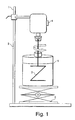
- FIG. 1 shows the schematic drawing of a matrix mixing device used in this invention. The preparation of the matrix formulations is described in detail in the EXAMPLES section of this invention.
- the restriction enzyme digest products from H. influenzae genomic DNA were used as a standard in order to determine a relative size of 250 bp and 340 bp fragments from the GeneScan TM ROX 500 ladder (Applied Biosystems, Foster City, CA)(see FIG. 2).
- the expected fragments from the enzymatic digest are known since the genome of H. influenzae has been completely sequenced and is a publicly available sequence (see, e . g ., Fleischmann et al ., Science, 1995. 269:496-512).
- a base to frame conversion algorithm was produced using the theoretical size of the peaks generated from the H. influenzae digest and the migration time (in frames) of the peaks on the electropherogram.
- FIG. 2 shows the relative size determined for the 250 base fragment in various formulations of this invention and compares it with commercially available matrix formulations.
- the apparent length of the 250 bp fragment is 243 bp when electrophoresed in the 3% linear polyacrylamide matrix with only 6 M urea as a denaturant.
- Addition of a second organic denaturing agent along with the 6 M urea in the matrix buffer affects the apparent length of the 250 bp fragment in a positive way, i . e ., making its apparent length closer to the theoretical value of 250 bp.
- 2-pyrrolidinone at 20% (w/w) concentration corrects the anomalous mobility of this fragment by 6 bp as compared to 3% LPA; 6 M urea matrix.
- N', N'-dimethylformamide, and N',N'-dimethylacetamide are highly effective as denaturants as well, resulting in apparent lengths of 248.3 and 248.4 bp, respectively.
- Formamide has been shown to improve DNA sequencing compressions in concentration of up to 30% in LPA matrixes for capillary electrophoresis (see, e.g. , Ruiz-Martinez et al ., Analytical Chemistry, 1993.
- formamide containing matrixes are highly unstable at room temperature or elevated column temperatures due to decomposition and also decrease the lifetime of capillary coatings.
- the comparison of matrixes in FIG. 2 may also reflect different properties of the two polymers (HEC vs. LPA) and their concentrations.
- 2-pyrrolidinone has been chosen as the most effective denaturant for more detailed investigation since the most positive tendency was observed on migration of ROX 250 bp fragment.
- concentration of 2-pyrrolidinone in a separation matrix was varied from 5% to 20%, while keeping the urea concentration constant at 6 M. Increasing the urea concentration above 6 M is not practical due to its limited solubility.
- up to 20% (weight) of 2-pyrrolidinone could be mixed into the starting 3% LPA solution, resulting in 2% LPA concentration and allowing for background electrolyte concentration compensation of 100 mM TAPS; 30 mM Tris; 1 mM EDTA.
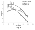
- FIG. 4 presents a plot of resolution versus fragment length for three sieving matrices including: two commercially available: LPA (Amhersham -Pharmacia) HEC ( Amhersham-Pharmacia) and the embodiment formulation (containing 20% 2-pyrrolidinone). From this figure, it can be observed that the 2% LPA matrix containing 20% 2-pyrrolidinone exhibits superior resolution to the commercial LPA matrix in the region beyond 450 bp while maintaining resolution > 2.0 between 100 and 350 bases, more than sufficient to obtain reliable sequence reads or fragment analysis data.
- LPA Amhersham -Pharmacia
- HEC Amhersham-Pharmacia
- separation matrixes were prepared with a background electrolyte concentrations 1 x (30 mM Tris, 100 mM TAPS, 1 mM EDTA), 0.86 x (26 mM Tris, 86 mM TAPS, 0.86 mM EDTA), 0.75 x (22.5 mM Tris, 75 mM TAPS, 0.75 mM EDTA), and 0.66 x (19.8 mM Tris, 67 mM TAPS, 0.67 mM EDTA). With decreasing background electrolyte concentration, the ROX 340 bp fragment migrated closer to its theoretical size, as shown in FIG. 5. The background electrolyte concentration of 0.66 x (19.8 mM Tris, 67 mM TAPS, 0.67 mM EDTA) has been chosen for subsequent experiments.
- STR polymorphism profiling requires single base resolution separations up to 400 bases, high sizing accuracy and precision. Sizing accuracy values reported in the literature using a capillary electrophoresis instruments were found to be 0.1 - 0.2 bp (within run) and between 0.2 to 0.8 bp (run-to-run) (see, e.g. , Vainer et al ., Genomics, 1997. 41(1):1-9; Deforce et al ., Journal of Chromatography, 1998. 806(1):149-155; Lazaruk et al ., Electrophoresis, 1998.
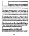
- the maximum and minimum values are 1.8 and -1.2 respectively while the 25% and 75% range points are at -0.3 and 0.2 respectively.
- the 5% and 95% points arc at -0.70 and 0.64 while the 1% and 99% points are at, respectively, -1.11 and 1.24. Sizing precision is better than 0.1 base on 24 capillaries of H . influenzae digest i0m0 from two separate runs; sixteen peaks were used for each trace.
- the first formulation contained 2% (w/w) linear polyacrylamide (MegaBACE Long read matrix), 20% (w/w) 2-pyrrolidinone: 6 M urea: 30 mM Tris: 100 mM TAPS: 1 mM EDTA and the second formulation contained 2% (w/w) linear polyacrylamide (MegaBACE Long read matrix), 20% (w/w) N',N'-dimethylacetamide: 6 M urea:30 mM Tris:100 mM TAPS: 1 mM EDTA.
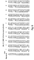
- FIG. 8 shows a portion of the electropherograms of the separation of M13 sequencing reaction products (around 400 bases) using the HEC and three different linear polyacrylamide formulations.
- the x-axis is in frames and the MegaBACE data acquisition rate is 1.8 hertz.
- the y-axis in FIG. 8 is arbitrary fluorescence units.
- the separation of the CGCT stretch is strongly affected by the separation media. This region is a well known compression in the sequence of M13 around 400 bases (see, e . g ., Kleparnik supra.). It can be clearly observed in FIG.
- both the formulation with 2-pyrrolidinone and N', N'-dimethylacetamide can resolve the CGCT compression more readily that the commercially available formulations. It can also be observed that the spacing between fragments in the inventive formulations in FIG. 8 is more uniform than in the commercial matrixes. Such an improved resolution of compressed sequences, especially in GC-rich DNA templates will greatly improve base calling accuracy.
- ROX standards were obtained from Perkin Elmer (Part No. 401734, Perkin Elmer, Applied Biosystems Division, Foster City, CA) and used in dilutions ranging from 1:20 to 1:100 in either deionized formamide or nanopure water for electrokinetic injection into capillaries.
- FAM-labeled fragment samples were prepared from Haemophilus influenzae genomic DNA by the QEATM chemistry process as described elsewhere (see, e.g., US Patent No. 5,871,697).
- the sequencing samples were the MegaBACETM sequencing standard (Amersham-Pharmacia) which contains 4 color M13 sequencing reaction generated with ThermoSequenaseTM (Amersham-Pharmacia) DNA polymerase and M13 (-40) forward primers (Sambrook, et al., Molecular Cloning: A Laboratory Manual, 2nd ed., Vols. 1, 2 and 3. Vol. 1, 2, 3. 1989, Cold Spring Harbor, NY: Cold Spring Harbor Laboratory Press. 1626) labeled with energy transfer dyes (Amersham-Pharmacia).
- MegaBACE 1000 DNA Sequencing System (Amersham - Molecular Dynamics, Sunnyvale, CA) was used in this invention.
- MegaBACE 1000 is a capillary array instrument utilizing 6 arrays of 16 capillaries, 100 ⁇ m I.D., 64 cm total length, 44 cm effective length. The instrument was operated at dual laser excitation mode (488 and 532 nm), FAM signals were detected in spectral channel 2 after a 520/20 band pass filter, ROX signals were detected in channel 3 after passing through a 610/LP long pass filter. Data was acquired at the rate of 1.8 Hz. Electrophoresis conditions: run voltage 12 kV, capillary temperature 44 °C, sample injection 20 s / 10 kV.
- the weight of a wide mouth (>1") glass jar (previously cleaned with deionized water) is obtained and set to zero.
- MegaBACETM Long read matrix (Amersham-Pharmacia, Piscataway, NJ) (provided in a 10 ml plastic syringe or one liter container) sufficient to prepare a 0.8-2.9%w/w solution was added to the jar.
- the weight of Long readTM (Amersham-Pharmacia) sieving matrix was recorded.
- a pre-determined amount of concentrated electrophoresis buffer was added to the glass vial to a final concentration between 10-50 mM Tris (preferred concentration 20 mM), 10-300 mM TAPS (preferred concentration 67 mM), 0.1-1.0 mM EDTA (preferred concentration 0.7 mM).
- Solid urea was also added to create a final concentration of 6 M). The maximum urea concentration is limited by its saturation level ( ⁇ 8 M).
- the organic denaturant was then added to a final concentration of 5-40%(w/w).
- the preferred denaturants include 2-pyrrolidinone, N',N'-dimethylacetamide, and N',N'-dimethylformamide.
- the most preferred denaturant combination was 20% 2-pyrrolidinone and 6 M urea.
- the solution was then brought to a final weight by the addition of deionized water.
- a 7/8" magnetic stirrer was added and the jar was capped and sealed using Para-filmTM.
- the solution was stirred at low speed on a magnetic stirrer at room temperature for about 12-18 hours prior to use.
- the mixture was withdrawn from the wide mouth jar using 10cc plastic syringes.
- the plastic syringes containing the polymer solution were fitted with pipet tips and 0.75 ml of the mixture was dispensed into 2 ml screw cap vials.
- the solution was degassed by centrifuging (5,000 X RPM) the tubes in a SpeedVac concentrator without vacuum for 4 minutes. The tubes were subsequently closed with an o-ring cap and stored at 4°C.
- Matrix preparation containing 20% of 2-pyrrolidinine was also scaled up to a production level as described below (see also FIG. 1): The procedure for preparation of the separation matrix was modified to mix quantities up to 1,000 g. The weight of a 2 liter Pyrex reactor (Corning, Corning, NY; 5. in FIG. 1) was obtained and set to zero. To the Pyrex reactor 400.0 g of MegaBACE Long Read matrix (Amersham-Pharmacia, Piscataway, NJ), 92.0 ml of 2-pyrrolidinone (Aldrich Chemical Company, Milwaukee, WI), 72.0 g of urea (Amresco, Solon, OH) were added and brought up to a final solution weight of 600 g with distilled water.
- MegaBACE Long Read matrix Amersham-Pharmacia, Piscataway, NJ
- 92.0 ml of 2-pyrrolidinone Aldrich Chemical Company, Milwaukee, WI
- 72.0 g of urea Amresco, Solon, OH
- the solution was mixed at 4°C in a refrigerator using a Heidolph over-head stirrer at 45 x RPM for 24-32 hours.
- the over-head stirrer was equipped with an Z-shaped blade (3. in FIG. 1) and a vacuum adapter (4. in FIG. 1) to minimize evaporation of the solution.
- the Z-shaped blade provided homogeneous mixing of the solution and minimized shearing of the high molecular weight polymers (see FIG. 1).
- the separation matrix was dispensed into 12 oz. cartridge retainers (Norlico Corporation, Hampton Falls, NH). The retainers are placed on a barrel loader (Norlico Corp.) and used to fill 55 cc syringes with the separation matrix.
- the syringes are used on a 2000 x L digital dispenser (Norlico Corp.) to dispense 1.5 ml of the matrix into 2 ml o-ring screw cap vials.
- the 2 ml vials containing the separation matrix were stored at 4°C.
- the vials were spun for 5 min at 5,000 x rpm prior to filling of the capillary arrays on the MegaBACE instrument (Molecular Dynamics, Sunnyvale, CA).
- H. influenzae restriction digests were prepared and run on the MegaBACE instrument in order to obtain a sizing calibration for the GeneScan 500 ROX ladder fragments.
- the successful double digests included i0m0 (BglII / BspHI), m1c0 (MfeI / Apa L1), m1i0 (MfeI / BglII), and m0p0 (BspHI / BstYI).
- a Mathematica program was written that allowed for a precise determination of a peak of interest coordinates in frame number (x, or time axis) and signal strength (y, or relative fluorescence axis). The peak maximum frame number was used to obtain the sizing values for the MegaBACE runs.
- the GeneScan fragment effective sizes accounting for mobility shift and electrophoretic mobility, were interpolated from a third order fit of H. influenzae p0r0.
- a preliminary calibration was obtained as follows. An association between frame number and H. influenzae i0m0 fragment size was obtained. Linear interpolation using four or five i0m0 peaks per ROX peak was used to obtain effective lengths of the ROX fragments, using the frame numbers of their peaks. Assignability of the H. influenzae p0r0 signal trace was checked, and found peaks could not be assigned with sufficient confidence to measure the accuracy. Upon checking the calibration, e.g. examining the H. influenzae i0m0 sizing, it was found that there were observable error trends.
- H. influenzae m1c0 digest was used for the primary calibration.
- a new assignment was obtained between frame number and fragment size using H. influenzae / m1c0. From four to eleven of the m1c0 peaks were used to obtain a linear interpolation for each ROX size. The number of peaks chosen for the linear fit was variable and chosen to include all peaks within a roughly +/- 50 base window. The sizing of H.
- influenzae i0m0 and m 1c0 was again examined to check accuracy and see if any trends were present. We found that the bands seemed to size low between 100 and 139. This observed trend was confirmed, for example with H. influenzae m1i0.
- Checking the entire m1c0 digest revealed that residual difference values (theoretical - measured) were all positive up to about 250 bases and that there were errors near 500 bases (and for values extrapolated to longer lengths). Using this information, corresponding adjustments were made to the calibration, for example: add 0.3 to each value up to 250. The sizing was checked and the values adjusted until better values (closer to zero) were obtained for (theoretical - measured).
- OGI® was used to view data from all 96 capillaries.
- GeneScape® an Internet based bioinformatics system was utilized for data analysis. Calculation of apparent fragment sizes based on standard peak mobilities, as well as calculations of resolution, efficiency, and selectivity were performed by a program written in Mathematica 3.0 (Wolfram Research, Champaign, IL). Sigma Stat (SPSS Inc., Chicago, IL) was used to perform statistical analysis on experimental data.
Landscapes
- Chemical & Material Sciences (AREA)
- Health & Medical Sciences (AREA)
- Life Sciences & Earth Sciences (AREA)
- Molecular Biology (AREA)
- General Health & Medical Sciences (AREA)
- Pathology (AREA)
- Electrochemistry (AREA)
- Physics & Mathematics (AREA)
- Analytical Chemistry (AREA)
- Biochemistry (AREA)
- Dispersion Chemistry (AREA)
- General Physics & Mathematics (AREA)
- Immunology (AREA)
- Chemical Kinetics & Catalysis (AREA)
- Measuring Or Testing Involving Enzymes Or Micro-Organisms (AREA)
- Investigating Or Analysing Biological Materials (AREA)
- Solid-Sorbent Or Filter-Aiding Compositions (AREA)
- Peptides Or Proteins (AREA)
- Medicines Containing Material From Animals Or Micro-Organisms (AREA)
- Electrostatic Separation (AREA)
- Compositions Of Macromolecular Compounds (AREA)
Abstract
Description
| Matrixes Used in the Invention | |||
| Matrix | Linear poly-acrylamide (%w/w) | Denaturant composition | Buffer Composition |
| MegaBACE Gel Matrix (HEC) (Amersham/ Pharmacia) | - | 6M urea, 10% (w/w) formamide | 30mM Tris:100mM TAPS: 1mM EDTA |
| MegaBACE™ Long read matrix (Amersham/ Pharmacia) | 3% | 6M urea | 30mM Tris:100mM TAPS: 1mM EDTA |
| LPA formulation LPA-PYR | 2.0% | 6M urea, 20% (w/w) 2-pyrrolidinone | 30mM Tris:100mM TAPS: 1mM EDTA |
| LPA formulation LPA-DMA | 2.0% | 6M urea, 20% (w/w) N',N'-dimethylaceta mide | 30mM Tris:100mM TAPS: 1mM EDTA |
| LPA formulation LPA-DMF | 2.0% | 6M urea, 20% (w/w) N', N'-dimethylformami de | 30mM Tris:100mM TAPS: 1mM EDTA |
In FIG. 8 the x-axis is in frames and the MegaBACE data acquisition rate is 1.8 hertz. The y-axis in FIG. 8 is arbitrary fluorescence units. As observed in FIG. 8 the separation of the CGCT stretch is strongly affected by the separation media. This region is a well known compression in the sequence of M13 around 400 bases (see, e.g., Kleparnik supra.). It can be clearly observed in FIG. 8 that both the formulation with 2-pyrrolidinone and N', N'-dimethylacetamide can resolve the CGCT compression more readily that the commercially available formulations. It can also be observed that the spacing between fragments in the inventive formulations in FIG. 8 is more uniform than in the commercial matrixes. Such an improved resolution of compressed sequences, especially in GC-rich DNA templates will greatly improve base calling accuracy.
Claims (16)
- A replaceable matrix formulation comprising:(a) a linear polyacrylamide solution;(b) at least one organic denaturant; wherein the organic denaturant is selected from the group consisting of: 2-pyrrolidinone; N',N'-dimethylacetamide; and N',N'-dimethylformamide.(c) a buffer; and(d) 6M urea.
- The matrix formulation according to Claim 1, wherein the linear polyacrylamide concentration is at least 1% (w/w).
- The matrix formulation according to Claim 2, wherein the linear polyacrylamide concentration ranges between 1%-3% (w/w).
- The matrix formulation according to Claim 1, wherein the organic denaturant concentration ranges between 5%-20% (w/w).
- The matrix formulation according to Claim 1, wherein the organic denaturant is 2-pyrrolidinone.
- A method of biomacromolecule separation using a capillary electrophoresis device comprising the matrix formulation according to Claim 1.
- The method according to Claim 6, wherein the electrophoresis device is operated at a run voltage of between 10kV-12kV.
- The method according to Claim 6, wherein the electrophoresis device is operated at a run, voltage of at least 12kV.
- The method according to Claim 6, wherein the electrophoresis device is operated at temperature ranging between 40°C and 60°C.
- The method according to Claim 9, wherein the electrophoresis device is operated at 44°C.
- The method according to Claim 6, wherein the buffer consists essentially of:(a) EDTA, with a concentration ranging between 0.6-1.0 mM;(b) Tris Base, with a concentration ranging between 12-30 mM; and(c) TAPS, with a concentration ranging between 50-85 mM.
- The method according to Claim 6, wherein the biomacromolecule separation is used in nucleic acid sequencing.
- The method according to Claim 6, wherein the biomacromolecule separation is used in the determination of the molecular size of a biomacromolecule.
- The method according to Claim 6, wherein the biomacromolecule separation is used in the differential display of mRNA.
- The method according to Claim 6, wherein the biomacromolecule separation is used in dideoxyfingerprinting.
- The method according to Claim 6, wherein the biomacromolecule separation is used in STR analysis.
Priority Applications (1)
| Application Number | Priority Date | Filing Date | Title |
|---|---|---|---|
| DK99958855T DK1129344T3 (en) | 1998-11-10 | 1999-11-10 | Methods and formulations for separating biological macromolecules |
Applications Claiming Priority (5)
| Application Number | Priority Date | Filing Date | Title |
|---|---|---|---|
| US10779898P | 1998-11-10 | 1998-11-10 | |
| US107798P | 1998-11-10 | ||
| US374174 | 1999-08-13 | ||
| US09/374,174 US6554985B2 (en) | 1998-11-10 | 1999-08-13 | Methods and formulations for the separation of biological macromolecules |
| PCT/US1999/026465 WO2000028314A1 (en) | 1998-11-10 | 1999-11-10 | Methods and formulations for the separation of biological macromolecules |
Publications (2)
| Publication Number | Publication Date |
|---|---|
| EP1129344A1 EP1129344A1 (en) | 2001-09-05 |
| EP1129344B1 true EP1129344B1 (en) | 2004-08-18 |
Family
ID=26805173
Family Applications (1)
| Application Number | Title | Priority Date | Filing Date |
|---|---|---|---|
| EP99958855A Expired - Lifetime EP1129344B1 (en) | 1998-11-10 | 1999-11-10 | Methods and formulations for the separation of biological macromolecules |
Country Status (11)
| Country | Link |
|---|---|
| US (1) | US6554985B2 (en) |
| EP (1) | EP1129344B1 (en) |
| JP (1) | JP2002529734A (en) |
| AT (1) | ATE274185T1 (en) |
| AU (1) | AU760638B2 (en) |
| CA (1) | CA2349439A1 (en) |
| DE (1) | DE69919568T2 (en) |
| DK (1) | DK1129344T3 (en) |
| ES (1) | ES2224723T3 (en) |
| HK (1) | HK1040284A1 (en) |
| WO (1) | WO2000028314A1 (en) |
Families Citing this family (9)
| Publication number | Priority date | Publication date | Assignee | Title |
|---|---|---|---|---|
| US6706162B1 (en) | 2000-09-25 | 2004-03-16 | Applera Corporation | High speed, high resolution compositions, methods, and kits for capillary electrophoresis |
| AU2002234252A1 (en) * | 2001-01-12 | 2002-07-24 | Curagen Corporation | Method and formulations for the separation of biological macromolecules |
| CA2453360A1 (en) | 2001-07-11 | 2003-01-23 | Applera Corporation | Internal calibration standards for electrophoretic analyses |
| JP4426261B2 (en) * | 2003-11-25 | 2010-03-03 | 株式会社日立製作所 | Channel adapter and disk array device |
| US7402229B2 (en) * | 2004-03-31 | 2008-07-22 | Intel Corporation | Fabrication and use of semipermeable membranes and gels for the control of electrolysis in a microfluidic device |
| US20070080064A1 (en) * | 2005-10-12 | 2007-04-12 | Baylor College Of Medicine | Micro-loading device |
| US8871524B2 (en) * | 2009-08-12 | 2014-10-28 | Caliper Life Sciences, Inc. | Methods of performing a sizing analysis using a corrected sizing ladder |
| WO2014148265A1 (en) * | 2013-03-21 | 2014-09-25 | 日本電気株式会社 | Microchip, method for dna analysis, and system for dna analysis |
| US20240418674A1 (en) * | 2021-12-17 | 2024-12-19 | Dh Technologies Development Pte. Ltd. | Capillary electrophoresis purity analysis of complementary strand nucleic acid molecules |
Family Cites Families (7)
| Publication number | Priority date | Publication date | Assignee | Title |
|---|---|---|---|---|
| US4999340A (en) * | 1988-08-04 | 1991-03-12 | Board Of Regents, The University Of Texas System | Rehydratable agarose gels |
| JPH07503542A (en) | 1992-01-31 | 1995-04-13 | ベックマン インスツルメンツ インコーポレーテッド | Capillary column containing removable separation gel composition and method of use thereof |
| US5455344A (en) | 1993-09-03 | 1995-10-03 | Fmc Corporation | Agarose compositions for nucleic acid sequencing |
| AU1254295A (en) * | 1993-12-17 | 1995-07-03 | Perkin-Elmer Corporation, The | Uncharged polymers for separation of biomolecules by capillary electrophoresis |
| US5503722A (en) | 1994-02-28 | 1996-04-02 | Beckman Instruments, Inc. | Rehydratable gels for capillary electrophoresis |
| US5534123A (en) | 1995-07-10 | 1996-07-09 | Molecular Dynamics | Denaturing separation matrix having hydroxyethyl cellulose for nucleic acid electrophoresis |
| JPH1123530A (en) | 1996-03-28 | 1999-01-29 | Fmc Corp | Stable denaturant used for electrophoretic technique and its using method |
-
1999
- 1999-08-13 US US09/374,174 patent/US6554985B2/en not_active Expired - Fee Related
- 1999-11-10 EP EP99958855A patent/EP1129344B1/en not_active Expired - Lifetime
- 1999-11-10 ES ES99958855T patent/ES2224723T3/en not_active Expired - Lifetime
- 1999-11-10 DE DE69919568T patent/DE69919568T2/en not_active Expired - Fee Related
- 1999-11-10 AT AT99958855T patent/ATE274185T1/en not_active IP Right Cessation
- 1999-11-10 CA CA002349439A patent/CA2349439A1/en not_active Abandoned
- 1999-11-10 AU AU16137/00A patent/AU760638B2/en not_active Ceased
- 1999-11-10 HK HK02101494.7A patent/HK1040284A1/en unknown
- 1999-11-10 WO PCT/US1999/026465 patent/WO2000028314A1/en not_active Ceased
- 1999-11-10 DK DK99958855T patent/DK1129344T3/en active
- 1999-11-10 JP JP2000581443A patent/JP2002529734A/en not_active Withdrawn
Also Published As
| Publication number | Publication date |
|---|---|
| US20020009721A1 (en) | 2002-01-24 |
| AU760638B2 (en) | 2003-05-22 |
| WO2000028314A1 (en) | 2000-05-18 |
| EP1129344A1 (en) | 2001-09-05 |
| DK1129344T3 (en) | 2004-12-06 |
| DE69919568T2 (en) | 2005-09-15 |
| DE69919568D1 (en) | 2004-09-23 |
| US6554985B2 (en) | 2003-04-29 |
| JP2002529734A (en) | 2002-09-10 |
| ES2224723T3 (en) | 2005-03-01 |
| CA2349439A1 (en) | 2000-05-18 |
| WO2000028314A9 (en) | 2001-03-22 |
| ATE274185T1 (en) | 2004-09-15 |
| HK1040284A1 (en) | 2002-05-31 |
| AU1613700A (en) | 2000-05-29 |
Similar Documents
| Publication | Publication Date | Title |
|---|---|---|
| US5370777A (en) | Capillary column containing removable separation gel composition and method of use | |
| Heiger et al. | Separation of DNA restriction fragments by high performance capillary electrophoresis with low and zero crosslinked polyacrylamide using continuous and pulsed electric fields | |
| Ruiz-Martinez et al. | DNA sequencing by capillary electrophoresis with replaceable linear polyacrylamide and laser-induced fluorescence detection | |
| Wenz et al. | High-precision genotyping by denaturing capillary electrophoresis | |
| US20020009721A1 (en) | Methods and formulations for the separation of biological macromolecul | |
| US20250189485A1 (en) | Compositions, methods, kits and devices for molecular analysis | |
| Righetti et al. | Recent advances in capillary electrophoresis of DNA fragments and PCR products in poly (N-substituted acrylamides) | |
| CN111344562B (en) | Compositions, methods, kits and devices for molecular analysis | |
| Kotler et al. | DNA sequencing of close to 1000 bases in 40 minutes by capillary electrophoresis using dimethyl sulfoxide and urea as denaturants in replaceable linear polyacrylamide solutions | |
| US20070141622A1 (en) | Buffers for electrophoresis and use thereof | |
| Smith et al. | Capillary electrophoresis of DNA | |
| EP0497448A1 (en) | Capillary gel electrophoresis in the presence of at least one neutral non-urea denaturing agent | |
| Gelfi et al. | Comparison of behavior of N‐substituted acrylamides and celluloses on double‐stranded DNA separations by capillary electrophoresis at 25° and 60° C | |
| EP0624248B1 (en) | Capillary column containing removable separation gel composition and method of use | |
| AU689648B2 (en) | Separation of nucleic acids by capillary electrophoresis in thermal gradients in viscous polymer solutions | |
| Ren | High throughput screening of genetic mutations/polymorphisms by capillary electrophoresis | |
| JP2898287B2 (en) | Polycationic buffer and gel electrophoresis of nucleic acid using the same | |
| Leclair et al. | Precision and accuracy in fluorescent short tandem repeat DNA typing: Assessment of benefits imparted by the use of allelic ladders with the AmpFlSTR® Profiler Plus™ kit | |
| Dai et al. | High-throughput separations using multiplexed capillary electrophoresis with laser-induced fluorescence detection | |
| US20040222095A1 (en) | Dna separation using linear polymer solutions with dimethyl sulfoxide | |
| Bienvenue et al. | Evaluation of sieving polymers for fast, reproducible electrophoretic analysis of short tandem repeats (STR) in capillaries | |
| Albany | Capillary Electrophoresis in DNA Analysis | |
| Mystic | Capillary Electrophoresis in DNA Analysis | |
| Dunn | ESSENTIAL GUIDES TO METHOD DEVELOPMENT IN TWO-DIMENSIONAL ELECTROPHORESIS |
Legal Events
| Date | Code | Title | Description |
|---|---|---|---|
| PUAI | Public reference made under article 153(3) epc to a published international application that has entered the european phase |
Free format text: ORIGINAL CODE: 0009012 |
|
| 17P | Request for examination filed |
Effective date: 20010521 |
|
| AK | Designated contracting states |
Kind code of ref document: A1 Designated state(s): AT BE CH CY DE DK ES FI FR GB GR IE IT LI LU MC NL PT SE |
|
| 17Q | First examination report despatched |
Effective date: 20020809 |
|
| GRAP | Despatch of communication of intention to grant a patent |
Free format text: ORIGINAL CODE: EPIDOSNIGR1 |
|
| GRAS | Grant fee paid |
Free format text: ORIGINAL CODE: EPIDOSNIGR3 |
|
| GRAA | (expected) grant |
Free format text: ORIGINAL CODE: 0009210 |
|
| AK | Designated contracting states |
Kind code of ref document: B1 Designated state(s): AT BE CH CY DE DK ES FI FR GB GR IE IT LI LU MC NL PT SE |
|
| PG25 | Lapsed in a contracting state [announced via postgrant information from national office to epo] |
Ref country code: FI Free format text: LAPSE BECAUSE OF FAILURE TO SUBMIT A TRANSLATION OF THE DESCRIPTION OR TO PAY THE FEE WITHIN THE PRESCRIBED TIME-LIMIT Effective date: 20040818 Ref country code: CY Free format text: LAPSE BECAUSE OF FAILURE TO SUBMIT A TRANSLATION OF THE DESCRIPTION OR TO PAY THE FEE WITHIN THE PRESCRIBED TIME-LIMIT Effective date: 20040818 |
|
| REG | Reference to a national code |
Ref country code: GB Ref legal event code: FG4D |
|
| REG | Reference to a national code |
Ref country code: CH Ref legal event code: EP |
|
| REG | Reference to a national code |
Ref country code: IE Ref legal event code: FG4D |
|
| REF | Corresponds to: |
Ref document number: 69919568 Country of ref document: DE Date of ref document: 20040923 Kind code of ref document: P |
|
| PGFP | Annual fee paid to national office [announced via postgrant information from national office to epo] |
Ref country code: AT Payment date: 20041021 Year of fee payment: 6 |
|
| REG | Reference to a national code |
Ref country code: SE Ref legal event code: TRGR |
|
| PG25 | Lapsed in a contracting state [announced via postgrant information from national office to epo] |
Ref country code: LU Free format text: LAPSE BECAUSE OF NON-PAYMENT OF DUE FEES Effective date: 20041110 |
|
| PGFP | Annual fee paid to national office [announced via postgrant information from national office to epo] |
Ref country code: IE Payment date: 20041111 Year of fee payment: 6 |
|
| PGFP | Annual fee paid to national office [announced via postgrant information from national office to epo] |
Ref country code: SE Payment date: 20041112 Year of fee payment: 6 Ref country code: DK Payment date: 20041112 Year of fee payment: 6 |
|
| PGFP | Annual fee paid to national office [announced via postgrant information from national office to epo] |
Ref country code: ES Payment date: 20041115 Year of fee payment: 6 |
|
| REG | Reference to a national code |
Ref country code: CH Ref legal event code: NV Representative=s name: ISLER & PEDRAZZINI AG |
|
| PG25 | Lapsed in a contracting state [announced via postgrant information from national office to epo] |
Ref country code: GR Free format text: LAPSE BECAUSE OF FAILURE TO SUBMIT A TRANSLATION OF THE DESCRIPTION OR TO PAY THE FEE WITHIN THE PRESCRIBED TIME-LIMIT Effective date: 20041118 |
|
| PGFP | Annual fee paid to national office [announced via postgrant information from national office to epo] |
Ref country code: CH Payment date: 20041122 Year of fee payment: 6 |
|
| PG25 | Lapsed in a contracting state [announced via postgrant information from national office to epo] |
Ref country code: MC Free format text: LAPSE BECAUSE OF NON-PAYMENT OF DUE FEES Effective date: 20041130 |
|
| REG | Reference to a national code |
Ref country code: DK Ref legal event code: T3 |
|
| PGFP | Annual fee paid to national office [announced via postgrant information from national office to epo] |
Ref country code: BE Payment date: 20041208 Year of fee payment: 6 |
|
| REG | Reference to a national code |
Ref country code: ES Ref legal event code: FG2A Ref document number: 2224723 Country of ref document: ES Kind code of ref document: T3 |
|
| ET | Fr: translation filed | ||
| PLBE | No opposition filed within time limit |
Free format text: ORIGINAL CODE: 0009261 |
|
| STAA | Information on the status of an ep patent application or granted ep patent |
Free format text: STATUS: NO OPPOSITION FILED WITHIN TIME LIMIT |
|
| 26N | No opposition filed |
Effective date: 20050519 |
|
| PG25 | Lapsed in a contracting state [announced via postgrant information from national office to epo] |
Ref country code: IT Free format text: LAPSE BECAUSE OF NON-PAYMENT OF DUE FEES Effective date: 20051110 Ref country code: AT Free format text: LAPSE BECAUSE OF NON-PAYMENT OF DUE FEES Effective date: 20051110 |
|
| PG25 | Lapsed in a contracting state [announced via postgrant information from national office to epo] |
Ref country code: SE Free format text: LAPSE BECAUSE OF NON-PAYMENT OF DUE FEES Effective date: 20051111 Ref country code: ES Free format text: LAPSE BECAUSE OF NON-PAYMENT OF DUE FEES Effective date: 20051111 |
|
| PG25 | Lapsed in a contracting state [announced via postgrant information from national office to epo] |
Ref country code: LI Free format text: LAPSE BECAUSE OF NON-PAYMENT OF DUE FEES Effective date: 20051130 Ref country code: DK Free format text: LAPSE BECAUSE OF NON-PAYMENT OF DUE FEES Effective date: 20051130 Ref country code: CH Free format text: LAPSE BECAUSE OF NON-PAYMENT OF DUE FEES Effective date: 20051130 Ref country code: BE Free format text: LAPSE BECAUSE OF NON-PAYMENT OF DUE FEES Effective date: 20051130 |
|
| REG | Reference to a national code |
Ref country code: DK Ref legal event code: EBP |
|
| REG | Reference to a national code |
Ref country code: CH Ref legal event code: PL |
|
| EUG | Se: european patent has lapsed | ||
| REG | Reference to a national code |
Ref country code: IE Ref legal event code: MM4A |
|
| REG | Reference to a national code |
Ref country code: ES Ref legal event code: FD2A Effective date: 20051111 |
|
| BERE | Be: lapsed |
Owner name: *CURAGEN CORP. Effective date: 20051130 |
|
| PG25 | Lapsed in a contracting state [announced via postgrant information from national office to epo] |
Ref country code: PT Free format text: LAPSE BECAUSE OF NON-PAYMENT OF DUE FEES Effective date: 20050118 |
|
| REG | Reference to a national code |
Ref country code: HK Ref legal event code: WD Ref document number: 1040284 Country of ref document: HK |
|
| PGFP | Annual fee paid to national office [announced via postgrant information from national office to epo] |
Ref country code: DE Payment date: 20080527 Year of fee payment: 9 |
|
| PGFP | Annual fee paid to national office [announced via postgrant information from national office to epo] |
Ref country code: NL Payment date: 20080529 Year of fee payment: 9 |
|
| PGFP | Annual fee paid to national office [announced via postgrant information from national office to epo] |
Ref country code: FR Payment date: 20080527 Year of fee payment: 9 |
|
| PGFP | Annual fee paid to national office [announced via postgrant information from national office to epo] |
Ref country code: GB Payment date: 20080523 Year of fee payment: 9 |
|
| GBPC | Gb: european patent ceased through non-payment of renewal fee |
Effective date: 20081110 |
|
| PG25 | Lapsed in a contracting state [announced via postgrant information from national office to epo] |
Ref country code: NL Free format text: LAPSE BECAUSE OF NON-PAYMENT OF DUE FEES Effective date: 20090601 |
|
| NLV4 | Nl: lapsed or anulled due to non-payment of the annual fee |
Effective date: 20090601 |
|
| REG | Reference to a national code |
Ref country code: FR Ref legal event code: ST Effective date: 20090731 |
|
| PG25 | Lapsed in a contracting state [announced via postgrant information from national office to epo] |
Ref country code: DE Free format text: LAPSE BECAUSE OF NON-PAYMENT OF DUE FEES Effective date: 20090603 |
|
| PG25 | Lapsed in a contracting state [announced via postgrant information from national office to epo] |
Ref country code: GB Free format text: LAPSE BECAUSE OF NON-PAYMENT OF DUE FEES Effective date: 20081110 |
|
| PG25 | Lapsed in a contracting state [announced via postgrant information from national office to epo] |
Ref country code: FR Free format text: LAPSE BECAUSE OF NON-PAYMENT OF DUE FEES Effective date: 20081130 |