EP1126859B1 - Blood leucocyte apheresis for treatment of hiv - Google Patents
Blood leucocyte apheresis for treatment of hiv Download PDFInfo
- Publication number
- EP1126859B1 EP1126859B1 EP00905345A EP00905345A EP1126859B1 EP 1126859 B1 EP1126859 B1 EP 1126859B1 EP 00905345 A EP00905345 A EP 00905345A EP 00905345 A EP00905345 A EP 00905345A EP 1126859 B1 EP1126859 B1 EP 1126859B1
- Authority
- EP
- European Patent Office
- Prior art keywords
- hiv
- apheresis
- cells
- treatment
- blood
- Prior art date
- Legal status (The legal status is an assumption and is not a legal conclusion. Google has not performed a legal analysis and makes no representation as to the accuracy of the status listed.)
- Expired - Lifetime
Links
- 238000002617 apheresis Methods 0.000 title claims abstract description 95
- 238000011282 treatment Methods 0.000 title claims abstract description 60
- 210000004369 blood Anatomy 0.000 title claims abstract description 44
- 239000008280 blood Substances 0.000 title claims abstract description 44
- 239000003463 adsorbent Substances 0.000 claims abstract description 37
- 229920002301 cellulose acetate Polymers 0.000 claims abstract description 12
- 239000011324 bead Substances 0.000 claims description 16
- 238000004519 manufacturing process Methods 0.000 claims description 14
- 239000003795 chemical substances by application Substances 0.000 claims description 3
- 239000002250 absorbent Substances 0.000 claims description 2
- 230000002745 absorbent Effects 0.000 claims description 2
- 238000000034 method Methods 0.000 abstract description 23
- 208000037265 diseases, disorders, signs and symptoms Diseases 0.000 abstract description 15
- 201000010099 disease Diseases 0.000 abstract description 10
- 230000002950 deficient Effects 0.000 abstract description 9
- 201000004792 malaria Diseases 0.000 abstract 1
- 244000052769 pathogen Species 0.000 abstract 1
- 102100036011 T-cell surface glycoprotein CD4 Human genes 0.000 description 80
- 210000004027 cell Anatomy 0.000 description 73
- 241000725303 Human immunodeficiency virus Species 0.000 description 39
- 210000004698 lymphocyte Anatomy 0.000 description 29
- 241000713772 Human immunodeficiency virus 1 Species 0.000 description 23
- 210000001744 T-lymphocyte Anatomy 0.000 description 23
- 108020004414 DNA Proteins 0.000 description 21
- 101001057504 Homo sapiens Interferon-stimulated gene 20 kDa protein Proteins 0.000 description 21
- 101001055144 Homo sapiens Interleukin-2 receptor subunit alpha Proteins 0.000 description 21
- 102100026878 Interleukin-2 receptor subunit alpha Human genes 0.000 description 21
- 230000000694 effects Effects 0.000 description 20
- 230000008859 change Effects 0.000 description 19
- 210000001616 monocyte Anatomy 0.000 description 19
- 238000011084 recovery Methods 0.000 description 17
- 102000003814 Interleukin-10 Human genes 0.000 description 15
- 108090000174 Interleukin-10 Proteins 0.000 description 15
- 210000003819 peripheral blood mononuclear cell Anatomy 0.000 description 15
- 230000000284 resting effect Effects 0.000 description 14
- 239000003814 drug Substances 0.000 description 13
- 229940079593 drug Drugs 0.000 description 13
- 102000000588 Interleukin-2 Human genes 0.000 description 12
- 108010002350 Interleukin-2 Proteins 0.000 description 12
- 230000007423 decrease Effects 0.000 description 12
- 210000002540 macrophage Anatomy 0.000 description 11
- 230000003612 virological effect Effects 0.000 description 10
- 101000914514 Homo sapiens T-cell-specific surface glycoprotein CD28 Proteins 0.000 description 9
- 102100027213 T-cell-specific surface glycoprotein CD28 Human genes 0.000 description 9
- 238000010241 blood sampling Methods 0.000 description 9
- 210000000987 immune system Anatomy 0.000 description 9
- 208000015181 infectious disease Diseases 0.000 description 9
- 208000030507 AIDS Diseases 0.000 description 8
- 102000017420 CD3 protein, epsilon/gamma/delta subunit Human genes 0.000 description 8
- -1 IL-1β Proteins 0.000 description 8
- 241000700605 Viruses Species 0.000 description 8
- MZOFCQQQCNRIBI-VMXHOPILSA-N (3s)-4-[[(2s)-1-[[(2s)-1-[[(1s)-1-carboxy-2-hydroxyethyl]amino]-4-methyl-1-oxopentan-2-yl]amino]-5-(diaminomethylideneamino)-1-oxopentan-2-yl]amino]-3-[[2-[[(2s)-2,6-diaminohexanoyl]amino]acetyl]amino]-4-oxobutanoic acid Chemical compound OC[C@@H](C(O)=O)NC(=O)[C@H](CC(C)C)NC(=O)[C@H](CCCN=C(N)N)NC(=O)[C@H](CC(O)=O)NC(=O)CNC(=O)[C@@H](N)CCCCN MZOFCQQQCNRIBI-VMXHOPILSA-N 0.000 description 7
- 102000004127 Cytokines Human genes 0.000 description 7
- 108090000695 Cytokines Proteins 0.000 description 7
- 208000031886 HIV Infections Diseases 0.000 description 7
- 108060008682 Tumor Necrosis Factor Proteins 0.000 description 7
- 102000000852 Tumor Necrosis Factor-alpha Human genes 0.000 description 7
- 230000000798 anti-retroviral effect Effects 0.000 description 7
- 238000002651 drug therapy Methods 0.000 description 7
- 230000006872 improvement Effects 0.000 description 7
- 208000037357 HIV infectious disease Diseases 0.000 description 6
- 210000003714 granulocyte Anatomy 0.000 description 6
- 208000033519 human immunodeficiency virus infectious disease Diseases 0.000 description 6
- 210000005087 mononuclear cell Anatomy 0.000 description 6
- 210000005105 peripheral blood lymphocyte Anatomy 0.000 description 6
- 230000001566 pro-viral effect Effects 0.000 description 6
- 108020003175 receptors Proteins 0.000 description 6
- 102000005962 receptors Human genes 0.000 description 6
- 230000009471 action Effects 0.000 description 5
- 210000000265 leukocyte Anatomy 0.000 description 5
- 210000005259 peripheral blood Anatomy 0.000 description 5
- 239000011886 peripheral blood Substances 0.000 description 5
- 239000002504 physiological saline solution Substances 0.000 description 5
- 238000001179 sorption measurement Methods 0.000 description 5
- 230000006433 tumor necrosis factor production Effects 0.000 description 5
- 210000003462 vein Anatomy 0.000 description 5
- 239000004743 Polypropylene Substances 0.000 description 4
- 230000006907 apoptotic process Effects 0.000 description 4
- 239000000969 carrier Substances 0.000 description 4
- 230000004087 circulation Effects 0.000 description 4
- 230000016396 cytokine production Effects 0.000 description 4
- 238000011010 flushing procedure Methods 0.000 description 4
- 230000001506 immunosuppresive effect Effects 0.000 description 4
- 210000002381 plasma Anatomy 0.000 description 4
- 239000004417 polycarbonate Substances 0.000 description 4
- 229920000515 polycarbonate Polymers 0.000 description 4
- 229920001155 polypropylene Polymers 0.000 description 4
- 238000002560 therapeutic procedure Methods 0.000 description 4
- INGWEZCOABYORO-UHFFFAOYSA-N 2-(furan-2-yl)-7-methyl-1h-1,8-naphthyridin-4-one Chemical compound N=1C2=NC(C)=CC=C2C(O)=CC=1C1=CC=CO1 INGWEZCOABYORO-UHFFFAOYSA-N 0.000 description 3
- KCXVZYZYPLLWCC-UHFFFAOYSA-N EDTA Chemical compound OC(=O)CN(CC(O)=O)CCN(CC(O)=O)CC(O)=O KCXVZYZYPLLWCC-UHFFFAOYSA-N 0.000 description 3
- 102000001554 Hemoglobins Human genes 0.000 description 3
- 108010054147 Hemoglobins Proteins 0.000 description 3
- 241000711549 Hepacivirus C Species 0.000 description 3
- 108090001005 Interleukin-6 Proteins 0.000 description 3
- 230000001413 cellular effect Effects 0.000 description 3
- 230000001684 chronic effect Effects 0.000 description 3
- 208000035475 disorder Diseases 0.000 description 3
- 210000003979 eosinophil Anatomy 0.000 description 3
- 230000001900 immune effect Effects 0.000 description 3
- 229940076144 interleukin-10 Drugs 0.000 description 3
- 239000003550 marker Substances 0.000 description 3
- 210000000440 neutrophil Anatomy 0.000 description 3
- 238000010926 purge Methods 0.000 description 3
- 230000002829 reductive effect Effects 0.000 description 3
- 206010001513 AIDS related complex Diseases 0.000 description 2
- 238000012286 ELISA Assay Methods 0.000 description 2
- 101000738771 Homo sapiens Receptor-type tyrosine-protein phosphatase C Proteins 0.000 description 2
- 108010038453 Interleukin-2 Receptors Proteins 0.000 description 2
- 102000010789 Interleukin-2 Receptors Human genes 0.000 description 2
- 239000004677 Nylon Substances 0.000 description 2
- 229940124158 Protease/peptidase inhibitor Drugs 0.000 description 2
- 102100037422 Receptor-type tyrosine-protein phosphatase C Human genes 0.000 description 2
- 230000004913 activation Effects 0.000 description 2
- 239000000427 antigen Substances 0.000 description 2
- 102000036639 antigens Human genes 0.000 description 2
- 108091007433 antigens Proteins 0.000 description 2
- 238000013459 approach Methods 0.000 description 2
- 238000003556 assay Methods 0.000 description 2
- 239000012298 atmosphere Substances 0.000 description 2
- 230000008901 benefit Effects 0.000 description 2
- 210000000601 blood cell Anatomy 0.000 description 2
- 230000017531 blood circulation Effects 0.000 description 2
- 210000004204 blood vessel Anatomy 0.000 description 2
- 201000011510 cancer Diseases 0.000 description 2
- 238000002512 chemotherapy Methods 0.000 description 2
- 239000000470 constituent Substances 0.000 description 2
- 238000012258 culturing Methods 0.000 description 2
- 230000006735 deficit Effects 0.000 description 2
- 210000004969 inflammatory cell Anatomy 0.000 description 2
- 230000000977 initiatory effect Effects 0.000 description 2
- 238000011835 investigation Methods 0.000 description 2
- 230000000670 limiting effect Effects 0.000 description 2
- 239000000463 material Substances 0.000 description 2
- 238000005259 measurement Methods 0.000 description 2
- 229920001778 nylon Polymers 0.000 description 2
- 238000012856 packing Methods 0.000 description 2
- 230000001575 pathological effect Effects 0.000 description 2
- 239000000137 peptide hydrolase inhibitor Substances 0.000 description 2
- 239000004800 polyvinyl chloride Substances 0.000 description 2
- 229920000915 polyvinyl chloride Polymers 0.000 description 2
- 230000000770 proinflammatory effect Effects 0.000 description 2
- 230000010076 replication Effects 0.000 description 2
- 229920002379 silicone rubber Polymers 0.000 description 2
- 239000004945 silicone rubber Substances 0.000 description 2
- 239000006228 supernatant Substances 0.000 description 2
- 208000024891 symptom Diseases 0.000 description 2
- 238000012360 testing method Methods 0.000 description 2
- 238000011269 treatment regimen Methods 0.000 description 2
- 206010001052 Acute respiratory distress syndrome Diseases 0.000 description 1
- 208000023275 Autoimmune disease Diseases 0.000 description 1
- 108010041397 CD4 Antigens Proteins 0.000 description 1
- 210000004366 CD4-positive T-lymphocyte Anatomy 0.000 description 1
- 206010006895 Cachexia Diseases 0.000 description 1
- 108010001857 Cell Surface Receptors Proteins 0.000 description 1
- 102000000844 Cell Surface Receptors Human genes 0.000 description 1
- 208000035473 Communicable disease Diseases 0.000 description 1
- 206010012735 Diarrhoea Diseases 0.000 description 1
- 206010018910 Haemolysis Diseases 0.000 description 1
- 206010061598 Immunodeficiency Diseases 0.000 description 1
- 208000029462 Immunodeficiency disease Diseases 0.000 description 1
- 206010061218 Inflammation Diseases 0.000 description 1
- 101000686985 Mouse mammary tumor virus (strain C3H) Protein PR73 Proteins 0.000 description 1
- 241000186362 Mycobacterium leprae Species 0.000 description 1
- 206010028980 Neoplasm Diseases 0.000 description 1
- 229940122313 Nucleoside reverse transcriptase inhibitor Drugs 0.000 description 1
- 206010037660 Pyrexia Diseases 0.000 description 1
- 238000010240 RT-PCR analysis Methods 0.000 description 1
- 208000013616 Respiratory Distress Syndrome Diseases 0.000 description 1
- PHMHDRYYFAYWEG-UHFFFAOYSA-N Rhapontigenin Natural products C1=C(O)C(OC)=CC=C1C=CC1=CC(O)=CC(O)=C1 PHMHDRYYFAYWEG-UHFFFAOYSA-N 0.000 description 1
- 241000191967 Staphylococcus aureus Species 0.000 description 1
- 230000006044 T cell activation Effects 0.000 description 1
- 230000024932 T cell mediated immunity Effects 0.000 description 1
- 108020005202 Viral DNA Proteins 0.000 description 1
- 201000000028 adult respiratory distress syndrome Diseases 0.000 description 1
- 230000002411 adverse Effects 0.000 description 1
- 208000026935 allergic disease Diseases 0.000 description 1
- 230000036436 anti-hiv Effects 0.000 description 1
- 239000003146 anticoagulant agent Substances 0.000 description 1
- 229940127219 anticoagulant drug Drugs 0.000 description 1
- 230000015572 biosynthetic process Effects 0.000 description 1
- 230000036770 blood supply Effects 0.000 description 1
- 238000012790 confirmation Methods 0.000 description 1
- 238000007796 conventional method Methods 0.000 description 1
- 230000007547 defect Effects 0.000 description 1
- 230000007812 deficiency Effects 0.000 description 1
- 238000010790 dilution Methods 0.000 description 1
- 239000012895 dilution Substances 0.000 description 1
- 230000008029 eradication Effects 0.000 description 1
- 238000011156 evaluation Methods 0.000 description 1
- 238000001914 filtration Methods 0.000 description 1
- 238000000684 flow cytometry Methods 0.000 description 1
- 239000012530 fluid Substances 0.000 description 1
- 230000006870 function Effects 0.000 description 1
- 230000036541 health Effects 0.000 description 1
- 230000008588 hemolysis Effects 0.000 description 1
- 210000002865 immune cell Anatomy 0.000 description 1
- 230000028993 immune response Effects 0.000 description 1
- 230000007813 immunodeficiency Effects 0.000 description 1
- 208000027866 inflammatory disease Diseases 0.000 description 1
- 230000004054 inflammatory process Effects 0.000 description 1
- 230000019734 interleukin-12 production Effects 0.000 description 1
- 239000005001 laminate film Substances 0.000 description 1
- 239000007788 liquid Substances 0.000 description 1
- 210000005229 liver cell Anatomy 0.000 description 1
- 239000003287 lymphocyte surface marker Substances 0.000 description 1
- 230000007246 mechanism Effects 0.000 description 1
- 210000003071 memory t lymphocyte Anatomy 0.000 description 1
- 208000010125 myocardial infarction Diseases 0.000 description 1
- 210000004296 naive t lymphocyte Anatomy 0.000 description 1
- 210000000653 nervous system Anatomy 0.000 description 1
- 201000001119 neuropathy Diseases 0.000 description 1
- 230000007823 neuropathy Effects 0.000 description 1
- 239000004745 nonwoven fabric Substances 0.000 description 1
- 229920006280 packaging film Polymers 0.000 description 1
- 239000012785 packaging film Substances 0.000 description 1
- 230000000242 pagocytic effect Effects 0.000 description 1
- 230000036961 partial effect Effects 0.000 description 1
- 230000008506 pathogenesis Effects 0.000 description 1
- 210000004976 peripheral blood cell Anatomy 0.000 description 1
- 208000033808 peripheral neuropathy Diseases 0.000 description 1
- 230000002085 persistent effect Effects 0.000 description 1
- 239000008177 pharmaceutical agent Substances 0.000 description 1
- 229920000139 polyethylene terephthalate Polymers 0.000 description 1
- 239000005020 polyethylene terephthalate Substances 0.000 description 1
- 230000003389 potentiating effect Effects 0.000 description 1
- 238000002360 preparation method Methods 0.000 description 1
- 230000002265 prevention Effects 0.000 description 1
- 230000008569 process Effects 0.000 description 1
- 102000004169 proteins and genes Human genes 0.000 description 1
- 108090000623 proteins and genes Proteins 0.000 description 1
- 238000005086 pumping Methods 0.000 description 1
- 239000002994 raw material Substances 0.000 description 1
- 238000003753 real-time PCR Methods 0.000 description 1
- 230000009467 reduction Effects 0.000 description 1
- 238000011160 research Methods 0.000 description 1
- 238000003757 reverse transcription PCR Methods 0.000 description 1
- 206010039073 rheumatoid arthritis Diseases 0.000 description 1
- 239000003419 rna directed dna polymerase inhibitor Substances 0.000 description 1
- 210000002966 serum Anatomy 0.000 description 1
- 238000010008 shearing Methods 0.000 description 1
- 239000000243 solution Substances 0.000 description 1
- 238000010561 standard procedure Methods 0.000 description 1
- 239000000126 substance Substances 0.000 description 1
- 231100000617 superantigen Toxicity 0.000 description 1
- 230000003746 surface roughness Effects 0.000 description 1
- 230000009885 systemic effect Effects 0.000 description 1
- 230000001225 therapeutic effect Effects 0.000 description 1
- 239000011800 void material Substances 0.000 description 1
- 208000016261 weight loss Diseases 0.000 description 1
- 230000004580 weight loss Effects 0.000 description 1
Images





Classifications
-
- C—CHEMISTRY; METALLURGY
- C12—BIOCHEMISTRY; BEER; SPIRITS; WINE; VINEGAR; MICROBIOLOGY; ENZYMOLOGY; MUTATION OR GENETIC ENGINEERING
- C12N—MICROORGANISMS OR ENZYMES; COMPOSITIONS THEREOF; PROPAGATING, PRESERVING, OR MAINTAINING MICROORGANISMS; MUTATION OR GENETIC ENGINEERING; CULTURE MEDIA
- C12N5/00—Undifferentiated human, animal or plant cells, e.g. cell lines; Tissues; Cultivation or maintenance thereof; Culture media therefor
- C12N5/0081—Purging biological preparations of unwanted cells
- C12N5/0087—Purging against subsets of blood cells, e.g. purging alloreactive T cells
-
- A—HUMAN NECESSITIES
- A61—MEDICAL OR VETERINARY SCIENCE; HYGIENE
- A61K—PREPARATIONS FOR MEDICAL, DENTAL OR TOILETRY PURPOSES
- A61K35/00—Medicinal preparations containing materials or reaction products thereof with undetermined constitution
- A61K35/12—Materials from mammals; Compositions comprising non-specified tissues or cells; Compositions comprising non-embryonic stem cells; Genetically modified cells
- A61K35/14—Blood; Artificial blood
-
- A—HUMAN NECESSITIES
- A61—MEDICAL OR VETERINARY SCIENCE; HYGIENE
- A61M—DEVICES FOR INTRODUCING MEDIA INTO, OR ONTO, THE BODY; DEVICES FOR TRANSDUCING BODY MEDIA OR FOR TAKING MEDIA FROM THE BODY; DEVICES FOR PRODUCING OR ENDING SLEEP OR STUPOR
- A61M1/00—Suction or pumping devices for medical purposes; Devices for carrying-off, for treatment of, or for carrying-over, body-liquids; Drainage systems
- A61M1/36—Other treatment of blood in a by-pass of the natural circulatory system, e.g. temperature adaptation, irradiation ; Extra-corporeal blood circuits
- A61M1/3679—Other treatment of blood in a by-pass of the natural circulatory system, e.g. temperature adaptation, irradiation ; Extra-corporeal blood circuits by absorption
-
- A—HUMAN NECESSITIES
- A61—MEDICAL OR VETERINARY SCIENCE; HYGIENE
- A61P—SPECIFIC THERAPEUTIC ACTIVITY OF CHEMICAL COMPOUNDS OR MEDICINAL PREPARATIONS
- A61P31/00—Antiinfectives, i.e. antibiotics, antiseptics, chemotherapeutics
- A61P31/04—Antibacterial agents
- A61P31/08—Antibacterial agents for leprosy
-
- A—HUMAN NECESSITIES
- A61—MEDICAL OR VETERINARY SCIENCE; HYGIENE
- A61P—SPECIFIC THERAPEUTIC ACTIVITY OF CHEMICAL COMPOUNDS OR MEDICINAL PREPARATIONS
- A61P31/00—Antiinfectives, i.e. antibiotics, antiseptics, chemotherapeutics
- A61P31/12—Antivirals
- A61P31/14—Antivirals for RNA viruses
- A61P31/18—Antivirals for RNA viruses for HIV
-
- A—HUMAN NECESSITIES
- A61—MEDICAL OR VETERINARY SCIENCE; HYGIENE
- A61M—DEVICES FOR INTRODUCING MEDIA INTO, OR ONTO, THE BODY; DEVICES FOR TRANSDUCING BODY MEDIA OR FOR TAKING MEDIA FROM THE BODY; DEVICES FOR PRODUCING OR ENDING SLEEP OR STUPOR
- A61M2202/00—Special media to be introduced, removed or treated
- A61M2202/04—Liquids
- A61M2202/0413—Blood
- A61M2202/0439—White blood cells; Leucocytes
-
- A—HUMAN NECESSITIES
- A61—MEDICAL OR VETERINARY SCIENCE; HYGIENE
- A61M—DEVICES FOR INTRODUCING MEDIA INTO, OR ONTO, THE BODY; DEVICES FOR TRANSDUCING BODY MEDIA OR FOR TAKING MEDIA FROM THE BODY; DEVICES FOR PRODUCING OR ENDING SLEEP OR STUPOR
- A61M2202/00—Special media to be introduced, removed or treated
- A61M2202/20—Pathogenic agents
- A61M2202/206—Viruses
-
- Y—GENERAL TAGGING OF NEW TECHNOLOGICAL DEVELOPMENTS; GENERAL TAGGING OF CROSS-SECTIONAL TECHNOLOGIES SPANNING OVER SEVERAL SECTIONS OF THE IPC; TECHNICAL SUBJECTS COVERED BY FORMER USPC CROSS-REFERENCE ART COLLECTIONS [XRACs] AND DIGESTS
- Y02—TECHNOLOGIES OR APPLICATIONS FOR MITIGATION OR ADAPTATION AGAINST CLIMATE CHANGE
- Y02A—TECHNOLOGIES FOR ADAPTATION TO CLIMATE CHANGE
- Y02A50/00—TECHNOLOGIES FOR ADAPTATION TO CLIMATE CHANGE in human health protection, e.g. against extreme weather
- Y02A50/30—Against vector-borne diseases, e.g. mosquito-borne, fly-borne, tick-borne or waterborne diseases whose impact is exacerbated by climate change
Definitions
- This invention relates to a method involving blood leucocyte apheresis for treatment of disease.
- therapeutic apheresis has been known as an alternative strategy for the management of disorders.
- the technique involves the removal of cells or proteins that are implicated in the pathogenesis of the disorder, for example rheumatoid factor, immune complexes and other humoral mediators in the plasma (1, 2).
- Procedures employing selective removal of blood cells or plasma components, without the use of replacement fluids, are associated with minimal side effects (1).
- An outpatient approach is possible, which is more convenient for the patient and reduces the cost of the procedure (2). Many of the unacceptable side-effects of chronic drug therapy are also reduced by use of the technique.
- EP-A-0-319961 there is described a method for the selective removal of granulocytes from blood by bringing the blood into contact with an adsorbent carrier having a higher affinity for granulocytes than for lymphocytes. That invention is concerned with cancer treatment. An apparatus for carrying out the method is also described in that application. A similar apparatus is described in US 5,725,768.
- JP 0193 481 and US 5567 443 there is described a method for the treatment of inflammatory disease which comprises bringing blood from a patient into contact with an adsorbent carrier that has an affinity for inflammatory cells (granulocytes and monocytes) which is greater than its affinity for lymphocytes.
- the method is reported to be useful for treating acute respiratory distress syndrome, rheumatoid arthritis, autoimmune diseases, allergic diseases and reflow disorders following myocardial infarction.
- HIV human immunodeficiency virus
- AIDS related complex AIDS related complex
- Certain cells infected with HIV harbour the virus and act as reservoirs of the HIV. Such cells include mononuclear cells (monocytes/macrophages) and resting CD4 + lymphocytes. While the virus is inside those cells, it is protected from clearance by the immune system. The virus is also shielded from contact with administered drugs so effective treatment of the condition is thus difficult.
- opportunistic infectious diseases and malignant tumours may develop and profoundly aggravate the patient's condition.
- Neuropathy may also become a serious problem and, depending on the patient, a disease of the cranial nervous system may also appear.
- HIV-induced disease Based on an immunological consideration of full blown HIV-induced disease (AIDS), the condition is characterised by a profound impairment of the immune response, including decline in CD4 + T lymphocytes, dysregulated cytokine production, increased production of proinflammatory or immunosuppressive cytokines such as TNF- ⁇ , IL-1 ⁇ , IL-6 and IL-10 by peripheral blood cells (3-6) including monocytes (3) and also by granulocytes (4) and enhanced susceptibility of immune cells to apoptosis. HIV antigens such as gp120 bound to immune complexes are believed to induce apoptosis of CD4 + cells (7-12). TNF- ⁇ , IL-1 ⁇ , IL-6 are reported to promote HIV replication (3).
- IL-12 an immunoregulator cytokine
- monocytes in healthy individuals, monocytes mainly produce IL-12, whereas in HIV-infected individuals, monocytes produce mainly IL-10.
- IL-10 was shown to have a pivotal role in the aforementioned changes in the production of cytokines (5, 6). In this context, it is also important to indicate that IL-2 and IL-12 reduce, whereas IL-10 increases susceptibility of CD4 + cells of HIV-infected patients to apoptosis (13-17).
- HAART highly active antiretroviral drug therapy
- HAART has been shown to increase CD4 + cell count and to suppress plasma HIV viral load to undetectable levels
- no drug treatment is known to affect cellular reservoirs of HIV, for example, in cells including mononuclear cells (monocytes/macrophages) and resting CD4 + lymphocytes. These cells serve as sites for an HIV reservoir and as sites for HIV replication and dissemination. This means that when drug treatment is ceased, there is a very strong possibility of relapse attributable to HIV propagation from the aforementioned reservoir cells and de novo infection.
- HIV infected resting cells
- latent HIV reservoirs the infected resting cells
- HCV hepatitis C virus
- the present invention is based on the surprising observation that leucocytes infected with HIV can be selectively removed from blood.
- the present invention provides the use of cellulose acetate beads as adsorbent carrier in the manufacture of an agent for the treatment by apheresis of a patient infected with HIV.
- the present invention effects selective adsorption of infected, defective and activated leucocytes including mononuclear cells i.e. monocytes/macrophages, and also activated lymphocytes, for example CD4 + lymphocytes, including resting CD4 + lymphocytes.
- leucocytes including mononuclear cells i.e. monocytes/macrophages
- lymphocytes for example CD4 + lymphocytes, including resting CD4 + lymphocytes.
- infection results in selective binding of a population of leucocytes to an adsorbent carrier.
- a change caused to a leucocyte by infection may be considered as activation of the leucocyte, or the leucocyte may be regarded as defective.
- leucocytes may be activated or rendered defective in other ways that result in the same or analogous selective binding as does infection.
- Infection, activation and/or defects may result in the same or similar cell surface changes, for example the changes may be in the same receptor type. However, this may not be the case, and the method of the invention is not dependant on the precise mechanism of the selective adsorption of the infected, activated and/or defective leucocytes.
- Immune recovery denotes an improvement in any one or more aspects of the immune system.
- One aspect of immune recovery is an immunological improvement. HIV has a particularly profound effect on a subject's immune system.
- Another aspect of immune recovery is the immuno reconstitution that results from removal of leucocytes and their replacement by new leucocytes. Yet a further aspect of immune recovery may result from reduced generation of immunosuppressive cytokines (TNF- ⁇ ).
- TNF- ⁇ immunosuppressive cytokines
- the CD4 leucocyte count is regarded as indicative of the status of a subject's immune system.
- a carrier as defined above the person's CD4 count increases, thereby demonstrating an immune improvement, and hence immune recovery.
- the present invention also provides use of an adsorbent carrier described above in the manufacture of an agent or adsorbent for use in apheresis for the various aspects of immune recovery described above.
- HIV infects monocytes/macrophages and CD4 + lymphocytes.
- Different viruses may infect different populations of leucocytes.
- Monocytes/macrophages may be particularly subject to infection owing to their phagocytic activity.
- different viruses may infect different populations of lymphocytes.
- HIV infects CD4 + lymphocytes because the virus effects entry via the CD4 receptor.
- Other viruses may effect entry into lymphocytes via different receptors and hence infect the population of lymphocytes that possess that particular receptor.
- the invention further provides, in use, flushing out (purging) infected leucocytes from resting sites in a subject.
- flushing out of cells is commonly known in the art as "purging".
- infected cells include mononuclear cells (monocytes/macrophages) and CD4 + lymphocytes. Flushing infected cells from resting sites results in the virus within the cells becoming vulnerable to drug therapy.
- adsorbent carrier is used herein to denote a material that is capable of adsorbing cells onto its surface.
- the adsorbent carrier used in accordance with the invention has a higher affinity for infected, activated and/or defective leucocytes than for uninfected, non-activated or non-defective leucocytes, whilst not adversely affecting the blood brought into contact with the adsorbent.
- Differential leucocyte counts are determined by a standard laboratory differential leucocyte counter, for example THMS H-1 (Technicon).
- the number of cells of a particular leucocyte type trapped by the carrier in the column is calculated by subtracting the appropriate leucocyte count in the blood at the column outlet from the corresponding leucocyte count in the blood at the column inlet.
- the number of cells exposed to the carrier is calculated from the flow rate (ml per minute), flow time (apheresis time) and the appropriate leucocyte count at the inflow.
- the adsorbent carrier has a size distinguishable from blood cells and a shape having a large contact area with the blood to be brought into contact therewith, to permit efficient contact.
- the adsorbent carrier may take the form of beads having a diameter of about 0.1-10mm.
- the beads may have a diameter in the range of from 0.2 to 5.0 mm.
- the beads may have a diameter in the range of from 0.5 to 4.0 mm. More preferably, the beads may have a diameter in the range of from 1.0 to 3.0 mm, for example 2.0 mm.
- the efficiency of cell removal by the adsorbent carrier may be enhanced when the adsorbent carrier has a roughened surface. This is particularly the case when the adsorbent carrier has on its surface roughness having a centre line average height Ra, being defined under Japanese and Industrial Standard B0601-1982, of 0.2 ⁇ m to 10 ⁇ m and a mean spacing Sm of unevenness within the range from 5 ⁇ m to 200 ⁇ m.
- the effectiveness of an apheresis adsorbent carrier with such surface characteristics has been described in US 5,593,586 and a method of making such an adsorbent carrier has been described in US 5,525,279.
- cellulose acetate beads have a diameter of in the range from about 0.1-10mm, for example, from 0.2 to 5.0 mm, for example from 0.5 to 4.0 mm, for example from 1.0 to 3.0 mm, for example 2.0 mm.
- the effectiveness of cell removal by a cellulose acetate adsorbent carrier is generally enhanced when the adsorbent carrier has a roughened surface, particularly as described above.
- a leucocyte removal device for use with the present invention comprises a means for bringing blood that comprises infected, activated and/or defective leucocytes into contact with the adsorbent carrier such that the blood is treated with the carrier and then recovered.
- the blood of the patient who is being treated is preferably peripheral blood.
- the device may also include a means for transporting blood back to the patient.
- One or both of the transferring means may include a pumping means.
- One or both of the transferring means may include a means for measuring the rate of flow and/or the pressure of the blood within the transferring means.
- a means for adding pharmaceutical agents to the blood may be present on one or both of the transferring means.
- a leucocyte apheresis column for use with the invention is a column filled with the adsorbent carrier in the form of beads bathed in physiological liquid, particularly sterile, physiological saline.
- Blood is supplied to the column via a blood inlet, and blood leaves the column via a blood outlet.
- the blood inlet is connected to a suitable blood vessel in a patient, such as a vein in an arm or a hand, generally via a transporting means, for example a tube.
- the blood outlet may be connected to a further suitable blood vessel in a patient, such as a vein in an arm or a hand.
- the flow rate of blood through a leucocyte apheresis device may be within the range of from 5 to 200 ml/min. Preferably it is from 20 to 50 ml/min. At high flow rates haemolysis owing to shearing may be disadvantageous.
- the duration of apheresis may be in the range of from 10 minutes to 10 hours. The exact duration will be a matter of clinical judgement in each individual case. The frequency of treatment will also be a matter of clinical judgement in each individual case. Typically, a regime of one apheresis of one hour's duration once per week at, for example, 30 ml/min flow rate may be used.
- Immune recovery denotes an improvement in any one or more aspects of the immune system.
- One aspect of immune recovery is an immunological improvement. HIV has a particularly profound effect on a subject's immune system.
- Another aspect of immune recovery is the immuno reconstitution that results from removal of leucocytes and their replacement by new leucocytes. Yet a further aspect of immune recovery may result from reduced generation of immunosuppressive cytokines (TNF- ⁇ ).
- TNF- ⁇ immunosuppressive cytokines
- the CD4 leucocyte count is regarded as indicative of the status of a subject's immune system.
- a carrier as defined above the person's CD4 count increases, thereby demonstrating an immune improvement, and hence immune recovery.
- the present invention has the following effects in the case of treatment of AIDS when the blood is contacted with cellulose acetate:
- An apheresis column is an adsorbent-type extracorporeal leucocyte apheresis device.
- the device column is filled with cellulose acetate beads (adsorbent carriers) of 2 mm diameter, bathed in sterile physiological saline.
- the finished assembly is then sterilized and thoroughly leak tested by standard procedures. Blood inlet and outlet are via suitable veins in the left and the right hands.
- the system has been developed for selective adsorption of activated or infected leucocytes including monocytes/macrophages and neutrophils by a process known as leucocyte apheresis. Normal, non-activated non-infected lymphocytes do not adhere to the column adsorbent carriers. Additionally, research results published in the literature suggest that IgG immune complexes bind to cellulose acetate surface (18), which may provide an additional advantage.
- G-1 column (Adacolumn) a device formed according to the present invention is presented in Figure 1 and the materials of its components are described in Table 1.
- An outline of the G-1 column leucocyte apheresis device is presented in Figure 2.
- the G-1 column comprises a polycarbonate column (Adacolumn (8)) which is filled with cellulose acetate beads (adsorbent carriers (7)) bathed in physiological saline (9).
- the column contents are sealed by inner cap (4), outer cap (5) and O-ring (6).
- the blood supply line is secured onto the column by a nozzle cap (2) and packing (3) at the blood inlet (1)
- the adsorbent carriers have a diameter of 2 mm with a total weight of 220 g.
- the polycarbonate column is 206 mm in length and 60mm in diameter and has a capacity of 335 ml.
- the total volume of the physiological saline is 130 ml (equal to the column void volume).
- the constituents of the G-1 column are shown below in Table 2. Table 2. Constituents of the G-1 Column (Adacolumn) Name Raw Material Adsorbent carrier* Cellulose acetate Main body* Polycarbonate Outer cap* Polypropylene Inner cap* Polypropylene O-ring* Silicone rubber Perforated stopper* Polyethylene terephthalate Packing Silicone rubber Nozzle cap Polypropylene Filling solution Isotonic physiological saline Circuit lines PVC (polyvinylchloride, plasticised) Packaging film Nylon/polypropylene laminate film, partially gas-permeable wood-free paper or nonwoven fabric (Nylon/C. P. P. film) *Indicates that the component comes into direct contact with the blood.
- PVC polyvinylchloride, plasticised
- the G-1 leucocyte apheresis device comprises a polycarbonate column (Adacolumn (26)), blood circuit lines and an Adamonitor (23) together with a pump (24).
- the blood outflow from an antecubital vein of the patient (21) passes through a bubble detector (22) to a pump (24) and an Adamonitor (23).
- the blood then passes an anticoagulant administration port (25) before entering an adacolumn (26).
- the blood enters drip chamber (29) to which are attached a drip chamber air outlet port (27) and venous pressure gauge (28).
- a bubble detector (30) the blood flows back into the patient via an antecubital vein (31).
- Patients under HAART received apheresis treatment using the G-1 column described above. Each patient received one apheresis per week over an 8 week period. Each apheresis is of 60 minutes duration at a flow rate of 30 ml/minute.
- Example 1 Treatment of an HIV-infected patient who had a low HIV DNA count at baseline.
- Baseline was the level of HIV-1 DNA before apheresis treatment was commenced. Immediately after baseline blood sampling, the patient received apheresis; one apheresis per week, at a flow rate of 30ml per minute, duration of each apheresis was 60 minutes.
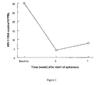
- the fall in HIV-1 DNA following apheresis with the G-1 column is shown in Figure 3.
- HIV-1 DNA was quantified using a standard quantitative polymerase chain reaction assay (Amplicor HIV MonitorTM , Roche, Basel, Switzerland) on peripheral blood mononuclear cells. The results indicate that apheresis with the G-1 column can flush out the HIV-infected resting cells. No drug is known that can produce such a rapid fall in HIV-DNA load.
- Example 2 Treatment of an HIV-infeoted patient who had a high HIV DNA count at baseline.
- Example 3 The fall in HIV-1 RNA during treatment of an HIV-infeoted patient.
- HIV-1 RNA was quantified using a standard quantitative reverse transcriptase-polymerase chain reaction competition assay. No drug or drug therapy is known to produce such a rapid sharp decline in HIV RNA.
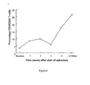
- Example 4 The rise in percentage CD4 + /CD25 + T-cells during treatment of an HIV-infected patient.
- CD4 + /CD25 + represents T-cell activity; CD4 + /CD25 + T-cells are activated T-cells.
- CD25 is the ⁇ -subunit of the IL-2 receptor which is required for high affinity binding of IL-2. It is expressed by activated but not by resting T-cells. The increase in CD4 + /CD25 + is very important for HIV patients because
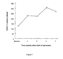
- Example 5 The rise in CD4 + T-cells following treatment of an HIV-infected patient.
- CD4 + T-cells following apheresis with the G-1 column is shown in Figure 7.
- the number of CD4 + lymphocytes was measured using an Epics,XL flow cytometer (Coulter Electronics, Inc.) using 100 ⁇ l of EDTA peripheral blood incubated for 30 minutes at 4°C with phycoerytrin-Texas-Red fluorochrome- labelled anti CD4 monoclonal antibody.
- CD4 + T-cell count is a key marker of HIV-disease symptoms; an increase in CD4 + T-cell count is often regarded as a marker of HIV decline and patient improvement.
- Example 6 The rise in CD8 + T-cells following treatment of an HIV-infected patient.
- CD8 + T-cells following apheresis with the G-1 column is shown in Figure 8.
- the number of CD8 4 lymphocytes was measured using an Epics XL flow cytometer (Coulter Electronics, Inc.) using 100 ⁇ l of EDTA peripheral blood incubated for 30 minutes at 4°C with fluorescin-isothiocyanate fluorochrome- labelled anti CD8 monoclonal antibody.
- CD8 + T-cells are caused to proliferate by raised levels of IL-2 (acting via the IL-2 receptor as described in relation to Example 4 above).
- Example 7 The decline in TNF- ⁇ production by peripheral blood mononuolear cells following treatment of an HIV-infected patient.
- TNF- ⁇ production by peripheral blood mononuclear cells was measured by culturing 3 ⁇ 10 6 PBMC per well in 24-well LINBRO plates (Flow laboratories, Inc. McLean, VA) at 37°C in a moist, 7% CO 2 atmosphere. The PBMC were then unstimulated or stimulated with LPS (SIGMA Diagnostics St. Louis, MO) at a concentration of 1 ⁇ g/ml. The supernatant was harvested after 48 hours of culture and cytokine production was then evaluated with a commercially available ELISA assay (Genzyme, Cambridge, MA).
- TNF- ⁇ is a major pathological factor in HIV-infection disease. No drug is known that can produce such a rapid reduction in TNF- ⁇ production by peripheral blood mononuclear cells.
- Example 8 The sharp decline in IL-10 (interleukin 10) production by peripheral blood mononuclear cells following treatment of an HIV-infected patient.
- IL-10 interleukin 10
- IL-10 interleukin 10
- PBMC peripheral blood mononuclear cells
- the PBMC were then unstimulated or stimulated with PHA (M form, Grand Island, NY) at a dilution 1:100.
- PHA M form, Grand Island, NY
- the supernatant was harvested after 48 hours of culture and cytokine production was then evaluated with a commercially available ELISA assay (Genzyme, Cambridge, MA).
- IL-10 is a major pathological factor in HIV-infection disease.
- IL-10 is a potent immune suppressor. It suppresses cytokine production by macrophages and thus indirectly suppresses T-cell activation. This counteracts the actions of IL-2 and IL-12 on cell-mediated immunity and IL-10 therefore effectively shuts down the immune system.
- Example 9 The change in HIV-1 DNA in two HIV patients and disease-matched controls.
- Example 10 further illustrates the invention.
- Example 10 describes a preliminary investigation into the method and apparatus of the invention.
- Examples 1 to 9 relate to subsequent and more thorough investigations.
- Example 10 The changes in viral load, CD4-positive cells, CD3 + cells, CD4 + CD28 + cells, CD4 + CD45RA + cells and CD4 + CD25 + cells in two HIV infected patients undergoing apheresis treatment.
- an external circulation apparatus composed in combination of a direct haemoperfusion column (capacity: about 335 ml) packed with cellulose acetate beads (220 g; diameter: 2 mm) and an adsorption-type haemocathartic apparatus ("ADAMONITOR MM6-P", tradename; manufactured by Otsuka Denshi K.K.) was used [clinical Study on Use of Granulocyte-Removing Apparatus (G-1) for Chronic Rheumatism", Ensho (Inflammation), 17(1), 57-80 (1997)].
- HIV-infected patients were chosen as subjects for treatment in accordance with the following standard: patients remaining under stable conditions over at least four months and generally one year owing to the ARV chemotherapy, having CD4-positive cell numbers not increased by 10% or greater from the corresponding base lines (the corresponding values before the initiation of the ARV therapy led to the stable conditions)(immunologically ARV-nonresponsive patients), and having more than 50,000 copies of HIV-RNA or showing a decrease in HIV-RNA not greater than 0.5 log ( ⁇ 0.5 log) since the beginning of HAART owing to the ARV chemotherapy led to the stable conditions (virologically ARV-nonresponsive patients).
- ARV anti-retroviral
- the treatment of the HIV-infected disease by the external circulation apparatus according to the present invention was conducted in substantially the same manner as in the clinical study on chronic rheumatism as described in the report referred to in the above.
- the treatment was conducted once a week by setting the amount of blood, which is to be circulated externally per treatment, on the basis of a.blood flow rate of 30ml/minute and an external circulation time of 60 minutes as standards.
- test items in Table 3 were all measured by methods commonly employed in the art, and include the number of white blood cells (WBC), the number of neutrophils, the number of monocytes, the number of eosinophils, the number of lymphocytes, the numbers of lymphocyte surface markers, the number of hemoglobins, the number of platelets, and the viral load.
- WBC white blood cells
- neutrophils the number of neutrophils
- monocytes the number of monocytes
- eosinophils the number of lymphocytes
- lymphocyte surface markers the number of lymphocyte surface markers
- hemoglobins the number of platelets
- viral load the viral load.
- the number of each lymphocyte surface marker was determined as a result of a measurement by flow cytometry while making use of a monoclonal antibody of the marker.
- "CD3" indicates the number of CD3-positive cells
- "#CD4" the number of CD4-positive cells
- "%CD4" the percentage of the CD4-positive cells based on lymphocytes
- "CD4/8" the ratio of the CD4-positive cells to CD8-positive cells
- "CD4+CD45RA+” the percentage of CD45RA-positive cells based on the CD4-positive cells
- CD4_CD25+(IL-2) the percentage of CD25-positive cells based on the CD4-positive cells
- “CD4+CD28+(Th-1) the percentage of CD28-positive cells based on the CD4-positive cells.
- each viral load is determined as a result of a measurement of a serum level of HIV-RNA by the RT-PCR method (cp: the number of copies per ml).
Landscapes
- Health & Medical Sciences (AREA)
- Life Sciences & Earth Sciences (AREA)
- Engineering & Computer Science (AREA)
- General Health & Medical Sciences (AREA)
- Biomedical Technology (AREA)
- Chemical & Material Sciences (AREA)
- Organic Chemistry (AREA)
- Animal Behavior & Ethology (AREA)
- Veterinary Medicine (AREA)
- Public Health (AREA)
- Zoology (AREA)
- Biotechnology (AREA)
- Hematology (AREA)
- Vascular Medicine (AREA)
- Cell Biology (AREA)
- Pharmacology & Pharmacy (AREA)
- Medicinal Chemistry (AREA)
- Virology (AREA)
- Wood Science & Technology (AREA)
- Genetics & Genomics (AREA)
- Bioinformatics & Cheminformatics (AREA)
- Heart & Thoracic Surgery (AREA)
- Molecular Biology (AREA)
- Developmental Biology & Embryology (AREA)
- Oncology (AREA)
- Chemical Kinetics & Catalysis (AREA)
- General Chemical & Material Sciences (AREA)
- Nuclear Medicine, Radiotherapy & Molecular Imaging (AREA)
- Epidemiology (AREA)
- General Engineering & Computer Science (AREA)
- Biochemistry (AREA)
- Anesthesiology (AREA)
- Communicable Diseases (AREA)
- Immunology (AREA)
- Microbiology (AREA)
- Cardiology (AREA)
- Tropical Medicine & Parasitology (AREA)
- AIDS & HIV (AREA)
- External Artificial Organs (AREA)
- Medicines Containing Material From Animals Or Micro-Organisms (AREA)
Abstract
Description
- This invention relates to a method involving blood leucocyte apheresis for treatment of disease.
- For some time, therapeutic apheresis has been known as an alternative strategy for the management of disorders. The technique involves the removal of cells or proteins that are implicated in the pathogenesis of the disorder, for example rheumatoid factor, immune complexes and other humoral mediators in the plasma (1, 2). Procedures employing selective removal of blood cells or plasma components, without the use of replacement fluids, are associated with minimal side effects (1). An outpatient approach is possible, which is more convenient for the patient and reduces the cost of the procedure (2). Many of the unacceptable side-effects of chronic drug therapy are also reduced by use of the technique.
- In EP-A-0-319961 there is described a method for the selective removal of granulocytes from blood by bringing the blood into contact with an adsorbent carrier having a higher affinity for granulocytes than for lymphocytes. That invention is concerned with cancer treatment. An apparatus for carrying out the method is also described in that application. A similar apparatus is described in US 5,725,768.
- In JP 0193 481 and US 5567 443 there is described a method for the treatment of inflammatory disease which comprises bringing blood from a patient into contact with an adsorbent carrier that has an affinity for inflammatory cells (granulocytes and monocytes) which is greater than its affinity for lymphocytes. The method is reported to be useful for treating acute respiratory distress syndrome, rheumatoid arthritis, autoimmune diseases, allergic diseases and reflow disorders following myocardial infarction.
- Bruisten et al. (Transfusion, 1990, 30, 833-837) described the efficiency of white cell filtration and a freeze-thaw procedure for removal of HIV-infected cells from blood.
- AIDS is a life-threatening immunodeficiency condition caused by HIV (human immunodeficiency virus). After confirmation and onset of HIV infection, the mortality rate is very high. No effective treatment strategy has been established yet for the treatment of AIDS and, owing to a long latent period, it is also difficult to establish an effective measure for the prevention of infection.
- After infection with HIV, anti-HIV antibody appears in the blood, generally within 6 to 8 weeks. Deficiency of or disturbance to the immune system proceeds, leading eventually to AIDS. For convenience, the intermediate stage is classified as AIDS related complex (ARC). Certain cells infected with HIV harbour the virus and act as reservoirs of the HIV. Such cells include mononuclear cells (monocytes/macrophages) and resting CD4+ lymphocytes. While the virus is inside those cells, it is protected from clearance by the immune system. The virus is also shielded from contact with administered drugs so effective treatment of the condition is thus difficult.
- The main symptoms of AIDS include fever, persistent systemic lymphadenoma, weight loss (cachexia) and diarrhoea. In addition, opportunistic infectious diseases and malignant tumours may develop and profoundly aggravate the patient's condition. Neuropathy may also become a serious problem and, depending on the patient, a disease of the cranial nervous system may also appear.
- Based on an immunological consideration of full blown HIV-induced disease (AIDS), the condition is characterised by a profound impairment of the immune response, including decline in CD4+ T lymphocytes, dysregulated cytokine production, increased production of proinflammatory or immunosuppressive cytokines such as TNF-α, IL-1β, IL-6 and IL-10 by peripheral blood cells (3-6) including monocytes (3) and also by granulocytes (4) and enhanced susceptibility of immune cells to apoptosis. HIV antigens such as gp120 bound to immune complexes are believed to induce apoptosis of CD4+ cells (7-12). TNF-α, IL-1β, IL-6 are reported to promote HIV replication (3). At the same time, there is a profound impairment of the ability of monocytes from HIV-infected patients to generate IL-12 (an immunoregulator cytokine). Thus, in healthy individuals, monocytes mainly produce IL-12, whereas in HIV-infected individuals, monocytes produce mainly IL-10. IL-10 was shown to have a pivotal role in the aforementioned changes in the production of cytokines (5, 6). In this context, it is also important to indicate that IL-2 and IL-12 reduce, whereas IL-10 increases susceptibility of CD4+ cells of HIV-infected patients to apoptosis (13-17).
- Currently, the treatment strategy for HIV infection consists primarily of highly active antiretroviral drug therapy (HAART) which includes a protease inhibitor. However, whilst in some patients, HAART has been shown to increase CD4+ cell count and to suppress plasma HIV viral load to undetectable levels, as yet no drug treatment is known to affect cellular reservoirs of HIV, for example, in cells including mononuclear cells (monocytes/macrophages) and resting CD4+ lymphocytes. These cells serve as sites for an HIV reservoir and as sites for HIV replication and dissemination. This means that when drug treatment is ceased, there is a very strong possibility of relapse attributable to HIV propagation from the aforementioned reservoir cells and de novo infection. In the light of this understanding and for complete eradication of HIV, it is considered to be essential to eliminate the infected resting cells (latent HIV reservoirs). However, at present, there is no drug that can affect the cellular HIV reservoirs, the HIV-1 proviral DNA load in the latently infected mononuclear cells (monocytes/macrophages) and resting CD4+ lymphocytes.
- The hepatitis C virus (HCV) infects cells, for example liver cells, and subsequently harbours in those cells and in leucocytes during an apparently latent period. At present, there is no drug that can affect the cellular HCV reservoirs in those latently infected cells.
- Mycobacterium leprae infects and then remains in mononuclear cells. Present drug treatments are largely ineffective against the organism when inside the cells.
- The present invention is based on the surprising observation that leucocytes infected with HIV can be selectively removed from blood.
- The present invention provides the use of cellulose acetate beads as adsorbent carrier in the manufacture of an agent for the treatment by apheresis of a patient infected with HIV.
- In use the present invention effects selective adsorption of infected, defective and activated leucocytes including mononuclear cells i.e. monocytes/macrophages, and also activated lymphocytes, for example CD4+ lymphocytes, including resting CD4+ lymphocytes. Normal, that is to say, non-infected, non-activated and non-defective leucocytes do not appear to adhere to the adsorbent carrier. This is surprising, as the previous uses of apheresis were based on the fact that granulocytes or inflammatory cells, classified as granulocytes and monocytes, were selectively adsorbed in preference to lymphocytes. Contrary to the previous observations that selective adsorption is based on cell type, we have now found that infection of a leucocyte with HIV, brings about adsorption to an apheresis column regardless of the leucocyte type.
- We presently believe that infection results in selective binding of a population of leucocytes to an adsorbent carrier. We believe that this is owing to a change in a property of the leucocytes, for example a change in the cell surface receptor population, for example a change in receptor types or receptor numbers. A change caused to a leucocyte by infection may be considered as activation of the leucocyte, or the leucocyte may be regarded as defective. However, leucocytes may be activated or rendered defective in other ways that result in the same or analogous selective binding as does infection. Infection, activation and/or defects may result in the same or similar cell surface changes, for example the changes may be in the same receptor type. However, this may not be the case, and the method of the invention is not dependant on the precise mechanism of the selective adsorption of the infected, activated and/or defective leucocytes.
- Whilst the aforementioned is the current theory for the functioning of the invention, the applicant is not bound by this theory.
- In use of the invention, the above described effects relating to leucocyte removal and the immune recovery both occur.
- Immune recovery denotes an improvement in any one or more aspects of the immune system. One aspect of immune recovery is an immunological improvement. HIV has a particularly profound effect on a subject's immune system.
- Another aspect of immune recovery is the immuno reconstitution that results from removal of leucocytes and their replacement by new leucocytes. Yet a further aspect of immune recovery may result from reduced generation of immunosuppressive cytokines (TNF-α).
- Certain aspects of immune recovery appear to result from removal of infected leucocytes whilst other aspects may be fully or partly independent of leucocyte removal.
- In AIDS, the CD4 leucocyte count is regarded as indicative of the status of a subject's immune system. When blood of an HIV positive person is contacted with a carrier as defined above the person's CD4 count increases, thereby demonstrating an immune improvement, and hence immune recovery. The present invention also provides use of an adsorbent carrier described above in the manufacture of an agent or adsorbent for use in apheresis for the various aspects of immune recovery described above.
- As explained above, HIV infects monocytes/macrophages and CD4+ lymphocytes. Different viruses may infect different populations of leucocytes. Monocytes/macrophages may be particularly subject to infection owing to their phagocytic activity. However, different viruses may infect different populations of lymphocytes. HIV infects CD4+ lymphocytes because the virus effects entry via the CD4 receptor. Other viruses may effect entry into lymphocytes via different receptors and hence infect the population of lymphocytes that possess that particular receptor.
- The invention further provides, in use, flushing out (purging) infected leucocytes from resting sites in a subject. Such flushing out of cells is commonly known in the art as "purging". Such infected cells include mononuclear cells (monocytes/macrophages) and CD4+ lymphocytes. Flushing infected cells from resting sites results in the virus within the cells becoming vulnerable to drug therapy.
- The above and other features and advantages of the present invention will become apparent from the following description and the appended claims, taken in conjunction with the accompanying drawings, in which:
- Figure 1 outlines the structural components of a leucocyte apheresis column according to one embodiment of the invention;
- Figure 2 illustrates the components of a leucocyte apheresis device according to one embodiment of the invention.
- Figure 3 to Figure 14 show biological changes in patients undergoing treatment using a method according to one embodiment of the invention.
- The term "adsorbent carrier" is used herein to denote a material that is capable of adsorbing cells onto its surface.
- The adsorbent carrier used in accordance with the invention has a higher affinity for infected, activated and/or defective leucocytes than for uninfected, non-activated or non-defective leucocytes, whilst not adversely affecting the blood brought into contact with the adsorbent.
- The efficiency with which a particular cell type is adsorbed by a particular adsorbent carrier is calculated as:

- Differential leucocyte counts (counts of a particular leucocyte type) are determined by a standard laboratory differential leucocyte counter, for example THMS H-1 (Technicon). The number of cells of a particular leucocyte type trapped by the carrier in the column is calculated by subtracting the appropriate leucocyte count in the blood at the column outlet from the corresponding leucocyte count in the blood at the column inlet. The number of cells exposed to the carrier is calculated from the flow rate (ml per minute), flow time (apheresis time) and the appropriate leucocyte count at the inflow.
- No particular limitation is imposed on the choice of shape and size of the adsorbent carrier. It is, however, preferable that the adsorbent carrier has a size distinguishable from blood cells and a shape having a large contact area with the blood to be brought into contact therewith, to permit efficient contact. For example, the adsorbent carrier may take the form of beads having a diameter of about 0.1-10mm. Suitably the beads may have a diameter in the range of from 0.2 to 5.0 mm. Preferably, the beads may have a diameter in the range of from 0.5 to 4.0 mm. More preferably, the beads may have a diameter in the range of from 1.0 to 3.0 mm, for example 2.0 mm.
- The efficiency of cell removal by the adsorbent carrier may be enhanced when the adsorbent carrier has a roughened surface. This is particularly the case when the adsorbent carrier has on its surface roughness having a centre line average height Ra, being defined under Japanese and Industrial Standard B0601-1982, of 0.2µm to 10µm and a mean spacing Sm of unevenness within the range from 5µm to 200µm. The effectiveness of an apheresis adsorbent carrier with such surface characteristics has been described in US 5,593,586 and a method of making such an adsorbent carrier has been described in US 5,525,279. cellulose acetate beads have a diameter of in the range from about 0.1-10mm, for example, from 0.2 to 5.0 mm, for example from 0.5 to 4.0 mm, for example from 1.0 to 3.0 mm, for example 2.0 mm.
- The effectiveness of cell removal by a cellulose acetate adsorbent carrier is generally enhanced when the adsorbent carrier has a roughened surface, particularly as described above.
- A leucocyte removal device for use with the present invention comprises a means for bringing blood that comprises infected, activated and/or defective leucocytes into contact with the adsorbent carrier such that the blood is treated with the carrier and then recovered. The blood of the patient who is being treated is preferably peripheral blood. The device may also include a means for transporting blood back to the patient. One or both of the transferring means may include a pumping means. One or both of the transferring means may include a means for measuring the rate of flow and/or the pressure of the blood within the transferring means. Optionally, a means for adding pharmaceutical agents to the blood may be present on one or both of the transferring means.
- A leucocyte apheresis column for use with the invention is a column filled with the adsorbent carrier in the form of beads bathed in physiological liquid, particularly sterile, physiological saline. Blood is supplied to the column via a blood inlet, and blood leaves the column via a blood outlet. Optionally, the blood inlet is connected to a suitable blood vessel in a patient, such as a vein in an arm or a hand, generally via a transporting means, for example a tube. Similarly, the blood outlet may be connected to a further suitable blood vessel in a patient, such as a vein in an arm or a hand.
- The precise protocol for use of a device according to the present invention and the method of its use will be a matter of clinical judgement in each individual case.
- The flow rate of blood through a leucocyte apheresis device according to the present invention may be within the range of from 5 to 200 ml/min. Preferably it is from 20 to 50 ml/min. At high flow rates haemolysis owing to shearing may be disadvantageous. The duration of apheresis may be in the range of from 10 minutes to 10 hours. The exact duration will be a matter of clinical judgement in each individual case. The frequency of treatment will also be a matter of clinical judgement in each individual case. Typically, a regime of one apheresis of one hour's duration once per week at, for example, 30 ml/min flow rate may be used.
- When the invention is put to use, the above described effects relating to leucocyte removal and the immune recovery both occur.
- Immune recovery denotes an improvement in any one or more aspects of the immune system. One aspect of immune recovery is an immunological improvement. HIV has a particularly profound effect on a subject's immune system.
- Another aspect of immune recovery is the immuno reconstitution that results from removal of leucocytes and their replacement by new leucocytes. Yet a further aspect of immune recovery may result from reduced generation of immunosuppressive cytokines (TNF-α).
- Certain aspects of immune recovery appear to result from removal of infected leucocytes whilst other aspects may be fully or partly independent of leucocyte removal.
- In AIDS, the CD4 leucocyte count is regarded as indicative of the status of a subject's immune system. When blood of an HIV positive person is contacted with a carrier as defined above the person's CD4 count increases, thereby demonstrating an immune improvement, and hence immune recovery.
- In use, the present invention has the following effects in the case of treatment of AIDS when the blood is contacted with cellulose acetate:
- a) Infected leucocytes including monocytes/macrophages are removed from the blood and as a consequence of this action production of leucocytes is increased and latently infected leucocytes are mobilized from resting sites in the patient.
- b) As a result of the action described in (a), a decrease in the plasma HIV-1 RNA and a decrease in total HIV-1 DNA (including integrated HIV-1 pro viral DNA load in latently infected cells) is achieved.
- c) Removal of infected leucocytes and increased production and mobilization of leucocytes results in a decrease in the number of resting memory T-cells (CD4+/CD45+RO), the latently infected CD4+ lymphocytes which can be reservoirs of HIV, and an increase the number of naive T-cells (CD4+/CD454RA).
- d) The actions of the present treatment approach described in (a), (b) and (c) above can flush out ("purge") the HIV-infected leucocytes from resting sites with the result that the integrated HIV-1 proviral DNA load in the latently infected CD4+ lymphocyte reservoir becomes vulnerable to drug therapy.
- e) In patients with HIV, peripheral blood leucocytes including monocytes/macrophages and neutrophils are known to produce increased amounts of proinflammatory or immunosuppressive cytokines, TNF-α, IL-1β, IL-6, IL-10 (see above). Production of these cytokines is suppressed by the present invention.
- f) As a result of actions in (a) and (e) above, T-cell activity increases, for example, CD4+/CD25+ increases. CD4+/CD25+ represents T-cell activity; these are activated T-cells (CD25 is the receptor for IL-2). The increase in CD4+/CD25+ is very important for HIV patients because:
- 1) they produce IL-2,
- 2) by being activated, they activate proviral DNA and make it vulnerable to HAART (drugs).
- g) By analogy to but independently of (f), the number of CD4+ T-cells and CD8+ T-cells may be increased by a method of the present invention. This demonstrates immune recovery.
- h) Patients with HIV infection are known to have increased levels of immune complexes bound to HIV antigens such as gp120; immune complex-gp120 is believed to induce apoptosis of CD4+ cells. Based on the evidence in the literature (18), we believe method of the present invention can remove IgG-containing immune complexes.
- Changes in TNFα- and IL-12 production as well as CD4 counts were maintained througout the follow-up period (7 weeks after cessation of the G-1 apheresis).
- Many drug therapies of HIV, are known to have side-effects, often severe side-effects which reduce the quality of life of the patient. Apheresis has minimal side effects as no substances are administered to the patient.
The disease state may therefore be improved with very little discomfort or health risk to the patient. Patients who have received apheresis treatment involving removal of leucocytes according to the present invention report no unpleasant effects from the treatment. - A leucocyte apheresis apparatus according to one embodiment of the invention will now be described by way of example only with reference to Figure 1 and Figure 2 of the accompanying drawings.
- Figure 1 outlines the structural components of a leucocyte apheresis column according to one embodiment of the invention;
- Figure 2 illustrates the components of a leucocyte apheresis device according to one embodiment of the invention.
- An apheresis column is an adsorbent-type extracorporeal leucocyte apheresis device. The device column is filled with cellulose acetate beads (adsorbent carriers) of 2 mm diameter, bathed in sterile physiological saline. The finished assembly is then sterilized and thoroughly leak tested by standard procedures. Blood inlet and outlet are via suitable veins in the left and the right hands. The system has been developed for selective adsorption of activated or infected leucocytes including monocytes/macrophages and neutrophils by a process known as leucocyte apheresis. Normal, non-activated non-infected lymphocytes do not adhere to the column adsorbent carriers. Additionally, research results published in the literature suggest that IgG immune complexes bind to cellulose acetate surface (18), which may provide an additional advantage.
- The structure of the G-1 column (Adacolumn) a device formed according to the present invention is presented in Figure 1 and the materials of its components are described in Table 1. An outline of the G-1 column leucocyte apheresis device is presented in Figure 2.
- As shown in Figure 1, the G-1 column comprises a polycarbonate column (Adacolumn (8)) which is filled with cellulose acetate beads (adsorbent carriers (7)) bathed in physiological saline (9). The column contents are sealed by inner cap (4), outer cap (5) and O-ring (6). The blood supply line is secured onto the column by a nozzle cap (2) and packing (3) at the blood inlet (1) The adsorbent carriers have a diameter of 2 mm with a total weight of 220 g. The polycarbonate column is 206 mm in length and 60mm in diameter and has a capacity of 335 ml. The total volume of the physiological saline is 130 ml (equal to the column void volume).
- The constituents of the G-1 column (Adacolumn) are shown below in Table 2.
Table 2. Constituents of the G-1 Column (Adacolumn) Name Raw Material Adsorbent carrier* Cellulose acetate Main body* Polycarbonate Outer cap* Polypropylene Inner cap* Polypropylene O-ring* Silicone rubber Perforated stopper* Polyethylene terephthalate Packing Silicone rubber Nozzle cap Polypropylene Filling solution Isotonic physiological saline Circuit lines PVC (polyvinylchloride, plasticised) Packaging film Nylon/polypropylene laminate film, partially gas-permeable wood-free paper or nonwoven fabric (Nylon/C. P. P. film) *Indicates that the component comes into direct contact with the blood. - As shown in Figure 2, the G-1 leucocyte apheresis device comprises a polycarbonate column (Adacolumn (26)), blood circuit lines and an Adamonitor (23) together with a pump (24). The blood outflow from an antecubital vein of the patient (21) passes through a bubble detector (22) to a pump (24) and an Adamonitor (23). The blood then passes an anticoagulant administration port (25) before entering an adacolumn (26). Following outflow from the column, the blood enters drip chamber (29) to which are attached a drip chamber air outlet port (27) and venous pressure gauge (28). After passing a bubble detector (30), the blood flows back into the patient via an antecubital vein (31).
- The following non-limiting Examples illustrate the invention.
-
- Examples 1 to 10:
HIV-1-infected patients who were receiving HAART, but had developed resistance or were partial responders to HAART were selected for apheresis therapy according to the selection criteria given below. The results of apheresis therapy are presented Examples 1 to 9 below and in Figures 3 to 14. - Figure 3. A graph showing the change in the number of copies of HIV-1 DNA per 105 peripheral blood lymphocytes (PBL) with time (in weeks) during the course of apheresis treatment of a patient with low HIV-1 DNA count before apheresis treatment.
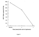
- Figure 4. A graph showing the change in the number of copies of HIV-1 DNA per 105 peripheral blood lymphocytes (PBL) with time (in weeks) during the course of apheresis treatment of a patient with high HIV-1 DNA count before apheresis treatment.
- Figure 5. A graph showing the change in the number of copies of HIV-1 RNA (x103) per ml blood with time during the course of apheresis treatment of an HIV-infected patient.
- Figure 6. A graph showing the change in the percentage of CD4+/CD25+ T-Cells with time (in weeks) during the course of apheresis treatment of an HIV-infected patient.
- Figure 7. A graph showing the change in the number of CD4+ T-cells per µl blood with time (in weeks) during the course of apheresis treatment of an HIV-infected patient.
- Figure 8. A graph showing the change in the number of CD8+ T-cells per µl blood with time (in weeks) during the course of apheresis treatment of an HIV-infected patient.
- Figure 9. A graph showing the change in SAC-stimulated TNF-α production by peripheral blood mononuclear cells (in pg/ml) with time (in weeks) during the course of apheresis treatment of an HIV-infected patient. SAC is the superantigen of staphilococcus aureus.
- Figure 10. A graph showing the change in SAC-stimulated IL-10 (
interleukin 10 in pg/ml) with time (in weeks) production by peripheral blood mononuclear cells during the course of apheresis treatment of an HIV-infected patient. - Figure 11. A graph showing the change in HIV-1 proviral DNA levels per 105 peripheral blood lymphocytes (PBL) in patients A and B with time (in weeks) during the course of apheresis treatment.
- Figure 12. A graph showing the change in the level of CD4-positive cells (% increase) and the change in viral load (copies per ml) with time in
patient 1 undergoing apheresis treatment. - Figure 13. A graph showing the change in the level of CD4-positive cells (% increase) and the change in viral load (copies per ml) with time in
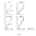
patient 2 undergoing apheresis treatment. - Figure 14. Graphs showing the changes in levels of CD3+ cells (as a percentage of lymphocytes), CD4+ CD28+ (TH-1) cells (the percentage of CD28+ cells based on CD4+ cells). CD5+CD45RA+ (the percentage of CD45RA+ cells based on the CD4+ cells) cells and CD4+CD25+ (IL-2) (the percentage of CD25+ cells based on CD4+ cells) cells before and after treatment in
patients - Patients under HAART received apheresis treatment using the G-1 column described above. Each patient received one apheresis per week over an 8 week period. Each apheresis is of 60 minutes duration at a flow rate of 30 ml/minute.
- Selection Criteria
- a) Documented HIV infection,
- b) Male or non-pregnant, non-lactating females over 18 years of age,
- c) Ability and willingness to give written informed consent, conforming to Italian guidelines, the declaration of Helsinki and international and institutional guidelines, and comply with the investigational nature of the study,
- d) On stable HAART drug therapy for at least 4 months
- Baseline was the level of HIV-1 DNA before apheresis treatment was commenced. Immediately after baseline blood sampling, the patient received apheresis; one apheresis per week, at a flow rate of 30ml per minute, duration of each apheresis was 60 minutes. The fall in HIV-1 DNA following apheresis with the G-1 column is shown in Figure 3. HIV-1 DNA was quantified using a standard quantitative polymerase chain reaction assay (Amplicor HIV Monitor™ , Roche, Basel, Switzerland) on peripheral blood mononuclear cells. The results indicate that apheresis with the G-1 column can flush out the HIV-infected resting cells. No drug is known that can produce such a rapid fall in HIV-DNA load.
- Immediately after baseline blood sampling, the patient received apheresis; one apheresis per week, at a flow rate of 30 ml per minute, duration of each apheresis was 60 minutes. The fall in HIV-1 DNA by apheresis with the G-1 column is shown in Figure 4. Again (as in the case of Example 1), the results indicate that apheresis with the G-1 column can flush out the HIV-infected resting cells. No drug is known that can produce such a rapid fall in HIV-DNA load.
- Immediately after baseline blood sampling, the patient received apheresis; one apheresis per week, at a flow rate of 30 ml per minute, duration of each apheresis was 60 minutes. The fall in HIV-1 RNA following apheresis with the G-1 column is shown in Figure 5. HIV-1 RNA was quantified using a standard quantitative reverse transcriptase-polymerase chain reaction competition assay. No drug or drug therapy is known to produce such a rapid sharp decline in HIV RNA.
- Immediately after baseline blood sampling, the patient received apheresis; one apheresis per week, at a flow rate of 30 ml per minute, duration of each apheresis was 60 minutes. The rise in percentage CD44/CD25+ T-cells following apheresis with the G-1 column is shown in Figure 6. The number of CD4+/CD25+ lymphocytes was measured using an Epics XL flow cytometer (Coulter Electronics, Inc.) using 100 µl of EDTA peripheral blood incubated for 30 minutes at 4°C with phycoerytrin fluorochrome-labelled anti CD25 monoclonal antibody.
- CD4+/CD25+ represents T-cell activity; CD4+/CD25+ T-cells are activated T-cells. CD25 is the α-subunit of the IL-2 receptor which is required for high affinity binding of IL-2. It is expressed by activated but not by resting T-cells.
The increase in CD4+/CD25+ is very important for HIV patients because - 1) they produce IL-2,
- 2) activated T-cells activate proviral DNA and make it susceptible to HAART (drugs). This facilitates the flushing out of latently infected lymphocytes (infected resting cells).
- Immediately after baseline blood sampling, the patient received apheresis; one apheresis per week, at a flow rate of 30 ml per minute, duration of each apheresis was 60 minutes. The rise in CD4+ T-cells following apheresis with the G-1 column is shown in Figure 7. The number of CD4+ lymphocytes was measured using an Epics,XL flow cytometer (Coulter Electronics, Inc.) using 100 µl of EDTA peripheral blood incubated for 30 minutes at 4°C with phycoerytrin-Texas-Red fluorochrome- labelled anti CD4 monoclonal antibody.
- CD4+ T-cell count is a key marker of HIV-disease symptoms; an increase in CD4+ T-cell count is often regarded as a marker of HIV decline and patient improvement.
- Immediately after baseline blood sampling, the patient received apheresis; one apheresis per week, at a flow rate of 30 ml per minute, duration of each apheresis was 60 minutes. The rise in CD8+ T-cells following apheresis with the G-1 column is shown in Figure 8. The number of CD84 lymphocytes was measured using an Epics XL flow cytometer (Coulter Electronics, Inc.) using 100 µl of EDTA peripheral blood incubated for 30 minutes at 4°C with fluorescin-isothiocyanate fluorochrome- labelled anti CD8 monoclonal antibody.
- CD8+ T-cells are caused to proliferate by raised levels of IL-2 (acting via the IL-2 receptor as described in relation to Example 4 above).
- Immediately after baseline blood sampling, the patient received apheresis; one apheresis per week, at a flow rate of 30 ml per minute, duration of each apheresis was 60 minutes. The sharp decline in TNF-α production by peripheral blood mononuclear cells is shown in Figure 9. The production of TNF-α by peripheral blood mononuclear cells(PBMC) was measured by culturing 3×10⁶ PBMC per well in 24-well LINBRO plates (Flow laboratories, Inc. McLean, VA) at 37°C in a moist, 7% CO2 atmosphere. The PBMC were then unstimulated or stimulated with LPS (SIGMA Diagnostics St. Louis, MO) at a concentration of 1 µg/ml. The supernatant was harvested after 48 hours of culture and cytokine production was then evaluated with a commercially available ELISA assay (Genzyme, Cambridge, MA).
- TNF-α is a major pathological factor in HIV-infection disease. No drug is known that can produce such a rapid reduction in TNF-α production by peripheral blood mononuclear cells.
- Immediately after baseline blood sampling, the patient received apheresis; one apheresis per week, at a flow rate of 30 ml per minute, duration of each apheresis was 60 minutes. The sharp decline in IL-10 (interleukin 10) production by peripheral blood mononuclear cells following apheresis with the G-1 column is shown in Figure 10. The production of IL-10 by peripheral blood mononuclear cells(PBMC) was measured by culturing 3x10⁶ PBMC per well in 24-well LINBRO plates (Flow laboratories, Inc.) at 37°C in a moist, 7% CO2 atmosphere. The PBMC were then unstimulated or stimulated with PHA (M form, Grand Island, NY) at a dilution 1:100. The supernatant was harvested after 48 hours of culture and cytokine production was then evaluated with a commercially available ELISA assay (Genzyme, Cambridge, MA).
- IL-10 is a major pathological factor in HIV-infection disease. IL-10 is a potent immune suppressor. It suppresses cytokine production by macrophages and thus indirectly suppresses T-cell activation. This counteracts the actions of IL-2 and IL-12 on cell-mediated immunity and IL-10 therefore effectively shuts down the immune system.
- Two pairs of HIV patients were matched within each pair for disease state (A and B). Control patients were treated with HAART only, whilst G-1 patients were treated with HAART and with apheresis. Baseline was the level of HIV-1 DNA at the start of the study before apheresis treatment was commenced. Immediately after blood sampling, the G-1 patients received apheresis; one apheresis per wee, at a flow rate of 30 ml per minute, duration of each apheresis was 60 minutes. The change in HIV-1 DNA over the 8 weeks of treatment is shown in Figure 11. The HIV-1 DNA falls sharply in the patients who received apheresis, but not in the patients treated with HAART alone. The HIV-1 DNA was measured as decribed in Example 1.
- The following additional, non-limiting Example 10 further illustrates the invention. Example 10 describes a preliminary investigation into the method and apparatus of the invention. Examples 1 to 9 relate to subsequent and more thorough investigations.
- As a blood treatment apparatus for the treatment of an HIV-infected disease, an external circulation apparatus composed in combination of a direct haemoperfusion column (capacity: about 335 ml) packed with cellulose acetate beads (220 g; diameter: 2 mm) and an adsorption-type haemocathartic apparatus ("ADAMONITOR MM6-P", tradename; manufactured by Otsuka Denshi K.K.) was used [clinical Study on Use of Granulocyte-Removing Apparatus (G-1) for Chronic Rheumatism", Ensho (Inflammation), 17(1), 57-80 (1997)].
- The results of clinical tests on the above-described external circulation apparatus, said results being to be described subsequently herein, are a part of the results of a phase II study which was conducted following legitimate procedures in Italy (A PHASE II SAFETY AND EFFICACY EVALUATION OF COMBINED ANTI-RETROVIRAL DRUGS AND G-1 COLUMN: A PILOT STUDY).
- From patients under treatment with an anti-retroviral (ARV) preparation composed in combination of two nucleoside reverse transcriptase inhibitors and one protease inhibitor, HIV-infected patients were chosen as subjects for treatment in accordance with the following standard: patients remaining under stable conditions over at least four months and generally one year owing to the ARV chemotherapy, having CD4-positive cell numbers not increased by 10% or greater from the corresponding base lines (the corresponding values before the initiation of the ARV therapy led to the stable conditions)(immunologically ARV-nonresponsive patients), and having more than 50,000 copies of HIV-RNA or showing a decrease in HIV-RNA not greater than 0.5 log (≤ 0.5 log) since the beginning of HAART owing to the ARV chemotherapy led to the stable conditions (virologically ARV-nonresponsive patients).
- The treatment of the HIV-infected disease by the external circulation apparatus according to the present invention was conducted in substantially the same manner as in the clinical study on chronic rheumatism as described in the report referred to in the above. The treatment was conducted once a week by setting the amount of blood, which is to be circulated externally per treatment, on the basis of a.blood flow rate of 30ml/minute and an external circulation time of 60 minutes as standards.
- The results of the treatment of Patient 1 (48 years old, male) and Patient 2 (43 years old, male) are shown in Table 3.
- The test items in Table 3 were all measured by methods commonly employed in the art, and include the number of white blood cells (WBC), the number of neutrophils, the number of monocytes, the number of eosinophils, the number of lymphocytes, the numbers of lymphocyte surface markers, the number of hemoglobins, the number of platelets, and the viral load.
- Specifically, the number of each lymphocyte surface marker was determined as a result of a measurement by flow cytometry while making use of a monoclonal antibody of the marker. "CD3" indicates the number of CD3-positive cells, "#CD4" the number of CD4-positive cells, "%CD4" the percentage of the CD4-positive cells based on lymphocytes, "CD4/8" the ratio of the CD4-positive cells to CD8-positive cells, "CD4+CD45RA+" the percentage of CD45RA-positive cells based on the CD4-positive cells, "CD4_CD25+(IL-2)" the percentage of CD25-positive cells based on the CD4-positive cells, and "CD4+CD28+(Th-1)" the percentage of CD28-positive cells based on the CD4-positive cells.
- Further each viral load is determined as a result of a measurement of a serum level of HIV-RNA by the RT-PCR method (cp: the number of copies per ml).
- Based on the results shown in Table 3 and Table 4, the rates (%) of increases in the number of CD4-positive cells from the respective base lines and the variations in viral load (the number of copies) are shown in Figure 12 (Patient 1) and Figure 13 (Patient 2), respectively. Further, the variations in the percentage of CD3-positive cells based on lymphocytes and the variations in "CD4+CD45RA+", CD4+CD25+(IL-2)" and "CD4+CD28+(th-1" are shown in Figure 14 (Patient 1: dots, Patient 2: circles).
- Concerning
Patient 1 andPatient 2, increases (50 to 70%) in the number of CD4-positive lymphcytes were already observed after the treatment by the present invention (as early as 60 minutes after the initiation), and this effect lasted for 3 to 7 days or even longer. In addition, by conducting the second therapy (treatment), this effect was observed to last for 7 days or longer in both of the patients.Table 3 Patient 11st apheresis 3 days later 2nd apheresis Baseline 60 min Baseline 60 min Number of WBC 5,300 4,200 Number of neurophils 2,300 2,500 2,800 2,900 2,400 Number of monocytes 500 300 200 700 400 Number of eosinophils 20 10 20 Number of lymphocytes 1,600 1,100 1,500 1,600 1,300 CD3 791 1,179 1,130 #CD4 219 323 365 234 333 %CD4 19.9 20.2 24.3 19.6 22.7 CD4/8 0.45 0.44 0.53 0.40 0.50 CD4+CD45RA+ 0.60 13.10 CD4+CD25+(IL-2) 10.2 18.02 CD4+CD28+(Th-1) 7.00 16.30 Number of hemoglobins 14.2 13.5 14.0 14.6 13.8 Number of platelets 130 124 127 143 129 Viral load 89,000cp 20,000cp Table 4 Patient 21st apheresis 2nd apheresis Baseline 60 min 75 min Baseline 60 min Number of WBC 6,000 5,000 Number of neurophils 2,400 2,300 2,500 1,700 2,000 Number of monocytes 700 600 500 700 500 Number of eosinophils 30 10 0 Number of lymphocytes 1,800 2,600 2,500 3,300 2,200 CD3 1,541 2,213 2,078 #CD4 284 478 488 468 451 %CD4 15.0 18.0 19.5 14.0 20.0 CD4/8 0.24 0.29 0.33 CD4+CD45RA+ 4.10 5.40 CD4+CD25+(IL-2) 12.02 19.02 CD4+CD28+(Th-1) 8.60 12.90 Number of hemoglobins 13.4 12.7 12.6 14.8 13.6 Number of platelets 187 169 166 253 194 Viral load 31,000cp 26,000cp - These prompt increases in the number of CD4-positive lymphocytes are postulated to be attributable to recovery and enhancement of myeloid function by the treatment in accordance with the present invention and formation of new CD3-positive cells, especially a group of naive cells such as CD45RA-positive cells, CD25-positive cells and CD28-positive cells, as shown in Figure 14. Such an increase is an outcome not observed at all from the conventional methods for the treatment of HIV-infected diseases. In addition, the treatment method according to the present invention has been recognised to bring about a decrease in viral load and hence to enhance its effect.
-
- 1. Lazarus H.M. et al. Selective in vivo removal of rheumatoid factor by an extracorporeal treatment device in rheumatoid arthritis patients. Transfusion, 1991; 31: 122-128.
- 2. Eshleman J.R. and Silberstein L.E. The development of selective plasmapheresis as a means for specific therapeutic intervention (editorial). Transfusion, 1991; 31: 96 - 98.
- 3. Fauci AS. Host factors and the pathogenesis of HIV-induced disease. Nature, 1996; 384: 529-533.
- 4. Cassone A., et al. (1997). Possible participation of polymorphonuclear cells stimulated by microbial immunomodulators in the dysregulated cytokine patterns of AIDS patients. J. Leukoc. Biol., 1997 62: 60-66.
- 5. Richman DD. HIV therapeutics. Science 1996; 272:1886-1888.
- 6. Saag MS. et al. HIV viral load markers in clinical practice. Nature Med. 1996; 2:625-629.
- 7. Oyaizu N and Pahwa S. Role of apoptosis in HIV disease pathogenesis. J. Clin. Immunol. 1995; 15:27-231.
- 8. Briant L. Binding of HIV-1 virions or gp120 anti-gp120 immune complexes to HIV-1 infected quiescent peripheral blood mononuclear cells reveals latent infection. J. Immunol. 1996; 156: 3994-4004.
- 9. Herbein G. et al. Apoptosis of CD8 T cells is mediated by macrophages through interaction of HIV gp120 with chemokine receptor CXCR4. Nature 1998; 395, 189-194.
- 10. Clerici, M and Shearer, G.M. A TH1--->TH2 switch is a critical step in the aetiology of HIV infection. Immunol Today 1993;14:107-111.
- 11. Clerici, M and Shearer, G.M (1994a) The TH1/TH2 hypothesis of HIV infection: new insights. Immunol Today 1994; 15:575-581.
- 12. Chehimi J. et al. IL-12 deficiency in HIV-infected patients. J. Exp. Med. 1994; 179: 1361-1366.
- 13. Connors M et al. HIV infection induces changes in the CD4 T cell phenotype and depletions within the CD4 T cell repertoire that are not immediately restored by antiviral or immune based therapies. Nature Med, 3:533-540, 1997.
- 14. Pakker NG. et al. Patterns of T cell repopulation, viral load reduction, and restoration of T cell function in HIV-infected persons during therapy with different antiretroviral agents. J AIDS Hum Retrovir 1997; 16:318-326.
- 15. Angel JB. et al.. Improvement in cell-mediated immune function, during potent anti-human immunodeficiency virus therapy with ritonavir plus saquinavir. J Inf Dis 1998; 177:808-904.
- 16. de Jong MD. et al. Summary of the international consensus symposium on management of HIV, CMV and hepatitis virus infections. Antiviral Res. 1998; 37:1-16.
- 17. Oyaizu N. et al. Accelerated apoptosis in PBMC from human immunodeficiency infected patients and in CD4 cross-linked PBMC from normal individuals. Blood 1993; 82:3392-3400.
- 18. D'Arrigo C., et al. Human neutrophil Fc receptor-mediated adhesion under flow: a hollow fiber model of intravascular arrest. Clin. Exp. Immunol., 1995; 100: 173-179.
Claims (9)
- Use of cellulose acetate beads as absorbent carrier in the manufacture of an agent for the treatment by apheresis of a patient injected with HIV.
- A use as claimed in claim 1 wherein the adsorbent carrier is a bead having a diameter in the range from 0.1 to 10.0 mm.
- A use as claimed in any one of claims 1 to 2 wherein the adsorbent carrier is a bead having a diameter in the range from 0.2 to 5.0 mm.
- A use as claimed in any one of claims 1 to 3 wherein the adsorbent carrier is a bead having a diameter in the range from 0.5 to 4.0 mm.
- A use as claimed in any one of claims 1 to 4wherein the adsorbent carrier is a bead having a diameter in the range from 1.0 to 3.0 mm.
- A use as described in any one of claims 1 to 5 wherein the adsorbent carrier has a roughened surface.
- A use as claimed in any one of claims 1 to 6 wherein the adsorbent carrier has on its surface irregularities having a centre line average height Ra, being defined under Japanese and Industrial Standard B0601-1982, of 0.2µm to 10µm and a mean spacing Sm of unevenness being within the range from 5µm to 200µm.
- A use as claimed in any one of claims 1 to 7 wherein the adsorbent carrier comprises cellulose acetate beads with a diameter in the range between 0.1 and 1.0 mm.
- A use as claimed in any one of claims 1 to 8 wherein the blood is separated into plasma and other components and the plasma is brought into contact with the adsorbent carrier.
Applications Claiming Priority (5)
| Application Number | Priority Date | Filing Date | Title |
|---|---|---|---|
| JP7214499 | 1999-03-17 | ||
| JP11072144A JP2000262612A (en) | 1999-03-17 | 1999-03-17 | Methods of treating HIV infectious diseases |
| GB9919565 | 1999-08-18 | ||
| GBGB9919565.3A GB9919565D0 (en) | 1999-03-17 | 1999-08-18 | Treatment of disease |
| PCT/JP2000/001099 WO2000055621A2 (en) | 1999-03-17 | 2000-02-25 | Blood leucocyte apheresis for treatment of disease |
Publications (2)
| Publication Number | Publication Date |
|---|---|
| EP1126859A2 EP1126859A2 (en) | 2001-08-29 |
| EP1126859B1 true EP1126859B1 (en) | 2006-05-10 |
Family
ID=26315857
Family Applications (1)
| Application Number | Title | Priority Date | Filing Date |
|---|---|---|---|
| EP00905345A Expired - Lifetime EP1126859B1 (en) | 1999-03-17 | 2000-02-25 | Blood leucocyte apheresis for treatment of hiv |
Country Status (10)
| Country | Link |
|---|---|
| US (1) | US6498007B1 (en) |
| EP (1) | EP1126859B1 (en) |
| JP (1) | JP4643830B2 (en) |
| AT (1) | ATE325613T1 (en) |
| AU (1) | AU2692900A (en) |
| DE (1) | DE60027863T2 (en) |
| DK (1) | DK1126859T3 (en) |
| ES (1) | ES2262506T3 (en) |
| PT (1) | PT1126859E (en) |
| WO (1) | WO2000055621A2 (en) |
Families Citing this family (38)
| Publication number | Priority date | Publication date | Assignee | Title |
|---|---|---|---|---|
| US20020197250A1 (en) * | 2001-04-10 | 2002-12-26 | Renal Tech International | Biocompatible devices, systems, and methods for reducing levels of pro-inflammatory or anti-inflammatory stimulators or mediators in the blood |
| US20020198487A1 (en) * | 2001-04-10 | 2002-12-26 | Renal Tech International | Devices, systems, and methods for reducing levels of pro-inflammatory or anti-inflammatory stimulators or mediators in physiologic fluids |
| US20020197249A1 (en) * | 2001-04-10 | 2002-12-26 | Renal Tech International | Devices, systems, and methods for reducing levels of pro-inflammatory or anti-inflammatory stimulators or mediators in blood products |
| US20020159995A1 (en) * | 1997-07-30 | 2002-10-31 | Renal Tech International | Devices, systems, and methods for reducing levels of pro-inflammatory or anti-inflammatory stimulators or mediators in the blood, generated as a result of extracorporeal blood processing |
| US8329388B2 (en) * | 1997-07-30 | 2012-12-11 | Cytosorbents, Inc. | Biocompatible devices, systems, and methods for reducing levels of proinflammatory of antiinflammatory stimulators or mediators in the blood |
| US6878127B2 (en) | 2001-04-10 | 2005-04-12 | Renaltech International, Llc | Devices, systems, and methods for reducing levels of pro-inflammatory or anti-inflammatory stimulators or mediators in the blood |
| US20020197252A1 (en) * | 2001-04-10 | 2002-12-26 | Renal Tech International | Selective adsorption devices and systems |
| DE10147638B4 (en) * | 2001-09-27 | 2004-05-27 | Leukocare Gmbh | Leukocyte Inactivation Module (LIM) |
| JP2003235959A (en) | 2002-02-14 | 2003-08-26 | Nippon Koutai Kenkyusho:Kk | How to remove and reduce hepatitis C virus |
| US7850858B2 (en) * | 2004-08-30 | 2010-12-14 | Kaneka Corporation | Granulocyte adsorbent |
| JP4958554B2 (en) | 2004-09-10 | 2012-06-20 | 株式会社カネカ | Adsorbent and treatment method for lymphocyte proliferation inhibitory factor |
| US20090204051A1 (en) * | 2005-03-30 | 2009-08-13 | Kaneka Corporation | Method of removing lymphocyte growth inhibitor |
| KR100647335B1 (en) | 2005-04-15 | 2006-11-23 | 삼성전자주식회사 | Cell separation method using hydrophobic solid supports |
| EP1712284B1 (en) * | 2005-04-15 | 2012-10-10 | Samsung Electronics Co., Ltd. | Cell separation method using hydrophobic solid supports |
| EP1931404B1 (en) | 2005-09-09 | 2011-08-03 | Membrana GmbH | Method for the elimination of leukocytes from blood |
| US20100135976A1 (en) * | 2006-06-16 | 2010-06-03 | Rune Nilsson | Adsorption device |
| KR100917465B1 (en) * | 2006-08-21 | 2009-09-14 | 삼성전자주식회사 | Method for separating microorganisms using non-planar solid support and microbial separation apparatus |
| KR100813265B1 (en) | 2006-08-21 | 2008-03-13 | 삼성전자주식회사 | Amplification of Nucleic Acids from Microorganisms Using Non-planar Solid Supports |
| US8158411B2 (en) | 2006-08-21 | 2012-04-17 | Samsung Electronics Co., Ltd. | Method of separating microorganism using nonplanar solid substrate and device for separating microorganism using the same |
| US20080070282A1 (en) * | 2006-08-21 | 2008-03-20 | Samsung Electronics Co., Ltd. | Method and device for obtaining or amplifying nucleic acid from a cell using a nonplanar solid substrate |
| KR100813264B1 (en) | 2006-08-21 | 2008-03-13 | 삼성전자주식회사 | Amplification of Nucleic Acids from Microorganisms Using Non-planar Solid Supports |
| WO2008038785A1 (en) * | 2006-09-29 | 2008-04-03 | Toray Industries, Inc. | Cell-adsorbing column |
| ES2581989T3 (en) * | 2007-01-13 | 2016-09-08 | 3M Innovative Properties Company | Device for the separation of leukocytes from the blood |
| AU2008293431B2 (en) | 2007-08-31 | 2013-07-04 | Cytopherx, Inc. | Selective cytopheresis devices and related methods thereof |
| US20090149802A1 (en) * | 2007-12-07 | 2009-06-11 | Ross Peter Jones | Surgical System Including a Trap for Noise-Inducing Materials |
| US8926543B2 (en) * | 2009-10-08 | 2015-01-06 | Otsuka Pharmaceutical Co., Ltd. | Immunoactivation blood perfusion filter for the treatment of malignant tumors |
| CN103260670A (en) | 2010-10-15 | 2013-08-21 | 塞托弗尔克斯股份有限公司 | Cytopheresic cartridge and use thereof |
| DE102010049790A1 (en) * | 2010-10-29 | 2012-05-03 | Fresenius Medical Care Deutschland Gmbh | Medical separator |
| US20130068691A1 (en) * | 2011-08-05 | 2013-03-21 | Henry John Smith | Targeted apheresis for the treatment of rheumatoid arthritis and immune disorders |
| EP2606921A1 (en) | 2011-12-21 | 2013-06-26 | Infomed SA | Device for purifying blood by extracorporeal circulation |
| CA2860831C (en) | 2012-01-09 | 2024-09-10 | Seastar Medical, Inc. | Cartridge and method for increasing myocardial function |
| EP3006060B1 (en) * | 2013-05-31 | 2017-11-22 | Toray Industries, Inc. | Adsorption carrier-packed column |
| CN106659834B (en) | 2014-08-26 | 2019-06-18 | 3M创新有限公司 | System for the removal of pro-inflammatory mediators and granulocytes and monocytes from the blood |
| US11383015B2 (en) * | 2016-01-11 | 2022-07-12 | Fenwal, Inc. | System and method for plasma purification prior to mononuclear cell collection |
| CN106267414B (en) * | 2016-07-01 | 2019-02-19 | 翁炳焕 | AIDS immune purifier |
| CN106166313B (en) * | 2016-07-01 | 2018-09-07 | 浙江大学 | A kind of AIDS pregnant woman blood clarifier |
| CN106267410B (en) * | 2016-07-01 | 2019-03-08 | 翁炳焕 | HIV infection cell separator |
| JP7429343B2 (en) | 2018-01-05 | 2024-02-08 | パス イーエックス,インク. | Device for the capture and removal of diseased material from fluids |
Family Cites Families (5)
| Publication number | Priority date | Publication date | Assignee | Title |
|---|---|---|---|---|
| US4906742A (en) * | 1986-07-31 | 1990-03-06 | Whitehead Institute For Biomedical Research | Encoding antigens of M. Leprae |
| JP2673567B2 (en) | 1987-12-10 | 1997-11-05 | 株式会社日本抗体研究所 | Method for removing granulocytes in blood and granulocyte removing apparatus used therefor |
| US5807881A (en) * | 1992-05-27 | 1998-09-15 | Quadra Logic Technologies, Inc. | Method for selectively reducing activated leukocyte cell population |
| US5567443A (en) | 1993-08-04 | 1996-10-22 | Japan Immunoresearch Laboratories Co., Ltd. | Method of treating inflammatory diseases |
| US5525279A (en) * | 1994-04-04 | 1996-06-11 | Sekisui Kagaku Kogyo K.K. | Method of forming a granulocyte adsorbing carrier and granulocyte remover |
-
2000
- 2000-02-25 JP JP2000605202A patent/JP4643830B2/en not_active Expired - Fee Related
- 2000-02-25 ES ES00905345T patent/ES2262506T3/en not_active Expired - Lifetime
- 2000-02-25 EP EP00905345A patent/EP1126859B1/en not_active Expired - Lifetime
- 2000-02-25 DK DK00905345T patent/DK1126859T3/en active
- 2000-02-25 US US09/700,632 patent/US6498007B1/en not_active Expired - Lifetime
- 2000-02-25 PT PT00905345T patent/PT1126859E/en unknown
- 2000-02-25 DE DE60027863T patent/DE60027863T2/en not_active Expired - Lifetime
- 2000-02-25 WO PCT/JP2000/001099 patent/WO2000055621A2/en not_active Ceased
- 2000-02-25 AT AT00905345T patent/ATE325613T1/en active
- 2000-02-25 AU AU26929/00A patent/AU2692900A/en not_active Abandoned
Also Published As
| Publication number | Publication date |
|---|---|
| DE60027863T2 (en) | 2006-12-07 |
| DE60027863D1 (en) | 2006-06-14 |
| US6498007B1 (en) | 2002-12-24 |
| DK1126859T3 (en) | 2006-08-28 |
| ES2262506T3 (en) | 2006-12-01 |
| WO2000055621A8 (en) | 2001-07-05 |
| AU2692900A (en) | 2000-10-04 |
| EP1126859A2 (en) | 2001-08-29 |
| JP4643830B2 (en) | 2011-03-02 |
| PT1126859E (en) | 2006-09-29 |
| WO2000055621A2 (en) | 2000-09-21 |
| WO2000055621A3 (en) | 2001-04-19 |
| ATE325613T1 (en) | 2006-06-15 |
| JP2002539178A (en) | 2002-11-19 |
Similar Documents
| Publication | Publication Date | Title |
|---|---|---|
| US6498007B1 (en) | Methods of treatment of disease using adsorbent carriers | |
| Leaf et al. | Thrombotic thrombocytopenic purpura associated with human immunodeficiency virus type 1 (HIV-1) infection | |
| Schmitz et al. | Cytokines and myelination in the central nervous system | |
| Younas et al. | Immune activation in the course of HIV‐1 infection: causes, phenotypes and persistence under therapy | |
| Heideman et al. | Terminal complement complexes and anaphylatoxins in septic and ischemic patients | |
| Prior et al. | Pulmonary sarcoidosis: patterns of cytokine release in vitro | |
| Cameron et al. | During HIV‐1 infection most blood dendritic cells are not productively infected and can induce allogeneic CD4+ T cells clonal expansion | |
| Hidaka et al. | Dynamic changes in cytokine levels in serum and synovial fluid following filtration leukocytapheresis therapy in patients with rheumatoid arthritis | |
| Kaser et al. | Interferon α augments activation-induced T cell death by upregulation of Fas (CD95/APO-1) and Fas ligand expression | |
| MO et al. | Expression patterns of the HIV type 1 coreceptors CCR5 and CXCR4 on CD4+ T cells and monocytes from cord and adult blood | |
| Vlahakis et al. | Chemokine-receptor activation by env determines the mechanism of death in HIV-infected and uninfected T lymphocytes | |
| Hashimoto et al. | Modulation of Bcl-2 protein by CD4 cross-linking: a possible mechanism for lymphocyte apoptosis in human immunodeficiency virus infection and for rescue of apoptosis by interleukin-2 | |
| Weichold et al. | Neither human immunodeficiency virus-1 (HIV-1) nor HIV-2 infects most-primitive human hematopoietic stem cells as assessed in long-term bone marrow cultures | |
| Sonza et al. | Susceptibility of human monocytes to HIV type 1 infection in vitro is not dependent on their level of CD4 expression | |
| Schenal et al. | Distinct patterns of HIV-specific memory T lymphocytes in HIV-exposed uninfected individuals and in HIV-infected patients | |
| Bernstein et al. | Neonatal natural killer cells produce chemokines and suppress HIV replication in vitro | |
| Creery et al. | Down-regulation of CXCR-4 and CCR-5 expression by interferon-γ is associated with inhibition of chemotaxis and human immunodeficiency virus (HIV) replication but not HIV entry into human monocytes | |
| Creery et al. | Differential regulation of CXCR4 and CCR5 expression by interleukin (IL)-4 and IL-13 is associated with inhibition of chemotaxis and human immunodeficiency virus (HIV) type 1 replication but not HIV entry into human monocytes | |
| US6713252B2 (en) | Method for removing and reducing hepatitis C virus | |
| Takeda et al. | Cellulose acetate beads induce release of interleukin-1 receptor antagonist, but not tumour necrosis factor-α or interleukin-1β in human peripheral blood | |
| Calabresi et al. | Chemokines in chronic progressive neurological diseases: HTLV-1 associated myelopathy and multiple sclerosis | |
| Mazza et al. | Recombinant interleukin‐2 (rIL‐2) in acquired immune deficiency syndrome (AIDS): Preliminary report in patients with lymphoma associated with HIV infection | |
| Bovolenta et al. | Expression and activation of a C-terminal truncated isoform of STAT5 (STAT5Δ) following interleukin 2 administration or AZT monotherapy in HIV-infected individuals | |
| Bolski et al. | The prevalence of AIDS-associated oral lesions in a cohort of patients with hemophilia | |
| Fu et al. | Comparison of haploidentical-hematopoietic stem cell transplantation using modified post-transplantation cyclophosphamide and eltrombopag combined with immunosuppression for the treatment of severe aplastic anemia |
Legal Events
| Date | Code | Title | Description |
|---|---|---|---|
| PUAI | Public reference made under article 153(3) epc to a published international application that has entered the european phase |
Free format text: ORIGINAL CODE: 0009012 |
|
| 17P | Request for examination filed |
Effective date: 20001215 |
|
| AK | Designated contracting states |
Kind code of ref document: A2 Designated state(s): AT BE CH CY DE DK ES FI FR GB GR IE IT LI LU MC NL PT SE |
|
| AX | Request for extension of the european patent |
Free format text: AL;LT;LV;MK;RO;SI |
|
| RIN1 | Information on inventor provided before grant (corrected) |
Inventor name: ADACHI, MASAKAZU Inventor name: HIBI, TOSHIFUMI |
|
| 17Q | First examination report despatched |
Effective date: 20031030 |
|
| GRAP | Despatch of communication of intention to grant a patent |
Free format text: ORIGINAL CODE: EPIDOSNIGR1 |
|
| RIC1 | Information provided on ipc code assigned before grant |
Ipc: 7A 61P 31/18 B Ipc: 7A 61M 1/36 B Ipc: 7A 61K 35/14 A |
|
| RIC1 | Information provided on ipc code assigned before grant |
Ipc: 7A 61K 35/14 A Ipc: 7A 61M 1/36 B Ipc: 7A 61P 31/18 B |
|
| RTI1 | Title (correction) |
Free format text: BLOOD LEUCOCYTE APHERESIS FOR TREATMENT OF HIV |
|
| GRAS | Grant fee paid |
Free format text: ORIGINAL CODE: EPIDOSNIGR3 |
|
| GRAA | (expected) grant |
Free format text: ORIGINAL CODE: 0009210 |
|
| RAP1 | Party data changed (applicant data changed or rights of an application transferred) |
Owner name: JIMRO CO., LTD. |
|
| AK | Designated contracting states |
Kind code of ref document: B1 Designated state(s): AT BE CH CY DE DK ES FI FR GB GR IE IT LI LU MC NL PT SE |
|
| PG25 | Lapsed in a contracting state [announced via postgrant information from national office to epo] |
Ref country code: IT Free format text: LAPSE BECAUSE OF FAILURE TO SUBMIT A TRANSLATION OF THE DESCRIPTION OR TO PAY THE FEE WITHIN THE PRESCRIBED TIME-LIMIT;WARNING: LAPSES OF ITALIAN PATENTS WITH EFFECTIVE DATE BEFORE 2007 MAY HAVE OCCURRED AT ANY TIME BEFORE 2007. THE CORRECT EFFECTIVE DATE MAY BE DIFFERENT FROM THE ONE RECORDED. Effective date: 20060510 |
|
| REG | Reference to a national code |
Ref country code: GB Ref legal event code: FG4D |
|
| REG | Reference to a national code |
Ref country code: CH Ref legal event code: EP |
|
| REF | Corresponds to: |
Ref document number: 60027863 Country of ref document: DE Date of ref document: 20060614 Kind code of ref document: P |
|
| REG | Reference to a national code |
Ref country code: IE Ref legal event code: FG4D |
|
| REG | Reference to a national code |
Ref country code: SE Ref legal event code: TRGR |
|
| REG | Reference to a national code |
Ref country code: CH Ref legal event code: NV Representative=s name: ISLER & PEDRAZZINI AG |
|
| REG | Reference to a national code |
Ref country code: DK Ref legal event code: T3 |
|
| REG | Reference to a national code |
Ref country code: GR Ref legal event code: EP Ref document number: 20060402514 Country of ref document: GR |
|
| REG | Reference to a national code |
Ref country code: PT Ref legal event code: SC4A Effective date: 20060809 |
|
| REG | Reference to a national code |
Ref country code: ES Ref legal event code: FG2A Ref document number: 2262506 Country of ref document: ES Kind code of ref document: T3 |
|
| ET | Fr: translation filed | ||
| PG25 | Lapsed in a contracting state [announced via postgrant information from national office to epo] |
Ref country code: MC Free format text: LAPSE BECAUSE OF NON-PAYMENT OF DUE FEES Effective date: 20070228 |
|
| PLBE | No opposition filed within time limit |
Free format text: ORIGINAL CODE: 0009261 |
|
| STAA | Information on the status of an ep patent application or granted ep patent |
Free format text: STATUS: NO OPPOSITION FILED WITHIN TIME LIMIT |
|
| 26N | No opposition filed |
Effective date: 20070213 |
|
| REG | Reference to a national code |
Ref country code: CH Ref legal event code: PCAR Free format text: ISLER & PEDRAZZINI AG;POSTFACH 1772;8027 ZUERICH (CH) |
|
| PG25 | Lapsed in a contracting state [announced via postgrant information from national office to epo] |
Ref country code: CY Free format text: LAPSE BECAUSE OF FAILURE TO SUBMIT A TRANSLATION OF THE DESCRIPTION OR TO PAY THE FEE WITHIN THE PRESCRIBED TIME-LIMIT Effective date: 20060510 |
|
| REG | Reference to a national code |
Ref country code: FR Ref legal event code: PLFP Year of fee payment: 17 |
|
| REG | Reference to a national code |
Ref country code: FR Ref legal event code: PLFP Year of fee payment: 18 |
|
| REG | Reference to a national code |
Ref country code: FR Ref legal event code: PLFP Year of fee payment: 19 |
|
| PGFP | Annual fee paid to national office [announced via postgrant information from national office to epo] |
Ref country code: LU Payment date: 20180208 Year of fee payment: 19 Ref country code: NL Payment date: 20180214 Year of fee payment: 19 |
|
| PGFP | Annual fee paid to national office [announced via postgrant information from national office to epo] |
Ref country code: DK Payment date: 20180212 Year of fee payment: 19 Ref country code: DE Payment date: 20180214 Year of fee payment: 19 Ref country code: GB Payment date: 20180221 Year of fee payment: 19 Ref country code: CH Payment date: 20180213 Year of fee payment: 19 Ref country code: FI Payment date: 20180209 Year of fee payment: 19 Ref country code: ES Payment date: 20180301 Year of fee payment: 19 |
|
| PGFP | Annual fee paid to national office [announced via postgrant information from national office to epo] |
Ref country code: PT Payment date: 20180221 Year of fee payment: 19 Ref country code: SE Payment date: 20180213 Year of fee payment: 19 Ref country code: IE Payment date: 20180212 Year of fee payment: 19 Ref country code: FR Payment date: 20180223 Year of fee payment: 19 Ref country code: GR Payment date: 20180214 Year of fee payment: 19 Ref country code: BE Payment date: 20180214 Year of fee payment: 19 Ref country code: AT Payment date: 20180209 Year of fee payment: 19 Ref country code: IT Payment date: 20180221 Year of fee payment: 19 |
|
| REG | Reference to a national code |
Ref country code: DE Ref legal event code: R119 Ref document number: 60027863 Country of ref document: DE |
|
| REG | Reference to a national code |
Ref country code: DK Ref legal event code: EBP Effective date: 20190228 |
|
| REG | Reference to a national code |
Ref country code: CH Ref legal event code: PL |
|
| REG | Reference to a national code |
Ref country code: SE Ref legal event code: EUG |
|
| REG | Reference to a national code |
Ref country code: NL Ref legal event code: MM Effective date: 20190301 |
|
| REG | Reference to a national code |
Ref country code: AT Ref legal event code: MM01 Ref document number: 325613 Country of ref document: AT Kind code of ref document: T Effective date: 20190225 |
|
| GBPC | Gb: european patent ceased through non-payment of renewal fee |
Effective date: 20190225 |
|
| PG25 | Lapsed in a contracting state [announced via postgrant information from national office to epo] |
Ref country code: LU Free format text: LAPSE BECAUSE OF NON-PAYMENT OF DUE FEES Effective date: 20190225 Ref country code: PT Free format text: LAPSE BECAUSE OF NON-PAYMENT OF DUE FEES Effective date: 20190826 Ref country code: SE Free format text: LAPSE BECAUSE OF NON-PAYMENT OF DUE FEES Effective date: 20190226 Ref country code: FI Free format text: LAPSE BECAUSE OF NON-PAYMENT OF DUE FEES Effective date: 20190225 |
|
| REG | Reference to a national code |
Ref country code: BE Ref legal event code: MM Effective date: 20190228 |
|
| REG | Reference to a national code |
Ref country code: IE Ref legal event code: MM4A |
|
| PG25 | Lapsed in a contracting state [announced via postgrant information from national office to epo] |
Ref country code: GR Free format text: LAPSE BECAUSE OF NON-PAYMENT OF DUE FEES Effective date: 20190904 |
|
| PG25 | Lapsed in a contracting state [announced via postgrant information from national office to epo] |
Ref country code: LI Free format text: LAPSE BECAUSE OF NON-PAYMENT OF DUE FEES Effective date: 20190228 Ref country code: CH Free format text: LAPSE BECAUSE OF NON-PAYMENT OF DUE FEES Effective date: 20190228 Ref country code: AT Free format text: LAPSE BECAUSE OF NON-PAYMENT OF DUE FEES Effective date: 20190225 |
|
| PG25 | Lapsed in a contracting state [announced via postgrant information from national office to epo] |
Ref country code: GB Free format text: LAPSE BECAUSE OF NON-PAYMENT OF DUE FEES Effective date: 20190225 Ref country code: NL Free format text: LAPSE BECAUSE OF NON-PAYMENT OF DUE FEES Effective date: 20190301 Ref country code: DK Free format text: LAPSE BECAUSE OF NON-PAYMENT OF DUE FEES Effective date: 20190228 Ref country code: IE Free format text: LAPSE BECAUSE OF NON-PAYMENT OF DUE FEES Effective date: 20190225 Ref country code: DE Free format text: LAPSE BECAUSE OF NON-PAYMENT OF DUE FEES Effective date: 20190903 |
|
| PG25 | Lapsed in a contracting state [announced via postgrant information from national office to epo] |
Ref country code: BE Free format text: LAPSE BECAUSE OF NON-PAYMENT OF DUE FEES Effective date: 20190228 Ref country code: FR Free format text: LAPSE BECAUSE OF NON-PAYMENT OF DUE FEES Effective date: 20190228 Ref country code: IT Free format text: LAPSE BECAUSE OF NON-PAYMENT OF DUE FEES Effective date: 20190225 |
|
| REG | Reference to a national code |
Ref country code: ES Ref legal event code: FD2A Effective date: 20200330 |
|
| PG25 | Lapsed in a contracting state [announced via postgrant information from national office to epo] |
Ref country code: ES Free format text: LAPSE BECAUSE OF NON-PAYMENT OF DUE FEES Effective date: 20190226 Ref country code: PT Free format text: LAPSE BECAUSE OF EXPIRATION OF PROTECTION Effective date: 20200305 |
|
| PG25 | Lapsed in a contracting state [announced via postgrant information from national office to epo] |
Ref country code: PT Free format text: LAPSE BECAUSE OF NON-PAYMENT OF DUE FEES Effective date: 20190225 |
|
| PG25 | Lapsed in a contracting state [announced via postgrant information from national office to epo] |
Ref country code: PT Free format text: LAPSE BECAUSE OF NON-PAYMENT OF DUE FEES Effective date: 20190225 |