EP1126782B1 - Method for improved imaging and photodynamic therapy - Google Patents
Method for improved imaging and photodynamic therapy Download PDFInfo
- Publication number
- EP1126782B1 EP1126782B1 EP99971283A EP99971283A EP1126782B1 EP 1126782 B1 EP1126782 B1 EP 1126782B1 EP 99971283 A EP99971283 A EP 99971283A EP 99971283 A EP99971283 A EP 99971283A EP 1126782 B1 EP1126782 B1 EP 1126782B1
- Authority
- EP
- European Patent Office
- Prior art keywords
- halogenated xanthene
- imaging
- agent
- diseased tissue
- halogenated
- Prior art date
- Legal status (The legal status is an assumption and is not a legal conclusion. Google has not performed a legal analysis and makes no representation as to the accuracy of the status listed.)
- Expired - Lifetime
Links
Images


Classifications
-
- A—HUMAN NECESSITIES
- A61—MEDICAL OR VETERINARY SCIENCE; HYGIENE
- A61K—PREPARATIONS FOR MEDICAL, DENTAL OR TOILETRY PURPOSES
- A61K49/00—Preparations for testing in vivo
- A61K49/001—Preparation for luminescence or biological staining
- A61K49/0013—Luminescence
- A61K49/0017—Fluorescence in vivo
- A61K49/0019—Fluorescence in vivo characterised by the fluorescent group, e.g. oligomeric, polymeric or dendritic molecules
- A61K49/0021—Fluorescence in vivo characterised by the fluorescent group, e.g. oligomeric, polymeric or dendritic molecules the fluorescent group being a small organic molecule
- A61K49/0041—Xanthene dyes, used in vivo, e.g. administered to a mice, e.g. rhodamines, rose Bengal
-
- A—HUMAN NECESSITIES
- A61—MEDICAL OR VETERINARY SCIENCE; HYGIENE
- A61F—FILTERS IMPLANTABLE INTO BLOOD VESSELS; PROSTHESES; DEVICES PROVIDING PATENCY TO, OR PREVENTING COLLAPSING OF, TUBULAR STRUCTURES OF THE BODY, e.g. STENTS; ORTHOPAEDIC, NURSING OR CONTRACEPTIVE DEVICES; FOMENTATION; TREATMENT OR PROTECTION OF EYES OR EARS; BANDAGES, DRESSINGS OR ABSORBENT PADS; FIRST-AID KITS
- A61F7/00—Heating or cooling appliances for medical or therapeutic treatment of the human body
-
- A—HUMAN NECESSITIES
- A61—MEDICAL OR VETERINARY SCIENCE; HYGIENE
- A61K—PREPARATIONS FOR MEDICAL, DENTAL OR TOILETRY PURPOSES
- A61K41/00—Medicinal preparations obtained by treating materials with wave energy or particle radiation ; Therapies using these preparations
- A61K41/0057—Photodynamic therapy with a photosensitizer, i.e. agent able to produce reactive oxygen species upon exposure to light or radiation, e.g. UV or visible light; photocleavage of nucleic acids with an agent
-
- A—HUMAN NECESSITIES
- A61—MEDICAL OR VETERINARY SCIENCE; HYGIENE
- A61K—PREPARATIONS FOR MEDICAL, DENTAL OR TOILETRY PURPOSES
- A61K41/00—Medicinal preparations obtained by treating materials with wave energy or particle radiation ; Therapies using these preparations
- A61K41/0057—Photodynamic therapy with a photosensitizer, i.e. agent able to produce reactive oxygen species upon exposure to light or radiation, e.g. UV or visible light; photocleavage of nucleic acids with an agent
- A61K41/008—Two-Photon or Multi-Photon PDT, e.g. with upconverting dyes or photosensitisers
-
- A—HUMAN NECESSITIES
- A61—MEDICAL OR VETERINARY SCIENCE; HYGIENE
- A61M—DEVICES FOR INTRODUCING MEDIA INTO, OR ONTO, THE BODY; DEVICES FOR TRANSDUCING BODY MEDIA OR FOR TAKING MEDIA FROM THE BODY; DEVICES FOR PRODUCING OR ENDING SLEEP OR STUPOR
- A61M25/00—Catheters; Hollow probes
- A61M25/10—Balloon catheters
- A61M25/104—Balloon catheters used for angioplasty
-
- A—HUMAN NECESSITIES
- A61—MEDICAL OR VETERINARY SCIENCE; HYGIENE
- A61N—ELECTROTHERAPY; MAGNETOTHERAPY; RADIATION THERAPY; ULTRASOUND THERAPY
- A61N5/00—Radiation therapy
- A61N5/06—Radiation therapy using light
- A61N5/0601—Apparatus for use inside the body
-
- A—HUMAN NECESSITIES
- A61—MEDICAL OR VETERINARY SCIENCE; HYGIENE
- A61N—ELECTROTHERAPY; MAGNETOTHERAPY; RADIATION THERAPY; ULTRASOUND THERAPY
- A61N5/00—Radiation therapy
- A61N5/06—Radiation therapy using light
- A61N5/0613—Apparatus adapted for a specific treatment
- A61N5/062—Photodynamic therapy, i.e. excitation of an agent
-
- A—HUMAN NECESSITIES
- A61—MEDICAL OR VETERINARY SCIENCE; HYGIENE
- A61P—SPECIFIC THERAPEUTIC ACTIVITY OF CHEMICAL COMPOUNDS OR MEDICINAL PREPARATIONS
- A61P35/00—Antineoplastic agents
-
- A—HUMAN NECESSITIES
- A61—MEDICAL OR VETERINARY SCIENCE; HYGIENE
- A61B—DIAGNOSIS; SURGERY; IDENTIFICATION
- A61B17/00—Surgical instruments, devices or methods
- A61B17/22—Implements for squeezing-off ulcers or the like on inner organs of the body; Implements for scraping-out cavities of body organs, e.g. bones; for invasive removal or destruction of calculus using mechanical vibrations; for removing obstructions in blood vessels, not otherwise provided for
- A61B2017/22051—Implements for squeezing-off ulcers or the like on inner organs of the body; Implements for scraping-out cavities of body organs, e.g. bones; for invasive removal or destruction of calculus using mechanical vibrations; for removing obstructions in blood vessels, not otherwise provided for with an inflatable part, e.g. balloon, for positioning, blocking, or immobilisation
- A61B2017/22062—Implements for squeezing-off ulcers or the like on inner organs of the body; Implements for scraping-out cavities of body organs, e.g. bones; for invasive removal or destruction of calculus using mechanical vibrations; for removing obstructions in blood vessels, not otherwise provided for with an inflatable part, e.g. balloon, for positioning, blocking, or immobilisation to be filled with liquid
-
- A—HUMAN NECESSITIES
- A61—MEDICAL OR VETERINARY SCIENCE; HYGIENE
- A61B—DIAGNOSIS; SURGERY; IDENTIFICATION
- A61B17/00—Surgical instruments, devices or methods
- A61B17/22—Implements for squeezing-off ulcers or the like on inner organs of the body; Implements for scraping-out cavities of body organs, e.g. bones; for invasive removal or destruction of calculus using mechanical vibrations; for removing obstructions in blood vessels, not otherwise provided for
- A61B2017/22051—Implements for squeezing-off ulcers or the like on inner organs of the body; Implements for scraping-out cavities of body organs, e.g. bones; for invasive removal or destruction of calculus using mechanical vibrations; for removing obstructions in blood vessels, not otherwise provided for with an inflatable part, e.g. balloon, for positioning, blocking, or immobilisation
- A61B2017/22065—Functions of balloons
- A61B2017/22069—Immobilising; Stabilising
-
- A—HUMAN NECESSITIES
- A61—MEDICAL OR VETERINARY SCIENCE; HYGIENE
- A61B—DIAGNOSIS; SURGERY; IDENTIFICATION
- A61B17/00—Surgical instruments, devices or methods
- A61B17/22—Implements for squeezing-off ulcers or the like on inner organs of the body; Implements for scraping-out cavities of body organs, e.g. bones; for invasive removal or destruction of calculus using mechanical vibrations; for removing obstructions in blood vessels, not otherwise provided for
- A61B2017/22082—Implements for squeezing-off ulcers or the like on inner organs of the body; Implements for scraping-out cavities of body organs, e.g. bones; for invasive removal or destruction of calculus using mechanical vibrations; for removing obstructions in blood vessels, not otherwise provided for after introduction of a substance
- A61B2017/22087—Implements for squeezing-off ulcers or the like on inner organs of the body; Implements for scraping-out cavities of body organs, e.g. bones; for invasive removal or destruction of calculus using mechanical vibrations; for removing obstructions in blood vessels, not otherwise provided for after introduction of a substance photodynamic
Definitions
- the present invention is directed to an apparatus and method of imaging and treatment using at least one photodynamic therapy (“PDT”) agent.
- PDT photodynamic therapy
- the apparatus and method is for imaging and treating diseased tissue.
- Imaging is typically performed to locate diseased tissue or tumors in a body. Once the diseased tissue is located, it is subsequently treated in some manner in order to destroy the diseased cells within this tissue. As explained infra, in the past, these were two separate procedures in a long, drawn out process that was frequently unsuccessful.
- Imaging is generally performed using an imaging device such as CAT (Computerized Axial Tomography) scan or MRI (Magnetic Resonance Imaging).
- CAT Computerized Axial Tomography
- MRI Magnetic Resonance Imaging
- fluorography using an image produced on a fluorescent screen by x-rays
- contrast agent for optimal performance.
- imaging contrast agents include iodinated agents such as OmnipaqueTM (Iohexol) and OmniscanTM (Gadodiamide) for x-ray based imaging or one of the various paramagnetic MRI contrast agents like gadolinium DPTA (Gd-DPTA).
- Photodynamic therapy is the combination of a photosensitive agent with site-specific illumination to produce a therapeutic response in certain tissues, such as a tumor.
- the agent attains an excited state when it absorbs a photon, and then is or becomes efficacious.
- conventional single-photon excitation (SPE) methods used for the illumination step in PDT have not allowed PDT to reach its potential, primarily because (1) the high-energy light required for such treatment is incapable of penetrating deeply into tissue and (2) such illumination affords the physician with minimal spatial control of the treatment site.
- SPE single-photon excitation
- TPE two-photon excitation
- Type-II agent porfimer sodium (or PHOTOFRINTM).
- This porphyrin-based agent is representative of a family of related agents (such as benzoporphyrin-derivative, SnEt 2 , and Lutex) that are commonly activated via single-photon methods using light between 500 nm and 730 nm in wavelength.
- Such Type-II agents produce a therapeutic effect through the light-activated conversion (photocatalytic conversion) of oxygen into an unstable and toxic form (singlet oxygen) that destroys biological material.
- this mechanism requires a rich supply of oxygen at the treatment site.
- porphyrin-based agents stem in part from the fact that these agents fail to achieve significant concentration in tumors. Rather, large doses administered systemically saturate all tissues. As a result, after a clearance time in the range of hours to days, single-photon excitation of residual agent at the treatment site produces not only the desired cytotoxic effect in the diseased tissue but can also damage healthy surrounding tissue by activation of the agent present there as well. It is this residual agent that also accounts for persistent skin photosensitization. Moreover, this family of agents is typified by relatively high toxicity without light activation (dark cytotoxicity). Light activation generally increases this toxicity only marginally (poor light-to-dark cytotoxicity ratio).
- the ability to realize such advantages requires that the size, location and depth of the target be known precisely so that the light used for TPE can be precisely delivered to the target. Therefore, a new method that allows tumors or other diseased tissues to be identified and located quickly and precisely is required. Additional characteristics of such a method should solve other current problems with PDT, including: improved light-to-dark cytotoxicity ratio for the agent (and more specifically a very low dark cytotoxicity); improved accumulation of agent into diseased tissue with strong contrast between diseased and healthy tissue; and capability of combining imaging and therapy (such as through photoactivation of the agent in imaged locations). Further characteristics should include significantly reducing the cost of the agent and rapidly clearing the agent from normal tissue.
- WO-A-00/207515 discloses a method and apparatus for topical treatment of diseased tissue, including topical or systemic application of a PDT agent to diseased tissue, followed by topical application of light.
- WO-A-99/45869 discloses the use of hardening agents in an enzyme orthokeratology method for correcting refractive errors in the eye of a subject mammal
- EP-A-175617 discloses antibody-therapeutic agent conjugates having a therapeutic agent covalently attached to an antibody or antibody fragment.
- WO-A-95/02324 discloses a method for inactivating viral and/or bacterial contamination in blood cellular matter, such as erythrocytes and platelets, or protein fractions. It is also disclosed that the cells or protein fractions are mixed with chemical sensitizers and irradiated with, for example UV, visible, gamma or X-ray radiation.
- US-A-5,435,991 discloses the use of a persistent ⁇ -system free radical for the manufacture of a contrast medium for use in magnetic resonance imaging, wherein the electron delocalising ⁇ -system of said radical comprises at least one homo or heterocyclic ring, said radical being other than the chloranil semiquinone anion radical or a trityl radical.
- the present invention provides a method of imaging diseased tissue, comprising the steps of: administering a halogenated xanthene to a patient prior to imaging, a portion of said halogenated xanthene being retained in said diseased tissue; magnetic resonance imaging the patient using a detected signal from said halogenated xanthene to contrast and identify said diseased tissue, wherein said halogenated xanthene is not paramagnetic.
- the present invention is directed to a method and apparatus for imaging and treating diseased tissue using at least one PDT agent.
- One embodiment of the method of the present invention includes the steps of administering a photo-active agent, the photo-active agent being retained in diseased tissue: and treating the diseased tissue with light sufficient to photo-activate the photo-active agent in the diseased tissue.
- the photo-active agent is a halogenated xanthene such as Rose Bengal.
- a further embodiment of the method of the present invention includes the steps of administering a photo-active agent to a patient prior to or following imaging, the photo-active agent being retained in the diseased tissue: imaging the patient to identify the diseased tissue; and treating the imaged diseased tissue with light sufficient to photo-activate the photo-active agent in the imaged diseased tissue.
- the photo-active agent is capable of acting as a contrast agent for MRI and being photo-activated in diseased tissue.
- the photo-active agent is mixed with MRI. targeting or contrast agents prior to use.
- the light source for performing PDT is integrated into or attached to an imaging device (e.g., MRI).
- the method uses a light source in the combined PDT/imaging apparatus which causes two-photon excitation.
- the light source in the combined PDT/imaging apparatus causes single photon excitation.
- the present invention is directed to the apparatus and use of at least one PDT agent in imaging and treating diseased tissue.
- the first embodiment of the present invention is directed to an improved method for photodynamic therapy which enhances performance through the use of a photo-active agent having superior light-to-dark cytotoxicity.
- This embodiment includes treating the diseased tissue with light so as to photo-activate the photo-active agent in the diseased tissue, thereby destroying the diseased tissue. Included in this embodiment is the step of administering a photo-active (PDT) agent to a patient.
- PDT photo-active
- the PDT agent will preferably accumulate in the diseased tissue.
- Rose Bengal 4,5,6,7-tetrachloro-2',4',5',7'-tetraiodofluorescein
- Rose Bengal is a Type-I PDT agent that is known to accumulate preferentially in (i.e. target) some tumors and other diseased tissues. Type-I agents produce a cytotoxic response through direct photochemical conversion into toxic substances, and their Type-I photodynamic action is thus oxygen independent. In the presence of oxygen. Rose Bengal is also capable of efficient singlet oxygen production (Type-II action), further enhancing its photodynamic potential.
- Rose Bengal is an extremely efficient PDT agent when compared to conventional PDT agents (such as porfimer sodium and other porphyrin-based agents that are limited to only Type-I or Type-II mechanism of action).
- conventional PDT agents such as porfimer sodium and other porphyrin-based agents that are limited to only Type-I or Type-II mechanism of action.
- Rose Bengal at a concentration of ⁇ 10 ⁇ g/mL is able to kill 10 7 bacteria/mL within 5 seconds of illumination.
- porfimer sodium requires several hours to kill only a few percent of these bacteria. Therefore, in relation to porfimer sodium, Rose Bengal has an extremely high light-induced cytotoxicity.
- Rose Bengal's dark cytotoxicity is negligible. Therefore, Rose Bengal has all the characteristics of a desirable replacement for porphyrin-based PDT agents: excellent biotargetting and high light-to-dark cytotoxicity ratio.
- Rose Bengal is a specific example of a class of photoactive agents that is preferably used in the present invention. These agents are referred to as halogenated xanthenes and are illustrated in Figure 1b , where the symbols X, Y, and Z represent various elements present at the designated positions, and the symbols R 1 and R 2 represent various functionalities present at the designated positions. Physical and photochemical properties of representative halogenated xanthenes are summarized in attached Table 1. Porfimer sodium, the most common PDT agent presently in use, is also listed for comparison of related properties.
- halogenated xanthenes are characterized by a low dark cytotoxicity, a high light cytotoxicity, a high single-photon cross-section extending from approximately 300 nm to 600 nm, and photochemical properties that are substantially unaffected by the local chemical environment or the attachment of functional derivatives at positions R 1 and R 2 . Moreover, the halogenated xanthenes will target some tumors or other diseased tissues based on selective partitioning properties.
- the facility with which the halogenated xanthenes target specific tissues or other sites can be further optimized by attachment of specific functional derivatives at positions R 1 and R 2 , so as to change the chemical partitioning or biological activity of the agent.
- attachment of one targeting moiety or more at positions R 1 or R 2 can improve targeting to specific tissues, such as cancerous tumor tissues or sites of localized infection.
- These targeting moieties include DNA, RNA. amino acids, proteins, antibodies, ligands, haptens, carbohydrate receptors or complexing agents, lipid receptors or complexing agents protein receptors or complexing agents, chelators, and encapsulating vehicles.
- one example of this feature would be to combine Rose Bengal with a lipid (at position R 1 , via esterification), so as to increase the lipophilicity of Rose Bengal, and thereby modify its targeting properties in the patient.
- a modified agent could be administered directly as a micelle suspension, or delivered in conjunction with a delivery vehicle, such as a surfactant, and would exhibit increased targeting to tumor cells.
- Suitable formulations of such an agent include topical creams and lotions, and liquids for intravenous, parenteral or intratumoral injection.
- the halogenated xanthenes afford attractive properties for TPE.
- this class of agent offers broad and intense TPE spectral response across a range wavelengths extending from greater than 730 nm to less than 1100 nm, as shown in Figure 2 .
- Ethyl Eosin OCH 2 CH 3
- the halogenated xanthenes constitute excellent PDT agents for both SPE and TPE activation mechanisms, and can be used directly or in derivatized form to improve, for example, solubility or biotargetting through attachment of various functionalities at positions R 1 and R 2 .
- at least one halogenated xanthene or halogenated xanthene derivative is used as a PDT agent.
- the PDT agent can be given orally, systemically (e.g. by an injection), or topically, in a manner well known in the art. It is further preferred that Rose Bengal be used as the PDT agent.
- Such agent can be activated using single-photon excitation, or preferably two-photon excitation.
- the selectivity of photodynamic activation is improved though use of conventional imaging methods to identify diseased tissue targets. For example.
- Magnetic Resonance Imaging is used to detect the location of diseased tissue.
- imaging works by detecting abnormalities in the distribution or properties of tissue components (such as density), the presence or absence of certain materials, or the uptake or exclusion of imaging contrast agents.
- tissue components such as density
- Such diseased tissue is then used as the target for selective optical activation of photodynamic agent administered to the patient, thereby selectively destroying such diseased tissue.
- PDT agents and more specifically the halogenated xanthenes, are not substantially photodynamically activated nor destroyed by exposure to the energies commonly used for MRI imaging. Accordingly, these agents are safe to administer prior to such diagnostic procedures. Hence, the PDT agent may be administered to the patient prior to diagnosis (thereby potentially reducing delay between diagnosis and treatment) or following diagnosis (thereby reducing unnecessary administration of agent in cases where no disease is detected).
- a preferred embodiment of the present invention comprises the steps of MRI imaging via conventional means to detect the presence of diseased tissue; administering a PDT agent, preferably a halogenated xanthene, prior to or upon detection of such diseased tissue, and directing light, appropriate for SPE or preferably TPE activation methods, as discussed infra_upon or to such detected diseased tissue sufficient to activate the PDT agent and thereby selectively destroy substantially only such diseased tissue.
- a PDT agent preferably a halogenated xanthene
- the efficacy of the detection or imaging step in the preceding embodiment is further improved through the use of an imaging contrast agent.
- an imaging contrast agent such as for example, x-ray contrast agents like OmnipaqueTM (Iohexol) and OmniscanTM (Gadodiamide) or one of the various paramagnetic MRI contrast agents like gadolinium DPTA (Gd-DPTA).
- x-ray contrast agents like OmnipaqueTM (Iohexol) and OmniscanTM (Gadodiamide) or one of the various paramagnetic MRI contrast agents like gadolinium DPTA (Gd-DPTA).
- Gd-DPTA gadolinium DPTA
- Rose Bengal is compatible in solution with agents such as OmnipaqueTM, OmniscanTM, and Gd-DPTA, and exhibits similar biotargetting properties.
- conventional imaging such as,
- MRI Magnetic resonance Imaging
- Rose Bengal is capable of selective photodynamic activation in a liver model, following administration of the agent in solution.
- Such a model is also known to accumulate MRI contrast agents.
- the inventors have shown that it is feasible to deliver conventional imaging contrast agents and PDT agents to target tissues, and that such agents will retain their respective activities in the target tissues, allowing combined detection and treatment of diseased tissue at locations indicated by imaging based on detected imaging contrast agents.
- one preferred embodiment of the present invention is to jointly administer, either sequentially (for example via injections or intravenous drip) or more preferably as a single, mixed solution, one or more MRI contrast agent with one or more PDT agent, preferably an halogenated xanthene agent, and subsequently to direct activation of the one or more PDT agent based on imaging data obtained utilizing the one or more contrast agent.
- the PDT agent also acts as a contrast agent for imaging.
- the same agent for both imaging and treatment procedures is highly advantageous. For example, it eliminates the need for a second dose of an agent. Such a second dose requires further time between imaging and treatment, as the second agent, after being administered, must accumulate in the diseased tissue before treatment can begin. Further, use of a second agent makes the process more costly and requires the patient to be subjected to a second application of a foreign substance. Halogenated xanthenes make attractive candidates as MRI contrast agents.
- the halogenated xanthenes contain aromatic protons which exhibit characteristic MRI signatures based on the chemical shift of such protons. Further, the presence of substantial densities of aromatic halides in the halogenated xanthenes constitutes a further unique and useful MRI signature based on detection of resonances from such aromatic halides. Since proton and halogen nuclear magnetic resonance are relatively sensitive phenomena (for example, F, Br and I have many-fold higher sensitivities relative to carbon-13 NMR, as shown in Table 2), MRI detection and imaging based on the presence of the halogenated xanthenes in diseased tissue represents a further unique and attractive medical application for such agents.
- halogenated xanthenes as MRI contrast agents, and to use imaging data based on detection of such agents to selectively direct the subsequent photoactivation of such agents present in diseased tissue using SPE and preferably TPE activation methods. Since the majority of installed MRI devices are based on detection of proton resonance, it is further preferred that such MRI detection be performed based on resonance of aromatic protons present in the halogenated xanthenes.
- light is applied via a light source to the disease site in order to photo-activate the agent associated with the diseased tissue.
- a light source Preferably, laser light is used.
- Alternate light sources include light emitting diodes, micro-lasers, monochromatic or continuum lasers or lamps for production of activating light, and continuous wave or pulsed lasers or lamps.
- Either single-photon or two-photon excitation methods can be used for agent activation. A more detailed explanation of such excitation methods is given in commonly assigned application serial no. 08/739,801 filed October 30, 1996 which is incorporated herein by reference.
- the excitation of the photo-active agent starts a process which eventually kills the cells in the diseased tissue.
- the light source used for photodynamic activation is integrated into or onto the imaging device, such as an MRI system or x-ray imaging device.
- the imaging device such as an MRI system or x-ray imaging device.
- An example of such a device for imaging and treatment 50 is illustrated in Figure 6 .
- Figure 6 shows a conventional imaging unit 52. such as a CAT scan or MRI system, used to identify a lesion 54 present in a patient 56. Imaging of this lesion can be done by one of the methods discussed supra or by other known methods of imaging.
- This lesion 54 then serves as the target for an integrated activation unit 58 that serves selectively to photoactivate PDT agent present in the lesion.
- the activation unit 58 preferably includes a light source 60, such as, for example, a laser capable of SPE activation of the agent and more preferably a laser capable of TPE activation of such agent, such as a mode-locked titanium:sapphire or neodymium YLF laser.
- a light source 60 such as, for example, a laser capable of SPE activation of the agent and more preferably a laser capable of TPE activation of such agent, such as a mode-locked titanium:sapphire or neodymium YLF laser.
- the activation unit 58 also includes an aiming system 62, such as, for example, a mirror-based galvanometer or other optical scanning system.
- the imaging unit 52 can be made to guide application of light 64 produced by the activation unit 58, for example under manual control of a physician or more preferably under automated or semi-automated computer control, such that the activating light 64 is applied substantially only to the site of the detected lesion 54, thereby improving safety and efficacy of the treatment process.
- a mode-locked titanium:sapphire laser is a preferred embodiment for the light source for the integrated activation unit. Such a laser is capable of producing a rapid series of high peak power pulses of NIR light that are well suited for TPE of the halogenated xanthenes.
- Standard, commercially available mode-locked titanium:sapphire lasers are capable of outputting mode-locked pulses with durations ⁇ 200 fs with pulse energies of about 1-20 nJ at pulse repetition frequencies in excess of 75 MHz. This constitutes a quasi-continuous beam of light having a relatively low average power (up to several Watts) but high peak power (on the order of 100 kW) that is continuously tunable over a NIR wavelength band from approximately 690-1080 nm.
- the pulse train from such a source is easily aimed using standard optical means, such as reflective or refractive optics, so as to be directed onto or into a lesion or other localized treatment target.
- Other light sources suitable for activation of photodynamic agents include: continuous wave and pulsed lamps, diode light sources, semiconductor lasers: other types of gas, dye, and solid-state continuous, pulsed or mode-locked lasers, including: argon ion lasers: krypton ion lasers; helium-neon lasers; helium-cadmium lasers; ruby lasers: Nd:YAG, Nd:YLF, Nd:YAP, Nd:YVO4, Nd:Glass, and Nd:CrGsGG lasers; Cr:LiSF lasers; Er:YAG lasers; F-center lasers; Ho:YAF and Ho:YLF lasers: copper vapor lasers; nitrogen lasers; optical parametric oscillators, amplifiers and generators; regeneratively amplified lasers:
Landscapes
- Health & Medical Sciences (AREA)
- Life Sciences & Earth Sciences (AREA)
- Animal Behavior & Ethology (AREA)
- General Health & Medical Sciences (AREA)
- Public Health (AREA)
- Veterinary Medicine (AREA)
- Biomedical Technology (AREA)
- Engineering & Computer Science (AREA)
- Chemical & Material Sciences (AREA)
- Epidemiology (AREA)
- Medicinal Chemistry (AREA)
- Nuclear Medicine, Radiotherapy & Molecular Imaging (AREA)
- Pharmacology & Pharmacy (AREA)
- Pathology (AREA)
- Molecular Biology (AREA)
- Radiology & Medical Imaging (AREA)
- Biochemistry (AREA)
- Biophysics (AREA)
- Heart & Thoracic Surgery (AREA)
- Vascular Medicine (AREA)
- Hematology (AREA)
- General Chemical & Material Sciences (AREA)
- Organic Chemistry (AREA)
- Child & Adolescent Psychology (AREA)
- Pulmonology (AREA)
- Anesthesiology (AREA)
- Chemical Kinetics & Catalysis (AREA)
- Medicines That Contain Protein Lipid Enzymes And Other Medicines (AREA)
- Medicines Containing Antibodies Or Antigens For Use As Internal Diagnostic Agents (AREA)
- Pharmaceuticals Containing Other Organic And Inorganic Compounds (AREA)
- Apparatus For Radiation Diagnosis (AREA)
- Magnetic Resonance Imaging Apparatus (AREA)
- Radiation-Therapy Devices (AREA)
Description
- The present invention is directed to an apparatus and method of imaging and treatment using at least one photodynamic therapy ("PDT") agent. In particular, the apparatus and method is for imaging and treating diseased tissue.
- Imaging is typically performed to locate diseased tissue or tumors in a body. Once the diseased tissue is located, it is subsequently treated in some manner in order to destroy the diseased cells within this tissue. As explained infra, in the past, these were two separate procedures in a long, drawn out process that was frequently unsuccessful.
- Imaging is generally performed using an imaging device such as CAT (Computerized Axial Tomography) scan or MRI (Magnetic Resonance Imaging). Alternatively, fluorography (using an image produced on a fluorescent screen by x-rays) or similar procedures can be used. Each of these imaging procedures requires a contrast agent for optimal performance. Examples of such imaging contrast agents include iodinated agents such as Omnipaque™ (Iohexol) and Omniscan™ (Gadodiamide) for x-ray based imaging or one of the various paramagnetic MRI contrast agents like gadolinium DPTA (Gd-DPTA).
- Once the diseased tissue has been located via imaging, it needs to be treated. Such treatments, however, are often unsuccessful.
- All current therapies for cancer (e.g., radiation and chemotherapy) function by attacking rapidly proliferating cells. Unfortunately, this targeting criterion does not limit the effects of treatment to cancer cells. As a consequence, such therapies are accompanied by undesirable side effects that may be life threatening. Furthermore, such therapies may actually reduce natural anti-tumor defenses. For example, radiation and chemotherapy damage the rapidly dividing cells of the immune system, suppressing anti-tumor and anti-infection responses.
- Besides producing undesirable side effects, current therapies are largely incapable of achieving the desired potency of effects since they do not specifically attack cancer cells. Consequently, radiation or chemotherapy alone or in combination rarely cures cancer. Thus, the primary treatment for cancer is currently surgical removal of the tumor. This is commonly paired with adjuvant radiation and chemotherapy. Hence, to achieve a cure, the patient is surgically mutilated and poisoned by highly toxic treatments in an effort to destroy all cancer cells.
- In an effort to minimize invasiveness of cancer treatment and improve overall efficacy, photodynamic therapy (PDT) has been developed. Photodynamic therapy is the combination of a photosensitive agent with site-specific illumination to produce a therapeutic response in certain tissues, such as a tumor. The agent attains an excited state when it absorbs a photon, and then is or becomes efficacious. Unfortunately, conventional single-photon excitation (SPE) methods used for the illumination step in PDT have not allowed PDT to reach its potential, primarily because (1) the high-energy light required for such treatment is incapable of penetrating deeply into tissue and (2) such illumination affords the physician with minimal spatial control of the treatment site. In contrast, the low-energy light used for two-photon excitation (TPE) PDT can safely penetrate tissue and provides three-dimensional control of treatment margins.
- A more detailed explanation of TPE and SPE is provided in commonly owned
U.S.S.N. 08/739,801 , which is incorporated herein by reference. - While the use of two-photon excitation in PDT substantially ameliorates the depth of penetration and spatial control issues plaguing conventional PDT, additional improvements can be achieved by improvement of therapeutic performance of PDT agents and improvement of disease specificity in the selection of activation site. This is the consequence of several shortcomings of currently used agents and activation targeting approaches.
- The only major PDT agent licensed by the Food and Drug Administration in the United States is the Type-II agent, porfimer sodium (or PHOTOFRIN™). This porphyrin-based agent is representative of a family of related agents (such as benzoporphyrin-derivative, SnEt2, and Lutex) that are commonly activated via single-photon methods using light between 500 nm and 730 nm in wavelength. Such Type-II agents produce a therapeutic effect through the light-activated conversion (photocatalytic conversion) of oxygen into an unstable and toxic form (singlet oxygen) that destroys biological material. Unfortunately, this mechanism requires a rich supply of oxygen at the treatment site. This supply, however, can be quickly depleted, for example due to compromised blood supply (as is common in the center of a large tumor) or intense illumination (which can consume all available oxygen, preventing continued conversion into singlet oxygen). Thus, treatment of large tumors and the use of aggressive illumination methods are not practical with such agents. Further, agents like porfimer sodium must typically be administered systemically (via intravenous injection) at high dose levels well in advance of illumination (typically at least 24 hours in advance - increasing cost and inconvenience to the patient). Moreover, the high doses required for systemic administration are very expensive (up to $5,000 or more per dose) and cause persistent skin photosensitization.
- The problems with porphyrin-based agents stem in part from the fact that these agents fail to achieve significant concentration in tumors. Rather, large doses administered systemically saturate all tissues. As a result, after a clearance time in the range of hours to days, single-photon excitation of residual agent at the treatment site produces not only the desired cytotoxic effect in the diseased tissue but can also damage healthy surrounding tissue by activation of the agent present there as well. It is this residual agent that also accounts for persistent skin photosensitization. Moreover, this family of agents is typified by relatively high toxicity without light activation (dark cytotoxicity). Light activation generally increases this toxicity only marginally (poor light-to-dark cytotoxicity ratio). While use of two-photon excitation can improve the performance of PDT with such agents, specifically by reducing or eliminating potential collateral damage during illumination, coupling TPE with an agent having improved biotargetting and light-to-dark cytotoxicity would dramatically enhance the safety and efficacy of PDT.
- However, the ability to realize such advantages requires that the size, location and depth of the target be known precisely so that the light used for TPE can be precisely delivered to the target. Therefore, a new method that allows tumors or other diseased tissues to be identified and located quickly and precisely is required. Additional characteristics of such a method should solve other current problems with PDT, including: improved light-to-dark cytotoxicity ratio for the agent (and more specifically a very low dark cytotoxicity); improved accumulation of agent into diseased tissue with strong contrast between diseased and healthy tissue; and capability of combining imaging and therapy (such as through photoactivation of the agent in imaged locations). Further characteristics should include significantly reducing the cost of the agent and rapidly clearing the agent from normal tissue.
- Therefore, it is an object of the present invention to meet these characteristics and to overcome the drawbacks in prior methods and agents.
-
WO-A-00/207515 -
WO-A-99/45869 -
EP-A-175617 -
WO-A-95/02324 -
US-A-5,435,991 discloses the use of a persistent π-system free radical for the manufacture of a contrast medium for use in magnetic resonance imaging, wherein the electron delocalising π-system of said radical comprises at least one homo or heterocyclic ring, said radical being other than the chloranil semiquinone anion radical or a trityl radical. - The present invention provides a method of imaging diseased tissue, comprising the steps of: administering a halogenated xanthene to a patient prior to imaging, a portion of said halogenated xanthene being retained in said diseased tissue; magnetic resonance imaging the patient using a detected signal from said halogenated xanthene to contrast and identify said diseased tissue, wherein said halogenated xanthene is not paramagnetic.
- The present invention is directed to a method and apparatus for imaging and treating diseased tissue using at least one PDT agent.
- One embodiment of the method of the present invention includes the steps of administering a photo-active agent, the photo-active agent being retained in diseased tissue: and treating the diseased tissue with light sufficient to photo-activate the photo-active agent in the diseased tissue.
- Preferably, the photo-active agent is a halogenated xanthene such as Rose Bengal.
- A further embodiment of the method of the present invention includes the steps of administering a photo-active agent to a patient prior to or following imaging, the photo-active agent being retained in the diseased tissue: imaging the patient to identify the diseased tissue; and treating the imaged diseased tissue with light sufficient to photo-activate the photo-active agent in the imaged diseased tissue.
- In a further embodiment, the photo-active agent is capable of acting as a contrast agent for MRI and being photo-activated in diseased tissue.
- In still a further embodiment, the photo-active agent is mixed with MRI. targeting or contrast agents prior to use.
- In another embodiment of the present invention, the light source for performing PDT is integrated into or attached to an imaging device (e.g., MRI). In a further embodiment, the method uses a light source in the combined PDT/imaging apparatus which causes two-photon excitation. In an alternative embodiment, the light source in the combined PDT/imaging apparatus causes single photon excitation.
-
-
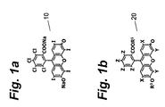
FIGURE 1 is an illustration of the chemical structure of Rose Bengal; -
FIGURE 1b is an illustration of the chemical structure of a halogenated xanthene; -
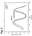
FIGURE 2 is an illustration of the two photon cross-section for several example halogenated xanthenes; -
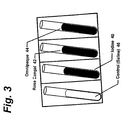
FIGURE 3 illustrates the CAT scan image of test tubes of Rose Bengal, x-ray contrast agents and a control; -
FIGURE 4 illustrates a CAT scan of a range of concentrations of the solutions ofFigure 3 ; -
FIGURE 5 is a graph of energy versus x-ray cross-section for halogens; -
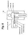
FIGURE 6 illustrates a combined imaging and treatment device in accordance with the present invention. - The present invention is directed to the apparatus and use of at least one PDT agent in imaging and treating diseased tissue.
- The first embodiment of the present invention is directed to an improved method for photodynamic therapy which enhances performance through the use of a photo-active agent having superior light-to-dark cytotoxicity. This embodiment includes treating the diseased tissue with light so as to photo-activate the photo-active agent in the diseased tissue, thereby destroying the diseased tissue. Included in this embodiment is the step of administering a photo-active (PDT) agent to a patient. The PDT agent will preferably accumulate in the diseased tissue. Each of these steps, the PDT agent and further embodiments of the present invention based thereon, will be discussed in more detail infra.
- One PDT agent which can be used in the present invention is Rose Bengal (4,5,6,7-tetrachloro-2',4',5',7'-tetraiodofluorescein); (see 10 in
Figure 1a ). Rose Bengal is a Type-I PDT agent that is known to accumulate preferentially in (i.e. target) some tumors and other diseased tissues. Type-I agents produce a cytotoxic response through direct photochemical conversion into toxic substances, and their Type-I photodynamic action is thus oxygen independent. In the presence of oxygen. Rose Bengal is also capable of efficient singlet oxygen production (Type-II action), further enhancing its photodynamic potential. Indeed, the inventors of the present application have found that Rose Bengal is an extremely efficient PDT agent when compared to conventional PDT agents (such as porfimer sodium and other porphyrin-based agents that are limited to only Type-I or Type-II mechanism of action). For example, in vitro tests have shown that Rose Bengal at a concentration of ≤ 10 µg/mL is able to kill 107 bacteria/mL within 5 seconds of illumination. Under similar conditions, porfimer sodium requires several hours to kill only a few percent of these bacteria. Therefore, in relation to porfimer sodium, Rose Bengal has an extremely high light-induced cytotoxicity. Moreover, Rose Bengal's dark cytotoxicity is negligible. Therefore, Rose Bengal has all the characteristics of a desirable replacement for porphyrin-based PDT agents: excellent biotargetting and high light-to-dark cytotoxicity ratio. - Rose Bengal is a specific example of a class of photoactive agents that is preferably used in the present invention. These agents are referred to as halogenated xanthenes and are illustrated in
Figure 1b , where the symbols X, Y, and Z represent various elements present at the designated positions, and the symbols R1 and R2 represent various functionalities present at the designated positions. Physical and photochemical properties of representative halogenated xanthenes are summarized in attached Table 1. Porfimer sodium, the most common PDT agent presently in use, is also listed for comparison of related properties. - In general, halogenated xanthenes are characterized by a low dark cytotoxicity, a high light cytotoxicity, a high single-photon cross-section extending from approximately 300 nm to 600 nm, and photochemical properties that are substantially unaffected by the local chemical environment or the attachment of functional derivatives at positions R1 and R2. Moreover, the halogenated xanthenes will target some tumors or other diseased tissues based on selective partitioning properties.
- The facility with which the halogenated xanthenes target specific tissues or other sites can be further optimized by attachment of specific functional derivatives at positions R1 and R2, so as to change the chemical partitioning or biological activity of the agent. For example, attachment of one targeting moiety or more at positions R1 or R2 can improve targeting to specific tissues, such as cancerous tumor tissues or sites of localized infection. These targeting moieties include DNA, RNA. amino acids, proteins, antibodies, ligands, haptens, carbohydrate receptors or complexing agents, lipid receptors or complexing agents protein receptors or complexing agents, chelators, and encapsulating vehicles.
- Thus, one example of this feature would be to combine Rose Bengal with a lipid (at position R1, via esterification), so as to increase the lipophilicity of Rose Bengal, and thereby modify its targeting properties in the patient. Such a modified agent could be administered directly as a micelle suspension, or delivered in conjunction with a delivery vehicle, such as a surfactant, and would exhibit increased targeting to tumor cells. Suitable formulations of such an agent include topical creams and lotions, and liquids for intravenous, parenteral or intratumoral injection.
- In addition to having desirable SPE characteristics, the halogenated xanthenes afford attractive properties for TPE. Specifically this class of agent offers broad and intense TPE spectral response across a range wavelengths extending from greater than 730 nm to less than 1100 nm, as shown in
Figure 2 . More specifically, attachment of moieties at positions R1 and R2 elicit insignificant changes in TPE spectral properties, as is clear, for example, by comparison of the spectral response of Eosin Y (wherein R1 = Na) and Ethyl Eosin (wherein R1 = OCH2CH3). Thus, attachment of targeting agents is possible without significantly affecting the photochemical properties of the agent. - Therefore, the halogenated xanthenes constitute excellent PDT agents for both SPE and TPE activation mechanisms, and can be used directly or in derivatized form to improve, for example, solubility or biotargetting through attachment of various functionalities at positions R1 and R2. Accordingly, in a preferred embodiment of the present invention, at least one halogenated xanthene or halogenated xanthene derivative is used as a PDT agent. The PDT agent can be given orally, systemically (e.g. by an injection), or topically, in a manner well known in the art. It is further preferred that Rose Bengal be used as the PDT agent. Such agent can be activated using single-photon excitation, or preferably two-photon excitation.
- In a further embodiment of the present invention, the selectivity of photodynamic activation is improved though use of conventional imaging methods to identify diseased tissue targets. For example.
- Magnetic Resonance Imaging (MRI) is used to detect the location of diseased tissue. Such imaging works by detecting abnormalities in the distribution or properties of tissue components (such as density), the presence or absence of certain materials, or the uptake or exclusion of imaging contrast agents. Such diseased tissue is then used as the target for selective optical activation of photodynamic agent administered to the patient, thereby selectively destroying such diseased tissue.
- The inventors of the present invention have discovered that certain PDT agents, and more specifically the halogenated xanthenes, are not substantially photodynamically activated nor destroyed by exposure to the energies commonly used for MRI imaging. Accordingly, these agents are safe to administer prior to such diagnostic procedures. Hence, the PDT agent may be administered to the patient prior to diagnosis (thereby potentially reducing delay between diagnosis and treatment) or following diagnosis (thereby reducing unnecessary administration of agent in cases where no disease is detected).
- Therefore, a preferred embodiment of the present invention comprises the steps of MRI imaging via conventional means to detect the presence of diseased tissue; administering a PDT agent, preferably a halogenated xanthene, prior to or upon detection of such diseased tissue, and directing light, appropriate for SPE or preferably TPE activation methods, as discussed infra_upon or to such detected diseased tissue sufficient to activate the PDT agent and thereby selectively destroy substantially only such diseased tissue.
- In a further embodiment of the present invention, the efficacy of the detection or imaging step in the preceding embodiment is further improved through the use of an imaging contrast agent. In particular, the PDT agent, and more specifically, a halogenated xanthene, is mixed with an imaging contrast agent, such as for example, x-ray contrast agents like Omnipaque™ (Iohexol) and Omniscan™ (Gadodiamide) or one of the various paramagnetic MRI contrast agents like gadolinium DPTA (Gd-DPTA). For example, Rose Bengal is compatible in solution with agents such as Omnipaque™, Omniscan™, and Gd-DPTA, and exhibits similar biotargetting properties. The mixture is then administered to the patient. Following administration of such a mixture of contrast agent and PDT agent, conventional imaging (such as,
- MRI) is used to locate diseased tissue based on response of the conventional contrast agent, then the PDT agent, co-located in the diseased tissue, would be activated at the site of the detected diseased tissue using SPE or more preferably TPE to destroy such diseased tissue.
- The inventors have shown that Rose Bengal is capable of selective photodynamic activation in a liver model, following administration of the agent in solution. Such a model is also known to accumulate MRI contrast agents. Thus, the inventors have shown that it is feasible to deliver conventional imaging contrast agents and PDT agents to target tissues, and that such agents will retain their respective activities in the target tissues, allowing combined detection and treatment of diseased tissue at locations indicated by imaging based on detected imaging contrast agents. Hence, one preferred embodiment of the present invention is to jointly administer, either sequentially (for example via injections or intravenous drip) or more preferably as a single, mixed solution, one or more MRI contrast agent with one or more PDT agent, preferably an halogenated xanthene agent, and subsequently to direct activation of the one or more PDT agent based on imaging data obtained utilizing the one or more contrast agent.
- In another embodiment of the present invention, the PDT agent also acts as a contrast agent for imaging. The use of the same agent for both imaging and treatment procedures is highly advantageous. For example, it eliminates the need for a second dose of an agent. Such a second dose requires further time between imaging and treatment, as the second agent, after being administered, must accumulate in the diseased tissue before treatment can begin. Further, use of a second agent makes the process more costly and requires the patient to be subjected to a second application of a foreign substance.
Halogenated xanthenes make attractive candidates as MRI contrast agents. - Although not paramagnetic like the majority of conventional MRI contrast agents, the halogenated xanthenes contain aromatic protons which exhibit characteristic MRI signatures based on the chemical shift of such protons. Further, the presence of substantial densities of aromatic halides in the halogenated xanthenes constitutes a further unique and useful MRI signature based on detection of resonances from such aromatic halides. Since proton and halogen nuclear magnetic resonance are relatively sensitive phenomena (for example, F, Br and I have many-fold higher sensitivities relative to carbon-13 NMR, as shown in Table 2), MRI detection and imaging based on the presence of the halogenated xanthenes in diseased tissue represents a further unique and attractive medical application for such agents. Hence, it is a further preferred embodiment of the present invention to utilize the halogenated xanthenes as MRI contrast agents, and to use imaging data based on detection of such agents to selectively direct the subsequent photoactivation of such agents present in diseased tissue using SPE and preferably TPE activation methods. Since the majority of installed MRI devices are based on detection of proton resonance, it is further preferred that such MRI detection be performed based on resonance of aromatic protons present in the halogenated xanthenes.
- Following imaging in the present invention, light is applied via a light source to the disease site in order to photo-activate the agent associated with the diseased tissue. Preferably, laser light is used. Alternate light sources include light emitting diodes, micro-lasers, monochromatic or continuum lasers or lamps for production of activating light, and continuous wave or pulsed lasers or lamps. Either single-photon or two-photon excitation methods can be used for agent activation. A more detailed explanation of such excitation methods is given in commonly assigned application serial no.
08/739,801 filed October 30, 1996 which is incorporated herein by reference. The excitation of the photo-active agent starts a process which eventually kills the cells in the diseased tissue. - In a further embodiment of the present invention, the light source used for photodynamic activation is integrated into or onto the imaging device, such as an MRI system or x-ray imaging device. An example of such a device for imaging and
treatment 50 is illustrated inFigure 6 . Such a combined imaging and treatment device allows more precise delivery of treatment to diseased tissue based on improved accuracy of registration between imagery data and treatment targets.Figure 6 , for example, shows aconventional imaging unit 52. such as a CAT scan or MRI system, used to identify alesion 54 present in apatient 56. Imaging of this lesion can be done by one of the methods discussed supra or by other known methods of imaging. Thislesion 54 then serves as the target for anintegrated activation unit 58 that serves selectively to photoactivate PDT agent present in the lesion. Theactivation unit 58 preferably includes alight source 60, such as, for example, a laser capable of SPE activation of the agent and more preferably a laser capable of TPE activation of such agent, such as a mode-locked titanium:sapphire or neodymium YLF laser. Preferably, theactivation unit 58 also includes an aimingsystem 62, such as, for example, a mirror-based galvanometer or other optical scanning system. Constructed and functioning in this manner, theimaging unit 52 can be made to guide application oflight 64 produced by theactivation unit 58, for example under manual control of a physician or more preferably under automated or semi-automated computer control, such that the activatinglight 64 is applied substantially only to the site of the detectedlesion 54, thereby improving safety and efficacy of the treatment process. - A mode-locked titanium:sapphire laser is a preferred embodiment for the light source for the integrated activation unit. Such a laser is capable of producing a rapid series of high peak power pulses of NIR light that are well suited for TPE of the halogenated xanthenes. Standard, commercially available mode-locked titanium:sapphire lasers are capable of outputting mode-locked pulses with durations <200 fs with pulse energies of about 1-20 nJ at pulse repetition frequencies in excess of 75 MHz. This constitutes a quasi-continuous beam of light having a relatively low average power (up to several Watts) but high peak power (on the order of 100 kW) that is continuously tunable over a NIR wavelength band from approximately 690-1080 nm. The pulse train from such a source is easily aimed using standard optical means, such as reflective or refractive optics, so as to be directed onto or into a lesion or other localized treatment target. Other light sources suitable for activation of photodynamic agents include: continuous wave and pulsed lamps, diode light sources, semiconductor lasers: other types of gas, dye, and solid-state continuous, pulsed or mode-locked lasers, including: argon ion lasers: krypton ion lasers; helium-neon lasers; helium-cadmium lasers; ruby lasers: Nd:YAG, Nd:YLF, Nd:YAP, Nd:YVO4, Nd:Glass, and Nd:CrGsGG lasers; Cr:LiSF lasers; Er:YAG lasers; F-center lasers; Ho:YAF and Ho:YLF lasers: copper vapor lasers; nitrogen lasers; optical parametric oscillators, amplifiers and generators; regeneratively amplified lasers: chirped-pulse amplified lasers: and sunlight.
- This description has been offered for illustrative purposes only and is not intended to limit the invention of this application, which is defined in the claims below.
- What is claimed as new and desired to be protected by Letters Patent is set forth in the appended claims.
Table 1. Physical and Photochemical Properties of Example Halogenated Xanthenes: Compound Substitution MW (g) λmax (nm) α (cm-1 mol1 L) λmax (nm) φ (fluor) φ (triplet) φ (singlet oxygen) X Y Z R1 R2 H2O EtOH MeOH H2O EtOH MeOH MeOH MeOH H2O EtOH MeOH Fluorescein H H H Na Na 376 490 499 492 6.4×10⁴ 519 519 518 0.93 0.03 0.03 0.03 0.09 Cl H H Na Na 445 502 511 0.04 0.07 H Cl H Na Na 445 502 511 0.04 0.07 H H Cl H H 470 515 2.9×10⁴ Cl Cl H Na Na 514 510 520 0.05 0.05 Dibromofluorescein Br H H Na Na 534 504 510 1.4×10⁴ 0.32 0.42 Solvent Red 72 H Br H H H 490 450 1.4×10⁴ Diiodofluorescein I H H Na Na 628 506 513 5.8×10⁴ 0.03 0.33 0.48 Eosin B NO2 Br H Na Na 624 522 3.9×10⁴ 0.00 Eosin Y Br Br H Na Na 692 517 523 527 9.1×10⁴ 538 544 541 0.63 0.28 0.32 0.57 0.39 Ethyl Eosin Br Br H C2H5 K 714 532 1.1×10⁴ 560 0.70 Erythrosin B I I H Na Na 880 528 532 529 9.1×10⁴ 547 553 551 0.08 0.62 0.69 0.63 0.62 Phloxine B Br Br Cl Na Na 830 541 548 547 1.0×10⁵ 556 565 563 0.30 0.40 0.63 Rose Bengal I I Cl Na Na 1018 547 557 556 1.0×10⁵ 568 566 573 0.08 0.76 0.86 0.75 0.76 I I Cl Li Li 986 559 572 568 I I Cl C2H5 (C2H4)3NH 1100 563 590 585 0.74 I I Cl (C2H3)3NH (C2H4)3NH 1166 559 569 573 0.72 Porlimer Sodium 1200 368 2.0×10³ >0.7 Table 2. Magnetic Resonance Properties of Elements: 1H 13C 19F 35Cl 37Cl 79Br 81Br 127 I Atomic Number 1 6 9 17 17 35 35 53 Atomic Mass (12C = 12.0000) 1.00 13.00 19.00 34.97 36.97 78.92 80.92 126.90 Natural Abundance (%) 99.98 1.02 100.00 75.53 24.47 50.54 49.46 100.00 Spin Quantum Number ½ ½ ½ 3/2 3/2 3/2 3/2 5/2 NRM Frequency (MHz at 1 Tesla) 42.58 10.70 40.06 4.17 3.47 10.67 11.50 8.52 NMR Sensitivity at Constant Field (relative to 1H) 1.00 1.6×10⁻² 0.83 4.7×10⁻³ 2.7×10⁻³ 7.9×10⁻² 9.8×10⁻² 9.3×10⁻² NMR Sensitivity at Constant Frequency (relative to 1H) 1.00 0.25 0.94 0.49 0.41 1.25 1.35 2.33
Claims (14)
- A method of imaging diseased tissue, comprising the steps of:administering a halogenated xanthene to a patient prior to imaging, a portion of said halogenated xanthene being retained in said diseased tissue;magnetic resonance imaging the patient using a detected signal from said halogenated xanthene to contrast and identify said diseased tissue, wherein said halogenated xanthene is not paramagnetic.
- The method according to claim 1, wherein said halogenated xanthene is detected based on resonance of aromatic protons in the halogenated xanthene.
- The method according to claim 1, wherein said halogenated xanthene is detected based on resonance of aromatic halides in the halogenated xanthene.
- The method according to any preceding claim, wherein said halogenated xanthene is an iodinated or brominated halogenated xanthene.
- The method according to any preceding claim, wherein said halogenated xanthene is Rose Bengal, Phloxine B, Erythrosin B or Eosin Y.
- The method according to any preceding claim, wherein said halogenated xanthene includes as a functional derivative a targeting moiety wherein the targeting moiety is deoxyribonucleic acid (DNA), ribonucleic acid (RNA), an amino acid, protein, antibody, ligand, hapten, carbohydrate receptor, carbohydrate complexing agent, lipid receptor, lipid complexing agent, protein receptor, protein complexing agent, chelator, or encapsulating vehicle.
- Use of a halogenated xanthene for the preparation of an image contrast agent for magnetic resonance imaging, wherein said halogenated xanthene is not paramagnetic.
- Use according to claim 7, wherein said halogenated xanthene is retained in said diseased tissue when administered to a patient.
- Use according to claim 7 or claim 8, wherein said imaging contrast agent is detected based on resonance of aromatic protons in a halogenated xanthene.
- Use according to claim 7 or claim 8, wherein said imaging contrast agent is detected based on resonance of aromatic halides in a halogenated xanthene.
- Use according to any of claims 7 to 10, wherein said halogenated xanthene is an iodinated or brominated halogenated xanthene.
- Use according to any of claims 7 to 11, wherein said halogenated xanthene is Rose Bengal, Phloxine B, Erythrosin B or Eosin Y.
- Use according to any of claims 7 to 12, wherein said halogenated xanthene includes as a functional derivative a targeting moiety wherein the targeting moiety is deoxyribonucleic acid (DNA), ribonucleic acid (RNA), an amino acid, protein, antibody, ligand, hapten, carbohydrate receptor, carbohydrate complexing agent, lipid receptor, lipid complexing agent, protein receptor, protein complexing agent, chelator, or encapsulating vehicle.
- A halogenated xanthene for use as an image contrast agent in magnetic resonance imaging, wherein said halogenated xanthene is not paramagnetic.
Applications Claiming Priority (3)
| Application Number | Priority Date | Filing Date | Title |
|---|---|---|---|
| US184388 | 1998-11-02 | ||
| US09/184,388 US6493570B1 (en) | 1998-11-02 | 1998-11-02 | Method for improved imaging and photodynamic therapy |
| PCT/US1999/025074 WO2000025665A1 (en) | 1998-11-02 | 1999-10-26 | Method for improved imaging and photodynamic therapy |
Publications (3)
| Publication Number | Publication Date |
|---|---|
| EP1126782A1 EP1126782A1 (en) | 2001-08-29 |
| EP1126782A4 EP1126782A4 (en) | 2003-06-04 |
| EP1126782B1 true EP1126782B1 (en) | 2008-06-04 |
Family
ID=22676684
Family Applications (1)
| Application Number | Title | Priority Date | Filing Date |
|---|---|---|---|
| EP99971283A Expired - Lifetime EP1126782B1 (en) | 1998-11-02 | 1999-10-26 | Method for improved imaging and photodynamic therapy |
Country Status (15)
| Country | Link |
|---|---|
| US (1) | US6493570B1 (en) |
| EP (1) | EP1126782B1 (en) |
| JP (1) | JP2002528472A (en) |
| KR (1) | KR20010080932A (en) |
| CN (1) | CN100382846C (en) |
| AR (1) | AR021061A1 (en) |
| AT (1) | ATE397408T1 (en) |
| AU (1) | AU1452100A (en) |
| BR (1) | BR9914969A (en) |
| CA (1) | CA2348094A1 (en) |
| DE (1) | DE69938870D1 (en) |
| ES (1) | ES2307344T3 (en) |
| HK (1) | HK1040899B (en) |
| IL (1) | IL142729A0 (en) |
| WO (1) | WO2000025665A1 (en) |
Families Citing this family (47)
| Publication number | Priority date | Publication date | Assignee | Title |
|---|---|---|---|---|
| US7390668B2 (en) * | 1996-10-30 | 2008-06-24 | Provectus Pharmatech, Inc. | Intracorporeal medicaments for photodynamic treatment of disease |
| US7648695B2 (en) * | 1998-08-06 | 2010-01-19 | Provectus Pharmatech, Inc. | Medicaments for chemotherapeutic treatment of disease |
| US6986740B2 (en) * | 1998-11-02 | 2006-01-17 | Xantech Pharmaceuticals, Inc. | Ultrasound contrast using halogenated xanthenes |
| US20020001567A1 (en) * | 1998-12-21 | 2002-01-03 | Photogen, Inc. | Intracorporeal medicaments for high energy phototherapeutic treatment of disease |
| US8470296B2 (en) * | 1998-12-21 | 2013-06-25 | Provectus Pharmatech, Inc. | Intracorporeal medicaments for high energy phototherapeutic treatment of disease |
| CN1372467A (en) | 1999-08-13 | 2002-10-02 | 福托金公司 | Improved topical medicaments and methods for photodynamic treatment of disease |
| WO2002003872A2 (en) * | 2000-07-11 | 2002-01-17 | Johns Hopkins University | Application of photochemotherapy for the treatment of cardiac arrhythmias |
| AU2002305148A1 (en) * | 2001-04-05 | 2002-10-21 | Johns Hopkins University | Imaging systems for in vivo protocols |
| WO2003000129A2 (en) * | 2001-06-22 | 2003-01-03 | University Of Florida | Microdialysis probes and methods of use |
| US7715901B2 (en) * | 2002-05-08 | 2010-05-11 | Yeda Research And Development Co. Ltd. | Sensitized online BOLD-MRI imaging method |
| AU2004220530A1 (en) * | 2003-03-10 | 2004-09-23 | Mpa Technologies, Inc. | Targeted agents for both photodiagnosis and photodynamic therapy |
| US20040253281A1 (en) * | 2003-06-12 | 2004-12-16 | Atrium Medical Corp. | Therapeutic markings applied to tissue |
| US20050053895A1 (en) | 2003-09-09 | 2005-03-10 | The Procter & Gamble Company Attention: Chief Patent Counsel | Illuminated electric toothbrushes emitting high luminous intensity toothbrush |
| US7282060B2 (en) * | 2003-12-23 | 2007-10-16 | Reliant Technologies, Inc. | Method and apparatus for monitoring and controlling laser-induced tissue treatment |
| US7184184B2 (en) * | 2003-12-31 | 2007-02-27 | Reliant Technologies, Inc. | High speed, high efficiency optical pattern generator using rotating optical elements |
| US7372606B2 (en) * | 2003-12-31 | 2008-05-13 | Reliant Technologies, Inc. | Optical pattern generator using a single rotating component |
| US7196831B2 (en) | 2003-12-31 | 2007-03-27 | Reliant Technologies, Inc. | Two-dimensional optical scan system using a counter-rotating disk scanner |
| US20050259779A1 (en) * | 2004-05-18 | 2005-11-24 | Siemens Aktiengesellschaft | Biomolecular contrast agents for therapy optimization in radiation therapy with proton or ion beams |
| US20050272967A1 (en) * | 2004-05-18 | 2005-12-08 | Siemens Aktiengesellschaft | Biomolecular contrast agents with multiple signal variance for therapy planning and control in radiation therapy with proton or ion beams |
| WO2005110495A1 (en) * | 2004-05-18 | 2005-11-24 | Siemens Aktiengesellschaft | Biomolecular contrast agents for therapy control in radiation therapy with proton or ion beams |
| US7585492B2 (en) * | 2004-05-18 | 2009-09-08 | Siemens Aktiengesellschaft | Biomolecular contrast agents for therapy success and dose monitoring in radiation therapy with proton or ion beams |
| US7427389B2 (en) * | 2004-06-10 | 2008-09-23 | Provectus Pharmatech, Inc. | Diagnostic agents for positron emission imaging using radiolabeled halogenated xanthenes |
| US7338652B2 (en) * | 2004-06-10 | 2008-03-04 | Provectus Pharmatech, Inc. | Methods for positron emission imaging with radiolabeled halogenated xanthenes diagnostic agents |
| AU2006209654A1 (en) * | 2005-01-28 | 2006-08-03 | Konica Minolta Medical & Graphic, Inc. | Pharmaceutical preparation containing magnetic vesicular particles, manufacturing method thereof and diagnostic therapeutic system |
| WO2007072800A1 (en) * | 2005-12-19 | 2007-06-28 | The University Of Tokyo | Photosensitizing compound |
| DE102005061847A1 (en) * | 2005-12-23 | 2007-07-05 | Siemens Ag | Operating method for an X-ray device |
| CN100366227C (en) * | 2006-03-03 | 2008-02-06 | 华南师范大学 | Method and device for monitoring photodynamic therapy effect of tumor by photoacoustic angiography |
| GB2453860B (en) | 2007-10-18 | 2011-03-16 | Searete Llc | Ionizing-radiation-responsive compositions,methods and systems |
| US9557635B2 (en) * | 2007-10-18 | 2017-01-31 | Gearbox, Llc | Ionizing-radiation-responsive compositions, methods, and systems |
| US8168958B2 (en) * | 2007-10-18 | 2012-05-01 | The Invention Science Fund I, Llc | Ionizing-radiation-responsive compositions, methods, and systems |
| US8227204B2 (en) * | 2007-10-18 | 2012-07-24 | The Invention Science Fund I, Llc | Ionizing-radiation-responsive compositions, methods, and systems |
| US8164074B2 (en) * | 2007-10-18 | 2012-04-24 | The Invention Science Fund I, Llc | Ionizing-radiation-responsive compositions, methods, and systems |
| US8529426B2 (en) | 2007-10-18 | 2013-09-10 | The Invention Science Fund I Llc | Ionizing-radiation-responsive compositions, methods, and systems |
| US20090104113A1 (en) * | 2007-10-18 | 2009-04-23 | Searete Llc | Ionizing-radiation-responsive compositions, methods, and systems |
| US8684898B2 (en) * | 2007-10-18 | 2014-04-01 | The Invention Science Fund I Llc | Ionizing-radiation-responsive compositions, methods, and systems |
| US20100284927A1 (en) * | 2007-10-26 | 2010-11-11 | University Of Utah Research Foundation | Use of mri contrast agents for evaluating the treatment of tumors |
| US9642687B2 (en) | 2010-06-15 | 2017-05-09 | The Procter & Gamble Company | Methods for whitening teeth |
| KR101336073B1 (en) * | 2012-01-04 | 2013-12-05 | 한국원자력의학원 | Composition for Dual SPECT/MRI contrast and a process for obtaining SPECT/MRI dual image using the same |
| WO2014157703A1 (en) | 2013-03-29 | 2014-10-02 | 国立大学法人三重大学 | Vital stain |
| EP3700520B1 (en) | 2017-11-29 | 2024-05-01 | The Rockefeller University | Combination of local and systemic therapies for enhanced treatment of dermatologic conditions |
| EP3773548B1 (en) | 2018-05-16 | 2023-08-09 | Provectus Pharmatech, Inc. | In vitro and xenograft anti-tumor activity of a halogenated-xanthene against refractory pediatric solid tumors |
| US12064507B2 (en) | 2019-11-19 | 2024-08-20 | Provectus Pharmatech, Inc. | Composition and method for oral treatment of leukemia |
| US11419844B2 (en) | 2019-11-19 | 2022-08-23 | Provectus Pharmatech, Inc. | Halogenated xanthene composition and method for treating hematologic cancers |
| CN111087376B (en) * | 2019-12-30 | 2021-08-03 | 厦门大学 | A kind of gadolinium-doped rose bengal nanodots and preparation method and application thereof |
| US11938182B2 (en) | 2020-03-26 | 2024-03-26 | Provectus Pharmatech, Inc. | Halogenated xanthenes as vaccine adjuvants |
| EP4103283A4 (en) | 2020-03-26 | 2024-02-28 | Provectus Pharmatech, Inc. | Novel uses of halogenated xanthenes in oncology and virology |
| CN111840550A (en) * | 2020-07-28 | 2020-10-30 | 清华大学 | A method and system for controlling drug release using pulsed laser |
Citations (2)
| Publication number | Priority date | Publication date | Assignee | Title |
|---|---|---|---|---|
| US5435991A (en) * | 1991-08-09 | 1995-07-25 | Nycomed Innovation Ab | Use of persistent heterocyclic free-radicals in magnetic resonance imaging |
| DE4403789A1 (en) * | 1994-02-03 | 1995-08-10 | Schering Ag | Means for visually marking body tissues |
Family Cites Families (22)
| Publication number | Priority date | Publication date | Assignee | Title |
|---|---|---|---|---|
| US4599227A (en) | 1983-11-07 | 1986-07-08 | Wisconsin Alumni Research Foundation | Injectable pharmaceutical preparation for the induction of multiple follicular growth |
| AU583854B2 (en) * | 1984-09-13 | 1989-05-11 | Cytogen Corporation | Antibody therapeutic agent conjugates |
| US4973848A (en) * | 1989-07-28 | 1990-11-27 | J. Mccaughan | Laser apparatus for concurrent analysis and treatment |
| US5418130A (en) * | 1990-04-16 | 1995-05-23 | Cryopharm Corporation | Method of inactivation of viral and bacterial blood contaminants |
| US5654423A (en) | 1990-11-21 | 1997-08-05 | Regents Of The University Of California | Boronated metalloporphyrine and therapeutic methods |
| US5149801A (en) | 1990-11-21 | 1992-09-22 | The Regents Of The University Of California | Boronated porphyrin compounds |
| US5128139A (en) | 1991-02-15 | 1992-07-07 | Nu Skin International, Inc. | Composition containing liposome-entrapped grapefruit seed extract and method for making |
| US5258453A (en) | 1992-01-21 | 1993-11-02 | University Of Utah | Drug delivery system for the simultaneous delivery of drugs activatable by enzymes and light |
| JP3369623B2 (en) * | 1993-03-16 | 2003-01-20 | 興和株式会社 | Laser scanning ophthalmic imaging device |
| US5445608A (en) * | 1993-08-16 | 1995-08-29 | James C. Chen | Method and apparatus for providing light-activated therapy |
| US5576013A (en) * | 1995-03-21 | 1996-11-19 | Eastern Virginia Medical School | Treating vascular and neoplastic tissues |
| US5591422A (en) | 1995-06-02 | 1997-01-07 | Pharmacyclics, Inc. | Texaphyrin complexes having improved functionalization |
| CA2218541A1 (en) * | 1995-06-07 | 1996-12-19 | Imarx Pharmaceutical Corp. | Novel targeted compositions for diagnostic and therapeutic use |
| IT1275571B (en) * | 1995-07-19 | 1997-08-07 | Consiglio Nazionale Ricerche | FLUOROGENIC SUBSTRATES SUSCEPTIBLE FOR PHOTOACTIVATION AFTER ENZYMATIC TRANSFORMATION SUITABLE FOR DIAGNOSIS AND PHOTODYNAMIC CANCER THERAPY |
| US5829448A (en) * | 1996-10-30 | 1998-11-03 | Photogen, Inc. | Method for improved selectivity in photo-activation of molecular agents |
| US6331286B1 (en) | 1998-12-21 | 2001-12-18 | Photogen, Inc. | Methods for high energy phototherapeutics |
| US5832931A (en) | 1996-10-30 | 1998-11-10 | Photogen, Inc. | Method for improved selectivity in photo-activation and detection of molecular diagnostic agents |
| US5827186A (en) * | 1997-04-11 | 1998-10-27 | Light Sciences Limited Partnership | Method and PDT probe for minimizing CT and MRI image artifacts |
| CN1166987A (en) * | 1997-06-18 | 1997-12-10 | 武汉大学 | Magnetic resonance imaging contrast medium of paramagnetism metal compound containing D-galactose group |
| JP2002506013A (en) * | 1998-03-09 | 2002-02-26 | イスタ ファーマシューティカルズ インコーポレイテッド | Use of corneal sclerosing agents in enzyme keratoplasty |
| KR20010072307A (en) * | 1998-08-06 | 2001-07-31 | 스무크 존 | Improved method for targeted topical treatment of disease |
| EP1124580A1 (en) | 1998-10-29 | 2001-08-22 | The General Hospital Corporation | Enhanced radiation therapy |
-
1998
- 1998-11-02 US US09/184,388 patent/US6493570B1/en not_active Expired - Lifetime
-
1999
- 1999-10-26 KR KR1020017005480A patent/KR20010080932A/en not_active Ceased
- 1999-10-26 HK HK02102739.0A patent/HK1040899B/en not_active IP Right Cessation
- 1999-10-26 AU AU14521/00A patent/AU1452100A/en not_active Abandoned
- 1999-10-26 CA CA002348094A patent/CA2348094A1/en not_active Abandoned
- 1999-10-26 DE DE69938870T patent/DE69938870D1/en not_active Expired - Lifetime
- 1999-10-26 ES ES99971283T patent/ES2307344T3/en not_active Expired - Lifetime
- 1999-10-26 BR BR9914969-9A patent/BR9914969A/en not_active IP Right Cessation
- 1999-10-26 AT AT99971283T patent/ATE397408T1/en not_active IP Right Cessation
- 1999-10-26 WO PCT/US1999/025074 patent/WO2000025665A1/en not_active Ceased
- 1999-10-26 EP EP99971283A patent/EP1126782B1/en not_active Expired - Lifetime
- 1999-10-26 IL IL14272999A patent/IL142729A0/en unknown
- 1999-10-26 CN CNB998129917A patent/CN100382846C/en not_active Expired - Lifetime
- 1999-10-26 JP JP2000579116A patent/JP2002528472A/en active Pending
- 1999-11-02 AR ARP990105533A patent/AR021061A1/en unknown
Patent Citations (2)
| Publication number | Priority date | Publication date | Assignee | Title |
|---|---|---|---|---|
| US5435991A (en) * | 1991-08-09 | 1995-07-25 | Nycomed Innovation Ab | Use of persistent heterocyclic free-radicals in magnetic resonance imaging |
| DE4403789A1 (en) * | 1994-02-03 | 1995-08-10 | Schering Ag | Means for visually marking body tissues |
Also Published As
| Publication number | Publication date |
|---|---|
| ES2307344T3 (en) | 2008-11-16 |
| US6493570B1 (en) | 2002-12-10 |
| IL142729A0 (en) | 2002-03-10 |
| JP2002528472A (en) | 2002-09-03 |
| ATE397408T1 (en) | 2008-06-15 |
| BR9914969A (en) | 2001-07-10 |
| AR021061A1 (en) | 2002-06-12 |
| EP1126782A4 (en) | 2003-06-04 |
| CN100382846C (en) | 2008-04-23 |
| CA2348094A1 (en) | 2000-05-11 |
| DE69938870D1 (en) | 2008-07-17 |
| CN1325284A (en) | 2001-12-05 |
| HK1040899A1 (en) | 2002-06-28 |
| HK1040899B (en) | 2008-12-24 |
| KR20010080932A (en) | 2001-08-25 |
| WO2000025665A1 (en) | 2000-05-11 |
| AU1452100A (en) | 2000-05-22 |
| EP1126782A1 (en) | 2001-08-29 |
Similar Documents
| Publication | Publication Date | Title |
|---|---|---|
| US6493570B1 (en) | Method for improved imaging and photodynamic therapy | |
| EP1192450B1 (en) | High energy phototherapeutic agents | |
| Dougherty | Photosensitization of malignant tumors | |
| US6750212B2 (en) | Photochemotherapeutic compositions | |
| US7354599B2 (en) | Liposomal formulations of hydrophobic photosensitizer for photodynamic therapy | |
| JPH04507236A (en) | Bacteriochlorophyll a derivative useful for photodynamic therapy | |
| EP1146912A1 (en) | Method for improved radiation therapy | |
| Powers et al. | Laser photochemotherapy of rhodamine-123 sensitized human glioma cells in vitro | |
| Pope et al. | Photodynamic | |
| Edell et al. | Photodynamic therapy: its use in the management of bronchogenic carcinoma | |
| EP1292298B1 (en) | Intracorporeal medicaments for high energy phototherapeutic treatment of disease | |
| US7384623B1 (en) | High energy phototherapeutic agents | |
| Pass | Photodynamic therapy for lung cancer | |
| DE60133394T2 (en) | Photosensitizing substances with ligand-targeting properties for tumor therapy | |
| US8986731B2 (en) | Pegylated liposomal formulations of hydrophobic photosensitizers for photodynamic therapy | |
| MXPA01004375A (en) | Method for improved imaging and photodynamic therapy | |
| Kaye et al. | A review of photoradiation therapy in the management of central nervous system tumours | |
| Barr | Selective tumor destruction with photodynamic therapy: exploitation of photodynamic thresholds | |
| MXPA01006368A (en) | High energy phototherapeutic agents | |
| Kato | The present status of photodynamic therapy | |
| Nelson | Photodynamic Therapy |
Legal Events
| Date | Code | Title | Description |
|---|---|---|---|
| PUAI | Public reference made under article 153(3) epc to a published international application that has entered the european phase |
Free format text: ORIGINAL CODE: 0009012 |
|
| 17P | Request for examination filed |
Effective date: 20010531 |
|
| AK | Designated contracting states |
Kind code of ref document: A1 Designated state(s): AT BE CH CY DE DK ES FI FR GB GR IE IT LI LU MC NL PT SE |
|
| AX | Request for extension of the european patent |
Free format text: AL;LT;LV;MK;RO;SI |
|
| A4 | Supplementary search report drawn up and despatched |
Effective date: 20030417 |
|
| 17Q | First examination report despatched |
Effective date: 20040325 |
|
| 17Q | First examination report despatched |
Effective date: 20040325 |
|
| 17Q | First examination report despatched |
Effective date: 20040325 |
|
| 17Q | First examination report despatched |
Effective date: 20040325 |
|
| GRAP | Despatch of communication of intention to grant a patent |
Free format text: ORIGINAL CODE: EPIDOSNIGR1 |
|
| RAP1 | Party data changed (applicant data changed or rights of an application transferred) |
Owner name: PROVECTUS PHARMATECH, INC. |
|
| GRAS | Grant fee paid |
Free format text: ORIGINAL CODE: EPIDOSNIGR3 |
|
| GRAA | (expected) grant |
Free format text: ORIGINAL CODE: 0009210 |
|
| AK | Designated contracting states |
Kind code of ref document: B1 Designated state(s): AT BE CH CY DE DK ES FI FR GB GR IE IT LI LU MC NL PT SE |
|
| REG | Reference to a national code |
Ref country code: GB Ref legal event code: FG4D |
|
| REG | Reference to a national code |
Ref country code: CH Ref legal event code: EP |
|
| REF | Corresponds to: |
Ref document number: 69938870 Country of ref document: DE Date of ref document: 20080717 Kind code of ref document: P |
|
| REG | Reference to a national code |
Ref country code: IE Ref legal event code: FG4D |
|
| PG25 | Lapsed in a contracting state [announced via postgrant information from national office to epo] |
Ref country code: FI Free format text: LAPSE BECAUSE OF FAILURE TO SUBMIT A TRANSLATION OF THE DESCRIPTION OR TO PAY THE FEE WITHIN THE PRESCRIBED TIME-LIMIT Effective date: 20080604 |
|
| REG | Reference to a national code |
Ref country code: ES Ref legal event code: FG2A Ref document number: 2307344 Country of ref document: ES Kind code of ref document: T3 |
|
| PG25 | Lapsed in a contracting state [announced via postgrant information from national office to epo] |
Ref country code: NL Free format text: LAPSE BECAUSE OF FAILURE TO SUBMIT A TRANSLATION OF THE DESCRIPTION OR TO PAY THE FEE WITHIN THE PRESCRIBED TIME-LIMIT Effective date: 20080604 Ref country code: AT Free format text: LAPSE BECAUSE OF FAILURE TO SUBMIT A TRANSLATION OF THE DESCRIPTION OR TO PAY THE FEE WITHIN THE PRESCRIBED TIME-LIMIT Effective date: 20080604 |
|
| NLV1 | Nl: lapsed or annulled due to failure to fulfill the requirements of art. 29p and 29m of the patents act | ||
| PG25 | Lapsed in a contracting state [announced via postgrant information from national office to epo] |
Ref country code: SE Free format text: LAPSE BECAUSE OF FAILURE TO SUBMIT A TRANSLATION OF THE DESCRIPTION OR TO PAY THE FEE WITHIN THE PRESCRIBED TIME-LIMIT Effective date: 20080904 Ref country code: PT Free format text: LAPSE BECAUSE OF FAILURE TO SUBMIT A TRANSLATION OF THE DESCRIPTION OR TO PAY THE FEE WITHIN THE PRESCRIBED TIME-LIMIT Effective date: 20081104 |
|
| PG25 | Lapsed in a contracting state [announced via postgrant information from national office to epo] |
Ref country code: BE Free format text: LAPSE BECAUSE OF FAILURE TO SUBMIT A TRANSLATION OF THE DESCRIPTION OR TO PAY THE FEE WITHIN THE PRESCRIBED TIME-LIMIT Effective date: 20080604 |
|
| PLBE | No opposition filed within time limit |
Free format text: ORIGINAL CODE: 0009261 |
|
| STAA | Information on the status of an ep patent application or granted ep patent |
Free format text: STATUS: NO OPPOSITION FILED WITHIN TIME LIMIT |
|
| PG25 | Lapsed in a contracting state [announced via postgrant information from national office to epo] |
Ref country code: DK Free format text: LAPSE BECAUSE OF FAILURE TO SUBMIT A TRANSLATION OF THE DESCRIPTION OR TO PAY THE FEE WITHIN THE PRESCRIBED TIME-LIMIT Effective date: 20080604 |
|
| 26N | No opposition filed |
Effective date: 20090305 |
|
| PG25 | Lapsed in a contracting state [announced via postgrant information from national office to epo] |
Ref country code: MC Free format text: LAPSE BECAUSE OF NON-PAYMENT OF DUE FEES Effective date: 20081031 |
|
| REG | Reference to a national code |
Ref country code: CH Ref legal event code: PL |
|
| PG25 | Lapsed in a contracting state [announced via postgrant information from national office to epo] |
Ref country code: LI Free format text: LAPSE BECAUSE OF NON-PAYMENT OF DUE FEES Effective date: 20081031 Ref country code: IE Free format text: LAPSE BECAUSE OF NON-PAYMENT OF DUE FEES Effective date: 20081026 Ref country code: CH Free format text: LAPSE BECAUSE OF NON-PAYMENT OF DUE FEES Effective date: 20081031 |
|
| PG25 | Lapsed in a contracting state [announced via postgrant information from national office to epo] |
Ref country code: LU Free format text: LAPSE BECAUSE OF NON-PAYMENT OF DUE FEES Effective date: 20081026 |
|
| PG25 | Lapsed in a contracting state [announced via postgrant information from national office to epo] |
Ref country code: CY Free format text: LAPSE BECAUSE OF FAILURE TO SUBMIT A TRANSLATION OF THE DESCRIPTION OR TO PAY THE FEE WITHIN THE PRESCRIBED TIME-LIMIT Effective date: 20080604 |
|
| PG25 | Lapsed in a contracting state [announced via postgrant information from national office to epo] |
Ref country code: GR Free format text: LAPSE BECAUSE OF FAILURE TO SUBMIT A TRANSLATION OF THE DESCRIPTION OR TO PAY THE FEE WITHIN THE PRESCRIBED TIME-LIMIT Effective date: 20080905 |
|
| REG | Reference to a national code |
Ref country code: FR Ref legal event code: PLFP Year of fee payment: 18 |
|
| REG | Reference to a national code |
Ref country code: FR Ref legal event code: PLFP Year of fee payment: 19 |
|
| REG | Reference to a national code |
Ref country code: FR Ref legal event code: PLFP Year of fee payment: 20 |
|
| PGFP | Annual fee paid to national office [announced via postgrant information from national office to epo] |
Ref country code: FR Payment date: 20180913 Year of fee payment: 20 |
|
| PGFP | Annual fee paid to national office [announced via postgrant information from national office to epo] |
Ref country code: GB Payment date: 20180924 Year of fee payment: 20 |
|
| PGFP | Annual fee paid to national office [announced via postgrant information from national office to epo] |
Ref country code: DE Payment date: 20181017 Year of fee payment: 20 |
|
| PGFP | Annual fee paid to national office [announced via postgrant information from national office to epo] |
Ref country code: IT Payment date: 20181018 Year of fee payment: 20 Ref country code: ES Payment date: 20181102 Year of fee payment: 20 |
|
| REG | Reference to a national code |
Ref country code: DE Ref legal event code: R071 Ref document number: 69938870 Country of ref document: DE |
|
| REG | Reference to a national code |
Ref country code: GB Ref legal event code: PE20 Expiry date: 20191025 |
|
| PG25 | Lapsed in a contracting state [announced via postgrant information from national office to epo] |
Ref country code: GB Free format text: LAPSE BECAUSE OF EXPIRATION OF PROTECTION Effective date: 20191025 |
|
| REG | Reference to a national code |
Ref country code: ES Ref legal event code: FD2A Effective date: 20200721 |
|
| PG25 | Lapsed in a contracting state [announced via postgrant information from national office to epo] |
Ref country code: ES Free format text: LAPSE BECAUSE OF EXPIRATION OF PROTECTION Effective date: 20191027 |