EP1118659B1 - Expression systems for functional nucleic acid expression - Google Patents
Expression systems for functional nucleic acid expression Download PDFInfo
- Publication number
- EP1118659B1 EP1118659B1 EP99940588A EP99940588A EP1118659B1 EP 1118659 B1 EP1118659 B1 EP 1118659B1 EP 99940588 A EP99940588 A EP 99940588A EP 99940588 A EP99940588 A EP 99940588A EP 1118659 B1 EP1118659 B1 EP 1118659B1
- Authority
- EP
- European Patent Office
- Prior art keywords
- ribozyme
- rna
- trna
- sequence
- seq
- Prior art date
- Legal status (The legal status is an assumption and is not a legal conclusion. Google has not performed a legal analysis and makes no representation as to the accuracy of the status listed.)
- Expired - Lifetime
Links
- 230000014509 gene expression Effects 0.000 title description 46
- 102000039446 nucleic acids Human genes 0.000 title description 3
- 108020004707 nucleic acids Proteins 0.000 title description 3
- 150000007523 nucleic acids Chemical class 0.000 title description 3
- 108091092562 ribozyme Proteins 0.000 claims abstract description 174
- 108090000994 Catalytic RNA Proteins 0.000 claims abstract description 172
- 102000053642 Catalytic RNA Human genes 0.000 claims abstract description 171
- 239000002773 nucleotide Substances 0.000 claims abstract description 60
- 125000003729 nucleotide group Chemical group 0.000 claims abstract description 60
- 108091032973 (ribonucleotides)n+m Proteins 0.000 claims description 99
- 241000713772 Human immunodeficiency virus 1 Species 0.000 claims description 27
- 238000000338 in vitro Methods 0.000 claims description 17
- 239000013604 expression vector Substances 0.000 claims description 13
- 238000000034 method Methods 0.000 claims description 10
- 108020005544 Antisense RNA Proteins 0.000 claims description 7
- 239000003184 complementary RNA Substances 0.000 claims description 6
- 208000030507 AIDS Diseases 0.000 claims description 4
- 239000008194 pharmaceutical composition Substances 0.000 claims description 4
- 230000002265 prevention Effects 0.000 claims description 4
- 208000011580 syndromic disease Diseases 0.000 claims description 3
- FWMNVWWHGCHHJJ-SKKKGAJSSA-N 4-amino-1-[(2r)-6-amino-2-[[(2r)-2-[[(2r)-2-[[(2r)-2-amino-3-phenylpropanoyl]amino]-3-phenylpropanoyl]amino]-4-methylpentanoyl]amino]hexanoyl]piperidine-4-carboxylic acid Chemical compound C([C@H](C(=O)N[C@H](CC(C)C)C(=O)N[C@H](CCCCN)C(=O)N1CCC(N)(CC1)C(O)=O)NC(=O)[C@H](N)CC=1C=CC=CC=1)C1=CC=CC=C1 FWMNVWWHGCHHJJ-SKKKGAJSSA-N 0.000 claims description 2
- 239000001257 hydrogen Substances 0.000 claims description 2
- 229910052739 hydrogen Inorganic materials 0.000 claims description 2
- 239000004615 ingredient Substances 0.000 claims description 2
- 210000004027 cell Anatomy 0.000 description 74
- 230000000694 effects Effects 0.000 description 55
- 238000001727 in vivo Methods 0.000 description 31
- 108090000623 proteins and genes Proteins 0.000 description 31
- 108020004414 DNA Proteins 0.000 description 30
- 238000003776 cleavage reaction Methods 0.000 description 25
- 239000013615 primer Substances 0.000 description 24
- 210000000805 cytoplasm Anatomy 0.000 description 23
- 230000007017 scission Effects 0.000 description 22
- 238000013518 transcription Methods 0.000 description 21
- 239000013598 vector Substances 0.000 description 21
- 108091034117 Oligonucleotide Proteins 0.000 description 20
- 230000003834 intracellular effect Effects 0.000 description 20
- 239000000523 sample Substances 0.000 description 20
- 239000013612 plasmid Substances 0.000 description 19
- 230000035897 transcription Effects 0.000 description 19
- 210000004940 nucleus Anatomy 0.000 description 18
- 239000000758 substrate Substances 0.000 description 18
- 241000725303 Human immunodeficiency virus Species 0.000 description 12
- 238000003556 assay Methods 0.000 description 12
- 230000005764 inhibitory process Effects 0.000 description 10
- 101710146423 Exportin-T Proteins 0.000 description 9
- 102100039207 Exportin-T Human genes 0.000 description 9
- 238000000636 Northern blotting Methods 0.000 description 9
- 241000700605 Viruses Species 0.000 description 9
- 239000012634 fragment Substances 0.000 description 9
- 108020004999 messenger RNA Proteins 0.000 description 9
- 238000004458 analytical method Methods 0.000 description 8
- 230000001086 cytosolic effect Effects 0.000 description 7
- 102000004190 Enzymes Human genes 0.000 description 6
- 108090000790 Enzymes Proteins 0.000 description 6
- 108060001084 Luciferase Proteins 0.000 description 6
- 239000005089 Luciferase Substances 0.000 description 6
- TWRXJAOTZQYOKJ-UHFFFAOYSA-L Magnesium chloride Chemical compound [Mg+2].[Cl-].[Cl-] TWRXJAOTZQYOKJ-UHFFFAOYSA-L 0.000 description 6
- RJURFGZVJUQBHK-UHFFFAOYSA-N actinomycin D Natural products CC1OC(=O)C(C(C)C)N(C)C(=O)CN(C)C(=O)C2CCCN2C(=O)C(C(C)C)NC(=O)C1NC(=O)C1=C(N)C(=O)C(C)=C2OC(C(C)=CC=C3C(=O)NC4C(=O)NC(C(N5CCCC5C(=O)N(C)CC(=O)N(C)C(C(C)C)C(=O)OC4C)=O)C(C)C)=C3N=C21 RJURFGZVJUQBHK-UHFFFAOYSA-N 0.000 description 6
- 238000013461 design Methods 0.000 description 6
- 230000006870 function Effects 0.000 description 6
- 208000015181 infectious disease Diseases 0.000 description 6
- 238000004519 manufacturing process Methods 0.000 description 6
- 239000006228 supernatant Substances 0.000 description 6
- 230000010474 transient expression Effects 0.000 description 6
- 102000007469 Actins Human genes 0.000 description 5
- 108010085238 Actins Proteins 0.000 description 5
- 101710177611 DNA polymerase II large subunit Proteins 0.000 description 5
- 101710184669 DNA polymerase II small subunit Proteins 0.000 description 5
- 239000006144 Dulbecco’s modified Eagle's medium Substances 0.000 description 5
- 108090001102 Hammerhead ribozyme Proteins 0.000 description 5
- GRRNUXAQVGOGFE-UHFFFAOYSA-N Hygromycin-B Natural products OC1C(NC)CC(N)C(O)C1OC1C2OC3(C(C(O)C(O)C(C(N)CO)O3)O)OC2C(O)C(CO)O1 GRRNUXAQVGOGFE-UHFFFAOYSA-N 0.000 description 5
- 102100022563 Tubulin polymerization-promoting protein Human genes 0.000 description 5
- 125000000266 alpha-aminoacyl group Chemical group 0.000 description 5
- 230000000692 anti-sense effect Effects 0.000 description 5
- 239000000074 antisense oligonucleotide Substances 0.000 description 5
- 238000012230 antisense oligonucleotides Methods 0.000 description 5
- 230000001413 cellular effect Effects 0.000 description 5
- 210000004748 cultured cell Anatomy 0.000 description 5
- 238000011156 evaluation Methods 0.000 description 5
- 238000002474 experimental method Methods 0.000 description 5
- GRRNUXAQVGOGFE-NZSRVPFOSA-N hygromycin B Chemical compound O[C@@H]1[C@@H](NC)C[C@@H](N)[C@H](O)[C@H]1O[C@H]1[C@H]2O[C@@]3([C@@H]([C@@H](O)[C@@H](O)[C@@H](C(N)CO)O3)O)O[C@H]2[C@@H](O)[C@@H](CO)O1 GRRNUXAQVGOGFE-NZSRVPFOSA-N 0.000 description 5
- 229940097277 hygromycin b Drugs 0.000 description 5
- 238000012545 processing Methods 0.000 description 5
- 102000004169 proteins and genes Human genes 0.000 description 5
- 230000032258 transport Effects 0.000 description 5
- 108090000331 Firefly luciferases Proteins 0.000 description 4
- 238000002105 Southern blotting Methods 0.000 description 4
- 230000009471 action Effects 0.000 description 4
- 230000008045 co-localization Effects 0.000 description 4
- 238000010276 construction Methods 0.000 description 4
- 239000003814 drug Substances 0.000 description 4
- 230000005714 functional activity Effects 0.000 description 4
- 230000001404 mediated effect Effects 0.000 description 4
- 239000002609 medium Substances 0.000 description 4
- 230000003169 placental effect Effects 0.000 description 4
- 239000000047 product Substances 0.000 description 4
- ATHGHQPFGPMSJY-UHFFFAOYSA-N spermidine Chemical compound NCCCCNCCCN ATHGHQPFGPMSJY-UHFFFAOYSA-N 0.000 description 4
- 238000001890 transfection Methods 0.000 description 4
- 102000040650 (ribonucleotides)n+m Human genes 0.000 description 3
- 108010092160 Dactinomycin Proteins 0.000 description 3
- 108700039691 Genetic Promoter Regions Proteins 0.000 description 3
- 108700007698 Genetic Terminator Regions Proteins 0.000 description 3
- 108091028043 Nucleic acid sequence Proteins 0.000 description 3
- 238000010240 RT-PCR analysis Methods 0.000 description 3
- RJURFGZVJUQBHK-IIXSONLDSA-N actinomycin D Chemical compound C[C@H]1OC(=O)[C@H](C(C)C)N(C)C(=O)CN(C)C(=O)[C@@H]2CCCN2C(=O)[C@@H](C(C)C)NC(=O)[C@H]1NC(=O)C1=C(N)C(=O)C(C)=C2OC(C(C)=CC=C3C(=O)N[C@@H]4C(=O)N[C@@H](C(N5CCC[C@H]5C(=O)N(C)CC(=O)N(C)[C@@H](C(C)C)C(=O)O[C@@H]4C)=O)C(C)C)=C3N=C21 RJURFGZVJUQBHK-IIXSONLDSA-N 0.000 description 3
- 230000006229 amino acid addition Effects 0.000 description 3
- 238000013459 approach Methods 0.000 description 3
- 230000030570 cellular localization Effects 0.000 description 3
- 238000006243 chemical reaction Methods 0.000 description 3
- 239000003153 chemical reaction reagent Substances 0.000 description 3
- 239000002299 complementary DNA Substances 0.000 description 3
- 230000000875 corresponding effect Effects 0.000 description 3
- 229960000640 dactinomycin Drugs 0.000 description 3
- 238000012217 deletion Methods 0.000 description 3
- 230000037430 deletion Effects 0.000 description 3
- VHJLVAABSRFDPM-QWWZWVQMSA-N dithiothreitol Chemical compound SC[C@@H](O)[C@H](O)CS VHJLVAABSRFDPM-QWWZWVQMSA-N 0.000 description 3
- 239000013613 expression plasmid Substances 0.000 description 3
- 230000004927 fusion Effects 0.000 description 3
- 229910001629 magnesium chloride Inorganic materials 0.000 description 3
- 229910021645 metal ion Inorganic materials 0.000 description 3
- 230000004048 modification Effects 0.000 description 3
- 238000012986 modification Methods 0.000 description 3
- 238000004806 packaging method and process Methods 0.000 description 3
- 238000002360 preparation method Methods 0.000 description 3
- 230000001629 suppression Effects 0.000 description 3
- 238000012360 testing method Methods 0.000 description 3
- 230000029812 viral genome replication Effects 0.000 description 3
- QKNYBSVHEMOAJP-UHFFFAOYSA-N 2-amino-2-(hydroxymethyl)propane-1,3-diol;hydron;chloride Chemical compound Cl.OCC(N)(CO)CO QKNYBSVHEMOAJP-UHFFFAOYSA-N 0.000 description 2
- 108091032955 Bacterial small RNA Proteins 0.000 description 2
- 108091003079 Bovine Serum Albumin Proteins 0.000 description 2
- 108091026890 Coding region Proteins 0.000 description 2
- 239000003155 DNA primer Substances 0.000 description 2
- 238000002965 ELISA Methods 0.000 description 2
- 241000282326 Felis catus Species 0.000 description 2
- 241000713869 Moloney murine leukemia virus Species 0.000 description 2
- 108010052090 Renilla Luciferases Proteins 0.000 description 2
- 108700008625 Reporter Genes Proteins 0.000 description 2
- 239000006146 Roswell Park Memorial Institute medium Substances 0.000 description 2
- 241001365914 Taira Species 0.000 description 2
- 108020004566 Transfer RNA Proteins 0.000 description 2
- XSQUKJJJFZCRTK-UHFFFAOYSA-N Urea Chemical compound NC(N)=O XSQUKJJJFZCRTK-UHFFFAOYSA-N 0.000 description 2
- 239000000427 antigen Substances 0.000 description 2
- 108091007433 antigens Proteins 0.000 description 2
- 102000036639 antigens Human genes 0.000 description 2
- 230000015572 biosynthetic process Effects 0.000 description 2
- 239000004202 carbamide Substances 0.000 description 2
- 230000002596 correlated effect Effects 0.000 description 2
- 108091092330 cytoplasmic RNA Proteins 0.000 description 2
- 238000010586 diagram Methods 0.000 description 2
- 230000029087 digestion Effects 0.000 description 2
- 239000012091 fetal bovine serum Substances 0.000 description 2
- 238000001415 gene therapy Methods 0.000 description 2
- 230000002401 inhibitory effect Effects 0.000 description 2
- 238000003670 luciferase enzyme activity assay Methods 0.000 description 2
- 239000006166 lysate Substances 0.000 description 2
- 230000007246 mechanism Effects 0.000 description 2
- 238000010606 normalization Methods 0.000 description 2
- 229920002401 polyacrylamide Polymers 0.000 description 2
- 239000011541 reaction mixture Substances 0.000 description 2
- 230000010076 replication Effects 0.000 description 2
- 230000001177 retroviral effect Effects 0.000 description 2
- 229940063673 spermidine Drugs 0.000 description 2
- 238000010361 transduction Methods 0.000 description 2
- 230000026683 transduction Effects 0.000 description 2
- 238000011144 upstream manufacturing Methods 0.000 description 2
- 101710132601 Capsid protein Proteins 0.000 description 1
- 229930182566 Gentamicin Natural products 0.000 description 1
- CEAZRRDELHUEMR-URQXQFDESA-N Gentamicin Chemical compound O1[C@H](C(C)NC)CC[C@@H](N)[C@H]1O[C@H]1[C@H](O)[C@@H](O[C@@H]2[C@@H]([C@@H](NC)[C@@](C)(O)CO2)O)[C@H](N)C[C@@H]1N CEAZRRDELHUEMR-URQXQFDESA-N 0.000 description 1
- 108091092195 Intron Proteins 0.000 description 1
- 241000124008 Mammalia Species 0.000 description 1
- 108020005196 Mitochondrial DNA Proteins 0.000 description 1
- 108020003217 Nuclear RNA Proteins 0.000 description 1
- 102000043141 Nuclear RNA Human genes 0.000 description 1
- 108020005187 Oligonucleotide Probes Proteins 0.000 description 1
- 238000010222 PCR analysis Methods 0.000 description 1
- 102000009572 RNA Polymerase II Human genes 0.000 description 1
- 108010009460 RNA Polymerase II Proteins 0.000 description 1
- 239000012980 RPMI-1640 medium Substances 0.000 description 1
- 102100037968 Ribonuclease inhibitor Human genes 0.000 description 1
- 102000039471 Small Nuclear RNA Human genes 0.000 description 1
- 108020004688 Small Nuclear RNA Proteins 0.000 description 1
- 210000001744 T-lymphocyte Anatomy 0.000 description 1
- 101150018082 U6 gene Proteins 0.000 description 1
- 108091026822 U6 spliceosomal RNA Proteins 0.000 description 1
- 108091023045 Untranslated Region Proteins 0.000 description 1
- 108020000999 Viral RNA Proteins 0.000 description 1
- 208000036142 Viral infection Diseases 0.000 description 1
- 235000010724 Wisteria floribunda Nutrition 0.000 description 1
- HMNZFMSWFCAGGW-XPWSMXQVSA-N [3-[hydroxy(2-hydroxyethoxy)phosphoryl]oxy-2-[(e)-octadec-9-enoyl]oxypropyl] (e)-octadec-9-enoate Chemical compound CCCCCCCC\C=C\CCCCCCCC(=O)OCC(COP(O)(=O)OCCO)OC(=O)CCCCCCC\C=C\CCCCCCCC HMNZFMSWFCAGGW-XPWSMXQVSA-N 0.000 description 1
- 230000004075 alteration Effects 0.000 description 1
- 150000001413 amino acids Chemical class 0.000 description 1
- 239000003443 antiviral agent Substances 0.000 description 1
- 238000000211 autoradiogram Methods 0.000 description 1
- 230000001580 bacterial effect Effects 0.000 description 1
- 230000015556 catabolic process Effects 0.000 description 1
- 238000006555 catalytic reaction Methods 0.000 description 1
- 239000013592 cell lysate Substances 0.000 description 1
- 210000003855 cell nucleus Anatomy 0.000 description 1
- 238000005119 centrifugation Methods 0.000 description 1
- 239000003795 chemical substances by application Substances 0.000 description 1
- 238000000975 co-precipitation Methods 0.000 description 1
- 230000000052 comparative effect Effects 0.000 description 1
- 238000005094 computer simulation Methods 0.000 description 1
- 108091036078 conserved sequence Proteins 0.000 description 1
- 238000011109 contamination Methods 0.000 description 1
- 230000006378 damage Effects 0.000 description 1
- 238000006731 degradation reaction Methods 0.000 description 1
- 238000009795 derivation Methods 0.000 description 1
- 230000001627 detrimental effect Effects 0.000 description 1
- 238000011161 development Methods 0.000 description 1
- 239000010432 diamond Substances 0.000 description 1
- 238000010790 dilution Methods 0.000 description 1
- 239000012895 dilution Substances 0.000 description 1
- 238000009826 distribution Methods 0.000 description 1
- 230000000464 effect on transcription Effects 0.000 description 1
- 238000001962 electrophoresis Methods 0.000 description 1
- 238000005516 engineering process Methods 0.000 description 1
- 230000003631 expected effect Effects 0.000 description 1
- 239000012894 fetal calf serum Substances 0.000 description 1
- 239000012737 fresh medium Substances 0.000 description 1
- 230000002068 genetic effect Effects 0.000 description 1
- 239000001963 growth medium Substances 0.000 description 1
- ZRALSGWEFCBTJO-UHFFFAOYSA-O guanidinium Chemical compound NC(N)=[NH2+] ZRALSGWEFCBTJO-UHFFFAOYSA-O 0.000 description 1
- 108090001052 hairpin ribozyme Proteins 0.000 description 1
- 238000009396 hybridization Methods 0.000 description 1
- 230000002779 inactivation Effects 0.000 description 1
- 238000010348 incorporation Methods 0.000 description 1
- 238000011534 incubation Methods 0.000 description 1
- 230000002458 infectious effect Effects 0.000 description 1
- 238000003780 insertion Methods 0.000 description 1
- 230000037431 insertion Effects 0.000 description 1
- 239000002502 liposome Substances 0.000 description 1
- 230000004807 localization Effects 0.000 description 1
- 239000012139 lysis buffer Substances 0.000 description 1
- 239000000463 material Substances 0.000 description 1
- 238000005259 measurement Methods 0.000 description 1
- 210000001577 neostriatum Anatomy 0.000 description 1
- 210000004492 nuclear pore Anatomy 0.000 description 1
- 239000002751 oligonucleotide probe Substances 0.000 description 1
- 238000002205 phenol-chloroform extraction Methods 0.000 description 1
- 108010024226 placental ribonuclease inhibitor Proteins 0.000 description 1
- 210000002729 polyribosome Anatomy 0.000 description 1
- 239000011148 porous material Substances 0.000 description 1
- 230000002035 prolonged effect Effects 0.000 description 1
- 238000002731 protein assay Methods 0.000 description 1
- 102000005912 ran GTP Binding Protein Human genes 0.000 description 1
- 108020003175 receptors Proteins 0.000 description 1
- 102000005962 receptors Human genes 0.000 description 1
- 230000004043 responsiveness Effects 0.000 description 1
- 108091008146 restriction endonucleases Proteins 0.000 description 1
- 238000003757 reverse transcription PCR Methods 0.000 description 1
- 239000003161 ribonuclease inhibitor Substances 0.000 description 1
- 238000012163 sequencing technique Methods 0.000 description 1
- 210000002966 serum Anatomy 0.000 description 1
- 239000000126 substance Substances 0.000 description 1
- 230000008685 targeting Effects 0.000 description 1
- 230000005945 translocation Effects 0.000 description 1
- 238000009966 trimming Methods 0.000 description 1
- 241001430294 unidentified retrovirus Species 0.000 description 1
- 230000009385 viral infection Effects 0.000 description 1
- 210000002845 virion Anatomy 0.000 description 1
Images





Classifications
-
- C—CHEMISTRY; METALLURGY
- C12—BIOCHEMISTRY; BEER; SPIRITS; WINE; VINEGAR; MICROBIOLOGY; ENZYMOLOGY; MUTATION OR GENETIC ENGINEERING
- C12N—MICROORGANISMS OR ENZYMES; COMPOSITIONS THEREOF; PROPAGATING, PRESERVING, OR MAINTAINING MICROORGANISMS; MUTATION OR GENETIC ENGINEERING; CULTURE MEDIA
- C12N15/00—Mutation or genetic engineering; DNA or RNA concerning genetic engineering, vectors, e.g. plasmids, or their isolation, preparation or purification; Use of hosts therefor
- C12N15/09—Recombinant DNA-technology
- C12N15/11—DNA or RNA fragments; Modified forms thereof; Non-coding nucleic acids having a biological activity
- C12N15/113—Non-coding nucleic acids modulating the expression of genes, e.g. antisense oligonucleotides; Antisense DNA or RNA; Triplex- forming oligonucleotides; Catalytic nucleic acids, e.g. ribozymes; Nucleic acids used in co-suppression or gene silencing
-
- A—HUMAN NECESSITIES
- A61—MEDICAL OR VETERINARY SCIENCE; HYGIENE
- A61P—SPECIFIC THERAPEUTIC ACTIVITY OF CHEMICAL COMPOUNDS OR MEDICINAL PREPARATIONS
- A61P31/00—Antiinfectives, i.e. antibiotics, antiseptics, chemotherapeutics
- A61P31/12—Antivirals
-
- A—HUMAN NECESSITIES
- A61—MEDICAL OR VETERINARY SCIENCE; HYGIENE
- A61P—SPECIFIC THERAPEUTIC ACTIVITY OF CHEMICAL COMPOUNDS OR MEDICINAL PREPARATIONS
- A61P37/00—Drugs for immunological or allergic disorders
- A61P37/02—Immunomodulators
- A61P37/04—Immunostimulants
-
- C—CHEMISTRY; METALLURGY
- C12—BIOCHEMISTRY; BEER; SPIRITS; WINE; VINEGAR; MICROBIOLOGY; ENZYMOLOGY; MUTATION OR GENETIC ENGINEERING
- C12N—MICROORGANISMS OR ENZYMES; COMPOSITIONS THEREOF; PROPAGATING, PRESERVING, OR MAINTAINING MICROORGANISMS; MUTATION OR GENETIC ENGINEERING; CULTURE MEDIA
- C12N15/00—Mutation or genetic engineering; DNA or RNA concerning genetic engineering, vectors, e.g. plasmids, or their isolation, preparation or purification; Use of hosts therefor
- C12N15/09—Recombinant DNA-technology
- C12N15/11—DNA or RNA fragments; Modified forms thereof; Non-coding nucleic acids having a biological activity
- C12N15/113—Non-coding nucleic acids modulating the expression of genes, e.g. antisense oligonucleotides; Antisense DNA or RNA; Triplex- forming oligonucleotides; Catalytic nucleic acids, e.g. ribozymes; Nucleic acids used in co-suppression or gene silencing
- C12N15/1131—Non-coding nucleic acids modulating the expression of genes, e.g. antisense oligonucleotides; Antisense DNA or RNA; Triplex- forming oligonucleotides; Catalytic nucleic acids, e.g. ribozymes; Nucleic acids used in co-suppression or gene silencing against viruses
- C12N15/1132—Non-coding nucleic acids modulating the expression of genes, e.g. antisense oligonucleotides; Antisense DNA or RNA; Triplex- forming oligonucleotides; Catalytic nucleic acids, e.g. ribozymes; Nucleic acids used in co-suppression or gene silencing against viruses against retroviridae, e.g. HIV
-
- A—HUMAN NECESSITIES
- A61—MEDICAL OR VETERINARY SCIENCE; HYGIENE
- A61K—PREPARATIONS FOR MEDICAL, DENTAL OR TOILETRY PURPOSES
- A61K38/00—Medicinal preparations containing peptides
-
- C—CHEMISTRY; METALLURGY
- C07—ORGANIC CHEMISTRY
- C07K—PEPTIDES
- C07K2319/00—Fusion polypeptide
-
- C—CHEMISTRY; METALLURGY
- C12—BIOCHEMISTRY; BEER; SPIRITS; WINE; VINEGAR; MICROBIOLOGY; ENZYMOLOGY; MUTATION OR GENETIC ENGINEERING
- C12N—MICROORGANISMS OR ENZYMES; COMPOSITIONS THEREOF; PROPAGATING, PRESERVING, OR MAINTAINING MICROORGANISMS; MUTATION OR GENETIC ENGINEERING; CULTURE MEDIA
- C12N2310/00—Structure or type of the nucleic acid
- C12N2310/10—Type of nucleic acid
- C12N2310/11—Antisense
- C12N2310/111—Antisense spanning the whole gene, or a large part of it
-
- C—CHEMISTRY; METALLURGY
- C12—BIOCHEMISTRY; BEER; SPIRITS; WINE; VINEGAR; MICROBIOLOGY; ENZYMOLOGY; MUTATION OR GENETIC ENGINEERING
- C12N—MICROORGANISMS OR ENZYMES; COMPOSITIONS THEREOF; PROPAGATING, PRESERVING, OR MAINTAINING MICROORGANISMS; MUTATION OR GENETIC ENGINEERING; CULTURE MEDIA
- C12N2310/00—Structure or type of the nucleic acid
- C12N2310/10—Type of nucleic acid
- C12N2310/12—Type of nucleic acid catalytic nucleic acids, e.g. ribozymes
- C12N2310/121—Hammerhead
Definitions
- the present invention relates to ribozymes and their expression systems.
- a hammerhead ribozyme is one of the smallest catalytic RNA Molecules (Kruger et al., 1982; Guerrier-Takada et al., 1983). Because of its small size and potential as an antiviral agent, numerous mechanistic studies (Dahm and Uhlenbech, 1991, Dahm et al. , 1993; Eckstein and Lilley, 1996; Pontius et al ., 1997; Lott et al., 1998 : Zhou et al., 1996. 1997; Zhou and Taira, 1998) and studies directed towards application in vivo have been performed (Erickson and Izant, 1992; Murray, 1992; Rossi, 1995; Eckstein and Lilley.
- RNA polymerase II (pol II) system, which is employed for transcription of mRNAs
- polymerase III (pol III) system, employed for transcription of small RNAs, such as tRNA and snRNA
- Transcripts driven by the pol II promoter have extra sequences at the 3' and 5' ends (for example, an untranslated region, a cap structure, and a polyA tail), in addition to the coding region. These extra sequences are essential for stability in vivo and functional recognition as mRNA.
- a transcript containing a ribozyme sequence driven by the pol II promoter includes all those sequences, unless such sequences are trimmed after transcription (Taira et al., 1991; Ohkawa et al., 1993). As a result, in some case, the site by which the ribozyme recognizes its target may be masked, for example, by a part of the coding sequence.
- the pol III system is suitable for expression of short RNAs and only very short extra sequences are generated. In addition, expression is at least one order of magnitude higher than that obtained with the pol II system (Cotten and Birnstiel, 1989).
- tRNA val -ribozyme ribozyme to which the sequence of tRNA val is added
- bulge refers to, in the case where RNA adopts a hairpin structure, a portion where there is a protruding single-stranded structure of unmatched base pairs) at the amino-acyl stem portion of the tRNA val structure
- bulge refers to, in the case where RNA adopts a hairpin structure, a portion where there is a protruding single-stranded structure of unmatched base pairs
- any RNA sequence comprising a ribozyme sequence connected to the 3' end can be made to exist intracellularly in a form where it is connected to tRNA val .
- Any RNA comprising a ribozyme sequence connected to the 3' end of the tRNA val structure of the present invention due to the properties of the tRNA structure, is transported stably and efficiently to the cytoplasm. This is of particular importance for the intracellular function of the ribozyme.
- a hammerhead ribozyme targeting a relatively conserved sequence of HIV-1 we designed a hammerhead ribozyme targeting a relatively conserved sequence of HIV-1, and constructed four expression systems by attaching this gene to downstream of the tRNA val promoter via various sequences.
- pUC19 Tropa
- other vectors such as pGREEN LANTERN (Life Technologies Oriental. Inc.) and pHaMDR (HUMAN GENE THERAPY 6:905-915 (July 1995) may also be used.
- oligonucleotide sequences necessary for the construction of these expression systems can be chemically synthesized with a DNA/RNA synthesizer (Model 394; Applied Biosystems, Division of Perkin Elmer Co. (ABI), Foster City, CA).
- a ribozyme is an RNA enzyme and both binding ability and disassociation ability with a substrate are important factors in its activity, Rz3 was expected to be the best in terms of cleavage ability.
- tRNA is first recognized intracellularly with its promoter sequence consisting of sequences known as A box and B box within its structural sequence, and transcribed with the extra sequences connected to the 5' and 3' ends.
- a box and B box sequences known as A box and B box within its structural sequence
- extra sequence are eliminated to form mature tRNA.
- the structure of the portion known as the amino-acyl stem becomes an important determinant of structure recognition. We found that by establishing a small bulge structure at the portion, it was possible to avoid the action of the mature enzyme.
- each of Rz1, Rz2 and Rz3 has a small bulge structure at the amino-acyl stem, and as a result, the ribozyme sequence of the transcribed RNA is not eliminated ( Figure 3B, 7A and determined base sequence).
- Figure 3B, 7A and determined base sequence Such a property is not due to the 3' end sequence being a ribozyme but is due to the bulge structure at the amino-acyl stem portion, consequently, any RNA sequence comprising antisense may be used.
- tRNA undergoes a series of processings before transportation from the nucleus including the removal of extra sequences at the 5' and 3' ends by a mature enzyme, removal of any introns by splicing mechanism, modification of specific bases, and addition of a sequence consisting of 5'-CCA-3' to the 3' terminus and in some cases addition of amino acids suitable for said tRNA (amino-acylation).
- the tRNA of the present invention is actively transported out of the nucleus without being subject to, from among the above series of modifications, at least, removal of extra sequences at the 5' and 3' ends, addition of a CCA sequence to the 3' end, and subsequent amino acylation (Figure 7A). This is likely because establishing a bulge in the amino acyl stem portion results in avoidance of action by mature enzymes and following addition of CCA sequence followed by amino acylation, and the entire structure resembles that of the original tRNA. This inference was also supported by the fact that the one with degenerated tRNA structure (Rz4), according to structure predictions using computers, was not transported to the cytoplasm. (Figure 7C). This property of being transported from the nucleus to the cytoplasm does not depend on the ribozyme sequence at the 3' end, and thus it is thought that any RNA sequence such as antisense RNA may be used similarly.
- RNAs of the present invention form stable tRNA-like structures by themselves, they have the function of definitely transporting to the cytoplasm without exerting great influence on the higher-order structure of the RNA linked to the 3' end (which is an extremely important property when the RNA linked to the 3' end is a functional RNA such as a ribozyme or antisense RNA).
- RNA molecules with functions not found in nature are recently being artificially created. If these molecules exhibit their functions intracellularly, particularly in the cytoplasm, then the present invention will be able to be used as an expression system for these RNA molecules.
- tRNA val -ribozyme of the present invention it is possible to specifically cleave a target RNA, particularly an HIV-1 RNA.
- the tRNA val -ribozyme of the present invention can be used as a medicine especially for the prevention and/or treatment of acquired immune deficiency syndrome.
- the transcription of HIV can be inhibited by encapsulating the tRNA val -ribozyme of the present invention in a liposome, administering this to an organism and allowing incorporation into cells comprising HIV.
- transcription of HIV can be inhibited by incorporating DNA encoding the tRNA val -ribozyme of the present invention in a vector such as virus, introducing the vector into cell comprising HIV to allow intracellular expression of the vector thereby effecting production of the tRNA val -ribozyme of the present invention.
- Administration of the tRNA val- ribozyme of the present invention will depend on severity of the conditions of the patient and responsiveness of the organism, and may be conducted in appropriate amount, form of administration and frequency, and over a period until the efficacy of prevention and/or treatment can be recognized, or until alleviation of the patient's condition is achieved.
- the plasmids that expressed each tRNA val -ribozyme were constructed as follows. Both sense and antisense oligonucleotide linkers encoding the sequence of the promoter region, derived from the human gene for placental tRNA val (pHtV1; Arnold et al., 1986), were annealed and ligated into the EcoRI/SalI site of pUC19.
- sequences of the oligonucleotide linkers were as follows: sense, 5'-aat tca gga cta gtc ttt tag gtc aaa aag aag aag ctt tgt aac cgt tgg ttt ccg tag tgt agt ggt tat cac gtt cgc cta aca cgc gaa agg tcc ccg gtt cga ag-3' (SEQ ID NO: 6); antisense, 5'-tcg act tcg aac cgg gga cct ttc, gcg tgt tag gcg aac gtg ata acc act aca cta cgg aaa cca acg gtt aca aag c
- both sense and antisense oligonucleotide linkers that encoded the terminator sequence were also annealed and ligated into the NspV/SalI site of pUC19 that contained the sequence of the promoter region.
- the sequences of oligonucleotide linkers were as follows: sense, 5'-cga aac cgg gca ccc ggg gaa tat aac ctc gag cgc ttt tttttct atc gcg tc-3' (SEQ ID NO: 8); antisense, 5'-tcg acg cga tag aaaaaaagc gct cga ggt tat att ccc cgg gtg ccc ggt ttc-3' (SEQ ID NO: 9).
- the resultant plasmid which contained the A and B boxes of tRNA val and
- DNA fragments encoding the sequence of each ribozyme and the tRNA val portion were amplified by PCR using pUCdt as a template with an upper primer (5'-cgc cag ggt ttc cca gtc acg ac-3') (SEQ ID NO: 10) and a lower primer that included the sequences of both the ribozyme and the terminator (Rz1, 5'-ctg cag gtc gac gcg ata gaa aaaag cgc tcg agg tgc ccg ttt cgt ccct cac gga ctc atc agt gtt gtgg gtg ccc ggt ttc gaa ccg gga ccttt-3' (SEQ ID NO: 11): Rz2, 5'-ctg cag gtc gac
- each fragment was ligated into the EcoRI/SalI site of pUC19 to yield pUCdt-Rz.
- the sequences of pUCdt and pUCdt-Rz series were confirmed by direct nucleotide sequencing.
- the members of pUC-Rr series which contained a reference gene-expression cassette in addition to the gene for the tRNA val -ribozyme (see Figure 3A), were constructed by inserting the PvuII fragment of pUCdt into the HincII site of each pUCdt-Rz. The direction of the inserted fragment was confirmed by digestion with restriction enzymes.
- the pHyg dt-Rz series which was used for generation of the ribozyme-transduced HeLa cells, was constructed by inserting each PvuII-SalI fragment of the pUCdt-Rz series into the EcoRV/SalI site of pHyg (Yates et al ., 1984). All oligonucleotide lingers and primers for PCR were synthesized by a DNA/RNA synthesizer (model 392;PE Applied Biosystems, Foster City, CA).
- Recombinant HIV vector plasmids were constructed as follows. A 2.0-kbp BamHI fragment that encoded the bacterial neo r gene cassette from pMC1 neo (Thomas and Capecchi, 1987) was inserted into the SalI site of an HIV-1-derived vector ( Figure 5A; Shimada et al. , 1991). Then, the tRNA val -ribozyme expression cassette was cloned into the SalI site, immediately upstream of TK-neo r , as shown in Figure 5B.
- HeLa and Cos cells were cultured in Dulbecco's modified Eagle's medium (DMEM; Gibco BRL, Gaithersburg, MD supplemented with 10% (v/v) fetal bovine serum (FBS; Gibco BRL) and 45 ⁇ g/ml gentamycin (Gibco BRL).
- DMEM Dulbecco's modified Eagle's medium
- FBS fetal bovine serum
- Gibco BRL fetal bovine serum
- hygromycin B was used at a final concentration of 300 ⁇ g/ml.
- H9 cells were cultured in RPMI (Gibco BRL) supplemented with 10% fetal calf serum (FCS; Gibco BRL).
- HIV vector plasmid HIV vector plasmid
- the steady-state level of each ribozyme was measured as follows. HeLa cells (1 x 10 6 cells/10-cm plate) were transfected with each pUC-Rr. Two days after transfection, total RNA was isolated from these cells. The amount of the reference RNA, located downstream of tRNA val -ribozyme in the isolated total RNA, was quantified first by Northern blotting analysis with a probe specific for the reference RNA (5'-aaa tcg cta taa aaa gcg ctc gag gtt atg ctc ccc ggg t-3') (SEQ ID NO: 14).
- each ribozyme was determined by Northern blotting analysis after treatment of cells with actinomycin D as described previously (Huang and Gerlach. 1996). In brief, cells were exposed to actinomycin D at a final concentration of 5 ⁇ g/ml for 0, 60, 120 or 180 min and, at each time point, total RNA was isolated ( Figure 3C). The amount of ribozyme in each preparation of isolated RNA was determined by Northern blotting.
- the substrate RNA that encoded the U5 LTR region of HIV-1 ( Figure 2A) was prepared by T7 transcription and radiolabeled with 32 P-Cleavage reactions were allowed to proceed in a 50- ⁇ ⁇ l reaction mixture [40mM Tris-HCl (pH 8.0), 8mM MgCl 2 , 5 mM dithiothreitol (DTT).
- Luciferase activity was measured with the Dual-LuciferaseTM Reporter Assay System (Promega, Madison, WI) according to the manufacturer's protocol.
- HeLa cells transfected with pUCdt-Rz and the target-expressing plasmid ( Figure 4A) or ribozyme-producing HeLa cells that had been transduced with the target-expressing plasmid ( Figure 4B) were lysed in 150 ⁇ l of 1 x Passive lysis buffer for 15 min and scraped off the plate. The cell debris was removed by centrifugation.
- Each normalized value of firefly luciferase activity was further normalized by reference to the concentration of protein in the lysate.
- the protein was quantitated with a Protein Assay Kit (Bio-Rad. California, USA) which was based is Bradford's method.
- HeLa cells stably transduced with a ribozyme
- Ribozyme-transduced cells were obtained by transfecting HeLa cells with pHyg dt or a member of the pHyg dt-Rz series and selection in DMEM that contained 300 ⁇ g/ml hygromycin B (Wako Chemicals. Osaka, Japan). Twelve h after transfection, the medium was replaced by growth medium and the cells were cultured for another 48 h. The cells were subcultured at a dilution of 1:5 in DMEM that contained 300 ⁇ g/ml hygromycin B (selection medium). The medium was replaced by fresh medium every three days. Cells resistant to hygromycin B were expanded in DMEM that contained 250 ⁇ g/ml hygromycin B.
- a supernatant containing recombinant virus was produced as described previously (Shimada, 1991). Cos cells (2 x 10 6 cells/10-cm dish) were cultured and transfected with 10 ⁇ g of the packaging vector plasmid and 10 ⁇ g of the recombinant HIV vector plasmid (HIVRib.N; shown in Figure 5). The supernatant, which contained recombinant virus, was collected after 48 h and filtered through a 0.22 ⁇ m pore filter. Then 2 x 10 6 H9 cells were incubated with 5 ml of the filtered supernatant that contained 6 ⁇ g/ml PolybreneTM (Abbott Laboratories).
- the medium was replaced with RPMI supplemented with 10% FCS and 1 mg/ml G418. These cells were cultured for a further 48 h and then G418 resistant clones were isolated. The transduction of the ribozyme gene was confirmed by RT-PCR analysis.
- cDNA was synthesized in a 20- ⁇ l reaction mixture [1 ⁇ g of total RNA, 20mM Tris-HCl (pH 8.3), 50 mM KCl, 5 mM MgCl 2 , 1 mM dNTP, 1 pmol of primer (for ⁇ -actin, 5'-gtg gcc atc tct tgc tcg aa-3' (SEQ ID NO: 16); for ribozyme : 5'-gac ctt tcg gtc ctc atc-3' (SEQ ID NO: 17)) and 0-25 U/ml Moloney murine leukemia virus Rtase (Takara Shu
- cDNA for ⁇ -actin was amplified by PCR with two oligonucleotide primers (upper, 5'-gac tac ctc atg aag atc ct-3' (SEQ ID NO: 18) ; lower: 5'-gtg gcc atc tct tgc tcg aa-3'(SEQ ID NO: 19)) with 13, 15 or 17 cycles of 94 °C for 1 min, 60°C for 1 min and 72°C for 2 min.
- Ribozyme cDNA was amplified by PCR with two oligonucleotide primers (upper, 5'-gtt atc acg ttc gcc taa-3' (SEQ ID NO: 20); lower: 5'-gac ctt tcg gtc ctc atc-3' (SEQ ID NO: 21)) with 13, 15 or 17 cycles of 94 °C for 1 min, 55°C for 1 min, and 72°C for 2 min.
- H9 cells transduced with the ribozyme by the HIV vector (HIVRib.N) and inock-tranduced control cells were incubated with NL432 at a m.o.i. (multiplicity of infection) of 0.01 for 4h. After two washes with PBS, these cells were cultured at 1 x 10 5 cells/ml in RPMI 1640 medium supplemented with 10% FCS. The supernatant was collected on days 3, 7, and 11 after viral infection. The level of the p24 antigen of HIV-1 in each supernatant was determined with an HIV-I antigen-capture ELISA test kit (DAINABOT, Tokyo. Japan) according to the manufacturer's protocol.
- DAINABOT HIV-I antigen-capture ELISA test kit
- Figure 1 shows the secondary structures of the tRNA val -ribozymes (sequences corresponding to A and B boxes are shaded), as predicted by computer modeling (Mulfold Biocomputing Office. Biology Department, Indiana University, IN, USA).
- Figure 1A the linker was inserted before the terminator sequence and restricted the flexibility of the 3' substrate recognition arm of the ribozyme.
- each tRNA val -ribozyme was normalized by adjusting the amount of the transcript of the reference gene, which was connected, in tandem, in the same plasmid (pUC-Rr; figure 3A). Transcripts of about 150 nucleotides in length (corresponding to the size of chimeric tRNA val -ribozyme) were detected in all samples of RNA that we isolated from HeLa cells that had been transfected with each plasmid that encoded a tRNA val ribozyme. The steady-state levels of the tRNA val -ribozymes differed over a 30-fold range of concentration.
- each tRNA val -ribozyme expression plasmid pUCdt-Rz
- a target gene expression plasmid which encoded a chimeric HIV-1 LTR (R-U5 region)-luciferase gene
- Rz3 had the highest cleavage activity in vitro, it failed to act more effectively than Rz2 in the cellular environment.
- RNA from HeLa cells transduced with the Rz2 expression cassette was separated into nuclear and cytoplasmic fractions. Then, transcribed Rz2 was detected by Northern blotting analysis with a probe specifc for the ribozyme. As shown in Figure 7A, Rz2 was found predominantly in the cytoplasmic fraction and it was not detected to any significant extent in the nuclear fraction.
- a ribozyme is a potentially useful tool for the suppression of the expression of specific gene since it can be engineered to act on other RNA molecules with high specificity (Uhlenbeck, 1987; Hasseloff and Gerlach, 1988). Although many trials have been successful (Eckstein and Lilley. 1996; Turner, 1997; Scalon. 1997), it remains difficult to design an effective ribozyme-expression system that can be used in vivo .
- One major challenge related to the use of ribozymes and antisense RNAs as therapeutics or genetic agents is the development of suitable expression vectors (Jennings and Molloy, 1987; Sullenger et al. , 1990; Bertrand et al., 1994, 1997; Thompson et al., 1995).
- Exportin A protein, designated Exportin(tRNA), which transports tRNA from the nucleus to the cytoplasm has recently been identified (Arts et al., 1998).
- Exportin (tRNA) binds RanGTP in the absence of tRNA but it does not bind tRNA in the absense of RanGTP. Therefore, a model for the transport of tRNAs was proposed wherein Exportin (tRNA) associates with RanGTP first in the nucleus and then the complex binds a mature tRNA molecule This final complex is translocated through a nuclear pore complex to the cytoplasm.
- ribozyme activity in vivo seems to be the association between the ribozyme and its target. A significant fraction of ribozymes must be degraded during their transport and also during their approach to their target site. For this reason, co-localization of a ribozyme and its target does not, by itself, guarantee the efficacy of ribozymes in vivo .
- the ribozyme Rz2 which was most stable in vivo ( Figures 3B, 3C, 6A and 6B), was more effective in the intracellular environment ( Figure 4) than Rz3, which had higher cleavage activity in vitro ( Figure 2). This difference in activity was magnified in the HIV-1 challenge ( Figure 6C). Although cells producing the more stable Rz2 were almost completely resistant to infection by HIV-1, other cells producing the less stable Rz3 were as sensitive as control cells to infection by HIV-1. Although Rz2 had a half-life, that was about twice that of Rz3, it is unclear, at present, which structural feature(s) made Rz2 more resistant to RNases. There were six more nucleotides within the linker in Rz3, as compared to Rz2, which must have influenced the higher-order structure.
- the tRNA val -vector may be useful for expression of functional RNAs other than ribozymes whose target molecules are localized in the cytoplasm.
- tRNA val- ribozymes In our hands, tRNA val- ribozymes have consistently high activities, at least in cultured cells. Therefore, properly designed tRNA val- ribozymes appeared to be very useful as tools in molecular biology, with potential utility in medicine as well.
- novel ribozymes and expression systems therefor there is provided novel ribozymes and expression systems therefor.
- the ribozyme of the present invention has high in vivo stability, and thereby exhibits high activity.
Landscapes
- Health & Medical Sciences (AREA)
- Life Sciences & Earth Sciences (AREA)
- Engineering & Computer Science (AREA)
- Genetics & Genomics (AREA)
- Biomedical Technology (AREA)
- Organic Chemistry (AREA)
- Chemical & Material Sciences (AREA)
- Bioinformatics & Cheminformatics (AREA)
- General Engineering & Computer Science (AREA)
- Biotechnology (AREA)
- Wood Science & Technology (AREA)
- Zoology (AREA)
- Molecular Biology (AREA)
- General Health & Medical Sciences (AREA)
- Virology (AREA)
- Plant Pathology (AREA)
- Biophysics (AREA)
- Microbiology (AREA)
- Physics & Mathematics (AREA)
- Biochemistry (AREA)
- Immunology (AREA)
- Nuclear Medicine, Radiotherapy & Molecular Imaging (AREA)
- Medicinal Chemistry (AREA)
- General Chemical & Material Sciences (AREA)
- Pharmacology & Pharmacy (AREA)
- Animal Behavior & Ethology (AREA)
- Public Health (AREA)
- Veterinary Medicine (AREA)
- AIDS & HIV (AREA)
- Chemical Kinetics & Catalysis (AREA)
- Communicable Diseases (AREA)
- Oncology (AREA)
- Medicines That Contain Protein Lipid Enzymes And Other Medicines (AREA)
- Micro-Organisms Or Cultivation Processes Thereof (AREA)
- Pharmaceuticals Containing Other Organic And Inorganic Compounds (AREA)
- Medicines Containing Material From Animals Or Micro-Organisms (AREA)
- Enzymes And Modification Thereof (AREA)
- Acyclic And Carbocyclic Compounds In Medicinal Compositions (AREA)
- Saccharide Compounds (AREA)
Abstract
Description
- The present invention relates to ribozymes and their expression systems.
- A hammerhead ribozyme is one of the smallest catalytic RNA Molecules (Kruger et al., 1982; Guerrier-Takada et al., 1983). Because of its small size and potential as an antiviral agent, numerous mechanistic studies (Dahm and Uhlenbech, 1991, Dahm et al., 1993; Eckstein and Lilley, 1996; Pontius et al., 1997; Lott et al., 1998 : Zhou et al., 1996. 1997; Zhou and Taira, 1998) and studies directed towards application in vivo have been performed (Erickson and Izant, 1992; Murray, 1992; Rossi, 1995; Eckstein and Lilley. 1996; Prislei et al., 1997; Turner, 1997; Scanlon, 1997). Many successful experiments, aimed at the use of ribozymes for suppression of gene expression in different organisms, have been reported (Sarver et al., 1990; Dropulic et al., 1992; Ojwang et al, 1992; Yu et al, 1593; Zhao and Pick, 1993; Inokuchi et al, 1994; Yamada et al, 1994; Ferbeyre et al, 1996; Fujita et al, 1997 ; Kawasaki et al. 1998). However, the efficacy of ribozymes in vitro is not necessarily correlated with functional activity in vivo. Some of the reasons for this ineffectiveness in vivo are as follows. i) Cellular proteins may inhibit the binding of the ribozyme to its target RNA or may disrupt the active conformation of the ribozyme. ii) The intracellular concentration of metal ions essential for ribozyme-mediated cleavage might not be sufficient for functional activity. iii) Ribozymes are easily attacked by RNases. However, we are now starting to understand the parameters that determine ribozyme activity in vivo (Bertrand and Rossi, 1996; Bertrand et al., 1997; Gebhard et al., 1997). Studies in vivo have suggested that the following factors are important for the effective ribozyme-mediated inactivation of genes: a high level of ribozyme expression (Yu et al., 1993); the intracellular stability of the ribozyme (Rossi and Sarver, 1990; Eckstein and Lilley, 1996); co-localization of the ribozyme and its target RNA in the same cellular compartment (Sullenger and Cech, 1993; Bertrand et al., 1997); and the cleavage activity of the transcribed ribozyme (Thompson et al., 1995). Recently, it was shown that these various features depend on the expression system that is used (Bertrand et al., 1997) .
- The RNA polymerase II (pol II) system, which is employed for transcription of mRNAs, and the polymerase III (pol III) system, employed for transcription of small RNAs, such as tRNA and snRNA, have been used as ribozyme expression systems (Turner, 1997). Transcripts driven by the pol II promoter have extra sequences at the 3' and 5' ends (for example, an untranslated region, a cap structure, and a polyA tail), in addition to the coding region. These extra sequences are essential for stability in vivo and functional recognition as mRNA. A transcript containing a ribozyme sequence driven by the pol II promoter includes all those sequences, unless such sequences are trimmed after transcription (Taira et al., 1991; Ohkawa et al., 1993). As a result, in some case, the site by which the ribozyme recognizes its target may be masked, for example, by a part of the coding sequence. By contrast, the pol III system is suitable for expression of short RNAs and only very short extra sequences are generated. In addition, expression is at least one order of magnitude higher than that obtained with the pol II system (Cotten and Birnstiel, 1989). Thus, it was suggested that the pol III system might be very useful for expression of ribozymes (Yu et al., 1993; Perriman et al., 1995). However, in many cases, the expected effects of ribozymes could not be achieved in spite of the apparently desirable features of the pol III system (Ilves et al., 1996; Bertrand et al., 1997)
- In order to investigate the parameters that determine ribozyme activity in vivo, we designed three types of ribozyme with an identical ribozyme sequence, driven by tRNAval promoter which is a pol III promoter, and demonstrated that the entire structure of the transcript (ribozyme to which the sequence of tRNAval is added (hereinafter termed "tRNAval-ribozyme")) determined not only cleavage activity but also the intracellular half-life of the ribozyme. All the chimeric tRNAval-ribozymes that were transcribed in the cell nucleus were exported to the cytoplasm. Thus, the ribozymes and their target were present within the same cellular compartment. Under these conditions, we found that the intracellular half-life and the steady-state level of each tRNAval-ribozyme were the major determinants of functional activity in vivo. Moreover, we demonstrated that cells that expressed a specifically designed ribozyme with the longest half-life in vivo were almost completely resistant to a challenge by HIV-1. Further, by establishing a small bulge structure ("bulge" refers to, in the case where RNA adopts a hairpin structure, a portion where there is a protruding single-stranded structure of unmatched base pairs) at the amino-acyl stem portion of the tRNAval structure, avoidance of recognition from the mature enzyme can be achieved and as a result, any RNA sequence comprising a ribozyme sequence connected to the 3' end can be made to exist intracellularly in a form where it is connected to tRNAval. Any RNA comprising a ribozyme sequence connected to the 3' end of the tRNAval structure of the present invention, due to the properties of the tRNA structure, is transported stably and efficiently to the cytoplasm. This is of particular importance for the intracellular function of the ribozyme.
- A summary of the present invention is presented as follows:
- 1. A ribozyme comprising a nucleotide sequence having the following base sequence (1) or (11):


- 2. An expression vector comprising DNA encoding a ribozyme according to 1 above.
- 3. A method of producing a ribozyme according to 1 above comprising transcribing to RNA with expression vector DNA as a template, in which the expression vector DNA comprises DNA encoding a ribozyme according to 1 above.
- 4. A pharmaceutical composition comprising a xibozyme according to 1 above or an expression vector according to 2 above, as an effective ingredient
- 5. A ribozyme according to 1 above, an expression vector according to 2 above or a pharmaceutical composition according to 4 above for the prevention and/or treatment of acquired immune deficiency syndrome.
- 6. A method of specifically cleaving a target RNA using a ribozyme according to 1 above.
- 7. A method according to 6 above, in which the target RNA is HIV-1 RNA.
- 8. An RNA derived from a tRNAVal having a nucleotide sequence depicted in SEQ ID NO: 4 and having a secondary structure (1) comprising a region in which hydrogen bonds are formed between nucleotides 8-14 and 73-79, the said derived RNA comprising a bulge structure introduced into said region of said tRNAVal nucleotide sequence, such that a transcript comprising said derived RNA and a sequence linked 3' thereto is capable of being transported out of the nucleus without the 3' sequence being eliminated.

- 9. An RNA according to 8 above, in which the bulge structure is introduced by substituting all or a part of the nucleotides 73-79 in the tRNAVal nucleotide sequence depicted in SEQ ID NO: 4.
- 10. An RNA according to 8 or 9 above, which consists of nucleotides 1-80 in the nucleotide sequence depicted in SEQ ID NO: 1.
- 11. An RNA according to 8 or 9 above, which consists of nucleotides 1-86 in the nucleotide sequence depicted in SEQ ID NO: 2.
- 12. An RNA comprising an RNA according to any of 8 to 11 above, together with a selected RNA chain linked thereto.
- 13. An RNA according to 12 above, in which the selected RNA chain is a ribozyme or an antisense RNA.
- 14. An RNA, according to 12 or 13 above, in which a bulge structure is fornned by base pairing between nucleotides in an RNA chain linked to the 3' terminus and nucleotides 8-14 in the nucleotide sequence depicted in SEQ ID NO: 4.
- 15. An expression vector comprising DNA encoding an RNA according to any of 8 to 14 above.
- Having consideration for the transcription amount, stability and post-transcription activity of ribozymes, we selected human tRNAval promoter which is involved in a polymerase III system, as an expression system therefor, and examined whether there was any difference in ribozyme effect in vivo due to the way in which the ribozyme was linked to this promoter. In other words, we focussed on intracellular stability which is an important factor in obtaining significant ribozyme effect in vivo, and post-transcription activity, and set out to clarify the relationship between the high-order structure of ribozymes and these factors.
- First, we designed a hammerhead ribozyme targeting a relatively conserved sequence of HIV-1, and constructed four expression systems by attaching this gene to downstream of the tRNAval promoter via various sequences. As a vector for the construction of these expression systems we used pUC19 (Takara), however, other vectors such as pGREEN LANTERN (Life Technologies Oriental. Inc.) and pHaMDR (HUMAN GENE THERAPY 6:905-915 (July 1995)) may also be used. Also, oligonucleotide sequences necessary for the construction of these expression systems can be chemically synthesized with a DNA/RNA synthesizer (Model 394; Applied Biosystems, Division of Perkin Elmer Co. (ABI), Foster City, CA).
- From predictions made using Zuker's method, it was thought that differences in the linker sequence used to connect the tRNAval promoter and hammerhead ribozyme would exert great influence on the secondary structure of the recognition site of the ribozyme (See Figure 1). According to this prediction map, it was clear that whereas the overall secondary structure of the ribozyme was almost the same, the degree of freedom at the substrate-binding site differed greatly. It is clear that whereas both substrate binding sites form a stem structure within the molecule in Rz1, one binding site in Rz2, and both binding sites in Rz3 protrude to the outside. In the case of Rz3, the protruding substrate binding site may be masked by protein. However, since a ribozyme is an RNA enzyme and both binding ability and disassociation ability with a substrate are important factors in its activity, Rz3 was expected to be the best in terms of cleavage ability. We performed a reaction using intracellularly transcribed ribozymes, in an in vitro system under the following conditions: 40mM Tris-Cl (pH8.0), 8mM MgCl2, 5mM DTT, 2mM Spermidine, 2 U/µl RNase inhibitor, 30 µg total RNA. At this time, the ribozyme content in total RNA was made constant. The results showed that ribozyme activity toward short substrates that were transcribed in vitro and radioactively labeled depended on the degree of freedom at the recognition site (See Figure 2). Further, stability of the ribozymes was examined. With the expression amount of a control gene made constant, we conducted a comparative study of each ribozyme amount. The difference in ribozyme structure also affected stability. The reason why structures having such little overall difference exert this influence is not clear, however, a difference of approximately 25 times was exhibited as between the most stable and the least (Figure 3B).
- We next examined the relationship between ribozyme activity in vitro and in vivo effect, which as discussed above is as yet unclear. First, using a luciferase gene as a reporter gene, a system for evaluating ribozyme effect was constructed wherein ribozymes are allowed to act on a fusion gene of this reporter gene and the sequence pNL4-3 (an HIV-1 clone), and luciferase activity in the cell extract is measured (See Figure 3A). A comparison of each of the ribozymes showed that the one with the highest intracellular stability exhibited highest activity suggesting the importance of stability (See Figure 4).
- The above discussion relates to an evaluation-of ribozyme effect on an artificial fusion gene of a luciferase gene and a HIV-1 sequence, in cultured cells. Therefore, it may be difficult to judge that these results are the equivalent of results that could be obtained in an organism. Thus, we performed an evaluation of ribozyme effect against actual HIV-1 (See figure 5). A ribozyme expression system transformant was infected-with HIV-1; and virus growth was measured by measuring the amount of p24 (core protein of the virus) produced in serum. Results indicating a similar trend to our evaluation in cultured cells were obtained. Further, it was clear that the ribozyme with the highest in vitro stability exhibited very high inhibitory effect with production of p24 inhibited by 99% (See Figure 6C). In contrast, the ribozyme expression system having the highest cleavage activity in vitro was mostly unable to inhibit growth of the virus.
- In this manner it became clear that the evaluation of ribozyme effect with the virus indicated the same trend as the evaluation with the artificial substrate in cultured cells. Thus, the results of the once-over evaluation we performed are thought to be a rough guide to the effect of the ribozymes in vivo. Further, from the results of these experiments both in vitro and those dealing with a virus, it is clear that to obtain significant intracellular effect of the ribozyme, while activity is important, intracellular stability is of greater importance. The fact shown in the case described herein, that even ribozymes whose sequences have little differences lead to great differences in effect in vivo depending on the linkage to the expression system, must be fully considered. The above result also suggests that it is important to design ribozymes with high stability while considering the influence of higher structures comprising added sequences for expression on their stability.
- tRNA is first recognized intracellularly with its promoter sequence consisting of sequences known as A box and B box within its structural sequence, and transcribed with the extra sequences connected to the 5' and 3' ends. Next, with to the action of a plurality of mature enzymes which exist within cells, extra sequence are eliminated to form mature tRNA. In the action of these enzymes, the structure of the portion known as the amino-acyl stem becomes an important determinant of structure recognition. We found that by establishing a small bulge structure at the portion, it was possible to avoid the action of the mature enzyme. For example, in Figure 1, each of Rz1, Rz2 and Rz3 has a small bulge structure at the amino-acyl stem, and as a result, the ribozyme sequence of the transcribed RNA is not eliminated (Figure 3B, 7A and determined base sequence). Such a property is not due to the 3' end sequence being a ribozyme but is due to the bulge structure at the amino-acyl stem portion, consequently, any RNA sequence comprising antisense may be used.
- Typically tRNA undergoes a series of processings before transportation from the nucleus including the removal of extra sequences at the 5' and 3' ends by a mature enzyme, removal of any introns by splicing mechanism, modification of specific bases, and addition of a sequence consisting of 5'-CCA-3' to the 3' terminus and in some cases addition of amino acids suitable for said tRNA (amino-acylation).
- However, the tRNA of the present invention is actively transported out of the nucleus without being subject to, from among the above series of modifications, at least, removal of extra sequences at the 5' and 3' ends, addition of a CCA sequence to the 3' end, and subsequent amino acylation (Figure 7A). This is likely because establishing a bulge in the amino acyl stem portion results in avoidance of action by mature enzymes and following addition of CCA sequence followed by amino acylation, and the entire structure resembles that of the original tRNA. This inference was also supported by the fact that the one with degenerated tRNA structure (Rz4), according to structure predictions using computers, was not transported to the cytoplasm. (Figure 7C). This property of being transported from the nucleus to the cytoplasm does not depend on the ribozyme sequence at the 3' end, and thus it is thought that any RNA sequence such as antisense RNA may be used similarly.
- In recent years, it has come to be understood that where antisense RNA and ribozyme RNA are expressed intracellularly, in order to elicit their function, it is important that their distribution in a cell is within the cytoplasm. Since RNAs of the present invention form stable tRNA-like structures by themselves, they have the function of definitely transporting to the cytoplasm without exerting great influence on the higher-order structure of the RNA linked to the 3' end (which is an extremely important property when the RNA linked to the 3' end is a functional RNA such as a ribozyme or antisense RNA). Further, when the RNA is made into DNA, it functions as a promoter irrespective of cell type and it has a broad range of hosts (originally of human derivation, but should be able to express in at least all mammals.) In short, the present invention is most suitable for an antisense RNA and ribozyme RNA expression systems, and can become an important tool for experiments using cultured cells and for gene therapy in the field of medicine. Further, through the technique of molecular evolution engineering, RNA molecules with functions not found in nature, are recently being artificially created. If these molecules exhibit their functions intracellularly, particularly in the cytoplasm, then the present invention will be able to be used as an expression system for these RNA molecules.
- Using the tRNAval-ribozyme of the present invention it is possible to specifically cleave a target RNA, particularly an HIV-1 RNA.
- The tRNAval-ribozyme of the present invention can be used as a medicine especially for the prevention and/or treatment of acquired immune deficiency syndrome. For example, the transcription of HIV can be inhibited by encapsulating the tRNAval-ribozyme of the present invention in a liposome, administering this to an organism and allowing incorporation into cells comprising HIV. Further, transcription of HIV can be inhibited by incorporating DNA encoding the tRNAval-ribozyme of the present invention in a vector such as virus, introducing the vector into cell comprising HIV to allow intracellular expression of the vector thereby effecting production of the tRNAval-ribozyme of the present invention. Administration of the tRNAval-ribozyme of the present invention will depend on severity of the conditions of the patient and responsiveness of the organism, and may be conducted in appropriate amount, form of administration and frequency, and over a period until the efficacy of prevention and/or treatment can be recognized, or until alleviation of the patient's condition is achieved.
-
- Figure 1 shows the secondary structures of tRNAVal-ribozymes that were predicted by computer folding. The sequence of hammerhead ribozyme (bold capital letters) was ligated with that of tRNAval sequence (capital letters) by means of various linker sequences. The sequences that correspond to the internal promoter of seven-base-deleted tRNAval, namely the A and B boxes, are indicated by shaded boxes. Diagrams A to D show the secondary structures of tRNAval-ribozyme 1 (Rz1), 2 (Rz2), 3 (Rz3) and 4 (Rz4), respectively. The recognition arms of ribozymes are indicated by underlining. Diagram E shows the secondary structure of the transcript of human placental tRNAval. The tRNA is processed at three sites (arrowheads) to yield in the mature tRNAval (capital letters).
- Figure 2 indicates the cleavages mediated by tRNAval-ribozyme in vitro. Panel A is a schematic representation of the substrate RNA (the substrate RNA corresponds to nucleotides 500-711 of pNL432, namely the U5 region of HIV-1 RNA). The substrate RNA was cleaved into two fragments by the tRNAval-ribozyme (5' fragment, 70-mer; 3' fragment 156-mer). Panel B is an autoradiogram showing the results of cleavage reactions. Lanes; M, markers; vector, tRNAval vector alone without a ribozyme; Rz1 -
ribozyme 1; Rz2 -ribozyme 2; and Rz3 -ribozyme 3. - Figure 3 indicates the stability of tRNAval-ribozyme in vivo. Panel A is a schematic representation of pUC-Rr that allowed normalization of the efficiency of transfection by the use of a reference gene. The reference gene was expressed downstream of the ribozyme-expression cassette. The sequences of the promoter and terminator were the same, respectively, in the two expression cassettes. Panel B shows steady-state levels of expression of tRNAval-ribozyme This figure shows Northern blotting analysis with the probe specific for the ribozyme (upper) and for the reference gene (bottom). Figure C indicates the half-lives of tRNAval-ribozymes in stably ribozyme-transduced cells. The circles indicate relative amounts of tRNAval-ribozyme 1 (Rz1). Squares and diamonds indicate relative amounts of ribozyme 2 (Rz2) and 3 (Rz3), respectively. Bars show S.E. of results from 3 assays.
- Figure 4 shows the inhibition of production of the U5 LTR-luciferase fusion gene in HeLa cells. Panel A. Transient expression in HeLa cells. Both the target-expressing plasmid and puCdt-Rz encoding a ribozyme were used to co-transfect HeLa cells. Panel B. Transient expression in stably ribozyme-transduced cells. Two independent clones were selected for each construct with similar levels of transcription of the inserted gene (tRNAval or tRNAval-ribozyme). Only the target-expressing plasmid was used to transfect ribozyme-producing HeLa cells. Bars show S.E. of results from 5 assays.
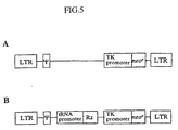
- Figure 5 is a schematic representation of the HIV vector. The expression cassette for each tRNAval-ribozyme was inserted into the SalI site immediately upstream of TK-neor in HIV-I-derived vector (A) to yield a retroviral vector, HIVRibo.N, that encoded a tRNAval-ribozyme(B). Ψ indicates a packaging signal.
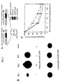
- Figure 6 shows quantitation of the expression of tRNAval-ribozyme in stably ribozyme-transduced H9 cells (CD4+ T cells) and inhibition of production of p24 in the transduced cells. Panel A. Quantitation of results, shown in B, of Southern blotting analysis of the RT-PCR-amplified ribozyme from two independent clones of ribozyme-transduced H9 cells. Products of PCR after 13, 15, and 17 cycles were analyzed by Southern blotting using a 32P-labeled oligonucleotide probe. Squares and circles indicate the results with transduced cells of the ribozyme 2 (Rz2) and the ribozyme 3 (Rz3), respectively. Panel B indicates the results of Southern blotting. Panel C. Cells were cultured for 11 days after infection with HIV-1 NL432. Small aliquots of supernatant were prepared from each culture on
days - Figure 7 shows intracellular localization of the tRNAval-ribozyme. Northern blotting analysis was performed using RNA from each intracellular fraction. Nucleic and cytoplasmic RNAs were prepared separately from cells that had been stably transduced with the gene for a ribozyme (the tRNAval-ribozyme-producing HeLa cells used in the experiments for which results are given in Figure 4B). A and C show the results obtained with a 32P-labeled probe specific for the tRNAval-ribozyme. B and D show controls: Contamination of the cytoplasmic fractions was examined with a probe specific for the transcript of the natural U6 gene.
- Below, the present invention will be explained in further detailed by means of examples. However, this is not intended to limit the scope of the present invention to these examples.
- The plasmids (pUCdt-Rz series) that expressed each tRNAval-ribozyme were constructed as follows. Both sense and antisense oligonucleotide linkers encoding the sequence of the promoter region, derived from the human gene for placental tRNAval (pHtV1; Arnold et al., 1986), were annealed and ligated into the EcoRI/SalI site of pUC19. The sequences of the oligonucleotide linkers were as follows: sense, 5'-aat tca gga cta gtc ttt tag gtc aaa aag aag aag ctt tgt aac cgt tgg ttt ccg tag tgt agt ggt tat cac gtt cgc cta aca cgc gaa agg tcc ccg gtt cga ag-3' (SEQ ID NO: 6); antisense, 5'-tcg act tcg aac cgg gga cct ttc, gcg tgt tag gcg aac gtg ata acc act aca cta cgg aaa cca acg gtt aca aag ctt ctt ctt ctt ttt gac cta aaa gac tag tcc tg-3' (SEQ ID NO: 7). Next, both sense and antisense oligonucleotide linkers that encoded the terminator sequence were also annealed and ligated into the NspV/SalI site of pUC19 that contained the sequence of the promoter region. The sequences of oligonucleotide linkers were as follows: sense, 5'-cga aac cgg gca ccc ggg gaa tat aac ctc gag cgc ttt ttt tct atc gcg tc-3' (SEQ ID NO: 8); antisense, 5'-tcg acg cga tag aaa aaa agc gct cga ggt tat att ccc cgg gtg ccc ggt ttc-3' (SEQ ID NO: 9). The resultant plasmid which contained the A and B boxes of tRNAval and a terminator was designated pUCdt.
- DNA fragments encoding the sequence of each ribozyme and the tRNAval portion were amplified by PCR using pUCdt as a template with an upper primer (5'-cgc cag ggt ttc cca gtc acg ac-3') (SEQ ID NO: 10) and a lower primer that included the sequences of both the ribozyme and the terminator (Rz1, 5'-ctg cag gtc gac gcg ata gaa aaa aag cgc tcg agg tgc ccg ttt cgt cct cac gga ctc atc agt gtt gtg tgg gtg ccc ggt ttc gaa ccg gga cct tt-3' (SEQ ID NO: 11): Rz2, 5'-ctg cag gtc gac gcg ata gaa aaa aac cgt ttc cga cgt gcc cgt ttc ggt cct ttc ggt cct cat cag tgt tgt gtt tgt agt gcc cgg ttt cga acc ggg gac ctt t-3' (SEQ ID NO: 12); Rz3, 5'-ctg cag gtc gac gcg ata gaa aaa aac cgt ttc cga cgt gcc cgt ttc ggt cct cat cag tgt tgt gtg ttg gtt tgt agt gcc cgg ttt cga acc ggg gac ctt t-3' (SEQ ID NO: 13)). After digestion of products of PCR with the EcoRI and SalI, each fragment was ligated into the EcoRI/SalI site of pUC19 to yield pUCdt-Rz. The sequences of pUCdt and pUCdt-Rz series were confirmed by direct nucleotide sequencing. The members of pUC-Rr series, which contained a reference gene-expression cassette in addition to the gene for the tRNAval-ribozyme (see Figure 3A), were constructed by inserting the PvuII fragment of pUCdt into the HincII site of each pUCdt-Rz. The direction of the inserted fragment was confirmed by digestion with restriction enzymes. The pHyg dt-Rz series, which was used for generation of the ribozyme-transduced HeLa cells, was constructed by inserting each PvuII-SalI fragment of the pUCdt-Rz series into the EcoRV/SalI site of pHyg (Yates et al., 1984). All oligonucleotide lingers and primers for PCR were synthesized by a DNA/RNA synthesizer (model 392;PE Applied Biosystems, Foster City, CA).
- Recombinant HIV vector plasmids were constructed as follows. A 2.0-kbp BamHI fragment that encoded the bacterial neor gene cassette from pMC1 neo (Thomas and Capecchi, 1987) was inserted into the SalI site of an HIV-1-derived vector (Figure 5A; Shimada et al., 1991). Then, the tRNAval-ribozyme expression cassette was cloned into the SalI site, immediately upstream of TK-neor, as shown in Figure 5B.
- HeLa and Cos cells were cultured in Dulbecco's modified Eagle's medium (DMEM; Gibco BRL, Gaithersburg, MD supplemented with 10% (v/v) fetal bovine serum (FBS; Gibco BRL) and 45 µg/ml gentamycin (Gibco BRL). To select ribozyme-transduced cells, hygromycin B was used at a final concentration of 300 µg/ml. H9 cells were cultured in RPMI (Gibco BRL) supplemented with 10% fetal calf serum (FCS; Gibco BRL).
- Cells were transfected using the Lipofectin reagent (Gibco BRL) according to the manufacturer's protocol. A recombinant HIV vector plasmid (HIVRib.N; shown in Figure 5B) was used to transfect H9 cells by the CaPO4 co-precipitation.
- Total RNA was extracted by the guanidinium thiocyanate-phenol-chloroform-method. Cytoplasmic RNA and nuclear RNA were separated as described previously (Huang and Carmichael, 1996).
- The steady-state level of each ribozyme was measured as follows. HeLa cells (1 x 10⁶ cells/10-cm plate) were transfected with each pUC-Rr. Two days after transfection, total RNA was isolated from these cells. The amount of the reference RNA, located downstream of tRNAval-ribozyme in the isolated total RNA, was quantified first by Northern blotting analysis with a probe specific for the reference RNA (5'-aaa tcg cta taa aaa gcg ctc gag gtt atg ctc ccc ggg t-3') (SEQ ID NO: 14). The amount of the reference DNA in each sample was maintained at a constant value and the level of total RNA in each sample was also kept constant by addition of RNA isolated from untransfected HeLa cells as necessary Finally, hybridization was repeated with a probe specific for the ribozyme (5'-etc atc tgt gtt gtg t-3') (SEQ ID NO: 15) or the probe specific for the reference RNA (Figure 3B).
- The half-life of each ribozyme was determined by Northern blotting analysis after treatment of cells with actinomycin D as described previously (Huang and Gerlach. 1996). In brief, cells were exposed to actinomycin D at a final concentration of 5 µg/ml for 0, 60, 120 or 180 min and, at each time point, total RNA was isolated (Figure 3C). The amount of ribozyme in each preparation of isolated RNA was determined by Northern blotting.
- Total RNA was isolated from HeLa cells transfected with each pUCdt-Rz or pUCdt. The amount of ribozyme in each preparation of isolated RNA was determined by Northern blotting with the probe that was specific for the ribozyme Then the concentration of each ribozyme was adjusted to the same value by addition of RNA isolated from untransfected HeLa cells The substrate RNA that encoded the U5 LTR region of HIV-1 (Figure 2A) was prepared by T7 transcription and radiolabeled with 32P-Cleavage reactions were allowed to proceed in a 50-· µl reaction mixture [40mM Tris-HCl (pH 8.0), 8mM MgCl2, 5 mM dithiothreitol (DTT). 2mM Spermidine. 40 U of placental RNase inhibitor, 30 µg of total RNA. 5kcpm of radiolabeled substrate RNA] at 37 °C for 12 h. Products were identified after electrophoresis on 6% polyacrylamide gel/7M urea gel (Figure 2B).
- Luciferase activity was measured with the Dual-Luciferase™ Reporter Assay System (Promega, Madison, WI) according to the manufacturer's protocol. HeLa cells transfected with pUCdt-Rz and the target-expressing plasmid (Figure 4A) or ribozyme-producing HeLa cells that had been transduced with the target-expressing plasmid (Figure 4B) were lysed in 150 µl of 1 x Passive lysis buffer for 15 min and scraped off the plate. The cell debris was removed by centrifugation. After addition of 20 µl of the centrofuged lysate to 100 µl of Luciferase Assay Reagent II, the luminescent signal was immediately quantitated with a luminometer (Lumant LB 9501; Berthold, Bad wildbad, Germany). Furthermore, for normalization of the activity of firefly luciferase, we measured the luminescent signal generated by Renilla luciferase by adding 100 µl of Stop & Glo™ Reagent to the sample tube immediately after quantitation of the reaction catalyzed by firefly luciferase. The recorded value of firefly luciferase activity was normalized by reference to the activity of Renilla luciferase (Figure 4).
- Each normalized value of firefly luciferase activity was further normalized by reference to the concentration of protein in the lysate. The protein was quantitated with a Protein Assay Kit (Bio-Rad. California, USA) which was based is Bradford's method.
- Ribozyme-transduced cells were obtained by transfecting HeLa cells with pHyg dt or a member of the pHyg dt-Rz series and selection in DMEM that contained 300 µg/ml hygromycin B (Wako Chemicals. Osaka, Japan). Twelve h after transfection, the medium was replaced by growth medium and the cells were cultured for another 48 h. The cells were subcultured at a dilution of 1:5 in DMEM that contained 300 µg/ml hygromycin B (selection medium). The medium was replaced by fresh medium every three days. Cells resistant to hygromycin B were expanded in DMEM that contained 250 µg/ml hygromycin B.
- A supernatant containing recombinant virus was produced as described previously (Shimada, 1991). Cos cells (2 x 10⁶ cells/10-cm dish) were cultured and transfected with 10 µg of the packaging vector plasmid and 10 µg of the recombinant HIV vector plasmid (HIVRib.N; shown in Figure 5). The supernatant, which contained recombinant virus, was collected after 48 h and filtered through a 0.22 µm pore filter. Then 2 x 10⁶ H9 cells were incubated with 5 ml of the filtered supernatant that contained 6 µg/ml Polybrene™ (Abbott Laboratories). After 24 h, the medium was replaced with RPMI supplemented with 10% FCS and 1 mg/ml G418. These cells were cultured for a further 48 h and then G418 resistant clones were isolated. The transduction of the ribozyme gene was confirmed by RT-PCR analysis.
- Quantitative RT-PCR was carried out as follows (Ozawa et al., 1990; Hamblet and Castora, 1995). Total RNA was extracted from H9 cells that had been stably transduced with a ribozyme. cDNA was synthesized in a 20- µl reaction mixture [1 µg of total RNA, 20mM Tris-HCl (pH 8.3), 50 mM KCl, 5 mM MgCl2, 1 mM dNTP, 1 pmol of primer (for β-actin, 5'-gtg gcc atc tct tgc tcg aa-3' (SEQ ID NO: 16); for ribozyme : 5'-gac ctt tcg gtc ctc atc-3' (SEQ ID NO: 17)) and 0-25 U/ml Moloney murine leukemia virus Rtase (Takara Shuzo, Kyoto, Japan)) at 42°C for 30 min.
- cDNA for β-actin was amplified by PCR with two oligonucleotide primers (upper, 5'-gac tac ctc atg aag atc ct-3' (SEQ ID NO: 18) ; lower: 5'-gtg gcc atc tct tgc tcg aa-3'(SEQ ID NO: 19)) with 13, 15 or 17 cycles of 94 °C for 1 min, 60°C for 1 min and 72°C for 2 min. Ribozyme cDNA was amplified by PCR with two oligonucleotide primers (upper, 5'-gtt atc acg ttc gcc taa-3' (SEQ ID NO: 20); lower: 5'-gac ctt tcg gtc ctc atc-3' (SEQ ID NO: 21)) with 13, 15 or 17 cycles of 94 °C for 1 min, 55°C for 1 min, and 72°C for 2 min.
- Products of PCR after 13, 15 and 17 cycles were analyzed by Southern blotting with a radiolabeled probe specific for the ribozyme (5'-acg cga aag gtc ccc ggt-3' (SEQ ID NO: 22)) or for β-actin (5'-gcg gga aaa tcg tgc gtg a-3' (SEQ ID NO: 23)). The radioactivity of each band (Figures 6 A and 6 B) was measured with BAS2000 system (Fuji Film, Tokyo, Japan).
- H9 cells transduced with the ribozyme by the HIV vector (HIVRib.N) and inock-tranduced control cells were incubated with NL432 at a m.o.i. (multiplicity of infection) of 0.01 for 4h. After two washes with PBS, these cells were cultured at 1 x 10⁵ cells/ml in RPMI 1640 medium supplemented with 10% FCS. The supernatant was collected on
days - To construct a pol III-driven ribozyme-expression cassette, we cloned a ribozyme sequence targeted to the 5' leader sequence of HIV-1 RNA (Adachi et al., 1986; Yu et al., 1993) adjacent to the sequence of a tRNAval promoter, with three kinds of short linker between them (linker sequences are indicated by lowercase letters and ribozyme sequences are indicated by bold capital letters in Figure 1), to yield a set of pUCdt-Rz plasmids. The insertion of the short linkers changed the overall structure of the transcripts and, thus, affected the accessibility of the recognition arms of the ribozyme (recognition arms are underlined). Naturally, it is important that both the 5' and 3' substrate-recognition arms of the ribozyme be available to the substrate so that the ribozyme can form the stem structures with the substrate RNA that ensure subsequent cleavage of the substrate. In order to clarify the relationship between structure and functional activity, we chose linkers that altered the extent of availability of the recognition arms. Figure 1 shows the secondary structures of the tRNAval-ribozymes (sequences corresponding to A and B boxes are shaded), as predicted by computer modeling (Mulfold Biocomputing Office. Biology Department, Indiana University, IN, USA). In one case (Figure 1A), the linker was inserted before the terminator sequence and restricted the flexibility of the 3' substrate recognition arm of the ribozyme. In addition, the 5' substrate-recognition arm was unavailable. Therefore, in the case of tRNAval-ribozyme 1 (Rz1 in Figure 1A), both 5' and 3' substrate-recognition arms were mostly embedded in a helical structure, tRNAval-ribozyme 2 (Rz2) has one restricted substrate-recognition arm on the 5' side. By contrast, tRNAval-ribozyme 3 (Rz3) had no restricted substrate-recognition arms and both arms were available for binding to the substrate- Judging from the flexibility of the substrate-recognition arms, we might expect that the cleavage activity of Rz3 would be the highest, followed by Rz2 and Rz1 in that order. The base sequences of Rz1-3 are represented in SEQ ID NOS: 3, 1 and 2, respectively.
- To examine whether the above ribozymes had the cleavage activity that we predicted from their secondary structures, (Figure 1) we first compared activities in vitro. Total RNA was isolated from HeLa cells that had been transfected with various pUCdt-Rz, plasmids that encoded the above ribozymes (tRNAval-ribozyme). We mixed a fixed amount (based on Northern blotting data) of each ribozyme within the isolated RNA and radiolabeled substrate RNA to initiate the cleavage reaction, and we monitored the progress of each reaction, after a 12-h incubation, on a 6% polyacrylamide/7 M urea gel (Figure 2). As expected, the cleavage activity of Rz3, with both recognition arms available, was the highest, followed by that of Rz2, while that of Rz1 with both recognition arms unavailable, was very low. It was, therefore, clear that the cleavage activity of tRNAval-ribozymes in vitro could be deduced from their computer-generated secondary structures.
- we expected that minor structural changes would occur in the entire structure as a result of the linker sequence. Thus, the linker should exert considerable influence on the stability of each ribozyme in vivo. We compared the intracellular stability of each tRNAval-ribozyme using two different approaches, as follows. We compared the steady-sate level of each transcript from HeLa cells that had been transiently transfected with pUC-Rr (a sequence of a reference gene was added to each ribozyme-coding pUCdt-Rz plasmid to yield pUC-Rr; Figure 3A) by Northern blotting analysis (transient expression assay). The level of expression of each tRNAval-ribozyme was normalized by adjusting the amount of the transcript of the reference gene, which was connected, in tandem, in the same plasmid (pUC-Rr; figure 3A). Transcripts of about 150 nucleotides in length (corresponding to the size of chimeric tRNAval-ribozyme) were detected in all samples of RNA that we isolated from HeLa cells that had been transfected with each plasmid that encoded a tRNAval ribozyme. The steady-state levels of the tRNAval-ribozymes differed over a 30-fold range of concentration. The level of Rz2, which was the highest, was about 26 times that of Rz1, which was the lowest, and the level of Rz3 was about 5 times that of Rz1. Since no modifications had been made in the promoter region of each ribozyme-expression cassette and, thus, since the efficiency of transcription was assumed to be the same in each case, we postulated that these differences among steady-state levels of transcripts were a consequence of the stability in vivo of each respective transcript.
- As a second approach and to test the above hypothesis, we attempted to compare the stability of each transcript under more natural, intracellular conditions We established stable HeLa transformants that produced each tRNAval-ribozyme and measured the intracellular half-life of each transcript directly by interrupting cellular transcription with actinomycin D. As shown in Figure 3C, the rate of degradation of Rz2 was lower than those that of Rz1 and of Rz3. The half-life of Rz2 (100±10 min) was more than twice that of Rz1 (35±2min) and Rz3 (40±15min). These results were in good agreement with the results of the transient expression assay and supported our hypothesis that the difference in the steady-state level of transcripts was due to the stability in vivo of each transcript rather than to any differences in the efficiency of transcription.
- In order to evaluate the intracellular activities of the tRNAval-ribozymes, we performed two types of assay. We first used each tRNAval-ribozyme expression plasmid (pUCdt-Rz) and a target gene expression plasmid, which encoded a chimeric HIV-1 LTR (R-U5 region)-luciferase gene, to co-transfect HeLa cells. After transient expression of both genes, in each cell lysate, we estimated the intracellular activity of each tRNAval-ribozyme by measuring the luciferase activity. The luciferase activity recorded when we used the control plasmid (pUCdt), with only minimal tRNAval promoter and terminator sequences instead of the ribozyme-expression plasmid, was taken as 100%. As shown in Figure 4A, Rz2, which had the highest stability in vivo, was most effective (>60% inhibition), followed by Rz3 (>40% inhibition). Rz1 was not very effective (about 10% inhibition), as expected from its low cleavage activity in vitro (figure 2B) and low stability in vivo (Figures 3B and 3C).
- In the second assay, only the target gene-expressing plasmid was used to transfect stable transformants that produced almost identical levels of tRNAval-ribozyme (stable HeLa transformants had been picked up arbitrarily and those clones with almost identical levels of expression of the ribozyme were selected for these studies). In this experiment (Figure 4B), with two independent stable transformants for each, ribozyme, we observed a similar trend to that described in the preceding paragraph. However, in this case, the effects of all of the ribozymes were stronger, most probably because all the transformed HeLa cells produced tRNAval-ribozyme constitutively. Rz2 inhibited expression of the target gene to a significant level, in some cases by as much as 97%.
- Although Rz3 had the highest cleavage activity in vitro, it failed to act more effectively than Rz2 in the cellular environment. These results suggest that, if a transcribed ribozyme is sufficiently stable within the cell, even if it does not have extremely high cleavage activity, it can have a remarkable effect in vivo.
- Since the above described studies demonstrated that Rz2 and Rz3 might have significant cleavage activity against the sequence of HIV-1 in vivo, we compared the abilities of that RNAval-ribozyme to inhibit replication of the HIV-1. Using an HIV vector (Figure 5; Shimada et al., 1991), we obtained stable transformants of the H9 cell line that expressed Rz2 or Rz3 (since Rz1 was inactive in studies described above, we above, we made no attempts to isolate stable transformants that produced Rz1). Cells transduced with the HIV vector without a ribozyme-expression cassette (Figure 5A) were used as a mock control. Two independent cell lines were used for subsequent analysis, and we detected no obvious changes in their growth rates over a period of 11 days, as compared with that of calls that did not produce either ribozyme (data not shown). Therefore, the ribozymes were not detrimental to host cells and probably only cleaved their target RNA with high specificity (Kawasaki et al., 1996, 1998).
- Before the virus-challenge assay, we measured the steady-state level of each tRNAval-ribozyme in the transduced H9 cells by quantitative RT-PCR analysis. The results of the transient expression assay in HeLa cells shown in Figure 3B, namely, that the difference in steady-state levels of Rz2 and Rz3 was about 5-fold, were confirmed by the RT-PCR analysis (Figures 6A and 6B). Clearly, Rz2 was more stable in vivo than Rz3.
- When we challenged stable H9 transformants that produced a tRNAval-ribozyme constitutively with HIV-1 virions, Rz2 inhibited viral replication almost completely (about 99%), as determined on
day 11 post-infection (Figure 6C). By contrast, to our surprise, Rz3 failed to inhibit viral replication at all under these experimental conditions. In the HIV-1 challenge assay, the difference between the effects of Rz2 and Rz3 was conspicuous. - Since the co-localization of a ribozyme with its target is clearly an important determinant of the ribozyme's efficiency (Sullenger and Cech, 1993; Bertrand et al., 1997), it was essential to determine the intracellular localization of tRNAval-ribozymes. Total RNA from HeLa cells transduced with the Rz2 expression cassette was separated into nuclear and cytoplasmic fractions. Then, transcribed Rz2 was detected by Northern blotting analysis with a probe specifc for the ribozyme. As shown in Figure 7A, Rz2 was found predominantly in the cytoplasmic fraction and it was not detected to any significant extent in the nuclear fraction. The other tRNA-ribozymes (Rz1 and Rz3) were also localized predominantly in cytoplasmic fractions (data not shown). U6 snRNA. which remains in the nucleus, was included as a control in these studies (Figure 7B).
- A ribozyme is a potentially useful tool for the suppression of the expression of specific gene since it can be engineered to act on other RNA molecules with high specificity (Uhlenbeck, 1987; Hasseloff and Gerlach, 1988). Although many trials have been successful (Eckstein and Lilley. 1996; Turner, 1997; Scalon. 1997), it remains difficult to design an effective ribozyme-expression system that can be used in vivo. One major challenge related to the use of ribozymes and antisense RNAs as therapeutics or genetic agents is the development of suitable expression vectors (Jennings and Molloy, 1987; Sullenger et al., 1990; Bertrand et al., 1994, 1997; Thompson et al., 1995). Two kinds of expression system have been used to date, as discussed in the Introduction, namely, the pol II system and pol III system. In this study, we used the pol III system and the promoter of a human gene for tRNAval for transcription of ribozymes (Yu et al., 1993). This promoter is not only suitable for transcription of small RNAs, but its use also facilitates prediction of secondary structure by computer folding. More importantly, it allows export of transcribed ribozymes from the nucleus to the cytoplasm so that the tRNAval-ribozymes can find with their mRNA targets.
- These secondary structure of a target mRNA determines its susceptibility to ribozyme-mediated cleavage, and the ribozyme must also fold into appropriate secondary and tertiary structures for maximal activity. Although there is no guarantee that a computer-predicted secondary structure really represents the corresponding structure after transcription, the structures predicted in this study (Figure 1A-1C) were well correlated with cleavage activities in vitro (Figure 2). In the expression cassettes, the last seven bases of the mature tRNAval (indicated by capital letters in Figure 1E) had been removed, without any effect on transcription, in order to block 3'-end processing of the transcript (Adeniyi-Jones et al., 1984). They were replaced by a linker (lowercase letters in Figure 1) followed by a ribozyme (bold capital letters). The freedom or availability of the substrate-recognition arms was adjusted by the linker sequence via formation of stable stem structures in combination with the sequence of the tRNAval which accounted for about two-thirds of the whole sequence. Thus, it was relatively easy to predict, by computer folding, the secondary structure and the accessibility of each recognition arm. Furthermore, even if the sequence of the substrate recognition arm is changed, as long as the same rules for predicting overall secondary structure are used, it is still possible to predict the accessibility of recognition arms. Indeed, we have succeeded in constructing a similar ribozyme-expression system for inhibition of the expression of other genes (Kawasaki et al., 1996 , 1998). Our expression system, as shown in figures 1A-1C, facilitates the design of an effective ribozyme-expression cassette.
- The ribozyme-expression cassettes shown in Figures 1A-1C allowed all the transcripts to be exported to the cytoplasm (Figure 7A) where they could find their mRNA targets, and significant inhibition by ribozymes of the expression of the target molecules was observed (Figures 4 and 6c). In a previous study (Bertrand et al., 1997), deletion of the last ten bases of mature tRNAMet not only blocked 3' processing but also inhibited the export of the transcript to cytoplasm (Tobian et al., 1985). These results suggested that 3' processing might be linked to export to the cytoplasm and that 3'-altered tRNA transcripts are not exported efficiently (Cotten and Birnstiel, 1989; Boelens et al., 1995). However, as demonstrated in Figure 7, the deletion of the last seven bases of mature tRNAval did not inhibit the export of the transcripts from the nucleus.
- A protein, designated Exportin(tRNA), which transports tRNA from the nucleus to the cytoplasm has recently been identified (Arts et al., 1998). Exportin (tRNA) binds RanGTP in the absence of tRNA but it does not bind tRNA in the absense of RanGTP. Therefore, a model for the transport of tRNAs was proposed wherein Exportin (tRNA) associates with RanGTP first in the nucleus and then the complex binds a mature tRNA molecule This final complex is translocated through a nuclear pore complex to the cytoplasm. There, the Ran-bound GTP is hydrolyzed, releasing the tRNA into the cytoplasm and allowing Exportin (tRNA) to be recycled to the nucleus (Arts et al., 1998). We do not yet know the minimal sequence or structure within a tRNA that can be recognized by Exportin (tRNA). However, since the ribozymes shown in Figures 1A-1C were successfully translocated to the cytoplasm, it is possible that they were recognized and transported by Exportin (tRNA) despite the deletions and alterations at the 3' end of the natural tRNA.
- It is clear, from our study, that even 3'-altered tRNA transcripts can be transported efficiently to the cytoplasm if their secondary structures resemble those in Figures 1A-1C. when we tried similarly to express an other kind of ribozyme (Rz4 in figure 1D(SEQ ID NO: 5)) in HeLa cells, the transcripts remained in the nucleus (Figure 7C). The secondary structure of Rz4 (Figure 1D) is quite different from that of ribozymes Rz1, Rz2 and Rz3, which were cytoplasmic, despite the fact that not only the A and B box promoter elements (shaded boxes in Figure 1) but also all the remaining sequence within the tRNAval segment were identical in transcripts Rz1 through Rz4. This observation suggest that, if Exportin (tRNA) can indeed recognize the ribozyme transcript, it is unlikely that it recognizes a specific nucleotide sequence. Exportin (tRNA) might, rather, recognize some specific higher-order structure of tRNA or some sequence within such a higher-order structure.
- Indeed, another ribozyme, constructed for mother purposes, whose secondary structure resembled that of Rz4 was found only in nuclei (data not shown). We have constructed more than ten other ribozymes for suppression of three other genes, keeping in mind that their secondary structures should resemble those of Rz1 through Rz3 in Figure 1 and adjusting linker sequences so that they might be transported to the cytoplasm. All of these ribozymes were found in the cytoplasm after transcription. They not only had high activities (>95% inhibition) but also high specificity (<5% inhibition by the inactive control). Thus, cytoplasmic ribozymes based on the design shown in Figures 1A-1C seem very attractive (Kawasaki et al., 1996, 1998). We should also mention that the secondary structures of Rossi' s tRNAMet-ribozymes, which remained in nuclei and were not very active (Bertrand et al., 1997), do not resemble our active secondary structures because of their different linker sequence. Their structures resemble that of Rz4 (computer-predicted structures not shown).
- It will be of interest to determine whether ribozymes such as Rz1 through Rz3 (but not Rz4 or Rossi's tRNAMet-ribozymes) form complexes with Exportin (tRNA) in the presence of RanGTP, that is, under conditions in which formation of a complex between an export receptor and its cargo would be expected (Arts et al., 1998)
- Sullenger and Cech (1993) and Rossi's group (Bertrand et al., 1997) clearly demonstrated the importance of intracellular co-localization of ribozymes with their targets. In the case of one specific expression cassette, both the ribozymes and its RNA target were located in the nucleus and the specific cleavage by the ribozyme of its target was detected (Bertrand et al., 1994). Thus, the critical parameter is not the localization of the ribozyme per se but it is, rather, the ability of the ribozyme to co-localize with its target (Bertrand et al., 1997). Since various proteinaceous factors are involved in the intracellular processing and transport of mRNAs and since such factors may bind promptly with mRNA immediately after transcription, such factors could inhibit the binding of the ribozyme with its RNA target in the nucleus. Also, in the cytoplasm, polysomes might inhibit the binding of the ribozyme with its RNA target. Moreover, since nuclear tRNAMet-ribozyme failed to inactivate a cytoplasmic mRNA that had originally been produced in the nucleus (Bertrand et al., 1997), the transport of mRNA from the nucleus to the cytoplasm seems to be much more rapid than the attack by the nuclear tRNAMet-ribozyme. One of the most critical factors determining ribozyme activity in vivo seems to be the association between the ribozyme and its target. A significant fraction of ribozymes must be degraded during their transport and also during their approach to their target site. For this reason, co-localization of a ribozyme and its target does not, by itself, guarantee the efficacy of ribozymes in vivo.
- The ribozyme Rz2, which was most stable in vivo (Figures 3B, 3C, 6A and 6B), was more effective in the intracellular environment (Figure 4) than Rz3, which had higher cleavage activity in vitro (Figure 2). This difference in activity was magnified in the HIV-1 challenge (Figure 6C). Although cells producing the more stable Rz2 were almost completely resistant to infection by HIV-1, other cells producing the less stable Rz3 were as sensitive as control cells to infection by HIV-1. Although Rz2 had a half-life, that was about twice that of Rz3, it is unclear, at present, which structural feature(s) made Rz2 more resistant to RNases. There were six more nucleotides within the linker in Rz3, as compared to Rz2, which must have influenced the higher-order structure.
- The half-lives of natural, tRNAs range from 50 to 60 hours (Smith and Weinberg, 1981), while that of Rz2 was only about 100 min. If the half-life of the tRNA-ribozyme could be prolonged, a higher inhibitory effect might be expected, while we still cannot predict the relative stabilities of transcripts in vivo, we can design ribozymes that can be transported into the cytoplasm by incorporating secondary structures such as the ones shown in Figure 1. Since we cannot accurately predict the stability of a transcript, we usually test several constructs and, in the case of various genes tested to date, we have always been able to obtain a cassette that can inactivate the gene of interest with >95% efficiency (Kawasaki et al., 1996, 1998)
- The tRNAval-vector may be useful for expression of functional RNAs other than ribozymes whose target molecules are localized in the cytoplasm. In our hands, tRNAval-ribozymes have consistently high activities, at least in cultured cells. Therefore, properly designed tRNAval-ribozymes appeared to be very useful as tools in molecular biology, with potential utility in medicine as well.
- By the present invention, there is provided novel ribozymes and expression systems therefor. The ribozyme of the present invention has high in vivo stability, and thereby exhibits high activity.
-
- Adachi, A. Gendelman, H. E. , Koenig, S., Folks, T., Willey, R., Rabson, A. and Martin, M. A. (1986) Production of acquired immunodeficiency syndrome-associated retrovirus in human and nonhuman cells transfected with an infectious molecular clone. J. virol., 59, 284-291.
- Adeniyi-Jones, S., Romeo, P. and Zasloff, M. (1984) Generation of long read through transcripts in vivo and in vitro by deletion of 3' termination and processing sequences in the human tRNAi met gene. Nucleic Acids Res., 12, 1101-1115.
- Arnold, G. J., Schmutzler, C., Thomann, U.. van Tol, H. and Gross, H. J. (1986) The human tRNAval gene family: organization, nucleotide sequences and homologous transcription of three single copy genes. Gene, 44, 287-297.
- Arts, G.-J.. Fornerod, M. and Mattaj, I. W. (1998) Identification of a nuclear export receptor for tRNA. Curr. Biol., 6, 305-314.
- Bertrand, E., Pictet. R. and Grange, T. (1994) Can hammerhead ribozymes be efficient tools for inactivating gene function? Nucleic Acids Res., 22, 293-300.
- Bertrand, E. and Rossi, J. J. (1996) Anti-HIV therapeutic hammerhead ribozymes: targeting strategies and optimization of intracellular function. In Eckstein. F. and. Lilley, D. M. J. (eds.), Nucleic Acids Mol. Biol., Vol. 10. Springer-verlag, Berlin, 301-313.
- Bertrand, E., Castanotto, D., Zhou, C., Carbonnelle, C., Lee, G. P., Chatterjee, S., Grange, T., Pictet, R., Kohn, D., Engelke, D. and Rossi, J. J. (1997) The expression cassette determines the functional activity of ribozymes in mammalian cells by controlling their intracellular localization. RNA, 3, 75-88.
- Boelens, W., Palacios, I. and Mattaj, I. W. (1995) Nuclear retention of RNA as a mechanism for localization. RNA, 1, 273-283.
- Cotten, M. and Birnstiel, M. (1989) Ribozyme mediated destruction of RNA in vivo. EMBO J., 8, 3861-3866.
- Dahm, S. C. and uhlenbeck, O.C. (1991) Role of divalent metal ions in the hammerhead RNA cleavage reaction. Biochemistry, 30, 9464-9469.
- Dahm, S. C., Derrick, W. B. and Uhlenbeck, O. C. (1993) Evidence for the role of solvated metal hydroxide in the hammerhead cleavage mechanism. Biochemistry, 32, 13040-13045.
- Dropulic. B., Lin, N.H., Martin, M.A. and Jeang, K. T. (1992) Functional characterization of a U5 ribozyme: intracellular suppression of human
immunodeficiency virus type 1 expression. J. Virol., 66, 1432-1441. - Eckstein, F. and Lilley, D. M. J. (eds.) (1996) Catalytic RNA, Nucleic Acids and Molecular Biology, Vol. 10. Springer-Verlag, Berlin.
- Erickson, R. P. and Izant, J. (eds.) (1992) Gene Reguration: Biology of Antisense RNA and DNA; Raven Perss, New York.
- Ferbeyre, G., Bratty, J., Chen, H. and Cedergren, R. (1996) Cell cycle arrest trans-hammerhead ribozyme action in Yeast. J. Biol. Chem., 271, 19318-19323.
- Fujita, S., Koguma, T., Ohkawa, J., Mori, K., Kohda, T. Kise, H., Nishikawa, S., Iwakura, M. K. and Taira, K. (1997) Discrimination of a single base change in a ribozyme using the gene for dihydrofolate reductase as a selective marker in Escherichia coli. Proc. Natl. Acad. Sci. USA, 94. 391-396.
- Gebhard, J. R., Perry, C. M., Mahadeviah, S. and Witton, J. L. (1997) Use of a nonviral vector to express a chimeric tRNA-ribozyme against lymphocytic choriomeningitis virus: cytoplasmic accumulation of a catalytically competent transcript but minimal antiviral effect. Antisense and Nucleic Acid Drug Dev. 7, 3-11.
- Good. P. D., Krikos, A. J., Li, S. X. L., Lee, N. S., Giver, L., Ellington, A., Zaia, J. A., Rossi, J. J. and Engelke, D. R. (1997) Expression of small, therapeutic RNAs in human cell nuclei. Gene Therapy, 4, 45-54.
- Guerrier-Takada, C., Gardiner, K., Marsh, T., Pace, N. and Altman, S. (1983) The RNA moiety of ribonuclease P is the catalytic subunit of the enzyme. Cell, 35, 849-857.
- Hamblet, N. S. and Castora, F. J. (1995) Mitochondrial DNA deletion analysis: a comparison of PCR quantitative methods. Biochem. Biophys. Res. Commun., 207, 839-847.
- Hasseloff, J. and Gerlach, W. L. (1988) Simple RNA enzymes with new and highly specific endoribonuclease activities. Nature, 334, 585-591.
- Huang, Y. and Carmichael, G. G. (1996) Role of polyadenylation in nucleocytoplasmic transport of mRNA. Mol. Cell. Biol., 16, 1534-1542.
- Inokuchi, Y., Yuyama, N., Hirashima, A., Nishikawa. S., Ohkawa, J., and Taira, K. (1994) A hammerhead ribozyme inhibits the proliferation of an RNA coliphage SP in E-coli. J. Biol. Chem., 269, 11361-11366.
- Ilves, H., Barske, C., Junker, U., Bohnlein, E. and Veres, G. (1996) Retroviral vectors designed for targeted expression of RNA polymerase III-driven transcripts: a comparative study. Gene, 171, 203-208.
- Jennings, P. A. and Molloy, P. L. (1987) Inhibition of SV40 replicon function by engineered anitisense RNA transcribed by RNA polymerase III. EMBO J, 6, 3043-3047.
- Kawasaki, H., Ohkawa, J., Tanishige, N., Yoshinari, K., Murata, T., Yokoyama, K. K. and Taira, K. (1996) Selection of the best target site for ribozyme-mediated cleavage within a fusion gene for adenovirus E1A-associated 300 kDa protein (p300) and luciferase. Nucleic Acids Res. 24, 3010 3016.
- Kawasaki. H., Ecker, R., Yao, T.-P., Taira, K., Chiu. R., Livingston, D.M. and Yokoyama, K. K. (1998) Distinct roles of the co-activators p300 and CBP in retinoic-acid-induced F9-cell differentiation Nature, 393, 284-289.
- Kruger, K., Grabowski, P. J., Zaug, A. J., Sands, J., Gottschling. D. E. and Cech, T. R. (1982) Self-splicing RNA: autoexcision and autocyclization of the ribosomal RNA intervening sequence of Tetrahymena. Cell, 31, 147-157.
- Lott, W. B., Pontius, B. W. and von Hippel, P. H. (1998) A two-metal ion mechanism operates in the hammerhead ribozyme-mediated cleavage of an RNA substrate, Proc. Natl. Acad. Sci. USA, 95, 542-547.
- Murray, J. A. H. (ed.) (1992) Antisense RNA and DNA; Wiley-Liss, Inc, New York.
- Ohkawa, J., Yuyama, N., Takebe, Y., Nishikawa, S. and Taira, K (1993) Importance of independence in ribozyme reactions: kinetic behavior of trimmed and of simply connected multiple ribozymes with potential activity against human immunodeficiency virus. Proc. Natl. Acad. Sci. USA, 90, 11302-11306.
- Ojwang, J. O.,Hampel, A., Looney, D. J., Wong-staal. F. and Rappaport, J. (1992) Inhibition of human
immunodeficiency virus type 1 expression by a hairpin ribozyme. Proc. Natl. Acad. Sci. USA, 89, 10802-10806. - Ozawa, T., Tanaka. M., Ikebe, S., Ohno, K., Kondo, T. and Mizuno, Y, (1990) Quantitative determination of deleted mitochondrial DNA relative to normal DNA in parkinsonian striatum by a kinetic PCR analysis. Biochem. Biophys. Res. Commun., 172, 483-489.
- Perriman, R., Bruening, G., Dennis E. S. and Peacock, W. J. (1995) Effective ribozyme delivery in plant cells. Proc. Natl. Acad. Sci. USA, 92, 6175-6179.
- Pontius, B. W., Lott, W. B. and von Hippel, P. H. (1997) Observations on catalysis by hammerhead ribozymes are consistent with a two-divalent-metal-ion mechanism. Proc. Natl. Acad. Sci. USA, 94, 2290-2294.
- Prislei, S., Buonomo, S. B. C., Michienzi, A. and Bozzoni, I. (1997) Use of adenoviral VAI small RNA as a carrier for cytoplasmic delivery of ribozymes. RNA, 3, 677-687.
- Rossi, J. J. and Sarver, N. (1990) RNA enzymes (ribozymes) as antiviral therapeutic agnts. TIBTECH, 8, 179-183.
- Rossi. J. J. (1995) Controlled, targeted, intracellular expression of ribozymes: progress and problems. TIBTECH, 13, 301-306.
- Sarver; N., Cantin, E. M., Chang, P. S., Zaida, J. A., Ladne, P. A., Stepenes, D. A. and Rossi, J. J. (1990) Ribozymes as potential anti-HIV-1 therapeutic agents Science, 247, 1222-1225.
- Scanlon, K. J. (ed.) (1997) Therapeutic Applications of Ribozymes; Methods in Molecular Medicine. Vol. 11. Humana Press, New Jersey.
- Shimada, T., Fujii, H., Mitsuya, H. and Nienhuis, A. W (1991) Targeted and highly efficient gene transfer into CD4+ cells by a recombinant human immunodeficiency virus retroviral vector. J. Clin. Invest., 88, 1043-1047.
- Smith, D. W. and weinberg, W. C. (1981) Transfer RNA in reticulocyte maturation. Biochem. Biophys. Acta., 655, 195-198.
- Sullenger, B. A., Lee, T. C., Smith. C. A. and Ungers. G. E. (1990) Expression of chimeric tRNA-driven antisense transcripts renders NIH 3T3 cells highly resistant to Moloney murine leukemia virus replication. Mol. Cell. Biol. 10, 6512-6523.
- Sullenger. B. A. and Cech, T. R. (1993) Tethering ribozymes to a retroviral packaging signal for destruction of viral RNA. Science, 262, 1566-1569.
- Taira. K., Nakagawa, K., Ni shikawa., S. and Furukawa, K. (1991) Construction of a novel RNA-transcript-trimming plasmid which can be used both in vitro in place of run-off and(G)-free transcriptions and in vivo ad multi-sequence transcription vectors. Nucleic Acids Res., 19, 5152-5130.
- Thomas, K. R. and Capecchi, M. R. (1987) Site-directed mutagenesis by gene targeting in mouse embryo-derived stem cells. Cell, 51, 503-512.
- Thompson, D. J., Ayers. F. D., Malmstrom, A. T., Ganousis, L. M., Chowrira. M. B., Couture, L. and Stinchcomb, T. D. (1995) Improved accumulation and activity of ribozymes expressed from a tRNA-based RNA polymerase III promoter. Nucleic Acids Res., 23, 2259-2268.
- Tobian, J. A., Drinkard, L. and Zaseloff, M. (1985) tRNA nuclear transport: defining the critical regions of human tRNA Met by point mutagenesis. Cell, 43, 415-422.
- Turner, P. C. (ed.) (1997) Ribozyme Protocols; Methods in Molecular Biology, Vol. 74, Humana Press, New Jersey.
- Uhlenbeck, O. C. (1987) A small catalytic oligoribonucleotide. Nature, 328,596-600.
- Yamada, O., Kraus, G. Leavitt, M. C., Yu, M. and Wong-Staal, F (1994) Activity and cleavage site specificity of an anti-HIV-1 hairpin ribozyme in human T cells. Virology, 205, 121-126.
- Yamada, O., Yu, M., Yee, J.-K., Kraus, G., Looney, D. and Wong-Staal, F. (1994) Intracellular immunization of human T cells with a hairpin ribozyme against human
immunodeficiency virus type 1. Gene Therapy, 1, 38-45. - Yates, J., Warren, N., Reisman, D. and Sugden. B. (1984) A cis-acting element from the Epstein-Barr viral genome that permits stable replication of recombinant plasmids in latently infected cells. Proc. Natl. Acad. Sci. USA, 81, 3806-3810.
- Yu, M., ojwang, J. O., Yamada, O., Hampel, A., Rappaport, J., Looney, D. and Wong-Staal. F.(1993) A hairpin ribozyme inhibits expression of diverse strains of human
immunodeficiency virus type 1. Proc. Natl. Acad. Sci. USA, 90, 6340-6344. - Zhao, J.J. and Pick. K, (1993) Generating loss-of function phenotypes of the fushitarazu gene with a targeted ribozyme in Drosophila. Nature, 365, 448-451.
- Zhou, D.-M., Zhang, L.-H., Kumar, P. K. R. and Taira, K. (1996) The ribozyme mechanism revisited: Evidence against direst coordination of a Mg2+ ion with the pro-R oxygen of the scissile phosphate in the transition state of a hammerhead ribozyine- catalyzed- reaction. J. Am. Chem. Soc., 118, 8969-8970.
- Zhou. D.-M., Zhang, L.-H. and Taira, K. (1997) Explanation by the double-metal-ion mechanism of catalysis for the differential metal ion effects on the cleavage rates of 5'-oxy and 5'-thio substrates by a hammerhead ribozyme. Proc. Natl. Acad. Sci. USA, 94. 14343-14348.
- Zhou, D.-M. and Taira, K.(1998) The hydrolysis of RNA: from theoretical calculations to the hammerhead ribozyme-mediated cleavage of RNA. Chem. Rev, 98, 991-1026.
-
- <110> Director-General of Agency of Industrial Science and Technology
- <120> Expression Systems for Transcription of Functional Nucleic Acids
- <130> 117F0059
- <140>
<141> - <150>
JP 10/244755
<151> 1998-8-31 - <160> 23
- <170> PatentIn Ver. 2.0
- <210> 1
<211> 135
<212> RNA
<213> Artificial Sequence - <220>
<223> Description of Artificial Sequence: the nucleotide sequence of Rz2 - <400> 1

- <210> 2
<211> 141
<212> RNA
<213> Artificial Sequence - <220>
<223> Description of Artificial Sequence: the nucleotide sequence of Rz3 - <400> 2

- <210> 3
<211> 128
<212> RNA
<213> Artificial Sequence - <220>
<223> Description of Artificial Sequence: the nucleotide sequence of Rz1 - <400> 3

- <210> 4
<211> 95
<212> RNA
<213> Artificial Sequence - <220>
<223> Description of Artificial Sequence: the nucleotide sequence of the transcript of human placental tRNAVal - <400> 4

- <210> 5
<211> 149
<212> RNA
<213> Artificial Sequence - <220>
<223> Description of Artificial Sequence: the nucleotide sequence of Rz4 - <400> 5


- <210> 6
<211> 110
<212> DNA
<213> Artificial Sequence - <220>
<223> Description of Artificial Sequence: the sequence of a sence oligonucleotide linker - <400> 6

- <210> 7
<211> 113
<212> DNA
<213> Artificial Sequence - <220>
<223> Description of Artificial Sequence: the sequence of an antisense oligonucleotide linker - <400> 7

- <210> 8
<211> 53
<212> DNA
<213> Artificial Sequence - <220>
<223> Description of Artificial Sequence: the sequence of a sense oligonucleotide linker - <400> 8

- <210> 9
<211> 54
<212> DNA
<213> Artificial Sequence - <220>
<223> Description of Artificial Sequence: the sequence of an antisense oligonuclotide linker - <400> 9

- <210> 10
<211> 23
<212> DNA
<213> Artificial Sequence - <220>
<223> Description of Artificial Sequence: the sequence of an upper primer - <400> 10

- <210> 11
<211> 101
<212> DNA
<213> Artificial Sequence - <220>
<223> Description of Artificial Sequence: the sequence of a lower primer including the sequences of Rz1 and a terminator - <400> 11

- <210> 12
<211> 109
<212> DNA
<213> Artificial Sequence - <220>
<223> Description of Artificial Sequence: the sequence of a lower primer including the sequences of Rz2 and a terminator - <400> 12

- <210> 13
<211> 106
<212> DNA
<213> Artificial Sequence - <220>
<223> Description of Artificial Sequence: the sequence of a lower primer including the sequences of Rz3 and a terminator - <400> 13

- <210> 14
<211> 40
<212> DNA
<213> Artificial Sequence - <220>
<223> Description of Artificial Sequence: the sequence of a probe specific for the reference RNA - <400> 14

- <210> 15
<211> 16
<212> DNA
<213> Artificial Sequence - <220>
<223> Description of Artificial Sequence: the sequence of a probe specific for the ribozyme - <400> 15

- <210> 16
<211> 20
<212> DNA
<213> Artificial Sequence - <220>
<223> Description of Artificial Sequence: the sequence of a primer for b-actin - <400> 16

- <210> 17
<211> 18
<212> DNA
<213> Artificial Sequence - <220>
<223> Description of Artificial Sequence: the sequence of a primer for the ribozyme - <400> 17

- <210> 18
<211> 20
<212> DNA
<213> Artificial Sequence - <220>
<223> Description of Artificial Sequence: the sequence of an upper oilgonucleotide primer - <400> 18

- <210> 19
<211> 20
<212> DNA
<213> Artificial Sequence - <220>
<223> Description of Artificial Sequence: the sequence of a lower oligonucleotide primer - <400> 19

- <210> 20
<211> 18
<212> DNA
<213> Artificial Sequence - <220>
<223> Description of Artificial Sequence: the sequence of an upper oligonucleotide primer - <400> 20

- <210> 21
<211> 18
<212> DNA
<213> Artificial Sequence - <220>
<223> Description of Artificial Sequence: the sequence of a lower oligonucleotide primer - <400> 21

- <210> 22
<211> 18
<212> DNA
<213> Artificial Sequence - <220>
<223> Description of Artificial Sequence: the sequence of a probe specific for the ribozyme - <400> 22

- <210> 23
<211> 19
<212> DNA
<213> Artificial Sequence - <220>
<223> Description of Artificial Sequence: the sequence of a probe specific for b-actin - <400> 23

-
- SEQ ID NO. 1: Nucleotide sequence of Rz2.
- SEQ ID NO. 2: Nucleotide sequence of Rz3.
- SEQ ID NO. 3: Nucleotide sequence of Rz1.
- SEQ ID NO. 4: Nucleotide sequence of human placental tRNAval transcript.
- SEQ ID NO. 5: Nucleotide sequence of Rz4.
- SEQ ID NO. 6: Nucleotide sequence of a sense oligonucleotide linker.
- SEQ ID NO.7: Nucleotide sequence of an antisense oligonucleotide linker.
- SEQ ID NO. 8: Nucleotide sequence of a sense oligonucleotide linker.
- SEQ ID NO. 9: Nucleotide sequence of an antisense oligonucleotide linker.
- SEQ ID NO. 10: Nucleotide sequence of an upper primer.
- SEQ ID NO. 11: Nucleotide sequence of a lower primer comprising sequences of Rz1 and a terminator.
- SEQ ID NO. 12: Nucleotide sequence of a lower primer comprising sequences of Rz2 and a terminator.
- SEQ ID NO. 13: Nucleotide sequence of a lower primer comprising sequences of Rz3 and a terminator.
- SEQ ID NO. 14: Nucleotide sequence of a reference RNA-specific probe.
- SEQ ID NO. 15: Nucleotide sequence of a ribozyme-specific probe.
- SEQ ID NO. 16: Nucleotide sequence of a primer for β-actin.
- SEQ ID NO. 17: Nucleotide sequence of a primer for ribozyme.
- SEQ ID NO. 18: Nucleotide sequence of an upper oligonucleotide primer.
- SEQ ID NO. 19: Nucleotide sequence of a lower oligonucleotide primer.
- SEQ ID NO. 20: Nucleotide sequence of an upper oligonucleotide primer.
- SEQ ID NO. 21: Nucleotide sequence of a lower oligonucleotide primer.
- SEQ ID NO. 22: Nucleotide sequence of a ribozyme-specific probe.
- SEQ ID NO. 23: Nucleotide sequence of a β-actin-specific probe.
Claims (15)
- A ribozyme comprising a nucleotide sequence having the following base sequence (I) or (II):


- An expression vector comprising DNA encoding a ribozyme according to Claim 1.
- A method of producing a ribozyme according to Claim 1 comprising transcribing to RNA with expression vector DNA as a template, in which the expression vector DNA comprises DNA encoding a ribozyme according to Claim 1.
- A pharmaceutical composition comprising a ribozyme according to Claim 1 or an expression vector according to Claim 2, as an effective ingredient.
- A ribozyme according to Claim 1, an expression vector according to Claim 2 or a pharmaceutical composition according to Claim 4 for the prevention and/or treatment of acquired immune deficiency syndrome.
- An in vitro method of specifically cleaving a target RNA using a ribozyme according to Claim 1.
- A method according to Claim 6, in which the target RNA is HIV-1 RNA.
- An RNA derived from a tRNAVal having a nucleotide sequence depicted in SEQ ID NO: 4 and having a secondary structure (I) comprising a region in which hydrogen bonds are formed between nucleotides 8-14 and 73-79, the said derived RNA comprising a bulge structure introduced into said region of said tRNAVal nucleotide sequence, such that a transcript comprising said derived RNA and a sequence linked 3' thereto is capable of being transported out of the nucleus without the 3' sequence being eliminated.

- An RNA according to Claim 8, in which the bulge structure is introduced by substituting all or a part of the nucleotides 73-79 in the tRNAval nucleotide sequence depicted in SEQ ID NO: 4.
- An RNA according to Claim 8 or 9, which consists of nucleotides 1-80 in the nucleotide sequence depicted in SEQ ID NO: 1.
- An RNA according to Claim 8 or 9, which consists of nucleotides 1-86 in the nucleotide sequence depicted in SEQ ID NO: 2.
- An RNA comprising an RNA according to any of Claims 8 to 11, together with a selected RNA chain linked thereto.
- An RNA according to Claim 12, in which the selected RNA chain is a ribozyme or an antisense RNA.
- An RNA according to Claim 12 or 13, in which a bulge structure is formed by base pairing between nucleotides in an RNA chain linked to the 3' terminus and nucleotides 8-14 in the nucleotide sequence depicted in SEQ ID NO: 4.
- An expression vector comprising DNA encoding an RNA according to any of Claims 8 to 14.
Applications Claiming Priority (3)
| Application Number | Priority Date | Filing Date | Title |
|---|---|---|---|
| JP10244755A JP2990268B1 (en) | 1998-08-31 | 1998-08-31 | Expression system for functional nucleic acid transcription |
| JP24475598 | 1998-08-31 | ||
| PCT/JP1999/004718 WO2000012686A1 (en) | 1998-08-31 | 1999-08-31 | Expression systems for functional nucleic acid expression |
Publications (3)
| Publication Number | Publication Date |
|---|---|
| EP1118659A1 EP1118659A1 (en) | 2001-07-25 |
| EP1118659A4 EP1118659A4 (en) | 2002-12-04 |
| EP1118659B1 true EP1118659B1 (en) | 2006-04-12 |
Family
ID=17123425
Family Applications (1)
| Application Number | Title | Priority Date | Filing Date |
|---|---|---|---|
| EP99940588A Expired - Lifetime EP1118659B1 (en) | 1998-08-31 | 1999-08-31 | Expression systems for functional nucleic acid expression |
Country Status (5)
| Country | Link |
|---|---|
| US (2) | US6740750B1 (en) |
| EP (1) | EP1118659B1 (en) |
| JP (2) | JP2990268B1 (en) |
| DE (1) | DE69930862T2 (en) |
| WO (1) | WO2000012686A1 (en) |
Families Citing this family (5)
| Publication number | Priority date | Publication date | Assignee | Title |
|---|---|---|---|---|
| US6995258B1 (en) | 2000-05-25 | 2006-02-07 | City Of Hope | Nucleolar targeting of therapeutics against HIV |
| AU2003237303A1 (en) * | 2002-05-30 | 2003-12-19 | Albert Einstein College Of Medicine Of Yeshiva University | Aptamer constructs |
| AU2004208057A1 (en) * | 2003-01-31 | 2004-08-12 | Dnavec Research Inc. | Paramyxovirus vector encoding ribozyme and utilization thereof |
| JPWO2005001082A1 (en) * | 2003-06-30 | 2006-08-10 | 株式会社ディナベック研究所 | Negative-strand RNA viral vector carrying a gene with a highly mutated region |
| WO2005086896A2 (en) * | 2004-03-10 | 2005-09-22 | Ulrich Thomann | Delivery vectors for short interfering rna, micro-rna and antisense rna |
Family Cites Families (7)
| Publication number | Priority date | Publication date | Assignee | Title |
|---|---|---|---|---|
| US5792608A (en) * | 1991-12-12 | 1998-08-11 | Gilead Sciences, Inc. | Nuclease stable and binding competent oligomers and methods for their use |
| ATE312510T1 (en) * | 1992-06-29 | 2005-12-15 | Gene Shears Pty Ltd | NUCLEIC ACIDS AND METHODS USING THEM TO FIGHT VIRAL PATHOGENS |
| WO1994008033A1 (en) * | 1992-10-02 | 1994-04-14 | The Board Of Trustees Of The Leland Stanford Junior University | tRNA GENES TO STABILIZE UNSTABLE PLASMID INHERITANCE IN GROWING CELLS |
| EP0702716A4 (en) | 1993-05-17 | 1999-05-26 | Univ California | Ribozyme gene therapy for hiv infection and aids |
| JPH10504442A (en) * | 1994-04-13 | 1998-05-06 | バイオトランスプラント,インコーポレイテッド | α (1,3) galactosyltransferase negative pig |
| US20020058636A1 (en) * | 1994-09-21 | 2002-05-16 | Geoffrey P. Symonds | Ribozymes targeting the retroviral packaging sequence expression constructs and recombinant retroviruses containing such constructs |
| US6132989A (en) * | 1996-06-03 | 2000-10-17 | University Of Washington | Methods and compositions for enhanced stability of non-adenoviral DNA |
-
1998
- 1998-08-31 JP JP10244755A patent/JP2990268B1/en not_active Expired - Lifetime
-
1999
- 1999-08-31 DE DE69930862T patent/DE69930862T2/en not_active Expired - Lifetime
- 1999-08-31 WO PCT/JP1999/004718 patent/WO2000012686A1/en not_active Ceased
- 1999-08-31 EP EP99940588A patent/EP1118659B1/en not_active Expired - Lifetime
- 1999-08-31 JP JP2000567673A patent/JP3829176B2/en not_active Expired - Lifetime
- 1999-08-31 US US09/763,590 patent/US6740750B1/en not_active Expired - Fee Related
-
2004
- 2004-04-09 US US10/820,820 patent/US20040198689A1/en not_active Abandoned
Also Published As
| Publication number | Publication date |
|---|---|
| EP1118659A4 (en) | 2002-12-04 |
| JP3829176B2 (en) | 2006-10-04 |
| JP2990268B1 (en) | 1999-12-13 |
| WO2000012686A1 (en) | 2000-03-09 |
| US20040198689A1 (en) | 2004-10-07 |
| DE69930862T2 (en) | 2007-05-16 |
| EP1118659A1 (en) | 2001-07-25 |
| JP2000069972A (en) | 2000-03-07 |
| US6740750B1 (en) | 2004-05-25 |
| DE69930862D1 (en) | 2006-05-24 |
Similar Documents
| Publication | Publication Date | Title |
|---|---|---|
| Koseki et al. | Factors governing the activity in vivo of ribozymes transcribed by RNA polymerase III | |
| Ojwang et al. | Inhibition of human immunodeficiency virus type 1 expression by a hairpin ribozyme. | |
| Birikh et al. | The structure, function and application of the hammerhead ribozyme | |
| Liu et al. | Inhibition of viral gene expression by the catalytic RNA subunit of RNase P from Escherichia coli. | |
| Chen et al. | Multitarget-ribozyme directed to cleave at up to nine highly conserved HIV-1 env RNA regions inhibits HIV-1 replication—potential effectiveness against most presently sequenced HIV-1 isolates | |
| JP3015463B2 (en) | Targeted cleavage of RNA using eukaryotic ribonuclease P and external guide sequences | |
| JP4330797B2 (en) | Ribozyme capable of inhibiting expression of CCR5 receptor | |
| Sun et al. | Target sequence-specific inhibition of Hiv-replication by ribozymes directed to tat RNA | |
| Marschall et al. | Inhibition of gene expression with ribozymes | |
| CA2433680A1 (en) | Double-stranded rna-mediated gene suppression | |
| Ohkawa et al. | Ribozymes: from mechanistic studies to applications in vivo | |
| GOILA et al. | Inhibition of hepatitis B virus X gene expression by novel DNA enzymes | |
| Kuwabara et al. | Significantly higher activity of a cytoplasmic hammerhead ribozyme than a corresponding nuclear counterpart: engineered tRNAs with an extended 3′ end can be exported efficiently and specifically to the cytoplasm in mammalian cells | |
| Tuschl et al. | RNA cleavage by small catalytic RNAs | |
| Biasolo et al. | A new antisense tRNA construct for the genetic treatment of human immunodeficiency virus type 1 infection | |
| US6740750B1 (en) | Expression systems for functional nucleic acid expression | |
| Koseki et al. | A simple assay system for examination of the inhibitory potential in vivo of decoy RNAs, ribozymes and other drugs by measuring the Tat-mediated transcription of a fusion gene composed of the long terminal repeat of HIV-1 and a gene for luciferase | |
| US20030199471A1 (en) | Functional chimeric molecules capable of sliding | |
| JPWO2000012686A1 (en) | Functional nucleic acid transcription expression system | |
| Ohkawa et al. | Factors Governing the Activity In Vivo of | |
| Gebhard et al. | Use of a nonviral vector to express a chimeric tRNA-ribozyme against lymphocytic choriomeningitis virus: cytoplasmic accumulation of a catalytically competent transcript but minimal antiviral effect | |
| Gu et al. | Use of a hammerhead ribozyme with cationic liposomes to reduce leukocyte type 12-lipoxygenase expression in vascular smooth muscle | |
| Castanotto et al. | Structural similarities between hammerhead ribozymes and the spliceosomal RNAs could be responsible for lack of ribozyme cleavage in yeast | |
| US20050260163A1 (en) | Triplex hairpin ribozyme | |
| JP3704037B2 (en) | Sliding functional chimeric molecule |
Legal Events
| Date | Code | Title | Description |
|---|---|---|---|
| PUAI | Public reference made under article 153(3) epc to a published international application that has entered the european phase |
Free format text: ORIGINAL CODE: 0009012 |
|
| 17P | Request for examination filed |
Effective date: 20010322 |
|
| AK | Designated contracting states |
Kind code of ref document: A1 Designated state(s): AT BE CH CY DE DK ES FI FR GB GR IE IT LI LU MC NL PT SE |
|
| A4 | Supplementary search report drawn up and despatched |
Effective date: 20021021 |
|
| AK | Designated contracting states |
Kind code of ref document: A4 Designated state(s): DE FR GB |
|
| 17Q | First examination report despatched |
Effective date: 20030703 |
|
| RBV | Designated contracting states (corrected) |
Designated state(s): DE FR GB |
|
| GRAP | Despatch of communication of intention to grant a patent |
Free format text: ORIGINAL CODE: EPIDOSNIGR1 |
|
| GRAS | Grant fee paid |
Free format text: ORIGINAL CODE: EPIDOSNIGR3 |
|
| GRAA | (expected) grant |
Free format text: ORIGINAL CODE: 0009210 |
|
| RAP1 | Party data changed (applicant data changed or rights of an application transferred) |
Owner name: NATIONAL INSTITUTE OF ADVANCED INDUSTRIAL SCIENCE |
|
| AK | Designated contracting states |
Kind code of ref document: B1 Designated state(s): DE FR GB |
|
| REG | Reference to a national code |
Ref country code: GB Ref legal event code: FG4D |
|
| REF | Corresponds to: |
Ref document number: 69930862 Country of ref document: DE Date of ref document: 20060524 Kind code of ref document: P |
|
| ET | Fr: translation filed | ||
| PLBE | No opposition filed within time limit |
Free format text: ORIGINAL CODE: 0009261 |
|
| STAA | Information on the status of an ep patent application or granted ep patent |
Free format text: STATUS: NO OPPOSITION FILED WITHIN TIME LIMIT |
|
| 26N | No opposition filed |
Effective date: 20070115 |
|
| PGFP | Annual fee paid to national office [announced via postgrant information from national office to epo] |
Ref country code: GB Payment date: 20090827 Year of fee payment: 11 Ref country code: DE Payment date: 20090821 Year of fee payment: 11 |
|
| GBPC | Gb: european patent ceased through non-payment of renewal fee |
Effective date: 20100831 |
|
| REG | Reference to a national code |
Ref country code: FR Ref legal event code: ST Effective date: 20110502 |
|
| REG | Reference to a national code |
Ref country code: DE Ref legal event code: R119 Ref document number: 69930862 Country of ref document: DE Effective date: 20110301 |
|
| PG25 | Lapsed in a contracting state [announced via postgrant information from national office to epo] |
Ref country code: FR Free format text: LAPSE BECAUSE OF NON-PAYMENT OF DUE FEES Effective date: 20100831 Ref country code: DE Free format text: LAPSE BECAUSE OF NON-PAYMENT OF DUE FEES Effective date: 20110301 |
|
| PG25 | Lapsed in a contracting state [announced via postgrant information from national office to epo] |
Ref country code: GB Free format text: LAPSE BECAUSE OF NON-PAYMENT OF DUE FEES Effective date: 20100831 |
|
| PGFP | Annual fee paid to national office [announced via postgrant information from national office to epo] |
Ref country code: FR Payment date: 20090914 Year of fee payment: 11 |