EP1104305B1 - Use of angiotensin in the treatment of cancer metastasis - Google Patents
Use of angiotensin in the treatment of cancer metastasis Download PDFInfo
- Publication number
- EP1104305B1 EP1104305B1 EP99940353A EP99940353A EP1104305B1 EP 1104305 B1 EP1104305 B1 EP 1104305B1 EP 99940353 A EP99940353 A EP 99940353A EP 99940353 A EP99940353 A EP 99940353A EP 1104305 B1 EP1104305 B1 EP 1104305B1
- Authority
- EP
- European Patent Office
- Prior art keywords
- angiotensin
- cells
- cell
- cancer
- integrin
- Prior art date
- Legal status (The legal status is an assumption and is not a legal conclusion. Google has not performed a legal analysis and makes no representation as to the accuracy of the status listed.)
- Expired - Lifetime
Links
- 206010028980 Neoplasm Diseases 0.000 title claims abstract description 52
- 201000011510 cancer Diseases 0.000 title claims abstract description 42
- 102000015427 Angiotensins Human genes 0.000 title claims abstract description 25
- 108010064733 Angiotensins Proteins 0.000 title claims abstract description 25
- 206010027476 Metastases Diseases 0.000 title claims description 21
- 230000009401 metastasis Effects 0.000 title claims description 20
- 230000002265 prevention Effects 0.000 claims abstract description 5
- 101800000733 Angiotensin-2 Proteins 0.000 claims description 36
- 229950006323 angiotensin ii Drugs 0.000 claims description 36
- 102000006495 integrins Human genes 0.000 claims description 36
- 108010044426 integrins Proteins 0.000 claims description 36
- CZGUSIXMZVURDU-JZXHSEFVSA-N Ile(5)-angiotensin II Chemical compound C([C@@H](C(=O)N[C@@H]([C@@H](C)CC)C(=O)N[C@@H](CC=1NC=NC=1)C(=O)N1[C@@H](CCC1)C(=O)N[C@@H](CC=1C=CC=CC=1)C([O-])=O)NC(=O)[C@@H](NC(=O)[C@H](CCCNC(N)=[NH2+])NC(=O)[C@@H]([NH3+])CC([O-])=O)C(C)C)C1=CC=C(O)C=C1 CZGUSIXMZVURDU-JZXHSEFVSA-N 0.000 claims description 35
- 239000003814 drug Substances 0.000 claims description 17
- 238000002360 preparation method Methods 0.000 claims description 12
- 210000000481 breast Anatomy 0.000 claims description 5
- 210000004072 lung Anatomy 0.000 claims description 4
- 210000002784 stomach Anatomy 0.000 claims description 4
- 238000007911 parenteral administration Methods 0.000 claims description 3
- 210000000664 rectum Anatomy 0.000 claims description 3
- 210000000988 bone and bone Anatomy 0.000 claims description 2
- 210000003679 cervix uteri Anatomy 0.000 claims description 2
- 230000006698 induction Effects 0.000 claims description 2
- 238000007918 intramuscular administration Methods 0.000 claims description 2
- 238000001990 intravenous administration Methods 0.000 claims description 2
- 210000002307 prostate Anatomy 0.000 claims description 2
- 238000007920 subcutaneous administration Methods 0.000 claims description 2
- 230000000699 topical effect Effects 0.000 claims description 2
- 102000005862 Angiotensin II Human genes 0.000 claims 1
- 238000000034 method Methods 0.000 abstract description 22
- 210000004027 cell Anatomy 0.000 description 71
- 102400000345 Angiotensin-2 Human genes 0.000 description 35
- 108090000623 proteins and genes Proteins 0.000 description 30
- 102000004169 proteins and genes Human genes 0.000 description 29
- 150000001413 amino acids Chemical class 0.000 description 26
- 235000001014 amino acid Nutrition 0.000 description 23
- 229940024606 amino acid Drugs 0.000 description 21
- 239000000499 gel Substances 0.000 description 20
- 235000018102 proteins Nutrition 0.000 description 20
- 230000000694 effects Effects 0.000 description 15
- XLYOFNOQVPJJNP-UHFFFAOYSA-N water Substances O XLYOFNOQVPJJNP-UHFFFAOYSA-N 0.000 description 15
- 239000008194 pharmaceutical composition Substances 0.000 description 14
- 239000000203 mixture Substances 0.000 description 13
- 239000000243 solution Substances 0.000 description 13
- 102000005962 receptors Human genes 0.000 description 11
- 108020003175 receptors Proteins 0.000 description 11
- 206010006187 Breast cancer Diseases 0.000 description 10
- FAPWRFPIFSIZLT-UHFFFAOYSA-M Sodium chloride Chemical compound [Na+].[Cl-] FAPWRFPIFSIZLT-UHFFFAOYSA-M 0.000 description 10
- LOKCTEFSRHRXRJ-UHFFFAOYSA-I dipotassium trisodium dihydrogen phosphate hydrogen phosphate dichloride Chemical compound P(=O)(O)(O)[O-].[K+].P(=O)(O)([O-])[O-].[Na+].[Na+].[Cl-].[K+].[Cl-].[Na+] LOKCTEFSRHRXRJ-UHFFFAOYSA-I 0.000 description 10
- 229940079593 drug Drugs 0.000 description 10
- 238000001114 immunoprecipitation Methods 0.000 description 10
- 239000002953 phosphate buffered saline Substances 0.000 description 10
- IJGRMHOSHXDMSA-UHFFFAOYSA-N Atomic nitrogen Chemical compound N#N IJGRMHOSHXDMSA-UHFFFAOYSA-N 0.000 description 8
- 208000026310 Breast neoplasm Diseases 0.000 description 8
- 229920002684 Sepharose Polymers 0.000 description 8
- DBMJMQXJHONAFJ-UHFFFAOYSA-M Sodium laurylsulphate Chemical compound [Na+].CCCCCCCCCCCCOS([O-])(=O)=O DBMJMQXJHONAFJ-UHFFFAOYSA-M 0.000 description 8
- 239000011324 bead Substances 0.000 description 8
- 239000003446 ligand Substances 0.000 description 8
- 239000007788 liquid Substances 0.000 description 8
- 239000003656 tris buffered saline Substances 0.000 description 8
- 239000000872 buffer Substances 0.000 description 7
- 239000002609 medium Substances 0.000 description 7
- 239000008188 pellet Substances 0.000 description 7
- 229920001184 polypeptide Polymers 0.000 description 7
- 230000008569 process Effects 0.000 description 7
- 235000019333 sodium laurylsulphate Nutrition 0.000 description 7
- 239000000126 substance Substances 0.000 description 7
- 239000000725 suspension Substances 0.000 description 7
- 210000001519 tissue Anatomy 0.000 description 7
- 108010067306 Fibronectins Proteins 0.000 description 6
- 102000016359 Fibronectins Human genes 0.000 description 6
- DHMQDGOQFOQNFH-UHFFFAOYSA-N Glycine Chemical compound NCC(O)=O DHMQDGOQFOQNFH-UHFFFAOYSA-N 0.000 description 6
- 239000004480 active ingredient Substances 0.000 description 6
- 239000012909 foetal bovine serum Substances 0.000 description 6
- 108090000765 processed proteins & peptides Proteins 0.000 description 6
- 238000003860 storage Methods 0.000 description 6
- 238000006467 substitution reaction Methods 0.000 description 6
- 102000004190 Enzymes Human genes 0.000 description 5
- 108090000790 Enzymes Proteins 0.000 description 5
- 108010085895 Laminin Proteins 0.000 description 5
- 102000007547 Laminin Human genes 0.000 description 5
- 108090000631 Trypsin Proteins 0.000 description 5
- 238000011161 development Methods 0.000 description 5
- 230000018109 developmental process Effects 0.000 description 5
- 239000012153 distilled water Substances 0.000 description 5
- 229940088598 enzyme Drugs 0.000 description 5
- 239000011521 glass Substances 0.000 description 5
- 239000001963 growth medium Substances 0.000 description 5
- 238000003365 immunocytochemistry Methods 0.000 description 5
- 230000009545 invasion Effects 0.000 description 5
- 239000000546 pharmaceutical excipient Substances 0.000 description 5
- 239000000843 powder Substances 0.000 description 5
- 102000004196 processed proteins & peptides Human genes 0.000 description 5
- 239000011780 sodium chloride Substances 0.000 description 5
- 239000003826 tablet Substances 0.000 description 5
- UUUHXMGGBIUAPW-UHFFFAOYSA-N 1-[1-[2-[[5-amino-2-[[1-[5-(diaminomethylideneamino)-2-[[1-[3-(1h-indol-3-yl)-2-[(5-oxopyrrolidine-2-carbonyl)amino]propanoyl]pyrrolidine-2-carbonyl]amino]pentanoyl]pyrrolidine-2-carbonyl]amino]-5-oxopentanoyl]amino]-3-methylpentanoyl]pyrrolidine-2-carbon Chemical compound C1CCC(C(=O)N2C(CCC2)C(O)=O)N1C(=O)C(C(C)CC)NC(=O)C(CCC(N)=O)NC(=O)C1CCCN1C(=O)C(CCCN=C(N)N)NC(=O)C1CCCN1C(=O)C(CC=1C2=CC=CC=C2NC=1)NC(=O)C1CCC(=O)N1 UUUHXMGGBIUAPW-UHFFFAOYSA-N 0.000 description 4
- HRPVXLWXLXDGHG-UHFFFAOYSA-N Acrylamide Chemical group NC(=O)C=C HRPVXLWXLXDGHG-UHFFFAOYSA-N 0.000 description 4
- 108010067225 Cell Adhesion Molecules Proteins 0.000 description 4
- 102000016289 Cell Adhesion Molecules Human genes 0.000 description 4
- MHAJPDPJQMAIIY-UHFFFAOYSA-N Hydrogen peroxide Chemical compound OO MHAJPDPJQMAIIY-UHFFFAOYSA-N 0.000 description 4
- 108010052285 Membrane Proteins Proteins 0.000 description 4
- 108090000882 Peptidyl-Dipeptidase A Proteins 0.000 description 4
- 102000004270 Peptidyl-Dipeptidase A Human genes 0.000 description 4
- 108090000783 Renin Proteins 0.000 description 4
- 102000004142 Trypsin Human genes 0.000 description 4
- 239000002775 capsule Substances 0.000 description 4
- 238000005119 centrifugation Methods 0.000 description 4
- 239000006071 cream Substances 0.000 description 4
- 208000037265 diseases, disorders, signs and symptoms Diseases 0.000 description 4
- 238000011534 incubation Methods 0.000 description 4
- 238000003780 insertion Methods 0.000 description 4
- 230000037431 insertion Effects 0.000 description 4
- 229910052757 nitrogen Inorganic materials 0.000 description 4
- 239000003921 oil Substances 0.000 description 4
- 239000000137 peptide hydrolase inhibitor Substances 0.000 description 4
- 239000000523 sample Substances 0.000 description 4
- 238000002415 sodium dodecyl sulfate polyacrylamide gel electrophoresis Methods 0.000 description 4
- 239000012588 trypsin Substances 0.000 description 4
- QKNYBSVHEMOAJP-UHFFFAOYSA-N 2-amino-2-(hydroxymethyl)propane-1,3-diol;hydron;chloride Chemical compound Cl.OCC(N)(CO)CO QKNYBSVHEMOAJP-UHFFFAOYSA-N 0.000 description 3
- CSCPPACGZOOCGX-UHFFFAOYSA-N Acetone Chemical compound CC(C)=O CSCPPACGZOOCGX-UHFFFAOYSA-N 0.000 description 3
- IAZDPXIOMUYVGZ-UHFFFAOYSA-N Dimethylsulphoxide Chemical compound CS(C)=O IAZDPXIOMUYVGZ-UHFFFAOYSA-N 0.000 description 3
- 239000004471 Glycine Substances 0.000 description 3
- 102000018697 Membrane Proteins Human genes 0.000 description 3
- OKKJLVBELUTLKV-UHFFFAOYSA-N Methanol Chemical compound OC OKKJLVBELUTLKV-UHFFFAOYSA-N 0.000 description 3
- KWYHDKDOAIKMQN-UHFFFAOYSA-N N,N,N',N'-tetramethylethylenediamine Chemical compound CN(C)CCN(C)C KWYHDKDOAIKMQN-UHFFFAOYSA-N 0.000 description 3
- 241000283973 Oryctolagus cuniculus Species 0.000 description 3
- 229920004890 Triton X-100 Polymers 0.000 description 3
- 239000013504 Triton X-100 Substances 0.000 description 3
- 238000010521 absorption reaction Methods 0.000 description 3
- 230000021164 cell adhesion Effects 0.000 description 3
- 230000010261 cell growth Effects 0.000 description 3
- 230000012292 cell migration Effects 0.000 description 3
- 238000010790 dilution Methods 0.000 description 3
- 239000012895 dilution Substances 0.000 description 3
- 229960001760 dimethyl sulfoxide Drugs 0.000 description 3
- 229940042399 direct acting antivirals protease inhibitors Drugs 0.000 description 3
- 201000010099 disease Diseases 0.000 description 3
- 238000002474 experimental method Methods 0.000 description 3
- 238000007710 freezing Methods 0.000 description 3
- 230000008014 freezing Effects 0.000 description 3
- 239000012160 loading buffer Substances 0.000 description 3
- 230000001404 mediated effect Effects 0.000 description 3
- ZIUHHBKFKCYYJD-UHFFFAOYSA-N n,n'-methylenebisacrylamide Chemical compound C=CC(=O)NCNC(=O)C=C ZIUHHBKFKCYYJD-UHFFFAOYSA-N 0.000 description 3
- 235000019198 oils Nutrition 0.000 description 3
- 229920005862 polyol Polymers 0.000 description 3
- 150000003077 polyols Chemical class 0.000 description 3
- 230000005855 radiation Effects 0.000 description 3
- 230000002829 reductive effect Effects 0.000 description 3
- 150000003839 salts Chemical class 0.000 description 3
- -1 semisolid Substances 0.000 description 3
- 210000003491 skin Anatomy 0.000 description 3
- 239000000758 substrate Substances 0.000 description 3
- 239000006228 supernatant Substances 0.000 description 3
- 238000011200 topical administration Methods 0.000 description 3
- 235000015112 vegetable and seed oil Nutrition 0.000 description 3
- 239000008158 vegetable oil Substances 0.000 description 3
- 239000004160 Ammonium persulphate Substances 0.000 description 2
- 101800000734 Angiotensin-1 Proteins 0.000 description 2
- 102400000344 Angiotensin-1 Human genes 0.000 description 2
- 102000004881 Angiotensinogen Human genes 0.000 description 2
- 108090001067 Angiotensinogen Proteins 0.000 description 2
- 108010039627 Aprotinin Proteins 0.000 description 2
- 208000024172 Cardiovascular disease Diseases 0.000 description 2
- 102000012422 Collagen Type I Human genes 0.000 description 2
- 108010022452 Collagen Type I Proteins 0.000 description 2
- KCXVZYZYPLLWCC-UHFFFAOYSA-N EDTA Chemical compound OC(=O)CN(CC(O)=O)CCN(CC(O)=O)CC(O)=O KCXVZYZYPLLWCC-UHFFFAOYSA-N 0.000 description 2
- LFQSCWFLJHTTHZ-UHFFFAOYSA-N Ethanol Chemical compound CCO LFQSCWFLJHTTHZ-UHFFFAOYSA-N 0.000 description 2
- 239000001828 Gelatine Substances 0.000 description 2
- PEDCQBHIVMGVHV-UHFFFAOYSA-N Glycerine Chemical compound OCC(O)CO PEDCQBHIVMGVHV-UHFFFAOYSA-N 0.000 description 2
- 108060003393 Granulin Proteins 0.000 description 2
- VEXZGXHMUGYJMC-UHFFFAOYSA-N Hydrochloric acid Chemical compound Cl VEXZGXHMUGYJMC-UHFFFAOYSA-N 0.000 description 2
- 206010020772 Hypertension Diseases 0.000 description 2
- 108060003951 Immunoglobulin Proteins 0.000 description 2
- 108090000723 Insulin-Like Growth Factor I Proteins 0.000 description 2
- 102000004218 Insulin-Like Growth Factor I Human genes 0.000 description 2
- QNAYBMKLOCPYGJ-REOHCLBHSA-N L-alanine Chemical compound C[C@H](N)C(O)=O QNAYBMKLOCPYGJ-REOHCLBHSA-N 0.000 description 2
- ZDXPYRJPNDTMRX-VKHMYHEASA-N L-glutamine Chemical compound OC(=O)[C@@H](N)CCC(N)=O ZDXPYRJPNDTMRX-VKHMYHEASA-N 0.000 description 2
- AGPKZVBTJJNPAG-WHFBIAKZSA-N L-isoleucine Chemical compound CC[C@H](C)[C@H](N)C(O)=O AGPKZVBTJJNPAG-WHFBIAKZSA-N 0.000 description 2
- ROHFNLRQFUQHCH-YFKPBYRVSA-N L-leucine Chemical compound CC(C)C[C@H](N)C(O)=O ROHFNLRQFUQHCH-YFKPBYRVSA-N 0.000 description 2
- KZSNJWFQEVHDMF-BYPYZUCNSA-N L-valine Chemical compound CC(C)[C@H](N)C(O)=O KZSNJWFQEVHDMF-BYPYZUCNSA-N 0.000 description 2
- ROHFNLRQFUQHCH-UHFFFAOYSA-N Leucine Natural products CC(C)CC(N)C(O)=O ROHFNLRQFUQHCH-UHFFFAOYSA-N 0.000 description 2
- TWRXJAOTZQYOKJ-UHFFFAOYSA-L Magnesium chloride Chemical compound [Mg+2].[Cl-].[Cl-] TWRXJAOTZQYOKJ-UHFFFAOYSA-L 0.000 description 2
- 229910021380 Manganese Chloride Inorganic materials 0.000 description 2
- GLFNIEUTAYBVOC-UHFFFAOYSA-L Manganese chloride Chemical compound Cl[Mn]Cl GLFNIEUTAYBVOC-UHFFFAOYSA-L 0.000 description 2
- CTQNGGLPUBDAKN-UHFFFAOYSA-N O-Xylene Chemical compound CC1=CC=CC=C1C CTQNGGLPUBDAKN-UHFFFAOYSA-N 0.000 description 2
- 102000003992 Peroxidases Human genes 0.000 description 2
- 102100028255 Renin Human genes 0.000 description 2
- KZSNJWFQEVHDMF-UHFFFAOYSA-N Valine Natural products CC(C)C(N)C(O)=O KZSNJWFQEVHDMF-UHFFFAOYSA-N 0.000 description 2
- 239000000443 aerosol Substances 0.000 description 2
- 235000004279 alanine Nutrition 0.000 description 2
- 125000001931 aliphatic group Chemical group 0.000 description 2
- ROOXNKNUYICQNP-UHFFFAOYSA-N ammonium persulfate Chemical compound [NH4+].[NH4+].[O-]S(=O)(=O)OOS([O-])(=O)=O ROOXNKNUYICQNP-UHFFFAOYSA-N 0.000 description 2
- 235000019395 ammonium persulphate Nutrition 0.000 description 2
- 238000004458 analytical method Methods 0.000 description 2
- ORWYRWWVDCYOMK-HBZPZAIKSA-N angiotensin I Chemical compound C([C@@H](C(=O)N[C@@H]([C@@H](C)CC)C(=O)N[C@@H](CC=1NC=NC=1)C(=O)N1[C@@H](CCC1)C(=O)N[C@@H](CC=1C=CC=CC=1)C(=O)N[C@@H](CC=1NC=NC=1)C(=O)N[C@@H](CC(C)C)C(O)=O)NC(=O)[C@@H](NC(=O)[C@H](CCCN=C(N)N)NC(=O)[C@@H](N)CC(O)=O)C(C)C)C1=CC=C(O)C=C1 ORWYRWWVDCYOMK-HBZPZAIKSA-N 0.000 description 2
- 239000003963 antioxidant agent Substances 0.000 description 2
- 235000006708 antioxidants Nutrition 0.000 description 2
- 229960004405 aprotinin Drugs 0.000 description 2
- 210000002469 basement membrane Anatomy 0.000 description 2
- 210000004369 blood Anatomy 0.000 description 2
- 239000008280 blood Substances 0.000 description 2
- 230000004709 cell invasion Effects 0.000 description 2
- 230000006037 cell lysis Effects 0.000 description 2
- 229940096422 collagen type i Drugs 0.000 description 2
- 230000009089 cytolysis Effects 0.000 description 2
- 230000003111 delayed effect Effects 0.000 description 2
- 238000012217 deletion Methods 0.000 description 2
- 230000037430 deletion Effects 0.000 description 2
- 239000003599 detergent Substances 0.000 description 2
- DOIRQSBPFJWKBE-UHFFFAOYSA-N dibutyl phthalate Chemical compound CCCCOC(=O)C1=CC=CC=C1C(=O)OCCCC DOIRQSBPFJWKBE-UHFFFAOYSA-N 0.000 description 2
- 239000002552 dosage form Substances 0.000 description 2
- 210000002919 epithelial cell Anatomy 0.000 description 2
- 210000000981 epithelium Anatomy 0.000 description 2
- 238000000605 extraction Methods 0.000 description 2
- 239000006260 foam Substances 0.000 description 2
- 238000009472 formulation Methods 0.000 description 2
- 210000001035 gastrointestinal tract Anatomy 0.000 description 2
- 229920000159 gelatin Polymers 0.000 description 2
- 235000019322 gelatine Nutrition 0.000 description 2
- 239000008187 granular material Substances 0.000 description 2
- 229940088597 hormone Drugs 0.000 description 2
- 239000005556 hormone Substances 0.000 description 2
- 230000002209 hydrophobic effect Effects 0.000 description 2
- 238000007654 immersion Methods 0.000 description 2
- 102000018358 immunoglobulin Human genes 0.000 description 2
- 229940072221 immunoglobulins Drugs 0.000 description 2
- 238000000099 in vitro assay Methods 0.000 description 2
- 238000011065 in-situ storage Methods 0.000 description 2
- 230000001965 increasing effect Effects 0.000 description 2
- 230000002401 inhibitory effect Effects 0.000 description 2
- ZPNFWUPYTFPOJU-LPYSRVMUSA-N iniprol Chemical compound C([C@H]1C(=O)NCC(=O)NCC(=O)N[C@H]2CSSC[C@H]3C(=O)N[C@@H](CCCCN)C(=O)N[C@@H](C)C(=O)N[C@@H](CCCNC(N)=N)C(=O)N[C@H](C(N[C@H](C(=O)N[C@@H](CCCNC(N)=N)C(=O)N[C@@H](CC=4C=CC(O)=CC=4)C(=O)N[C@@H](CC=4C=CC=CC=4)C(=O)N[C@@H](CC=4C=CC(O)=CC=4)C(=O)N[C@@H](CC(N)=O)C(=O)N[C@@H](C)C(=O)N[C@@H](CCCCN)C(=O)N[C@@H](C)C(=O)NCC(=O)N[C@@H](CC(C)C)C(=O)N[C@@H](CSSC[C@H](NC(=O)[C@H](CC(O)=O)NC(=O)[C@H](CCC(O)=O)NC(=O)[C@H](C)NC(=O)[C@H](CO)NC(=O)[C@H](CCCCN)NC(=O)[C@H](CC=4C=CC=CC=4)NC(=O)[C@H](CC(N)=O)NC(=O)[C@H](CC(N)=O)NC(=O)[C@H](CCCNC(N)=N)NC(=O)[C@H](CCCCN)NC(=O)[C@H](C)NC(=O)[C@H](CCCNC(N)=N)NC2=O)C(=O)N[C@@H](CCSC)C(=O)N[C@@H](CCCNC(N)=N)C(=O)N[C@@H]([C@@H](C)O)C(=O)N[C@@H](CSSC[C@H](NC(=O)[C@H](CC=2C=CC=CC=2)NC(=O)[C@H](CC(O)=O)NC(=O)[C@H]2N(CCC2)C(=O)[C@@H](N)CCCNC(N)=N)C(=O)N[C@@H](CC(C)C)C(=O)N[C@@H](CCC(O)=O)C(=O)N2[C@@H](CCC2)C(=O)N2[C@@H](CCC2)C(=O)N[C@@H](CC=2C=CC(O)=CC=2)C(=O)N[C@@H]([C@@H](C)O)C(=O)NCC(=O)N2[C@@H](CCC2)C(=O)N3)C(=O)NCC(=O)NCC(=O)N[C@@H](C)C(O)=O)C(=O)N[C@@H](CCC(N)=O)C(=O)N[C@H](C(=O)N[C@@H](CC=2C=CC=CC=2)C(=O)N[C@H](C(=O)N1)C(C)C)[C@@H](C)O)[C@@H](C)CC)=O)[C@@H](C)CC)C1=CC=C(O)C=C1 ZPNFWUPYTFPOJU-LPYSRVMUSA-N 0.000 description 2
- 238000002347 injection Methods 0.000 description 2
- 239000007924 injection Substances 0.000 description 2
- 229960000310 isoleucine Drugs 0.000 description 2
- AGPKZVBTJJNPAG-UHFFFAOYSA-N isoleucine Natural products CCC(C)C(N)C(O)=O AGPKZVBTJJNPAG-UHFFFAOYSA-N 0.000 description 2
- 238000002372 labelling Methods 0.000 description 2
- 210000004324 lymphatic system Anatomy 0.000 description 2
- 230000003211 malignant effect Effects 0.000 description 2
- 239000011565 manganese chloride Substances 0.000 description 2
- 235000002867 manganese chloride Nutrition 0.000 description 2
- 229940099607 manganese chloride Drugs 0.000 description 2
- 238000004519 manufacturing process Methods 0.000 description 2
- 239000003550 marker Substances 0.000 description 2
- 210000004379 membrane Anatomy 0.000 description 2
- 239000012528 membrane Substances 0.000 description 2
- 239000002674 ointment Substances 0.000 description 2
- 239000006072 paste Substances 0.000 description 2
- 230000001575 pathological effect Effects 0.000 description 2
- FAXGPCHRFPCXOO-LXTPJMTPSA-N pepstatin A Chemical compound OC(=O)C[C@H](O)[C@H](CC(C)C)NC(=O)[C@H](C)NC(=O)C[C@H](O)[C@H](CC(C)C)NC(=O)[C@H](C(C)C)NC(=O)[C@H](C(C)C)NC(=O)CC(C)C FAXGPCHRFPCXOO-LXTPJMTPSA-N 0.000 description 2
- 108040007629 peroxidase activity proteins Proteins 0.000 description 2
- 229920002401 polyacrylamide Polymers 0.000 description 2
- 238000002203 pretreatment Methods 0.000 description 2
- 230000002035 prolonged effect Effects 0.000 description 2
- 230000001737 promoting effect Effects 0.000 description 2
- 238000000163 radioactive labelling Methods 0.000 description 2
- 230000036454 renin-angiotensin system Effects 0.000 description 2
- 210000002966 serum Anatomy 0.000 description 2
- 239000012679 serum free medium Substances 0.000 description 2
- 239000002002 slurry Substances 0.000 description 2
- DAEPDZWVDSPTHF-UHFFFAOYSA-M sodium pyruvate Chemical compound [Na+].CC(=O)C([O-])=O DAEPDZWVDSPTHF-UHFFFAOYSA-M 0.000 description 2
- 239000011537 solubilization buffer Substances 0.000 description 2
- 239000007921 spray Substances 0.000 description 2
- UCSJYZPVAKXKNQ-HZYVHMACSA-N streptomycin Chemical compound CN[C@H]1[C@H](O)[C@@H](O)[C@H](CO)O[C@H]1O[C@@H]1[C@](C=O)(O)[C@H](C)O[C@H]1O[C@@H]1[C@@H](NC(N)=N)[C@H](O)[C@@H](NC(N)=N)[C@H](O)[C@H]1O UCSJYZPVAKXKNQ-HZYVHMACSA-N 0.000 description 2
- 239000013589 supplement Substances 0.000 description 2
- 238000001356 surgical procedure Methods 0.000 description 2
- 239000006188 syrup Substances 0.000 description 2
- 235000020357 syrup Nutrition 0.000 description 2
- LENZDBCJOHFCAS-UHFFFAOYSA-N tris Chemical compound OCC(N)(CO)CO LENZDBCJOHFCAS-UHFFFAOYSA-N 0.000 description 2
- RYFMWSXOAZQYPI-UHFFFAOYSA-K trisodium phosphate Chemical compound [Na+].[Na+].[Na+].[O-]P([O-])([O-])=O RYFMWSXOAZQYPI-UHFFFAOYSA-K 0.000 description 2
- 239000004474 valine Substances 0.000 description 2
- 239000008096 xylene Substances 0.000 description 2
- WZUVPPKBWHMQCE-XJKSGUPXSA-N (+)-haematoxylin Chemical compound C12=CC(O)=C(O)C=C2C[C@]2(O)[C@H]1C1=CC=C(O)C(O)=C1OC2 WZUVPPKBWHMQCE-XJKSGUPXSA-N 0.000 description 1
- CUKWUWBLQQDQAC-VEQWQPCFSA-N (3s)-3-amino-4-[[(2s)-1-[[(2s)-1-[[(2s)-1-[[(2s,3s)-1-[[(2s)-1-[(2s)-2-[[(1s)-1-carboxyethyl]carbamoyl]pyrrolidin-1-yl]-3-(1h-imidazol-5-yl)-1-oxopropan-2-yl]amino]-3-methyl-1-oxopentan-2-yl]amino]-3-(4-hydroxyphenyl)-1-oxopropan-2-yl]amino]-3-methyl-1-ox Chemical compound C([C@@H](C(=O)N[C@@H]([C@@H](C)CC)C(=O)N[C@@H](CC=1NC=NC=1)C(=O)N1[C@@H](CCC1)C(=O)N[C@@H](C)C(O)=O)NC(=O)[C@@H](NC(=O)[C@H](CCCN=C(N)N)NC(=O)[C@@H](N)CC(O)=O)C(C)C)C1=CC=C(O)C=C1 CUKWUWBLQQDQAC-VEQWQPCFSA-N 0.000 description 1
- KZDCMKVLEYCGQX-UDPGNSCCSA-N 2-(diethylamino)ethyl 4-aminobenzoate;(2s,5r,6r)-3,3-dimethyl-7-oxo-6-[(2-phenylacetyl)amino]-4-thia-1-azabicyclo[3.2.0]heptane-2-carboxylic acid;hydrate Chemical compound O.CCN(CC)CCOC(=O)C1=CC=C(N)C=C1.N([C@H]1[C@H]2SC([C@@H](N2C1=O)C(O)=O)(C)C)C(=O)CC1=CC=CC=C1 KZDCMKVLEYCGQX-UDPGNSCCSA-N 0.000 description 1
- VADKRMSMGWJZCF-UHFFFAOYSA-N 2-bromophenol Chemical compound OC1=CC=CC=C1Br VADKRMSMGWJZCF-UHFFFAOYSA-N 0.000 description 1
- APKFDSVGJQXUKY-KKGHZKTASA-N Amphotericin-B Natural products O[C@H]1[C@@H](N)[C@H](O)[C@@H](C)O[C@H]1O[C@H]1C=CC=CC=CC=CC=CC=CC=C[C@H](C)[C@@H](O)[C@@H](C)[C@H](C)OC(=O)C[C@H](O)C[C@H](O)CC[C@@H](O)[C@H](O)C[C@H](O)C[C@](O)(C[C@H](O)[C@H]2C(O)=O)O[C@H]2C1 APKFDSVGJQXUKY-KKGHZKTASA-N 0.000 description 1
- 102400000347 Angiotensin 1-7 Human genes 0.000 description 1
- QMMRCKSBBNJCMR-KMZPNFOHSA-N Angiotensin III Chemical compound C([C@@H](C(=O)N[C@@H]([C@@H](C)CC)C(=O)N[C@@H](CC=1NC=NC=1)C(=O)N1[C@@H](CCC1)C(=O)N[C@@H](CC=1C=CC=CC=1)C(O)=O)NC(=O)[C@@H](NC(=O)[C@@H](N)CCCN=C(N)N)C(C)C)C1=CC=C(O)C=C1 QMMRCKSBBNJCMR-KMZPNFOHSA-N 0.000 description 1
- 102400000348 Angiotensin-3 Human genes 0.000 description 1
- 101800000738 Angiotensin-3 Proteins 0.000 description 1
- 239000004475 Arginine Substances 0.000 description 1
- DCXYFEDJOCDNAF-UHFFFAOYSA-N Asparagine Natural products OC(=O)C(N)CC(N)=O DCXYFEDJOCDNAF-UHFFFAOYSA-N 0.000 description 1
- 102000004506 Blood Proteins Human genes 0.000 description 1
- 108010017384 Blood Proteins Proteins 0.000 description 1
- 206010005949 Bone cancer Diseases 0.000 description 1
- 208000018084 Bone neoplasm Diseases 0.000 description 1
- 108050007957 Cadherin Proteins 0.000 description 1
- 102000000905 Cadherin Human genes 0.000 description 1
- UXVMQQNJUSDDNG-UHFFFAOYSA-L Calcium chloride Chemical compound [Cl-].[Cl-].[Ca+2] UXVMQQNJUSDDNG-UHFFFAOYSA-L 0.000 description 1
- 108090000712 Cathepsin B Proteins 0.000 description 1
- 102000004225 Cathepsin B Human genes 0.000 description 1
- 102000003908 Cathepsin D Human genes 0.000 description 1
- 108090000258 Cathepsin D Proteins 0.000 description 1
- 108090000317 Chymotrypsin Proteins 0.000 description 1
- 108010035532 Collagen Proteins 0.000 description 1
- 102000008186 Collagen Human genes 0.000 description 1
- 102000029816 Collagenase Human genes 0.000 description 1
- 108060005980 Collagenase Proteins 0.000 description 1
- 229920002261 Corn starch Polymers 0.000 description 1
- 241000792859 Enema Species 0.000 description 1
- 201000003741 Gastrointestinal carcinoma Diseases 0.000 description 1
- CEAZRRDELHUEMR-URQXQFDESA-N Gentamicin Chemical compound O1[C@H](C(C)NC)CC[C@@H](N)[C@H]1O[C@H]1[C@H](O)[C@@H](O[C@@H]2[C@@H]([C@@H](NC)[C@@](C)(O)CO2)O)[C@H](N)C[C@@H]1N CEAZRRDELHUEMR-URQXQFDESA-N 0.000 description 1
- 229930182566 Gentamicin Natural products 0.000 description 1
- WQZGKKKJIJFFOK-GASJEMHNSA-N Glucose Natural products OC[C@H]1OC(O)[C@H](O)[C@@H](O)[C@@H]1O WQZGKKKJIJFFOK-GASJEMHNSA-N 0.000 description 1
- 102000003886 Glycoproteins Human genes 0.000 description 1
- 108090000288 Glycoproteins Proteins 0.000 description 1
- WZUVPPKBWHMQCE-UHFFFAOYSA-N Haematoxylin Natural products C12=CC(O)=C(O)C=C2CC2(O)C1C1=CC=C(O)C(O)=C1OC2 WZUVPPKBWHMQCE-UHFFFAOYSA-N 0.000 description 1
- PVHLMTREZMEJCG-GDTLVBQBSA-N Ile(5)-angiotensin II (1-7) Chemical compound C([C@@H](C(=O)N[C@@H]([C@@H](C)CC)C(=O)N[C@@H](CC=1NC=NC=1)C(=O)N1[C@@H](CCC1)C([O-])=O)NC(=O)[C@@H](NC(=O)[C@H](CCCNC(N)=[NH2+])NC(=O)[C@@H]([NH3+])CC([O-])=O)C(C)C)C1=CC=C(O)C=C1 PVHLMTREZMEJCG-GDTLVBQBSA-N 0.000 description 1
- 206010061218 Inflammation Diseases 0.000 description 1
- XUJNEKJLAYXESH-REOHCLBHSA-N L-Cysteine Chemical compound SC[C@H](N)C(O)=O XUJNEKJLAYXESH-REOHCLBHSA-N 0.000 description 1
- 150000008575 L-amino acids Chemical class 0.000 description 1
- ODKSFYDXXFIFQN-BYPYZUCNSA-P L-argininium(2+) Chemical compound NC(=[NH2+])NCCC[C@H]([NH3+])C(O)=O ODKSFYDXXFIFQN-BYPYZUCNSA-P 0.000 description 1
- DCXYFEDJOCDNAF-REOHCLBHSA-N L-asparagine Chemical compound OC(=O)[C@@H](N)CC(N)=O DCXYFEDJOCDNAF-REOHCLBHSA-N 0.000 description 1
- CKLJMWTZIZZHCS-REOHCLBHSA-N L-aspartic acid Chemical compound OC(=O)[C@@H](N)CC(O)=O CKLJMWTZIZZHCS-REOHCLBHSA-N 0.000 description 1
- WHUUTDBJXJRKMK-VKHMYHEASA-N L-glutamic acid Chemical compound OC(=O)[C@@H](N)CCC(O)=O WHUUTDBJXJRKMK-VKHMYHEASA-N 0.000 description 1
- 229930182816 L-glutamine Natural products 0.000 description 1
- HNDVDQJCIGZPNO-YFKPBYRVSA-N L-histidine Chemical compound OC(=O)[C@@H](N)CC1=CN=CN1 HNDVDQJCIGZPNO-YFKPBYRVSA-N 0.000 description 1
- KDXKERNSBIXSRK-YFKPBYRVSA-N L-lysine Chemical compound NCCCC[C@H](N)C(O)=O KDXKERNSBIXSRK-YFKPBYRVSA-N 0.000 description 1
- FFEARJCKVFRZRR-BYPYZUCNSA-N L-methionine Chemical compound CSCC[C@H](N)C(O)=O FFEARJCKVFRZRR-BYPYZUCNSA-N 0.000 description 1
- COLNVLDHVKWLRT-QMMMGPOBSA-N L-phenylalanine Chemical compound OC(=O)[C@@H](N)CC1=CC=CC=C1 COLNVLDHVKWLRT-QMMMGPOBSA-N 0.000 description 1
- QIVBCDIJIAJPQS-VIFPVBQESA-N L-tryptophane Chemical compound C1=CC=C2C(C[C@H](N)C(O)=O)=CNC2=C1 QIVBCDIJIAJPQS-VIFPVBQESA-N 0.000 description 1
- OUYCCCASQSFEME-QMMMGPOBSA-N L-tyrosine Chemical compound OC(=O)[C@@H](N)CC1=CC=C(O)C=C1 OUYCCCASQSFEME-QMMMGPOBSA-N 0.000 description 1
- GUBGYTABKSRVRQ-QKKXKWKRSA-N Lactose Natural products OC[C@H]1O[C@@H](O[C@H]2[C@H](O)[C@@H](O)C(O)O[C@@H]2CO)[C@H](O)[C@@H](O)[C@H]1O GUBGYTABKSRVRQ-QKKXKWKRSA-N 0.000 description 1
- 102100022744 Laminin subunit alpha-3 Human genes 0.000 description 1
- GDBQQVLCIARPGH-UHFFFAOYSA-N Leupeptin Natural products CC(C)CC(NC(C)=O)C(=O)NC(CC(C)C)C(=O)NC(C=O)CCCN=C(N)N GDBQQVLCIARPGH-UHFFFAOYSA-N 0.000 description 1
- 206010058467 Lung neoplasm malignant Diseases 0.000 description 1
- KDXKERNSBIXSRK-UHFFFAOYSA-N Lysine Natural products NCCCCC(N)C(O)=O KDXKERNSBIXSRK-UHFFFAOYSA-N 0.000 description 1
- 239000004472 Lysine Substances 0.000 description 1
- 235000019759 Maize starch Nutrition 0.000 description 1
- 102000005741 Metalloproteases Human genes 0.000 description 1
- 108010006035 Metalloproteases Proteins 0.000 description 1
- 206010061309 Neoplasm progression Diseases 0.000 description 1
- 206010061535 Ovarian neoplasm Diseases 0.000 description 1
- 108090000526 Papain Proteins 0.000 description 1
- 102000035195 Peptidases Human genes 0.000 description 1
- 108091005804 Peptidases Proteins 0.000 description 1
- 108010039918 Polylysine Proteins 0.000 description 1
- 239000004793 Polystyrene Substances 0.000 description 1
- 206010060862 Prostate cancer Diseases 0.000 description 1
- 208000000236 Prostatic Neoplasms Diseases 0.000 description 1
- 239000004365 Protease Substances 0.000 description 1
- 229940124158 Protease/peptidase inhibitor Drugs 0.000 description 1
- 208000015634 Rectal Neoplasms Diseases 0.000 description 1
- 206010039491 Sarcoma Diseases 0.000 description 1
- 108090000184 Selectins Proteins 0.000 description 1
- 102000003800 Selectins Human genes 0.000 description 1
- XUIMIQQOPSSXEZ-UHFFFAOYSA-N Silicon Chemical compound [Si] XUIMIQQOPSSXEZ-UHFFFAOYSA-N 0.000 description 1
- 208000000453 Skin Neoplasms Diseases 0.000 description 1
- 235000021355 Stearic acid Nutrition 0.000 description 1
- 208000005718 Stomach Neoplasms Diseases 0.000 description 1
- NINIDFKCEFEMDL-UHFFFAOYSA-N Sulfur Chemical compound [S] NINIDFKCEFEMDL-UHFFFAOYSA-N 0.000 description 1
- 239000005864 Sulphur Substances 0.000 description 1
- GLNADSQYFUSGOU-GPTZEZBUSA-J Trypan blue Chemical compound [Na+].[Na+].[Na+].[Na+].C1=C(S([O-])(=O)=O)C=C2C=C(S([O-])(=O)=O)C(/N=N/C3=CC=C(C=C3C)C=3C=C(C(=CC=3)\N=N\C=3C(=CC4=CC(=CC(N)=C4C=3O)S([O-])(=O)=O)S([O-])(=O)=O)C)=C(O)C2=C1N GLNADSQYFUSGOU-GPTZEZBUSA-J 0.000 description 1
- QIVBCDIJIAJPQS-UHFFFAOYSA-N Tryptophan Natural products C1=CC=C2C(CC(N)C(O)=O)=CNC2=C1 QIVBCDIJIAJPQS-UHFFFAOYSA-N 0.000 description 1
- 206010064390 Tumour invasion Diseases 0.000 description 1
- 208000006105 Uterine Cervical Neoplasms Diseases 0.000 description 1
- 108010000134 Vascular Cell Adhesion Molecule-1 Proteins 0.000 description 1
- 102100023543 Vascular cell adhesion protein 1 Human genes 0.000 description 1
- 241000700605 Viruses Species 0.000 description 1
- 108010031318 Vitronectin Proteins 0.000 description 1
- 102100035140 Vitronectin Human genes 0.000 description 1
- GPKUGWDQUVWHIC-UHFFFAOYSA-N [4-(4-hydrazinylphenyl)phenyl]hydrazine tetrahydrochloride Chemical compound Cl.Cl.Cl.Cl.NNC1=CC=C(C=C1)C1=CC=C(NN)C=C1 GPKUGWDQUVWHIC-UHFFFAOYSA-N 0.000 description 1
- 230000002378 acidificating effect Effects 0.000 description 1
- 239000013543 active substance Substances 0.000 description 1
- 230000001070 adhesive effect Effects 0.000 description 1
- 230000002411 adverse Effects 0.000 description 1
- 230000002776 aggregation Effects 0.000 description 1
- 238000004220 aggregation Methods 0.000 description 1
- 238000013019 agitation Methods 0.000 description 1
- 150000001298 alcohols Chemical class 0.000 description 1
- 150000001408 amides Chemical group 0.000 description 1
- APKFDSVGJQXUKY-INPOYWNPSA-N amphotericin B Chemical compound O[C@H]1[C@@H](N)[C@H](O)[C@@H](C)O[C@H]1O[C@H]1/C=C/C=C/C=C/C=C/C=C/C=C/C=C/[C@H](C)[C@@H](O)[C@@H](C)[C@H](C)OC(=O)C[C@H](O)C[C@H](O)CC[C@@H](O)[C@H](O)C[C@H](O)C[C@](O)(C[C@H](O)[C@H]2C(O)=O)O[C@H]2C1 APKFDSVGJQXUKY-INPOYWNPSA-N 0.000 description 1
- 229960003942 amphotericin b Drugs 0.000 description 1
- 108010021281 angiotensin I (1-7) Proteins 0.000 description 1
- 210000004102 animal cell Anatomy 0.000 description 1
- 125000000129 anionic group Chemical group 0.000 description 1
- 239000005557 antagonist Substances 0.000 description 1
- 239000000427 antigen Substances 0.000 description 1
- 239000002246 antineoplastic agent Substances 0.000 description 1
- 239000003125 aqueous solvent Substances 0.000 description 1
- ODKSFYDXXFIFQN-UHFFFAOYSA-N arginine Natural products OC(=O)C(N)CCCNC(N)=N ODKSFYDXXFIFQN-UHFFFAOYSA-N 0.000 description 1
- 125000003118 aryl group Chemical group 0.000 description 1
- 229960001230 asparagine Drugs 0.000 description 1
- 235000009582 asparagine Nutrition 0.000 description 1
- 229940009098 aspartate Drugs 0.000 description 1
- 239000012298 atmosphere Substances 0.000 description 1
- QVGXLLKOCUKJST-UHFFFAOYSA-N atomic oxygen Chemical compound [O] QVGXLLKOCUKJST-UHFFFAOYSA-N 0.000 description 1
- 230000002238 attenuated effect Effects 0.000 description 1
- 238000000376 autoradiography Methods 0.000 description 1
- 230000008901 benefit Effects 0.000 description 1
- PXXJHWLDUBFPOL-UHFFFAOYSA-N benzamidine Chemical compound NC(=N)C1=CC=CC=C1 PXXJHWLDUBFPOL-UHFFFAOYSA-N 0.000 description 1
- 238000002306 biochemical method Methods 0.000 description 1
- 230000004071 biological effect Effects 0.000 description 1
- 230000031018 biological processes and functions Effects 0.000 description 1
- 230000033228 biological regulation Effects 0.000 description 1
- 230000015572 biosynthetic process Effects 0.000 description 1
- 238000009835 boiling Methods 0.000 description 1
- 239000001110 calcium chloride Substances 0.000 description 1
- 229910001628 calcium chloride Inorganic materials 0.000 description 1
- 230000009400 cancer invasion Effects 0.000 description 1
- 125000003178 carboxy group Chemical group [H]OC(*)=O 0.000 description 1
- 239000003183 carcinogenic agent Substances 0.000 description 1
- 230000009084 cardiovascular function Effects 0.000 description 1
- 238000005266 casting Methods 0.000 description 1
- 230000015556 catabolic process Effects 0.000 description 1
- 238000004113 cell culture Methods 0.000 description 1
- 239000013592 cell lysate Substances 0.000 description 1
- 239000006285 cell suspension Substances 0.000 description 1
- 230000017455 cell-cell adhesion Effects 0.000 description 1
- 230000035289 cell-matrix adhesion Effects 0.000 description 1
- 238000006243 chemical reaction Methods 0.000 description 1
- 239000003795 chemical substances by application Substances 0.000 description 1
- 239000002975 chemoattractant Substances 0.000 description 1
- 229960002376 chymotrypsin Drugs 0.000 description 1
- 239000011248 coating agent Substances 0.000 description 1
- 229920001436 collagen Polymers 0.000 description 1
- 229960002424 collagenase Drugs 0.000 description 1
- 238000012790 confirmation Methods 0.000 description 1
- 239000013068 control sample Substances 0.000 description 1
- 238000013270 controlled release Methods 0.000 description 1
- XUJNEKJLAYXESH-UHFFFAOYSA-N cysteine Natural products SCC(N)C(O)=O XUJNEKJLAYXESH-UHFFFAOYSA-N 0.000 description 1
- 235000018417 cysteine Nutrition 0.000 description 1
- 229940127089 cytotoxic agent Drugs 0.000 description 1
- 238000006731 degradation reaction Methods 0.000 description 1
- 230000018044 dehydration Effects 0.000 description 1
- 238000006297 dehydration reaction Methods 0.000 description 1
- 229940009976 deoxycholate Drugs 0.000 description 1
- KXGVEGMKQFWNSR-LLQZFEROSA-N deoxycholic acid Chemical compound C([C@H]1CC2)[C@H](O)CC[C@]1(C)[C@@H]1[C@@H]2[C@@H]2CC[C@H]([C@@H](CCC(O)=O)C)[C@@]2(C)[C@@H](O)C1 KXGVEGMKQFWNSR-LLQZFEROSA-N 0.000 description 1
- 238000001514 detection method Methods 0.000 description 1
- 230000001627 detrimental effect Effects 0.000 description 1
- 229960002380 dibutyl phthalate Drugs 0.000 description 1
- 230000004069 differentiation Effects 0.000 description 1
- 208000035475 disorder Diseases 0.000 description 1
- 238000010494 dissociation reaction Methods 0.000 description 1
- 230000005593 dissociations Effects 0.000 description 1
- 239000006196 drop Substances 0.000 description 1
- 239000003937 drug carrier Substances 0.000 description 1
- 238000001962 electrophoresis Methods 0.000 description 1
- 239000003995 emulsifying agent Substances 0.000 description 1
- 239000000839 emulsion Substances 0.000 description 1
- 239000007920 enema Substances 0.000 description 1
- 229940079360 enema for constipation Drugs 0.000 description 1
- 230000002255 enzymatic effect Effects 0.000 description 1
- 210000002615 epidermis Anatomy 0.000 description 1
- 239000003797 essential amino acid Substances 0.000 description 1
- 230000007717 exclusion Effects 0.000 description 1
- 239000000284 extract Substances 0.000 description 1
- 239000003889 eye drop Substances 0.000 description 1
- 229940012356 eye drops Drugs 0.000 description 1
- 239000003925 fat Substances 0.000 description 1
- 210000002950 fibroblast Anatomy 0.000 description 1
- 239000010419 fine particle Substances 0.000 description 1
- 235000003599 food sweetener Nutrition 0.000 description 1
- 239000003205 fragrance Substances 0.000 description 1
- 230000006870 function Effects 0.000 description 1
- 108020001507 fusion proteins Proteins 0.000 description 1
- 102000037865 fusion proteins Human genes 0.000 description 1
- 230000002496 gastric effect Effects 0.000 description 1
- 230000002068 genetic effect Effects 0.000 description 1
- 229960002518 gentamicin Drugs 0.000 description 1
- 238000009650 gentamicin protection assay Methods 0.000 description 1
- 230000000762 glandular Effects 0.000 description 1
- 239000008103 glucose Substances 0.000 description 1
- 229930195712 glutamate Natural products 0.000 description 1
- ZDXPYRJPNDTMRX-UHFFFAOYSA-N glutamine Natural products OC(=O)C(N)CCC(N)=O ZDXPYRJPNDTMRX-UHFFFAOYSA-N 0.000 description 1
- 235000011187 glycerol Nutrition 0.000 description 1
- 230000012010 growth Effects 0.000 description 1
- 239000003102 growth factor Substances 0.000 description 1
- 230000001435 haemodynamic effect Effects 0.000 description 1
- 239000000833 heterodimer Substances 0.000 description 1
- HNDVDQJCIGZPNO-UHFFFAOYSA-N histidine Natural products OC(=O)C(N)CC1=CN=CN1 HNDVDQJCIGZPNO-UHFFFAOYSA-N 0.000 description 1
- 238000009396 hybridization Methods 0.000 description 1
- 239000008309 hydrophilic cream Substances 0.000 description 1
- 239000012133 immunoprecipitate Substances 0.000 description 1
- 238000000338 in vitro Methods 0.000 description 1
- 238000010348 incorporation Methods 0.000 description 1
- 230000001939 inductive effect Effects 0.000 description 1
- 208000015181 infectious disease Diseases 0.000 description 1
- 230000004054 inflammatory process Effects 0.000 description 1
- 239000003112 inhibitor Substances 0.000 description 1
- 230000005764 inhibitory process Effects 0.000 description 1
- 201000002313 intestinal cancer Diseases 0.000 description 1
- 230000003834 intracellular effect Effects 0.000 description 1
- 230000026045 iodination Effects 0.000 description 1
- 230000019948 ion homeostasis Effects 0.000 description 1
- 108010028309 kalinin Proteins 0.000 description 1
- 210000003734 kidney Anatomy 0.000 description 1
- 239000008101 lactose Substances 0.000 description 1
- 108010018737 laminin P1 Proteins 0.000 description 1
- GDBQQVLCIARPGH-ULQDDVLXSA-N leupeptin Chemical compound CC(C)C[C@H](NC(C)=O)C(=O)N[C@@H](CC(C)C)C(=O)N[C@H](C=O)CCCN=C(N)N GDBQQVLCIARPGH-ULQDDVLXSA-N 0.000 description 1
- 108010052968 leupeptin Proteins 0.000 description 1
- 230000000670 limiting effect Effects 0.000 description 1
- 210000004185 liver Anatomy 0.000 description 1
- 230000007774 longterm Effects 0.000 description 1
- 239000006210 lotion Substances 0.000 description 1
- 239000007937 lozenge Substances 0.000 description 1
- 201000005202 lung cancer Diseases 0.000 description 1
- 208000020816 lung neoplasm Diseases 0.000 description 1
- 210000001165 lymph node Anatomy 0.000 description 1
- 239000006166 lysate Substances 0.000 description 1
- 239000012139 lysis buffer Substances 0.000 description 1
- 229910001629 magnesium chloride Inorganic materials 0.000 description 1
- 239000000463 material Substances 0.000 description 1
- 239000011159 matrix material Substances 0.000 description 1
- 201000001441 melanoma Diseases 0.000 description 1
- 230000001394 metastastic effect Effects 0.000 description 1
- 206010061289 metastatic neoplasm Diseases 0.000 description 1
- 229930182817 methionine Natural products 0.000 description 1
- 238000000386 microscopy Methods 0.000 description 1
- 239000007758 minimum essential medium Substances 0.000 description 1
- 230000000877 morphologic effect Effects 0.000 description 1
- 239000002324 mouth wash Substances 0.000 description 1
- CHVZPRDGLWBEMJ-UHFFFAOYSA-N n-chlorobenzenesulfonamide Chemical compound ClNS(=O)(=O)C1=CC=CC=C1 CHVZPRDGLWBEMJ-UHFFFAOYSA-N 0.000 description 1
- 239000007923 nasal drop Substances 0.000 description 1
- 229940100662 nasal drops Drugs 0.000 description 1
- 229940097496 nasal spray Drugs 0.000 description 1
- 239000007922 nasal spray Substances 0.000 description 1
- QIQXTHQIDYTFRH-UHFFFAOYSA-N octadecanoic acid Chemical compound CCCCCCCCCCCCCCCCCC(O)=O QIQXTHQIDYTFRH-UHFFFAOYSA-N 0.000 description 1
- OQCDKBAXFALNLD-UHFFFAOYSA-N octadecanoic acid Natural products CCCCCCCC(C)CCCCCCCCC(O)=O OQCDKBAXFALNLD-UHFFFAOYSA-N 0.000 description 1
- 239000012053 oil suspension Substances 0.000 description 1
- 239000003883 ointment base Substances 0.000 description 1
- 230000005305 organ development Effects 0.000 description 1
- 230000003647 oxidation Effects 0.000 description 1
- 238000007254 oxidation reaction Methods 0.000 description 1
- 239000001301 oxygen Substances 0.000 description 1
- 229910052760 oxygen Inorganic materials 0.000 description 1
- 229940055729 papain Drugs 0.000 description 1
- 235000019834 papain Nutrition 0.000 description 1
- 239000002245 particle Substances 0.000 description 1
- 235000010603 pastilles Nutrition 0.000 description 1
- 230000008506 pathogenesis Effects 0.000 description 1
- 230000035515 penetration Effects 0.000 description 1
- 108010091212 pepstatin Proteins 0.000 description 1
- 238000010647 peptide synthesis reaction Methods 0.000 description 1
- COLNVLDHVKWLRT-UHFFFAOYSA-N phenylalanine Natural products OC(=O)C(N)CC1=CC=CC=C1 COLNVLDHVKWLRT-UHFFFAOYSA-N 0.000 description 1
- YBYRMVIVWMBXKQ-UHFFFAOYSA-N phenylmethanesulfonyl fluoride Chemical compound FS(=O)(=O)CC1=CC=CC=C1 YBYRMVIVWMBXKQ-UHFFFAOYSA-N 0.000 description 1
- 230000035790 physiological processes and functions Effects 0.000 description 1
- 229940012957 plasmin Drugs 0.000 description 1
- 229920000656 polylysine Polymers 0.000 description 1
- 229920002223 polystyrene Polymers 0.000 description 1
- 239000011148 porous material Substances 0.000 description 1
- 238000004321 preservation Methods 0.000 description 1
- 239000003755 preservative agent Substances 0.000 description 1
- 230000017854 proteolysis Effects 0.000 description 1
- 238000000746 purification Methods 0.000 description 1
- 238000011002 quantification Methods 0.000 description 1
- 238000001959 radiotherapy Methods 0.000 description 1
- 238000011084 recovery Methods 0.000 description 1
- 230000009467 reduction Effects 0.000 description 1
- 230000011768 regulation of aldosterone secretion Effects 0.000 description 1
- 238000011160 research Methods 0.000 description 1
- 230000004044 response Effects 0.000 description 1
- 238000010079 rubber tapping Methods 0.000 description 1
- 239000012146 running buffer Substances 0.000 description 1
- 230000028327 secretion Effects 0.000 description 1
- 238000013207 serial dilution Methods 0.000 description 1
- 229910052710 silicon Inorganic materials 0.000 description 1
- 239000010703 silicon Substances 0.000 description 1
- 238000002741 site-directed mutagenesis Methods 0.000 description 1
- 201000000849 skin cancer Diseases 0.000 description 1
- 229940054269 sodium pyruvate Drugs 0.000 description 1
- 159000000000 sodium salts Chemical class 0.000 description 1
- 239000007787 solid Substances 0.000 description 1
- 239000007790 solid phase Substances 0.000 description 1
- 125000006850 spacer group Chemical group 0.000 description 1
- 239000003381 stabilizer Substances 0.000 description 1
- 238000010186 staining Methods 0.000 description 1
- 238000010561 standard procedure Methods 0.000 description 1
- 239000008117 stearic acid Substances 0.000 description 1
- 201000000498 stomach carcinoma Diseases 0.000 description 1
- 229960005322 streptomycin Drugs 0.000 description 1
- 235000000346 sugar Nutrition 0.000 description 1
- 150000008163 sugars Chemical class 0.000 description 1
- 239000000829 suppository Substances 0.000 description 1
- 239000000375 suspending agent Substances 0.000 description 1
- 239000003765 sweetening agent Substances 0.000 description 1
- 239000008399 tap water Substances 0.000 description 1
- 235000020679 tap water Nutrition 0.000 description 1
- 239000002562 thickening agent Substances 0.000 description 1
- 229940100611 topical cream Drugs 0.000 description 1
- 229940100615 topical ointment Drugs 0.000 description 1
- 231100000331 toxic Toxicity 0.000 description 1
- 230000002588 toxic effect Effects 0.000 description 1
- 238000013518 transcription Methods 0.000 description 1
- 230000035897 transcription Effects 0.000 description 1
- 102000027257 transmembrane receptors Human genes 0.000 description 1
- 108091008578 transmembrane receptors Proteins 0.000 description 1
- 210000004881 tumor cell Anatomy 0.000 description 1
- OUYCCCASQSFEME-UHFFFAOYSA-N tyrosine Natural products OC(=O)C(N)CC1=CC=C(O)C=C1 OUYCCCASQSFEME-UHFFFAOYSA-N 0.000 description 1
- 239000008215 water for injection Substances 0.000 description 1
- 239000001993 wax Substances 0.000 description 1
- 239000000080 wetting agent Substances 0.000 description 1
Images

Classifications
-
- A—HUMAN NECESSITIES
- A61—MEDICAL OR VETERINARY SCIENCE; HYGIENE
- A61K—PREPARATIONS FOR MEDICAL, DENTAL OR TOILETRY PURPOSES
- A61K38/00—Medicinal preparations containing peptides
- A61K38/04—Peptides having up to 20 amino acids in a fully defined sequence; Derivatives thereof
- A61K38/08—Peptides having 5 to 11 amino acids
- A61K38/085—Angiotensins
-
- A—HUMAN NECESSITIES
- A61—MEDICAL OR VETERINARY SCIENCE; HYGIENE
- A61P—SPECIFIC THERAPEUTIC ACTIVITY OF CHEMICAL COMPOUNDS OR MEDICINAL PREPARATIONS
- A61P35/00—Antineoplastic agents
- A61P35/04—Antineoplastic agents specific for metastasis
Definitions
- the present invention relates to the use of angiotensin for preparing a medicament for the treatment or prevention of metastasis of cancer cells.
- Renin is a proteolytic enzyme secreted into the bloodstream by the juxtaglomerula cells of the kidney. It cleaves a substrate, angiotensinogen, which is a component of the ⁇ 2 -globulin fraction of the plasma proteins to yield a decapeptide called angiotensin-I. Two amino acids from the carboxyl terminus of this peptide are, in turn, cleaved by angiotensin converting enzyme (ACE) to produce the active octapeptide angiotensin-II:
- ACE angiotensin converting enzyme
- angiotensin-I or -II may act on angiotensin-I or -II to yield angiotensin-III (des-Asp'-angiotensin-II) or angiotensin 1-7, but is generally thought that angiotensin-II has the greatest biological significance (Vinson et al Mol. Medicine Today 1 35-38 (1995)).
- in situ hybridisation methods it has been shown that the prorenin gene is actively transcribed in fibroblasts surrounding the breast ducts in normal tissue. This arrangement becomes distorted in the cancerous breast, in which prorenin gene transcription becomes attenuated, and may cease altogether (Tahmasebi et al Eur. J. Cancer - in press (1998)).
- angiotensin-II has been recognised for its central role in mammalian electrolyte homeostasis and haemodynamics (Peach, M. T., Physiol. Rev. 57 313-370 (1977)), particularly through the regulation of aldosterone secretion and cardiovascular function.
- the RAS has a significant role in hypertension: ACE antagonists are a valuable tool in the treatment of this disease (Ferrario, C. M., J. Cardiovasc . Pharmacol . 15 (Suppl. 3) S1-S5 (1990)).
- the genes encoding both ACE and angiotensin are considered to be candidates contributing to the pathogenesis of hypertension and cardiovascular disease (Jeunemaitre et al Cell 71 169-180 (1992)).
- the AT 1 receptor is widely distributed in many tissue types and appears to be particularly abundant in epithelial tissue.
- the AT 1 receptor has also found to be present at a higher than normal density in breast cancer epithelial cells leading to the suggestion that AT 1 receptors may have a functional bearing on the development of breast cancer (Vinson et al Mol . Medicine Today 1 35-38 (1995)), or that angiotensin may be involved in the development of cancer (Vinson et al B. J. Cancer 75 (9) 1279-1283 (1997)).
- cancer As a cause of mortality, cancer is second only to cardiovascular disease.
- the commonest sites of cancer in populations in Europe or North America are lung, skin, large bowel, prostate, stomach, rectum in men, and breast, large bowel, skin, lung and cervix in women.
- the overall mortality rates may, though, be different from the incidence rates.
- Cancer incidence also shows a variation geographically with certain countries or areas having rates of incidence for certain types of cancer.
- many factors have been identified as being responsible for the development of cancer and these include: chemical carcinogens, irradiation (ionising radiation and UV radiation, including sunlight), viruses and genetic factors. Cancer cells that proliferate in the body but stay together form benign tumours; those that not only proliferate but also shed cells, e.g. via the blood or lymphatic system (metastasis) to form colonies elsewhere form malignant tumours.
- Metastasis is a remarkable process and one which is still poorly understood.
- the risk of metastases increases as tumours become larger.
- the cells must survive tissue invasion, circulation, passage across the capillary wall, and establishment in tissues.
- the process of tissue penetration appears to be by secretion of enzymes known as metalloproteinases (such as collagenase).
- metalloproteinases such as collagenase
- the precise location of a metastasis is probably due in part to chance.
- clinical patterns of blood-borne metastasis have been observed. For example, gut cancers spread through the portal venous system to the liver; ovarian cancers seed into the peritoneal space; breast cancer has a tendency to spread to the bones of the axial skeleton; and sarcomas often spread into the lung (Souhami, R. L. and Moxham, J., Textbook of Medicine, Second edition, Churchill Livingstone, New York (1994)).
- a long term goal in the treatment of cancer is the prevention of
- a key feature of metastasis is a disruption of the normal regulation of the integrin class of cell adhesion molecules.
- the integrins are members of a large family of cell adhesion molecules which include the cadherins, selectins and immunoglobulins.
- the integrins are receptors that normally modulate cell-matrix as well as cell-cell adhesion and play an important part in a diverse range of biological processes including organogenesis, growth and inflammation by influencing cell migration, anchorage and differentiation (Albelda, S. M. and Buck, C. A., FASEB J. 4 2868-2880 (1990)).
- a disturbance in the normal control of integrin function predisposes to pathological conditions, including tumour invasion and metastasis (Hart, I. R.
- Integrins are transmembrane receptors, each one being a glycoprotein heterodimer consisting of varying ⁇ - and ⁇ -subunits. Fourteen ⁇ - and eight ⁇ -subunits have been described and these associate to form 20 known integrins (Hynes, R. O. Cell 69 11-25 (1992)). The integrins recognise a variety of important basement membrane and matrix proteins including laminin, collagen, fibronectin and vitronectin (Ruoslahti, E. and Pierschbacher, M. D., Science 238 491-497 (1987); Yamada et al Cancer Res . 50 4485-4496 (1990)).
- the principle integrins are members of the ⁇ 1 , ⁇ 3 , ⁇ 4 and ⁇ 5 subfamilies and selective loss of their expression in primary breast cancer cells has been described (Koukoulis et al Am. J. Pathol. 137 863-870 (1990); Pignatelli et al Hum. Pathol. 23 1159-1166 (1992); Gui et al Surgery 117 102-108 (1995)).
- a significant relationship has been shown between loss of specific integrin expression on primary breast cancer cells and the presence of axillary nodal metastasis (Gui et al Surgery 117 102-108 (1995)).
- angiotensin induces integrin production in cancer cells in contrast to its previously supposed role and that as a result angiotensin can inhibit cancer cell invasiveness.
- an angiotensin in the preparation of a medicament for the prevention or treatment of metastasis of cancer cells.
- the present invention therefore offers a significant advance in the treatment of cancer which should permit the early and effective treatment of aggressive malignant tumours in preventing or inhibiting the spread from the primary tumour location.
- Angiotensin is a naturally occurring biologically active molecule which should be tolerated well by the body in contrast to existing chemotherapeutic agent or radiotherapy currently used to treat cancer.
- the angiotensin molecule may be angiotensin-II, although it is envisaged that alternative synthetic forms of the hormone could be made by substitution of one or more amino acids in the molecule.
- the invention therefore extends to the use of a molecule having angiotensin activity.
- various amino acids have similar properties.
- One or more such amino acids of a substance can often be substituted by one or more other such amino acids without eliminating a desired activity of that substance.
- the amino acids glycine, alanine, valine, leucine and isoleucine can often be substituted for one another (amino acids having aliphatic side chains).
- glycine and alanine are used to substitute for one another (since they have relatively short side chains) and that valine, leucine and isoleucine are used to substitute for one another (since they have larger aliphatic side chains which are hydrophobic).
- amino acids which can often be substituted for one another include: phenylalanine, tyrosine and tryptophan (amino acids having aromatic side chains); lysine, arginine and histidine (amino acids having basic side chains); aspartate and glutamate (amino acids having acidic side chains); asparagine and glutamine (amino acids having amide side chains); and cysteine and methionine (amino acids having sulphur containing side chains). Substitutions of this nature are often referred to as “conservative” or “semi-conservative" amino acid substitutions.
- Amino acid deletions or insertions may also be made relative to the amino acid sequence of angiotensin.
- amino acids which do not have a substantial effect on the activity of angiotensin, or at least which do not eliminate such activity may be deleted.
- Amino acid insertions relative to the sequence of angiotensin can also be made . This may be done to alter the properties of a substance of the present invention (e.g. to assist in identification, purification or expression, where the protein is obtained from a recombinant source, including a fusion protein.
- Such amino acid changes relative to the sequence of angiotensin from a recombinant source can be made using any suitable technique e.g. by using site-directed mutagenesis.
- the angiotensin molecule may, of course, be prepared by standard chemical synthetic techniques, e.g. solid phase peptide synthesis, or by available biochemical techniques, e.g. enzymatic treatment of angiotensinogen with renin.
- amino acid substitutions or insertions can be made using naturally occurring or non-naturally occurring amino acids. Whether or not natural or synthetic amino acids are used, it is preferred that only L-amino acids are present.
- preferred polypeptides of the present invention have at least 50% sequence identity with a polypeptide as defined above more preferably the degree of sequence identity is at least 75%. Sequence identities of at least 90% or at least 95 % are most preferred.
- the degree of amino acid sequence identity can be calculated using a program such as "bestfit” (Smith and Waterman, Advances in Applied Mathematics, 482-489 (1981)) to find the best segment of similarity between any two sequences.
- the alignment is based on maximising the score achieved using a matrix of amino acid similarities, such as that described by Schwarz and Dayhof (1979) Atlas of Protein Sequence and Structure, Dayhof, M.O., Ed pp 353-358. Where high degrees of sequence identity are present there will be relatively few differences in amino acid sequence.
- Metastasis of cancer cells is the process by which cancer cells from a malignant primary tumour invade the surrounding tissue and spread out into the body to seed secondary tumours. Secondary tumours are also capable of undergoing metastasis to spread further. Metastasis can also be characterised as the invasiveness potential of a cancer tumour. The invention can therefore be used to reduce and/or inhibit the invasiveness potential of a cancer cell.
- the present invention will be generally applicable to all forms of cancer, but it is to breast cancer that the invention should find particular utility.
- Other cancers which may be treated include, but are not limited to, skin cancer (melanoma), large bowel cancer, prostate cancer, lung cancer, bone cancer, or cancer of the cervix, stomach, or rectum.
- a medicament comprising an angiotensin may be prepared by standard pharmaceutical techniques known in the art depending upon the mode of administration and the particular disease to be treated.
- the medicament will usually be supplied as part of a sterile, pharmaceutical composition which will normally include a pharmaceutically acceptable carrier.
- This pharmaceutical composition may be in any suitable form, (depending upon the desired method of administering it to a patient). It may be provided in unit dosage form, will generally be provided in a sealed container and may be provided as part of a kit. Such a kit would normally (although not necessarily) include instructions for use. It may include a plurality of said unit dosage forms.
- the pharmaceutical composition may be adapted for administration by any appropriate route, for example by the oral (including buccal or sublingual), rectal, nasal, topical (including buccal, sublingual or transdermal), vaginal or parenteral (including subcutaneous, intramuscular, intravenous or intradermal) route.
- Such compositions may be prepared by any method known in the art of pharmacy, for example by admixing the active ingredient with the carrier(s) or excipient(s) under sterile conditions.
- compositions adapted for oral administration may be presented as discrete units such as capsules or tablets; as powders or granules; as solutions, syrups or suspensions (in aqueous or non-aqueous liquids; or as edible foams or whips; or as emulsions).
- Suitable excipients for tablets or hard gelatine capsules include lactose, maize starch or derivatives thereof, stearic acid or salts thereof.
- Suitable excipients for use with soft gelatine capsules include for example vegetable oils, waxes, fats, semisolid, or liquid polyols etc.
- excipients which may be used include for example water, polyols and sugars.
- suspensions oils e.g. vegetable oils
- delayed release preparations may be advantageous and compositions which can deliver an angiotensin in a delayed or controlled release manner may also be prepared.
- Prolonged gastric residence brings with it the problem of degradation by the enzymes present in the stomach and so enteric-coated capsules may also be prepared by standard techniques in the art where the angiotensin is for release lower down in the gastro-intestinal tract.
- compositions adapted for transdermal administration may be presented as discrete patches intended to remain in intimate contact with the epidermis of the recipient for a prolonged period of time.
- the active ingredient may be delivered from the patch by iontophoresis as generally described in Pharmaceutical Research, 3(6) :318 (1986).
- compositions adapted for topical administration may be formulated as ointments, creams, suspensions, lotions, powders, solutions, pastes, gels, sprays, aerosols or oils.
- the compositions are preferably applied as a topical ointment or cream.
- the active ingredient may be employed with either a paraffinic or a water-miscible ointment base.
- the active ingredient may be formulated in a cream with an oil-in-water cream base or a water-in-oil base.
- compositions adapted for topical administration to the eye include eye drops wherein the active ingredient is dissolved or suspended in a suitable carrier, especially an aqueous solvent.
- Pharmaceutical compositions adapted for topical administration in the mouth include lozenges, pastilles and mouth washes.
- compositions adapted for rectal administration may be presented as suppositories or enemas.
- compositions adapted for nasal administration wherein the carrier is a solid include a coarse powder having a particle size for example in the range 20 to 500 microns which is administered in the manner in which snuff is taken, i.e. by rapid inhalation through the nasal passage from a container of the powder held close up to the nose.
- suitable compositions wherein the carrier is a liquid, for administration as a nasal spray or as nasal drops, include aqueous or oil solutions of the active ingredient.
- compositions adapted for administration by inhalation include fine particle dusts or mists which may be generated by means of various types of metered dose pressurised aerosols, nebulizers or insufflators.
- compositions adapted for vaginal administration may be presented as pessaries, tampons, creams, gels, pastes, foams or spray formulations.
- compositions adapted for parenteral administration include aqueous and non-aqueous sterile injection solution which may contain anti-oxidants, buffers, bacteriostats and solutes which render the formulation substantially isotonic with the blood of the intended recipient; and aqueous and non-aqueous sterile suspensions which may include suspending agents and thickening agents.
- Excipients which may be used for injectable solutions include water, alcohols, polyols, glycerine and vegetable oils, for example.
- compositions may be presented in unit-dose or multi-dose containers, for example sealed ampoules and vials, and may be stored in a freeze-dried (lyophilized) condition requiring only the addition of the sterile liquid carried, for example water for injections, immediately prior to use.
- sterile liquid carried, for example water for injections, immediately prior to use.
- Extemporaneous injection solutions and suspensions may be prepared from sterile powders, granules and tablets.
- compositions may contain preserving agents, solubilising agents, stabilising agents, wetting agents, emulsifiers, sweeteners, colourants, odourants, salts (substances of the present invention may themselves be provided in the form of a pharmaceutically acceptable salt), buffers, coating agents or antioxidants. They may also contain therapeutically active agents in addition to the substance of the present invention.
- Dosages of the substance to be used according to the present invention can vary between wide limits, depending upon the disease or disorder to be treated, the age and condition of the individual to be treated, etc. and a physician will ultimately determine appropriate dosages to be used.
- angiotensin molecule is promoting cell adhesion by inducing the expression of integrin production in cancer cells.
- An increase in cell adhesion being mediated by integrin molecules thus leading to a reduced chance for metastasis of cancer cells from a tumour.
- an angiotensin in the preparation of a medicament for the induction of expression of ⁇ 1 integrin molecules in cancer cells.
- ⁇ 1 integrin subunit is found in combination with various ⁇ -subunits as follows: ⁇ 1 ⁇ 1 (ligands include collage type-I, -IV, -VI, laminin P1 and E1); ⁇ 1 ⁇ 2 (ligands include collagen type-I, -IV, laminin, fibronectin); ⁇ 1 ⁇ 3 (ligands include fibronectin, collagen type-I, laminin (E3), epiligrin; ⁇ 1 ⁇ 4 (ligands include fibronectin, VCAM-1); ⁇ 1 ⁇ 5 (ligands include fibronectin); ⁇ 1 ⁇ 6 (ligands include laminin (E8)); ⁇ 1 ⁇ 7 (ligands include laminin (E8)); and ⁇ 1 ⁇ 8 (ligands include fibronectin).
- ⁇ 1 ⁇ 1 ligands include collage type-I, -IV, -VI, laminin P1 and E1
- ⁇ 1 ⁇ 2 ligands include
- a use in accordance with this aspect of the present invention has the advantage of promoting the expression of integrin molecules on cancer cells which can reduce or inhibit the invasiveness potential of the cancer cells.
- the promotion of expression of integrin expression can prevent or treat metastasis of cancer cells.
- MCF-7 cells were obtained from the European Collection of Animal Cell Cultures (ECACC) (Porton Down, Salisbury, Wiltshire). Monoclonal antibodies used were as follows: Anti- ⁇ 1 integrin, MAB1977 (Chemicon, Harrow, Middlesex), Anti-AT 1 R, 6313/G2 (our own).
- BCM Basic culture medium
- MEM minimum essential medium Eagle
- DMSO dimethyl sulphoxide
- the flask was incubated at 37°C in a 5% CO 2 -enriched atmosphere until cell growth was established when the content of FBS was reduced to 5%. When the cells had become sub-confluent they could be harvested for use in the in vitro assays.
- Colonies of MCF-7 cells were grown in sterile, 75cm 3 vented-cap culture flasks containing BCM. Incubation was performed in a clean, sterile, humidified, 5% CO 2 - supplemented environment at a constant temperature of 37°C. Medium was removed and replaced with fresh basic culture medium every 2-3 days and cells were used for functional studies when sub-confluent.
- Cells were harvested enzymatically with trypsin (0.05% w/v) in EDTA solution (0.02%). Firstly, the effete medium was withdrawn by pipette and the cells washed with phosphate-buffered saline (PBS) (0.15 M sodium chloride, 0.01 M sodium monophosphate, pH 7.5). This was removed and 5ml trypsin/EDTA added to each flask which was placed into the incubator for 1-2 minutes. The trypsin was removed and the flask replaced in the incubator for a further 2-5 minutes until the cells were either free-floating or could be detached from the flask by gentle tapping. BCM was added and cell pellet recovered by centrifugation (900 rpm, 10 minutes).
- PBS phosphate-buffered saline
- the pellet was resuspended in 5 ml fresh BCM with 1ml being added to each of five fresh culture flasks containing 9ml BCM.
- the cell pellet was suspended in serum-free medium to avoid the potential influence of FBS on receptor expression or affinity.
- Immunocytochemical straining of both receptors was based on the standard streptavidinbiotin peroxidase method. For the optimal working concentration of both antibodies serial dilutions were performed. The AT 1 R antibody 6313/G2 in the range 1:1 to 1:500 and for anti- ⁇ 1 integrin between 1:10 and 1:500. It was found that 1:100 and 1:400 respectively yielded the best results.
- harvested cells were plated onto sterile microscope slides within slide culture wells (Heraeus Instruments GmbH, Osterode am Harz, Germany) and incubated with fresh BCM. When sub-confluent the medium was replaced with serum-free BCM to which was added Angiotensin-II (A-II), or not in the case of controls which were subjected to otherwise identical preparation and straining. Angiotensin-II (A-II) was added in the concentration range 10 -5 M to 10 -9 M and incubated for 24 hours. The medium was then removed, the slides rinsed with PBS, air dried and stored frozen as above.
- Angiotensin-II A-II
- TBS Tris-buffered sa
- the secondary layer biotinylated rabbit anti-mouse antibody (Dako), at a dilution of 1:400 in TBS, was applied and allowed to incubate as above for 30 minutes.
- Dako biotinylated rabbit anti-mouse antibody
- the substrate diaminobenzidine tetrahydrochloride solution (DAB) was made fresh immediately prior to use by the addition of one tablet of hydrogen peroxide tablet and one of DAB per 1 ml distilled water.
- the resultant solution was passed through a 2 ⁇ m filter, protected from light exposure and added to the specimens for 10 minutes.
- the slides were then washed in running water for 10 minutes and counterstained with Harris' haematoxylin for two minutes.
- Two five minute soaks in xylene (British Drug House) completed the procedure and the strained sections were mounted with Distrene 80 dibutylphthalate xylene (DPX) (British Drug House).
- Immunoprecipitation is a technique for the detection and quantification of target protein. It comprises the five steps of radiolabelling of the cell-membrane proteins, cell lysis, formation of the specific antibody-antigen complexes, extraction of these complexes and analysis of the radiolabelled target proteins contained within the immunoprecipitation. All experiments were performed in triplicate.
- MCF-7 cells were cultured in 5% FBS-supplemented MEM. When sub-confluent, the medium was removed and the cells washed with phosphate buffered saline (PBS) (0.15M sodium chloride, 20 mM sodium monophosphate; pH 7.5). The medium was then replaced with serum-free MEM and treated with angiotensin-II (A-II) at a concentration of 10 -5 M for 24 hours. Control cells were cultured in serum-free medium only. On this occasion the cells were harvested not by trypsinisation, which may impair the immunoprecipitation process, but with a cell scraper. They were then gently passed repeatedly through a narrow pipette, to break down cell clusters, and centrifuged at 900 rpm per ten minutes.
- PBS phosphate buffered saline
- A-II angiotensin-II
- the cell pellets as above were taken up in PBS and counted with the haemocytometer. Trypan blue exclusion was employed so that 1-2x10 6 viable cells were taken up in each 1ml aliquot.
- To each cell suspension was added 1mCi of 125 I (Amersham International PLC, Little Chalfont, Bucks.) and five IODO-BEADS (Pierce & Warriner (UK) Ltd, Chester).
- the latter comprises N-chlorobenzenesulfonamide (sodium salt) immobilised on non-porous, uniform, polystyrene beads which induce protein iodination through oxidation.
- the beads were washed with PBS and dried on filter paper prior to use as their efficacy can be adversely effected by moisture.
- the reaction was allowed to proceed at room temperature and stopped after 20 minutes by removal of the IODO-BEADS with forceps.
- protease inhibitors used in immunoprecipitation Inhibitor Activity
- Aprotinin Trypsin Chymotrypsin Plasma Kallikrein Leupeptin Trypsin Plasmin Papain Cathepsin B Pepstatin A Pepsin Cathepsin D Phenlymethyl-sulphonylfluoride Trypsin Chymotrypsin
- the radio-iodinated cells were washed three times with 500 ⁇ l cold PBS and incubated on a vertical rotator for 30 minutes with 100 ⁇ l of solubilisation buffer (1 mmol/l calcium chloride, 1 mmol/l magnesium chloride, 10 mmol/l Tris -HCl and 1% Triton X-100; pH 8) containing protease inhibitor cocktail (1% aprotinin, 1 ⁇ g/ml benzamidine, 2 mmol/l leupeptin, 1 ⁇ g/ml pepstatinA, 1 mmol/l phenylmethyl sulphonyl fluoride).
- the samples were the centrifuged at 15,000g for 30 minutes and 1 ⁇ l of the supernatant was sampled to confirm, and calculate the degree of, incorporation of 125 I into solubilised membrane proteins prior to immunoprecipitation.
- the tertiary protein A/Sepharose 4B-antibody complexes were collected by centrifugation at 10,000g for five minutes. These were then washed to displace proteins non-specifically absorbed to the protein A/Sepharose 4B five times with buffer 1 (1 mmol/l manganese chloride, 500 mmol/l Sodium chloride, 10 mmol/l Tris-HCl, 1% Triton X-100; pH 7.4), three times with buffer 2 (0.5% deoxycholate, 1 mmol/l manganese chloride, 100 mmol/I sodium chloride, 10 mmol/l Tris-HCl, 1 % Triton X-100; pH 7.6) and trice with PBS.
- buffer 1 (1 mmol/l manganese chloride, 500 mmol/l Sodium chloride, 10 mmol/l Tris-HCl, 1% Triton X-100; pH 7.4
- buffer 2 (0.5% deoxycholate, 1 mmol/
- SDS sodium dodecylsulphate
- Bio-rad Laboratories Ltd, Hemel Hempsted, Hertfordshire loading buffer was added to the final pellet and the mixture was boiled for 5 minutes to denature and detach the proteins from the Sepharose beads.
- a final centrifugation at 12,000g for 20 seconds allowed the protein contained within the supernatant to be separated from the protein A/Sepharose 4B beads.
- Proteins are analysed by electrophoresis in polyacrylamide gels under conditions that ensure dissociation into component polypeptide subunits with minimal aggregation.
- the detergent SDS in combination with boiling, dissociates the proteins before they are loaded onto the gel.
- the strongly anionic SDS binds the denatured polypeptides which themselves become negatively charged.
- the amount of SDS bound is proportional to the molecular weight of the polypeptide irrespective of the sequence and thus distance that the SDS-polypeptide complexes migrate through the gel is inversely proportional to the molecular weight.
- the concomitant running of marker proteins with known molecular weights allows confirmation of the molecular weight of the sample.
- the SDS-polypeptide complexes migrate in response to an electric current.
- the samples (15 ⁇ l) are first loaded into wells created in the stacking buffer. This is highly porous and allows concentration of the sample in a very thin zone at the surface of the running gel: this has the effect of increasing the resolution.
- Polyacrylamide gels comprise polymerised and cross-linked acrylamide chains.
- Bisacrylamide cross-links not only add rigidity and tensile strength, but also create pores through which sample complexes pass.
- Large molecular weight proteins such as the integrin ⁇ 1 subunit (115,000 non-reducing, 105,000 reducing) are optionally resolved using an acrylamide concentration of 7.5%.
- the water was poured away and stacking gel (1.37ml acrylamide/bisacrylamide, 50 ⁇ l ammonium persulphate, 6ml distilled water, 2.5 ml stacking buffer (0.5 M Tris-HC1; pH 6.8), 100 ⁇ l 10% SDS solution, 10 ⁇ l TEMED) added with the comb in situ to form the sample wells.
- the gel was left for a further 15-30 minutes and, when fully set, was removed from the casting stand and placed in the tank. This was filled with electrode buffer (200mM glycine, 3 mM sodium dodecylsulphate, 25 mM Trizma-base), ensuring coverage of the upper electrode.
- the power was disconnected and the glass plates were separated.
- the gel was wrapped in cling film and, in a dark room, placed adjacent to photographic film (BioMax Light-1) thus allowing the emitted ⁇ radiation to produce an image of the 125 I-labelled integrin ⁇ 1 proteins within the gel.
- the ensemble was placed into a film cassette and stored at -80°C for 2 to 4 days. Once the film had been adequately exposed it was developed automatically.
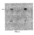
- FIG. 1 shows the result of an autoradiograph demonstrating the increased expression of ⁇ 1 integrin after treatment with angiotensin-II (A-II) compared to a control sample.
- A-II angiotensin-II
- angiotensin-II A-II
- the effect of angiotensin-II (A-II) in inhibiting growth factor-induced stimulated cancer cell invasiveness was studied as a model system and the results are shown in Figure 3.
- the study was carried out using an invasion chamber in which two compartments are separated by a perforated membrane coated in matrix protein.
- a chemoattractant, IGF-I was added to the medium on one side of the chamber, and the breast cancer cells to the other.
- the invasiveness potential of the cancer cells was therefore markedly inhibited by angiotensin-II.
- angiotensin-II did not stimulate cell growth suggesting that it had a role in cell-cell adgesion and cell migration.
Landscapes
- Health & Medical Sciences (AREA)
- Life Sciences & Earth Sciences (AREA)
- Chemical & Material Sciences (AREA)
- Veterinary Medicine (AREA)
- Public Health (AREA)
- Medicinal Chemistry (AREA)
- General Health & Medical Sciences (AREA)
- Animal Behavior & Ethology (AREA)
- Pharmacology & Pharmacy (AREA)
- Immunology (AREA)
- Proteomics, Peptides & Aminoacids (AREA)
- Epidemiology (AREA)
- Bioinformatics & Cheminformatics (AREA)
- Engineering & Computer Science (AREA)
- Vascular Medicine (AREA)
- Gastroenterology & Hepatology (AREA)
- Chemical Kinetics & Catalysis (AREA)
- General Chemical & Material Sciences (AREA)
- Oncology (AREA)
- Organic Chemistry (AREA)
- Nuclear Medicine, Radiotherapy & Molecular Imaging (AREA)
- Medicines That Contain Protein Lipid Enzymes And Other Medicines (AREA)
- Pharmaceuticals Containing Other Organic And Inorganic Compounds (AREA)
- Medicines Containing Material From Animals Or Micro-Organisms (AREA)
- Peptides Or Proteins (AREA)
Abstract
Description

| Supplements added to MEM as basic culture medium | |
| Supplement | Concentration |
| Amphotericin B | 5 U/ml |
| Foetal bovine serum | 5% |
| Gentamicin | 20 µg/l |
| Glucose | 4.5 g/l |
| L-glutamine | 4 mM |
| Non-essential amino acids | 10ml/l |
| Penicillin | 100 U/ml |
| Sodium pyruvate | 2mM |
| Streptomycin | 100 µg/ml |
| Properties of protease inhibitors used in immunoprecipitation | |
| Inhibitor | Activity |
| Aprotinin | Trypsin |
| Chymotrypsin | |
| Plasma | |
| Kallikrein | |
| Leupeptin | Trypsin |
| Plasmin | |
| Papain | |
| Cathepsin B | |
| Pepstatin A | Pepsin |
| Cathepsin D | |
| Phenlymethyl-sulphonylfluoride | Trypsin |
| Chymotrypsin |
Claims (6)
- The use of an angiotensin in the preparation of a medicament for the prevention or treatment of metastasis of cancer cells.
- A use as claimed in claim 1, in which the cancer cell is derived from breast, skin, large bowel, prostate, lung, bone, cervix, stomach, or rectum.
- A use as claimed in claim 1 or claim 2, in which the angiotensin is angiotensin-II.
- A use as claimed in any one of claims 1 to 3, in which the medicament is for oral, rectal, nasal, topical, vaginal or parenteral administration.
- A use as claimed in claim 4, in which the parenteral administration is subcutaneous, intramuscular, intravenous or intradermal.
- The use as claimed in claim 1, in which metastasis of cancer cells is treated or prevented by the induction of expression of β1 integrin molecules in the cancer cells.
Applications Claiming Priority (5)
| Application Number | Priority Date | Filing Date | Title |
|---|---|---|---|
| GBGB9818023.5A GB9818023D0 (en) | 1998-08-18 | 1998-08-18 | Cancer treatment |
| GB9818023 | 1998-08-18 | ||
| GBGB9820000.9A GB9820000D0 (en) | 1998-09-14 | 1998-09-14 | Cancer treatment |
| GB9820000 | 1998-09-14 | ||
| PCT/GB1999/002727 WO2000010590A2 (en) | 1998-08-18 | 1999-08-18 | Cancer treatment |
Publications (2)
| Publication Number | Publication Date |
|---|---|
| EP1104305A2 EP1104305A2 (en) | 2001-06-06 |
| EP1104305B1 true EP1104305B1 (en) | 2005-03-23 |
Family
ID=26314231
Family Applications (1)
| Application Number | Title | Priority Date | Filing Date |
|---|---|---|---|
| EP99940353A Expired - Lifetime EP1104305B1 (en) | 1998-08-18 | 1999-08-18 | Use of angiotensin in the treatment of cancer metastasis |
Country Status (7)
| Country | Link |
|---|---|
| US (1) | US6852316B2 (en) |
| EP (1) | EP1104305B1 (en) |
| AT (1) | ATE291434T1 (en) |
| AU (1) | AU5434899A (en) |
| DE (1) | DE69924385T2 (en) |
| ES (1) | ES2239454T3 (en) |
| WO (1) | WO2000010590A2 (en) |
Families Citing this family (4)
| Publication number | Priority date | Publication date | Assignee | Title |
|---|---|---|---|---|
| EP2043675A2 (en) * | 2006-07-07 | 2009-04-08 | Washington State University Research Foundation | C-met receptor regulation by angiotensin iv (at4) receptor ligands |
| US8236761B2 (en) * | 2006-07-07 | 2012-08-07 | Washington State University Research Foundation | C-Met receptor regulation by angiotensin IV (AT4) receptor ligands |
| US20090042296A1 (en) * | 2007-03-16 | 2009-02-12 | Marie Callahan | Transfection ready eukaryotic cells |
| US8378100B2 (en) * | 2008-01-09 | 2013-02-19 | University Of Virginia Patent Foundation | Phosphonate derivatives as autotaxin inhibitors |
Family Cites Families (1)
| Publication number | Priority date | Publication date | Assignee | Title |
|---|---|---|---|---|
| WO1997034627A2 (en) * | 1996-03-19 | 1997-09-25 | Societe De Conseils De Recherches Et D'applications Scientifiques S.A. | Protection of hemopoietic cells during chemotherapy or radiotherapy |
-
1999
- 1999-08-18 AT AT99940353T patent/ATE291434T1/en not_active IP Right Cessation
- 1999-08-18 DE DE69924385T patent/DE69924385T2/en not_active Expired - Lifetime
- 1999-08-18 AU AU54348/99A patent/AU5434899A/en not_active Abandoned
- 1999-08-18 WO PCT/GB1999/002727 patent/WO2000010590A2/en not_active Ceased
- 1999-08-18 ES ES99940353T patent/ES2239454T3/en not_active Expired - Lifetime
- 1999-08-18 EP EP99940353A patent/EP1104305B1/en not_active Expired - Lifetime
-
2001
- 2001-02-16 US US09/784,005 patent/US6852316B2/en not_active Expired - Fee Related
Also Published As
| Publication number | Publication date |
|---|---|
| AU5434899A (en) | 2000-03-14 |
| US20020119142A1 (en) | 2002-08-29 |
| ES2239454T3 (en) | 2005-09-16 |
| DE69924385D1 (en) | 2005-04-28 |
| US6852316B2 (en) | 2005-02-08 |
| DE69924385T2 (en) | 2006-01-19 |
| WO2000010590A2 (en) | 2000-03-02 |
| WO2000010590A3 (en) | 2000-05-18 |
| ATE291434T1 (en) | 2005-04-15 |
| EP1104305A2 (en) | 2001-06-06 |
Similar Documents
| Publication | Publication Date | Title |
|---|---|---|
| Caillou et al. | Na+/I− symporter distribution in human thyroid tissues: an immunohistochemical study | |
| Jin et al. | Expression of neural cell adhesion molecule in normal and neoplastic human neuroendocrine tissues | |
| Ishiwata et al. | Neuroepithelial stem cell marker nestin regulates the migration, invasion and growth of human gliomas | |
| Sosne et al. | Thymosin ß4 promotes human conjunctival epithelial cell migration | |
| US20120164148A1 (en) | Compositions Containing JARID1B Inhibitors and Methods for Treating Cancer | |
| Beaulieu et al. | Immunolocalization of extracellular matrix components during organogenesis in the human small intestine | |
| Kim et al. | Role of alternative splicing of periostin in human bladder carcinogenesis | |
| Mueller et al. | Transforming growth factor α in arterioles: cell surface processing of its precursor by elastases | |
| Petty et al. | Overexpression of carcinoma and embryonic cytotrophoblast cell-specific Mig-7 induces invasion and vessel-like structure formation | |
| Waleh et al. | Selective down-regulation of integrin receptors in spheroids of squamous cell carcinoma | |
| Tuguzbaeva et al. | PEP06 polypeptide 30 is a novel cluster-dissociating agent inhibiting αv integrin/FAK/Src signaling in oral squamous cell carcinoma cells | |
| Löhr et al. | Expression and function of receptors for extracellular matrix proteins in human ductal adenocarcinomas of the pancreas | |
| Wu et al. | Expression of E-cadherin in immature rat and mouse testis and in rat Sertoli cell cultures | |
| US10034913B2 (en) | Anti-angiogenic peptides and uses thereof | |
| US6852316B2 (en) | Cancer treatment | |
| Juillerat-Jeanneret et al. | Dexamethasone selectively regulates the activity of enzymatic markers of cerebral endothelial cell lines | |
| Motti et al. | Loss of p27 expression through RAS-> BRAF-> MAP kinase-dependent pathway in human thyroid carcinomas | |
| Montagnani Marelli et al. | Gonadotropin-releasing hormone agonists reduce the migratory and the invasive behavior of androgen-independent prostate cancer cells by interfering with the activity of IGF-I | |
| Bastholm et al. | MConfocal Fluorescence Microscopy of Urokinase Plasminogen Activator Receptor and Cathepsin D in Human MDA-MB-231 Breast Cancer Cells Migrating in Reconstituted Basement Membrane | |
| Pechoux et al. | Localization of thrombospondin, CD36 and CD51 during prenatal development of the human mammary gland | |
| Lemaire et al. | Bombesin-like peptides in alveolar macrophage: increased release in pulmonary inflammation and fibrosis | |
| Moretti et al. | Gonadotropin-releasing hormone agonists suppress melanoma cell motility and invasiveness through the inhibition of α3 integrin and MMP-2 expression and activity | |
| CN121002053A (en) | Peptide inhibitors of cathepsin D/LRP-1 interaction | |
| Luther et al. | Expression of α1-adrenergic receptor subtypes in heart cell culture | |
| Haberern-Blood et al. | Laminin expression by human pancreatic carcinoma cells in the nude mouse and in culture |
Legal Events
| Date | Code | Title | Description |
|---|---|---|---|
| PUAI | Public reference made under article 153(3) epc to a published international application that has entered the european phase |
Free format text: ORIGINAL CODE: 0009012 |
|
| 17P | Request for examination filed |
Effective date: 20010316 |
|
| AK | Designated contracting states |
Kind code of ref document: A2 Designated state(s): AT BE CH CY DE DK ES FI FR GB GR IE IT LI LU MC NL PT SE |
|
| AX | Request for extension of the european patent |
Free format text: AL;LT;LV;MK;RO;SI |
|
| 17Q | First examination report despatched |
Effective date: 20030722 |
|
| GRAP | Despatch of communication of intention to grant a patent |
Free format text: ORIGINAL CODE: EPIDOSNIGR1 |
|
| RTI1 | Title (correction) |
Free format text: USE OF ANGIOTENSIN IN THE TREATMENT OF CANCER METASTASIS |
|
| GRAS | Grant fee paid |
Free format text: ORIGINAL CODE: EPIDOSNIGR3 |
|
| GRAA | (expected) grant |
Free format text: ORIGINAL CODE: 0009210 |
|
| AK | Designated contracting states |
Kind code of ref document: B1 Designated state(s): AT BE CH CY DE DK ES FI FR GB GR IE IT LI LU MC NL PT SE |
|
| PG25 | Lapsed in a contracting state [announced via postgrant information from national office to epo] |
Ref country code: NL Free format text: LAPSE BECAUSE OF FAILURE TO SUBMIT A TRANSLATION OF THE DESCRIPTION OR TO PAY THE FEE WITHIN THE PRESCRIBED TIME-LIMIT Effective date: 20050323 Ref country code: LI Free format text: LAPSE BECAUSE OF FAILURE TO SUBMIT A TRANSLATION OF THE DESCRIPTION OR TO PAY THE FEE WITHIN THE PRESCRIBED TIME-LIMIT Effective date: 20050323 Ref country code: FI Free format text: LAPSE BECAUSE OF FAILURE TO SUBMIT A TRANSLATION OF THE DESCRIPTION OR TO PAY THE FEE WITHIN THE PRESCRIBED TIME-LIMIT Effective date: 20050323 Ref country code: CH Free format text: LAPSE BECAUSE OF FAILURE TO SUBMIT A TRANSLATION OF THE DESCRIPTION OR TO PAY THE FEE WITHIN THE PRESCRIBED TIME-LIMIT Effective date: 20050323 Ref country code: BE Free format text: LAPSE BECAUSE OF FAILURE TO SUBMIT A TRANSLATION OF THE DESCRIPTION OR TO PAY THE FEE WITHIN THE PRESCRIBED TIME-LIMIT Effective date: 20050323 Ref country code: AT Free format text: LAPSE BECAUSE OF FAILURE TO SUBMIT A TRANSLATION OF THE DESCRIPTION OR TO PAY THE FEE WITHIN THE PRESCRIBED TIME-LIMIT Effective date: 20050323 |
|
| REG | Reference to a national code |
Ref country code: GB Ref legal event code: FG4D |
|
| REG | Reference to a national code |
Ref country code: CH Ref legal event code: EP |
|
| REG | Reference to a national code |
Ref country code: IE Ref legal event code: FG4D |
|
| REF | Corresponds to: |
Ref document number: 69924385 Country of ref document: DE Date of ref document: 20050428 Kind code of ref document: P |
|
| PG25 | Lapsed in a contracting state [announced via postgrant information from national office to epo] |
Ref country code: GR Free format text: LAPSE BECAUSE OF FAILURE TO SUBMIT A TRANSLATION OF THE DESCRIPTION OR TO PAY THE FEE WITHIN THE PRESCRIBED TIME-LIMIT Effective date: 20050623 Ref country code: DK Free format text: LAPSE BECAUSE OF FAILURE TO SUBMIT A TRANSLATION OF THE DESCRIPTION OR TO PAY THE FEE WITHIN THE PRESCRIBED TIME-LIMIT Effective date: 20050623 |
|
| PG25 | Lapsed in a contracting state [announced via postgrant information from national office to epo] |
Ref country code: LU Free format text: LAPSE BECAUSE OF NON-PAYMENT OF DUE FEES Effective date: 20050818 Ref country code: IE Free format text: LAPSE BECAUSE OF NON-PAYMENT OF DUE FEES Effective date: 20050818 Ref country code: CY Free format text: LAPSE BECAUSE OF FAILURE TO SUBMIT A TRANSLATION OF THE DESCRIPTION OR TO PAY THE FEE WITHIN THE PRESCRIBED TIME-LIMIT Effective date: 20050818 |
|
| PG25 | Lapsed in a contracting state [announced via postgrant information from national office to epo] |
Ref country code: MC Free format text: LAPSE BECAUSE OF NON-PAYMENT OF DUE FEES Effective date: 20050831 |
|
| NLV1 | Nl: lapsed or annulled due to failure to fulfill the requirements of art. 29p and 29m of the patents act | ||
| PG25 | Lapsed in a contracting state [announced via postgrant information from national office to epo] |
Ref country code: PT Free format text: LAPSE BECAUSE OF FAILURE TO SUBMIT A TRANSLATION OF THE DESCRIPTION OR TO PAY THE FEE WITHIN THE PRESCRIBED TIME-LIMIT Effective date: 20050907 |
|
| REG | Reference to a national code |
Ref country code: ES Ref legal event code: FG2A Ref document number: 2239454 Country of ref document: ES Kind code of ref document: T3 |
|
| REG | Reference to a national code |
Ref country code: CH Ref legal event code: PL |
|
| PLBE | No opposition filed within time limit |
Free format text: ORIGINAL CODE: 0009261 |
|
| STAA | Information on the status of an ep patent application or granted ep patent |
Free format text: STATUS: NO OPPOSITION FILED WITHIN TIME LIMIT |
|
| ET | Fr: translation filed | ||
| 26N | No opposition filed |
Effective date: 20051227 |
|
| REG | Reference to a national code |
Ref country code: IE Ref legal event code: MM4A |
|
| PG25 | Lapsed in a contracting state [announced via postgrant information from national office to epo] |
Ref country code: SE Free format text: LAPSE BECAUSE OF FAILURE TO SUBMIT A TRANSLATION OF THE DESCRIPTION OR TO PAY THE FEE WITHIN THE PRESCRIBED TIME-LIMIT Effective date: 20050623 |
|
| PGFP | Annual fee paid to national office [announced via postgrant information from national office to epo] |
Ref country code: ES Payment date: 20100830 Year of fee payment: 12 |
|
| PGFP | Annual fee paid to national office [announced via postgrant information from national office to epo] |
Ref country code: IT Payment date: 20100826 Year of fee payment: 12 Ref country code: FR Payment date: 20100901 Year of fee payment: 12 |
|
| PGFP | Annual fee paid to national office [announced via postgrant information from national office to epo] |
Ref country code: GB Payment date: 20100831 Year of fee payment: 12 |
|
| PGFP | Annual fee paid to national office [announced via postgrant information from national office to epo] |
Ref country code: DE Payment date: 20100922 Year of fee payment: 12 |
|
| GBPC | Gb: european patent ceased through non-payment of renewal fee |
Effective date: 20110818 |
|
| REG | Reference to a national code |
Ref country code: FR Ref legal event code: ST Effective date: 20120430 |
|
| PG25 | Lapsed in a contracting state [announced via postgrant information from national office to epo] |
Ref country code: IT Free format text: LAPSE BECAUSE OF NON-PAYMENT OF DUE FEES Effective date: 20110818 |
|
| REG | Reference to a national code |
Ref country code: DE Ref legal event code: R119 Ref document number: 69924385 Country of ref document: DE Effective date: 20120301 |
|
| PG25 | Lapsed in a contracting state [announced via postgrant information from national office to epo] |
Ref country code: FR Free format text: LAPSE BECAUSE OF NON-PAYMENT OF DUE FEES Effective date: 20110831 Ref country code: GB Free format text: LAPSE BECAUSE OF NON-PAYMENT OF DUE FEES Effective date: 20110818 |
|
| PG25 | Lapsed in a contracting state [announced via postgrant information from national office to epo] |
Ref country code: DE Free format text: LAPSE BECAUSE OF NON-PAYMENT OF DUE FEES Effective date: 20120301 |
|
| REG | Reference to a national code |
Ref country code: ES Ref legal event code: FD2A Effective date: 20131029 |
|
| PG25 | Lapsed in a contracting state [announced via postgrant information from national office to epo] |
Ref country code: ES Free format text: LAPSE BECAUSE OF NON-PAYMENT OF DUE FEES Effective date: 20110819 |