EP1095280B1 - System for the quantification of platelet activation - Google Patents
System for the quantification of platelet activation Download PDFInfo
- Publication number
- EP1095280B1 EP1095280B1 EP99933539A EP99933539A EP1095280B1 EP 1095280 B1 EP1095280 B1 EP 1095280B1 EP 99933539 A EP99933539 A EP 99933539A EP 99933539 A EP99933539 A EP 99933539A EP 1095280 B1 EP1095280 B1 EP 1095280B1
- Authority
- EP
- European Patent Office
- Prior art keywords
- platelets
- fluorochrome
- activation
- labeled
- ligand
- Prior art date
- Legal status (The legal status is an assumption and is not a legal conclusion. Google has not performed a legal analysis and makes no representation as to the accuracy of the status listed.)
- Expired - Lifetime
Links
- 230000010118 platelet activation Effects 0.000 title claims abstract description 51
- 238000011002 quantification Methods 0.000 title abstract 2
- 238000000034 method Methods 0.000 claims abstract description 80
- 210000004369 blood Anatomy 0.000 claims abstract description 65
- 239000008280 blood Substances 0.000 claims abstract description 65
- 239000000556 agonist Substances 0.000 claims abstract description 32
- BFMYDTVEBKDAKJ-UHFFFAOYSA-L disodium;(2',7'-dibromo-3',6'-dioxido-3-oxospiro[2-benzofuran-1,9'-xanthene]-4'-yl)mercury;hydrate Chemical compound O.[Na+].[Na+].O1C(=O)C2=CC=CC=C2C21C1=CC(Br)=C([O-])C([Hg])=C1OC1=C2C=C(Br)C([O-])=C1 BFMYDTVEBKDAKJ-UHFFFAOYSA-L 0.000 claims description 56
- 239000003446 ligand Substances 0.000 claims description 55
- 230000004913 activation Effects 0.000 claims description 53
- YBJHBAHKTGYVGT-ZKWXMUAHSA-N (+)-Biotin Chemical compound N1C(=O)N[C@@H]2[C@H](CCCCC(=O)O)SC[C@@H]21 YBJHBAHKTGYVGT-ZKWXMUAHSA-N 0.000 claims description 42
- XTWYTFMLZFPYCI-KQYNXXCUSA-N 5'-adenylphosphoric acid Chemical group C1=NC=2C(N)=NC=NC=2N1[C@@H]1O[C@H](COP(O)(=O)OP(O)(O)=O)[C@@H](O)[C@H]1O XTWYTFMLZFPYCI-KQYNXXCUSA-N 0.000 claims description 41
- 108010049003 Fibrinogen Proteins 0.000 claims description 33
- 102000008946 Fibrinogen Human genes 0.000 claims description 33
- 229940012952 fibrinogen Drugs 0.000 claims description 33
- WSFSSNUMVMOOMR-UHFFFAOYSA-N Formaldehyde Chemical group O=C WSFSSNUMVMOOMR-UHFFFAOYSA-N 0.000 claims description 30
- 238000005497 microtitration Methods 0.000 claims description 26
- 108010035766 P-Selectin Proteins 0.000 claims description 21
- 229960002685 biotin Drugs 0.000 claims description 21
- 235000020958 biotin Nutrition 0.000 claims description 21
- 239000011616 biotin Substances 0.000 claims description 21
- 239000003795 chemical substances by application Substances 0.000 claims description 11
- 230000009257 reactivity Effects 0.000 claims description 11
- 108010090804 Streptavidin Proteins 0.000 claims description 9
- 230000001419 dependent effect Effects 0.000 claims description 9
- 239000000834 fixative Substances 0.000 claims description 9
- 102000004169 proteins and genes Human genes 0.000 claims description 7
- 108090000623 proteins and genes Proteins 0.000 claims description 7
- OXHYRVSBKWIFES-WWSDOYNLSA-N trap-14 peptide Chemical group C([C@@H](C(=O)N[C@@H](CC(C)C)C(=O)N[C@@H](CC(C)C)C(=O)N[C@@H](CCCNC(N)=N)C(=O)N[C@@H](CC(N)=O)C(=O)N1[C@@H](CCC1)C(=O)N[C@@H](CC(N)=O)C(=O)N[C@@H](CC(O)=O)C(=O)N[C@@H](CCCCN)C(=O)N[C@@H](CC=1C=CC(O)=CC=1)C(=O)N[C@@H](CCC(O)=O)C(=O)N1[C@@H](CCC1)C(=O)N[C@@H](CC=1C=CC=CC=1)C(O)=O)NC(=O)[C@@H](N)CO)C1=CC=CC=C1 OXHYRVSBKWIFES-WWSDOYNLSA-N 0.000 claims description 6
- 238000005406 washing Methods 0.000 claims description 5
- 108090001008 Avidin Proteins 0.000 claims description 4
- 238000005119 centrifugation Methods 0.000 claims description 4
- 238000002360 preparation method Methods 0.000 claims description 3
- 239000007787 solid Substances 0.000 claims 21
- 102000008212 P-Selectin Human genes 0.000 claims 4
- 230000003213 activating effect Effects 0.000 claims 3
- 239000003146 anticoagulant agent Substances 0.000 abstract description 10
- 230000000702 anti-platelet effect Effects 0.000 abstract description 4
- 230000015572 biosynthetic process Effects 0.000 abstract description 4
- 238000012544 monitoring process Methods 0.000 abstract description 2
- 239000002831 pharmacologic agent Substances 0.000 abstract description 2
- 102000003886 Glycoproteins Human genes 0.000 description 26
- 108090000288 Glycoproteins Proteins 0.000 description 26
- 102100023472 P-selectin Human genes 0.000 description 19
- 238000003556 assay Methods 0.000 description 16
- JKMHFZQWWAIEOD-UHFFFAOYSA-N 2-[4-(2-hydroxyethyl)piperazin-1-yl]ethanesulfonic acid Chemical compound OCC[NH+]1CCN(CCS([O-])(=O)=O)CC1 JKMHFZQWWAIEOD-UHFFFAOYSA-N 0.000 description 15
- BSYNRYMUTXBXSQ-UHFFFAOYSA-N Aspirin Chemical compound CC(=O)OC1=CC=CC=C1C(O)=O BSYNRYMUTXBXSQ-UHFFFAOYSA-N 0.000 description 14
- 229960001138 acetylsalicylic acid Drugs 0.000 description 14
- MHMNJMPURVTYEJ-UHFFFAOYSA-N fluorescein-5-isothiocyanate Chemical compound O1C(=O)C2=CC(N=C=S)=CC=C2C21C1=CC=C(O)C=C1OC1=CC(O)=CC=C21 MHMNJMPURVTYEJ-UHFFFAOYSA-N 0.000 description 14
- PHWBOXQYWZNQIN-UHFFFAOYSA-N ticlopidine Chemical compound ClC1=CC=CC=C1CN1CC(C=CS2)=C2CC1 PHWBOXQYWZNQIN-UHFFFAOYSA-N 0.000 description 14
- 230000004044 response Effects 0.000 description 12
- 229940028869 ticlid Drugs 0.000 description 12
- 102000012750 Membrane Glycoproteins Human genes 0.000 description 11
- 108010090054 Membrane Glycoproteins Proteins 0.000 description 11
- 230000037406 food intake Effects 0.000 description 11
- 239000000243 solution Substances 0.000 description 11
- TWRXJAOTZQYOKJ-UHFFFAOYSA-L Magnesium chloride Chemical compound [Mg+2].[Cl-].[Cl-] TWRXJAOTZQYOKJ-UHFFFAOYSA-L 0.000 description 10
- UIIMBOGNXHQVGW-UHFFFAOYSA-M Sodium bicarbonate Chemical compound [Na+].OC([O-])=O UIIMBOGNXHQVGW-UHFFFAOYSA-M 0.000 description 10
- FAPWRFPIFSIZLT-UHFFFAOYSA-M Sodium chloride Chemical compound [Na+].[Cl-] FAPWRFPIFSIZLT-UHFFFAOYSA-M 0.000 description 10
- 239000000872 buffer Substances 0.000 description 10
- 239000000306 component Substances 0.000 description 10
- 239000007790 solid phase Substances 0.000 description 10
- 108010004729 Phycoerythrin Proteins 0.000 description 9
- 208000032843 Hemorrhage Diseases 0.000 description 8
- 208000034158 bleeding Diseases 0.000 description 8
- 230000000740 bleeding effect Effects 0.000 description 8
- 230000015271 coagulation Effects 0.000 description 8
- 238000005345 coagulation Methods 0.000 description 8
- 239000007850 fluorescent dye Substances 0.000 description 8
- 230000003993 interaction Effects 0.000 description 8
- 229940107685 reopro Drugs 0.000 description 8
- 108010059382 Zea mays trypsin inhibitor Proteins 0.000 description 6
- 229940127219 anticoagulant drug Drugs 0.000 description 6
- 229940127218 antiplatelet drug Drugs 0.000 description 6
- 102000015081 Blood Coagulation Factors Human genes 0.000 description 5
- 108010039209 Blood Coagulation Factors Proteins 0.000 description 5
- OYPRJOBELJOOCE-UHFFFAOYSA-N Calcium Chemical compound [Ca] OYPRJOBELJOOCE-UHFFFAOYSA-N 0.000 description 5
- UXVMQQNJUSDDNG-UHFFFAOYSA-L Calcium chloride Chemical compound [Cl-].[Cl-].[Ca+2] UXVMQQNJUSDDNG-UHFFFAOYSA-L 0.000 description 5
- WQZGKKKJIJFFOK-GASJEMHNSA-N Glucose Natural products OC[C@H]1OC(O)[C@H](O)[C@@H](O)[C@@H]1O WQZGKKKJIJFFOK-GASJEMHNSA-N 0.000 description 5
- 239000007995 HEPES buffer Substances 0.000 description 5
- 208000007536 Thrombosis Diseases 0.000 description 5
- 238000013176 antiplatelet therapy Methods 0.000 description 5
- 239000003114 blood coagulation factor Substances 0.000 description 5
- 229910052791 calcium Inorganic materials 0.000 description 5
- 239000011575 calcium Substances 0.000 description 5
- 239000001110 calcium chloride Substances 0.000 description 5
- 229910001628 calcium chloride Inorganic materials 0.000 description 5
- 239000008121 dextrose Substances 0.000 description 5
- 231100000673 dose–response relationship Toxicity 0.000 description 5
- 210000003743 erythrocyte Anatomy 0.000 description 5
- 230000012953 feeding on blood of other organism Effects 0.000 description 5
- 239000008187 granular material Substances 0.000 description 5
- 238000011534 incubation Methods 0.000 description 5
- 229910001629 magnesium chloride Inorganic materials 0.000 description 5
- 239000000106 platelet aggregation inhibitor Substances 0.000 description 5
- 239000011541 reaction mixture Substances 0.000 description 5
- 229910000030 sodium bicarbonate Inorganic materials 0.000 description 5
- 239000011780 sodium chloride Substances 0.000 description 5
- AJPJDKMHJJGVTQ-UHFFFAOYSA-M sodium dihydrogen phosphate Chemical compound [Na+].OP(O)([O-])=O AJPJDKMHJJGVTQ-UHFFFAOYSA-M 0.000 description 5
- 229910000162 sodium phosphate Inorganic materials 0.000 description 5
- 238000002560 therapeutic procedure Methods 0.000 description 5
- 108010071241 Factor XIIa Proteins 0.000 description 4
- 208000024248 Vascular System injury Diseases 0.000 description 4
- 208000012339 Vascular injury Diseases 0.000 description 4
- 238000000684 flow cytometry Methods 0.000 description 4
- 230000023597 hemostasis Effects 0.000 description 4
- 230000005764 inhibitory process Effects 0.000 description 4
- 239000003550 marker Substances 0.000 description 4
- 208000006011 Stroke Diseases 0.000 description 3
- 230000002439 hemostatic effect Effects 0.000 description 3
- 238000005259 measurement Methods 0.000 description 3
- 239000012528 membrane Substances 0.000 description 3
- 208000010125 myocardial infarction Diseases 0.000 description 3
- 239000005528 B01AC05 - Ticlopidine Substances 0.000 description 2
- KRKNYBCHXYNGOX-UHFFFAOYSA-K Citrate Chemical compound [O-]C(=O)CC(O)(CC([O-])=O)C([O-])=O KRKNYBCHXYNGOX-UHFFFAOYSA-K 0.000 description 2
- 108010080865 Factor XII Proteins 0.000 description 2
- 102000000429 Factor XII Human genes 0.000 description 2
- 101000622137 Homo sapiens P-selectin Proteins 0.000 description 2
- 108090000190 Thrombin Proteins 0.000 description 2
- 239000012190 activator Substances 0.000 description 2
- 230000002776 aggregation Effects 0.000 description 2
- 238000004220 aggregation Methods 0.000 description 2
- 238000004458 analytical method Methods 0.000 description 2
- 230000008859 change Effects 0.000 description 2
- 239000002738 chelating agent Substances 0.000 description 2
- -1 citrate Chemical compound 0.000 description 2
- 230000003247 decreasing effect Effects 0.000 description 2
- 229940079593 drug Drugs 0.000 description 2
- 239000003814 drug Substances 0.000 description 2
- 230000000694 effects Effects 0.000 description 2
- 238000001727 in vivo Methods 0.000 description 2
- 230000037361 pathway Effects 0.000 description 2
- 210000004623 platelet-rich plasma Anatomy 0.000 description 2
- 230000031915 positive regulation of coagulation Effects 0.000 description 2
- 230000008569 process Effects 0.000 description 2
- QZAYGJVTTNCVMB-UHFFFAOYSA-N serotonin Chemical compound C1=C(O)C=C2C(CCN)=CNC2=C1 QZAYGJVTTNCVMB-UHFFFAOYSA-N 0.000 description 2
- 229960004072 thrombin Drugs 0.000 description 2
- 230000001732 thrombotic effect Effects 0.000 description 2
- 229960005001 ticlopidine Drugs 0.000 description 2
- 206010003210 Arteriosclerosis Diseases 0.000 description 1
- 208000037260 Atherosclerotic Plaque Diseases 0.000 description 1
- 206010049993 Cardiac death Diseases 0.000 description 1
- 208000020446 Cardiac disease Diseases 0.000 description 1
- 206010010356 Congenital anomaly Diseases 0.000 description 1
- 206010011906 Death Diseases 0.000 description 1
- 108010014172 Factor V Proteins 0.000 description 1
- 108010054265 Factor VIIa Proteins 0.000 description 1
- 102000009123 Fibrin Human genes 0.000 description 1
- 108010073385 Fibrin Proteins 0.000 description 1
- BWGVNKXGVNDBDI-UHFFFAOYSA-N Fibrin monomer Chemical compound CNC(=O)CNC(=O)CN BWGVNKXGVNDBDI-UHFFFAOYSA-N 0.000 description 1
- 102000018697 Membrane Proteins Human genes 0.000 description 1
- 108010052285 Membrane Proteins Proteins 0.000 description 1
- 208000037273 Pathologic Processes Diseases 0.000 description 1
- 208000001647 Renal Insufficiency Diseases 0.000 description 1
- 108010000499 Thromboplastin Proteins 0.000 description 1
- 102000002262 Thromboplastin Human genes 0.000 description 1
- 208000011775 arteriosclerosis disease Diseases 0.000 description 1
- 239000012503 blood component Substances 0.000 description 1
- 210000004204 blood vessel Anatomy 0.000 description 1
- 208000026106 cerebrovascular disease Diseases 0.000 description 1
- 239000013522 chelant Substances 0.000 description 1
- 238000004132 cross linking Methods 0.000 description 1
- 238000011161 development Methods 0.000 description 1
- 230000028023 exocytosis Effects 0.000 description 1
- 238000002474 experimental method Methods 0.000 description 1
- 229940012414 factor viia Drugs 0.000 description 1
- 229950003499 fibrin Drugs 0.000 description 1
- 230000004927 fusion Effects 0.000 description 1
- 230000036541 health Effects 0.000 description 1
- 208000019622 heart disease Diseases 0.000 description 1
- 238000003018 immunoassay Methods 0.000 description 1
- 230000001771 impaired effect Effects 0.000 description 1
- 238000000338 in vitro Methods 0.000 description 1
- 239000003112 inhibitor Substances 0.000 description 1
- 230000002401 inhibitory effect Effects 0.000 description 1
- 230000000977 initiatory effect Effects 0.000 description 1
- 102000006495 integrins Human genes 0.000 description 1
- 108010044426 integrins Proteins 0.000 description 1
- 201000006370 kidney failure Diseases 0.000 description 1
- 210000003712 lysosome Anatomy 0.000 description 1
- 230000001868 lysosomic effect Effects 0.000 description 1
- 238000002483 medication Methods 0.000 description 1
- 230000010807 negative regulation of binding Effects 0.000 description 1
- 239000002773 nucleotide Substances 0.000 description 1
- 125000003729 nucleotide group Chemical group 0.000 description 1
- 230000009054 pathological process Effects 0.000 description 1
- 150000003904 phospholipids Chemical class 0.000 description 1
- 230000002947 procoagulating effect Effects 0.000 description 1
- 229940076279 serotonin Drugs 0.000 description 1
- 238000012360 testing method Methods 0.000 description 1
- RZWIIPASKMUIAC-VQTJNVASSA-N thromboxane Chemical compound CCCCCCCC[C@H]1OCCC[C@@H]1CCCCCCC RZWIIPASKMUIAC-VQTJNVASSA-N 0.000 description 1
Images



Classifications
-
- G—PHYSICS
- G01—MEASURING; TESTING
- G01N—INVESTIGATING OR ANALYSING MATERIALS BY DETERMINING THEIR CHEMICAL OR PHYSICAL PROPERTIES
- G01N33/00—Investigating or analysing materials by specific methods not covered by groups G01N1/00 - G01N31/00
- G01N33/48—Biological material, e.g. blood, urine; Haemocytometers
- G01N33/50—Chemical analysis of biological material, e.g. blood, urine; Testing involving biospecific ligand binding methods; Immunological testing
- G01N33/80—Chemical analysis of biological material, e.g. blood, urine; Testing involving biospecific ligand binding methods; Immunological testing involving blood groups or blood types or red blood cells
-
- G—PHYSICS
- G01—MEASURING; TESTING
- G01N—INVESTIGATING OR ANALYSING MATERIALS BY DETERMINING THEIR CHEMICAL OR PHYSICAL PROPERTIES
- G01N33/00—Investigating or analysing materials by specific methods not covered by groups G01N1/00 - G01N31/00
- G01N33/48—Biological material, e.g. blood, urine; Haemocytometers
- G01N33/50—Chemical analysis of biological material, e.g. blood, urine; Testing involving biospecific ligand binding methods; Immunological testing
- G01N33/86—Chemical analysis of biological material, e.g. blood, urine; Testing involving biospecific ligand binding methods; Immunological testing involving blood coagulating time or factors, or their receptors
-
- G—PHYSICS
- G01—MEASURING; TESTING
- G01N—INVESTIGATING OR ANALYSING MATERIALS BY DETERMINING THEIR CHEMICAL OR PHYSICAL PROPERTIES
- G01N2333/00—Assays involving biological materials from specific organisms or of a specific nature
- G01N2333/435—Assays involving biological materials from specific organisms or of a specific nature from animals; from humans
- G01N2333/705—Assays involving receptors, cell surface antigens or cell surface determinants
- G01N2333/7056—Selectin superfamily, e.g. LAM-1, GlyCAM, ELAM-1, PADGEM
-
- G—PHYSICS
- G01—MEASURING; TESTING
- G01N—INVESTIGATING OR ANALYSING MATERIALS BY DETERMINING THEIR CHEMICAL OR PHYSICAL PROPERTIES
- G01N2333/00—Assays involving biological materials from specific organisms or of a specific nature
- G01N2333/435—Assays involving biological materials from specific organisms or of a specific nature from animals; from humans
- G01N2333/745—Assays involving non-enzymic blood coagulation factors
- G01N2333/75—Fibrin; Fibrinogen
-
- Y—GENERAL TAGGING OF NEW TECHNOLOGICAL DEVELOPMENTS; GENERAL TAGGING OF CROSS-SECTIONAL TECHNOLOGIES SPANNING OVER SEVERAL SECTIONS OF THE IPC; TECHNICAL SUBJECTS COVERED BY FORMER USPC CROSS-REFERENCE ART COLLECTIONS [XRACs] AND DIGESTS
- Y10—TECHNICAL SUBJECTS COVERED BY FORMER USPC
- Y10S—TECHNICAL SUBJECTS COVERED BY FORMER USPC CROSS-REFERENCE ART COLLECTIONS [XRACs] AND DIGESTS
- Y10S435/00—Chemistry: molecular biology and microbiology
- Y10S435/971—Capture of complex after antigen-antibody reaction
Definitions
- the present invention relates to a method for quantifying and assessing platelet activation in whole blood samples and monitoring the dosage of antiplatelet pharmacologic agents according to claim 1, 21 and 37.
- the method comprises exposing platelets to a physiological concentration of an agonist that activates some of the platelets, resulting in the formation of at least one binding site on the surface of the activated platelets, and measuring the activated platelets.
- the present invention permits the assessment of specific components of platelet activation.
- Platelet activation is a critical component of hemostasis, the process which prevents bleeding following vascular injury. During hemostasis, platelets adhere to the site of the vascular injury. The platelets undergo activation, which leads to a change in their shape, facilitating the formation of a hemostatic plug. Platelet activation also involves a process known as degranulation, which provides the following components: proteins (e.g., Factor V and fibrinogen) and calcium necessary for the coagulation cascade; agonists (e.g., serotonin, thromboxane, and ADP) which activate and recruit additional platelets; and a negatively charged phospholipid surface on the platelet which promotes assembly and the activation of coagulation factors.
- proteins e.g., Factor V and fibrinogen
- agonists e.g., serotonin, thromboxane, and ADP
- a negatively charged phospholipid surface on the platelet which promotes assembly and the activation of coagulation factors.
- Platelet activation is also critical in thrombosis, a pathologic process in which the activation of platelets and of the coagulation cascade leads to an occlusion of the blood vessel in response to atherosclerotic plaque rupture.
- Thrombosis complicating plaque rupture is responsible for the majority of myocardial infarctions (heart attacks) and cerebrovascular accidents (strokes).
- Therapy with antiplatelet agents such as aspirin, ticlopidine, and ReoPro has been shown to reduce the incidence of myocardial infarctions and cardiac death in patients having a known cardiac disease.
- Therapy with aspirin and ticlopidine has also been shown to reduce the incidence of stroke in patients having a cerebrovascular disease.
- platelet aggregometry assesses the aggregation of platelets and reflects the activation of the surface glycoprotein IIb-IIIa and the degranulation of platelets. This method is based on an in vitro phenomenon which has little to do with platelet function in vivo .
- U.S. Patent No. 5,529,902 discloses a fluorescence immunoassay for platelet function based on the determination of P-selectin expression.
- This assay suffers from the following drawbacks: (1) as in the conventional assays of platelet function, the method is performed using a platelet-rich plasma, and cannot be performed directly on whole blood samples; (2) the method is complicated by the fact that the number of platelets in each sample must be quantitated independently prior to performing the assay; (3) the assay requires that each sample be split into two portions, one of which serves as a control; and (4) the assay permits the determination of only one marker of platelet activation.
- the present invention permits the assessment of specific components of platelet activation.
- this assay facilitates a determination of increased platelet reactivity and thus the risk associated with increased reactivity (thrombotic occlusion of vessels) and decreased reactivity (excess bleeding).
- This assay also facilitates a determination of the adequacy of inhibition of reactivity secondary to treatment with antiplatelet regimens with regard to specific components of platelet activation. Accordingly, because each individual subject has a unique response to antiplatelet therapy, this assay allows therapy to be tailored to the needs of each individual patient and facilitate appropriate dosage adjustment over time in each.
- the specific components of platelet activation include the following: activation of the surface glycoprotein IIb-IIIa which reveals a binding site for fibrinogen and thus allows for the crosslinking of platelets by fibrinogen [see, e.g., S.J. Shattil et al., Blood, 70,307(1987 ), and C.S. Abrams et al., Blood, 75, 128 (1990 )]; the release of the contents of the alpha granules, in which degranulation leads to the release of stored proteins and to the expression of the integrin P-selectin on the surface of the platelet [see, e.g., J.N. George et al., J. Clin.
- the present invention provides a method of determining the extent of platelet activation in response to physiologic concentrations of an agonist.
- the platelets After exposure to such an agonist, the platelets are exposed to solutions containing the following: fluorochrome-labeled ligands which bind to all platelets, both activated and quiescent; fluorochrome-labeled ligands which bind only to activated platelets; and biotin conjugated ligands which facilitate the binding of platelets to a solid phase.
- a fixative e.g., formaldehyde
- the platelets are bound to a solid phase by exposure to a streptavidin or avidin coated surface.
- the interaction of platelets with the coated surface is preferably facilitated by centrifugation. Unbound fluorochrome-labeled ligands are preferably removed by washing.
- the specific components of platelet activation are preferably determined by measuring the fluorescent signal intensities emitted by each of the bound activation-dependent fluorochrome-labeled ligands, and emitted by the fluorochrome-labeled marker of all platelets.
- the present invention provides a method for the assessment of specific components of platelet activation in blood samples.
- whole blood samples are used because platelet reactivity may be altered by the methods used to obtain purified platelet preparations or by the removal of selected blood components.
- the artificial activation of coagulation which occurs after blood is exposed to foreign surfaces must be prevented because this artificial activation alters platelet reactivity. Exposure of blood to a foreign surface activates a protein, Factor XII, which in turn initiates the coagulation cascade leading to the generation of thrombin; thrombin activates platelets and forms fibrin. On the other hand, the initiation of the coagulation cascade in vivo after vascular injury does not involve the activation of Factor XII. Vascular injury causes blood to be exposed to tissue factor, which complexes with Factor VIIa and initiates the coagulation cascade. Accordingly, in the preferred embodiment of the invention, artificial activation of the coagulation cascade is prevented by inhibiting Factor XIIa, while the remainder of the coagulation cascade is left intact due to its interplay with the activation of platelets.
- the assessment of platelet activation is carried out using whole blood samples preferably treated only with corn trypsin inhibitor, a specific inhibitor of Factor XIIa with no effect on other coagulation factors.
- Other anticoagulants such as chelate calcium, would be avoided as they have been shown to cause artificial enhancement of ADP-induced platelet activation.
- the extent of platelet activation is determined in response to physiologic concentrations of an agonist, such as, but not limited to, ADP (adenosine 5'-diphosphate) or TRAP (thrombin receptor agonist peptide).
- an agonist such as, but not limited to, ADP (adenosine 5'-diphosphate) or TRAP (thrombin receptor agonist peptide).
- ADP adenosine 5'-diphosphate
- TRAP thrombin receptor agonist peptide
- Interaction between platelets and the solid phase may be enhanced by centrifuging the samples after adding the samples to a vessel such as microtitration plates.
- unbound fluorochrome-labeled ligands are removed by washing, and the specific components of platelet activation are determined by measuring the fluorescent signal intensities emitted by each of the bound activation-dependent fluorochrome-labeled ligands, and emitted by the fluorochrome-labeled marker of all platelets.
- P-selectin is an integral membrane protein in the alpha granules of platelets. During platelet activation, alpha granule exocytosis results in fusion of the granule membrane with the platelet membrane, bringing P-selectin to the platelet membrane surface.
- the expression of P-selectin can be measured by capturing platelets with an immobilized antibody in a microtitration well, and then using two fluorescent markers: CD42*PerCP (an IgG antibody to glycoprotein IX, with PerCP as the fluorescent label) which marks all platelets, and CD62*PE (an IgG antibody to P-selectin, with phycoerythrin as the fluorescent label) which marks platelets expressing P-selectin and thus marks those platelets which are activated.
- CD42*PerCP an IgG antibody to glycoprotein IX, with PerCP as the fluorescent label
- CD62*PE an IgG antibody to P-selectin, with phycoerythrin as the fluorescent label
- Phlebotomy was performed by using a two syringe technique whereby the first 3 ml of blood were discarded.
- Activation of platelets was delineated in blood drawn directly into a syringe containing 32 ⁇ g/ml of corn trypsin inhibitor (CTI, 9:1 v/v), an agent which inhibits Factor XIIa without affecting other coagulation factors.
- CTI corn trypsin inhibitor
- the CTI was used as an anticoagulant to prevent activation of the contact factor pathway and permit analysis of activation of platelets dependent on addition of exogenous activators.
- other anticoagulants particularly chelators of calcium such as citrate, alter the activation of platelets.
- CD 42a binds to an activation independent epitope on glycoprotein IX and thus was used to tag and hence identify all platelets.
- the biotin conjugated CD 41 binds to an activation independent epitope on glycoprotein IIb and thus serves as the ligand to capture platelets.
- P-selectin is expressed on the surface of only activated platelets and thus serves to mark platelets which have degranulated.
- the plate was centrifuged (1000 X g for 15 minutes) to optimize the interaction between the platelets and the solid phase. The wells were then washed three times with Hepes Tyrodes buffer. The intensity of fluorescence for each ligand was determined with a microplate fluorescent plate reader.
- Platelet activation is also reflected in the activation of the surface glycoprotein IIb-IIIa which reveals a binding site for fibrinogen.
- the extent of fibrinogen binding can be measured by capturing platelets with an immobilized antibody in a microtitration well, and then using two fluorescent markers: CD42*PerCP (an IgG antibody to glycoprotein IX, with PerCP as the fluorescent label) which marks all platelets; and fibrinogen*FITC (fibrinogen conjugated with fluorescein isothiocyanate as the fluorescent label) which marks platelets in which activation of surface glycoprotein IIb-IIIa has occurred.
- CD42*PerCP an IgG antibody to glycoprotein IX, with PerCP as the fluorescent label
- fibrinogen*FITC fibrinogen conjugated with fluorescein isothiocyanate as the fluorescent label
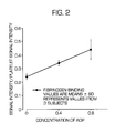
- the efficacy of antiplatelet therapy can be determined by analyzing the slope of individual response curves or by the activation in response to a selected concentration of the agonist, as shown in FIG. 2.
- Phlebotomy was performed by using a two syringe technique whereby the first 3 ml of blood were discarded.
- Activation of platelets was delineated in blood drawn directly into a syringe containing 32 ⁇ g/ml of corn trypsin inhibitor (CTI, 9:1 v/v), an agent which inhibits Factor XIIa without affecting other coagulation factors.
- CTI corn trypsin inhibitor
- the CTI was used as an anticoagulant to prevent the activation of the contact factor pathway and permit an analysis of platelet activation dependent on the addition of exogenous activators.
- CTI was used rather than other anticoagulants because unlike CTI, other anticoagulants, particularly chelators of calcium such as citrate, alter the activation of platelets.
- CD 42a binds to an activation independent epitope on glycoprotein IX and thus was used to tag and hence identify all platelets.
- the biotin conjugated CD 41 binds to an activation independent epitope on glycoprotein IIb and thus serves as the ligand to capture platelets.
- Fibrinogen binds to the surface of platelets only when the surface glycoprotein IIb-IIIa is activated and thus serves to identify activated platelets. After adding blood, the reaction solution was mixed gently and incubated at room temperature for 15 minutes. The platelets were fixed, and the red blood cells were lysed by adding Optilyse-C (1.5% formaldehyde) after the 15 minute incubation.
- the reaction mixture was then added to a streptavidin-coated well in a microtitration plate.
- the plate was centrifuged (1000 X g for 15 minutes) to optimize the interaction between the platelets and the solid phase.
- the wells were then washed three times with Hepes Tyrodes buffer. The intensity of fluorescence for each ligand was determined with a microplate fluorescent plate reader.
- Platelet activation is inhibited by the drug ReoPro, and is reflected by the inhibition of binding of fibrinogen to the activated conformer of the surface glycoprotein IIb-IIIa.
- the extent of fibrinogen binding can be measured by capturing platelets with an immobilized antibody in a microtitration well, and then using two fluorescent markers: CD42*PerCP (an IgG antibody to glycoprotein IX, with PerCP as the fluorescent label) which marks all platelets; and fibrinogen*FITC (fibrinogen conjugated with fluorescein isothiocyanate as the fluorescent label) which marks platelets in which an activation of surface glycoprotein IIb-IIIa has occurred.
- CD42*PerCP an IgG antibody to glycoprotein IX, with PerCP as the fluorescent label
- fibrinogen*FITC fibrinogen conjugated with fluorescein isothiocyanate as the fluorescent label
- ADP concentration of ADP
- concentration of ReoPro used is that known to inhibit the aggregation of platelets as determined with the use of aggregometry.
- pretreatment of blood with ReoPro inhibits binding of fibrinogen*FITC to platelets.
- Phlebotomy was performed by using a two syringe technique whereby the first 3 ml of blood were discarded.
- Activation of platelets was delineated in blood drawn directly into a syringe containing 32 ⁇ g/ml of corn trypsin inhibitor (CTI, 9:1 v/v) and into a syringe containing CTI (32 ⁇ g/ml) plus ReoPro (3 ⁇ g/ml) .
- CTI corn trypsin inhibitor
- CD 42a binds to an activation independent epitope on glycoprotein IX and thus was used to tag and hence identify all platelets.
- the biotin conjugated CD 41 binds to an activation independent epitope on glycoprotein IIb and thus serves as the ligand to capture platelets.
- Fibrinogen binds to the surface of platelets only when the surface glycoprotein IIb-IIIa is activated and thus serves to identify activated platelets. After adding blood, the reaction solution was mixed gently and incubated at room temperature for 15 minutes. The platelets were fixed, and the red blood cells were lysed by adding Optilyse-C (1.5% formaldehyde) after the 15 minute incubation.
- the reaction mixture was then added to a streptavidin-coated well in a microtitration plate.
- the plate was centrifuged (1000 X g for 15 minutes) to optimize the interaction between the platelets and the solid phase.
- the wells were then washed three times with Hepes Tyrodes buffer. The intensity of fluorescence for each ligand was determined with a microplate fluorescent plate reader.
- Platelet activation is inhibited after ingestion of two antiplatelet agents, aspirin and Ticlid.
- Degranulation in response to ADP was determined before and after ingestion of aspirin and Ticlid by capturing platelets with an immobilized antibody in a microtitration well, and then using two fluorescent markers: CD42*PerCP (an IgG antibody to glycoprotein IX, with PerCP as the fluorescent label) which marks all platelets; and CD62*PE (a phycoerythrin [PE] conjugated IgG directed against P-selectin).
- P-selectin is expressed on the surface of only activated platelets and thus serves to mark platelets which have degranulated.
- Phlebotomy was performed by using a two syringe technique whereby the first 3 ml of blood were discarded. Activation of platelets was delineated in blood drawn directly into a syringe containing 32 ⁇ g/ml of corn trypsin inhibitor (CTI, 9:1 v/v).
- CD 42a binds to an activation independent epitope on glycoprotein IX and thus was used to tag and hence identify all platelets.
- the biotin conjugated CD 41 binds to an activation independent epitope on glycoprotein IIb and thus serves as the ligand to capture platelets.
- CD 42 binds to the surface of platelets only when degranulation has occurred and P-selectin is expressed on the surface of the platelet.
- the plate was centrifuged (1000 X g for 15 minutes) to optimize the interaction between the platelets and the solid phase. The wells were then washed three times with Hepes Tyrodes buffer. The intensity of fluorescence for each ligand was determined with a microplate fluorescent plate reader.
- Platelet activation is inhibited after ingestion of two antiplatelet agents, aspirin and Ticlid.
- Activation of glycoprotein IIb-IIIa in response to ADP was determined before and after ingestion of aspirin and Ticlid by capturing platelets with an immobilized antibody in a microtitration well, and then using two fluorescent markers: CD42*PerCP (an IgG antibody to glycoprotein IX, with PerCP as the fluorescent label) which marks all platelets; and fibrinogen*FITC (fibrinogen conjugated to fluorescein isothiocyanate [FITC]). Fibrinogen binds to the activated conformer of the surface glycoprotein IIb-IIIa.
- Phlebotomy was performed by using a two syringe technique whereby the first 3 ml of blood were discarded.
- Activation of platelets was delineated in blood drawn directly into a syringe containing 32 ⁇ g/ml of corn trypsin inhibitor (CTI, 9:1 v/v). 5 ⁇ l of blood were added to microcentrifuge tubes containing Hepes Tyrodes buffer (5 mM HEPES, 137 mM NaCl , 2.
- CD 42a PerCP conjugated IgG directed against glycoprotein IX
- FITC fluorescein isothiocyanate
- CD 41 biotin conjugated antibody directed against glycoprotein IIb
- the biotin conjugated CD 41 binds to an activation independent epitope on glycoprotein IIb and thus serves as the ligand to capture platelets. Fibrinogen binds to the surface of platelets when the surface glycoprotein IIb-IIIa is activated. After adding blood, the reaction solution was mixed gently and incubated at room temperature for 15 minutes. The platelets were fixed, and the red blood cells were lysed by adding Optilyse-C (1.5% formaldehyde) after the 15 minute incubation. The reaction mixture was then added to a streptavidin-coated well in a microtitration plate. The plate was centrifuged (1000 X g for 15 minutes) to optimize the interaction between the platelets and the solid phase. The wells were then washed three times with Hepes Tyrodes buffer. The intensity of fluorescence for each ligand was determined with a microplate fluorescent plate reader.
Landscapes
- Health & Medical Sciences (AREA)
- Life Sciences & Earth Sciences (AREA)
- Hematology (AREA)
- Engineering & Computer Science (AREA)
- Molecular Biology (AREA)
- Biomedical Technology (AREA)
- Chemical & Material Sciences (AREA)
- Immunology (AREA)
- Urology & Nephrology (AREA)
- Biotechnology (AREA)
- Microbiology (AREA)
- Cell Biology (AREA)
- Food Science & Technology (AREA)
- Medicinal Chemistry (AREA)
- Physics & Mathematics (AREA)
- Analytical Chemistry (AREA)
- Biochemistry (AREA)
- General Health & Medical Sciences (AREA)
- General Physics & Mathematics (AREA)
- Pathology (AREA)
- Investigating Or Analysing Biological Materials (AREA)
- Investigating Or Analysing Materials By The Use Of Chemical Reactions (AREA)
Abstract
Description
- The present invention relates to a method for quantifying and assessing platelet activation in whole blood samples and monitoring the dosage of antiplatelet pharmacologic agents according to claim 1, 21 and 37. The method comprises exposing platelets to a physiological concentration of an agonist that activates some of the platelets, resulting in the formation of at least one binding site on the surface of the activated platelets, and measuring the activated platelets. The present invention permits the assessment of specific components of platelet activation.
- Platelet activation is a critical component of hemostasis, the process which prevents bleeding following vascular injury. During hemostasis, platelets adhere to the site of the vascular injury. The platelets undergo activation, which leads to a change in their shape, facilitating the formation of a hemostatic plug. Platelet activation also involves a process known as degranulation, which provides the following components: proteins (e.g., Factor V and fibrinogen) and calcium necessary for the coagulation cascade; agonists (e.g., serotonin, thromboxane, and ADP) which activate and recruit additional platelets; and a negatively charged phospholipid surface on the platelet which promotes assembly and the activation of coagulation factors. An accurate assessment of platelet activation would allow clinicians to determine the risk of bleeding after invasive procedures in patients having impaired platelet function, both congenital and acquired (e.g., in association with renal failure or after ingestion of medications which alter platelet function).
- Platelet activation is also critical in thrombosis, a pathologic process in which the activation of platelets and of the coagulation cascade leads to an occlusion of the blood vessel in response to atherosclerotic plaque rupture. Thrombosis complicating plaque rupture is responsible for the majority of myocardial infarctions (heart attacks) and cerebrovascular accidents (strokes). Therapy with antiplatelet agents such as aspirin, ticlopidine, and ReoPro has been shown to reduce the incidence of myocardial infarctions and cardiac death in patients having a known cardiac disease. Therapy with aspirin and ticlopidine has also been shown to reduce the incidence of stroke in patients having a cerebrovascular disease.
- The major complication of antiplatelet therapy is bleeding. An accurate assessment of platelet activation would facilitate determining an optimal therapy for each patient and help reduce the incidence of thrombosis, while maintaining the hemostatic response necessary to prevent bleeding.
- Conventional assays of platelet function are performed in platelet-rich plasma rather than in whole blood. Preparation of platelets for such purposes can alter their functional properties and cause assay results to be spurious. For example, platelet aggregometry assesses the aggregation of platelets and reflects the activation of the surface glycoprotein IIb-IIIa and the degranulation of platelets. This method is based on an in vitro phenomenon which has little to do with platelet function in vivo.
-
U.S. Patent No. 5,529,902 discloses a fluorescence immunoassay for platelet function based on the determination of P-selectin expression. This assay suffers from the following drawbacks: (1) as in the conventional assays of platelet function, the method is performed using a platelet-rich plasma, and cannot be performed directly on whole blood samples; (2) the method is complicated by the fact that the number of platelets in each sample must be quantitated independently prior to performing the assay; (3) the assay requires that each sample be split into two portions, one of which serves as a control; and (4) the assay permits the determination of only one marker of platelet activation. - Flow cytometry has been used in conjunction with platelet surface glycoprotein-specific antibodies to determine the extent of platelet activation [see, e.g., R.E. Scharf et al., Arteriosclerosis and Thrombosis, 12, 1475 (1992)]. Although whole blood samples can be tested using flow cytometry-based assays, the procedures are time consuming and require expensive instrumentation, and therefore are not practical for rapidly and economically testing large numbers of samples.
- Another commonly used assay of overall platelet function is the measurement of bleeding time. This procedure assesses hemostasis and only indirectly reflects the contribution of platelets and the coagulation cascade. It is notoriously insensitive regarding actual platelet function.
- In summary, the critical role of platelets in hemostasis and thrombosis and the development of new, more effective antiplatelet regimens, as well as the inadequacies associated with conventional assessments of platelet function, have defined the need for an accurate assay of platelet activation. This assay would allow health care providers to define platelet reactivity and thus the risk associated with increased reactivity (thrombotic occlusion of vessels) and decreased reactivity (excess bleeding). Because each individual has a unique response to antiplatelet therapy, this assay would allow therapy to be tailored to the needs of that individual and facilitate needed adjustment of dosage over time.
- To solve the above problems, it is an object of the present invention to provide an improved method for the assessment of platelet activation in whole blood samples. The problem is solved according to claims 1,21 and 27. Unlike conventional assays of platelet function, such as platelet aggregrometry and bleeding times, the present invention permits the assessment of specific components of platelet activation. Thus, this assay facilitates a determination of increased platelet reactivity and thus the risk associated with increased reactivity (thrombotic occlusion of vessels) and decreased reactivity (excess bleeding). This assay also facilitates a determination of the adequacy of inhibition of reactivity secondary to treatment with antiplatelet regimens with regard to specific components of platelet activation. Accordingly, because each individual subject has a unique response to antiplatelet therapy, this assay allows therapy to be tailored to the needs of each individual patient and facilitate appropriate dosage adjustment over time in each.
- The specific components of platelet activation include the following: activation of the surface glycoprotein IIb-IIIa which reveals a binding site for fibrinogen and thus allows for the crosslinking of platelets by fibrinogen [see, e.g., S.J. Shattil et al., Blood, 70,307(1987), and C.S. Abrams et al., Blood, 75, 128 (1990)]; the release of the contents of the alpha granules, in which degranulation leads to the release of stored proteins and to the expression of the integrin P-selectin on the surface of the platelet [see, e.g., J.N. George et al., J. Clin. Invest., 78, 340 (1986)]; the release of the contents of the dense granules, in which degranulation leads to the release of nucleotides and calcium; the release of the contents of lysosomes, in which degranulation leads to the release of stored proteins; the procoagulant effect whereby platelets facilitate assembly and activity of coagulation factors; and shape change whereby platelets increase surface area thereby facilitating formation of a hemostatic plug.
- In order to accurately reflect the reactivity of platelets, the present invention provides a method of determining the extent of platelet activation in response to physiologic concentrations of an agonist. After exposure to such an agonist, the platelets are exposed to solutions containing the following: fluorochrome-labeled ligands which bind to all platelets, both activated and quiescent; fluorochrome-labeled ligands which bind only to activated platelets; and biotin conjugated ligands which facilitate the binding of platelets to a solid phase. After exposing the platelets to a fixative (e.g., formaldehyde), the platelets are bound to a solid phase by exposure to a streptavidin or avidin coated surface.
- The interaction of platelets with the coated surface is preferably facilitated by centrifugation. Unbound fluorochrome-labeled ligands are preferably removed by washing.
- The specific components of platelet activation are preferably determined by measuring the fluorescent signal intensities emitted by each of the bound activation-dependent fluorochrome-labeled ligands, and emitted by the fluorochrome-labeled marker of all platelets.
- The present invention will be understood and appreciated more fully from the following detailed description taken in conjunction with the drawings in which:
- FIG. 1 is a graphic illustration of the extent of platelet activation caused by exposure of a whole blood sample to selected concentrations of the agonist ADP (adenosine 5'-diphosphate), in which platelet activation is defined as the ratio between the fluorescent signal produced by fluorochrome-labeled antibodies bound to a protein, P-selectin, on the surface of activated platelets and the fluorescent signal produced by fluorochrome-labeled antibodies bound to all platelets in the reaction vessel.
- FIG. 2 is a graphic illustration of the extent of platelet activation caused by exposure of a whole blood sample to selected concentrations of the agonist ADP, in which platelet activation is defined as the ratio between the fluorescent signal produced by fluorochrome-labeled fibrinogen bound to the surface of activated platelets and the fluorescent signal produced by fluorochrome-labeled antibodies bound to all platelets in the reaction vessel.
- FIG. 3 is a graphic illustration of the decrease in platelet activation caused by the antiplatelet drug ReoPro, where the extent of platelet activation, caused by exposure of a whole blood sample to selected concentrations of the agonist ADP, is defined as the ratio between the fluorescent signal produced by fluorochrome-labeled fibrinogen bound to the surface of activated platelets and the fluorescent signal produced by fluorochrome-labeled antibodies bound to all platelets in the reaction vessel.
- FIG.4 is a graphic illustration of the decrease in platelet activation caused by the antiplatelet drugs aspirin and Ticlid, where the extent of platelet activation, caused by exposure of a whole blood sample to selected concentrations of the agonist ADP, is defined as the ratio between the fluorescent signal produced by fluorochrome-labeled antibodies bound to P-selectin on the surface of activated platelets and the fluorescent signal produced by fluorochrome-labeled antibodies bound to all platelets in the reaction vessel.
- FIG. 5 is a graphic illustration of the decrease in platelet activation caused by the antiplatelet drugs aspirin and Ticlid, where the extent of platelet activation, caused by exposure of a whole blood sample to selected concentrations of the agonist ADP, is defined as the ratio between the fluorescent signal produced by fluorochrome-labeled fibrinogen bound to the surface of activated platelets and the fluorescent signal produced by fluorochrome-labeled antibodies bound to all platelets in the reaction vessel.
- The present invention provides a method for the assessment of specific components of platelet activation in blood samples. In the preferred embodiment of this invention, whole blood samples are used because platelet reactivity may be altered by the methods used to obtain purified platelet preparations or by the removal of selected blood components.
- In the preferred embodiment of the invention, the artificial activation of coagulation which occurs after blood is exposed to foreign surfaces must be prevented because this artificial activation alters platelet reactivity. Exposure of blood to a foreign surface activates a protein, Factor XII, which in turn initiates the coagulation cascade leading to the generation of thrombin; thrombin activates platelets and forms fibrin. On the other hand, the initiation of the coagulation cascade in vivo after vascular injury does not involve the activation of Factor XII. Vascular injury causes blood to be exposed to tissue factor, which complexes with Factor VIIa and initiates the coagulation cascade. Accordingly, in the preferred embodiment of the invention, artificial activation of the coagulation cascade is prevented by inhibiting Factor XIIa, while the remainder of the coagulation cascade is left intact due to its interplay with the activation of platelets.
- Thus, in the preferred embodiment of the invention, the assessment of platelet activation is carried out using whole blood samples preferably treated only with corn trypsin inhibitor, a specific inhibitor of Factor XIIa with no effect on other coagulation factors. Other anticoagulants, such as chelate calcium, would be avoided as they have been shown to cause artificial enhancement of ADP-induced platelet activation.
- In the preferred embodiment of the invention, the extent of platelet activation is determined in response to physiologic concentrations of an agonist, such as, but not limited to, ADP (adenosine 5'-diphosphate) or TRAP (thrombin receptor agonist peptide). After exposure to such an agonist, the platelets are exposed to the following solutions (A-D), either simultaneously or sequentially:
- (A) A solution containing fluorochrome-labeled ligands which bind to all platelets, both activated and quiescent; examples of such fluorochrome-labeled ligands include, but are not limited to, fluorochrome-labeled antibodies which bind to all platelets.
- (B) A solution containing fluorochrome-labeled ligands which bind only to activated platelets; examples of such fluorochrome-labeled ligands include, but are not limited to, fluorochrome-labeled antibodies which bind to P-selectin on the surface of activated platelets, or fluorochrome-labeled fibrinogen which binds to sites on the surface of activated platelets.
- (C) A solution containing ligands which facilitate the binding of all platelets to a solid phase; examples of such ligands include, but are not limited to, biotin-labeled antibodies which bind to all platelets.
- (D) A solution containing a fixative, which prevents further platelet activation; examples of such a fixative include, but are not limited to, 1.5% formaldehyde.
- Interaction between platelets and the solid phase may be enhanced by centrifuging the samples after adding the samples to a vessel such as microtitration plates.
- After binding the platelets to a solid phase including, but not limited to, at least one well of a microtitration plate, unbound fluorochrome-labeled ligands are removed by washing, and the specific components of platelet activation are determined by measuring the fluorescent signal intensities emitted by each of the bound activation-dependent fluorochrome-labeled ligands, and emitted by the fluorochrome-labeled marker of all platelets.
- The manner in which the assessment of specific components of platelet activation is performed can be understood more fully by reference to the following illustrative examples, which are not intended to limit the scope of the invention in any way.
- P-selectin is an integral membrane protein in the alpha granules of platelets. During platelet activation, alpha granule exocytosis results in fusion of the granule membrane with the platelet membrane, bringing P-selectin to the platelet membrane surface. The expression of P-selectin can be measured by capturing platelets with an immobilized antibody in a microtitration well, and then using two fluorescent markers: CD42*PerCP (an IgG antibody to glycoprotein IX, with PerCP as the fluorescent label) which marks all platelets, and CD62*PE (an IgG antibody to P-selectin, with phycoerythrin as the fluorescent label) which marks platelets expressing P-selectin and thus marks those platelets which are activated. Two concentrations of ADP, a weak agonist, are used to activate the platelets, and the dose response of platelets to ADP is characterized in whole blood. By using fixed doses of ADP, it is possible to determine the extent of platelet activation in each sample, and the efficacy of antiplatelet therapy can be determined by analyzing the slope of individual response curves or by the activation in response to a selected concentration of the agonist, as shown in FIG. 1.
- Similar results were obtained with parallel experiments in which the association of labeled ligands with platelets was determined by using flow cytometry. When a flow cytometry determination of the association of ligands with platelets is made, in the absence of added agonists, minimal platelet activation is observed (i.e., less than 1 percent of platelets are activated in the absence of agonists when P-selectin is used as the marker for activation).
- Phlebotomy was performed by using a two syringe technique whereby the first 3 ml of blood were discarded. Activation of platelets was delineated in blood drawn directly into a syringe containing 32 µg/ml of corn trypsin inhibitor (CTI, 9:1 v/v), an agent which inhibits Factor XIIa without affecting other coagulation factors. The CTI was used as an anticoagulant to prevent activation of the contact factor pathway and permit analysis of activation of platelets dependent on addition of exogenous activators. Unlike CTI, other anticoagulants, particularly chelators of calcium such as citrate, alter the activation of platelets. 5 µl of blood were added to microcentrifuge tubes containing Hepes Tyrodes buffer (5 mM HEPES, 137 mM NaCl, 2.7 mM NaHCO3, 0.36 mM NaH2PO4, 2 mM CaCl2, 4 mM MgCl2 , and 5 mM dextrose, pH 7.4), a PerCP conjugated IgG directed against glycoprotein IX (CD 42a), a phycoerythrin (PE) conjugated IgG directed against P-selectin (CD 62), a biotin conjugated antibody directed against glycoprotein IIb (CD 41), and selected concentrations of ADP (0, 0.4, 0.8 µM). CD 42a binds to an activation independent epitope on glycoprotein IX and thus was used to tag and hence identify all platelets. The biotin conjugated CD 41 binds to an activation independent epitope on glycoprotein IIb and thus serves as the ligand to capture platelets. P-selectin is expressed on the surface of only activated platelets and thus serves to mark platelets which have degranulated. After adding blood, the reaction solution was mixed gently and incubated at room temperature for 15 minutes. The platelets were fixed, and the red blood cells were lysed by adding Optilyse-C (1.5% formaldehyde) after a 15 minute incubation. The reaction mixture was then added to a streptavidin-coated well in a microtitration plate. The plate was centrifuged (1000 X g for 15 minutes) to optimize the interaction between the platelets and the solid phase. The wells were then washed three times with Hepes Tyrodes buffer. The intensity of fluorescence for each ligand was determined with a microplate fluorescent plate reader.
- Platelet activation is also reflected in the activation of the surface glycoprotein IIb-IIIa which reveals a binding site for fibrinogen. The extent of fibrinogen binding can be measured by capturing platelets with an immobilized antibody in a microtitration well, and then using two fluorescent markers: CD42*PerCP (an IgG antibody to glycoprotein IX, with PerCP as the fluorescent label) which marks all platelets; and fibrinogen*FITC (fibrinogen conjugated with fluorescein isothiocyanate as the fluorescent label) which marks platelets in which activation of surface glycoprotein IIb-IIIa has occurred. Two concentrations of ADP are used to activate the platelets, and the dose response of platelets to ADP is characterized in whole blood. By using fixed doses of ADP, it is possible to determine the extent of platelet activation in each sample. The efficacy of antiplatelet therapy can be determined by analyzing the slope of individual response curves or by the activation in response to a selected concentration of the agonist, as shown in FIG. 2.
- Phlebotomy was performed by using a two syringe technique whereby the first 3 ml of blood were discarded. Activation of platelets was delineated in blood drawn directly into a syringe containing 32 µg/ml of corn trypsin inhibitor (CTI, 9:1 v/v), an agent which inhibits Factor XIIa without affecting other coagulation factors. The CTI was used as an anticoagulant to prevent the activation of the contact factor pathway and permit an analysis of platelet activation dependent on the addition of exogenous activators. CTI was used rather than other anticoagulants because unlike CTI, other anticoagulants, particularly chelators of calcium such as citrate, alter the activation of platelets. 5 µl of blood were added to microcentrifuge tubes containing Hepes Tyrodes buffer (5 mM HEPES, 137 mM NaCl, 2.7 mM NaHCO3, 0.36 mM NaH2PO4, 2 mM CaCl2, 4 mM MgCl2, and 5 mM dextrose, pH 7.4), a PerCP conjugated IgG directed against glycoprotein IX (CD 42a), fibrinogen conjugated to fluorescein isothiocyanate (FITC), a biotin conjugated antibody directed against glycoprotein IIb (CD 41), and selected concentrations of ADP (0, 0.4, 0.8 µM). CD 42a binds to an activation independent epitope on glycoprotein IX and thus was used to tag and hence identify all platelets. The biotin conjugated CD 41 binds to an activation independent epitope on glycoprotein IIb and thus serves as the ligand to capture platelets. Fibrinogen binds to the surface of platelets only when the surface glycoprotein IIb-IIIa is activated and thus serves to identify activated platelets. After adding blood, the reaction solution was mixed gently and incubated at room temperature for 15 minutes. The platelets were fixed, and the red blood cells were lysed by adding Optilyse-C (1.5% formaldehyde) after the 15 minute incubation. The reaction mixture was then added to a streptavidin-coated well in a microtitration plate. The plate was centrifuged (1000 X g for 15 minutes) to optimize the interaction between the platelets and the solid phase. The wells were then washed three times with Hepes Tyrodes buffer. The intensity of fluorescence for each ligand was determined with a microplate fluorescent plate reader.
- Platelet activation is inhibited by the drug ReoPro, and is reflected by the inhibition of binding of fibrinogen to the activated conformer of the surface glycoprotein IIb-IIIa. The extent of fibrinogen binding can be measured by capturing platelets with an immobilized antibody in a microtitration well, and then using two fluorescent markers: CD42*PerCP (an IgG antibody to glycoprotein IX, with PerCP as the fluorescent label) which marks all platelets; and fibrinogen*FITC (fibrinogen conjugated with fluorescein isothiocyanate as the fluorescent label) which marks platelets in which an activation of surface glycoprotein IIb-IIIa has occurred. Two concentrations of ADP are used to activate the platelets, and the dose response of platelets to ADP is characterized in whole blood and in whole blood spiked with ReoPro. The concentration of ReoPro used is that known to inhibit the aggregation of platelets as determined with the use of aggregometry. As shown in FIG. 3, pretreatment of blood with ReoPro inhibits binding of fibrinogen*FITC to platelets.
- Phlebotomy was performed by using a two syringe technique whereby the first 3 ml of blood were discarded. Activation of platelets was delineated in blood drawn directly into a syringe containing 32 µg/ml of corn trypsin inhibitor (CTI, 9:1 v/v) and into a syringe containing CTI (32 µg/ml) plus ReoPro (3 µg/ml) . 5 µl of blood were added to microcentrifuge tubes containing Hepes Tyrodes buffer (5 mM HEPES, 137 mM NaCl, 2.7 mM NaHCO3, 0.36 mM NaH2PO4, 2 mM CaCl2, 4 mM MgCl2, and 5 mM dextrose, pH 7.4), a PerCP conjugated IgG directed against glycoprotein IX (CD 42a), fibrinogen conjugated to fluorescein isothiocyanate (FITC), a biotin conjugated antibody directed against glycoprotein IIb (CD 41), and selected concentrations of ADP (0, 0.4, 0.8 µM). CD 42a binds to an activation independent epitope on glycoprotein IX and thus was used to tag and hence identify all platelets. The biotin conjugated CD 41 binds to an activation independent epitope on glycoprotein IIb and thus serves as the ligand to capture platelets. Fibrinogen binds to the surface of platelets only when the surface glycoprotein IIb-IIIa is activated and thus serves to identify activated platelets. After adding blood, the reaction solution was mixed gently and incubated at room temperature for 15 minutes. The platelets were fixed, and the red blood cells were lysed by adding Optilyse-C (1.5% formaldehyde) after the 15 minute incubation. The reaction mixture was then added to a streptavidin-coated well in a microtitration plate. The plate was centrifuged (1000 X g for 15 minutes) to optimize the interaction between the platelets and the solid phase. The wells were then washed three times with Hepes Tyrodes buffer. The intensity of fluorescence for each ligand was determined with a microplate fluorescent plate reader.
- Platelet activation is inhibited after ingestion of two antiplatelet agents, aspirin and Ticlid. Degranulation in response to ADP was determined before and after ingestion of aspirin and Ticlid by capturing platelets with an immobilized antibody in a microtitration well, and then using two fluorescent markers: CD42*PerCP (an IgG antibody to glycoprotein IX, with PerCP as the fluorescent label) which marks all platelets; and CD62*PE (a phycoerythrin [PE] conjugated IgG directed against P-selectin). P-selectin is expressed on the surface of only activated platelets and thus serves to mark platelets which have degranulated. Two concentrations of ADP are used to activate the platelets, and the dose response of platelets to ADP is characterized in whole blood obtained from subjects before and after 4 days of ingestion of aspirin (325 mg/day) and Ticlid (250 mg BID). As shown in FIG. 4, ingestion of aspirin and Ticlid inhibits ADP-induced degranulation of platelets.
- Phlebotomy was performed by using a two syringe technique whereby the first 3 ml of blood were discarded. Activation of platelets was delineated in blood drawn directly into a syringe containing 32 µg/ml of corn trypsin inhibitor (CTI, 9:1 v/v). 5 µl of blood were added to microcentrifuge tubes containing Hepes Tyrodes buffer (5 mM HEPES, 137 mM NaCl , 2.7 mM NaHCO3, 0.36 mM NaH2PO4, 2 mM CaCl2, 4 mM MgCl2, and 5 mM dextrose, pH 7.4), a PerCP conjugated IgG directed against glycoprotein IX (CD 42a), CD 42 conjugated to phycoerythrin (PE), a biotin conjugated antibody directed against glycoprotein IIb (CD 41), and selected concentrations of ADP (0 , 0.4, 0.8 µM). CD 42a binds to an activation independent epitope on glycoprotein IX and thus was used to tag and hence identify all platelets. The biotin conjugated CD 41 binds to an activation independent epitope on glycoprotein IIb and thus serves as the ligand to capture platelets. CD 42 binds to the surface of platelets only when degranulation has occurred and P-selectin is expressed on the surface of the platelet. After adding blood, the reaction solution was mixed gently and incubated at room temperature for 15 minutes. The platelets were fixed, and the red blood cells were lysed by adding Optilyse-C (1.5% formaldehyde) after the 15 minute incubation. The reaction mixture was then added to a streptavidin-coated well in a microtitration plate. The plate was centrifuged (1000 X g for 15 minutes) to optimize the interaction between the platelets and the solid phase. The wells were then washed three times with Hepes Tyrodes buffer. The intensity of fluorescence for each ligand was determined with a microplate fluorescent plate reader.
- Platelet activation is inhibited after ingestion of two antiplatelet agents, aspirin and Ticlid. Activation of glycoprotein IIb-IIIa in response to ADP was determined before and after ingestion of aspirin and Ticlid by capturing platelets with an immobilized antibody in a microtitration well, and then using two fluorescent markers: CD42*PerCP (an IgG antibody to glycoprotein IX, with PerCP as the fluorescent label) which marks all platelets; and fibrinogen*FITC (fibrinogen conjugated to fluorescein isothiocyanate [FITC]). Fibrinogen binds to the activated conformer of the surface glycoprotein IIb-IIIa. Two concentrations of ADP are used to activate the platelets, and the dose response of platelets to ADP is characterized in whole blood obtained from subjects before and after 4 days of ingestion of aspirin (325 mg/day) and Ticlid (250 mg BID). As shown in FIG. 5, the ingestion of aspirin and Ticlid inhibits ADP-induced binding of fibrinogen to platelets.
- Phlebotomy was performed by using a two syringe technique whereby the first 3 ml of blood were discarded. Activation of platelets was delineated in blood drawn directly into a syringe containing 32 µg/ml of corn trypsin inhibitor (CTI, 9:1 v/v). 5 µl of blood were added to microcentrifuge tubes containing Hepes Tyrodes buffer (5 mM HEPES, 137 mM NaCl , 2. 7 mM NaHCO3, 0.36 mM NaH2PO4, 2 mM CaCl2 , 4 mM MgCl2, and 5 mM dextrose, pH 7.4), a PerCP conjugated IgG directed against glycoprotein IX (CD 42a), fibrinogen conjugated to fluorescein isothiocyanate (FITC), a biotin conjugated antibody directed against glycoprotein IIb (CD 41), and selected concentrations of ADP (0, 0.4,0.8 µM). CD 42a binds to an activation independent epitope on glycoprotein IX and thus was used to tag and hence identify all platelets. The biotin conjugated CD 41 binds to an activation independent epitope on glycoprotein IIb and thus serves as the ligand to capture platelets. Fibrinogen binds to the surface of platelets when the surface glycoprotein IIb-IIIa is activated. After adding blood, the reaction solution was mixed gently and incubated at room temperature for 15 minutes. The platelets were fixed, and the red blood cells were lysed by adding Optilyse-C (1.5% formaldehyde) after the 15 minute incubation. The reaction mixture was then added to a streptavidin-coated well in a microtitration plate. The plate was centrifuged (1000 X g for 15 minutes) to optimize the interaction between the platelets and the solid phase. The wells were then washed three times with Hepes Tyrodes buffer. The intensity of fluorescence for each ligand was determined with a microplate fluorescent plate reader.
Claims (53)
- A method for measuring platelet reactivity in a whole blood sample without preparing a platelet-rich preparation, comprising(A) activating a portion of the platelets in the whole blood sample by adding to the sample an amount of platelet agonist, thereby providing the whole blood sample containing activated platelets and unactivated platelets;(B) exposing the whole blood sample from (A) to the following agents:an activation-dependent fluorochrome-labeled ligand comprising a ligand that binds one or more activated platelets;an activation-independent fluorochrome-labeled ligand comprising a ligand that binds both said activated platelets and said unactivated platelets;a binding ligand which binds all platelets to a solid surface;a fixative which prevents further platelet activation;(C) exposing the blood sample from (B) to a solid surface, wherein said binding ligand binds all platelets to the solid surface;(D) measuring fluorescence of the activation-dependent fluorochrome-labeled ligand and the activation-independent fluorochrome-labeled ligand.
- The method of claim 1, wherein the platelet agonist is adenosine 5'-diphosphate (ADP).
- The method of claim 2, wherein the concentration of ADP is 0.4 µM.
- The method of claim 2, wherein the concentration of ADP is 0.8 µM.
- The method of claim 1, wherein the platelet agonist is thrombin receptor agonist peptide (TRAP).
- The method of claim 1, wherein the activation-dependent fluorochrome-labeled ligand binds to a protein expressed by the activated platelets as a result of activation.
- The method of claim 6, wherein the protein expressed by the activated platelets as a result of activation is P-selectin.
- The method of claim 1, wherein the activation-dependent fluorochrome-labeled ligand is a fluorochrome-labeled anti-P-selectin antibody.
- The method of claim 1, wherein the activation-dependent fluorochrome-labeled ligand is fluorochrome-labeled fibrinogen.
- The method of claim 1, wherein the activation-independent fluorochrome-labeled ligand is a fluorochrome-labeled antibody.
- The method of claim 10, wherein the fluorochrome-labeled antibody is a fluorochrome-labeled anti-glycoprotein IX antibody.
- The method of claim 1, wherein the binding ligand which binds all platelets to a solid surface is a biotin-conjugated antibody, and wherein said solid surface is coated with avidin or streptavidin.
- The method of claim 12, wherein the biotin-conjugated antibody is biotin conjugated anti-glycoprotein IIb antibody.
- The method of claim 1, wherein the fixative is formaldehyde.
- The method of claim 1, wherein the solid surface is at least one well of a microtitration plate.
- The method of claim 14, further comprising centrifuging the microtitration plate to increase binding of all platelets to the solid surface during the exposing step (C).
- The method of claim 16, further comprising post-centrifugation washing of the at least one well of the microtitration plate prior to measuring fluorescence.
- The method of claim 1, further comprising using a microplate fluorescence reader to measure fluorescence.
- The method of claim 1, wherein the agonist in step (A) and the agents set forth in step (B) are added to the whole blood sample simultaneously.
- The method of claim 1, wherein the agonist in step (A) and the agents set forth in step (B) are added to the whole blood sample sequentially.
- The method of claim 1 comprising:(A) activating a portion of the platelets in the whole blood sample by adding to the sample an amount of a platelet agonist that also induces expression of at least one binding site for fibrinogen on the surface of each activated platelet, thereby providing the whole blood sample containing activated platelets and unactivated platelets;(B) exposing the whole blood sample from (A) to the following agents:fluorochrome-labeled fibrinogen;an activation-independent fluorochrome-labeled ligand comprising a ligand that binds both said activated platelets and said unactivated platelets;a binding ligand which binds all platelets to a solid surface;a fixative which prevents further platelet activation;(C) exposing the whole blood sample from (B) to a solid surface, wherein said binding ligand binds all platelets to the solid surface;(D) measuring fluorescence of the fluorochrome-labeled fibrinogen and the activation-independent fluorochrome-labeled ligand.
- The method of claim 21, wherein the platelet agonist is ADP.
- The method of claim 22, wherein the concentration of ADP is 0.4 µM.
- The method of claim 22, wherein the concentration of ADP is 0.8 µM.
- The method of claim 21, wherein the platelet agonist is TRAP.
- The method of claim 21, wherein the activation-independent fluorochrome-labeled ligand is a fluorochrome-labeled antibody.
- The method of claim 26, wherein the fluorochrome-labeled antibody is a fluorochrome-labeled anti-glycoprotein IX antibody.
- The method of claim 21, wherein the binding ligand which binds all platelets to a solid surface is a biotin-conjugated antibody, and wherein said solid surface is coated with avidin or streptavidin.
- The method of claim 28, wherein the biotin-conjugated antibody is biotin conjugated anti-glycoprotein IIb antibody.
- The method of claim 21, wherein the fixative is formaldehyde.
- The method of claim 21, wherein the solid surface is at least one well of a microtitration plate.
- The method of claim 31, further comprising centrifuging the microtitration plate to increase binding of all platelets to the solid surface during the exposing step (C).
- The method of claim 32, further comprising post-centrifugation washing of the at least one well of the microtitration plate prior to measuring fluorescence.
- The method of claim 21, further comprising using a microplate fluorescence reader to measure fluorescence.
- The method of claim 21, wherein the agonist in step (A) and the agents set forth in step (B) are added to the whole blood sample simultaneously.
- The method of claim 21, wherein the agonist in step (A) and the agents set forth in step (B) are added to the whole blood sample sequentially.
- The method of claim 1 comprising:(A) activating a portion of the platelets in the whole blood sample by adding to the sample an amount of a platelet agonist that also induces expression of P-selectin on the surface of each activated platelet, thereby providing the whole blood sample containing activated platelets and unactivated platelets;(B) exposing the whole blood sample from (A) to the following agents:a fluorochrome-labeled ligand that binds P-selectin;an activation-independent fluorochrome-labeled ligand comprising a ligand that binds both said activated platelets and said unactivated platelets;a binding ligand which binds all platelets to a solid surface;a fixative which prevents further platelet activation;(C) exposing the whole blood sample from (B) to a solid surface, wherein said binding ligand binds all platelets to the solid surface;(D) measuring fluorescence of the fluorochrome-labeled ligand that binds P-selection and the activation-independent fluorochrome-labeled ligand.
- The method of claim 37, wherein the platelet agonist is ADP.
- The method of claim 38, wherein the concentration of ADP is 0.4 µM.
- The method of claim 38, wherein the concentration of ADP is 0.8 µM.
- The method of claim 37, wherein the platelet agonist is TRAP.
- The method of claim 37, wherein the fluorochrome-labeled ligand that binds P-selectin is a fluorochrome-labeled anti-P-selectin antibody.
- The method of claim 37, wherein the activation-independent fluorochrome-labeled ligand is a fluorochrome-labeled antibody.
- The method of claim 43, wherein the fluorochrome-labeled antibody is a fluorochrome-labeled anti-glycoprotein IX antibody.
- The method of claim 37, wherein the binding ligand which binds all platelets to a solid surface is a biotin-conjugated antibody, and wherein said solid surface is coated with avidin or streptavidin.
- The method of claim 45, wherein the biotin-conjugated antibody is biotin conjugated anti-glycoprotein IIb antibody.
- The method of claim 37, wherein the fixative is formaldehyde.
- The method of claim 37, wherein the solid surface is at least one well of a microtitration plate.
- The method of claim 48, further comprising centrifuging the microtitration plate to increase binding of all platelets to the solid surface during the exposing step (C).
- The method of claim 49, further comprising post-centrifugation washing of the at least one well of the microtitration plate prior to measuring fluorescence.
- The method of claim 37, further comprising using a microplate fluorescence reader to measure fluorescence.
- The method of claim 37, wherein the agonist in step (A) and the agents set forth in step (B) are added to the whole blood sample simultaneously.
- The method of claim 37, wherein the agonist in step (A) and the agents set forth in step (B) are added to the whole blood sample sequentially.
Applications Claiming Priority (3)
| Application Number | Priority Date | Filing Date | Title |
|---|---|---|---|
| US115992 | 1998-07-15 | ||
| US09/115,992 US6391568B1 (en) | 1998-07-15 | 1998-07-15 | Method for determining platelet reactivity in a whole blood sample |
| PCT/US1999/014126 WO2000004388A2 (en) | 1998-07-15 | 1999-07-02 | System for the quantification of platelet activation |
Publications (2)
| Publication Number | Publication Date |
|---|---|
| EP1095280A2 EP1095280A2 (en) | 2001-05-02 |
| EP1095280B1 true EP1095280B1 (en) | 2007-09-19 |
Family
ID=22364609
Family Applications (1)
| Application Number | Title | Priority Date | Filing Date |
|---|---|---|---|
| EP99933539A Expired - Lifetime EP1095280B1 (en) | 1998-07-15 | 1999-07-02 | System for the quantification of platelet activation |
Country Status (5)
| Country | Link |
|---|---|
| US (1) | US6391568B1 (en) |
| EP (1) | EP1095280B1 (en) |
| AT (1) | ATE373828T1 (en) |
| DE (1) | DE69937146T2 (en) |
| WO (1) | WO2000004388A2 (en) |
Cited By (1)
| Publication number | Priority date | Publication date | Assignee | Title |
|---|---|---|---|---|
| CN121856545A (en) * | 2026-03-16 | 2026-04-14 | 珠海朗泰生物科技有限公司 | Biosensor-based platelet-rich plasma detection method |
Families Citing this family (24)
| Publication number | Priority date | Publication date | Assignee | Title |
|---|---|---|---|---|
| US6221672B1 (en) * | 1996-04-30 | 2001-04-24 | Medtronic, Inc. | Method for determining platelet inhibitor response |
| GB9901504D0 (en) * | 1999-01-22 | 1999-03-17 | Gray Charles R W | Accelrrated whole blood clotting time (A.W.B.C.T.) |
| JP2003534291A (en) * | 2000-05-19 | 2003-11-18 | ザ センター フォー ブラッド リサーチ インク | Methods for diagnosing and treating hemostatic disorders by modulating P-selectin activity |
| US6913932B2 (en) * | 2002-08-23 | 2005-07-05 | Beckman Coulter, Inc. | Formaldehyde-ammonium salt complexes for the stabilization of blood cells |
| JP2005538383A (en) * | 2002-09-10 | 2005-12-15 | プラコア・インコーポレーテッド | Method and apparatus for monitoring platelet function |
| US7071001B2 (en) | 2003-01-10 | 2006-07-04 | Dnk Associates, Inc. | System and method for in vitro bleeding time testing |
| US7439069B2 (en) * | 2004-02-27 | 2008-10-21 | Nippoldt Douglas D | Blood coagulation test cartridge, system, and method |
| US7422905B2 (en) * | 2004-02-27 | 2008-09-09 | Medtronic, Inc. | Blood coagulation test cartridge, system, and method |
| US7399637B2 (en) * | 2004-04-19 | 2008-07-15 | Medtronic, Inc. | Blood coagulation test cartridge, system, and method |
| US20060269978A1 (en) * | 2005-04-25 | 2006-11-30 | Haworth William S | Method and device for monitoring platelet function |
| RU2432577C2 (en) * | 2005-10-18 | 2011-10-27 | Фудзимори Когио Ко., Лтд. | Thrombosis monitor and method of thrombosis monitoring |
| JP5036547B2 (en) | 2005-10-18 | 2012-09-26 | 藤森工業株式会社 | Thrombus monitoring device and thrombus monitoring method |
| EP1890155A1 (en) * | 2006-08-17 | 2008-02-20 | F. Hoffmann-Roche AG | Methods and means for determining platelet function and diagnosing platelet-related and cardiovascular disorders |
| WO2008070324A2 (en) * | 2006-10-25 | 2008-06-12 | Placor, Inc. | Methods and devices for monitoring platelet function |
| AU2009280612B2 (en) * | 2008-08-11 | 2014-11-13 | Fujimori Kogyo Co., Ltd. | Blood-platelet test method and blood-platelet test device |
| FR2945126B1 (en) * | 2009-04-29 | 2019-11-01 | Commissariat A L'energie Atomique | METHOD AND APPARATUS FOR COUNTING THROMBOCYTES |
| ES2968371T3 (en) | 2013-10-10 | 2024-05-09 | Eastern Virginia Medical School | 4-((2-hydroxy-3-methoxybenzyl)amino)benzenesulfonamide derivatives as 12-lipoxygenase inhibitors |
| EP3700419A4 (en) * | 2017-10-26 | 2021-11-10 | Essenlix Corporation | Rapid measurement of platelets |
| WO2019084515A1 (en) * | 2017-10-26 | 2019-05-02 | Essenlix Corporation | Devices and methods for tissue and cell staining |
| CA3110278A1 (en) | 2018-08-24 | 2020-02-27 | Beth Israel Deaconess Medical Center, Inc. | Compositions and methods for evaluation of antiplatelet drug sensitivity |
| EP4264282A1 (en) * | 2020-12-21 | 2023-10-25 | Sedicidodici Srl | Method for evaluating blood parameters |
| CN115792222B (en) * | 2022-12-29 | 2025-08-05 | 北京乐普诊断科技股份有限公司 | An immunochromatographic detection kit for platelet P2Y12 receptor inhibition rate, preparation method, and detection method |
| CN115902203B (en) * | 2023-01-03 | 2025-07-11 | 北京乐普诊断科技股份有限公司 | Immunochromatographic detection kit for platelet GP IIb/IIIa receptor inhibition rate, preparation method and detection method |
| CN116840485B (en) * | 2023-05-18 | 2025-03-14 | 河络新图生物科技(上海)有限公司 | A detection system, a detection method and a detection kit for detecting platelet function subgroup PAC-1 marker, Annexin-V marker and CD62P marker |
Family Cites Families (6)
| Publication number | Priority date | Publication date | Assignee | Title |
|---|---|---|---|---|
| GB8631084D0 (en) | 1986-12-31 | 1987-02-04 | Amersham Int Plc | Shutter assembly |
| NZ235778A (en) | 1989-10-25 | 1992-11-25 | Russell M Jaffe | Enzyme assay and assay kit to measure cellular activation |
| WO1994005310A1 (en) * | 1992-09-08 | 1994-03-17 | Centocor, Inc. | Peptide inhibitors of cellular adhesion |
| WO1994013307A1 (en) | 1992-12-11 | 1994-06-23 | Steritech, Inc. | Platelet activation and function |
| AU1128195A (en) | 1993-10-26 | 1995-05-22 | Mayo Foundation For Medical Education And Research | Direct fluorescence-conjugated immunoassay for platelet activation |
| ATE202942T1 (en) * | 1994-04-15 | 2001-07-15 | Cobe Cardiovascular Inc | BLOOD COMPATIBLE COATED ITEM |
-
1998
- 1998-07-15 US US09/115,992 patent/US6391568B1/en not_active Expired - Fee Related
-
1999
- 1999-07-02 DE DE69937146T patent/DE69937146T2/en not_active Expired - Fee Related
- 1999-07-02 EP EP99933539A patent/EP1095280B1/en not_active Expired - Lifetime
- 1999-07-02 WO PCT/US1999/014126 patent/WO2000004388A2/en not_active Ceased
- 1999-07-02 AT AT99933539T patent/ATE373828T1/en not_active IP Right Cessation
Cited By (1)
| Publication number | Priority date | Publication date | Assignee | Title |
|---|---|---|---|---|
| CN121856545A (en) * | 2026-03-16 | 2026-04-14 | 珠海朗泰生物科技有限公司 | Biosensor-based platelet-rich plasma detection method |
Also Published As
| Publication number | Publication date |
|---|---|
| WO2000004388A3 (en) | 2000-06-29 |
| DE69937146D1 (en) | 2007-10-31 |
| ATE373828T1 (en) | 2007-10-15 |
| WO2000004388A2 (en) | 2000-01-27 |
| EP1095280A2 (en) | 2001-05-02 |
| DE69937146T2 (en) | 2008-08-28 |
| US6391568B1 (en) | 2002-05-21 |
Similar Documents
| Publication | Publication Date | Title |
|---|---|---|
| US6391568B1 (en) | Method for determining platelet reactivity in a whole blood sample | |
| Rand et al. | Platelet function assays | |
| Yardumian et al. | Laboratory investigation of platelet function: a review of methodology. | |
| Hechler et al. | Platelet preparation for function testing in the laboratory and clinic: Historical and practical aspects | |
| Carr Jr | Development of platelet contractile force as a research and clinical measure of platelet function | |
| Mammen et al. | PFA-100™ system: a new method for assessment of platelet dysfunction | |
| US20080299587A1 (en) | Methods of measuring inhibition of platelet aggregation by thrombin receptor antagonists | |
| US5246832A (en) | Platelet analysis in whole blood | |
| Smith et al. | Circulating actin-gelsolin complexes following oleic acid-induced lung injury | |
| BRPI0612967B1 (en) | method for the in vitro determination of thrombin activity in a biological sample and kit | |
| Bruhn et al. | Multicentric evaluation of a new assay for prothrombin fragment F1+ 2 determination | |
| AU9139798A (en) | Method for determining the anticoagulatory potential of a sample | |
| US5939276A (en) | Flow cytometry assay for heparin-induced thrombocytopenia | |
| US6924114B2 (en) | Method for monitoring resting and activated platelets in unfixed blood samples | |
| Cimmino et al. | Antiplatelet therapy in acute coronary syndromes. Lights and shadows of platelet function tests to guide the best therapeutic approach | |
| WO1996015449A1 (en) | Detection of procoagulant platelet-derived microparticles in whole blood | |
| WO2003085400A1 (en) | Onset of force development as a marker of thrombin generation | |
| WO1994004704A1 (en) | Effects of actin filaments on fibrin clot structure and lysis | |
| Rohrer et al. | High-dose heparin suppresses platelet alpha granule secretion | |
| EP1123508B1 (en) | Method for measuring cellular adhesion | |
| Oda et al. | Heterogeneity in filamentous actin content among individual human blood platelets | |
| US5215888A (en) | Kit for assaying the cellular integrity of blood platelets | |
| Crowley et al. | The volume of blood shed during the bleeding time correlates with the peripheral venous hematocrit | |
| WO1999018442A1 (en) | Diagnosis of thrombotic events by detecting p-selectin | |
| Berkowitz et al. | Progress in point-of-care laboratory testing for assessing platelet function |
Legal Events
| Date | Code | Title | Description |
|---|---|---|---|
| PUAI | Public reference made under article 153(3) epc to a published international application that has entered the european phase |
Free format text: ORIGINAL CODE: 0009012 |
|
| 17P | Request for examination filed |
Effective date: 20010214 |
|
| AK | Designated contracting states |
Kind code of ref document: A2 Designated state(s): AT BE CH CY DE DK ES FI FR GB GR IE IT LI LU MC NL PT SE |
|
| 17Q | First examination report despatched |
Effective date: 20030901 |
|
| GRAP | Despatch of communication of intention to grant a patent |
Free format text: ORIGINAL CODE: EPIDOSNIGR1 |
|
| GRAS | Grant fee paid |
Free format text: ORIGINAL CODE: EPIDOSNIGR3 |
|
| GRAA | (expected) grant |
Free format text: ORIGINAL CODE: 0009210 |
|
| AK | Designated contracting states |
Kind code of ref document: B1 Designated state(s): AT BE CH CY DE DK ES FI FR GB GR IE IT LI LU MC NL PT SE |
|
| REG | Reference to a national code |
Ref country code: GB Ref legal event code: FG4D |
|
| REG | Reference to a national code |
Ref country code: CH Ref legal event code: EP |
|
| REF | Corresponds to: |
Ref document number: 69937146 Country of ref document: DE Date of ref document: 20071031 Kind code of ref document: P |
|
| REG | Reference to a national code |
Ref country code: IE Ref legal event code: FG4D |
|
| PG25 | Lapsed in a contracting state [announced via postgrant information from national office to epo] |
Ref country code: FI Free format text: LAPSE BECAUSE OF FAILURE TO SUBMIT A TRANSLATION OF THE DESCRIPTION OR TO PAY THE FEE WITHIN THE PRESCRIBED TIME-LIMIT Effective date: 20070919 |
|
| PG25 | Lapsed in a contracting state [announced via postgrant information from national office to epo] |
Ref country code: CH Free format text: LAPSE BECAUSE OF FAILURE TO SUBMIT A TRANSLATION OF THE DESCRIPTION OR TO PAY THE FEE WITHIN THE PRESCRIBED TIME-LIMIT Effective date: 20070919 Ref country code: LI Free format text: LAPSE BECAUSE OF FAILURE TO SUBMIT A TRANSLATION OF THE DESCRIPTION OR TO PAY THE FEE WITHIN THE PRESCRIBED TIME-LIMIT Effective date: 20070919 Ref country code: AT Free format text: LAPSE BECAUSE OF FAILURE TO SUBMIT A TRANSLATION OF THE DESCRIPTION OR TO PAY THE FEE WITHIN THE PRESCRIBED TIME-LIMIT Effective date: 20070919 |
|
| NLV1 | Nl: lapsed or annulled due to failure to fulfill the requirements of art. 29p and 29m of the patents act | ||
| ET | Fr: translation filed | ||
| PG25 | Lapsed in a contracting state [announced via postgrant information from national office to epo] |
Ref country code: BE Free format text: LAPSE BECAUSE OF FAILURE TO SUBMIT A TRANSLATION OF THE DESCRIPTION OR TO PAY THE FEE WITHIN THE PRESCRIBED TIME-LIMIT Effective date: 20070919 |
|
| REG | Reference to a national code |
Ref country code: CH Ref legal event code: PL |
|
| PG25 | Lapsed in a contracting state [announced via postgrant information from national office to epo] |
Ref country code: NL Free format text: LAPSE BECAUSE OF FAILURE TO SUBMIT A TRANSLATION OF THE DESCRIPTION OR TO PAY THE FEE WITHIN THE PRESCRIBED TIME-LIMIT Effective date: 20070919 Ref country code: GR Free format text: LAPSE BECAUSE OF FAILURE TO SUBMIT A TRANSLATION OF THE DESCRIPTION OR TO PAY THE FEE WITHIN THE PRESCRIBED TIME-LIMIT Effective date: 20071220 Ref country code: ES Free format text: LAPSE BECAUSE OF FAILURE TO SUBMIT A TRANSLATION OF THE DESCRIPTION OR TO PAY THE FEE WITHIN THE PRESCRIBED TIME-LIMIT Effective date: 20071230 |
|
| PG25 | Lapsed in a contracting state [announced via postgrant information from national office to epo] |
Ref country code: PT Free format text: LAPSE BECAUSE OF FAILURE TO SUBMIT A TRANSLATION OF THE DESCRIPTION OR TO PAY THE FEE WITHIN THE PRESCRIBED TIME-LIMIT Effective date: 20080219 |
|
| PG25 | Lapsed in a contracting state [announced via postgrant information from national office to epo] |
Ref country code: SE Free format text: LAPSE BECAUSE OF FAILURE TO SUBMIT A TRANSLATION OF THE DESCRIPTION OR TO PAY THE FEE WITHIN THE PRESCRIBED TIME-LIMIT Effective date: 20071219 |
|
| PLBE | No opposition filed within time limit |
Free format text: ORIGINAL CODE: 0009261 |
|
| STAA | Information on the status of an ep patent application or granted ep patent |
Free format text: STATUS: NO OPPOSITION FILED WITHIN TIME LIMIT |
|
| PG25 | Lapsed in a contracting state [announced via postgrant information from national office to epo] |
Ref country code: DK Free format text: LAPSE BECAUSE OF FAILURE TO SUBMIT A TRANSLATION OF THE DESCRIPTION OR TO PAY THE FEE WITHIN THE PRESCRIBED TIME-LIMIT Effective date: 20070919 |
|
| 26N | No opposition filed |
Effective date: 20080620 |
|
| PG25 | Lapsed in a contracting state [announced via postgrant information from national office to epo] |
Ref country code: MC Free format text: LAPSE BECAUSE OF NON-PAYMENT OF DUE FEES Effective date: 20080731 |
|
| PG25 | Lapsed in a contracting state [announced via postgrant information from national office to epo] |
Ref country code: IE Free format text: LAPSE BECAUSE OF NON-PAYMENT OF DUE FEES Effective date: 20080702 Ref country code: CY Free format text: LAPSE BECAUSE OF FAILURE TO SUBMIT A TRANSLATION OF THE DESCRIPTION OR TO PAY THE FEE WITHIN THE PRESCRIBED TIME-LIMIT Effective date: 20070919 |
|
| PGFP | Annual fee paid to national office [announced via postgrant information from national office to epo] |
Ref country code: FR Payment date: 20090716 Year of fee payment: 11 |
|
| PGFP | Annual fee paid to national office [announced via postgrant information from national office to epo] |
Ref country code: GB Payment date: 20090720 Year of fee payment: 11 Ref country code: DE Payment date: 20090722 Year of fee payment: 11 |
|
| PG25 | Lapsed in a contracting state [announced via postgrant information from national office to epo] |
Ref country code: LU Free format text: LAPSE BECAUSE OF NON-PAYMENT OF DUE FEES Effective date: 20080702 |
|
| PG25 | Lapsed in a contracting state [announced via postgrant information from national office to epo] |
Ref country code: IT Free format text: LAPSE BECAUSE OF NON-PAYMENT OF DUE FEES Effective date: 20080731 |
|
| GBPC | Gb: european patent ceased through non-payment of renewal fee |
Effective date: 20100702 |
|
| REG | Reference to a national code |
Ref country code: FR Ref legal event code: ST Effective date: 20110331 |
|
| PG25 | Lapsed in a contracting state [announced via postgrant information from national office to epo] |
Ref country code: DE Free format text: LAPSE BECAUSE OF NON-PAYMENT OF DUE FEES Effective date: 20110201 |
|
| REG | Reference to a national code |
Ref country code: DE Ref legal event code: R119 Ref document number: 69937146 Country of ref document: DE Effective date: 20110201 |
|
| PG25 | Lapsed in a contracting state [announced via postgrant information from national office to epo] |
Ref country code: FR Free format text: LAPSE BECAUSE OF NON-PAYMENT OF DUE FEES Effective date: 20100802 |
|
| PG25 | Lapsed in a contracting state [announced via postgrant information from national office to epo] |
Ref country code: GB Free format text: LAPSE BECAUSE OF NON-PAYMENT OF DUE FEES Effective date: 20100702 |