FIELD OF THE INVENTION
The invention relates to Hepatitis B surface antigen ("HBs
antigen" or "HBsAG") particles which are composed of
polypeptides prepared by recombinant DNA processes, DNA
sequences coding for these polypeptides and cell lines for the
expression of the same. The present invention relates
especially to new particles having increased immunogenicity.
BACKGROUND OF THE INVENTION
Expression in Host Cells
Advances in vaccine production techniques have made it possible
to synthesize polypeptides corresponding to the HBs antigen in
bacteria, yeast and mammalian cells. Transcription of
eukaryotic genes in bacteria and yeast, however, adversely
affects the efficaciousness of these polypeptides as antigens
due to several drawbacks concerning the glycosilation and
secretion of the polypeptides and composition of the particle
formed therefrom.
For example, in the case of the Hepatitis B virus, the
polypeptide antigens produced in vivo are heavily glycosilated
(Gerlich, 1984: J. Virol.: 52 (2), 396). In prokaryotes,
glycosilation is not an essential process so that polypeptides
produced by genetically engineered bacteria are either not
glycosilated or are incompletely glycosilated. In either case,
polypeptides corresponding to HBsAg, when expressed in
bacteria, do not raise antibodies which will see HBsAg
sufficiently well for an effective vaccine. Although yeast as
a eukaryotic host is capable of more complete glycosilation,
polypeptides corresponding to HbsAg expressed in yeast share
the same deficiency as in the case of bacterial expression.
(Murray et al., 1979: Nature, 282, 575; Valenzuela et al.,
1982: Nature, 298, 347; Miyanohara et al., 1983: PNAS, 80, 1).
As a further example, in bacteria the eukaryotic structural
gene of the HBsAg is in most cases not efficiently
transcribed. Furthermore the structure and function of the
eukaryotic HBsAg gene product may be dependent on the
additional post-translational processes of the linkage of
disulfide bonds which can not be accomplished by the bacterial
host.
Still further, the expressed polypeptide is rarely secreted
from the bacterial host cells. They must be lysed to harvest
the expressed polypeptide. During the purification process
bacterial wall components may contaminate the polypeptide and
cause serious allergic reactions or lead to anaphylactic shock
in patients.
Finally, eukaryotic promoters usually do not work in bacteria
and must be substituted by a bacterial promoter which can
result in modification of the polypeptide expressed.
(Offensperger et al., 1985: PNAS, 82, 7540; Valenzuela et al.,
1980: ICN-UCLA Symp, Mol. Cell. Biol., 18 57).
FORMATION AND SECRETION OF PARTICLES
The natural forms of Hepatitis B virus ("HBV") and HBV protein
occur in three distinct morphologies:
- the HBV-virion (Dane particle), which is thought to be the
infectious material,
- the filaments, and
- the 20 or 22 nm particles (hereinafter "20 nm particle")
which consist only of a protein envelope.
The most interesting form for an efficient vaccine is the 20 nm
particle because 1) the coding sequences are entirely known, 2)
it is completely uninfectious, and 3) it causes some useful
immunogenicity in a human organism.
The three known components of HBV particles differ in their
relative amounts of the protein composition. There are three
monomers called the major protein with 226 amino acids, the
middle protein with 281 amino acids, and the large protein with
389 or 400 amino acids, depending on the subtype ayw and adw,
respectively. The large protein is encoded by the complete
sequence of the pre-S1-, pre-S2- and S-regions, whereas the
middle protein is derived from only the pre-S2- and S-regions,
and finally the major protein from only the S-region
(Tiollais et al., 1985: Nature, 317, 489; Dubois et al., 1980:
PNAS, 77, 4549; McAlzer et al., 1984: Nature, 307, 178).
The infectious virion of HBV (Dane particle) contains 40-80
times more of the high molecular monomers - the pre-S1 and
pre-S2 peptides - compared to the 20 nm particle. It is now
known that these pre-S polypeptides may be associated with some
biological and clinical implications. The polyalbumin receptor
on the pre-S polypeptides can bind polymerized albumins from
humans and chimpanzees which are susceptible to HBV (Thung et
al., 1983: Liver, 3, 290; Machida et al., 1984:
Gastroenterology, 86, 910). This narrow host range and the
known receptor for poly human serum albumin on human
hepatocytes explain the hepatotropism of HBV: Dane particles
are able to contact hepatocytes via poly human serum albumin
taken up by hepatocytes from circulation. Based on this
evidence the pre-S peptides should be helpful for an efficient
vaccine against HBV because its antibody could be expected to
block the significant site on Dane particles that are required
for entering hepatocytes (Tiollais et al., 1985: Nature, 317,
489; Millich et al., 1985: Science, 228, 1195).
Literature data would also suggest a better protection against
the infectious Dane-particle where the pre-S1 epitope is
present in much higher ratio than on the envelope particles.
The vaccine obtained from natural sources (e.g., donor blood),
which causes a limited immunogenic protection, contains
(almost) none of the pre-S proteins; this is due to two
different reasons. First, the purification process is focused
on the noninfectious 20 nm particles. These contain at most 1%
pre-S1 peptide compared to 15-20% in the Dane particle
(Gerlich, 1984: J. Vir., 52 (2), 396; Tiollais et al., 1985:
Nature, 317, 489; Gerlich, 1982: virology, 123, 436). Second,
the 20 nm particles are isolated from sera of anti-HBE positive
carriers (Hevac B, HepaVac B) or are digested by proteases
during the purification process. This proteolytic digestion has
been shown to cut the pre-S-polypeptides leaving only the S
monomers. As a result these vaccines contain none or very
little pre-S polypeptides.
Therefore there is a demand for a vaccine in the form of HBs
antigen particles which possess a high immunogenicity due to
the composition of the particle, which undergo glycosilation in
the cell and which are secreted continuously from the
particle-producing cell.
REFERENCES AND PATENTS
EP-A-72 318 describes the expression of HBsAg in yeast cells,
which have been transformed by a vector comprising a yeast
replicon, a yeast promoter and a DNA sequence coding for the S
peptide.
Laub et al., J. Virol., Vol. 48, No. 1, pp. 271-280, 1983,
disclose the construction of a vector starting from simian
virus 40 into which the HBsAg including the 163 codon precursor
sequence was incorporated. Laub et al. report that CV-l cells
transformed with said vector yield a better expression when the
vector contains only the coding sequence for the S protein as
compared to the above vector which comprises additionally also
the 163 codon precursor sequence.
Also Takeda Chemical Ind., Japanese Patent Application No.
J5-8194-897-A describes the expression of the entire pre-S and
S peptides. Reference is also made to the expression of the adw
subtype.
Feitilson et al., Virology, Vol. 130, pp. 75-90, 1983, have
described the partial expression of polypeptides within the
pre-S coding sequence, including species with 24000, 28000,
32000, 43000 and 50000 dalton.
Further, DE-OS 34 39 400 describes the expression of an
immunogenic polypeptide sequence of Hepatitis B virus.
Said sequence represents a partial sequence of the pre-S1
polypeptide, comprises 108 or 119 codons and starts with the
first starting codon of HBsAg, and terminates 281 codons in
front of the stop codon.
EP-A-154 902 discloses a Hepatitis B vaccine which contains a
peptide with an amino acid chain of at least six consecutive
amino acids within the pre-S chain coding region of the
envelope of Hepatitis B virus. This vaccine is free of an
amino acid sequence corresponding to the naturally occurring
envelope proteins of Hepatitis B virus.
Also Kent et al. have described in Pept. Chem., Vol 22, pp.
16770, 1984, that a chemically synthesized peptide comprising
the N-terminal 26 amino acids of the pre-S2 region can serve
as an antigen and may therefore be suitable as a synthetic
vaccine.
OBJECTS OF THE INVENTION
None of the above discussed references consider the possibility
that, by altering the composition of the monomers making up the
20 nm particles and approaching thereby the natural composition
of the Dane particle, the antigenicity of the particle can be
improved.
As discussed mentioned above, the immunogenicity of the peptide
monomers of the virus envelope protein is very poor compared to
assembled protein particles. The object of this invention is
the development of protein particles which contain an amount of
the pre-S polypeptide epitopes comparable to the natural
composition of the surface structure of the infectious Dane
particle.
It is a further object to utilize additional pre-S peptides
containing important protective epitopes in the development of
a better immune response, a longer protection and lower
non-responder rate as compared to all the other products either
already marketed or under development.
It is a further object to express HBsAg in mammalian cells.
This requires overcoming known difficulties where expression of
the desired peptide in a mammalian cell can result in:
- different regulatory mechanisms for the three
translational/(transcriptional) products
- promoter-promoter inhibition
- different strength of the start codons
- not all peptides expressed.
SUMMARY OF THE INVENTION
The term "HBV S peptide" as used herein refers to the
peptide encoded by the entire S region of the HBV genome. The
term "HBV pre-S2 peptide" as used herein refers to the
peptide encoded by the entire pre-S2 and S regions of the HBV
genome. The term "HBV pre-S1 peptide" as used herein refers
to the polypeptide encoded by the entire pre-S1, pre-S2 and
S regions of the HBV genome. The term "epitope" as used herein
refers to a sequence of at least six consecutive amino acids
encoded by the designated genome region (e.g., a "HBV pre-S2
epitope" refers to a sequence of at least six amino acids
encoded by the pre-S2 region of the HBV genome). As used
herein "antigenicity" means the ability to provoke an immune
response (e.g., acting as a vaccine or an antigen), the ability
to cause the production of antibodies (e.g. acting as an
antigen) and/or the ability to interact with a cell surface
receptor so as to enhance an immune response or production of
antibodies (e.g., reacting with a T-cell surface receptor to
enhance immune response).
The term "HBV" means any subtype of the virus, particularly
adw, ayw, adr and ayr, described in the literature (P. Valenzuela,
Nature Vol. 280, p. 815 (1979), Gerlich, EP-A-85 111 361, Neurath,
EP-A-85 102 250). Examples of peptide sequences thereof, from
which the epitopes of this invention can be derived, are shown in
Figures XVI to XX.
In accordance with the present invention, recombinant DNA
molecules are disclosed which comprise a first DNA sequence and
a second DNA sequence. The first DNA sequence encodes for
expression of an amino acid sequence a portion of which
displays the antigenicity of an epitope selected from the group
consisting of an HBV pre-S1 epitope and an HBV pre-S2
epitope. The second DNA sequence encodes for expression of a
peptide which upon secretion will form particles which are at
least 10 nm in diameter. These particles are believed to be
the smallest particles which will effectively form a good
vaccine. Preferably the peptide which upon secretion will form
particles which are at least 10 nm in diameter is either HBV S
peptide, HBV core antigen, polio surface antigen, Hepatitis A
surface antigen, Hepatitis A core antigen, HIV surface antigen
and HIV core antigen. A substantial portion or all of the HBV
S peptide is especially preferred as the peptide encoded by the
second DNA sequence. In the recombinant DNA molecules encoding
for the first and second DNA sequences must be (1) in the same
reading frame, (2) encode for respective discrete regions of a
single peptide, and (3) be operatively linked to an expression
control sequence. Finally, these recombinant DNA molecules are
free of DNA sequences encoding for the expression of the entire
HBV pre-S1 peptide or HBV pre-S2 peptide.
Specific recombinant DNA molecules of the present invention
are also disclosed wherein the first DNA sequence comprises a
nucleotide sequence corresponding to the nucleotide sequence of
(1) the HBV pre-S1 and pre-S2 regions from which the
pre-S2 start codon ATG has been deleted, (2) the HBV pre-S1
and pre-S2 regions and wherein the sequences flanking the
pre-S1 ATG have been changed from the natural sequence, (3)
the HBV pre-S1 and pre-S2 regions and wherein the sequences
flanking the pre-S2 ATG have been changed from the natural
sequence, (4) the HBV pre-S1 and pre-S2 regions and wherein
the 5' terminus of the pre-S1 region has been deleted, (5)
the HBV pre-S1 and pre-S2 regions and wherein the 5'
terminus of the pre-S2 region has been deleted, (6) the HBV
pre-S1 region and wherein the 3' terminus of the pre-S1
region has been deleted, (7) the HBV pre-S2 region and
wherein the 3' terminus of the pre-S2 region has been
deleted, (8) the HBV pre-S1 and pre-S2 regions from which
the pre-S2 ATG has been deleted and the second DNA sequence
comprises a sequence corresponding to the nucleotide sequence
of the HBV S region from which the S ATG has been deleted,
and/or (a) an oligonucleotide described in Table I.
Host cells transfected with the recombinant DNA molecules
of the present invention are also disclosed. As used herein,
"transfected" or "transfection" refer to the addition of
exogenous DNA to a host cell whether by transfection,
transformation or other means. Host cells include any
unicellular organism capable of transcribing and translating
recombinant DNA molecules including without limitation
mammalian cells, bacteria and yeast. Host cells of the present
invention may also be cotransfected with a second recombinant
DNA molecule comprising a DNA sequence encoding for expression
of an amino acid sequence corresponding to a substantial
portion or all of the amino acid sequence of the HBV S peptide.
Peptides are also disclosed comprising a first discrete
region and a second discrete region. The first region displays
the antigenicity of an epitope of an HBV pre-S1 epitope or an
HBV pre-S2 epitope. The second region correspond to a
substantial portion of a peptide which upon secretion will form
particles which are at least 10 nm in diameter. Preferably the
peptide which upon secretion will form particles which are at
least 10 nm in diameter is either HBV S peptide, HBV core
antigen, polio surface antigen, Hepatitis A surface antigen,
Hepatitis A core antigen, HIV surface antigen and HIV core
antigen. A substantial portion or all of the HBV S peptide is
especially preferred. Preferably, the first region is located
closer to the N-terminus of the peptide than the second region.
Immunogenic particles are also disclosed which comprise a
plurality of first peptide monomers. Each of said first
peptide monomers comprises a first discrete region and a second
discrete region which can be the same as the first and second
discrete regions of the peptides described above. Immunogenic
particles are also disclosed which further comprise a plurality
of second peptide monomers arid wherein the first and second
peptide monomers are bound together by interactive forces
between the monomers. Each of said second peptide monomers
comprising an amino acid sequence corresponding to a
substantial portion of or all of the amino acid sequence of the
HBV S peptide.
Immunogenic particles are also disclosed which contain
substantially more than one percent, preferably more than five
percent, of the pre-S1 epitope. As used herein, a particle
"contains one percent" of a designated epitope if peptide
monomers having the designated epitope constitute one percent
of all protein in the particle. Immunogenic particles which
contain substantially more than ten percent, preferably more
than fifteen percent, of the pre-S2 epitope are also
disclosed.
Pharmaceutical preparations and preparations useful for
production of antibodies comprising the above-described
immunogenic particles in sufficient concentration to elicit an
immune response upon administration of said preparation and a
suitable carrier are also disclosed. Suitable carriers are
known to those skilled in the art and may include simple buffer
solutions.
Other preparations useful for production of antibodies are
disclosed comprising the above-described immunogenic particles
in sufficient concentration to elicit an immune response upon
administration of said preparation and a suitable carrier.
Suitable carriers are known to those skilled in the art and may
include simple buffer solutions.
A process for producing a transfected host cell is
disclosed which comprises providing host cells which have been
made competent for uptake of DNA, exposing the host cells to a
first preparation of DNA comprising one of the above-described
recombinant DNA molecules, allowing under suitable conditions
the host cells to take up DNA from the first preparation of
DNA, and selecting for host cells which have taken up exogenous
DNA. The process may further comprise exposing the host cells
to a second preparation of DNA comprising a DNA molecule
encoding for a peptide including the amino acid sequence of the
HBV S peptide and allowing under suitable conditions the host
cells to take up DNA from the second preparation of DNA. The
exposure and uptake of the second preparation of DNA can be
done before or after exposure to and uptake of the first DNA
preparation. Alternatively, the first DNA preparation can also
include a DNA molecule encoding for a peptide including the
amino acid sequence of the HBV S peptide.
A method for producing a peptide is also disclosed which
comprises preparing an above-described recombinant DNA
molecule, transfecting a host cell with the recombinant DNA
molecule, culturing the host cell under conditions allowing
expression and secretion of protein by the host cell, and
collecting the peptide produced as a result of expression of
DNA sequences within the recombinant DNA molecule. The peptide
produced by such method can contain less than the entire amino
acid encoded by the coding region of the recombinant DNA
molecule. This may result from transcription and/or
translation of only a portion of the coding region of the
recombinant molecule or by deletions made in the peptide after
translation.
A method of producing immunogenic particles is disclosed
comprising preparing an above-described recombinant DNA
molecule, transfecting a host cell with the recombinant DNA
molecule, culturing the host cell under conditions allowing
expression and secretion of protein by the host cell, and
allowing under suitable conditions the aggregation of peptide
monomers produced as a result of expression of exogenous DNA
sequences within the host cell. A method of producing
immunogenic particles is also disclosed which further comprises
transfecting (cotransfection) the host cell with a DNA molecule
encoding for a peptide including the amino acid sequence of the
HBV S peptide. The cotransfection can occur before, after or
simultaneous with the transfection of the above-described
recombinant DNA molecule. Presence of peptides encoded by the
cotransfected DNA molecule are necessary to obtain more than
trace amounts of particles secreted from the host cell.
Methods of manufacturing a pharmaceutical preparation and a
preparation useful for production of antibodies are disclosed
comprising preparing an above-described recombinant DNA
molecule, transfecting a host cell with the recombinant DNA
molecule, culturing the host cell under conditions allowing
expression and secretion of protein by the host cell, allowing
under suitable conditions the aggregation of peptides produced
as a result of expression of DNA sequences within the host cell
to form immunogenic particles, and combining the immunogenic
particles with a suitable carrier such that the immunogenic
particles are present in sufficient concentration to cause
production of antibodies upon administration of a preparation
to an individual. Host cells used in these methods can also be
cotransfected as previously described.
BRIEF DESCRIPTION OF THE FIGURES
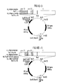
Figures I to X show gene constructs according to the present
invention.
Figures XI and XII show the results obtained by caesium chloride
sedimentation of immunogenic particles according to example 10.
Figure XIII shows a prior art construct shown in Table XI.
Figure XIV shows the characterisation of particles derived from a
construct according to a further embodiment of the present
invention.
Figure XV shows the oligo sequence of Figure X-2.
Figure XVI to XX show examples of peptide sequences from which
the epitopes of this invention can be derived.
DESCRIPTION OF THE PREFERRED EMBODIMENTS
Preferred DNA constructs of the present invention are
characterized by the presence of a selection marker selected
from the group consisting of dhfr (dihydrofolate reductase),
MT-neo (a neomycin resistance sequence coupled to a
methallothionein and MT-ecogpt (a resistance sequence
coupled to a methallothionein promoter). The expression rate
may be further enhanced by adding to the constructs a dhfr gene
as an amplification gene.
HBV nucleotide sequences used in certain constructs of the
present invention can be formed or isolated by any means
including isolation and ligation of restriction fragments and
synthetic oligonucleotides. Constructs specifically described
herein were formed by the ligation of synthetic
oligonucleotides to a 5' XbaI-BglII 3' fragment from the S
region of the HBV genome shown in Figure IX (hereinafter the
"XbaI-BglII fragment") which is derived from a BglII-BglII HBV
fragment including the entire pro-S
1-pre-S
2-S regions (the
"Bg1II-Bg1II Fragment"). The pre-S
1-pre-S
2-S region of the
HBV genome is shown in Figure IX. Oligonucleotides used in
making such constructs are summarized in Table I.
The oligonucleotides in Table I were combined with the
XbaI-BglII fragment to produce constructs with desired
features. In certain constructs adapter oligonucleotide
sequences (Table II) were used to create proper matching sticky
ends on the oligonucleotides and other construct components.
Other adapter sequences may be used to combine desired
oligonucleotides from Table I with the XbaI-BglII fragment,
other restriction fragments, oligonucleotides and other
construct components. The necessary sequences of such other
adapter sequences will be readily apparent to those skilled in
the art from consideration of tables of restriction sites
[e.g., that found at pages 121-128 of Methods in Enzymology,
volume 152, "Guide to Molecular Cloning Techniques," ed. Berger
and Kimmel (Academic Press 1987) which is incorporated herein
in its entirety by reference] and the sequences of the various
nucleotides to be combined. Adapter sequences can also be used
to introduce additional restriction sites into constructs of
the present invention. It should be noted that adapter
sequences must be selected or designed so that the proper
reading frame is maintained throughout the HBV sequence.
Preferred gene constructs which were used to transfect host
cells were prepared by recombinant DNA techniques in accordance
with the present invention. Preferred embodiments of
constructs with an enhanced expression rate are shown in
Figures I-VIII and are schematically represented by the
following:
Each of the constructs shown in Figures I-VIII contain, in
addition to a HBV sequence, a neomycin selection marker with
the MT promoter, an ampicillin selection marker, a dhfr
selection/amplification gene and a promoter for the HBV
sequence. The promoter for the HBV sequence is preferably the
U2 promoter, the MT promoter or the H2K promoter. Isolation of
fragments containing the various promoters, the selection
markers and amplification gene is described below. The HBV
sequences in the constructs of Figures I-VIII are schematically
represented by a rectangular bar in each figure which indicates
the oligonucleotides and/or adapter sequences from Tables I and
II which were combined with the XbaI-BglII fragment. Shaded
areas within the bar indicate generally regions of the entire
pre-S1-pre-S2-S region which are not found in the specific
construct. Oligonucleotides from Table I which can be used to
construct each type of HBV sequence are indicated in the
figures.
Figure X depicts two additional constructs for expression of
peptides including sequence from the pre-S2 region under the
control of the MT promoter.
Constructs have also been made which include the entire
BglII-BglII fragment from the HBV genome under the control of the
US promoter. These constructs have produced peptides which include
a deletion in the S region as indicated by Western blot analysis.
The above-cited promoters are specially preferable when their
use is coupled with a modulation method using the dhfr gene and
methotrexate to enhance the expression. This is achieved when
in addition to the selection marker the dhfr minigene is also
introduced into the plasmid sequence. It is essential that the
dhfr gene is located on the same plasmid together with the
structural gene to be expressed. An enhancement of the
expression rate of the structural gene can then be obtained by
adding methotrexate in the micromolar concentration range.
Thereby a manyfold enhancement of the expression rate is
achieved.
Suitable cells are e.g. VERO cells (monkey kidney cell line),
3T3-cells (murine fibroblast line), C127-cells (murine fibroblast
line), L-cells and CHO - cells (Chinese hamster cells, which
are either positive or negative in dehydrofolate reductase).
As a stop signal it is preferred to use a stop signal from a
eukaryotic cell. Preferably the stop signal of the caseine
DNA-sequence is used. As used throughout the following
examples, "HBV protein" refers generically to any protein
produced in accordance with the present invention which
corresponds to HBsAg sequences.
EXAMPLE 1
Particle Purification Procedures
1. Fractionated precipitation with polyethylene glycol (PEG)
The supernatant of HBV protein producing cultures was
collected and split into portions of 2,400 ml. To each
portion 144 g of PEG 6000 (Serva) were added and dissolved
by stirring at room temperature for 20 minutes and was
stirred for another 6 hours at 4°C. The precipitate was
separated by centrifugation in 500 ml bottles in a GS 3
rotor at 9,000 rpm (15,000 x g) for 30 minutes at 10 C. The
supernatant was collected and 144 g of PEG 6000 were added
and dissolved as described above. The solution was stirred
at 4 C for 3 hours. The precipitate from this solution was
harvested as described above except that centrifugation was
continued for 60 minutes.
2. Gel Chromatography
The material obtained after PEG precipitation was
redissolved in 20 ml PBS and submitted to gel
chromatography on A-5m (BioRad). Column dimensions were 25
x 1000 mm and 480 ml bed volume. In a typical fractionation
run 1,000 ug of PEG precipitated HBV protein in 10 to 15 ml
was loaded and eluted with PBS at a speed of 6 drops/min
(18 ml/h) 3 ml fractions were collected. HBV protein
eluted with the first peak. Collected fractions were
submitted to a CsCl gradient.
3. Sedimentation in CsCl Gradient
About 30 fractions covering the first peak in column
chromatography on A-5m and containing prepurified HBV
protein were collected to approximately 100 ml. This
solution was adjusted to a density of 1.30 g/cc with CsCl
and subsequently transferred to a nitrocellulose tube
fitting into a SW 27/28 rotor (Beckman). A gradient was set
by underlaying 4 ml of a CsCl solution of 1.35 g/cc and by
overlaying 4 ml of 1.25 g/cc followed by 4 ml of 1.20 g/cc
density. This gradient had been run at 28,000 rpm for 50
hours at 10 C. Thereafter the gradient was fractionated and
purified HBV protein floating in the 1.20 g/cc density
layer was collected. The solution was desalted by three
cycles of dialysis in bags against water.
Example 2
Quantitative Determination of HBV protein
1, with Radioimmunoassay
In the AUSRIA II-125 "sandwich" radioimmunoassay
(commercially available from Abbot), beads coated with
guinea pig antibody to Hepatitis B Surface Antigen
(Anti-HBs) were incubated with serum or plasma or purified
protein and appropriate controls. Any HBsAg present was
bound to the solid phase antibody. After aspiration of the
unbound material and washing of the bead, human
125T-Anti-HBs was allowed to react with the
antibody-antigen complex on the bead. The beads were then
washed to remove unbound 125I-Anti-HBs.
)-Anti-HBs HBsAg
)-Anti-HBs . HBsAg 125I-Anti-HBs
)-Anti-HBs . HBsAg . 125-Anti-HBs
The radioactivity remaining on the beads was counted in a
gamma scintillation counter.
2. with ELISA
In the Enzygnost HBsAg micro "sandwich" assay (commercially
available from Behring), wells were coated with anti-HBs.
Serum plasma or purified protein and appropriate controls
were added to the wells and incubated. After washing,
peroxidase-labelled antibodies to HBsAg were reacted with
the remaining antigenic determinants. The unbound
enzyme-linked antibodies; are removed by washing and the
enzyme activity on the solid phase is determined. The
enzymatically catalyzed reaction of hydrogen peroxide and
chromogen was stopped by adding diluted sulfuric acid. The
colour intensity was proportional to the HBsAg
concentration of the sample and was obtained by photometric
comparison of the colour intensity of the unknown samples
with the colour intensities of the accompanying negative
and positive control sera.
Example 3
Preparation of a construct of the present invention containing
the methallothionein promoter.
1) Isolation of the MI promoter
The plasmid pBPV-342-12 (commercially available from ATCC)
was digested with the endonucleases Bg1II and BamHI. Three
DNA molecules were generated. The fragment of interest
contains the methallothionein promoter and a pBR322
sequence comprising 4.5 kb and is easily detectable from
the other fragments (2.0 kb and 7.6 kb).
The reaction was performed in a total volume of 200 ul of
reaction buffer at a final concentration of 0.5 ug/ul DNA
including 100 units of each restriction enzyme. The
completion of the digestion was checked after incubation at
37°C for three hours by agarose gel electrophoresis at a
0.8% agarose gel. The reaction was stopped by adding 4 ul
0.5 M EDTA.
The 4.5 kb fragment was separated from the other fragments
by preparative 1.2% agarose gel electrophoresis. The DNA
was eluted from the agarose gel on DE-81 Whatman filter
paper from which the DNA was removed in a high salt
buffer. The DNA was purified by a phenol/chloroform
extraction and two ethanol precipitations.
2) Ligation of the 2.3 kb MBV Bg1II-Bg1II fragment
A 2.3 kb BglII-BglII fragment containing the HBV
pre-S1,pre-S2 and S coding regions was isolated from
HBV-containing DNA. The 2-3kb fragment was ligated together
with the 4.5 kb fragment (obtained as described in Cl)
containing the methallothionein promoter.
2 ul of the 2.3 kb fragment were mixed with 3 ul of the 4.5
kb fragment and ligated together in a total volume of 10 ul
ligation buffer, containing 2 units T4-DNA ligase and 2mM
ATP at 14°C overnight.
The ligation mixture was added to 150 ul competent
bacterial cell suspension for DNA up-take. After the DNA
up-date the bacterial cells were spread on LB agar plate
containing 50 ug/ml ampicillin at volumes of 50 to 300 ul
cell suspension per plate. The agar plates were incubated
at 37°C overnight. Single isolated bacterial colonies were
screened for the presence of a plasmid containing the
desired fragments.
3) Screening for desired plasmid containing bacterial colonies.
Single colonies were picked with a toothpick and
transferred to a LB-ampicillin media containing tube (5
ml). The tubes were incubated overnight at 37°C by shaking
rapidly. A mini-plasmid preparation of each grown
bacterial suspension was made. The different resulting
DNAs were proved by digestion with the restriction
endonuclease EcoRI. Two molecules were expected, a 2.2 kb
fragment and S 4.6 kb fragment. The digestion was analysed
by agarose gel electrophoresis. Plasmid DNA was isolated
from the bacterial cells.
4) Conversion of a part of the HBV-gene sequence.
The plasmid resulting from (3) above was digested with the
endonucleases BglII and XbaI. Two molecules were expected,
one 550 bp fragment and one 6.250 kb fragment which was
isolated after agarose gel electrophoresis.
The 6.250 kb fragment was ligated together with
oligomecleotide No.55 from Table I. The ligation mixture
was added to 150 ul competent bacterial cell suspension for
DNA up-take. Single isolated bacterial colonies were
screened for the presence of the desired plasmid. The new
plasmid was proved by a digestion with the endonucleases
EcoRI and BglII. Two molecules were expected, one 1.9 kb
and one 4.450 kb.
5) Insertion of a neomycin selection marker.
The plasmid resulting from (4) above was linearized by
digestion with the restriction enzyme EcoRI. The reaction
was performed in a total volume of 50 ul and a final
concentration of 1 ug/ul plasmid DNA. 50 units of ECORI
were added and the digestion was proved after incubation at
37°C for three hours by agarose gel electrophoresis. The
reaction was stopped by adding 1 ul of 0.5 M EDTA and the
DNA was precipitated with a final concentration of 0.3 M
sodium acetate and 3-4 volumes of ethanol at -80°C for 30
minutes. The precipitated DNA was dissolved in 50 ul
distilled water.
2 ul of the linearized plasmid were mixed with 3 ul of the
DNA fragment containing the methallothionein promoter and
the neomycin selection gene [isolated from the plasmid
pMT-neo-E (available from ATCC ) by digestion with
the endonuclease EcoRI as a 4kb fragment], and ligated
together. Single bacterial colonies were screened for the
presence of the desired plasmid.
6) Additional of the dhfr Amplification Gene dhfr
The plasmid pdhfr3.2 (available from ATCC) was digested
with the restriction endonuclease HindIII. Two molecules
were generated, one of 3,000 bp containing the dhfr gene
sequence and one of 3,400 bp. The 3,000 bp fragment was
isolated and ligated into the plasmid resulting from (5)
above which was previously opened by digestion with
HindIII. The resulting plasmid is represented by Fig. I-2.
Example 4
1) Isolation of a fragment containing the U2 promoter sequence.
The plasmid pUC-8-42 (available from Exogene ) was digested
with the restriction endonucleases EcoRI and Apal. Two DNA
molecules were generated. The fragment of interest contains
the U2-promoter comprising 340 bp and is easily detectable
from the other fragment (3160 bp). The digestion was
performed in a total volume of 200 ul of reaction buffer at
a final concentration of 0.5 ug/ul DNA including 100 Units
of each restriction enzyme. The completion of the digest
was checked after incubation at 37°C for three hours by
agarose gel electrophoresis in a 0.7% agarose gel. The
reaction was stopped by adding 4 ul 0.5 M EDTA. The 340 bp
fragment was separated from the plasmid DNA by preparative
1.2% agarose gel electrophoresis. The DNA was eluted from
the agarose gel on DE-81 Whatman filter paper from which
the DNA was removed in a high salt buffer. The DNA was
purified by a phenol/chloroform extraction and two ethanol
precipitations.
2) Insertion of the fragment containing the promoter sequence
into a polylinker plasmid.
The plasmid pSP165 (commercially available from Promega Biotec)
containing a polylinker sequence (containing the following
restriction sites: EcoRI, SacI, SmaI, AvaI, BamHI, BglII,
SalI, PstI, HindIII) was linearized with the restriction
enzyme EcoRI. The reaction was performed in a total volume
of 50 ul and a final concentration of lug/ul plasmid DNA.
50 Units of EcoRI were added an the digestion was proved
after incubation at 37°C for three hours by agarose gel
electrophores. The reaction was stopped by adding 1 ul of
0.5 M EDTA and the DNA was precipitated with a final
concentration of 0.3 M sidium acetate and 3-4 volumes of
ethanol at -80°C for 30 minutes. The precipitated DNA was
dissolved in 50 ul distilled water.
2 ul of plasmid DNA were mixed with 10 ul of the fragment
DNA containing the V2 promoter sequence, and ligated
together in a total volume of 25 ul of ligation buffer
containing 2 units T4-DNA ligase and 2 mM ATP at 14°C
overnight. Thereafter the DNA was purified by phenol/
chloroform extractions followed by two ethanol
precipitations and dissolved in 10 ul distilled water. The
resulting sticky ends of EcoRI and ApaI had to be converted
into blunt ends and ligated. The blunt ends were converted
by a removing reaction with the Mung bean nuclease as
follows: to 25 ul DNA (1 ug/ul concentration) reaction
buffer, 20 units of enzyme and a final concentration of 1%
glycerol to the reaction volume of 35 ul were added. After
an incubation for 30 minutes at 30 C the DNA was purified
by phenol/chloroform extractions followed by two ethanol
precipitations. The DNA was dissolved again in 5 ul
distilled water. The resulting blunt ends were ligated
together in 15 ul reaction volume containing 10 x more T4
ligase then used above and 2 mM ATP at 14°C overnight.
The ligation mixture was added to 150 ul competent
bacterial cell suspension for DNA up-take. After the DNA
up-take the bacterial cells were spread on LB agar plates
containing 50 ug/ml ampicillin at volumes of 50 to 300 ul
cell suspension per plate. The agar plates were incubated
at 37°C overnight. Single isolated bacterial colonies were
screened for the presence of a plasmid containing the
desired U2-promoter fragment.
3. Screening for desired plasmid containing bacterial colonies
Single colonies were picked with a toothpick and
transferred to a LB-ampicillin containing tube (5 ml). The
tubes were incubated overnight at 37°C by shaking rapidly.
A mini plasmid preparation of each grown bacterial
suspension was made. The different resulting plasmid was
proved by digestion with both restriction endonucleases
EcoRI and HindIII. Two molecules were found, a 400 bp
fragment containing the U2 promoter sequence and the
plasmid of 2,700 bp. The digestion was analysed by agarose
gel electrophoresis. The resulting plasmid was isolated
from the bacterial cells.
4) Insertion of the neomycine selection marker.
The plasmid pBPV-342-12 (commercially available from ATCC)
was digested with the endonucleases EcoRI and BamHI. Two
molecules were isolated, one containing the MT promoter
together with the neomycin selection gene of 4,000 bp and
the plasmid of 10,000 bp.
The plasmid resulting from (3) above was linearized with
EcoRI and ligated together with the 4,000 bp fragment
containing the MT-promoter together with the neomycin
selection gene. The resulting sticky ends were also
converted into blunt ends and ligated together as described
above.
After bacterial transformation, colony selection and mini
plasmid preparation, the resulting plasmids were analysed
by a digestion with the restriction enzymes ECORI and
HindIII. Two DNA molecules were isolated, a 400 bp fragment
and a 6,700 bp fragment.
5) Ligation of the BglII-BglII fragment
The plasmid resulting from (4) above was linearized with
Bg1II. The 2.3 kb-BgIII-BgIII fragment was ligated
together with the linearized plasmid. Bacterial colonies
were analysed to find the resulting plasmid. The
plasmid-DNA was digested with EcoRI and two resulting
fragments were obtained, a 700 bp fragment (containing the
promoter and a part of the HBV-sequence) and a 8,700 bp
fragment (containing the rest of the HBV-sequence, MT-neo
and plasmid).
6) Alterations within the HBV-sequence
The plasmid resulting from (5) above was digested with the
endonucleases BglII and MstII. Two molecules were
generated, one of 300 bp containing part of the pre-S
sequence and the other (9,100 bp) which was eluted as
described above. This 9,100 bp fragment was ligated to
another BglII/MstII 216 bp fragment (sequence
coding for an altered pre-S
1 gene sequence.
The desired plasmid was digested with EcoRI and
two resulting fragments were isolated, a 616 bp fragment
and a 8,700 bp fragment.
Example 5
Isolation of the H2K Promoter
The H2K promoter was isolated as an EcoRI/BglII fragment
(2kb) from psp65H2 (available from Exogene).
Isolation of the egpt selection marker
The fragment containing the methallothionein promoter and
the egpt-selection gene was isolated by digestion of the
plasmid pMSG (available from Pharmacia) with the
restriction enzyme EcoRI as a 3.6 kb fragment.
All other plasmid constructions were made in similar ways
by combining fragments containing the necessary components
and employing desired oligonucleotides and adapter
sequences (where necessary).
Example 6
Transfection of Mammalian Cells with Constructs of the Present
Invention.
In order to achieve secretion of substantial amounts of the
HBV peptides encoded by constructs of the present
invention, mammalian cells must be transfected with both
the construct of the present invention and a construct
which will express entire S protein. The cotransfection
was performed in two steps (i.e., a separate transfection
for each construct) or in a single step (i.e., one
transfection using preparation of both constructs).
Cotransfection was confirmed either by use of different
selection markers on the two constructs or by detection of
secretion of expression products of both constructs by
immunoassay.
Alternatively, a sequence encoding the HBV peptide sequence
of the present invention and a separate sequence encoding
the entire S protein could be combined in a single
construct.
Example 7
General Procedures
General procedures useful in practicing the present
invention may be found in (1) Methods of Enzymology, volume
152, "Guide to Molecular Cloning Techniques," ed. Berger
and Kimmel (Academic Press 1987), and (2) Maniatis et al.,
"Molecular Cloning: A Laboratory Manual," (Cold Spring
Harber Laboratory 1982), both of which are incorporated
herein in their antirety by reference. Specific techniques
employed are described below.
1) Digestion with Endonucleases and Isolation of Fragments
The restriction endonucleases used were:
BglII, BamHI, HindIII, EcoRI, XbaI, MstII, XhoI, PflMI,
commercially available from Gibco/BRL with their respective
restriction buffers (10x).
Unless otherwire indicated, restriction digests were
performed and fragments were isolated as follows.
Reactions typically contained 1-5 ug DNA.
- distilled water was added to the DNA in an eppendorf
tube to a final volume of 8 ul
- 1 ul of the appropriate 10x digestion buffer was added
- 1 ul (containing 5-10 U) restriction enzyme was added
and mixed carefully
- the reaction tube was incubated for 1 hour at 37°C
- digestion was stopped by adding 0.5 M EDTA (pH 8.0) to
a final concentration of 10 mM
- if the DNA was analysed directly on a gel, 1 ul of
gel-loading dye III (Maniatis) was added, mixed and
the sample was loaded into the slots of a 0.8% agarose
gel.
The agarose gel normally contains 0.8% agarose 1 x running
buffer (TBE, Maniatis). Where a fragment (about
100-1000bp) was isolated from an agarose gel the agarose
was increased to 1.2 to 1.4%.
2) Competent Bacterial Cells
From a dense overnight culture, 1 ml of the bacterial cell
suspension was added to 100 ml fresh growth medium
(L-broth). The cells were grown at 37°C to a density of
OD600 = 0.7 which was reached within 2 hours with
vigorous shaking in a 500 ml Erlenmeyer flask. Growth was
stopped by chilling the culture on ice for 10 minutes. From
this
culture, 3 ml were taken for harvesting the exponential
bacterial cells at 3,000 rpm for 5 minutes. The cells were
resuspended in 1.5 ml of 50 mM CaCl2 in 10 mM Tris, pH
8.0, and incubated on ice for another 15 minutes. The cells
were harvested once more by centrifugation at 3,000 rpm for
5 minutes and resuspended in 200 ul of 50 mM CaCl2 in 10
mM Tris, pH 8.0, and used directly.
3) Transformation of Competent Bacterial Cells
The DNA to be transformed was suspended in 10 mM Tris, pH
7.5, 1 mM EDT 70 ul and added to the 200 ul bacterial cell
suspension for DNA take-up. The mixture was incubated on
ice for 30 minutes and then 1 ml L-broth was added. The
mixture was incubated at 42°C for 2 minutes and at 37°C
for 40 minutes.
After the incubation, the cells were spread on agar plates
containing 50 ug ampicillin/ml agar at volumes of 50-300 ul
cell suspension per plate. The agar plates were incubated
at 37°C overnight. After this incubation period, single
isolated bacterial colonies were formed.
4) Plasmid DNA Isolation
1 liter of plasmid-bearing cells was grown to 0.5 OD600
in L-broth and amplified for 20 hours with 200 ug/ml
chloramphenicol. The culture was then centrifuged at 4,000
rpm for 20 minutes in JA-10 rotor, 4°C. The pellet was
resuspended in 18 ml cold 25% sucrose, 50 mM Tris, pH 8.0,
transferred to a 250 ml Erlenmeyer flask and kept on ice.
6 ml 5mg/ml lysozyme in 250 mM Tris, pH 8.0 was added and
the mixture was left to stand 10-15 minutes. 6 ml 250 mM
EDTA, pH 8.0, was added, mixed gently and incubated for 15
minutes on ice. 30 ml detergent (0.01% Triton X-100; 60 mM
EDTA, pH 8.0; 50 mM Tris, pH 8.0) was added and the mixture
was incubated for 30 minutes on ice. After incubation, the
mixture was centrifuged at 25,000 rpm 90 minutes in SW28
rotor, 4°C.
Pronase was added to supernatant fluid to 250 ug/ml and
incubated 30 minutes, 37°C. The solution was extracted
with phenol once with 1/2 volume phenol equilibrated with
10 mM Tris, pH 8.0, 1 mM EDTA. The aqueous layer was
removed. Sodium acetate was then added to a final
concentration of 300 mM, followed by the addition of 3
volumes cold 100% ethanol and thorough mixing. The mixture
was stored at -20°C overnight.
The mixture was thawed and centrifuged. The pellet was
resuspended in 6 ml 10 mM Tris, 10 mM EDTA, pH 8.0. 9.4 g
CsCl and 0.65 ml of 6 mg/ml ethidium bromide were added and
the volume was brought up to 10 ml with sterile
double-distilled water. The 10 ml alignots were put into
Beckman heat-sealable gradient tubes and centrifuged,
50,000 rpm, 48 hours in Ti70.1 Beckman rotor.
Plasmid bands were visualized with UV and removed with
syringe and 18 gauge needle by piercing the side of the
tube. Ethidium bromide was removed from the plasmid
fractions by 3 successive extractions with equal volumes of
isobutanol. Fractions were then (1) dialyzed against one
2-liter lot of 10 mM Tris, pH 7.4, 1 mM EDTA, pH 7.5, 5 mM
NaCl for 2 hours or more at 4°C; and (2) phenol extracted
once with 1/3 volume phenol equilibrated as above. Sodium
acetate was then added to a final concentration of 300 mM,
followed by addition of two volumes of 100% ethanol.
Precipitate formed at -20°C overnight, or at -70°C for 30
minutes.
5) Mini-Plasmid Preparation
1 ml of an overnight bacteria culture was put into an
eppendorf tube and centrifugated for 20 minutes. The
supernatant was removed. 100 ul of 50 mM glucose, 25 mM
Tris (pH 8.0), 10 mM EDTA (pH 8.0) was added to the pellet,
mixed by vortex and incubated for 5 minutes at room
temperature. 200 ul of 0.2 N NaOH, 1% SDS was added, mixed
by vortex and incubated for 5 minutes on ice. 150 ul 3 M
Sodium acetate (pH 4.8) was added, mixed by vortex and
incubated for 5 minutes on ice. After centrifugation for 5
minutes at 13,000 rpm the supernatant was decanted into a
fresh eppendorf tube. 3 volumes of 100% ethanol were
supplemented, mixed well and incubated for 30 minutes at
-80°C, then centrifuged for 10 minutes at 13,000 rpm. The
ethanol was removed, the pellet washed with 70% ethanol,
lyophilized and dissolved in 20 ul distilled water. 5 ul
of this plasmid DNA solution were used directly for
restriction analysis.
6) Nick Translation
Nick translation was performed according to Rigby et al.,
J. Mol. Biol., Vol. 113, pp. 237-251, 1977, which is
incorporated herein by reference. The reaction mixture for
32P-labeling of DNA contained 0.5 ug of a HBV fragment,
in a total volume of 30 ul with 50 mM Tris, pH 7.8, 5 mM
MgCl2, 10 mM mercaptoethanol, 0.1 mM dATP, 0.1 mM dGTP,
0.1 mM dTTP, 50 uCi 32P-dCTP, 10 units DNA polymerase I,
3 ul of a 2 x 10⁻⁵ fold dilution of 1 mg/ml DNase I and
is incubated for 90 minutes at 15°C, yielding 3 x 10⁶ to
12 x 106 total cpm, i.e. 1 x 10⁷ to 5 x 10⁷ cpm/ug
DNA.
7) Southern Blot Analysis
To characterize the organization within the host cell
genome of the vectors of this invention, chromosomal DNA
from cell lines producing particles of this invention were
isolated and digested with the appropriate restriction
enzyme(s) and analysed by the method of Southern (J. Mol.
Biol., Vol. 98, pp. 503-517, 1975), which is incorporated
herein by reference, using a 32P-labeled DNA probe.
Following digestion of the chromosomal DNA (20 ug) with the
restriction enzyme BglII, the resulting fragments were
separated by 0.7% agarose gel electrophoresis. Thereafter,
the DNA was denatured by exposing to 366 nm UV light for 10
minutes and by incubation in a solution of 0.5 M NaOH and 1
M NaCl for 45 minutes. The gels were neutralized by
incubation in 0.5 M Tris, 1.5 M NaCl, pH 7.5 for 60
minutes. The DNA was transferred to a nitrocellulose
filter by soaking in 3 M NaCl, 0.3 M Sodiumcitrate (20 x
SSC) for 20 hours through the gel by covering the top of
the nitrocellulose filter with a staple of dry paper
towels. The nitrocellulose filter was kept for 2 hours in
a vacuum oven at 80 C. A radioactive DNA probe from the
BglII fragment of the pHBV (2.3 kb) was prepared by nick
translation.
For hybridization with the DNA probe, the nitrocellulose
filter was sealed in a plastic bag containing 10 ml of
prehybridization mixture: 50% formamide, 5 x SSC, 50 mM
Sodiumphosphate, pH 7.0, 5 x Denhardt's solution, 250 ug/ml
denatured salmon sperm DNA. The filter was incubated in
this mixture for 4 hours at 45°C, after which the
pre-hybridization mixture was replaced by the hybridization
mixture: 50% formamide, 5 x SSC, 20 mM Sodiumphosphate, pH
7.0, 1 x Denhardt's solution, 100 ug/ml denatured salmon
sperm DNA, 5 x 10⁵ cmp/ml 32P-probe. The filter, after
incubating in the hybridization mix for 18 hours at 45°C,
was washed three times, 5 minutes each, in 0.1 x SSC, 0.1%
SDS at 50°C. The filter was dried at 60°C for 10 minutes
and exposed to two X-ray films (XAR-5, KODAK) between two
intensifying screens and kept at -80°C. The first X-ray
film is developed after 3 days' exposure; the second film
after 7 days' exposure.
8) Preparation of Mammalian Cells and DNA Precipitate for
Transfection
The recipient cells (C127 or CHO-cells available from ATCC) were
seeded in normal growth medium (DMEM+10% Fetal Calf Serum, Glycose and
Glutamin) into petri-dishes (1-2 x 10⁶ cells per dish,
10 cm) at day 1. The next day the medium was removed (4
hours before the DNA precipitate was added onto the cells),
and the cells were washed twice with 1 x PBS. Then 8 ml DMEM
without FCS were added. 4 hours later the DNA precipitate
(prepared as described below) was added to the cells.
Again after 4 hours the medium was removed, 3 ml of
Glycerol-Mix (50 ml 2 x TBS buffer, 30 ml glycerol, 120 ml
distilled water) were added. The Glycerol-Mix was
immediately removed after an incubation at 37°C for 3
minutes and the cells were washed with 1 x PBS. The cells
were cultivated overnight with 8 ml of DMEM with 10% FCS.
After 48 hours, the cells were recovered from the dish by
treating with Trypsin-EDTA-Solution (0.025% Trypsin + 1 mM
EDTA). Afterwards, to remove the Trypsin-EDTA the cells
were washed with 1 x PBS, suspended in DMEM with 10% FCS
and distributed into 24 costar-well-plates (cells from one
dish into four 24-well-plates). When the cells had grown
well, selection medium was added (concentration 0.5 - 1mg/ml
of neomycin,or xanthine: 250 µg/ml, hypoxanthine: 15 µg/ml (or
adenine: 25 µg/ml), thymidine: 10 µg/ml, aminopterine 2 µg/ml
mycophenolic acid: 25 µg/ml for eco-gpt, for example).
The medium was changed every week. The first growing cell
colonies were seen after 2 weeks.
To 10 ug of plasmid DNA and 20 ug of carrier-DNA
(salmon-sperm DNA, calf-thymus DNA) TE-buffer (10 mM
Trix-HCl, 1 mM EDTA, pH 7.05) was added to a final volume
of 440 ul and mixed together with 60 ul 2 M CaCl2. Then
the same amount of 2x TBS (Hepes 50 mM, NaCl 280 mM,
Na2HPO4 1.5 mM, pH 7.05) was added and mixed well.
The precipitation solution was incubated for 30 minutes at
37°C and added directly to the cells which should be
transfected.
Example 8
Culturing of Transfected Cells to Secrete Protein
The selected cells are treated for further cultivation in
normal growth medium as described in section 8.
Example 9
F) Preparation of the Adjuvant of Purified Particles
To the desired concentration of antigen particles suspended
in sterile saline, 1 : 10,000 volume Thimerosol, 1/10
volume of filter-sterilised 0.2 M Al K(SO4)2 : 12 H 20
were added. The pH was adjusted to 5.0 with sterile 1 N
NaOH and the suspension was stirred at room temperature for
3 hours. The alum-precipitated antigen was recovered by
centrifugation for 10 minutes at 2,000 rpm, resuspended in
sterile normal saline containing 1;10,000 Thimerosol and
aliquoted under sterile conditions.
Example 10
Tables III - X give some of the results of ELISA analysis of
immunogenic particles of the present invention as described
below:
- Table III:
- shows the ELISA data of the purified HBs
antigen particle produced from any HBV
sequence construct of the present invention
including the pre-S1 region with total
deletion of pre-S2 and deletions upstream
of the pre-S2 ATG and the S region with
deletion of the S ATG and downstream the
S ATG through the XBaI site (e.g. the
construct of Fig. I-1) with the
anti-pre-S1 monoclonal antibody MA 18/7.
The fractions 9-15 (Fig. XI) were pooled
after CsCl sedimentation.
- Table IV:
- shows the ELISA data of the purified HBS
antigen particle produced from any HBV
sequence construct of the present invention
including the pre-S1 region with total
deletion of pre-S2 and deletions upstream
of the pre-S2 ATG and the S region with
deletion of the S ATG and downstream the
S ATG through the XBaI site
(e.g., the construct of Fig. I-1) with the
anti-pre-S2 monoclonal antibody MQ 19/10.
The fractions 9-15 (Fig. XI) were pooled
after CsCl sedimentation.
- Table V:
- shows the ELISA data of the purified HBs
antigen particle produced from an HBV
sequence construct of the present invention
including the pre-S2 region with none of the
pre-S1 region and deletions upstream of the
S ATG and downstream of the S ATG through
the XBaI site, and the S region with
deletion of the S ATG (e.g.
the construct of Fig. II-1) with the
anti-pre-S1 monoclonal antibody MA 18/7.
The fractions 9-15 (Fig. XII) were pooled
after CsCl sedimentation.
- Table VI:
- shows the ELISA data of the purified HBS
antigen particle produced from an HBV
sequence construct of the present invention
including the pre-S2 region with none of the
pre-S1 region and deletions upstream of the
S ATG and downstream of the S ATG through
the XBaI site, and the S region with
deletion of the S ATG (e.g.
the construct of Fig. II-1) with the
anti-pre-S2 monoclonal antibody MQ 19/10.
The fractions 9-15 (Fig. XII) were pooled
after CsCl sedimentation.
| CsCl-gradient | ELISA Measurement |
| | Monoclonal Antibody MA 18/7 |
| Fraction No. 9-15 (pooled) | E492 = 0.839 |
| CsCl-gradient | ELISA Measurement |
| | Monoclonal Antibody MQ 19/10 |
| Fraction No. 9-15 (pooled) | E492 = 0.000 |
| CsCl-gradient | ELISA Measurement |
| | Monoclonal Antibody MA 18/7 |
| Fraction No. 9-15 (pooled) | E492 = 0.000 |
| CsCl-gradient | ELISA Measurement |
| | Monoclonal Antibody MQ 19/10 |
| Fraction No. 9-15 (pooled) | E492 = 1.028 |
- Table VII:
- shows the ELISA data of the purified HBs
antigen particle produced from any HBV
sequence construct of the present invention
including the pre-S1 region with total
deletion of pre-S2 and deletions upstream
of the pre-S2 ATG and the S region with
deletion of the S ATG
(e.g., the construct of Fig. VI-2)
with the anti-pre-S1 monoclonal antibody MA
18/7. The fractions 9-15 (Fig. XI) were
pooled after CsCl sedimentation.
- Table VIII:
- shows the ELISA data of the purified HBs
antigen particle produced from any HBV
sequence construct of the present invention
including the pre-S1 region with deletions
upstream of the pre-S2 ATG with deletion of
the S ATG (e.g., the construct of Fig. VI-4)
with the anti-pre-S2 monoclonal antibody MQ
19/10. The fractions 9-15 (Fig. XI) were
pooled after CsCl sedimentation.
- Table IX:
- shows the ELISA data of the purified HBs
antigen particle produced from an HBV
sequence construct of the present invention
including the pre-S2 region with none of the
pre-S1 region and deletions upstream of the
S ATG
and the S region with
deletion of the S ATG
(e.g., the construct of Fig. VII-2) with
the anti-pre-S1 monoclonal antibody MA 18/7.
The fractions 9-15 (Fig. XII) were pooled
after CsCl sedimentation.
- Table X:
- shows the ELISA data of the purified HBs
antigen particle produced from an HBV
sequence construct of the present invention
including the pre-S2 region with deletions
upstream of the S ATG with deletion of the S
ATG (e.g., the construct of Fig. VII-4) with
the anti-pre-S2 monoclonal antibody MQ 19/10.
The fractions 9-15 (Fig. XII) were pooled
after CsCl sedimentation.
| CsCl-gradient | ELISA Measurement |
| | Monoclonal Antibody MA 18/7 |
| Fraction No. 9-15 (pooled) | E492 = 1.273 |
| CsCl-gradient | ELISA Measurement |
| | Monoclonal Antibody MQ 19/10 |
| Fraction No. 9-15 (pooled) | E492 = 0.000 |
| CsCl-gradient | ELISA Measurement |
| | Monoclonal Antibody MA 18/7 |
| Fraction No. 9-15 (pooled) | E492 = 0.000 |
| CsCl-gradient | ELISA Measurement |
| | Monoclonal Antibody MQ 19/10 |
| Fraction No. 9-15 (pooled) | E492 = 0.985 |
- Table XI
- shows the ELISA data of purified HBs antigen
particles produced by construct
including the entire pre-S1 - pre-S2 - S
region under control of the LTR region of
rous sarcoma virus after stimulation with
stimulating substances (e.g. PMA) and the
additional cotransfection with S (Fig. XIII).
| CsCl-gradient | ELISA Measurement |
| | Monoclonal Antibody MA 18/7 |
| Fraction No. 9-15 (pooled) | E492 = 0.125 |
- Figure XIV
- shows the characterisation of the
particles derived from gene constructs
according to table III (Fig. I-1) and
table V (Fig. II-1) cotransfected in
C127 after purification in the CsCl
gradient. The fraction collected had
a smaller volume.
- Table XII
- shows the serotyping of particles
according to Fig. I-1 having the
S sequence done in the Pettenkofer
Institute.
| Results: |
| adw / ayw : positive |
From the foregoing, it will be obvious to those skilled in
the art that various modifications in the above-described
compositions and methods can be made without departing from the
spirit and scope of the invention. Accordingly, the invention
may be embodied in other specific forms without departing from
the spirit or essential characteristics thereof. Present
embodiments, therefore, are to be considered in all respects as
illustrative and not restrictive, the scope of the invention
being indicated by the appended claims rather than by the
foregoing description, and all changes which come within the
meaning and range of equivalency of the claims are therefore
intended to be embraced therein.