EP1088227B1 - Method for the quantitative release of natural or recombinant proteins, polypeptides or peptides - Google Patents
Method for the quantitative release of natural or recombinant proteins, polypeptides or peptides Download PDFInfo
- Publication number
- EP1088227B1 EP1088227B1 EP99928306A EP99928306A EP1088227B1 EP 1088227 B1 EP1088227 B1 EP 1088227B1 EP 99928306 A EP99928306 A EP 99928306A EP 99928306 A EP99928306 A EP 99928306A EP 1088227 B1 EP1088227 B1 EP 1088227B1
- Authority
- EP
- European Patent Office
- Prior art keywords
- sample
- spa
- heating step
- protein
- range
- Prior art date
- Legal status (The legal status is an assumption and is not a legal conclusion. Google has not performed a legal analysis and makes no representation as to the accuracy of the status listed.)
- Expired - Lifetime
Links
- 238000000034 method Methods 0.000 title claims abstract description 56
- 108090000765 processed proteins & peptides Proteins 0.000 title abstract description 22
- 102000004196 processed proteins & peptides Human genes 0.000 title abstract description 21
- 229920001184 polypeptide Polymers 0.000 title abstract description 15
- 108010008281 Recombinant Fusion Proteins Proteins 0.000 title abstract description 5
- 102000007056 Recombinant Fusion Proteins Human genes 0.000 title abstract description 5
- 238000010438 heat treatment Methods 0.000 claims abstract description 39
- 239000003153 chemical reaction reagent Substances 0.000 claims abstract description 30
- 108060003951 Immunoglobulin Proteins 0.000 claims abstract description 13
- 150000001875 compounds Chemical class 0.000 claims abstract description 13
- 102000018358 immunoglobulin Human genes 0.000 claims abstract description 13
- 229940072221 immunoglobulins Drugs 0.000 claims abstract description 9
- 238000001816 cooling Methods 0.000 claims abstract description 7
- 238000002156 mixing Methods 0.000 claims abstract description 6
- 102000004169 proteins and genes Human genes 0.000 claims description 52
- 108090000623 proteins and genes Proteins 0.000 claims description 52
- DBMJMQXJHONAFJ-UHFFFAOYSA-M Sodium laurylsulphate Chemical compound [Na+].CCCCCCCCCCCCOS([O-])(=O)=O DBMJMQXJHONAFJ-UHFFFAOYSA-M 0.000 claims description 31
- 239000000872 buffer Substances 0.000 claims description 29
- 229910019142 PO4 Inorganic materials 0.000 claims description 7
- 239000002738 chelating agent Substances 0.000 claims description 7
- 150000002500 ions Chemical class 0.000 claims description 7
- NBIIXXVUZAFLBC-UHFFFAOYSA-K phosphate Chemical compound [O-]P([O-])([O-])=O NBIIXXVUZAFLBC-UHFFFAOYSA-K 0.000 claims description 7
- 239000010452 phosphate Substances 0.000 claims description 7
- -1 0.1 to 2 mM Chemical class 0.000 claims description 3
- BTBUEUYNUDRHOZ-UHFFFAOYSA-N Borate Chemical compound [O-]B([O-])[O-] BTBUEUYNUDRHOZ-UHFFFAOYSA-N 0.000 claims description 3
- 229910001385 heavy metal Inorganic materials 0.000 claims description 3
- 230000002209 hydrophobic effect Effects 0.000 claims description 2
- 238000003556 assay Methods 0.000 abstract description 28
- 108010032595 Antibody Binding Sites Proteins 0.000 abstract description 5
- 230000000984 immunochemical effect Effects 0.000 abstract description 4
- 101000582398 Staphylococcus aureus Replication initiation protein Proteins 0.000 description 70
- 239000000523 sample Substances 0.000 description 41
- 239000000243 solution Substances 0.000 description 40
- 239000012895 dilution Substances 0.000 description 18
- 238000010790 dilution Methods 0.000 description 18
- FAPWRFPIFSIZLT-UHFFFAOYSA-M Sodium chloride Chemical compound [Na+].[Cl-] FAPWRFPIFSIZLT-UHFFFAOYSA-M 0.000 description 16
- 239000002953 phosphate buffered saline Substances 0.000 description 16
- 239000003446 ligand Substances 0.000 description 13
- 238000002965 ELISA Methods 0.000 description 12
- 229920001213 Polysorbate 20 Polymers 0.000 description 12
- 235000010486 polyoxyethylene sorbitan monolaurate Nutrition 0.000 description 12
- 239000000256 polyoxyethylene sorbitan monolaurate Substances 0.000 description 12
- QPCDCPDFJACHGM-UHFFFAOYSA-N N,N-bis{2-[bis(carboxymethyl)amino]ethyl}glycine Chemical compound OC(=O)CN(CC(O)=O)CCN(CC(=O)O)CCN(CC(O)=O)CC(O)=O QPCDCPDFJACHGM-UHFFFAOYSA-N 0.000 description 11
- 210000002966 serum Anatomy 0.000 description 11
- 238000006243 chemical reaction Methods 0.000 description 10
- 239000012460 protein solution Substances 0.000 description 9
- 239000012898 sample dilution Substances 0.000 description 9
- 108091003079 Bovine Serum Albumin Proteins 0.000 description 8
- 229940098773 bovine serum albumin Drugs 0.000 description 8
- 229920000036 polyvinylpyrrolidone Polymers 0.000 description 8
- 235000013855 polyvinylpyrrolidone Nutrition 0.000 description 8
- 239000011780 sodium chloride Substances 0.000 description 8
- 230000009257 reactivity Effects 0.000 description 7
- 238000004153 renaturation Methods 0.000 description 7
- 238000012360 testing method Methods 0.000 description 7
- 239000002253 acid Substances 0.000 description 6
- 238000001514 detection method Methods 0.000 description 6
- 238000002360 preparation method Methods 0.000 description 6
- 108091008324 binding proteins Proteins 0.000 description 5
- 102000023732 binding proteins Human genes 0.000 description 5
- 238000010494 dissociation reaction Methods 0.000 description 5
- 230000005593 dissociations Effects 0.000 description 5
- 238000002474 experimental method Methods 0.000 description 5
- 238000011534 incubation Methods 0.000 description 5
- 239000011159 matrix material Substances 0.000 description 5
- 239000001267 polyvinylpyrrolidone Substances 0.000 description 5
- TWRXJAOTZQYOKJ-UHFFFAOYSA-L Magnesium chloride Chemical compound [Mg+2].[Cl-].[Cl-] TWRXJAOTZQYOKJ-UHFFFAOYSA-L 0.000 description 4
- 241000283973 Oryctolagus cuniculus Species 0.000 description 4
- UIIMBOGNXHQVGW-UHFFFAOYSA-M Sodium bicarbonate Chemical compound [Na+].OC([O-])=O UIIMBOGNXHQVGW-UHFFFAOYSA-M 0.000 description 4
- 239000008351 acetate buffer Substances 0.000 description 4
- 125000003277 amino group Chemical group 0.000 description 4
- 239000000427 antigen Substances 0.000 description 4
- 102000036639 antigens Human genes 0.000 description 4
- 108091007433 antigens Proteins 0.000 description 4
- UQLDLKMNUJERMK-UHFFFAOYSA-L di(octadecanoyloxy)lead Chemical compound [Pb+2].CCCCCCCCCCCCCCCCCC([O-])=O.CCCCCCCCCCCCCCCCCC([O-])=O UQLDLKMNUJERMK-UHFFFAOYSA-L 0.000 description 4
- 239000013024 dilution buffer Substances 0.000 description 4
- LOKCTEFSRHRXRJ-UHFFFAOYSA-I dipotassium trisodium dihydrogen phosphate hydrogen phosphate dichloride Chemical compound P(=O)(O)(O)[O-].[K+].P(=O)(O)([O-])[O-].[Na+].[Na+].[Cl-].[K+].[Cl-].[Na+] LOKCTEFSRHRXRJ-UHFFFAOYSA-I 0.000 description 4
- 238000003018 immunoassay Methods 0.000 description 4
- 239000000463 material Substances 0.000 description 4
- 239000000203 mixture Substances 0.000 description 4
- 229960003330 pentetic acid Drugs 0.000 description 4
- IAZDPXIOMUYVGZ-UHFFFAOYSA-N Dimethylsulphoxide Chemical compound CS(C)=O IAZDPXIOMUYVGZ-UHFFFAOYSA-N 0.000 description 3
- KCXVZYZYPLLWCC-UHFFFAOYSA-N EDTA Chemical compound OC(=O)CN(CC(O)=O)CCN(CC(O)=O)CC(O)=O KCXVZYZYPLLWCC-UHFFFAOYSA-N 0.000 description 3
- HEMHJVSKTPXQMS-UHFFFAOYSA-M Sodium hydroxide Chemical compound [OH-].[Na+] HEMHJVSKTPXQMS-UHFFFAOYSA-M 0.000 description 3
- 150000001413 amino acids Chemical class 0.000 description 3
- 230000003139 buffering effect Effects 0.000 description 3
- 239000000356 contaminant Substances 0.000 description 3
- 229960001484 edetic acid Drugs 0.000 description 3
- RAXXELZNTBOGNW-UHFFFAOYSA-N imidazole Natural products C1=CNC=N1 RAXXELZNTBOGNW-UHFFFAOYSA-N 0.000 description 3
- 238000000746 purification Methods 0.000 description 3
- 241000894007 species Species 0.000 description 3
- 238000011282 treatment Methods 0.000 description 3
- 239000011534 wash buffer Substances 0.000 description 3
- XLYOFNOQVPJJNP-UHFFFAOYSA-N water Substances O XLYOFNOQVPJJNP-UHFFFAOYSA-N 0.000 description 3
- JBFLYOLJRKJYNV-MASIZSFYSA-N (1z)-1-[(3,4-diethoxyphenyl)methylidene]-6,7-diethoxy-3,4-dihydro-2h-isoquinoline;hydron;chloride Chemical compound Cl.C1=C(OCC)C(OCC)=CC=C1\C=C/1C2=CC(OCC)=C(OCC)C=C2CCN\1 JBFLYOLJRKJYNV-MASIZSFYSA-N 0.000 description 2
- 108010088751 Albumins Proteins 0.000 description 2
- 102000009027 Albumins Human genes 0.000 description 2
- 241000287828 Gallus gallus Species 0.000 description 2
- DHMQDGOQFOQNFH-UHFFFAOYSA-N Glycine Chemical compound NCC(O)=O DHMQDGOQFOQNFH-UHFFFAOYSA-N 0.000 description 2
- 239000007836 KH2PO4 Substances 0.000 description 2
- 206010028980 Neoplasm Diseases 0.000 description 2
- 108010058846 Ovalbumin Proteins 0.000 description 2
- CDBYLPFSWZWCQE-UHFFFAOYSA-L Sodium Carbonate Chemical compound [Na+].[Na+].[O-]C([O-])=O CDBYLPFSWZWCQE-UHFFFAOYSA-L 0.000 description 2
- 101710120037 Toxin CcdB Proteins 0.000 description 2
- 239000000654 additive Substances 0.000 description 2
- 238000001042 affinity chromatography Methods 0.000 description 2
- 230000001580 bacterial effect Effects 0.000 description 2
- 230000008033 biological extinction Effects 0.000 description 2
- 239000008366 buffered solution Substances 0.000 description 2
- 201000011510 cancer Diseases 0.000 description 2
- 238000004587 chromatography analysis Methods 0.000 description 2
- 230000000052 comparative effect Effects 0.000 description 2
- 230000001419 dependent effect Effects 0.000 description 2
- 230000000694 effects Effects 0.000 description 2
- 238000009472 formulation Methods 0.000 description 2
- 239000012634 fragment Substances 0.000 description 2
- 238000002523 gelfiltration Methods 0.000 description 2
- XLYOFNOQVPJJNP-ZSJDYOACSA-N heavy water Substances [2H]O[2H] XLYOFNOQVPJJNP-ZSJDYOACSA-N 0.000 description 2
- 229910001629 magnesium chloride Inorganic materials 0.000 description 2
- MYWUZJCMWCOHBA-VIFPVBQESA-N methamphetamine Chemical compound CN[C@@H](C)CC1=CC=CC=C1 MYWUZJCMWCOHBA-VIFPVBQESA-N 0.000 description 2
- 229910000402 monopotassium phosphate Inorganic materials 0.000 description 2
- GNSKLFRGEWLPPA-UHFFFAOYSA-M potassium dihydrogen phosphate Chemical compound [K+].OP(O)([O-])=O GNSKLFRGEWLPPA-UHFFFAOYSA-M 0.000 description 2
- 238000001556 precipitation Methods 0.000 description 2
- 238000004445 quantitative analysis Methods 0.000 description 2
- 238000003127 radioimmunoassay Methods 0.000 description 2
- 230000009467 reduction Effects 0.000 description 2
- 230000004044 response Effects 0.000 description 2
- 239000011734 sodium Substances 0.000 description 2
- 229910000030 sodium bicarbonate Inorganic materials 0.000 description 2
- 238000010186 staining Methods 0.000 description 2
- 239000000126 substance Substances 0.000 description 2
- YMXHPSHLTSZXKH-RVBZMBCESA-N (2,5-dioxopyrrolidin-1-yl) 5-[(3as,4s,6ar)-2-oxo-1,3,3a,4,6,6a-hexahydrothieno[3,4-d]imidazol-4-yl]pentanoate Chemical compound C([C@H]1[C@H]2NC(=O)N[C@H]2CS1)CCCC(=O)ON1C(=O)CCC1=O YMXHPSHLTSZXKH-RVBZMBCESA-N 0.000 description 1
- MIAKOEWBCMPCQR-YBXAARCKSA-N (2s,3r,4s,5r,6r)-2-(4-aminophenoxy)-6-(hydroxymethyl)oxane-3,4,5-triol Chemical compound C1=CC(N)=CC=C1O[C@H]1[C@H](O)[C@@H](O)[C@@H](O)[C@@H](CO)O1 MIAKOEWBCMPCQR-YBXAARCKSA-N 0.000 description 1
- QTBSBXVTEAMEQO-UHFFFAOYSA-M Acetate Chemical compound CC([O-])=O QTBSBXVTEAMEQO-UHFFFAOYSA-M 0.000 description 1
- 239000010755 BS 2869 Class G Substances 0.000 description 1
- 241000894006 Bacteria Species 0.000 description 1
- 241000282465 Canis Species 0.000 description 1
- 239000004471 Glycine Substances 0.000 description 1
- 241000282412 Homo Species 0.000 description 1
- 108091009325 IgG binding proteins Proteins 0.000 description 1
- 102000028555 IgG binding proteins Human genes 0.000 description 1
- 108010021625 Immunoglobulin Fragments Proteins 0.000 description 1
- 102000008394 Immunoglobulin Fragments Human genes 0.000 description 1
- HNDVDQJCIGZPNO-YFKPBYRVSA-N L-histidine Chemical compound OC(=O)[C@@H](N)CC1=CN=CN1 HNDVDQJCIGZPNO-YFKPBYRVSA-N 0.000 description 1
- 241000124008 Mammalia Species 0.000 description 1
- 101100533558 Mus musculus Sipa1 gene Proteins 0.000 description 1
- 206010041925 Staphylococcal infections Diseases 0.000 description 1
- 241000191967 Staphylococcus aureus Species 0.000 description 1
- 241000193998 Streptococcus pneumoniae Species 0.000 description 1
- 239000007983 Tris buffer Substances 0.000 description 1
- 239000003463 adsorbent Substances 0.000 description 1
- 230000002411 adverse Effects 0.000 description 1
- 150000001412 amines Chemical class 0.000 description 1
- 239000012491 analyte Substances 0.000 description 1
- 238000010171 animal model Methods 0.000 description 1
- 210000003719 b-lymphocyte Anatomy 0.000 description 1
- 102000005936 beta-Galactosidase Human genes 0.000 description 1
- 108010005774 beta-Galactosidase Proteins 0.000 description 1
- 230000008436 biogenesis Effects 0.000 description 1
- 230000004071 biological effect Effects 0.000 description 1
- YBJHBAHKTGYVGT-ZKWXMUAHSA-N biotin Natural products N1C(=O)N[C@@H]2[C@H](CCCCC(=O)O)SC[C@@H]21 YBJHBAHKTGYVGT-ZKWXMUAHSA-N 0.000 description 1
- 239000007853 buffer solution Substances 0.000 description 1
- 210000004027 cell Anatomy 0.000 description 1
- 230000008859 change Effects 0.000 description 1
- 239000012504 chromatography matrix Substances 0.000 description 1
- 239000011248 coating agent Substances 0.000 description 1
- 238000000576 coating method Methods 0.000 description 1
- 230000009918 complex formation Effects 0.000 description 1
- 230000000536 complexating effect Effects 0.000 description 1
- 238000004132 cross linking Methods 0.000 description 1
- 238000004925 denaturation Methods 0.000 description 1
- 230000036425 denaturation Effects 0.000 description 1
- 238000003795 desorption Methods 0.000 description 1
- 239000012153 distilled water Substances 0.000 description 1
- FMWAXKQEIXRUTI-UHFFFAOYSA-N dodecyl hydrogen sulfate;sodium Chemical compound [Na].CCCCCCCCCCCCOS(O)(=O)=O FMWAXKQEIXRUTI-UHFFFAOYSA-N 0.000 description 1
- 231100000655 enterotoxin Toxicity 0.000 description 1
- 238000010353 genetic engineering Methods 0.000 description 1
- HNDVDQJCIGZPNO-UHFFFAOYSA-N histidine Natural products OC(=O)C(N)CC1=CN=CN1 HNDVDQJCIGZPNO-UHFFFAOYSA-N 0.000 description 1
- 102000028557 immunoglobulin binding proteins Human genes 0.000 description 1
- 108091009323 immunoglobulin binding proteins Proteins 0.000 description 1
- 229940027941 immunoglobulin g Drugs 0.000 description 1
- 244000005700 microbiome Species 0.000 description 1
- 230000002297 mitogenic effect Effects 0.000 description 1
- 238000012986 modification Methods 0.000 description 1
- 230000004048 modification Effects 0.000 description 1
- 230000003287 optical effect Effects 0.000 description 1
- 230000020477 pH reduction Effects 0.000 description 1
- 230000007918 pathogenicity Effects 0.000 description 1
- 238000010647 peptide synthesis reaction Methods 0.000 description 1
- 239000008363 phosphate buffer Substances 0.000 description 1
- 108010055837 phosphocarrier protein HPr Proteins 0.000 description 1
- 238000002264 polyacrylamide gel electrophoresis Methods 0.000 description 1
- 238000012529 protein A ELISA Methods 0.000 description 1
- 230000006920 protein precipitation Effects 0.000 description 1
- 238000001742 protein purification Methods 0.000 description 1
- HSSLDCABUXLXKM-UHFFFAOYSA-N resorufin Chemical compound C1=CC(=O)C=C2OC3=CC(O)=CC=C3N=C21 HSSLDCABUXLXKM-UHFFFAOYSA-N 0.000 description 1
- 238000012552 review Methods 0.000 description 1
- 238000003118 sandwich ELISA Methods 0.000 description 1
- 230000035945 sensitivity Effects 0.000 description 1
- 229910000029 sodium carbonate Inorganic materials 0.000 description 1
- 238000002415 sodium dodecyl sulfate polyacrylamide gel electrophoresis Methods 0.000 description 1
- 239000012192 staining solution Substances 0.000 description 1
- 238000011272 standard treatment Methods 0.000 description 1
- 230000000638 stimulation Effects 0.000 description 1
- 229940031000 streptococcus pneumoniae Drugs 0.000 description 1
- 231100000331 toxic Toxicity 0.000 description 1
- 230000002588 toxic effect Effects 0.000 description 1
Images




Classifications
-
- G—PHYSICS
- G01—MEASURING; TESTING
- G01N—INVESTIGATING OR ANALYSING MATERIALS BY DETERMINING THEIR CHEMICAL OR PHYSICAL PROPERTIES
- G01N33/00—Investigating or analysing materials by specific methods not covered by groups G01N1/00 - G01N31/00
- G01N33/48—Biological material, e.g. blood, urine; Haemocytometers
- G01N33/50—Chemical analysis of biological material, e.g. blood, urine; Testing involving biospecific ligand binding methods; Immunological testing
- G01N33/68—Chemical analysis of biological material, e.g. blood, urine; Testing involving biospecific ligand binding methods; Immunological testing involving proteins, peptides or amino acids
-
- G—PHYSICS
- G01—MEASURING; TESTING
- G01N—INVESTIGATING OR ANALYSING MATERIALS BY DETERMINING THEIR CHEMICAL OR PHYSICAL PROPERTIES
- G01N33/00—Investigating or analysing materials by specific methods not covered by groups G01N1/00 - G01N31/00
- G01N33/48—Biological material, e.g. blood, urine; Haemocytometers
- G01N33/50—Chemical analysis of biological material, e.g. blood, urine; Testing involving biospecific ligand binding methods; Immunological testing
- G01N33/53—Immunoassay; Biospecific binding assay; Materials therefor
- G01N33/563—Immunoassay; Biospecific binding assay; Materials therefor involving antibody fragments
-
- Y—GENERAL TAGGING OF NEW TECHNOLOGICAL DEVELOPMENTS; GENERAL TAGGING OF CROSS-SECTIONAL TECHNOLOGIES SPANNING OVER SEVERAL SECTIONS OF THE IPC; TECHNICAL SUBJECTS COVERED BY FORMER USPC CROSS-REFERENCE ART COLLECTIONS [XRACs] AND DIGESTS
- Y10—TECHNICAL SUBJECTS COVERED BY FORMER USPC
- Y10S—TECHNICAL SUBJECTS COVERED BY FORMER USPC CROSS-REFERENCE ART COLLECTIONS [XRACs] AND DIGESTS
- Y10S435/00—Chemistry: molecular biology and microbiology
- Y10S435/814—Enzyme separation or purification
-
- Y—GENERAL TAGGING OF NEW TECHNOLOGICAL DEVELOPMENTS; GENERAL TAGGING OF CROSS-SECTIONAL TECHNOLOGIES SPANNING OVER SEVERAL SECTIONS OF THE IPC; TECHNICAL SUBJECTS COVERED BY FORMER USPC CROSS-REFERENCE ART COLLECTIONS [XRACs] AND DIGESTS
- Y10—TECHNICAL SUBJECTS COVERED BY FORMER USPC
- Y10S—TECHNICAL SUBJECTS COVERED BY FORMER USPC CROSS-REFERENCE ART COLLECTIONS [XRACs] AND DIGESTS
- Y10S435/00—Chemistry: molecular biology and microbiology
- Y10S435/961—Chemistry: molecular biology and microbiology including a step of forming, releasing, or exposing the antigen or forming the hapten-immunogenic carrier complex or the antigen per se
-
- Y—GENERAL TAGGING OF NEW TECHNOLOGICAL DEVELOPMENTS; GENERAL TAGGING OF CROSS-SECTIONAL TECHNOLOGIES SPANNING OVER SEVERAL SECTIONS OF THE IPC; TECHNICAL SUBJECTS COVERED BY FORMER USPC CROSS-REFERENCE ART COLLECTIONS [XRACs] AND DIGESTS
- Y10—TECHNICAL SUBJECTS COVERED BY FORMER USPC
- Y10S—TECHNICAL SUBJECTS COVERED BY FORMER USPC CROSS-REFERENCE ART COLLECTIONS [XRACs] AND DIGESTS
- Y10S436/00—Chemistry: analytical and immunological testing
- Y10S436/825—Pretreatment for removal of interfering factors from sample
-
- Y—GENERAL TAGGING OF NEW TECHNOLOGICAL DEVELOPMENTS; GENERAL TAGGING OF CROSS-SECTIONAL TECHNOLOGIES SPANNING OVER SEVERAL SECTIONS OF THE IPC; TECHNICAL SUBJECTS COVERED BY FORMER USPC CROSS-REFERENCE ART COLLECTIONS [XRACs] AND DIGESTS
- Y10—TECHNICAL SUBJECTS COVERED BY FORMER USPC
- Y10S—TECHNICAL SUBJECTS COVERED BY FORMER USPC CROSS-REFERENCE ART COLLECTIONS [XRACs] AND DIGESTS
- Y10S436/00—Chemistry: analytical and immunological testing
- Y10S436/826—Additives, e.g. buffers, diluents, preservatives
-
- Y—GENERAL TAGGING OF NEW TECHNOLOGICAL DEVELOPMENTS; GENERAL TAGGING OF CROSS-SECTIONAL TECHNOLOGIES SPANNING OVER SEVERAL SECTIONS OF THE IPC; TECHNICAL SUBJECTS COVERED BY FORMER USPC CROSS-REFERENCE ART COLLECTIONS [XRACs] AND DIGESTS
- Y10—TECHNICAL SUBJECTS COVERED BY FORMER USPC
- Y10T—TECHNICAL SUBJECTS COVERED BY FORMER US CLASSIFICATION
- Y10T436/00—Chemistry: analytical and immunological testing
- Y10T436/25—Chemistry: analytical and immunological testing including sample preparation
-
- Y—GENERAL TAGGING OF NEW TECHNOLOGICAL DEVELOPMENTS; GENERAL TAGGING OF CROSS-SECTIONAL TECHNOLOGIES SPANNING OVER SEVERAL SECTIONS OF THE IPC; TECHNICAL SUBJECTS COVERED BY FORMER USPC CROSS-REFERENCE ART COLLECTIONS [XRACs] AND DIGESTS
- Y10—TECHNICAL SUBJECTS COVERED BY FORMER USPC
- Y10T—TECHNICAL SUBJECTS COVERED BY FORMER US CLASSIFICATION
- Y10T436/00—Chemistry: analytical and immunological testing
- Y10T436/25—Chemistry: analytical and immunological testing including sample preparation
- Y10T436/25125—Digestion or removing interfering materials
Definitions
- the present invention relates to a method for the quantitative release of, thermostable immunoligands able to bind to the Fc-part of immunoglobulins (antibodies, in particular of the IgG class and primarily becoming bound outside the paratope) from complexes in various sample matrixes in order to make these released natural or recombinant proteins, polypeptides or peptides quantitatively available in immunochemical assays and to keep them quantitatively available.
- the method of release is primarily intended to be used as a prestep in the immunoassay of the kind of proteins, polypeptides and peptides just mentioned when they exist as contaminants in various immunoglobulin/antibody preparations or as more or less pure preparations.
- the term “of bacterial origin” refers to a polypeptide or protein which is derived from polypeptide or protein that is produced naturally by bacteria or other microorganisms.
- “Recombinant” designates a protein, a polypeptide or a shorter antibody binding fragment expressed in any type of cells by means of genetic engineering.
- “Synthetic peptide” means that the part of a protein or polypeptide which contains the functional region is produced by means of chemical peptide synthesis.
- Naturally occurring proteins that are able to bind to the Fc part of primarily class G immunoglobulins (Ig) (IgG) of most mammals are mainly of bacterial origin.
- the binding strength differs according to species and subclass. This property was first described regarding protein A (SPA) of staphylococcus aureus in 1966 ( Forsgren A., Sjöquist J. J. Immunol. 1966; 17:822-27 ) and, a little later, regarding protein G (SPG) of streptococcus pneumoniae in 1973 ( Kronvall G., J. Immunol. 1973; 111:1401-06 ). These proteins bind to antibodies in the region between the constant domains 2 and 3 (CH2 and CH3) of the heavy chains.
- SPA protein A
- SPG protein G of streptococcus pneumoniae
- Proteins A and G are known to be able to bind additionally to the variable region of the heavy chains of IgA, IgE, IgG and IgM, which Igs belong to the VH3 family ( Inganäs M. et al., Scand. J. Immunol. 1981; 14:379-88 and Erntell M. et al., Scand. J. Immunol. 1983; 17:201-09 ).
- Amino acids of three sections in the variable region of the heavy chains of the antibodies (FR1, CDR2 and FR3) are involved in this so-called "alternative reactivity" ( Potter KN. et al., J. Immunol. 1996; 157(7):2982-88 and Potter KN.
- Proteins A and G bind antibodies either at the same site or, at least, at closely overlapping sites ( Eliasson M. et al., J. Immunol. 1989; 142(2):575-81 ).
- Protein A has 5 (A-E) IgG binding subunits ( Moks T. et al., Eur.J.Biochem. 1986; 153(3):537-43 ).
- the "alternative reactivity" may be a function of all single domains, however, it has successfully been examined for two fragments (domains), for domain B ( Inganäs M. et al., Scand.J.Immunol.
- proteins A and G Apart from proteins A and G, further antibody binding proteins are known, which, however, have different specificities and binding regions, such as, e.g., protein H ( Akesson P. et al., Mol. Immunol. 1990; 27(6):523-31 ) or clusterine ( Wilson MR. et al., Biochim. Biophys. Acta 1992; 1159(3):319-326 ).
- the above mentioned IgG binding proteins, polypeptides and peptides (binding outside the paratope) are relatively resistant to heat treatment (in solution) and will therefore in the present document be referred to as thermostable immunoligands.
- proteins are used in a variety of applications and are employed on a large scale mainly in the purification of monoclonal and polyclonal antibodies. Both the native and recombinasnt forms are used as ligands in immunoaffinity chromatography.
- This refined technology provides highly effective purification of antibodies from complex solutions.
- Protein A (the same applies to protein G) is assumed to have high biological activity, and many publications describe toxic effects in animal models and in humans ( Bensinger WI. et al., J. Biol. Response Mod. 1984; 3 (3):347-51 , Messerschmidt GL. et al., J. Biol. Response Mod. 1984; 3(3):325-29 , Terman DS. et al., Eur. J. Cancer Clin. Oncol. 1985 Oct; 21(10):1115-22 , Ventura GJ. et al., Cancer Treat. Rep. 1987 Apr.; 71(4):411-13 ).
- enterotoxines A and B protein A is also thought to play a role in the pathogenicity of staphylococcus aureus infections. Due to its "alternative reactivity", it can also cause mitogenic stimulation of family VH3 B-cells. Therefore, it is essential that these ligands can be identified sensitively, specifically and, above all, correctly, in immunoglobulin preparations.
- immunochemical assays such as ELISA (Enzyme Linked Immuno Sorbent Assay) or RIA (Radio Immuno Assay) have been employed in different variations to detect and quantify Ig-binding proteins and/or polypeptides.
- this object is achieved by mixing the sample with a reagent compound that is able to bind non-specifically to immunoglobulins, and thereafter subjecting the sample to a heat treatment step followed by a cooling step.
- the sample may be subjected to further treatments, for instance assaying for the presence and/or amount of a thermostable immunoligand that is able to bind to immunoglobulins outside the paratope.
- a buffer and protein are added before the heating step in order to compensate for sample variations in pH and in protein content, respectively.
- a chelator may be added before the heating step in order to bind heavy metal ions that may be present.
- the reagent compound that is able to bind non-specifically to immunoglobulins may be any compound that has this property and that may remain bound during the heating step.
- Examples are compounds that may be used in polyacryl amide gel electrophoresis for separating proteins according to molecular weight, for instance SDS-PAGE, and that are active by encapsulating protein molecules in order to give them an overall charge.
- the reagent compounds have hydrophobic as well as negatively charged ends. They will further on be referred to as SDS-like compounds based on the fact that the most preferred compound is sodium dodecyl sulfate (SDS).
- SDS-like compounds based on the fact that the most preferred compound is sodium dodecyl sulfate (SDS).
- the concentration range for the SDS-like compounds in the sample before the heating step may be within the range of 10 to 40 mM, preferrably 30 to 40 mM.
- the heating step is normally applied during a time period within the range of 3 to 180 minutes and at a temperature within the range of 60 to 100°C, the important matter being that the conformation in the Fc part is changed so that the thermostable ligand no longer binds to any significant degree to immunoglobulin after cooling the sample.
- Cooling means that the temperature after the heating step is allowed to go down to the temperature that is to be applied for the subsequent step. This means within the range of 0° to 40°C, for instance 4° to 35°C, which in particular applies to immunoassays.
- the pH during the heating step is normally within the range of 5 to 8, the preferred range being 6 to 7,5.
- the buffering components are preferably selected so as to provide sufficient buffering capacity within these ranges. This also means that the preferred acid-base systems have pKa values within the same pH ranges or close thereto.
- thermostable ligands often contain buffering components which are derived from amines and/or amino acids and therefore may appear as positive ions. This type of components easily neutralize negatively charged SDS-like molecules. This means that among buffer substances those that are negatively charged are preferred.
- phosphate or borate buffers are preferred to be added to the sample before the heating step.
- the concentration of the buffer in the sample before the heating step should be selected within the range of 5 to 100 mM, such as 20 mM, which particularly applies for borate and phosphate buffers.
- the chelating compound may be any chelator that efficiently chelates the various heavy metal ions that may be present in the samples contemplated or as contaminants in any of the added reagents. Examples are EDTA (ethylene diamine tetraacetic acid) and DETAPAC (diethylene triamine pentaacetic acid).
- EDTA ethylene diamine tetraacetic acid
- DETAPAC diethylene triamine pentaacetic acid
- the added chelator should be present in the sample before the heating step within the concentration range of 0.5 to 5 mM.
- Proteins may be added before the heating step so that the protein concentration will be in the range of 1/20 to 1/100 of the concentration of added SDS, i.e. 0.1 to 2 mM, preferably 0,3 to 2 mM.
- Suitable proteins may be any proteins that do not interfere with the releasing step or with any subsequent step, for instance the immunoassay steps.
- the proteins to be used are best represented by albumin of various origins.
- thermostable immunoligands Possible samples are, for instance, antibody and other immunoglobulin eluates obtained from affinity adsorbents containing any of the above mentioned thermostable immunoligands as affinity ligands, further processed solutions in various buffers and with and without additives, final formulations for clinical purposes; and native plasma or serum samples and any other samples that may be contaminated with the above mentioned thermostable immunoligands.
- the samples may also be various preparations of purified forms of the thermostable immunoligands.
- the samples are brought into a uniform sample matrix by means of predilution with a preferably phosphate buffered solution having a defined protein content, and mixed with a defined reagent adjusted to this sample matrix in order to release thermostable immunoligands.
- This defined reagent comprises sodium dodecyl sulfate (SDS) and a chelating agent, such as DTPA or EDTA.
- SDS sodium dodecyl sulfate
- a chelating agent such as DTPA or EDTA.
- Possible samples are, for instance, all antibody eluates in immunoaffinity chromatography, processed antibody solutions in various buffer solutions with and without additives, final formulations for clinical purposes, and native plasma and serum samples.
- the sample is preferably mixed with the phosphate buffered protein solution at a ratio of 1+4, and this mixture is then mixed with the reagent solution at a ratio of 1+2.
- the method can be easily adapted to the consumption by the present protein matrix, preventing disturbing influences caused by the reaction solution components in subsequent assays, e.g. ELISAs.
- the dilution ratio can be reduced, maintaining an appropriate protein:SDS ratio.
- the reagent is added proportionately to the sample dilution buffer used in the assay for quantitative analysis. This keeps the SDS molecules adsorbed to the heavy chains from being desorbed by the dilution of the samples to such an extent that the reduction in the number of negative charges would allow renaturation (refolding) of the binding sites. Renaturation is effectively prevented at a free SDS concentration of 0.02% without adversely affecting the assay.
- the method according to the present invention is carried out as follows.
- Samples containing ligands entirely or partly in bound form as antibody-ligand-complexes are mixed with a phosphate buffered defined protein solution at a ratio of 1+4 and subsequently mixed with the reagent solution in sealable reaction vessels at a ratio of 1+2. Subsequently, the reaction vessels are subjected to heat treatment (2 h at 100°C) and then cooled to room temperature.
- the present invention allows constant release of the ligands from the antibody-ligand-complexes.
- the method of the present invention offers sufficient buffer capacity for differences in pH, protein content and composition of the samples in order to allow uniform use of the reagent solution.
- the pH of the two reaction solutions i.e. the defined protein solution and the reagent solution, is particularly desirably adjusted to 6.0-7.5, such as 6.0-7.0, e.g. by means of 1N NaOH or 1N HCl.
- heat treatment is preferably carried out at 100°C for up to 120 min.
- the stated mixing ratio will also buffer a varying amount of remaining free SDS molecules in order to prevent negative effects caused by excessively high concentrations in the subsequent quantitative immunochemical assay (e.g. ELISA).
- reagent solution to the sample dilution buffer in the assay will correspondingly work against renaturation of the heavy chains of antibodies, preventing desorption of SDS molecules and thus re-binding of the ligands.
- the protein solution and the reagent solution are used at the following optimum concentrations: Phosphate buffered solution having defined protein content:
- the method according to the present invention aims at eliminating negative and, in addition, very variable influences by different sample components, which prevent quantitative analysis in the assay.
- this aim is reached if the measured extinctions of the dilution series of the standard protein and of the samples containing antibodies are similar in slope.
- protein A e.g. human and canine IgG
- protein A e.g. human and canine IgG
- very high antibody concentrations >1 mg/ml
- the object of this example is the determination of reactivity of native staphylococcus aureus protein A (SPA) in a specific ELISA in the absence and in the presence of human IgG antibodies having high affinity, the determination of protein release at various incubation periods at 100°C (cf. Fig. 1a - 1d ), the determination of the extent of renaturation of binding sites if no reagent is added to the sample dilution buffer at an appropriate concentration ( Fig. 1e ), and the comparison of the method according to the present invention with the method according to prior art (acid dissociation, Fig. 1f ).
- SPA native staphylococcus aureus protein A
- a sensitive anti protein A ELISA was established at the IAM (Institute of Applied Microbiology, Vienna). Assay scheme and implementation are shown in fig. 5 .
- a monoclonal mouse anti-SPA (P-2921, Sigma, St. Louis, U.S.A.) was used as a capture antibody.
- the second antibody in the assay (chicken anti-protein A, Biogenesis 7839-9006) was biotinylated with NHS-biotin (N-hydroxy succin imide activated biotin, Amersham Pharmacia Biotech, England). This ELISA is similarly suitable for both native and recombinant SPA from Amersham Pharmacia Biotech.
- the various standards for protein A were obtained in lyophilized form.
- the standard proteins were weighed in, dissolved in distilled water, then the optical density at 275 nm was determined, and the protein A content was calculated by means of the appropriate molar extinction coefficient.
- the standard proteins were prediluted in PBS-TP, aliquoted and stored at -30°C.
- SPA negative human plasma was obtained from Octapharma, Vienna.
- EAR400 was used as ELISA Reader, and D-Soft from SLT (Grödig, Austria) was used as evaluation software.
- SDS was from Merck, Darmstadt, DTPA and polyvinylpyrrolidone (PVP-T40) from Sigma, St. Louis, U.S.A., bovine serum albumin from Hämosan (Ilz, Austria).
- Samples are diluted 1:5 in washing buffer + 5% BSA, then diluted 1:3 in reagent solution (2% SDS 0.1% DTPA), mixed well and incubated at 100°C for 120 min.
- Resorufin ⁇ -D-galactopyranside 1 mg dissolved in 100 ⁇ l of DMSO was diluted in 10 ml of staining buffer. 100 ⁇ l of staining solution/well applied
- the samples were prediluted 1:5 in PBS with 5% BSA (bovine serum albumin) or egg albumin (50 ⁇ l of sample + 200 ⁇ l of BSA solution), then diluted 1:3 with reaction solution, mixed and incubated in screw-top reaction vessels in a water bath at 100°C for 120 min. Aliquots of the dilution series (50 ⁇ l) in PBS-TP + 7 mM SDS and 25 ⁇ M DTPA were transferred to 96 well test plates and incubated at room temperature for 60 min.
- BSA bovine serum albumin
- egg albumin 50 ⁇ l of sample + 200 ⁇ l of BSA solution
- the reagent solution was added to the sample dilution buffer at a ratio of 1:100 in the assay. Human plasma was spiked with native SPA (545 ng and 54.5 ng/ml, respectively) and diluted 1:5 in 5% BSA solution and then 1:3 in 70 mM SDS + 2.5 mM DTPA solution.
- the samples containing 545 ng of SPA were subsequently diluted 1:20 in PBS-TP + reagent (1:100), samples containing 54,5 ng of SPA 1:2, and subsequently eightfold 1:2 in series.
- SPA tends to dimerize in solution. This can lead to reduced detection in the assay. Therefore, SPA was applied diluted only in PBS + 0.1% Tween20 + 1% PVP (standard in PBS) in order to demonstrate that monomerization will also take place under heat treatment for an appropriately long period and at appropriate SDS concentration.
- samples - protein solution - reaction solution (1 + 4 + 10) is generally suitable for all.common sample matrixes (in protein purification), for plasma and serum samples in order to obtain quantitative release and detection of bound SPA. Due to the high dilution ratio of sample : protein solution (1:5), possible reductions in dissociation, e.g. by too many SDS molecules adsorbing to buffer ions containing amino groups, are sufficiently buffered. If required for sensitivity, the dilution ratio may be reduced, observing the protein : SDS ratio (in the case of samples without buffer systems containing amino groups and having a pH of 5 - 8.5).
- rabbit anti-SPA serum (Amersham Pharmacia Biotech, Uppsala, Sweden; serum no. 1183) was spiked with native SPA at 2 different concentrations (545 ng/ml and 54.5 ng/ml). Otherwise, this example was carried out as example 1.
- SPA standard (3 mg/ml) was diluted in phosphate buffered saline solution (PBS) with 0.1% Tween20 (PBST) and with 1% polyvinylpyrrolidone (PBS-TP) in a parallel fashion and added to rabbit anti SPA serum in 10 times concentrated form (10 serum + 1 SPA solution). The samples were mixed and incubated at room temperature for 1h.
- PBS phosphate buffered saline solution
- PBST 0.1% Tween20
- PBS-TP polyvinylpyrrolidone
- Polyclonal IgG (Cohn fraction, Immuno, Vienna) was concentrated dissolved in PBS (30 mg/ml) and gel filtrated. The concentration was determined by means of anti-human IgG ELISA.
- Recombinant SPA standard (3 mg/ml) in phosphate buffered saline solution (PBS) was diluted with 0.1% Tween20 (PBST) and with 1% polyvinylpyrrolidone (PBS-TP) in a parallel fashion, and added, in 10 times concentrated form (6 ⁇ g/ml and 600 ng/ml, respectively), to the IgG solution at 1.1 mg/ml (10 parts of IgG solution + 1 part of SPA solution). The samples were mixed and incubated at room temperature for 1h.
- the sample containing 545 ng of SPA/ml was prediluted 1:300, and the sample containing 54.5 ng of SPA/ml 1:30, in 0.5M acetate buffer with 0.1M NaCl and 0.1% Tween20.
- Polyclonal IgG (Cohn fraction, Immuno, Vienna) was concentrated dissolved in PBS (30 mg/ml) and gel filtrated. The concentration was determined by means of anti human IgG ELISA.
- Recombinant SPA standard (3 mg/ml) was diluted in a parallel fashion in phosphate buffered saline solution (PBS) with 0.1% Tween 20 (PBST) and with 1% polyvinylpyrrolidone (PBS-TP), and added, in 10 times concentrated form (6 ⁇ g/ml and 600 ng/ml, respectively), to the IgG solution at 11 mg/ml (10 parts of IgG solution + 1 part of SPA solution). The samples were mixed and incubated at room temperature for 1h.
Landscapes
- Health & Medical Sciences (AREA)
- Life Sciences & Earth Sciences (AREA)
- Engineering & Computer Science (AREA)
- Immunology (AREA)
- Molecular Biology (AREA)
- Chemical & Material Sciences (AREA)
- Biomedical Technology (AREA)
- Urology & Nephrology (AREA)
- Hematology (AREA)
- Biotechnology (AREA)
- Microbiology (AREA)
- Cell Biology (AREA)
- Food Science & Technology (AREA)
- Medicinal Chemistry (AREA)
- Physics & Mathematics (AREA)
- Analytical Chemistry (AREA)
- Biochemistry (AREA)
- General Health & Medical Sciences (AREA)
- General Physics & Mathematics (AREA)
- Pathology (AREA)
- Proteomics, Peptides & Aminoacids (AREA)
- Peptides Or Proteins (AREA)
Abstract
Description
- The present invention relates to a method for the quantitative release of, thermostable immunoligands able to bind to the Fc-part of immunoglobulins (antibodies, in particular of the IgG class and primarily becoming bound outside the paratope) from complexes in various sample matrixes in order to make these released natural or recombinant proteins, polypeptides or peptides quantitatively available in immunochemical assays and to keep them quantitatively available. The method of release is primarily intended to be used as a prestep in the immunoassay of the kind of proteins, polypeptides and peptides just mentioned when they exist as contaminants in various immunoglobulin/antibody preparations or as more or less pure preparations.
- In the context of the present application, the term "of bacterial origin" refers to a polypeptide or protein which is derived from polypeptide or protein that is produced naturally by bacteria or other microorganisms. "Recombinant" designates a protein, a polypeptide or a shorter antibody binding fragment expressed in any type of cells by means of genetic engineering. "Synthetic peptide" means that the part of a protein or polypeptide which contains the functional region is produced by means of chemical peptide synthesis.
- Naturally occurring proteins that are able to bind to the Fc part of primarily class G immunoglobulins (Ig) (IgG) of most mammals are mainly of bacterial origin. The binding strength differs according to species and subclass. This property was first described regarding protein A (SPA) of staphylococcus aureus in 1966 (Forsgren A., Sjöquist J. J. Immunol. 1966; 17:822-27) and, a little later, regarding protein G (SPG) of streptococcus pneumoniae in 1973 (Kronvall G., J. Immunol. 1973; 111:1401-06). These proteins bind to antibodies in the region between the constant domains 2 and 3 (CH2 and CH3) of the heavy chains. Proteins A and G are known to be able to bind additionally to the variable region of the heavy chains of IgA, IgE, IgG and IgM, which Igs belong to the VH3 family (Inganäs M. et al., Scand. J. Immunol. 1981; 14:379-88 and Erntell M. et al., Scand. J. Immunol. 1983; 17:201-09). Amino acids of three sections in the variable region of the heavy chains of the antibodies (FR1, CDR2 and FR3) are involved in this so-called "alternative reactivity" (Potter KN. et al., J. Immunol. 1996; 157(7):2982-88 and Potter KN. et al., Int. Rev. Immunol. 1997; 14(4):291-308). Proteins A and G bind antibodies either at the same site or, at least, at closely overlapping sites (Eliasson M. et al., J. Immunol. 1989; 142(2):575-81). Protein A has 5 (A-E) IgG binding subunits (Moks T. et al., Eur.J.Biochem. 1986; 153(3):537-43). The "alternative reactivity" may be a function of all single domains, however, it has successfully been examined for two fragments (domains), for domain B (Inganäs M. et al., Scand.J.Immunol. 1980; 12:23-31, Inganäs M. et al., Scand.J.Immunol. 1981; 13:343-352, Inganäs M. et al., Scand.J.Immunol. 1981; 14:379-388) and for domain D (Roben PW. et al., J. Immunol. 1995; 154(12):6437-45).
- Apart from proteins A and G, further antibody binding proteins are known, which, however, have different specificities and binding regions, such as, e.g., protein H (Akesson P. et al., Mol. Immunol. 1990; 27(6):523-31) or clusterine (Wilson MR. et al., Biochim. Biophys. Acta 1992; 1159(3):319-326). The above mentioned IgG binding proteins, polypeptides and peptides (binding outside the paratope) are relatively resistant to heat treatment (in solution) and will therefore in the present document be referred to as thermostable immunoligands.
- These proteins (particularly protein A) are used in a variety of applications and are employed on a large scale mainly in the purification of monoclonal and polyclonal antibodies. Both the native and recombinasnt forms are used as ligands in immunoaffinity chromatography. This refined technology provides highly effective purification of antibodies from complex solutions. The antibodies are usually bound at moderate pH to a chromatographic matrix carrying as ligand one of these immunoglobulin-binding proteins and desorbed in an acid environment (pH=2.7-3.5, or 2.7-3.2). Under these conditions, however, it has been impossible so far to avoid a certain degree of ligand leakage.
- This is of major importance for antibody preparations used for clinical applications. Protein A (the same applies to protein G) is assumed to have high biological activity, and many publications describe toxic effects in animal models and in humans (Bensinger WI. et al., J. Biol. Response Mod. 1984; 3 (3):347-51, Messerschmidt GL. et al., J. Biol. Response Mod. 1984; 3(3):325-29, Terman DS. et al., Eur. J. Cancer Clin. Oncol. 1985 Oct; 21(10):1115-22, Ventura GJ. et al., Cancer Treat. Rep. 1987 Apr.; 71(4):411-13). Along with enterotoxines A and B, protein A is also thought to play a role in the pathogenicity of staphylococcus aureus infections. Due to its "alternative reactivity", it can also cause mitogenic stimulation of family VH3 B-cells. Therefore, it is essential that these ligands can be identified sensitively, specifically and, above all, correctly, in immunoglobulin preparations.
- Many publications and review articles deal with possible ways to use polypeptides able to bind to the Fc-part of antibodies, and with their potential dangers when present as contaminants in products for clinical applications (e.g. Langone JJ. et al., J. Adv. Immunol. 1982; 17:157-252).
- So far, immunochemical assays, such as ELISA (Enzyme Linked Immuno Sorbent Assay) or RIA (Radio Immuno Assay) have been employed in different variations to detect and quantify Ig-binding proteins and/or polypeptides.
- One problem with this type of assays is that an Fc(IgG)-binding protein (analyte) will form complexes with the Fc part of antibodies/immunoglobulin G with different affinity depending on the species, IgG subclass and even antibody. This has previously been solved by using either antibody fragments (Fab, Fab2') or specific chicken antibodies (which are not bound by proteins A or G at the Fc-part) for detection (e.g. Langone JJ. et al., J. Immunol. Meth. 1982;63:145-57).
- In order to release Fc(IgG)-binding proteins and polypeptides from the Fc part of IgG in order to identify them in the presence of IgG, the samples have conventionally been assayed in an acid environment (pH = 3.2 or 3.5, respectively) (
Berglund A. and Inganäs M., U.S. patent 4,752,571, 1988 ; Knicker S. et al., J. Immunol. Meth. 1991; 142:53-59). This method is useful, for instance, to determine protein A in samples containing mouse IgG (various subclasses) at a concentration of up to 250 µg of IgG/ml. - This previously known method has the following limitations:
- 1. Not all antibody-ligand complexes can be broken up under the conditions used previously. The efficiency of this method depends on the species, the amount of antibodies and the sample matrix. This method is particularly unsuitable for human IgG and plasma and serum samples.
- 2. It is not possible to further reduce the pH, as would be necessary in many cases, because this would also prevent the reaction with the antibodies used in the assay.
- 3. Acidification of the samples leads to protein precipitation of various intensity depending on the sample matrix, which causes variable degrees of loss of the proteins or peptides to be detected.
- 4. Slight deviations in pH lead to different reaction behaviour in the assay and may cause the standard protein dilutions and sample dilutions to react with different intensity in the assay.
- It has been suggested to use a combination of SDS (sodium dodecyl hydrogensulfate) and DETAPAC (diethylene triamine-pentaacetic acid) combined with a heating step in order to make immune complexed antigen molecules (bound by the Fab parts of antibodies) available for immunoassays (
AT 403,378, Al, Steindl F. - Accordingly, for the detection in general of proteins or polypeptides (thermostable immunoligands) recognizing only regions in individual chains of antibodies, however, this method is unsuitable for the following reasons:
- 1. The renaturation of the heavy chains in the Fc region and thus the re-binding of such proteins and polypeptides to the heavy chains is not prevented in an optimal way.
- 2. Various molecules used as buffer ions in affinity chromatography, particularly those containing amino groups (amino acids, e.g. glycine, histidine, or molecules, such as TRIS) also adsorb SDS during heat treatment and thus reduce efficiency. These effects make it hard to adapt the SDS concentration to the protein concentration, particularly in samples having a low protein content.
- 3. The reagent solution used has only very little buffer capacity (regarding pH). As this method has been optimized primarily for plasma and serum samples, optimum adjustment of the SDS concentration requires previous knowledge of the samples (protein content, type of buffer ions in the sample). This means that the method is not generally applicable. In the case of unknown samples, it is usually impossible to find the optimum SDS concentration at the first attempt.
- 4. The period of heat treatment is too short and the SDS concentration is not high enough to optimally denaturate the tertiary structure of antibodies and thus of the specific antigen binding regions (paratopes) and to maintain quantitative dissociation in the sample dilutions for the assay.
- It is an object of the present invention to overcome the disadvantages of the prior art methods for dissociating complexes between Fc-binding proteins and the Fc part of antibodies/IgG.
- According to the present invention, this object is achieved by mixing the sample with a reagent compound that is able to bind non-specifically to immunoglobulins, and thereafter subjecting the sample to a heat treatment step followed by a cooling step. After the cooling step the sample may be subjected to further treatments, for instance assaying for the presence and/or amount of a thermostable immunoligand that is able to bind to immunoglobulins outside the paratope. A buffer and protein are added before the heating step in order to compensate for sample variations in pH and in protein content, respectively. Furthermore, a chelator may be added before the heating step in order to bind heavy metal ions that may be present.
- The reagent compound that is able to bind non-specifically to immunoglobulins may be any compound that has this property and that may remain bound during the heating step. Examples are compounds that may be used in polyacryl amide gel electrophoresis for separating proteins according to molecular weight, for instance SDS-PAGE, and that are active by encapsulating protein molecules in order to give them an overall charge. The reagent compounds have hydrophobic as well as negatively charged ends. They will further on be referred to as SDS-like compounds based on the fact that the most preferred compound is sodium dodecyl sulfate (SDS). The concentration range for the SDS-like compounds in the sample before the heating step may be within the range of 10 to 40 mM, preferrably 30 to 40 mM.
- The heating step is normally applied during a time period within the range of 3 to 180 minutes and at a temperature within the range of 60 to 100°C, the important matter being that the conformation in the Fc part is changed so that the thermostable ligand no longer binds to any significant degree to immunoglobulin after cooling the sample.
- Cooling means that the temperature after the heating step is allowed to go down to the temperature that is to be applied for the subsequent step. This means within the range of 0° to 40°C, for instance 4° to 35°C, which in particular applies to immunoassays.
- The pH during the heating step is normally within the range of 5 to 8, the preferred range being 6 to 7,5. The buffering components are preferably selected so as to provide sufficient buffering capacity within these ranges. This also means that the preferred acid-base systems have pKa values within the same pH ranges or close thereto.
- Many of the samples contemplated, in particular those obtained in affinity chromatography utilizing thermostable ligands often contain buffering components which are derived from amines and/or amino acids and therefore may appear as positive ions. This type of components easily neutralize negatively charged SDS-like molecules. This means that among buffer substances those that are negatively charged are preferred.
- Generally, this means that phosphate or borate buffers are preferred to be added to the sample before the heating step.
- The concentration of the buffer in the sample before the heating step should be selected within the range of 5 to 100 mM, such as 20 mM, which particularly applies for borate and phosphate buffers.
- The chelating compound may be any chelator that efficiently chelates the various heavy metal ions that may be present in the samples contemplated or as contaminants in any of the added reagents. Examples are EDTA (ethylene diamine tetraacetic acid) and DETAPAC (diethylene triamine pentaacetic acid). The added chelator should be present in the sample before the heating step within the concentration range of 0.5 to 5 mM.
- Proteins may be added before the heating step so that the protein concentration will be in the range of 1/20 to 1/100 of the concentration of added SDS, i.e. 0.1 to 2 mM, preferably 0,3 to 2 mM. Suitable proteins may be any proteins that do not interfere with the releasing step or with any subsequent step, for instance the immunoassay steps. The proteins to be used are best represented by albumin of various origins.
- Possible samples are, for instance, antibody and other immunoglobulin eluates obtained from affinity adsorbents containing any of the above mentioned thermostable immunoligands as affinity ligands, further processed solutions in various buffers and with and without additives, final formulations for clinical purposes; and native plasma or serum samples and any other samples that may be contaminated with the above mentioned thermostable immunoligands. The samples may also be various preparations of purified forms of the thermostable immunoligands.
- According to a preferred embodiment of the present invention, the samples are brought into a uniform sample matrix by means of predilution with a preferably phosphate buffered solution having a defined protein content, and mixed with a defined reagent adjusted to this sample matrix in order to release thermostable immunoligands. This defined reagent comprises sodium dodecyl sulfate (SDS) and a chelating agent, such as DTPA or EDTA. The samples are mixed with the reagent, heated, and subsequently cooled to room temperature. Possible samples are, for instance, all antibody eluates in immunoaffinity chromatography, processed antibody solutions in various buffer solutions with and without additives, final formulations for clinical purposes, and native plasma and serum samples.
- In the case of unknown (not defined) samples, the sample is preferably mixed with the phosphate buffered protein solution at a ratio of 1+4, and this mixture is then mixed with the reagent solution at a ratio of 1+2. Thus, the method can be easily adapted to the consumption by the present protein matrix, preventing disturbing influences caused by the reaction solution components in subsequent assays, e.g. ELISAs.
- In the case of defined samples containing no buffer systems containing amino groups, the dilution ratio can be reduced, maintaining an appropriate protein:SDS ratio.
- In order to prevent renaturation of the heavy chains of IgG antibodies and thus to keep ligands and heavy chains from re-binding before or during detection, the reagent is added proportionately to the sample dilution buffer used in the assay for quantitative analysis. This keeps the SDS molecules adsorbed to the heavy chains from being desorbed by the dilution of the samples to such an extent that the reduction in the number of negative charges would allow renaturation (refolding) of the binding sites. Renaturation is effectively prevented at a free SDS concentration of 0.02% without adversely affecting the assay.
- For instance, the method according to the present invention is carried out as follows.
- Samples containing ligands entirely or partly in bound form as antibody-ligand-complexes are mixed with a phosphate buffered defined protein solution at a ratio of 1+4 and subsequently mixed with the reagent solution in sealable reaction vessels at a ratio of 1+2. Subsequently, the reaction vessels are subjected to heat treatment (2 h at 100°C) and then cooled to room temperature.
- The present invention allows constant release of the ligands from the antibody-ligand-complexes. Thanks to the, e.g., phosphate buffered defined protein solution, the method of the present invention offers sufficient buffer capacity for differences in pH, protein content and composition of the samples in order to allow uniform use of the reagent solution. The pH of the two reaction solutions, i.e. the defined protein solution and the reagent solution, is particularly desirably adjusted to 6.0-7.5, such as 6.0-7.0, e.g. by means of 1N NaOH or 1N HCl. Furthermore, heat treatment is preferably carried out at 100°C for up to 120 min. The stated mixing ratio will also buffer a varying amount of remaining free SDS molecules in order to prevent negative effects caused by excessively high concentrations in the subsequent quantitative immunochemical assay (e.g. ELISA).
- Moreover, the addition of reagent solution to the sample dilution buffer in the assay will correspondingly work against renaturation of the heavy chains of antibodies, preventing desorption of SDS molecules and thus re-binding of the ligands.
- At a preferred mixing ratio of 1 (sample) : 4 (protein solution) : 10 (reagent solution), the protein solution and the reagent solution are used at the following optimum concentrations:
Phosphate buffered solution having defined protein content: - 1.15 g/l of Na2HPO4.2H2O
- 0.2 g/l of KH2PO4
- 0.2 g/l of KCl
- 8 g of NaCl
- 50 g/l of bovine serum albumin or egg albumin or another albumin per litre.
- pH = 6-7.5, such as 6-7 (adjust with 1N HCl)
-
- SDS 15 g/l
- DTPA 1 g/l
- Possible denaturations of ligands are compensated for by prediluting and treating the standard in the defined protein solution and reagent solution in exactly the same way.
- The method according to the present invention aims at eliminating negative and, in addition, very variable influences by different sample components, which prevent quantitative analysis in the assay. E.g. in ELISA, this aim is reached if the measured extinctions of the dilution series of the standard protein and of the samples containing antibodies are similar in slope. This problem has remained unsolved in protein A detection for antibodies reacting most intensively with protein A (e.g. human and canine IgG), or, generally, in the case of very high antibody concentrations (>1 mg/ml). Furthermore, ever so often it has not been possible to quantify protein A correctly if specific (anti-protein A) antibodies are present in the sample.
- The figures show:
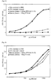
- Fig. 1a - 1d:
- A comparison of retrieval of native staphylococcus aureus protein A (SPA) by means of ELISA added to human plasma in relation to the incubation period at 100°C (in a water bath, 0, 30, 60, and 120 min). These samples were diluted according to the method of the present invention.
- Fig. 1e:
- A comparison of the availablity of SPA in the assay if the sample aliquots shown in
Fig. 1d were diluted in dilution buffer in the assay without adding reagent. - Fig. 1f:
- The results obtained according to the prior art method from these samples (acid dissociation).
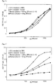
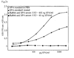
- Fig. 2a - 2c:
- A comparison of the method according to the present invention with the prior art method in the case of protein A retrieval from complexes having formed in rabbit anti-protein A serum.
- Fig. 3a - 3b:
- A comparison of the method according to the present invention with the prior art method in the case of protein A retrieval from complexes having formed in the presence of 1 mg of human polyclonal IgG/ml.
- Fig. 4a - 4b:
- A comparison of the method according to the present invention with the prior art method in the case of protein A retrieval from complexes having formed in the presence of 10 mg of human polyclonal IgG/ml.
- Fig. 5:
- Schematic representation of assay setup and implementation of anti-protein A double antibody sandwich ELISAs.
- The following examples and comparative experiments are intended to further explain the present invention.
- The object of this example is the determination of reactivity of native staphylococcus aureus protein A (SPA) in a specific ELISA in the absence and in the presence of human IgG antibodies having high affinity, the determination of protein release at various incubation periods at 100°C (cf.
Fig. 1a - 1d ), the determination of the extent of renaturation of binding sites if no reagent is added to the sample dilution buffer at an appropriate concentration (Fig. 1e ), and the comparison of the method according to the present invention with the method according to prior art (acid dissociation,Fig. 1f ). - A sensitive anti protein A ELISA was established at the IAM (Institute of Applied Microbiology, Vienna). Assay scheme and implementation are shown in
fig. 5 . A monoclonal mouse anti-SPA (P-2921, Sigma, St. Louis, U.S.A.) was used as a capture antibody. The second antibody in the assay (chicken anti-protein A, Biogenesis 7839-9006) was biotinylated with NHS-biotin (N-hydroxy succin imide activated biotin, Amersham Pharmacia Biotech, England). This ELISA is similarly suitable for both native and recombinant SPA from Amersham Pharmacia Biotech. - Eight 1:2 dilutions of the standard SPA protein were prepared as duplicates on each test plate (standard range 2000-15.6 pg SPA/ml). In these examples, highly pure native SPA (Amersham Pharmacia Biotech, Uppsala, Sweden, Lot no. T-249789) was used as SPA standard and to spike different sample matrixes with antibodies.
- The various standards for protein A (native protein A lot T-249789, recombinant protein A lot T-238881) were obtained in lyophilized form. The standard proteins were weighed in, dissolved in distilled water, then the optical density at 275 nm was determined, and the protein A content was calculated by means of the appropriate molar extinction coefficient. The standard proteins were prediluted in PBS-TP, aliquoted and stored at -30°C.
- SPA negative human plasma was obtained from Octapharma, Vienna.
- EAR400 was used as ELISA Reader, and D-Soft from SLT (Grödig, Austria) was used as evaluation software. SDS was from Merck, Darmstadt, DTPA and polyvinylpyrrolidone (PVP-T40) from Sigma, St. Louis, U.S.A., bovine serum albumin from Hämosan (Ilz, Austria).
-
- 0.1N NaHCO3 buffer, pH = 9.6-9.8
- 8.4 g/l of NaHCO3
- 4.0 g/l of Na2CO3
- storage life: 1 week at 20°C
-
- PBS, pH = 7.2-7.4
- 1.15 g/l of Na2HPO4.2H2O
- 0.2 g/l of KH2PO4
- 0.2 g/l of KCl
- 8.0 g/l of NaCl
- 1 ml of Tween20
- storage life: 1 day at 20°C
- Samples are diluted 1:5 in washing buffer + 5% BSA, then diluted 1:3 in reagent solution (2% SDS 0.1% DTPA), mixed well and incubated at 100°C for 120 min.
- Washing buffer + 1% PVP + reagent solution 1:100 storage life: 1 day at 20°C
- Dilution buffer + 2 mM MgCl2
-
- 10 mM imidazole
- 2 mM MgCl2
- 1M NaCl
- adjust with 1N HCl
- Resorufin β-D-galactopyranside: 1 mg dissolved in 100 µl of DMSO was diluted in 10 ml of staining buffer. 100 µl of staining solution/well applied
- SPA standard (3 mg/ml) was diluted in phosphate buffered saline solution (PBS) with 0.1% Tween20 (PBST) and with 1% polyvinylpyrrolidone (PBS-TP) in a parallel fashion and added to human plasma in 10 times concentrated form (10 plasma + 1 SPA solution). The samples were mixed and incubated at room temperature for 1 h. [To each 2 ml of human plasma were added 200 µl of SPA solution (6 µg/ml or 600 ng/ml) in PBS + 0.1% Tween20 + 1% PVP = 545.4 ng of SPA/ml and 54.5 ng of SPA/ml, respectively.]
- Subsequently, aliquots were treated according to the prior art method and according to the method of the present invention (as described below). The untreated samples were each diluted in the appropriate medium. A 1:2 dilution series was prepared from all treated and untreated samples and standards (8 dilution steps), and aliquots were transferred to the test plates.
- The samples and standards were prediluted in 0.5 M acetate/HCl buffer (pH = 3,5) with 0.1 M NaCl and 0.1% Tween20, and the dilution series for the assays were similarly prepared in this buffer. Aliquots of the dilution series (50 µl) were transferred to 96 well test plates and incubated at room temperature for 60 min.
- The samples were prediluted 1:5 in PBS with 5% BSA (bovine serum albumin) or egg albumin (50 µl of sample + 200 µl of BSA solution), then diluted 1:3 with reaction solution, mixed and incubated in screw-top reaction vessels in a water bath at 100°C for 120 min. Aliquots of the dilution series (50 µl) in PBS-TP + 7 mM SDS and 25 µM DTPA were transferred to 96 well test plates and incubated at room temperature for 60 min.
- The concentration of the reaction solution used in this 1:5 predilution with 5% BSA solution was 70 mM SDS (=2%), 2.5 mM DTPA (=0.1%). This corresponds to a 100-fold molar excess of SDS over protein. In order to prevent renaturation of the antibody heavy chains, the reagent solution was added to the sample dilution buffer at a ratio of 1:100 in the assay. Human plasma was spiked with native SPA (545 ng and 54.5 ng/ml, respectively) and diluted 1:5 in 5% BSA solution and then 1:3 in 70 mM SDS + 2.5 mM DTPA solution. The samples containing 545 ng of SPA were subsequently diluted 1:20 in PBS-TP + reagent (1:100), samples containing 54,5 ng of SPA 1:2, and subsequently eightfold 1:2 in series. The standard protein A (60 ng/ml) was diluted and treated like the sample containing 54.5 ng of SPA/ml (=SPA standard treatment). SPA tends to dimerize in solution. This can lead to reduced detection in the assay. Therefore, SPA was applied diluted only in PBS + 0.1% Tween20 + 1% PVP (standard in PBS) in order to demonstrate that monomerization will also take place under heat treatment for an appropriately long period and at appropriate SDS concentration.
- This mixing ratio of "samples - protein solution - reaction solution" (1 + 4 + 10) is generally suitable for all.common sample matrixes (in protein purification), for plasma and serum samples in order to obtain quantitative release and detection of bound SPA. Due to the high dilution ratio of sample : protein solution (1:5), possible reductions in dissociation, e.g. by too many SDS molecules adsorbing to buffer ions containing amino groups, are sufficiently buffered. If required for sensitivity, the dilution ratio may be reduced, observing the protein : SDS ratio (in the case of samples without buffer systems containing amino groups and having a pH of 5 - 8.5).
-
- Fig. 1a
- shows the results of the anti SPA ELISA without heat treatment. It is obvious that in the human plasma sample, to which 60 ng SPA/ml has been added, no, and in the sample containing 600 ng of SPA/ml, only very little amounts are available in the assay.
- Fig. 1b
- shows that after 30 min of heat treatment at 100°C, SPA in the sample containing 54.5 ng/ml is already quantitatively available, in the sample containing 545 ng of SPA/ml, however, only about 50% are available.
- Fig. 1c
- shows that after 60 min of heat treatment, SPA is almost quantitatively available even in the sample containing 545 ng/ml.
- Fig. 1d
- shows the quantitative release of SPA in both samples (54.5 ng and 545 ng of SPA/ml, respectively) after 120 min of heat treatment at 100°C.
- Figs. 1a to 1d
- clearly show how after 120 min the samples and standard curves become congruent, resulting in 100% retrieval. In order to show that the heavy chains of IgG antibodies may renaturate proportionately after this treatment in the region where protein A binds and may again react with SPA if the SDS concentration is too low, the samples shown in
Fig. 1d were diluted in the assay dilution buffer without adding reagent in a parallel fashion and tested analoguously. - Fig. 1e
- shows how the slope of the sample dilution series and the retrieval changes as compared to the SPA standard treated in the same way if SDS is excessively diluted. This can be prevented by adding 0.7 mM SDS to the sample dilution buffer in the assay, as can be seen in
fig. 1e . - Fig. 1f
- shows how reactivity (slope) of the sample dilution series and retrieval in an acid environment (pH = 3.5) change as compared to the standard in PBS (pH = 7.3) and the SPA standard treated in a similar way. In this method, even very small differences in the pH of the samples will affect the results. The sample containing 545 ng of SPA/ml was prediluted 1:300, and the sample containing 54.5 ng of SPA/ml 1:30, in 0.5M acetate buffer with 0.1M NaCl and 0.1% Tween20.
- Determination of retrieval of SPA from complexes having formed in rabbit anti-SPA serum after addition of SPA and incubation (1 h at 37°C). The presence of many specific antibodies allows even stronger and more intensive crosslinking and complex formation.
- Instead of human plasma, rabbit anti-SPA serum (Amersham Pharmacia Biotech, Uppsala, Sweden; serum no. 1183) was spiked
with native SPA at 2 different concentrations (545 ng/ml and 54.5 ng/ml). Otherwise, this example was carried out as example 1. - SPA standard (3 mg/ml) was diluted in phosphate buffered saline solution (PBS) with 0.1% Tween20 (PBST) and with 1% polyvinylpyrrolidone (PBS-TP) in a parallel fashion and added to rabbit anti SPA serum in 10 times concentrated form (10 serum + 1 SPA solution). The samples were mixed and incubated at room temperature for 1h.
- Subsequently, aliquots were treated according to the prior art method and according to the method of the present invention (as described below). The untreated samples were each diluted in the appropriate medium. A 1:2 dilution series was prepared from all treated and untreated samples and standards (8 dilution steps), and aliquots were transferred to the test plates.
-
- Fig. 2a
- shows the results without heat treatment. It is obvious that no SPA is detectable even in the sample containing 545 ng of SPA/ml.
- Fig. 2b
- shows that after 120 min of heat treatment at 100°C, similarly to the results from human plasma (
Fig. 1d ), SPA added to the two samples (545 ng and 54.5 ng of SPA/ml, respectively) is quantitatively released and detectable. - Fig. 2c
- shows that in this case, no SPA is released and detectable according to the prior art method. The 545 ng of SPA/ml sample was prediluted 1:300, the 54.5 ng of SPA/ml sample 1:30, in 0.5M acetate buffer with 0.1M NaCl and 0.1% Tween 20.
- Determination of retrieval of recombinant SPA from complexes having formed with polyclonal human IgG (1 mg of IgG/ml) after addition of SPA and incubation (1h at 37°C).
- Native as well as differently modified recombinant SPA are used as immunoligands for IgG purification. Therefore, it is important that the method according to the invention for dissociation and the assay system are equally suitable for the various modifications.
- Polyclonal human IgG, pre-purified by means of Cohn precipitation and gel filtration, was spiked with recombinant SPA (Amersham Pharmacia Biotech, Uppsala, Sweden, lot no. 238881) at 2 different concentrations (545 ng/ml and 54.5 ng/ml). Otherwise this example was carried out according to example 1.
- Polyclonal IgG (Cohn fraction, Immuno, Vienna) was concentrated dissolved in PBS (30 mg/ml) and gel filtrated. The concentration was determined by means of anti-human IgG ELISA. Recombinant SPA standard (3 mg/ml) in phosphate buffered saline solution (PBS) was diluted with 0.1% Tween20 (PBST) and with 1% polyvinylpyrrolidone (PBS-TP) in a parallel fashion, and added, in 10 times concentrated form (6 µg/ml and 600 ng/ml, respectively), to the IgG solution at 1.1 mg/ml (10 parts of IgG solution + 1 part of SPA solution). The samples were mixed and incubated at room temperature for 1h.
- Then aliquots were treated according to the prior art method and according to the method of the present invention (as described below). The untreated samples were diluted in the corresponding medium. A 1:2 dilution series (8 dilution steps) was prepared from all treated and untreated samples and standards, and aliquots were transferred to the test plates.
-
- Fig. 3a
- shows that after 120 min of heat treatment at 100°C the added recombinant SPA in both samples (545 ng and 54.5 ng of SPA/ml) is quantitatively released and detectable even in samples containing prepurified polyclonal human IgG at an antibody concentration of 1 mg of IgG/ml.
- Fig. 3b
- shows that reactivity at a pH of 3.5 is highly dependent on the total ionic strength and on slight changes in pH in the samples, and that, above all, undissolved complexes can even give the illusion of an excessively high SPA content as compared to the standard.
- The sample containing 545 ng of SPA/ml was prediluted 1:300, and the sample containing 54.5 ng of SPA/ml 1:30, in 0.5M acetate buffer with 0.1M NaCl and 0.1% Tween20.
- Determination of retrieval of recombinant SPA from complexes having formed at high polyclonal human IgG (10 mg of IgG/ml) content after addition of SPA and incubation (1 h at 37°C).
- Polyclonal human IgG, prepurified by means of Cohn precipitation and gel filtration, was spiked with recombinant SPA (Amersham Pharmacia Biotech, Uppsala, Sweden. lot no. 238881) at 2 different concentrations (545 ng/ml and 54.5 ng/ml). Otherwise, this example was carried out according to example 1.
- Polyclonal IgG (Cohn fraction, Immuno, Vienna) was concentrated dissolved in PBS (30 mg/ml) and gel filtrated. The concentration was determined by means of anti human IgG ELISA. Recombinant SPA standard (3 mg/ml) was diluted in a parallel fashion in phosphate buffered saline solution (PBS) with 0.1% Tween 20 (PBST) and with 1% polyvinylpyrrolidone (PBS-TP), and added, in 10 times concentrated form (6 µg/ml and 600 ng/ml, respectively), to the IgG solution at 11 mg/ml (10 parts of IgG solution + 1 part of SPA solution). The samples were mixed and incubated at room temperature for 1h.
- Then aliquots were treated according to the prior art method and according to the method of the present invention (as described below). The untreated samples were each diluted in the appropriate medium. A 1:2 dilution series (8 dilution steps) was prepared from all treated and untreated samples and standards, and aliquots were transferred to the test plates.
-
- Fig. 4a
- shows that after 120 min of heat treatment at 100°C the added recombinant SPA in both samples (545 ng and 54.5 ng of SPA/ml) is quantitatively released and detectable even in samples containing prepurified polyclonal human IgG at an antibody concentration of 10 mg of IgG/ml.
- Fig. 4b
- shows that reactivity of SPA with IgG is dependent on the concentration ratio (cf.
Fig. 3a ). As opposed to the method according to the present invention, the SPA content in these samples can not be determined correctly by the prior art method (cf.figs. 1-3 ). The sample containing 545 ng of SPA/ml was prediluted 1:300, and the sample containing 54.5 ng of SPA/ml 1:30, in 0.5M acetate buffer with 0.1M NaCl and 0.1% of Tween 20.
Claims (11)
- Method for the quantitative release of thermostable immunoligands able to bind to the Fc-part of immunoglobulins from complexes in various sample matrixes, characterized by mixing the sample with a reagent compound that is able to adsorb or bind non-specifically to immunoglobulins and which has hydrophobic as well as negatively charged ends, and thereafter subjecting the sample to a heat treatment step followed by a cooling step, wherein a buffer and protein are added before the heating step in order to compensate for sample variations in pH and in protein content, respectively.
- Method according to claim 1, characterized in that a chelator is added before the heating step in order to bind heavy metal ions that may be present.
- Method according to one of claims 1 and 2, characterized in that the reagent compound is sodium dodecyl sulfate (SDS).
- Method according to one of claims 1 to 3, characterized in that the concentration range for the reagent compound in the sample before the heating step is within the range of 10 to 40 mM, preferably 30 to 40 mM.
- Method according to one of claims 1 to 4, characterized in that the heating step is applied during a time period within the range of 3 to 180 minutes and at a temperature within the range of 60 to 100°C.
- Method according to one of claims 1 to 5, characterized in that in the cooling step the temperature after the heating step is allowed to go down to a temperature within the range of 0 to 40°C, preferably 4 to 35°C.
- Method according to one of claims 1 to 6, characterized in that the pH during the heating step is within the range of 5 to 8, preferably between 6 to 7.5.
- Method according to one of claims 1 to 7, characterized in that phosphate or borate buffers are added to the sample before the heating step.
- Method according to one of claims 1 to 8, characterized in that the concentration of the buffer in the sample before the heating step is selected within the range of 5 to 100 mM.
- Method according to one of claims 1 to 9, characterized in that the concentration of the added proteins in the sample before the heating step is selected within the range of 1/20 to 1/100 of the concentration of added reagent compound, i.e. 0.1 to 2 mM, preferably 0.3 to 2 mM.
- Method according to one of claims 2 to 10, characterized in that the chelator is present in the sample before the heating step within the concentration range of 0.5 to 5 mM.
Applications Claiming Priority (3)
| Application Number | Priority Date | Filing Date | Title |
|---|---|---|---|
| AT106898 | 1998-06-19 | ||
| AT106898 | 1998-06-19 | ||
| PCT/SE1999/000828 WO1999066325A1 (en) | 1998-06-19 | 1999-05-14 | Method for the quantitative release of natural or recombinant proteins, polypeptides or peptides |
Publications (2)
| Publication Number | Publication Date |
|---|---|
| EP1088227A1 EP1088227A1 (en) | 2001-04-04 |
| EP1088227B1 true EP1088227B1 (en) | 2009-12-16 |
Family
ID=3505919
Family Applications (1)
| Application Number | Title | Priority Date | Filing Date |
|---|---|---|---|
| EP99928306A Expired - Lifetime EP1088227B1 (en) | 1998-06-19 | 1999-05-14 | Method for the quantitative release of natural or recombinant proteins, polypeptides or peptides |
Country Status (7)
| Country | Link |
|---|---|
| US (1) | US6706486B1 (en) |
| EP (1) | EP1088227B1 (en) |
| JP (1) | JP4399115B2 (en) |
| AT (1) | ATE452337T1 (en) |
| DE (1) | DE69941815D1 (en) |
| ES (1) | ES2337639T3 (en) |
| WO (1) | WO1999066325A1 (en) |
Families Citing this family (3)
| Publication number | Priority date | Publication date | Assignee | Title |
|---|---|---|---|---|
| JP2003194775A (en) * | 2001-12-28 | 2003-07-09 | Japan Science & Technology Corp | Electrophoresis of proteins |
| EP2124056A4 (en) * | 2006-12-27 | 2010-06-02 | Japan Science & Tech Agency | IMMUNOLOGICAL DETECTION METHOD USING AN AVIAN ANTIBODY |
| JP5785959B2 (en) * | 2010-01-26 | 2015-09-30 | オブチェストヴォ エス オグラニチェノワ オトヴェトストヴェノステュ “ナウクノ− プロイズヴォドストヴェナヤ ファルマ “マテリア メディカ ホールディング” | Method for measuring autoantibody concentration using enzyme immunoassay |
Family Cites Families (6)
| Publication number | Priority date | Publication date | Assignee | Title |
|---|---|---|---|---|
| US4092114A (en) | 1976-10-20 | 1978-05-30 | Fisher Scientific Company | Indirect latex test for determination of immunoglobulins |
| SE448190B (en) * | 1985-06-03 | 1987-01-26 | Pharmacia Ab | SET TO DETERMINE FC PART BINDING Bacterial POLYPEPTIDES |
| EP0338591A3 (en) | 1988-04-21 | 1991-09-04 | Microprobe Corporation | Nucleic acid extraction method |
| CA2176496C (en) | 1993-11-29 | 1999-09-28 | Kathleen A. Clark | Method for extracting nucleic acids from a wide range of organisms |
| US5556745A (en) * | 1994-06-03 | 1996-09-17 | Sch+E,Uml U+Ee Pbach; J+E,Uml O+Ee Rg | Method for the detection and quantitative determination of antigen in a test sample containing immune complexes of antigen bound to antibodies and to rheumatoid factors |
| AT403378B (en) * | 1995-09-11 | 1998-01-26 | Steindl Franz Dipl Ing Dr | Immune and protein complex dissociation |
-
1999
- 1999-05-14 EP EP99928306A patent/EP1088227B1/en not_active Expired - Lifetime
- 1999-05-14 DE DE69941815T patent/DE69941815D1/en not_active Expired - Lifetime
- 1999-05-14 JP JP2000555093A patent/JP4399115B2/en not_active Expired - Fee Related
- 1999-05-14 AT AT99928306T patent/ATE452337T1/en not_active IP Right Cessation
- 1999-05-14 US US09/719,512 patent/US6706486B1/en not_active Expired - Lifetime
- 1999-05-14 WO PCT/SE1999/000828 patent/WO1999066325A1/en not_active Ceased
- 1999-05-14 ES ES99928306T patent/ES2337639T3/en not_active Expired - Lifetime
Also Published As
| Publication number | Publication date |
|---|---|
| JP4399115B2 (en) | 2010-01-13 |
| ATE452337T1 (en) | 2010-01-15 |
| US6706486B1 (en) | 2004-03-16 |
| WO1999066325A1 (en) | 1999-12-23 |
| EP1088227A1 (en) | 2001-04-04 |
| DE69941815D1 (en) | 2010-01-28 |
| ES2337639T3 (en) | 2010-04-27 |
| JP2002518675A (en) | 2002-06-25 |
Similar Documents
| Publication | Publication Date | Title |
|---|---|---|
| Jeon et al. | Biotin–avidin mediated competitive enzyme-linked immunosorbent assay to detect residues of tetracyclines in milk | |
| Hsu et al. | Protein A, avidin, and biotin in immunohistochemistry. | |
| EP0185870B2 (en) | Immunoassays for denatured protein analytes, particularly HB Alc, and monoclonal antibodies thereto | |
| Thompson et al. | Detection of staphylococcal enterotoxins by enzyme-linked immunosorbent assays and radioimmunoassays: comparison of monoclonal and polyclonal antibody systems | |
| EP0930366A3 (en) | Binding domains in delta proteins | |
| JPH068822B2 (en) | Protected binding assay | |
| Masuho et al. | Development of an intravenous γ-globulin with Fc activities | |
| Stiffler-Rosenberg et al. | Simple assay for staphylococcal enterotoxins A, B, and C: modification of enzyme-linked immunosorbent assay | |
| Rungsiwongse et al. | Development of an ELISA to assess the potency of horse therapeutic antivenom against Thai cobra venom | |
| KR100249752B1 (en) | Interference-suppressing reagent specific to certain classes of antibodies | |
| US6686169B2 (en) | Reagent for the detection of Staphylococcus aureus by agglutination | |
| US6706486B1 (en) | Method for the quantitative release of natural or recombinant proteins, polypeptides or peptides | |
| Herrmann et al. | Quantitation of immunoglobulin adsorption to plastics | |
| JPS6120815B2 (en) | ||
| GB2175906A (en) | Assay methods and reagents | |
| Peng et al. | Binding properties of protein A and protein G for human IgE | |
| Pachmann et al. | Insolubilization of protein antigens on polyacrylic plastic beads using poly-L-lysine | |
| Klonisch et al. | Enhancement in antigen binding by a combination of synergy and antibody capture | |
| Limet et al. | Particle counting immunoassay. IV. The use of F (ab′) 2 fragments and Nϵ-chloroacetyl lysine N-carboxyanhydride for their coupling to polystyrene latex particles | |
| JPS58146855A (en) | Method for testing immune | |
| Rej et al. | Direct immunological determination of aspartate aminotransferase isoenzymes. | |
| Wilson | Studies on the products of peptic digestion of IgA | |
| JPH06100603B2 (en) | Method for quantifying specific bacterial polypeptide and antibody thereto | |
| Lindahl et al. | Nonimmune binding of Ig to Clostridium perfringens. Preferential binding of IgM and aggregated IgG. | |
| EA200100287A1 (en) | HIGH-AFFINITY ANTIBODIES |
Legal Events
| Date | Code | Title | Description |
|---|---|---|---|
| PUAI | Public reference made under article 153(3) epc to a published international application that has entered the european phase |
Free format text: ORIGINAL CODE: 0009012 |
|
| 17P | Request for examination filed |
Effective date: 20001211 |
|
| AK | Designated contracting states |
Kind code of ref document: A1 Designated state(s): AT BE CH DE DK ES FR GB IE IT LI NL SE |
|
| RAP1 | Party data changed (applicant data changed or rights of an application transferred) |
Owner name: AMERSHAM BIOSCIENCES AB |
|
| RAP1 | Party data changed (applicant data changed or rights of an application transferred) |
Owner name: GE HEALTHCARE BIO-SCIENCES AB |
|
| 17Q | First examination report despatched |
Effective date: 20060306 |
|
| GRAP | Despatch of communication of intention to grant a patent |
Free format text: ORIGINAL CODE: EPIDOSNIGR1 |
|
| GRAS | Grant fee paid |
Free format text: ORIGINAL CODE: EPIDOSNIGR3 |
|
| GRAA | (expected) grant |
Free format text: ORIGINAL CODE: 0009210 |
|
| AK | Designated contracting states |
Kind code of ref document: B1 Designated state(s): AT BE CH DE DK ES FR GB IE IT LI NL SE |
|
| REG | Reference to a national code |
Ref country code: GB Ref legal event code: FG4D |
|
| REG | Reference to a national code |
Ref country code: CH Ref legal event code: EP |
|
| REG | Reference to a national code |
Ref country code: IE Ref legal event code: FG4D |
|
| REF | Corresponds to: |
Ref document number: 69941815 Country of ref document: DE Date of ref document: 20100128 Kind code of ref document: P |
|
| REG | Reference to a national code |
Ref country code: CH Ref legal event code: NV Representative=s name: ISLER & PEDRAZZINI AG |
|
| REG | Reference to a national code |
Ref country code: NL Ref legal event code: VDEP Effective date: 20091216 |
|
| REG | Reference to a national code |
Ref country code: ES Ref legal event code: FG2A Ref document number: 2337639 Country of ref document: ES Kind code of ref document: T3 |
|
| PG25 | Lapsed in a contracting state [announced via postgrant information from national office to epo] |
Ref country code: SE Free format text: LAPSE BECAUSE OF FAILURE TO SUBMIT A TRANSLATION OF THE DESCRIPTION OR TO PAY THE FEE WITHIN THE PRESCRIBED TIME-LIMIT Effective date: 20091216 |
|
| PG25 | Lapsed in a contracting state [announced via postgrant information from national office to epo] |
Ref country code: AT Free format text: LAPSE BECAUSE OF FAILURE TO SUBMIT A TRANSLATION OF THE DESCRIPTION OR TO PAY THE FEE WITHIN THE PRESCRIBED TIME-LIMIT Effective date: 20091216 |
|
| PG25 | Lapsed in a contracting state [announced via postgrant information from national office to epo] |
Ref country code: NL Free format text: LAPSE BECAUSE OF FAILURE TO SUBMIT A TRANSLATION OF THE DESCRIPTION OR TO PAY THE FEE WITHIN THE PRESCRIBED TIME-LIMIT Effective date: 20091216 |
|
| PG25 | Lapsed in a contracting state [announced via postgrant information from national office to epo] |
Ref country code: BE Free format text: LAPSE BECAUSE OF FAILURE TO SUBMIT A TRANSLATION OF THE DESCRIPTION OR TO PAY THE FEE WITHIN THE PRESCRIBED TIME-LIMIT Effective date: 20091216 |
|
| PLBE | No opposition filed within time limit |
Free format text: ORIGINAL CODE: 0009261 |
|
| STAA | Information on the status of an ep patent application or granted ep patent |
Free format text: STATUS: NO OPPOSITION FILED WITHIN TIME LIMIT |
|
| 26N | No opposition filed |
Effective date: 20100917 |
|
| PG25 | Lapsed in a contracting state [announced via postgrant information from national office to epo] |
Ref country code: DK Free format text: LAPSE BECAUSE OF FAILURE TO SUBMIT A TRANSLATION OF THE DESCRIPTION OR TO PAY THE FEE WITHIN THE PRESCRIBED TIME-LIMIT Effective date: 20091216 |
|
| PG25 | Lapsed in a contracting state [announced via postgrant information from national office to epo] |
Ref country code: IT Free format text: LAPSE BECAUSE OF FAILURE TO SUBMIT A TRANSLATION OF THE DESCRIPTION OR TO PAY THE FEE WITHIN THE PRESCRIBED TIME-LIMIT Effective date: 20091216 |
|
| PGFP | Annual fee paid to national office [announced via postgrant information from national office to epo] |
Ref country code: DE Payment date: 20120529 Year of fee payment: 14 Ref country code: CH Payment date: 20120525 Year of fee payment: 14 Ref country code: IE Payment date: 20120525 Year of fee payment: 14 |
|
| PGFP | Annual fee paid to national office [announced via postgrant information from national office to epo] |
Ref country code: FR Payment date: 20120607 Year of fee payment: 14 Ref country code: GB Payment date: 20120525 Year of fee payment: 14 |
|
| PGFP | Annual fee paid to national office [announced via postgrant information from national office to epo] |
Ref country code: ES Payment date: 20120528 Year of fee payment: 14 |
|
| REG | Reference to a national code |
Ref country code: CH Ref legal event code: PL |
|
| GBPC | Gb: european patent ceased through non-payment of renewal fee |
Effective date: 20130514 |
|
| PG25 | Lapsed in a contracting state [announced via postgrant information from national office to epo] |
Ref country code: DE Free format text: LAPSE BECAUSE OF NON-PAYMENT OF DUE FEES Effective date: 20131203 Ref country code: CH Free format text: LAPSE BECAUSE OF NON-PAYMENT OF DUE FEES Effective date: 20130531 Ref country code: LI Free format text: LAPSE BECAUSE OF NON-PAYMENT OF DUE FEES Effective date: 20130531 |
|
| REG | Reference to a national code |
Ref country code: IE Ref legal event code: MM4A |
|
| REG | Reference to a national code |
Ref country code: DE Ref legal event code: R119 Ref document number: 69941815 Country of ref document: DE Effective date: 20131203 |
|
| REG | Reference to a national code |
Ref country code: FR Ref legal event code: ST Effective date: 20140131 |
|
| PG25 | Lapsed in a contracting state [announced via postgrant information from national office to epo] |
Ref country code: IE Free format text: LAPSE BECAUSE OF NON-PAYMENT OF DUE FEES Effective date: 20130514 Ref country code: GB Free format text: LAPSE BECAUSE OF NON-PAYMENT OF DUE FEES Effective date: 20130514 |
|
| PG25 | Lapsed in a contracting state [announced via postgrant information from national office to epo] |
Ref country code: FR Free format text: LAPSE BECAUSE OF NON-PAYMENT OF DUE FEES Effective date: 20130531 |
|
| REG | Reference to a national code |
Ref country code: ES Ref legal event code: FD2A Effective date: 20140606 |
|
| PG25 | Lapsed in a contracting state [announced via postgrant information from national office to epo] |
Ref country code: ES Free format text: LAPSE BECAUSE OF NON-PAYMENT OF DUE FEES Effective date: 20130515 |