EP1085469A2 - Methods and apparatus for pre-filtering in image reconstruction - Google Patents
Methods and apparatus for pre-filtering in image reconstruction Download PDFInfo
- Publication number
- EP1085469A2 EP1085469A2 EP00307191A EP00307191A EP1085469A2 EP 1085469 A2 EP1085469 A2 EP 1085469A2 EP 00307191 A EP00307191 A EP 00307191A EP 00307191 A EP00307191 A EP 00307191A EP 1085469 A2 EP1085469 A2 EP 1085469A2
- Authority
- EP
- European Patent Office
- Prior art keywords
- weighting function
- panel
- data
- accordance
- edge
- Prior art date
- Legal status (The legal status is an assumption and is not a legal conclusion. Google has not performed a legal analysis and makes no representation as to the accuracy of the status listed.)
- Withdrawn
Links
Images

Classifications
-
- G—PHYSICS
- G06—COMPUTING OR CALCULATING; COUNTING
- G06T—IMAGE DATA PROCESSING OR GENERATION, IN GENERAL
- G06T12/00—Tomographic reconstruction from projections
- G06T12/10—Image preprocessing, e.g. calibration, positioning of sources or scatter correction
-
- G—PHYSICS
- G06—COMPUTING OR CALCULATING; COUNTING
- G06T—IMAGE DATA PROCESSING OR GENERATION, IN GENERAL
- G06T2211/00—Image generation
- G06T2211/40—Computed tomography
- G06T2211/432—Truncation
-
- Y—GENERAL TAGGING OF NEW TECHNOLOGICAL DEVELOPMENTS; GENERAL TAGGING OF CROSS-SECTIONAL TECHNOLOGIES SPANNING OVER SEVERAL SECTIONS OF THE IPC; TECHNICAL SUBJECTS COVERED BY FORMER USPC CROSS-REFERENCE ART COLLECTIONS [XRACs] AND DIGESTS
- Y10—TECHNICAL SUBJECTS COVERED BY FORMER USPC
- Y10S—TECHNICAL SUBJECTS COVERED BY FORMER USPC CROSS-REFERENCE ART COLLECTIONS [XRACs] AND DIGESTS
- Y10S378/00—X-ray or gamma ray systems or devices
- Y10S378/901—Computer tomography program or processor
Definitions
- This invention relates generally to image reconstruction using data collected in a computed tomography scan and, more particularly, to generating an volumetric computed tomography imaging using half-projection data.
- an x-ray source projects a fan-shaped beam which is collimated to lie within an X-Y plane of a Cartesian coordinate system and generally referred to as the "imaging plane".
- the x-ray beam passes through the object being imaged, such as a patient.
- the beam after being attenuated by the object, impinges upon an array of radiation detectors.
- the intensity of the attenuated beam radiation received at the detector array is dependent upon the attenuation of the x-ray beam by the object.
- Each detector element of the array produces a separate electrical signal that is a measurement of the beam attenuation at the detector location.
- the attenuation measurements from all the detectors are acquired separately to produce a transmission profile.
- the x-ray source and the detector array are rotated with a gantry within the imaging plane and around the object to be imaged so that the angle at which the x-ray beam intersects the object constantly changes.
- a group of x-ray attenuation measurements, i.e., projection data, from the detector array at one gantry angle is referred to as a "view”.
- a "scan" of the object comprises a set of views made at different gantry angles, or view angles, during one revolution of the x-ray source and detector.
- the projection data is processed to construct an image that corresponds to a two dimensional slice taken through the object.
- CT numbers integers called "CT numbers” or “Hounsfield units”, which are used to control the brightness of a corresponding pixel on a cathode ray tube display.
- volumetric CT data is collected using a large flat panel digital x-ray device, or detector, having a plurality of pixels arranged in rows and columns.
- the imaging system includes an x-ray source and at least one solid-state x-ray detector.
- the x-ray source and the x-ray detector are rotated around the object of interest. For each identified view, x-rays are emitted from the x-ray source toward the x-ray detector and projection data is collected for the view.
- the panel can be offset from iso-center to increase scan field of view by utilizing 2 ⁇ rotation of projections. In such a configuration, each projection is truncated and image artifacts typically result.
- the method includes the steps of selecting a weighting function which satisfies the following criteria.
- a computed tomograph (CT) imaging system 10 is shown as including a gantry 12 representative of a "third generation" CT scanner.
- Gantry 12 has an x-ray source 14 that projects a beam of x-rays 16 toward a detector array 18 on the opposite side of gantry 12.
- Detector array 18 is formed by detector elements 20 which together sense the projected x-rays that pass through an object 22, for example a medical patient.
- Detector array 18 may be fabricated in a single slice or multi-slice configuration.
- Each detector element 20 produces an electrical signal that represents the intensity of an impinging x-ray beam and hence the attenuation of the beam as it passes through patient 22.
- gantry 12 and the components mounted thereon rotate about a center of rotation 24.
- Control mechanism 26 includes an x-ray controller 28 that provides power and timing signals to x-ray source 14 and a gantry motor controller 30 that controls the rotational speed and position of gantry 12.
- a data acquisition system (DAS) 32 in control mechanism 26 samples analog data from detector elements 20 and converts the data to digital signals for subsequent processing.
- An image reconstructor 34 receives sampled and digitized x-ray data from DAS 32 and performs high speed image reconstruction. The reconstructed image is applied as an input to a computer 36 which stores the image in a mass storage device 38.
- DAS data acquisition system
- Computer 36 also receives commands and scanning parameters from an operator via console 40 that has a keyboard.
- An associated cathode ray tube display 42 allows the operator to observe the reconstructed image and other data from computer 36.
- the operator supplied commands and parameters are used by computer 36 to provide control signals and information to DAS 32, x-ray controller 28 and gantry motor controller 30.
- computer 36 operates a table motor controller 44 which controls a motorized table 46 to position patient 22 in gantry 12. Particularly, table 46 moves portions of patient 22 through gantry opening 48.
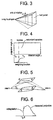
- Figure 3 illustrates the geometry of a half panel CT system.
- a single panel covers slightly over half the field of view, and the x-ray source and the panel rotate 360 degrees. By reducing the data collection requirement to about one-half the data collected by a panel, the useful scan field of view is increased.
- the weighting algorithm may be performed, for example, by a processor in image reconstructor 34 or by computer 36.
- a weighing function using the redundant samples of the projection data in 2 ⁇ range can be utilized to weight the collected data.
- the samples ( ⁇ , ⁇ ) and ( - ⁇ , ⁇ + ⁇ -2 ⁇ ) form a conjugate sampling pair where ⁇ is a fan angle and ⁇ a projection view angle.
- Figure 4 illustrates a Radon space sampling pattern.
- the weighting function satisfies the following three conditions:
- conjugate is used instead of conjugate because the two rays are not redundant samples.
- the rays merely share the property that their ⁇ angles are 180 degrees apart.
- each projection sample is denoted as ( ⁇ , ⁇ , ⁇ ,) where ⁇ , ⁇ , and ⁇ are the fan angle, projection view angle, and tilt angle, respectively.
- ⁇ , ⁇ , and ⁇ are the fan angle, projection view angle, and tilt angle, respectively.
- Equation 1 A simple example of the weighting function that satisfies this condition is described in Equation 1. The weights remain constant for the entire range of ⁇ .
- the size of the panel is slightly larger than the radius of the field of view (to cover the range - ⁇ o ⁇ ⁇ 0).
- the weighting algorithm can further be configured so that no additional overlapped channels are required.
- the projection data at the edge of the panel is further extrapolated for the range - ⁇ o ⁇ ⁇ 0.
- Such extrapolation can be performed using many known extrapolation techniques.
- the extrapolated data could be formed based on the slope of the projection data near the edge or a polynomial fit of the projection data at the edge of the projection can be performed. The results of the extrapolation is illustrated in Figure 6.
- the above weighting scheme is then applied to suppress the discontinuity of the projection at the edge. Since the extrapolated data is, in general, less reliable than the real measurement, the weighting function can be configured to provide higher weights to the measured data than to the extrapolated data.
- CT system described herein is a "third generation” system in which both the x-ray source and detector rotate with the gantry.
- CT systems including "fourth generation” systems wherein the detector is a full-ring stationary detector and only the x-ray source rotates with the gantry, may be used if individual detector elements are corrected to provide substantially uniform responses to a given x-ray beam.
- the system described herein performs an axial scan, however, the invention may be used with a helical scan although more than 360° of data are required.
Landscapes
- Physics & Mathematics (AREA)
- General Physics & Mathematics (AREA)
- Engineering & Computer Science (AREA)
- Theoretical Computer Science (AREA)
- Apparatus For Radiation Diagnosis (AREA)
- Image Processing (AREA)
Abstract
Description
- (a)
- A sum of weights of conjugate samples equals unity.
- (b)
- The weighting function is continuous and differentiable along γ where γ is the fan angle.
- (c)
- The weighting function approaches zero near an edge of the panel and approaches unity near a panel boundary.
- (d)
- The weighting function remains constant for a range ξ1<ξ<ξ2, where ξ1 and ξ2 are determined based on an end intersecting point of a complimentary ray within a reconstruction field of view.



Claims (13)
- A method for weighting data collected from a portion of a digital flat panel in a computed tomograph system, said method comprising the steps of:selecting a weighting function in which a sum of weights of complementary samples equals unity, the weighting function is continuous and differentiable along γ where γ is the fan angle, the weighting function approaches zero near an edge of the panel and approaches unity near a panel boundary, and the weighting function remains constant for a range ξ1< ξ<ξ2 where ξ1 and ξ2 are determined based on an end intersecting point of a complimentary ray within a reconstruction field of view; andapplying the weighting function to the data.
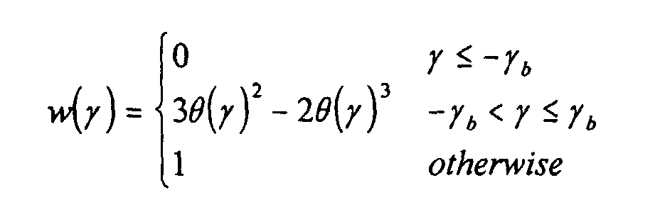
- A method in accordance with Claim 1 wherein the weighting function is:where:

- A method in accordance with Claim 1 further comprising the step of extrapolating projection data at the edge of the panel for a range -γo<γ <0.
- A method in accordance with Claim 3 wherein the extrapolation is based on a slope of the projection data near the panel edge.
- A method in accordance with Claim 3 wherein the extrapolation is based on a polynomial fit of the projection data at the edge of the panel
- A method in accordance with Claim 3 wherein the weighting function provides higher weights to the measured data than to the extrapolated data.
- Apparatus for reconstructing an image using projection data collected in a computed tomograph scan, said apparatus comprising a processor programmed to:obtain the projection data; andapply a weighting function to the projection data, the weighting function providing that a sum of weights of complementary samples equals unity, being continuous and differentiable along γ where γ is the fan angle, approaching zero near an edge of the panel and approaching unity near a panel boundary, and remaining constant for a range ξ1<ξ<ξ2, where ξ1 and ξ2 are determined based on an end intersecting point of a complimentary ray within a reconstruction field of view.
- A computed tomograph apparatus comprising a flat panel digital detector for collecting data, and a processor programmed to weight the collected data by applying a weighting function to the projection data, said weighting function providing that a sum of weights of complementary samples equals unity, said weighting function being continuous and differentiable along γ where γ is the fan angle, said weighting function approaching zero near an edge of said panel and approaching unity near a panel boundary, said weighting function remaining constant for a range ξ1<ξ<ξ2, where ξ1 and ξ2 are determined based on an end intersecting point of a complimentary ray within a reconstruction field of view.
- Apparatus in accordance with Claim 7 or claim 8 wherein the weighting function is:where
- Apparatus in accordance with Claim 7 or claim 8 wherein the processor is further programmed to extrapolate projection data at the edge of the panel for a range -γo <γ <0.
- Apparatus in accordance with Claim 10 wherein the processor is programmed to perform the extrapolation based on a slope of the projection data near the panel edge.
- Apparatus in accordance with Claim 10 wherein the processor is programmed to perform the extrapolation based on a polynomial fit of the projection data at the edge of the panel.
- Apparatus in accordance with Claim 10 wherein the processor is programmed to provide higher weights to the measured data than to the extrapolated data.
Applications Claiming Priority (2)
| Application Number | Priority Date | Filing Date | Title |
|---|---|---|---|
| US390449 | 1995-02-17 | ||
| US09/390,449 US6269139B1 (en) | 1999-09-07 | 1999-09-07 | Methods and apparatus for pre-filtering weighting in image reconstruction |
Publications (2)
| Publication Number | Publication Date |
|---|---|
| EP1085469A2 true EP1085469A2 (en) | 2001-03-21 |
| EP1085469A3 EP1085469A3 (en) | 2004-03-31 |
Family
ID=23542499
Family Applications (1)
| Application Number | Title | Priority Date | Filing Date |
|---|---|---|---|
| EP00307191A Withdrawn EP1085469A3 (en) | 1999-09-07 | 2000-08-22 | Methods and apparatus for pre-filtering in image reconstruction |
Country Status (5)
| Country | Link |
|---|---|
| US (1) | US6269139B1 (en) |
| EP (1) | EP1085469A3 (en) |
| JP (1) | JP2001128964A (en) |
| CN (1) | CN1287327A (en) |
| IL (1) | IL138112A (en) |
Cited By (1)
| Publication number | Priority date | Publication date | Assignee | Title |
|---|---|---|---|---|
| WO2008024327A2 (en) | 2006-08-22 | 2008-02-28 | Analog Devices, Inc. | Method for determining the change of a signal, and an apparatus including a circuit arranged to implement the method |
Families Citing this family (12)
| Publication number | Priority date | Publication date | Assignee | Title |
|---|---|---|---|---|
| US6980681B1 (en) | 2000-04-24 | 2005-12-27 | Ge Medical Systems Global Technology Company, Llc | Methods and apparatus for helical reconstruction for multislice CT scan |
| US6463118B2 (en) | 2000-12-29 | 2002-10-08 | Ge Medical Systems Global Technology Company, Llc | Computed tomography (CT) weighting for high quality image recontruction |
| US7072436B2 (en) * | 2001-08-24 | 2006-07-04 | The Board Of Trustees Of The Leland Stanford Junior University | Volumetric computed tomography (VCT) |
| US6678346B2 (en) * | 2001-10-11 | 2004-01-13 | Ge Medical Systems Global Technology Company Llc | Cone-beam CT scanner with image reconstruction using multiple sub-images |
| US6404842B1 (en) | 2001-10-15 | 2002-06-11 | Ge Medical Systems Global Technology Company, Llc | Method and apparatus for image reconstruction in twin helical computed tomographic systems |
| US6587537B1 (en) | 2002-04-01 | 2003-07-01 | Ge Medical Systems Global Technology Company, Llc | Methods and apparatus for multi-slice image reconstruction |
| US7415145B2 (en) * | 2003-12-30 | 2008-08-19 | General Electric Company | Methods and apparatus for artifact reduction |
| US7050527B2 (en) * | 2004-02-09 | 2006-05-23 | G.E. Medical Systems Global Technology Company, Llc | Methods and apparatus for artifact reduction in cone beam CT image reconstruction |
| US20050201605A1 (en) * | 2004-03-11 | 2005-09-15 | Jianying Li | Methods and apparatus for CT smoothing to reduce artifacts |
| US7173996B2 (en) * | 2004-07-16 | 2007-02-06 | General Electric Company | Methods and apparatus for 3D reconstruction in helical cone beam volumetric CT |
| US8284892B2 (en) * | 2008-12-22 | 2012-10-09 | General Electric Company | System and method for image reconstruction |
| EP3254623B1 (en) * | 2016-06-09 | 2020-09-16 | Agfa Nv | Method and system for correcting geometric misalignment during image reconstruction in chest tomosynthesis |
Family Cites Families (2)
| Publication number | Priority date | Publication date | Assignee | Title |
|---|---|---|---|---|
| US4703424A (en) * | 1985-01-03 | 1987-10-27 | General Electric Company | Method and means of correcting for a shift in the center of rotation of a rotating fan beam CT system |
| EP0489907A1 (en) * | 1990-07-02 | 1992-06-17 | Varian Associates, Inc. | Partial fan beam tomographic apparatus and data reconstruction method |
-
1999
- 1999-09-07 US US09/390,449 patent/US6269139B1/en not_active Expired - Lifetime
-
2000
- 2000-08-22 EP EP00307191A patent/EP1085469A3/en not_active Withdrawn
- 2000-08-27 IL IL13811200A patent/IL138112A/en not_active IP Right Cessation
- 2000-09-06 JP JP2000269439A patent/JP2001128964A/en not_active Withdrawn
- 2000-09-07 CN CN00126337A patent/CN1287327A/en active Pending
Cited By (1)
| Publication number | Priority date | Publication date | Assignee | Title |
|---|---|---|---|---|
| WO2008024327A2 (en) | 2006-08-22 | 2008-02-28 | Analog Devices, Inc. | Method for determining the change of a signal, and an apparatus including a circuit arranged to implement the method |
Also Published As
| Publication number | Publication date |
|---|---|
| IL138112A0 (en) | 2001-10-31 |
| JP2001128964A (en) | 2001-05-15 |
| EP1085469A3 (en) | 2004-03-31 |
| US6269139B1 (en) | 2001-07-31 |
| CN1287327A (en) | 2001-03-14 |
| IL138112A (en) | 2004-03-28 |
Similar Documents
| Publication | Publication Date | Title |
|---|---|---|
| US6421412B1 (en) | Dual cardiac CT scanner | |
| US6754299B2 (en) | Methods and apparatus for weighting of computed tomography data | |
| JP4367884B2 (en) | Method and apparatus for leveling calcification | |
| US6421411B1 (en) | Methods and apparatus for helical image artifact reduction | |
| US6452996B1 (en) | Methods and apparatus utilizing generalized helical interpolation algorithm | |
| US6366638B1 (en) | Methods and apparatus for CT scout image processing | |
| EP1113396B1 (en) | Method and apparauts for multislice CT using partial scan | |
| US6266388B1 (en) | Methods and apparatus for two-pass cone beam image reconstruction | |
| US5606585A (en) | Methods and apparatus for multislice helical image reconstruction in a computer tomography system | |
| EP1149558A2 (en) | Apparatus for region of interest multislice CT scan | |
| US6341154B1 (en) | Methods and apparatus for fast CT imaging helical weighting | |
| IL124014A (en) | Methods and apparatus for scanning an object in a computed tomography system | |
| US6381297B1 (en) | High pitch reconstruction of multislice CT scans | |
| US6269139B1 (en) | Methods and apparatus for pre-filtering weighting in image reconstruction | |
| US5812628A (en) | Methods and apparatus for detecting partial volume image artifacts | |
| US6343110B1 (en) | Methods and apparatus for submillimeter CT slices with increased coverage | |
| EP0989521B1 (en) | Fluoroscopy image reconstruction | |
| US5974110A (en) | Helical reconstruction algorithm | |
| US6980681B1 (en) | Methods and apparatus for helical reconstruction for multislice CT scan | |
| US5974109A (en) | Methods and apparatus for cell ganging in a multislice computed tomography system | |
| IL128035A (en) | Methods and apparatus to desensitize incident angle errors on a multi-slice computed tomograph detector | |
| US6798860B1 (en) | Methods and apparatus for deconvolving imaging data | |
| JPH09182745A (en) | Computer tomography | |
| US6418183B1 (en) | Methods and apparatus for two-pass CT imaging | |
| US6263040B1 (en) | Methods and apparatus for cone-tilted parallel sampling and reconstruction |
Legal Events
| Date | Code | Title | Description |
|---|---|---|---|
| PUAI | Public reference made under article 153(3) epc to a published international application that has entered the european phase |
Free format text: ORIGINAL CODE: 0009012 |
|
| AK | Designated contracting states |
Kind code of ref document: A2 Designated state(s): AT BE CH CY DE DK ES FI FR GB GR IE IT LI LU MC NL PT SE |
|
| AX | Request for extension of the european patent |
Free format text: AL;LT;LV;MK;RO;SI |
|
| PUAL | Search report despatched |
Free format text: ORIGINAL CODE: 0009013 |
|
| AK | Designated contracting states |
Kind code of ref document: A3 Designated state(s): AT BE CH CY DE DK ES FI FR GB GR IE IT LI LU MC NL PT SE |
|
| AX | Request for extension of the european patent |
Extension state: AL LT LV MK RO SI |
|
| AKX | Designation fees paid | ||
| REG | Reference to a national code |
Ref country code: DE Ref legal event code: 8566 |
|
| STAA | Information on the status of an ep patent application or granted ep patent |
Free format text: STATUS: THE APPLICATION IS DEEMED TO BE WITHDRAWN |
|
| 18D | Application deemed to be withdrawn |
Effective date: 20041001 |