EP1065278A2 - Detection of partly complementary nucleic acid fragment - Google Patents
Detection of partly complementary nucleic acid fragment Download PDFInfo
- Publication number
- EP1065278A2 EP1065278A2 EP00112235A EP00112235A EP1065278A2 EP 1065278 A2 EP1065278 A2 EP 1065278A2 EP 00112235 A EP00112235 A EP 00112235A EP 00112235 A EP00112235 A EP 00112235A EP 1065278 A2 EP1065278 A2 EP 1065278A2
- Authority
- EP
- European Patent Office
- Prior art keywords
- fragment
- nucleic acid
- dna
- electric current
- pna
- Prior art date
- Legal status (The legal status is an assumption and is not a legal conclusion. Google has not performed a legal analysis and makes no representation as to the accuracy of the status listed.)
- Withdrawn
Links
- 0 C*1C=CC*1 Chemical compound C*1C=CC*1 0.000 description 3
Images


Classifications
-
- C—CHEMISTRY; METALLURGY
- C12—BIOCHEMISTRY; BEER; SPIRITS; WINE; VINEGAR; MICROBIOLOGY; ENZYMOLOGY; MUTATION OR GENETIC ENGINEERING
- C12Q—MEASURING OR TESTING PROCESSES INVOLVING ENZYMES, NUCLEIC ACIDS OR MICROORGANISMS; COMPOSITIONS OR TEST PAPERS THEREFOR; PROCESSES OF PREPARING SUCH COMPOSITIONS; CONDITION-RESPONSIVE CONTROL IN MICROBIOLOGICAL OR ENZYMOLOGICAL PROCESSES
- C12Q1/00—Measuring or testing processes involving enzymes, nucleic acids or microorganisms; Compositions therefor; Processes of preparing such compositions
- C12Q1/68—Measuring or testing processes involving enzymes, nucleic acids or microorganisms; Compositions therefor; Processes of preparing such compositions involving nucleic acids
- C12Q1/6813—Hybridisation assays
- C12Q1/6816—Hybridisation assays characterised by the detection means
- C12Q1/6825—Nucleic acid detection involving sensors
-
- Y—GENERAL TAGGING OF NEW TECHNOLOGICAL DEVELOPMENTS; GENERAL TAGGING OF CROSS-SECTIONAL TECHNOLOGIES SPANNING OVER SEVERAL SECTIONS OF THE IPC; TECHNICAL SUBJECTS COVERED BY FORMER USPC CROSS-REFERENCE ART COLLECTIONS [XRACs] AND DIGESTS
- Y10—TECHNICAL SUBJECTS COVERED BY FORMER USPC
- Y10T—TECHNICAL SUBJECTS COVERED BY FORMER US CLASSIFICATION
- Y10T436/00—Chemistry: analytical and immunological testing
- Y10T436/14—Heterocyclic carbon compound [i.e., O, S, N, Se, Te, as only ring hetero atom]
- Y10T436/142222—Hetero-O [e.g., ascorbic acid, etc.]
- Y10T436/143333—Saccharide [e.g., DNA, etc.]
Definitions
- This invention relates to a detection of a partly complementary nucleic acid fragment.
- the detection of a partly complementary nucleic acid fragment is of value in the gene analyses such as analysis of gene polymorphism and analysis of variation of abnormal gene.
- the gene analysis is recently paid an attention in developing gene technology.
- SSCP single-stranded conformation polymorphism
- Fig. 1 The procedure of SSCP for differentiating an abnormal gene fragment from a normal gene fragment is illustrated in Fig. 1 attached to the specification.
- Fig. 1 Each of double stranded normal gene fragments (11a) and double stranded abnormal gene fragments (11b, which is different from the normal gene fragment 11a in the base unit positioned in the crossed place) is treated (31) to give a set of two single stranded gene fragments (21a, 21b), as illustrated in Fig. 1-(A). Both of the single stranded gene fragments (21a, 21b) are subjected to electrophoresis on a polyacrylamide gel.
- Each gene fragment has a specific high-order structure differing from each other. Accordingly, each of the single stranded gene fragments (21a, 21b) moves differently from each other in the electrophoresis, as illustrated in Fig. 1-(B). The detection of the movement of the gene fragment gives an information of variation of gene fragments.
- the SSCP method sometimes fails to differentiate an abnormal gene fragment from a normal gene fragment, because a certain abnormal gene fragment moves similarly to the corresponding normal gene fragment. Particularly, if the variation of base takes place in the vicinity of a gene fragment, it is likely that the abnormal gene fragment takes almost the same high-order structure as that of the corresponding normal gene fragment, and hence that there is caused almost no difference of movement in the electrophoresis.
- the variation of gene fragment from a single stranded normal gene fragment to a single stranded abnormal gene fragment can be also detected by way of hybridization.
- a hybrid structure is formed differently between the normal gene fragment and the abnormal gene fragment.
- Japanese Patent PCT Publication 9-501561 describes detection of a double-stranded nucleic acid hybrid having a specific structure by means of a fluorescent microscope.
- Japanese Patent Provisional Publication No. 9-288080 describes a method for detecting a double stranded DNA fragment of a full-match structure by way of an electrochemical detection method.
- a sample DNA fragment is electrochemically detected using a DNA probe which is complementary to the sample DNA fragment in the presence of an electrochemically active thread intercalator.
- PNA Peptide Nucleic Acid or Polyamide Nucleic Acid
- PNA has no negative charge and functions in the same manner as DNA does.
- PNA has a polyamide skeleton of N-(2-aminoethyl)glycine units and has neither glucose units nor phosphate groups.
- a representative PNA as well as a representative DNA are illustrated below:
- PNA Since PNA is electrically neutral and is not charged in the absence of an electrolytic salt, PNA is able to hybridize with a complementary nucleic acid fragment to form a hybrid which is more stable than hybrid given by a DNA prove and its complementary nucleic acid fragment (Preprint of the 74th Spring Conference of Japan Chemical Society, pp. 1287, reported by mecanic Sugimoto).
- Japanese Patent Provisional Publication No.11-332595 describes a PNA probe fixed on a electroconductive substrate at its one end and a detection method utilizing the PNA probe.
- the PNA probe is fixed onto the electroconductive substrate by the avidin-biotin method.
- the PNA probe Since the PNA probe shows no electric repulsion to a target nucleic acid fragment in a sample liquid, an improved high detection sensitivity is expected.
- This analyzing method is of value in the gene analysis such as analysis of gene polymorphism or analysis of variation of abnormal gene.
- the present invention resides in a method of analyzing a nucleic acid fragment sample to judge whether the nucleic acid fragment sample is uncomplementary, partly complementary or complementary to a DNA fragment in its specific base sequence, which comprises the steps of:
- the invention also resides in a method of analyzing a nucleic acid fragment sample to judge whether the nucleic acid fragment sample is uncomplementary, partly complementary or complementary to a PNA fragment in its specific base sequence, which comprises the steps of:
- the above-defined detection methods of the invention preferably further comprises the steps of:
- the specific base sequence of the DNA fragment (or PNA fragment) is preferably predetermined.
- the DNA chip (or PNA chip) employed in the detection method of the invention preferably has a plurality of short chain spacer molecules having a hydrophilic moiety at each one end which are fixed at each another end onto a surface area of the electroconductive substrate having no DNA fragments (or no PNA fragments) thereon.
- the electrochemical thread intercalator preferably is a ferrocene-containing thread intercalator having an oxidative-reductive activity.
- the measurement of the electric current in each step in the detection method of the invention is preferably conducted by differential pulse voltamography.
- the DNA chip and PNA chip preferably employed in the detection method of the invention are described below. Since the DNA chip and PNA chip have essentially the same structure, the following description is mainly addressed to the structure and preparation of the DNA chip.
- the DNA chip employed in the invention generally has an electroconductive substrate (e.g., electrode).
- the electroconductive substrate may be provided on an electro-insulative support material.
- the electroconductive substrate and the support material preferably have a less hydrophilic surface or a hydrophobic surface.
- the electroconductive substrate may have a plain surface or a surface having many fine concaves and convexes.
- the electro-insulative support material can be prepared from glass, ceramics, polymer materials (e.g., polyethylene terephthalate, cellulose acetate, polycarbonate of Bisphenol A, polystyrene, poly(methyl methacrylate), silicon, active carbon, and porous materials (e.g., porous glass, porous ceramics, porous silicon, porous active carbon, cloth, knitted cloth, non-woven cloth, filter paper, and membrane filter).
- polymer materials, glass, and silicon are preferably employed.
- the electro-insulative support material is employed in the form of a sheet (or a film).
- the sheet of the support material preferably has a thickness in the range of 100 to 1,000 ⁇ m.
- the electroconductive substrate can be made of electrode material, optical fiber, photodiode, thermistor, piezo electrical element, or surface elasticity element.
- the electrode material is generally employed.
- the electrode can be carbon electrode of graphite or glassy carbon, noble metal electrode of platinum, gold, palladium, or rhodium, metal oxide electrode of titanium dioxide, tin oxide, manganese oxide, or lead oxide, semiconductor electrode of Si, Ge, ZnO, or Cds, or electron conductor of titanium. Preferred are glassy carbon electrode and gold electrode.
- the electrode may be covered with electroconductive polymer film or monomolecular film.
- the DNA chip of the invention is preferably composed of a hydrophobic, electro-insulative support material, a plurality of hydrophobic electroconductive substrates placed on the support material, a plurality of DNA fragments fixed on each of the electroconductive substrates, and, optionally, a plurality of spacer molecules fixed on the substrates at free areas (i.e., areas where no DNA fragments are present).
- Each of the electroconductive substrates is preferably arranged apart from the adjoining electroconductive substrates so that each substrate is insulated from the adjoining substrates.
- the electroconductive substrate may be placed on the support material via an intermediate layer such as a hydrophilic intermediate layer which nay have electron charges.
- An example of the stricture composed of an electro-insulative support material and a plurality of electrodes arranged on the support material is a silicon chip described in Sosnowski, R.G., et al., Proc. Natl. Acad. USA, 94, 1119-1123(1997).
- the electrode may be produced on a polymer film using a composite sheet of a polymer film and a metal film.
- the detection method of the invention is preferably employed for detecting an abnormal gene fragment having one or more different bases from its corresponding normal gene fragment.
- the normal gene fragment or the abnormal gene fragment is preferably employed as the DNA fragment (DNA probe) to be fixed on the electroconductive substrate. It is preferred that the normal gene fragment is used as the DNA probe.
- DNA fragment to be fixed onto the electroconductive substrate may be oligonucleotide, which is favorably employed for studying variations and polymorphism of gene.
- the DNA fragment to be fixed onto the substrate preferably is one of 3 to 50 mers, more preferably 10 to 25 mers.
- the DNA chip of the invention comprises plural DNA chip units each of which has an electroconductive substrate (e.g., electrode) and DNA fragments fixed onto the substrate
- the plural DNA chip units can have the same DNA fragments or different DNA fragments.
- Fixation of DNA fragments onto the substrate can be done by any of known methods. For instance, DNA fragments having a reactive group on one end can be fixed onto the substrate through covalent bond by the reaction of the reactive group and the functional group of the surface of the substrate. For instance, a mercapto group is attached to DNA fragment at its 5'- or 3'-terminal, and the mercapto group is then caused to react with a gold electrode, so that an electroconductive substrate having DNA fragments fixed thereon is produced.
- the procedure for attaching a mercapto group to DNA fragments is described in M. Maeda, et al., Chem. Lett., 1805-1808(1994) and B. A. Connolly, Nucleic Acids Res., 13, 4484(1985).
- the electroconductive substrate is made of glassy carbon electrode
- the glassy carbon electrode is oxidized by potassium permanganate to produce a carboxylic acid group on the electrode.
- the carboxylic acid group on the electrode forms an amide bonding with DNA fragment so that the DNA fragment is fixed onto the substrate. See K.M. Millan, et al,., Analytical Chemistry, 65, 2317-2323(1993).
- DNA fragments can be fixed to the substrate, initially, in the form of hybrid DNA fragments.
- the hybrid DNA fragments are combined with a mercapto group at their 5'- or 3'-terminals (preferably 5'-terminals) of their single fragment, and are brought into contact with a gold electrode, so that the hybrid DNA fragments are fixed on the electrode.
- the hybrid DNA fragment fixed on the electrode is then processed to dissociate a single fragment having no mercapto group, so that the desired DNA chip is produced.
- the covalent bond between the DNA fragment and the substrate can be formed using an amino group, an aldehyde group, a mercapto group, or a biotin molecule which is attached to the DNA fragment.
- DNA fragments can be synthesized on the substrate by a known method.
- the DNA fragment having a reactive group on one end can be fixed onto an electroconductive substrate by spotting onto the substrate an aqueous solution containing the DNA fragment.
- the aqueous solution preferably contains the DNA fragment in a concentration of several pM to several mM.
- the volume for the spotting generally is in the range of 1 to 100 nL, preferably 1 to 10 nL.
- the aqueous solution may contain a viscosity increasing additive such as sucrose, polyethylene glycol, or glycerol.
- the spotting can be made manually or utilizing a commercially available spotter.
- the spotted solution is then kept on the electroconductive substrate at a predetermined temperature for several hours (namely, incubation), whereby the DNA fragment is fixed onto the substrate by covalent bonding. After the incubation is complete, free DNA fragment (which is not fixed onto the substrate) is preferably washed out.
- the DNA fragment is preferably fixed onto the electroconductive substrate in an amount of 10 -20 to 10 -12 mol./mm 2 of the surface area of the substrate.
- the amount of the fixed DNA fragment can be determined by means of HPLC (high performance liquid chromatography) or other analytical apparatuses.
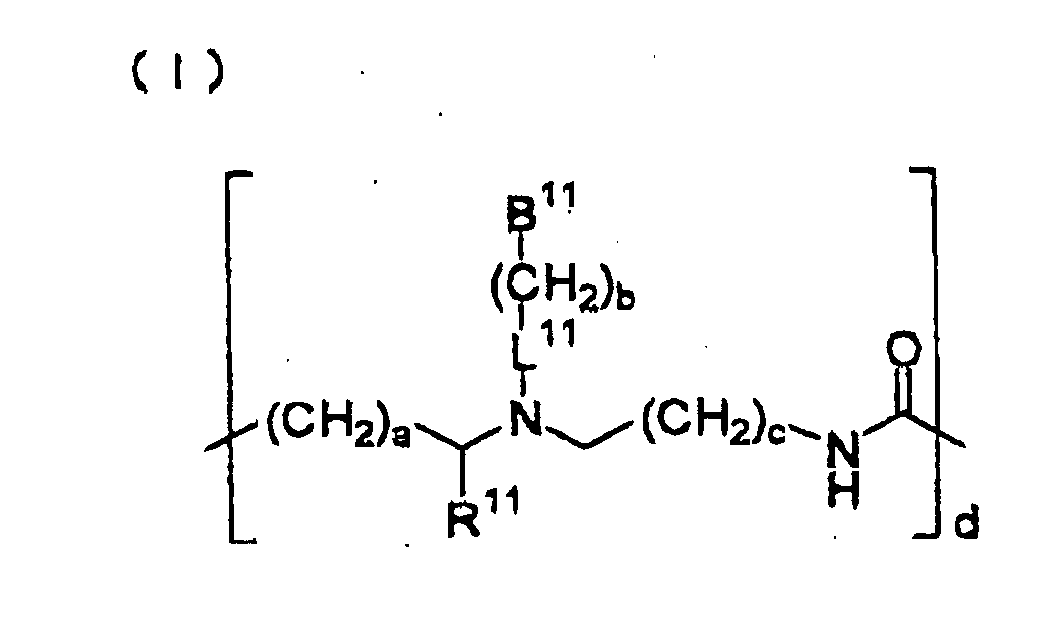
- the PNA fragment preferably employable in the invention has the following formula (I):
- B 11 is a ligand and represents one of bases of natural nucleic acids (i.e., A, T, C, G, I, or U) or its analogue. B 11 is bonded through the 9th position in the case that the base is a purine base such as adenine, guanine or inosine, and through the 1st position in the case that the base is a pyrimidine base such as thymine, uracil or cytosine.
- the base analogue is an organic base which is similar to the base of natural origin in its chemical structure, for instance, a base group which is prepared by replacing the carbon or nitrogen atom of the purine or pyrimidine ring with a nitrogen or carbon atom, respectively, or a base group modifying the purine or pyrimidine ring with a substituent such as a sulfhydryl group or a halogen atom.
- B 11 can be an aromatic moiety containing no nucleic acid base, an alkanoyl group having 1 to 4 carbon atoms, a hydroxyl group, or a hydrogen atom.
- Examples of the base analogues include 7-deazaadenine, 6-azauracil, and 5-azacytosine.
- B 11 also can be a DNA intercalator, a reporter ligand, a protein label such as hapten or biotin, a spin label, or a radioactive label.
- a reporter ligand e.g., a reporter ligand
- a protein label such as hapten or biotin
- a spin label e.g., a radioactive label.
- nucleic acid bases i.e., A, T, C, G, and U).
- R 11 is a hydrogen atom or a group derived from a side-chain of an ⁇ -amino acid of natural origin.
- groups include an alkyl group having 1 to 6 carbon atoms 1 an aryl group having 6 to 20 carbon atoms, an aralkyl group having an alkyl group of 1 to 6 carbon atoms, a heteroaryl group having 6 to 20 carbon atoms, a hydroxyl group, an alkoxy group having 1 to 6 carbon atoms, an alkylthio group having 1 to 6 carbon atoms, a group of -NR 13 R 14 [each of R 13 and R 14 independently is a hydrogen atom, an alkyl group having 1 to 3 carbon atoms, an alkoxy group having 1 to 3 carbon atoms, an alkylthio group having 1 to 3 carbon atoms, or a hydroxyl group], and a mercapto group.
- R 11 may form an alicyclic ring or a heterocyclic ring in combination with the carbon
- L 11 is a linking group such as a divalent group represented by the group of -CO- or -CONR 12 - [R 12 is a hydrogen atom, an alkylene group having 1 to 4 carbon atoms, a hydroxyl group, an alkoxy group having 1 to 4 carbon atoms, or an amino group], or an alkylene group having 1 to 4 carbon atoms.
- the alkoxy group and amino group may have one or more substituents such as alkyl of 1-4 carbon atoms, alkoxy of 1-4 carbon atoms, and hydroxyl.
- Each of a, b and c independently is an integer of 0 to 5, preferably 1, and d is an integer of 1 to 60, preferably an integer of 1 to 40.
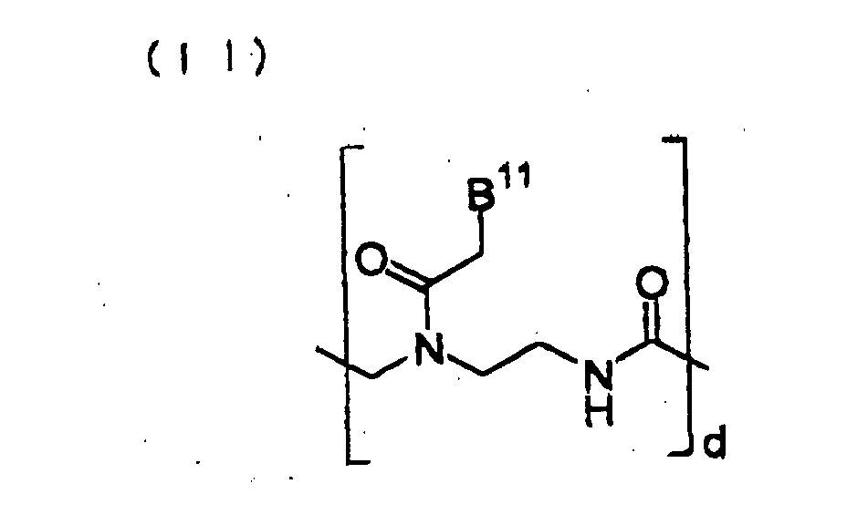
- a particularly preferred PNA fragment has the following formula (II), in which each of B 11 and d has the same meaning as described above for the formula (I):
- the PNA fragment can be fixed onto an electroconductive substrate in a known manner (see Protein, Nucleic Acid, Enzyme, Vol.43, No.13, 2004-2011(1988) and Japanese Patent Provisional Publication 9-288080. Also employable are procedures similar to the aforementioned DNA fragment-fixing procedures.
- the DNA chip preferably has a plurality of short chain spacer molecules having a hydrophilic moiety at each one end which are fixed at each another end onto a surface area of the electroconductive substrate having no DNA fragments thereon.
- the coverage of the substrate using the spacer molecules can be expressed in terms of "masking treatment”.
- the short chain of the spacer molecule means that the molecular length of the spacer molecule is sufficiently short, as compared with the length of the DNA fragment fixed onto the substrate in their vicinity.
- the spacer molecule preferably has a main skeleton composed of an alkylene group having 1 to 6 carbon atoms.
- the spacer molecule may have a cyclic group in the molecular chain or as a substituent.
- the cyclic groups include an aryl group having 6 to 12 carbon atoms, a cycloalkyl group having 3 to 6 carbon atoms, and a heterocyclic group containing 1 to 4 hetero atoms (e.g., N, S, O, or P) and 2 to 20 carbon atoms.
- the cyclic group can contain the reactive group which can be reacted with the substrate surface to fix the spacer atoms to the substrate.
- One example is a imidazole-2-thione group.
- the hydrophilic moiety can be attached to the spacer molecule in the terminal position or in the vicinity of the terminal position.
- One or more hydrophilic moieties may be attached to the spacer molecule.
- the hydrophilic moieties include hydroxyl, carboxyl, amido, phosphoryl, and sulfonyl. Preferred is hydroxyl.
- the other end of the spacer molecule is fixed onto a surface area of the substrate where the DNA fragment is not attached.
- the fixation of the spacer molecule is preferably performed utilizing a reactive group which is attached to the spacer molecule at the other end.
- the reactive groups include mercapto, sulfide, disultide, thiocarbonyl, and thiocarboxyl. Preferred are mercapto, thiocarbonyl, and sulfide. Most preferred is mercapto.
- the electroconductive electrode has a reactive group, on its surface, such as amino, imino, hydrazino, hydrazide, amide, carboxyl, aldehyde, epoxy, or peroxy.
- Examples of the reactive compounds serving as the spacer molecules include mercaptomethanol, 2-mercaptoethanol, 3-mercaptopropanol, 4-mercaptobutanol, 5-mercaptopentanol, 6-mercaptohexanol, N,N'-di-(3-hydroxy-n-propyl) imidazole-2-thione, and various imidazole-2-thione derivatives described in A.J. Arduengo, et al., J. Am. Chem. Soc., 1990, 112, 6153-6154.
- Preferred are 2-mercaptoethanol, 3-mercaptopropanol-, 4-mercaptobutanol, 5-mercaptopentanol, and 6-mercaptohexanol.
- active compounds may be in the form of their salts with sodium or potassium.
- the alkylene chain of the active compound can be substituted with one or more substituents such as a hydrocarbyl group having 1 to 6 carbon atoms (e.g., methyl, ethyl, or phenyl).
- two or more different spacer molecules can be provided.
- the hybridization can be performed essentially in the same manner as that employed in various assay procedures utilizing the conventional DNA chip.
- an electrochemically active molecule specifically, an electrochemically active thread intercalator
- an electrochemically active thread intercalator is preferably employed for insertion into a hybrid formed by the DNA fragment and a sample nucleic acid fragment on the electroconductive substrate.
- the thread intercalator assists easy flowing of electric current from or to the substrate along the formed hybrid structure.
- the electrochemical thread intercalator can be present when hybridization takes place. Otherwise, the thread intercalator can be brought into contact with a previously formed hybrid structure.
- a free nucleic acid fragment which is not hybridized with the fixed DNA fragment is preferably removed from the substrate by washing with a mixture of a surfactant (preferably sodium dodecylsulfate) and a buffer (preferably a citrate buffer) in advance of the contact with the intercalator.
- a surfactant preferably sodium dodecylsulfate
- a buffer preferably a citrate buffer
- the hybridization is preferably performed at a temperature between room temperature and approximately 70°C, for 0.5 to 20 hours.
- the electrochemically active thread intercalators favorably employed for the electrochemical analysis of nucleic acid fragments are already known.
- a representative example of the intercalator is a thread intercalator having an electroconductive group at one end or both ends.
- the thread intercalator having the electroconductive group preferably has an oxidative-reductive activity.
- the oxidative-reductive activity can be imparted to the thread intercalator by incorporating into the intercalator a ferrocene group, a catechol amine group, a metal bipyridine complex group, a metal phenathroline complex group, or a viologen group.
- the intercalator moiety preferably comprises a naphthaleneimide moiety, an anthracene moiety, or an anthraquinone moiety.
- Preferred electrochemically active thread intercalator is a ferrocene-containing naphthalene diimide compound [NDIFc 2 -1, which is prepared from carboxylic acid ester of N-hydroxysuccinimide and a corresponding amine compound, see S. Takenaka et al., J. Chem. Soc. Commun., 1111 (1998)]:
- a ferrocene-containing naphthalene diimide derivative having the following formula is also preferably employed:
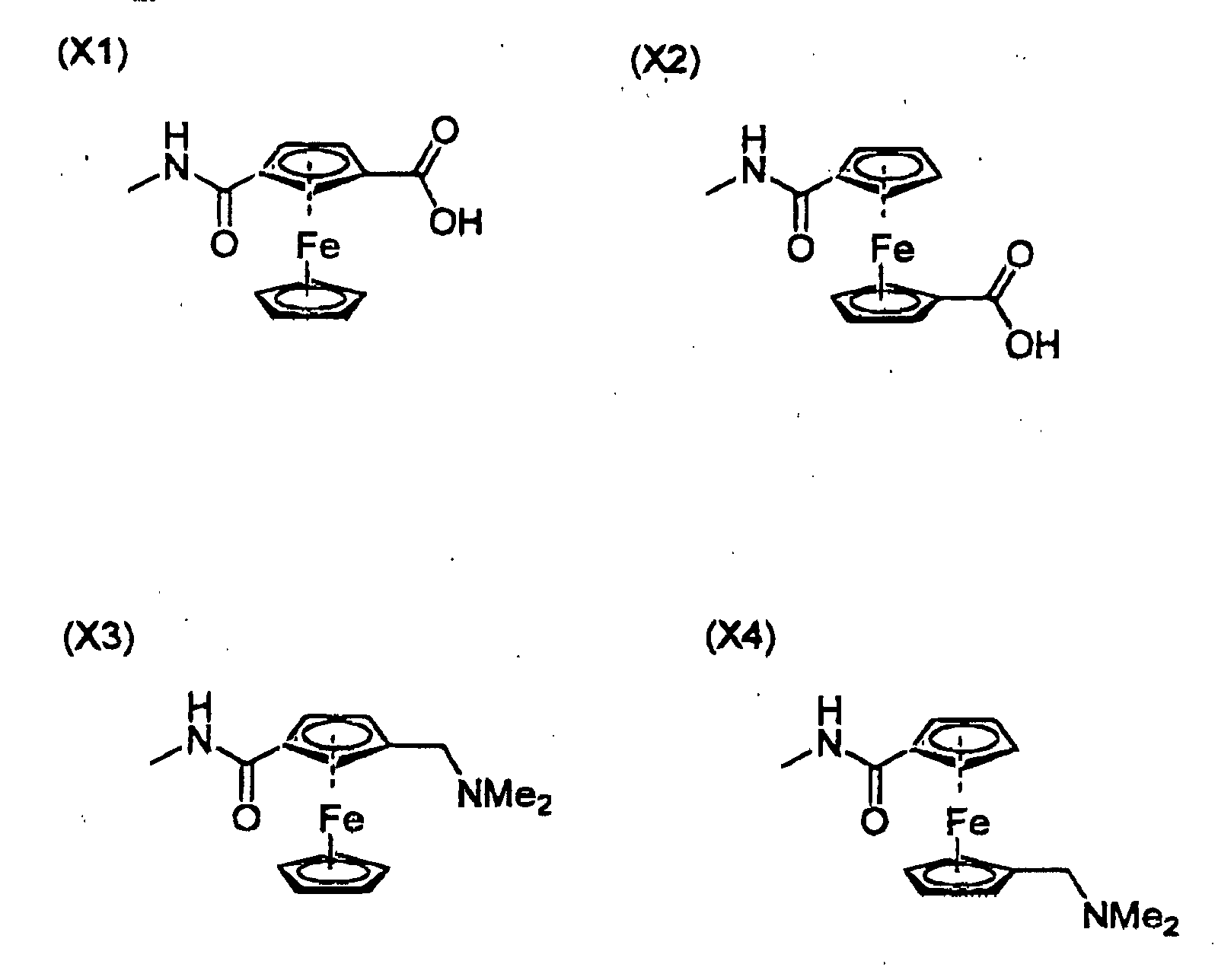
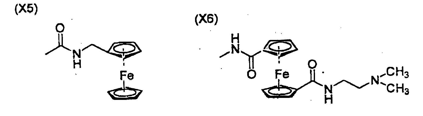
- X is one of the following ferrocene derivative groups:
- the thread intercalator having an electroconductive group comprises not only the oxidative-reductive active moiety and the intercalator moiety but also a linker moiety placed between these moieties.
- the 1,4-dipropylpiperazinyl group of the formula is an example of the linker moiety.
- the piperazinyl group can be replaced with an quaternary imino group.
- An intercalator of the below-illustrated formula which has a quaternary imino group always is cationic regardless of pH condition. This means that the intercalator is firmly fixed to the DNA hybrid and PNA hybrid. Accordingly, it is favorably employed in the invention.
- the intercalator having a quaternary imino group is preferred in the use in combination with the PNA chip.
- the linker can be an N-alkyl group having 1 to 6 carbon atoms (e.g., methyl, ethyl, or n-propyl).
- the oxidative-reductive potential of the ferrocene moiety of the intercalator varies depending upon the nature of the linker moiety.
- the detection method of the invention is preferably employed for detecting an abnormal gene fragment having one or more different bases from its corresponding normal gene fragment.
- the normal gene fragment or the abnormal gene fragment is preferably employed as the sample nucleic acid fragment. It is preferred that the abnormal gene fragment is used as the sample nucleic acid fragment.
- the abnormal gene fragment can be defective and different from the corresponding normal gene fragment.
- Examples of the abnormal gene fragments include a gene fragment in which one or more base units are replaced with different base units, a gene fragment in which one or more base units are missing from the normal gene fragment, and a gene fragment in which one or more base units are added to the normal gene fragment.
- the abnormal gene fragment is generally obtained from a living body having the double stranded abnormal gene.
- the double stranded abnormal gene is cleaved and treated in the conventional manner to give a single stranded abnormal gene fragment for performing the detection of the invention.
- the single stranded abnormal gene fragment can be chemically synthesized.
- a method of the invention for analyzing a nucleic acid fragment sample to judge whether the nucleic acid fragment sample is uncomplementary, partly complementary or complementary to a DNA fragment in its specific base sequence is described by referring to Fig. 2.
- the detection method of the invention typically comprises the following steps.
- a DNA chip (61) On an electroconductive substrate (41) is fixed a single stranded normal DNA fragment (51, namely DNA probe) to give a DNA chip (61).
- the DNA chip (61) is brought into contact with an aqueous solution containing a single stranded normal nucleic acid fragment (71a) which is completely hybridizable with the DNA fragment (51), an aqueous solution containing a single stranded abnormal nucleic acid fragment (71b) which is different from the fragment (71a) in one base unit, or an aqueous solution containing a single stranded abnormal nucleic acid fragment (71c or 71d) which is different from the fragment (71a) in two base units.
- the abnormal nucleic acid fragments (71b, 71c, 71d) differ from the normal nucleic acid fragment (71a) in the base unit or units at the crossed point.
- each of the fragments (71a, 71b, 71c, 71d) hybridizes with the DNA probe (51) to give a full-match hybrid structure (91a), a mis-match hybrid structure (91b), and bulge hybrid structures (91c, 91d), respectively.
- the full-match hybrid structure (91a) is the completely hybridized structure.
- the mis-match hybrid structure (91b) has a missing bond at one defective base unit.
- the bulge hybrid structures (91c, 91d) has a missing bond at two defective base units.
- the hybrid stricture (91a, 101a) is most stable, as compared with the other hybrid structures (91b, 101b, 91c, 101c, 91d, 101d). This means that much electric current flows on the DNA chip having the hybrid structure (91a, 101a) formed with the normal DNA fragment (71a), as compared with other DNA chips having the other hybrid structures.
- DNA chips having the other hybrid structures can be detected by comparing the electric currents flowing on the DNA chips.
- the electric current measured on the DNA chip having the hybrid structure (91a, 101a) formed with the normal DNA fragment (71a) is previously or separately prepared.
- the detection method of the invention nay further comprises the steps for obtaining a background electric current data (i e., blank data) and comparing the blank data with the electric current measured in the above-described step.
- a background electric current data i e., blank data
- the additional comparing procedure can be performed by the steps of:
- the fixation of the electrochemically active thread intercalator to the hybrid structure can be detected by measuring an electric current flowing from or to the electroconductive substrate (i.e., electroconductive substrate) along the hybrid structure having the electrochemically active thread intercalator.
- the measurement of electric current can be performed by any of known methods such as cyclic voltamography (CV), differential pulse voltamography (DPD), and potentiostat.
- CV cyclic voltamography
- DPD differential pulse voltamography
- potentiostat potentiostat.
- the differential pulse voltamography is most preferred.
- the base having "*" on its shoulder means a base changed from the normal base in the normal gene.
- Example 1-1 Detection of partly complementary nucleic acid using a synthetically prepared nucleic acid fragment sample for the formation of mis-match hybrid structure
- a gold electrode (surface area: 2 mm 2 ) was immersed in an aqueous 2N sodium hydroxide solution for one hour, washed with super pure water, and immersed in conc. nitric acid. The nitric acid was stirred for 15 min. The gold electrode was then washed with super pure water and dried. On thus treated gold electrode was spotted 1.0 ⁇ L of an aqueous solution containing 20 mers of adenine having a mercaptohexyl group at its 5 '-terminal (HS-dA 20 , in an amount of 10 pico mol./ ⁇ L), and the electrode was allowed to stand for 2 hours. The electrode was washed with super pure water to give a DNA chip. The preparation of HS-dA 20 was made in the manner described in Japanese Patent Provisional Publication No. 9-288080.
- a nucleic acid fragment of the following dT 9 dAdT 10 was prepared in the manner described in the above-mentioned patent publication: 5'-TTTTTTTTTTTATTTTTTTTT-3'
- aqueous electrolytic solution [mixture of aqueous 0.1 M acetic acid/potassium acetate solution (pH 5.6) and aqueous 0.1 M potassium chloride solution) containing 50 ⁇ M of the ferrocene-containing naphthalene diimide (prepared in (4) above)] was placed in a thermostat cell maintained at 20°C.
- aqueous electrolytic solution were placed tri-electrodes composed of the DNA chip (i.e., working electrode), a platinum electrode (opposite electrode), and a silver/silver chloride referential electrode), and differential pulse voltamography (DPV) was performed at 42°C.
- Fig. 3 indicates the voltamography data (DVP-2) for the mis-match hybrid structure by the chain curve connecting the blank squares.
- Fig. 3 indicates the voltamography data (DVP-1) by the curve connecting the circles.
- the peak current at 460 mV of DPV-2 is higher than the corresponding peak current of DPV-1 by 22.2%.
- Example 1-2 Detection of partly complementary nucleic acid using a synthetically prepared nucleic acid fragment sample for the formation of bulge hybrid structure
- Example 1-1 The procedures of Example 1-1 were repeated except for replacing the sample nucleic acid fragment with the following dT 8 dA 4 dT 8 to obtain a bulge hybrid structure of a voltamographic curve of DPV-4 represented by -blank square- in Fig. 4: 5'-TTTTTTTTAAAATTTTTTTT-3'.
- the peak current at 460 my of DPV-4 is higher than the corresponding peak current of DPV-3 by 15.1%.
- Example 1-3 Detection of complementary nucleic acid using a synthetically prepared nucleic acid fragment sample for the formation of full-match hybrid structure
- Example 1-1 The procedures of Example 1-1 were repeated except for replacing the sample nucleic acid fragment with the thymine 20 mers (dT 20 ) to obtain a full-match hybrid structure of a voltamographic curve of DPV-6 represented by -blank square- in Fig. 5.
- the peak current at 460 mV of DPV-6 is higher than the corresponding peak current of DPV-5 by 37.3%.
- Example 2-1 Detection of partly complementary nucleic acid using a LPL abnormal gene fragment sample for the formation of mis-match hybrid structure
- Example 1-1 The preparation procedures of DNA chip described in Example 1-1 were repeated except for using an aqueous solution of 10 pico mol. of HS-d(CAGCCTGACTTCT) having a mercaptohexyl group at 5' -terminal of oligonucleotide WT-, to prepare a DNA chip.
- the oligonucleotide was prepared in the manner described in the aforementioned patent publication.
- S447X+ (a model gene of a LPL abnormal gene) was prepared in the manner described in the aforementioned patent publication.
- Example 1-1 The corresponding procedures of Example 1-1 were repeated except for using the DNA chip prepared in (1) above and the sample nucleic acid fragment prepared in (2) above, to obtain a mis-match hybrid structure of a voltamographic curve of DPV-8.
- the peak current at 460 mV of DPV-8 differs from the corresponding peak current of DPV-7 by 6.3%.
- Example 2-2 Detection of complementary nucleic acid using a LPL normal gene fragment sample for the formation of full-match hybrid structure
- Example 2-1 The corresponding procedures of Example 2-1 were repeated except for using the sample nucleic acid fragment of oligonucleotide WT+, to obtain a full-match hybrid structure of a voltamographic curve of DPV-10.
- the peak current at 460 mV of DPV-10 differs from the corresponding peak current of DPV-9 by 65.4%.
- Example 3-1 Detection of partly complementary nucleic acid using a LPL abnormal gene fragment sample for the formation of mis-match hybrid structure
- Example 1-1 The corresponding procedures of Example 1-1 were repeated except for using the DNA chip prepared in (1) above and the sample nucleic acid fragment of nucleotide WT+, to obtain a mis-match hybrid structure of a voltamographic curve of DPV-12.
- the peak current at 460 mV of DPV-12 differs from the corresponding peak current of DPV-11 by 9.7%.
- Example 3-2 Detection of complementary nucleic acid using a LPL normal gene fragment sample for the formation of full-match hybrid structure
- Example 3-1 The corresponding procedures of Example 3-1 were repeated except for using the sample nucleic acid fragment of oligonucleotide S447X+, to obtain a full-match hybrid structure of a voltamographic curve of DPV-14.
- the peak current at 460 mV of DPV-14 differs from the corresponding peak current of DPV-13 by 29.4%.
- Example 4-1 Detection of partly complementary nucleic acid using a LPL abnormal gene fragment sample
- a PLP gene portion of a chromosome DNA obtained by purification of leucocyte (1 mL) of a patient having abnormal LPL gene was multiplied (40 cycles) by PCR method to obtain oligonucleotide S447+/S447X+.
- the obtained oligonucleotide was purified by dialysis to prepare a sample nucleic acid fragment for hybridization.
- Example 1-1 The correspondind procedures of Example 1-1 were repeated except for using the DNA chip prepared in (1) above and the sample nucleic acid fragment of (2) above, to obtain a partly complementary hybrid structure of a voltamographic curve of DPV-16.
- the peak current at 460 mV of DPV-16 adversely differs from the corresponding peak current of DPV-15 by 1.4%.
- Example 4-2 Detection of partly complementary nucleic acid using a LPL abnormal/normal mixed gene fragment sample
- a PLP gene portion of a chromosome DNA obtained by purification of leucocyte (1 mL) of a patient having normal LPL gene was multiplied (40 cycles) by PCR method to obtain oligonucleotide WT+/WT+.
- the obtained oligonucleotide WT+/WT+ was combined with the oligonucleotide S447X+/S447X+ obtained in Example 4-1 at a ratio of 1:1, and purified by dialysis to prepare a sample nucleic acid fragment for hybridization.
- Example 4-1 The corresponding procedures of Example 4-1 were repeated except for using the sample nucleic acid fragment of (1) above, to obtain a partly complementary hybrid structure of a voltamographic curve of DPV-18.
- the peak current at 460 mV of DPV-18 differs from the corresponding peak current of DPV-17 by 16.4%.
- Example 4-3 Detection of complementary nucleic acid using a LPL normal gene fragment sample for the formation of full-match hybrid structure
- Example 4-1 The corresponding procedures of Example 4-1 were repeated except for using the sample nucleic acid fragment of oligonucleotide WT+/WT+, to obtain a full-match hybrid structure of a voltamographic curve of DPV-20.
- the peak current at 460 mV of DPV-20 differs from the corresponding peak current of DPV-19 by 38.4%.
- Examples 1 to 4 indicate that the partly complementary nucleic acid fragments can be well detected by the method of the invention even if a hybrid structure is a mis-match structure or a bulge structure.
- Example 1-1 The hybridization and the measurement of electric current were performed in the same manner as in Example 1-1, except for varying the temperature for the measurement of DPV to 15°C, 25°C, 35°C, and 45°C.
- the DPV was then examined to determine a peak current at 450 mV and plotted in Fig. 6 by a blank triangle to give a chain curve.
- the results shown in Fig. 6 are understood to mean that the peak current corresponds to an amount of the thread intercalator attached to the hybrid structure, and does not correspond to the amount of formation of the hybrid structure on the electrode of the DNA chip. Further, it is understood that the hybridization is preferably performed at a low temperature around 20°C.
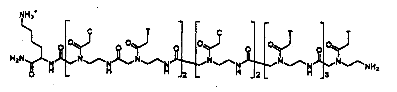
- a PNA fragment [PNA-CTCT(C) 2 (T) 4 , in which T stands for thymine, and C stands for cytosine) having the below-illustrated formula was prepared in the manner as described in P.E. Nielsen et al., Journal of American Chemical Society, 114, 1895-1897(1992) and ibid., 114, 9677-9678(1992).
- DNA-(A) 4 GTAGAG was prepared in the manner described in Japanese Patent Provisional Publication No. 10-288080 to use as a sample nucleic acid fragment.
- aqueous electrolytic solution [mixture of aqueous 0.1 M acetic acid/potassium acetate solution (pH 5.6) and aqueous 0.1 M potassium chloride solution) containing 50 ⁇ M of the ferrocene-containing naphthalene diimide (prepared in Example 1-1)] was placed in a thermostat cell maintained at 38°C.
- the differential pulse voltamography (DPV) was performed at a voltage varying from 100 mV to 700 mV.
- a responsive current value (back ground value) was measured at a voltage of 460 mM to give a back ground value of -1.3 ⁇ A.
- the measurement was performed at a pulse amplitude of 50 mV, a pulse width of 50 mS, and a scanning rate of 100 mV/sec.
- Example 6-1 A sample nucleic acid fragment of DNA-(A) 4 (G) 2 AGAG which was prepared in the manner described in Japanese Patent Provisional Publication No. 10-288080 was spotted on the PNA chip prepared in Example 6-1. Subsequently, the procedures of (4) in Example 6-1 were repeated to determine a background value and a responsive current value. -1.3 ⁇ A and -2.7 ⁇ A were obtained, respectively. The variation of the responsive current value from the background value was 108%.
- Examples 6-1 and 6-2 indicate that the PNA chip is also effective to detect a partly complementary sample nucleic acid fragment and that the detection Sensitivity is higher when the PNA chip is employed because the PNA chip gives a lower background value.
Landscapes
- Chemical & Material Sciences (AREA)
- Life Sciences & Earth Sciences (AREA)
- Organic Chemistry (AREA)
- Analytical Chemistry (AREA)
- Zoology (AREA)
- Wood Science & Technology (AREA)
- Proteomics, Peptides & Aminoacids (AREA)
- Health & Medical Sciences (AREA)
- Engineering & Computer Science (AREA)
- Microbiology (AREA)
- Immunology (AREA)
- Molecular Biology (AREA)
- Biotechnology (AREA)
- Biophysics (AREA)
- Physics & Mathematics (AREA)
- Biochemistry (AREA)
- Bioinformatics & Cheminformatics (AREA)
- General Engineering & Computer Science (AREA)
- General Health & Medical Sciences (AREA)
- Genetics & Genomics (AREA)
- Measuring Or Testing Involving Enzymes Or Micro-Organisms (AREA)
Abstract
Description

and



- Oligonucleotide WT+:
- d(GTCGGACTGAAGA) which is a sense chain of normal lipoprotein lipase (LPL) gene
- Oligonucleotide WT-:
- d(CAGCCTGACTTCT) which is an anti-sense chain of the above LPL gene
- Oligonucleotide S447X+:
- d(GTCGGAG*TGAAGA) which is a sense chain of gene which is produced by change in the normal LPL gene from the 447th serine (TCA) to the termination codon (TGA)
- Oligonucleotide S447X-:
- d(CAGCCTC*ACTTCT) which is an antisense chain of the above-mentioned gene
5'-TTTTTTTTTTTATTTTTTTTT-3'

5'-TTTTTTTTAAAATTTTTTTT-3'.
| Example | Probe DNA | Sample nucleic acid fragment | Variation of current |
| Examp1e 1-1 | dA20 | dT9dAdT10 | 22.2% |
| Example 1-2 | dA20 | dT8dA4dT8 | 15.1% |
| Example 1-3 | dA20 | dT20 | 37.3% |
| Example 2-1 | WT- | S447X+ | 6.3% |
| Example 2-2 | WT- | WT+ | 65.4% |
| Example 3-1 | S447X- | WT+ | 9.7% |
| Example 3-2 | S447X- | S447X+ | 29.4% |
| Example 4-1 | WT- | S447X+/S447X+ | -1.4% |
| EXample 4-2 | WT- | WT+/S447X+ | 16.4% |
| Example 4-3 | WT- | WT+/WT+ | 38.4% |


Claims (20)
- A method of analyzing a nucleic acid fragment sample to judge whether the nucleic acid fragment sample is uncomplementary, partly complementary or complementary to a DNA fragment in its specific base sequence, which comprises the steps of:bringing an aqueous solution of the nucleic acid fragment sample into contact with a DNA chip comprising an electroconductive substrate and the DNA fragment fixed onto the substrate in the presence of an electrochemical thread intercalator;measuring an electric current flowing from or to the electroconductive substrate along the DNA fragment under application of a potential to the substrate; andcomparing the electric current measured above with a referential electric current which is prepared employing a combination of a DNA chip equivalent to the above-mentioned DNA chip, the electrochemical thread intercalator, and an aqueous solution of a nucleic acid fragment which is complementary to the DNA fragment of the DNA chip.
- The method of claim 1, which further comprises the steps of:bringing an aqueous solution not containing the nucleic acid fragment sample into contact with a DNA chip equivalent of the DNA chip in the presence of the electrochemical thread intercalator;measuring an electric current flowing from or to the electroconductive substrate along the DNA fragment under application of a potential to the substrate so as to obtain a background electric current; andcomparing the background electric current with the electric current which is measured employing the aqueous solution containing the nucleic acid fragment sample.
- The method of claim 1, wherein the specific base sequence of the DNA fragment is predetermined.
- The method of claim 2, wherein the specific base sequence of the DNA fragment is predetermined.
- The method of claim 1, wherein the DNA chip has a plurality of short chain spacer molecules having a hydrophilic moiety at each one end which are fixed at each another end onto a surface area of the electroconductive substrate having no DNA fragments thereon.
- The method of claim 2, wherein the DNA chip has a plurality of short chain spacer molecules having a hydrophilic moiety at each one end which are fixed at each another end onto a surface area of the electroconductive substrate having no DNA fragments thereon.
- The method of claim 1, wherein the electrochemical thread intercalator is a ferrocene-containing thread intercalator having an oxidative-reductive activity.
- The method of claim 2, wherein the electrochemical thread intercalator is a ferrocene-containing thread intercalator having an oxidative-reductive activity.
- The method of claim 1, wherein the measurement of the electric current in each step is conducted by differential pulse voltamography.
- The method of claim 2, wherein the measurement of the electric current in each step is conducted by differential pulse voltamography.
- A method of analyzing a nucleic acid fragment sample to judge whether the nucleic acid fragment sample is uncomplementary, partly complementary or complementary to a PNA fragment in its specific base sequence, which comprises the steps of:bringing an aqueous solution of the nucleic acid fragment sample into contact with a PNA chip comprising an electroconductive substrate and the PNA fragment fixed onto the substrate in the presence of an electrochemical thread intercalator;measuring an electric current flowing from or to the electroconductive substrate along the PNA fragment under application of a potential to the substrate; andcomparing the electric current measured above with a referential electric current which is prepared employing a combination of a PNA chip equivalent to the above-mentioned PNA chip, the electrochemical thread intercalator, and an aqueous solution of a nucleic acid fragment which is complementary to the PNA fragment of the PNA chip.
- The method of claim 11, which further comprises the steps of:bringing an aqueous solution not containing the nucleic acid fragment sample into contact with a PNA chip equivalent of the PNA chip in the presence of the electrochemical thread intercalator;measuring an electric current flowing from or to the electroconductive substrate along the PNA fragment under application of a potential to the substrate so as to obtain a background electric current; andcomparing the background electric current with the electric current which is measured employing the aqueous solution containing the nucleic acid fragment sample.
- The method of claim 11, wherein the specific base sequence of the PNA fragment is predetermined.
- The method of claim 12, wherein the specific base sequence of the PNA fragment is predetermined.
- The method of claim 11, wherein the PNA chip has a plurality of short chain spacer molecules having a hydrophilic moiety at each one end which are fixed at each another end onto a surface area of the electroconductive substrate having no PNA fragments thereon.
- The method of claim 12, wherein the PNA chip has a plurality of short chain spacer molecules having a hydrophilic moiety at each one end which are fixed at each another end onto a surface area of the electroconductive substrate having no PNA fragments thereon.
- The method of claim 11, wherein the electrochemical thread intercalator is a ferrocene-containing thread intercalator having an oxidative-reductive activity.
- The method of claim 12, wherein the electrochemical thread intercalator is a ferrocene-containing thread intercalator having an oxidative-reductive activity.
- The method of claim 11, wherein the measurement of the electric current in each step is conducted by differential pulse voltamography.
- The method of claim 12, wherein the measurement of the electric current in each step is conducted by differential pulse voltamography.
Applications Claiming Priority (2)
| Application Number | Priority Date | Filing Date | Title |
|---|---|---|---|
| JP15933999 | 1999-06-07 | ||
| JP15933999 | 1999-06-07 |
Publications (2)
| Publication Number | Publication Date |
|---|---|
| EP1065278A2 true EP1065278A2 (en) | 2001-01-03 |
| EP1065278A3 EP1065278A3 (en) | 2004-02-04 |
Family
ID=15691682
Family Applications (1)
| Application Number | Title | Priority Date | Filing Date |
|---|---|---|---|
| EP00112235A Withdrawn EP1065278A3 (en) | 1999-06-07 | 2000-06-07 | Detection of partly complementary nucleic acid fragment |
Country Status (2)
| Country | Link |
|---|---|
| US (1) | US6399305B1 (en) |
| EP (1) | EP1065278A3 (en) |
Cited By (7)
| Publication number | Priority date | Publication date | Assignee | Title |
|---|---|---|---|---|
| WO2002063041A1 (en) * | 2001-02-06 | 2002-08-15 | Mitocon Ltd. | Mixed intercalator and electrochemical detection of dna using same |
| WO2002066679A1 (en) * | 2001-02-20 | 2002-08-29 | Mitocon Ltd. | Intercalator suitable for electrochemical detection of double strand dnas and a process for preparing thereof |
| EP1132484A3 (en) * | 2000-03-08 | 2003-05-28 | Fuji Photo Film Co., Ltd. | Method for testing complementation of nucleic acid |
| EP1462528A1 (en) * | 2003-03-26 | 2004-09-29 | Seiko Epson Corporation | Nucleic acid immobilization and use as a biosensor |
| EP1445609A4 (en) * | 2002-08-23 | 2005-04-06 | Toshiba Kk | Base sequencing electrode, base sequencing device and base sequencing method |
| US7576205B2 (en) | 2005-06-13 | 2009-08-18 | Agency For Science, Technology & Research | Detectable threading intercalator |
| US8105477B2 (en) | 2003-04-16 | 2012-01-31 | Handylab, Inc. | System and method for electrochemical detection of biological compounds |
Families Citing this family (4)
| Publication number | Priority date | Publication date | Assignee | Title |
|---|---|---|---|---|
| EP1390382A4 (en) * | 2001-04-30 | 2007-10-31 | Benjamin J Doranz | Elucidation of gene function |
| US20040132008A1 (en) * | 2002-04-30 | 2004-07-08 | Doranz Benjamin J | Elucidation of gene function |
| JP4317744B2 (en) * | 2001-05-30 | 2009-08-19 | ナノキャリア株式会社 | Method for binding an introduced substance to the end of a polymer compound |
| JP4142027B2 (en) * | 2005-03-18 | 2008-08-27 | 株式会社東芝 | Base sequence determination method and base sequence determination system |
Family Cites Families (7)
| Publication number | Priority date | Publication date | Assignee | Title |
|---|---|---|---|---|
| US5965452A (en) * | 1996-07-09 | 1999-10-12 | Nanogen, Inc. | Multiplexed active biologic array |
| US5591578A (en) * | 1993-12-10 | 1997-01-07 | California Institute Of Technology | Nucleic acid mediated electron transfer |
| JP3233851B2 (en) * | 1996-04-24 | 2001-12-04 | 繁織 竹中 | Electrochemical detection method of gene and its device |
| US6221586B1 (en) * | 1997-04-09 | 2001-04-24 | California Institute Of Technology | Electrochemical sensor using intercalative, redox-active moieties |
| US6203758B1 (en) * | 1997-11-10 | 2001-03-20 | Bio-Pixel Ltd. | Micro-circuit system with array of functionalized micro-electrodes |
| JP2967197B1 (en) * | 1998-08-11 | 1999-10-25 | 九州大学長 | Probe for detecting special single-stranded nucleic acid site of gene, method and apparatus for detecting special single-stranded nucleic acid site of gene |
| US6261780B1 (en) * | 1999-01-06 | 2001-07-17 | Fuji Photo Film Co., Ltd. | Quantitative analysis of biochemical compound utilizing electrochemical reaction |
-
2000
- 2000-06-07 EP EP00112235A patent/EP1065278A3/en not_active Withdrawn
- 2000-06-07 US US09/588,950 patent/US6399305B1/en not_active Expired - Lifetime
Cited By (10)
| Publication number | Priority date | Publication date | Assignee | Title |
|---|---|---|---|---|
| EP1132484A3 (en) * | 2000-03-08 | 2003-05-28 | Fuji Photo Film Co., Ltd. | Method for testing complementation of nucleic acid |
| WO2002063041A1 (en) * | 2001-02-06 | 2002-08-15 | Mitocon Ltd. | Mixed intercalator and electrochemical detection of dna using same |
| WO2002066679A1 (en) * | 2001-02-20 | 2002-08-29 | Mitocon Ltd. | Intercalator suitable for electrochemical detection of double strand dnas and a process for preparing thereof |
| EP1445609A4 (en) * | 2002-08-23 | 2005-04-06 | Toshiba Kk | Base sequencing electrode, base sequencing device and base sequencing method |
| US7097751B2 (en) | 2002-08-23 | 2006-08-29 | Kabushiki Kaisha Toshiba | Base sequence detecting electrode, base sequence detecting device and base sequence detecting method |
| US7718048B2 (en) | 2002-08-23 | 2010-05-18 | Kabushiki Kaisha Toshiba | Base sequence detecting electrode, base sequence detecting device and base sequence detecting method |
| EP1462528A1 (en) * | 2003-03-26 | 2004-09-29 | Seiko Epson Corporation | Nucleic acid immobilization and use as a biosensor |
| CN1532293B (en) * | 2003-03-26 | 2011-05-11 | 精工爱普生株式会社 | Nucleic acid fixing method and method for producing biosensor by said method |
| US8105477B2 (en) | 2003-04-16 | 2012-01-31 | Handylab, Inc. | System and method for electrochemical detection of biological compounds |
| US7576205B2 (en) | 2005-06-13 | 2009-08-18 | Agency For Science, Technology & Research | Detectable threading intercalator |
Also Published As
| Publication number | Publication date |
|---|---|
| US6399305B1 (en) | 2002-06-04 |
| EP1065278A3 (en) | 2004-02-04 |
Similar Documents
| Publication | Publication Date | Title |
|---|---|---|
| EP1077264B1 (en) | DNA chip, PNA chip, and their preparation methods | |
| US20250346950A1 (en) | Tagged nucleoside compounds useful for nanopore detection | |
| US7615348B2 (en) | Electrochemical method to measure DNA attachment to an electrode surface in the presence of molecular oxygen | |
| US6399305B1 (en) | Protection of partial complementary nucleic acid fragment using a electroconductive chip and intercalator | |
| JP2002532386A (en) | Method for electrochemically detecting nucleic acid oligomer hybrid | |
| US6635426B2 (en) | Mixed intercalator and electrochemical detection of DNA using same | |
| US20010021504A1 (en) | Method for testing complementation of nucleic acid | |
| US6864055B2 (en) | Method for detecting nucleic acids | |
| US20100133118A1 (en) | Electrochemical methods of detecting nucleic acid hybridization | |
| US20010053522A1 (en) | DNA chip and reactive electrode | |
| JP4320104B2 (en) | Method for detecting partially complementary nucleic acid fragments | |
| JP4426700B2 (en) | Highly sensitive quantification method of complementary nucleic acid fragments using DNA analysis element or PNA analysis element | |
| US7321831B2 (en) | Nucleic acid fragment-fixed electrode and its use | |
| US6809201B2 (en) | Intercalator suitable for electrochemical detection of double strand DNAs and a process for preparing thereof | |
| JP2001013103A (en) | Method for detecting and quantitatively determining sample nucleic acid fragment by scanning electrochemical microscope | |
| JP2001321198A (en) | Method for assaying complementarity of sample nucleic acid fragment | |
| EP1620570B1 (en) | Electrochemical method to measure dna attachment to an electrode surface in the presence of molecular oxygen | |
| JP2001226376A (en) | Stitch in-type intercalator having oxidation-reduction activity | |
| JP2003125798A (en) | Method for confirming base sequence in gene of specimen |
Legal Events
| Date | Code | Title | Description |
|---|---|---|---|
| PUAI | Public reference made under article 153(3) epc to a published international application that has entered the european phase |
Free format text: ORIGINAL CODE: 0009012 |
|
| AK | Designated contracting states |
Kind code of ref document: A2 Designated state(s): AT BE CH CY DE DK ES FI FR GB GR IE IT LI LU MC NL PT SE |
|
| AX | Request for extension of the european patent |
Free format text: AL;LT;LV;MK;RO;SI |
|
| PUAL | Search report despatched |
Free format text: ORIGINAL CODE: 0009013 |
|
| AK | Designated contracting states |
Kind code of ref document: A3 Designated state(s): AT BE CH CY DE DK ES FI FR GB GR IE IT LI LU MC NL PT SE |
|
| AX | Request for extension of the european patent |
Extension state: AL LT LV MK RO SI |
|
| 17P | Request for examination filed |
Effective date: 20040301 |
|
| 17Q | First examination report despatched |
Effective date: 20040414 |
|
| AKX | Designation fees paid |
Designated state(s): DE FR GB SE |
|
| STAA | Information on the status of an ep patent application or granted ep patent |
Free format text: STATUS: THE APPLICATION IS DEEMED TO BE WITHDRAWN |
|
| 18D | Application deemed to be withdrawn |
Effective date: 20051112 |