EP1036324B1 - Identification and characterization of interacting molecules by automated interaction mating - Google Patents
Identification and characterization of interacting molecules by automated interaction mating Download PDFInfo
- Publication number
- EP1036324B1 EP1036324B1 EP98965193A EP98965193A EP1036324B1 EP 1036324 B1 EP1036324 B1 EP 1036324B1 EP 98965193 A EP98965193 A EP 98965193A EP 98965193 A EP98965193 A EP 98965193A EP 1036324 B1 EP1036324 B1 EP 1036324B1
- Authority
- EP
- European Patent Office
- Prior art keywords
- host cells
- protein
- cells
- interaction
- clones
- Prior art date
- Legal status (The legal status is an assumption and is not a legal conclusion. Google has not performed a legal analysis and makes no representation as to the accuracy of the status listed.)
- Expired - Lifetime
Links
Images








Classifications
-
- C—CHEMISTRY; METALLURGY
- C40—COMBINATORIAL TECHNOLOGY
- C40B—COMBINATORIAL CHEMISTRY; LIBRARIES, e.g. CHEMICAL LIBRARIES
- C40B30/00—Methods of screening libraries
- C40B30/04—Methods of screening libraries by measuring the ability to specifically bind a target molecule, e.g. antibody-antigen binding, receptor-ligand binding
-
- C—CHEMISTRY; METALLURGY
- C12—BIOCHEMISTRY; BEER; SPIRITS; WINE; VINEGAR; MICROBIOLOGY; ENZYMOLOGY; MUTATION OR GENETIC ENGINEERING
- C12N—MICROORGANISMS OR ENZYMES; COMPOSITIONS THEREOF; PROPAGATING, PRESERVING, OR MAINTAINING MICROORGANISMS; MUTATION OR GENETIC ENGINEERING; CULTURE MEDIA
- C12N15/00—Mutation or genetic engineering; DNA or RNA concerning genetic engineering, vectors, e.g. plasmids, or their isolation, preparation or purification; Use of hosts therefor
- C12N15/09—Recombinant DNA-technology
- C12N15/10—Processes for the isolation, preparation or purification of DNA or RNA
- C12N15/1034—Isolating an individual clone by screening libraries
- C12N15/1055—Protein x Protein interaction, e.g. two hybrid selection
-
- C—CHEMISTRY; METALLURGY
- C12—BIOCHEMISTRY; BEER; SPIRITS; WINE; VINEGAR; MICROBIOLOGY; ENZYMOLOGY; MUTATION OR GENETIC ENGINEERING
- C12Q—MEASURING OR TESTING PROCESSES INVOLVING ENZYMES, NUCLEIC ACIDS OR MICROORGANISMS; COMPOSITIONS OR TEST PAPERS THEREFOR; PROCESSES OF PREPARING SUCH COMPOSITIONS; CONDITION-RESPONSIVE CONTROL IN MICROBIOLOGICAL OR ENZYMOLOGICAL PROCESSES
- C12Q1/00—Measuring or testing processes involving enzymes, nucleic acids or microorganisms; Compositions therefor; Processes of preparing such compositions
- C12Q1/68—Measuring or testing processes involving enzymes, nucleic acids or microorganisms; Compositions therefor; Processes of preparing such compositions involving nucleic acids
- C12Q1/6897—Measuring or testing processes involving enzymes, nucleic acids or microorganisms; Compositions therefor; Processes of preparing such compositions involving nucleic acids involving reporter genes operably linked to promoters
-
- G—PHYSICS
- G01—MEASURING; TESTING
- G01N—INVESTIGATING OR ANALYSING MATERIALS BY DETERMINING THEIR CHEMICAL OR PHYSICAL PROPERTIES
- G01N33/00—Investigating or analysing materials by specific methods not covered by groups G01N1/00 - G01N31/00
- G01N33/48—Biological material, e.g. blood, urine; Haemocytometers
- G01N33/50—Chemical analysis of biological material, e.g. blood, urine; Testing involving biospecific ligand binding methods; Immunological testing
- G01N33/5005—Chemical analysis of biological material, e.g. blood, urine; Testing involving biospecific ligand binding methods; Immunological testing involving human or animal cells
-
- G—PHYSICS
- G01—MEASURING; TESTING
- G01N—INVESTIGATING OR ANALYSING MATERIALS BY DETERMINING THEIR CHEMICAL OR PHYSICAL PROPERTIES
- G01N33/00—Investigating or analysing materials by specific methods not covered by groups G01N1/00 - G01N31/00
- G01N33/48—Biological material, e.g. blood, urine; Haemocytometers
- G01N33/50—Chemical analysis of biological material, e.g. blood, urine; Testing involving biospecific ligand binding methods; Immunological testing
- G01N33/5005—Chemical analysis of biological material, e.g. blood, urine; Testing involving biospecific ligand binding methods; Immunological testing involving human or animal cells
- G01N33/5008—Chemical analysis of biological material, e.g. blood, urine; Testing involving biospecific ligand binding methods; Immunological testing involving human or animal cells for testing or evaluating the effect of chemical or biological compounds, e.g. drugs, cosmetics
-
- G—PHYSICS
- G01—MEASURING; TESTING
- G01N—INVESTIGATING OR ANALYSING MATERIALS BY DETERMINING THEIR CHEMICAL OR PHYSICAL PROPERTIES
- G01N33/00—Investigating or analysing materials by specific methods not covered by groups G01N1/00 - G01N31/00
- G01N33/48—Biological material, e.g. blood, urine; Haemocytometers
- G01N33/50—Chemical analysis of biological material, e.g. blood, urine; Testing involving biospecific ligand binding methods; Immunological testing
- G01N33/5005—Chemical analysis of biological material, e.g. blood, urine; Testing involving biospecific ligand binding methods; Immunological testing involving human or animal cells
- G01N33/5008—Chemical analysis of biological material, e.g. blood, urine; Testing involving biospecific ligand binding methods; Immunological testing involving human or animal cells for testing or evaluating the effect of chemical or biological compounds, e.g. drugs, cosmetics
- G01N33/502—Chemical analysis of biological material, e.g. blood, urine; Testing involving biospecific ligand binding methods; Immunological testing involving human or animal cells for testing or evaluating the effect of chemical or biological compounds, e.g. drugs, cosmetics for testing non-proliferative effects
-
- G—PHYSICS
- G01—MEASURING; TESTING
- G01N—INVESTIGATING OR ANALYSING MATERIALS BY DETERMINING THEIR CHEMICAL OR PHYSICAL PROPERTIES
- G01N33/00—Investigating or analysing materials by specific methods not covered by groups G01N1/00 - G01N31/00
- G01N33/48—Biological material, e.g. blood, urine; Haemocytometers
- G01N33/50—Chemical analysis of biological material, e.g. blood, urine; Testing involving biospecific ligand binding methods; Immunological testing
- G01N33/68—Chemical analysis of biological material, e.g. blood, urine; Testing involving biospecific ligand binding methods; Immunological testing involving proteins, peptides or amino acids
-
- G—PHYSICS
- G01—MEASURING; TESTING
- G01N—INVESTIGATING OR ANALYSING MATERIALS BY DETERMINING THEIR CHEMICAL OR PHYSICAL PROPERTIES
- G01N33/00—Investigating or analysing materials by specific methods not covered by groups G01N1/00 - G01N31/00
- G01N33/48—Biological material, e.g. blood, urine; Haemocytometers
- G01N33/50—Chemical analysis of biological material, e.g. blood, urine; Testing involving biospecific ligand binding methods; Immunological testing
- G01N33/68—Chemical analysis of biological material, e.g. blood, urine; Testing involving biospecific ligand binding methods; Immunological testing involving proteins, peptides or amino acids
- G01N33/6803—General methods of protein analysis not limited to specific proteins or families of proteins
- G01N33/6845—Methods of identifying protein-protein interactions in protein mixtures
-
- G—PHYSICS
- G01—MEASURING; TESTING
- G01N—INVESTIGATING OR ANALYSING MATERIALS BY DETERMINING THEIR CHEMICAL OR PHYSICAL PROPERTIES
- G01N2800/00—Detection or diagnosis of diseases
- G01N2800/52—Predicting or monitoring the response to treatment, e.g. for selection of therapy based on assay results in personalised medicine; Prognosis
Definitions
- the present invention relates to an improved method for the identification and optionally the characterisation of interacting molecules designed to detect positive clones from the rather large numbers of false positive clones isolated by conventional two-hybrid systems.
- the method of the invention relies on a novel combination of selection steps used to detect clones that express interacting molecules from false positive clones.
- the present invention provides for high-throughput interaction screens for the reliable identification of interacting molecules, which in turn can lead to the identification of substances inhibiting said interactions. Such inhibitors can find their use in the formulation of a pharmaceutical composition.
- the present invention further relates to kits useful for carrying out the method of the invention.
- Protein-protein interactions are essential for nearly all biological processes like replication, transcription, secretion, signal transduction and metabolism.
- Classical methods for identifying such interactions like co-immunoprecipitation or cross-linking are not available for all proteins or may not be sufficiently sensitive.
- Said methods further have the disadvantage that only by a great deal of energy, potentially interacting partners and corresponding nucleic acid fragments or sequences may be identified. Usually, this is effected by protein sequencing or production of antibodies, followed by the screening of an expression library.
- yeast two-hybrid (2H) system presented by Fields and Song (1989). This genetic procedure not only allows the rapid demonstration of in vivo interactions, but also the simple isolation of corresponding nucleic acid sequences encoding for the interacting partners.
- the yeast 2H system makes use of the features of a wide variety of eukaryotic transcription factors which carry two separable functional domains: one DNA binding domain as well as a second domain which activates the RNA-polymerase complex (activation domain).
- a so-called “bait” protein comprising of a DNA binding domain (GAL4bd or lex A), and a protein of interest "X” are expressed as a fusion protein in yeast ("bait hybrid”).
- the same yeast cell also simultaneously expresses a so called “fish” protein comprising of an activation domain (GAL4ad or VP16) and a protein "Y” ("fish hybrid”).
- the DNA binding and activation domains of the fusion proteins are brought into close proximity and the resulting protein complex triggers the expression of the reporter genes, e.g. HIS3 or lacZ.
- Said expression can be easily monitored by cultivation of the yeast cells on selective medium without histidine as well as upon the activation of the lacZ gene.
- the genetic sequence encoding, for example, an unknown fish protein may easily be identified by isolating the corresponding plasmid and subsequent sequence analysis. Meanwhile, a number of variants of the 2H system have been developed.
- yeast is not the host cell of choice in a variety of investigations (e.g. when a mammalian protein suspected to interact with a second protein requires substantial posttranslational modifications), it would be desirable for a high-throughput 2H system to be versatile with regard to the type of host cell employed. All systems put forward so far that are geared to eliminate the difficulties of 2H screening, although mostly claiming to be applicable to all types of cells, have been designed towards the specific biological properties of the yeast two hybrid system, and cannot necessarily be transferred to, for example, bacterial or mammalian cell systems.
- the technical problem underlying the present invention was therefore to provide a method that allows the fast and reliable elimination of false positives.
- This method should, moreover, be suitable for large-scale library vs. library screens using a high-throughput approach.
- this method would be applicable to a range of different host cell systems, such as yeast, bacterial, mammalian, plant and insect cells.
- Such method could routinely be applied to the identification of pathways of molecular interactions in biological systems, and the interconnections between such .pathways.
- the identification of molecules involved in interactions that form part of such pathways can be employed in order to pinpoint targets for pharmaceuticals.
- the present invention relates to a method for the identification of at least one member of a pair or complex of interacting molecules from a pool of potentially interacting molecules, comprising:
- identification and identifying relate to the ability of the person skilled in the art to detect positive clones that express interacting molecules from false positive clones due to the activation of the readout system on the selective media and optionally additionally to characterise at least one of said interacting molecules by one or a set of unambiguous features.
- said molecules are characterised by the DNA sequence-encoding them, upon nucleic acid hybridisation or isolation and sequencing of the respective DNA molecules.
- said molecules may be characterised by different features such as molecular weight, isoelectric point and, in the case of proteins, the N-terminal amino acid sequence etc. Methods for determining such parameters are well known in the art.
- nucleic acids, peptides, domains of proteins or proteins that can be formed upon the transcription and/or translation of genetic information, and which may, but are not required to be able to interact with one or more other such nucleic acids, peptides or proteins, together forming a pair or complex of interacting molecules.
- said potentially interacting molecules represent nucleic acids, peptides, domains of proteins or proteins which occur in cells from which the genetic information was derived.
- said potentially interacting molecules specified by said genetic information are connected to a further entity that will upon the interaction activate or contribute to the activation of said read-out system. It is further preferred that said entity is conserved for each type of genetic element and that different types of genetic elements comprise different entities. It is additionally preferred that said potentially interacting molecule forms, when transcribed as RNA from said genetic element, an RNA transcript fused with RNA specifying said entity. Most preferably, said fused RNA transcript is translated to form a fusion protein comprising said potentially interacting molecule fused to said entity.
- said entity may be in one type of genetic element a DNA sequence encoding a DNA-binding domain and in a different type of genetic element a transactivating protein domain.
- said genetic elements are vectors such as plasmids.
- the at least two genetic elements comprised in said host cell preferentially contain genetic information from a library such as a cDNA or genomic library.
- a library such as a cDNA or genomic library.
- the method of the invention allows the screening of a variety of host cells wherein the vector portion of said genetic elements is preferably the same for each type of genetic element whereas the potentially interacting molecules are representatives of a library and, thus, as a rule and in case that the library has not been amplified, may differ in each host cell or in a majority of host cells.
- type of genetic element refers to an element characterised by comprising the same entity, selectable and, optionally, counterselectable markers.
- the genetic elements specified in the present invention may further and advantageously be equipped with selection markers functional in bacteria such as E.coli.
- the selection markers for example aphA (Pansegrau et al., 1987) or bla allow the easy separation of said genetic elements upon retransformation into E.coli strains.
- the interaction according to the invention is a specific interaction.
- the "interaction" of said molecules is characterised by a high binding constant.
- the term “interaction” may also refer to a binding between molecules with a lower binding constant which, however, must be sufficient to activate the readout system.
- the interaction that is detectable by the method of the invention preferably leads to the formation of a functional entity having a biological, physical or chemical activity which was not present in said host cell before said interaction occurred. More preferably, such activity is a detectable activity. Most preferably, such functional entity is a protein.
- Said interaction may preferably lead to the formation of a functional transcriptional activator comprising a DNA-binding and a transactivating protein domain and which is capable of activating a responsive moiety driving the activation of said readout system.
- said moiety may be a promoter.
- said interaction may lead to a detectable fluorescence resonance energy transfer obtained by the interaction of fusion proteins containing, for example, the GFP type a and GFP type b fluorescent proteins (Cubbitt et al., 1995).
- growth on selective media refers to the fact that yeast cells containing one genetic element are placed on selective media that preclude growth of said cells upon auto-activation of said read-out system, or the visual differentiation between cells whose read-out system has been auto-activated and those cells whose read-out system has not been auto-activated.
- yeast cells when a ura3 yeast strain which contains a URA3 reporter system and which also contains a plasmid expressing a LexA fusion protein that activates the URA3 reporter system is selected on selective medium containing 5-fluoroorotic acid (5-FOA), the yeast cells cannot grow on this medium because the URA3 reporter system synthesises the enzyme orotidine-5'-phosphate decarboxylase that converts 5-FOA into the toxic compound 5-fluorouracil (Boeke et al., 1984). In contrast, on a selective medium lacking for example tryptophan and which contains X-Gal, yeast cells that contain plasmids for the expression of LexA fusion proteins that either activate or do not activate the read-out system, can grow. However, the yeast cells in which the lacZ reporter system is activated will turn blue because the substrate X-Gal is cleaved into the coloured compound 5-bromo-4-chloro-indigo.
- 5-FOA 5-fluoroorotic acid
- growth on selective medium also refers to the fact that host cells containing two genetic elements expressing interacting molecules which do not activate the read-out system on their own, are selected on selective medium. For example, clones that express interacting LexA and GAL4ad fusion proteins which activate a URA3 and HIS3 reporter system can be selected on selective media lacking tryptophan, leucine, histidine and uracil. On this selective medium, only those yeast cells that contain interacting LexA and GAL4ad fusion proteins that activate the URA3 and HIS3 readout system can grow.
- each of the selective media would comprise at least one counterselectable compound such as 5-FOA or cycloheximide wherein the counterselectable compound would be different in different selective media; they would further typically lack a compound complementing for an auxotrophic marker or comprise an antibiotic.
- the compound or antibiotic may be the same for the various selective media. Preferably, at least one is different.
- a counterselection against clones that express a single molecule able to activate the URA3 read-out system can be carried out on culture media preferably comprising 5 fluoroorotic acid (5-FOA).
- culture media preferably comprising 5 fluoroorotic acid (5-FOA)
- 5-FOA 5 fluoroorotic acid
- auto-activate or “auto-activation” relate to the fact that certain molecules encoded by said genetic elements are able to activate the readout system without the need for an interacting molecule.
- the single fusion protein LexA-HIP1 is capable of activating the HIS3 and lacZ read-out system without any corresponding interacting activation domain fusion protein (Wanker et al., 1997).
- preselection relates to the selection of a set of host cells comprising a genetic element and a read-out system for those host cells expressing a single fusion protein unable to auto-activate said read-out system.
- genetic preselection as used in accordance with the present invention, relates to preselection employing counterselection which makes use of a read-out system comprising a counterselectable gene.
- the present invention provides a highly effective method to perform 2H screens in a variety of host cell types.
- the invention provides a reliable method for the detection of false positive clones that express fusion proteins which are able to activate the read-out system without an interaction with a second molecule.
- Further aspects provide methods to produce pharmaceutical compositions employing large scale 2H methods.
- kits are provided that will allow performing the method of the invention.
- the present invention provides for three methods to achieve such removal of false positives employing preselection. Inclusion of at least one preselection step as a feature of the invention has a number of significant advantages as compared to prior art methods that we addressed in more detail below.
- a preselection step for host cells expressing a single molecule unable to activate the readout system employs visual differentiation between host cells whose readout system has been activated and host cells, whose readout system has not been activated.
- the readout system comprises at least one detectable protein. More preferably, said detectable protein is encoded from at least one of the genes lacZ, gfp, yfp, bfp, cat, luxAB, HPRT or a surface marker gene. Other such genes exist and the person skilled in the art will readily identify other such genes that can be employed according to this embodiment.
- said visual differentiation in step (B) is based on a difference between host cells in different activation states of the readout system which can be detected by visual means. It is particularly preferred that said difference between host cells in different activation states, that can be detected by visual means, is brought about by activation of one of the genes lacZ, gfp, yfp, bfp, CAT, luxAB, or of a surface marker.
- said visual means include digital image capture, storage, processing and/or analysis.
- Such visual means may incorporate a camera, a sensitive CCD camera that is suitable for luminescent and fluorescent detection, or may be colourimetric detection systems including computer-based scanners or specialised fluorescent, luminescent or colourimetric plate readers such as the Victor II system from Wallace (Finland).
- Preselection employing one or more counterselective reporter genes or, alternatively, by visually detecting a host cell expressing a single fusion protein which is able to activate the readout system can equally be used to remove false positive clones in the 2H system.
- Using a counterselective reporter gene is in some cases unsatisfactory for a number of reasons, particularly when applied to a large-scale library vs. library screen with the aim of generating protein interaction networks of a eukaryotic system.
- the amount of false-positive clones passing through the preselection step can potentially be reduced. Furthermore, by allowing all clones that carry a plasmid to grow and using visual differentiation to distinguish false positive clones, false positive clones could be ignored using an automated colony picking system. This would significantly reduce the problem of false positive clones being carried through the preselective step compared to a counterselective system that is "leaky" since the location of dormant yet viable cells is unknown.
- read-out systems exhibiting visual differentiation between activation and non-activation states, such as ⁇ -galactosidase, green fluorescent protein, luciferase, secreted alkaline phosphatase and ⁇ -glucuronidase, are detectable when expressed in different host-cell types including yeast, bacteria, plant and insect cells. Therefore, systems to preselect for false positive clones would be easier to transfer to other host-cell types if based on these readout systems.
- said pair or complex of interacting molecules is selected from the group consisting of RNA-RNA, RNA-DNA, RNA-protein, DNA-DNA, DNA-protein, protein-protein, protein-peptide, or peptide-peptide interactions.
- the method of the invention is applicable in a wide range of biological interactions.
- the invention will be useful in identifying peptide-protein or peptide-peptide-interactions by employing synthetic peptide libraries (Yang et al., 1995).
- Two applications of interest are the application of a large scale 2H system for the detection of protein-protein interactions involved in medically relevant pathways which may be useful as diagnostic or therapeutic targets for the treatment of disease, and a large scale tri-hybrid system which is one example of said complex of interacting molecules mentioned herein above for the identification of, for example, novel post-transciptional regulators and their binding sites (SenGupta et al., 1996; Putz et al., 1996).
- a complex in accordance with the invention may comprise more than three interacting molecules.
- such a complex may be composed of biologically or chemically different members.
- a plasmid expressing a LexA-HIV-1Rev protein a plasmid transcribing an RNA sequence in fusion with the responsive element and a plasmid expressing a potentially RNA-interacting protein in fusion with an activation domain may be present in one cell.
- the plasmids encoding the RNA fusion molecule and the activation domain fusion protein must contain different selectable and counterselectable markers-according to the method of the invention. If the RNA fusion molecule interacts with the respective two fusion proteins, the read-out system is activated. To test whether the RNA fusion molecule or the activation domain fusion protein interact, the method of the invention is used to investigate the activation of the read-out system in the absence of either of these fusion molecules.
- said genetic elements are plasmids, artificial chromosomes, viruses or other extrachromosomal elements.
- said read-out system additionally comprises at least one counterselectable gene.
- the person skilled in the art may choose from a variety of such counterselectable genes.
- said genes are URA3, LYS2, sacB, CAN1, CYH2, rpsL, or lacY.
- the person skilled in the art will be able to choose the appropriate marker for a given cell system, e.g. URA3 in a yeast 2H system or sacB in a bacterial system.
- said selective medium in step (B) of the method of the invention comprises a counterselective compound.
- said counterselective compound is chosen from 5-FOA, canavanine, cycloheximide, sucrose, streptomycin or.tONPG.
- the URA3 gene is incorporated as a component of the read-out system.
- Clones containing only one of said genetic elements are placed on a selective medium comprising 5-FOA.
- 5-FOA is converted into the toxic 5-fluorouracil. Accordingly, host cells containing auto-activating molecules will die on the selective medium containing 5-FOA (Le Douarin, 1995, Vidal et al., 1996a). Surviving cells are then collected by scraping or washing off colonies from the surface.
- the read-out system according to the invention comprises at least one detectable protein.
- a number of read-out systems are known in the art and may, if necessary, be adapted to be useful in the method of the invention.
- said detectable protein is that encoded by the genes lacZ, HIS3, URA3, LYS2, sacB, tetA, gfp, yfp, bfp, CAT, luxAB, HPRT or a surface marker, respectively.
- the expression of the ⁇ -gal enzyme in yeast can be used for the formation of a detectable blue colony after incubation in X-Gal solution. Proteins which confer resistance to an antibiotic represent a popular choice for bacterial cell systems and can be detected by selection for growth in the presence of the antibiotic.
- fluorescent proteins as well as the expression of a surface marker and subsequent visualisation with a fluorescently marked antibody, can preferentially be employed in mammalian systems in conjunction with fluorescence-assisted cell sorting (FACS) or laser scanning confocal microscopy.
- FACS fluorescence-assisted cell sorting
- the method of the invention is not restricted for use of only one read-out system.
- a number of such read-out systems may be combined.
- Said combination of a number of read-out systems is, in accordance with the present invention, also comprised by the term "read-out system". Such a combination will provide an additional safeguard for the identification of clones containing interacting partners.
- yeast cells Although the 2H system has been developed in yeast, the method of the invention can be carried out in a variety of host systems. Preferred of those are yeast cells, bacterial cells, mammalian cells (Wu et al. 1996), insect cells, plant cells or hybrid cells. Preferably, the bacterial cells are E. coli cells.
- prokaryotic systems have certain advantages over eukaryotic systems including the ease of genetic, laboratory and automated procedures. Additionally, unlike conventional yeast two-hybrid systems, nuclear localisation of fusion proteins is irrelevant for prokaryotic cells and the entry of small molecules into the cell is typically easier than for a yeast cell. However, some protein-protein interactions depend on post-translational modifications such as mRNA splicing or glycosylation that are not available in prokaryotic or yeast cells, respectively. Therefore, in order to uncover many, if not most, protein-protein interactions that exist in biological systems, library vs.
- Vidal et al (1996a) describes a genetic method to preselect against cells expressing single fusion proteins able to activate the read-out system, but no solution is provided as to how a person skilled in the art may conduct this preselection in for example a prokaryotic or mammalian two-hybrid system.
- detectable proteins such as GFP or ⁇ -galactosidase that are appropriate for a broad range of host-types as one part of the read-out system, a substantially similar procedure and method can be used to visually differentiate against false positive clones in a variety of host-types.
- the genetic elements may be engineered and prepared in one host organism and then, e.g. by employing shuttle vectors, be transferred to a different host organism where it is employed in the method of the invention.
- the person skilled in the art may initiate the identification method of the invention starting from fully transformed or transfected host cells, he may wish to first generate such host cells in accordance with the aim of his research or commercial interest. For example, he may wish to generate a certain type of library first that he intends to screen against a second library already present in said host cells. Alternatively, he may have in mind to generate two or more different libraries that he wants to screen against each other. In this case, he would need to first transform said host cells, simultaneously or successively, with both or all types of genetic elements.
- the method of the present invention comprises transforming, infecting or transfecting at least one set of host cells of said sets of host cells with said genetic element or genetic elements prior to step (D).
- the method of the invention further comprises transforming, infecting or transfecting each set of host cells of said sets of host cells with dais genetic elements prior to step (D).
- the method of the present invention comprises transforming, infecting or transfecting one set of host cells of said sets of host cells with at least one genetic element prior to step (A), selecting against host cells in said one set of host cells expressing a molecule able to auto-activate said read-out system as specified in step (B), and transforming, infecting or transfecting said set of host cells with at least one further genetic element prior to step (D).
- said host cells with said genetic elements are generated by cell fusion, conjugation or interaction mating prior to step (D), preferably in step (C).
- said cell fusion, conjugation or interaction mating is effected or assisted by automation. More preferably, said automation is effected by an automated picking, spotting, rearraying, pipetting, micropipetting or cell sorting device. Most preferably, said device is a picking robot, spotting robot, rearraying robot, pipetting system, micropipetting system or fluorescence assisted cell sorting (FACS) system.
- automation is effected by an automated picking, spotting, rearraying, pipetting, micropipetting or cell sorting device.
- said device is a picking robot, spotting robot, rearraying robot, pipetting system, micropipetting system or fluorescence assisted cell sorting (FACS) system.
- FACS fluorescence assisted cell sorting
- Interaction mating is well known as a tool for use in the yeast 2H system to combine genetic elements that express potentially interacting fusion proteins (Bendixen et al., 1994). Although cell fusion, conjugation or interaction mating are efficient in combining genetic material between different cell strains, such an approach would only be of use in a large-scale library vs. library screen if it could be conducted at high-throughput, due to the large number of colonies that needs to be harvested.
- automated systems which had been designed to speed the handling of E.coli cells for the analysis of DNA (Lehrach et al, 1997), it is possible to conduct automated and high-throughput interaction mating in bacteria and yeast cells. Pipetting or micropipetting systems could be used for example in the handling of mammalian cells. Alternatively, FACS could be employed to the same task.
- the illumination of the agar-trays containing plated colonies was changed from the dark-field sub-illumination typically used when picking E . coli clones to dark-field top-illumination to successfully visualise yeast colonies by differentiation from the lawn of non-transformant cells.
- the existing vision guided motion system Karlnaswamy & Agapakis 1997) was modified to allow for a larger range of "blob" size when selecting yeast colonies to pick from the blobs features returned by connectivity algorithms when applied to a digital image of the agar tray containing colonies.
- the clone inoculation routine was re-programmed to ensure that cell material which had dried on the picking pins during the picking routine was initially re-hydrated by 10 seconds of immersion in the wells of a microtiter plate before vigorous pin-motion with the well.
- This robotic procedure ensured that sufficient cell material was inoculated from each picking pin into an individual.well of a microtiter plate.
- the picking pins were sterilised after inoculation to allow the picking cycle to be repeated by programming the robot to brush the picking pins in a 0.3% (v/v) solution of hydrogen peroxide, followed by a 70% ethanol rinse from a second wash-bath and finally a heat-gun treatment to evaporate any remaining ethanol from the pins.
- the combination at high-throughput of genetic material from all combinations of pairs of cells expressing fusion proteins could also be conducted in a systematic, rather than a random manner.
- Said lawn may be applied as a layer of cells suspended in a solid or semisolid growth medium or may be applied by spraying a thin and uniform layer of cells of one mating type onto the surface where contact with the cell of the alternative mating type is made.
- systems where individual clones can be individually positioned or contacted with other particular clones This can be enabled for example by individually addressable multi-head dispensing units, or by a transfer head with individually addressable and moveable transfer pins.
- Such a system can easily be brought forward by a person skilled in the art using the disclosures in this invention using systems such as the rearraying robots as described by Stanton et al (1995) and Lehrach et al (1997), or from those supplied by commercial robot suppliers such as Genetix (UK). It should be recognised that said combination strategy may be conducted on a planar carrier as disclosed herein below. It may also be conducted directly on solid growth agar, or within the wells of microtiter plates.
- Clones from a given library would be pooled in numbers of 2 to 10, 10 to 100, 100 to 500 or 500 to 1000, and pools contacted with clones or pools from a second library.
- said pools of clones shall be designed using multidimensional pooling strategies as are commonly known in the art (Barillot et al, 1991; Strauss, et al, 1992; Liu et al. 1995) such that the individual identity of the two clones that contacted and caused activation of the readout system can be subsequently deconvoluted. It is of advantage that most or all false positive clones are removed from the two libraries prior to combination such that said deconvolution can be conducted most efficiently.
- the selectable markers are auxotrophic or antibiotic markers.
- markers that are used as a read-out system may also be used as selectable markers. It is further important to note that one and the same marker cannot be used as selectable marker and as part of the read-out system at the same time.
- auxotrophic or antibiotic markers are selected from LEU2, TRP1, URA3, ADE2, HIS3, LYS2, kan, bla, Zeocin, neomycin, hygromycin, pyromycin or G418.
- a further preferred embodiment of the invention relates to a method wherein progeny of host cells obtained in step (C) are transferred to a storage compartment.
- said transfer to a storage compartment is advantageously effected or assisted by automation.
- said automation is effected by an automated arraying, picking, spotting, pipetting, micropipetting or cell sorting device.
- said device is an arraying robot, picking robot, spotting robot, automated pipetting or micropipetting system or FACS system.
- a pipetting, micropipetting or FACS system may be advantageously applied to the transfer of mammalian cells.
- Other automation or robot systems that reliably transfer progeny of said host cells into predetermined arrays in the storage compartments may also be employed. As the person skilled in the art will realise, the choice of said device will largely depend on the host cell system under investigation.
- the host cells may, in this embodiment, be propagated in said storage compartment and provide further progeny for the additional tests.
- replicas of said storage compartment maintaining the array of clones are set up.
- Said storage compartments comprising the transformed host cells and the appropriate media may be maintained in accordance with conventional cultivation protocols.
- said storage compartments may comprise an anti-freeze agent and therefore be appropriate for storage in a deep-freezer. This embodiment is particularly useful when the evaluation of potential interacting partners is to be postponed.
- frozen host cells may easily be recovered upon thawing and further tested in accordance with the invention.
- said anti-freeze agent is glycerol which is preferably present in said media in an amount of 3 - 25% (vol/vol), or DMSO.
- said storage compartment is at least one microtiter plate.
- said at least one microtiter plate comprises 96, 384, 846 or 1536 wells.
- Microtiter plates have the particular advantage of providing a pre-fixed array that allows the easy replicating of clones and furthermore the unambiguous identification and assignment of clones throughout the various steps of the experiment. 384, 846 or 1536 well microtiter plates are, due to comparatively small size and large number of compartments, particularly suitable for experiments where large numbers of clones need to be screened, but plates with lower numbers of cells may be required depending on the host cell system.
- the host cells may be grown in the storage compartment such as the above microtiter plate to logarithmic or stationary phase. Growth conditions may be established by the person skilled in the art according to conventional procedures. Cell growth is usually performed at between 15 and 45 degrees Celsius.
- the transfer of said host cells or progeny of host cells is effected or assisted by automation' using a regular grid pattern. More preferably, said automation using a regular grid pattern transfer of said host cells or progeny of host cells is effected by an automated picking, spotting, replicating, pipetting or micropipetting device. Most preferably that device is a picking robot, replicating robot, spotting robot, pipetting system, micropipetting system or fluorescent assisted cell sorting (FACS) system. How such a robot or automated system may be devised and equipped is, for example, described in Lehrach et al. (1997).
- FACS fluorescent assisted cell sorting
- planar carriers with a high-density pattern of yeast clones from the defined interaction library contained within 384-well microtiter plates are provided by using a high-throughput spotting robot such as that described by Lehrach et al. (1997).
- a regular grid pattern of yeast cells expressing fusion proteins at a density greater than 18 clones per square centimetre within 1536-well microtiter plates, which have a well every 2.25 mm in a 32 by 48 well arrangement, a regular grid, is provided.
- the choice of said device will largely depend on the host cell system under investigation.
- step (E) it is most advantageous to make multiple transfers that carry additional host cells of the same clone to the same position in said regular grid.
- the number of said multiple transfers is between 2 and 20 times. If said multiple transfer is from a microtiter plate and effected or assisted by automation it is most advantageous to be made from a slightly different position of the microtiter plate well containing said clone.
- said transfer is made to at least one carrier.
- this at least one carrier is a microtiter plate, and the regular grid pattern is at densities greater than 1, preferably greater than 4, more preferably greater than 10, most preferably greater than 18 clones per centimeter square.
- said at least one carrier is a porous support and the regular grid pattern is at densities in the range of 1 to 10, preferably 10 to 50, more preferably 50 to 100, most preferably greater than 100 clones per centimeter square.
- said at least one carrier is a non-porous support and the regular grid pattern is at densities in the range of 1 to 100, preferably 100 to 500, more preferably 500 to 1,000, most preferably greater than 1,000 clones per centimeter square.
- the progeny of said host cells may be transferred to a variety of carriers. It is well known in the art that many enzymatic screens can be conducted at high throughput in microtiter plates. Microtiter plates are robotically handled, filled, incubated and any signal from the enzymatic screen measured. Indeed, this approach forms the basis of most high-throughout screen in the pharmaceutical industry to identify primary hits from large chemical libraries. Each well in such a screen contains identical cells or other biological system, and it is only the small amount of test chemical that differs in each well of the microtiter plate.
- a library of host-cells expressing fusion proteins effectively comprises a different biological system in every well (host-cell expressing two potentially interacting fusion proteins) that must be screened for activity of the read-out system. If a screen to identify interacting positive cells that express interacting molecules could be conducted using microtiter plates, then it would be possible to use substantially similar robotic systems to that currently developed for high-throughput enzymatic screens.
- Yeast One Step Yeast Lysis Buffer supplied by Tropix USA is a convenient method to lyse cells for a microtiter plate format screen, other methods are appropriate.
- Other methods to lyse host cells are well known in the art and include lysis of cells stored in a microtiter plate without anti-freeze medium by a freeze-thaw procedure, or by addition of a small amount of toluene/chloroform mixture.
- Other ⁇ -galactosidase substrates equally may be used including X-Gal, and the activity of the reporter gene measured by colourimetric means from the density of the blue-colour produced.
- readout systems may be utilised that do not depend on cell lysis.
- secreted enzymes such as secreted alkaline phosphatase, or cell-surface or secreted proteins that may be detected by ELISA assay.
- Read-out systems that do not depend on additional substrates for example green fluorescent protein, may also be utilised.
- the method of detection used will depend on the read-out system used, and may include a sensitive CCD camera that is suitable for luminescent and fluorescent detection, or may be colourimetric detection systems including computer-based scanners or specialised fluorescent, luminescent or colourimetric plate readers such as the Victor II system from Wallace (Finland).
- a person skilled in the art would also be able to design a read-out system based on radioactive detection using for example a scintillation counter or phosphor storage imaging (Johnston et al., 1990).
- This carrier might also be a porous support, e.g. a membrane manufactured from nylon, nitro-cellulose, cellulose acetate or PVDF, which membrane would be particularly advantageous for bacterial cells or yeast cells.
- Said solid support could, for example be a glass slide coated with poly-lysine, which glass slide would be particularly advantageous for mammalian cells.
- Solid supports can be advantageous, as they allow the highest spotting densities. In general, higher spotting densities are advantageous in large scale screening and, hence, preferred. As the person skilled in the art will realise, the choice of said carrier will largely depend on the host cell system under investigation.
- the selective media used for growth of appropriate clones may be in liquid or in solid form.
- said selective media when used in conjunction with a spotting robot and membranes as planar carriers are solidified with agar on which said spotted membranes are subsequently placed.
- said selective media when in liquid form are held within microtiter plates and said transfer is made by replication.
- the activation state of the read-out system can be analysed by a variety of means.
- it can be analysed by visual inspection, radioactive, chemiluminescent, fluorescent, photometric, spectrometric, infrared, colourimetric or resonant detection.
- step (F) of host cells that express interacting fusion proteins from consideration of the activation state of said read-out system of host cells grown on the selective medium as specified in step (F) is effected or assisted by using visual means.
- said identification of host cells that express interacting fusion proteins in step (F) from consideration of the activation state of said read-out system is effected or assisted by automated digital image capture, storage, analysis or processing.
- automation stands for the use of electronic devices such as computers in conjunction with complex instruction sets such as software, commercially available or self devised, which performs or assists in performing large numbers of calculations on images converted to a digital format.
- positive clones which are preferably arrayed on a planar carrier such as a membrane are identified by comparison of digital images obtained from the carrier after activation of said read-out system on said selective media specified in (E).
- the analysis of a small number of clones or grids for activity of the read-out system can be conducted by manual inspection of the activation state for each individual clone.
- manual inspection becomes time consuming to the extent of almost being impossible.
- identification of the at least one member of the pair or complex of interacting molecules may be effected by a variety of means.
- at least one member of said pair or complex of interacting molecules is characterised by nucleic acid hybridisation, oligonucleotide hybridisation, nucleic acid or protein sequencing, restriction digestion, spectrometry or antibody reactions, determining the genetic information encoding said at least one member.
- the second member or further members can also be characterised by any of the above methods.
- the identification of at least one member of an interaction is effected by nucleic acid hybridisation, antibody binding or nucleic acid sequencing.
- said identification of at least one member of said pair or complex interacting molecules is effected using regular grid patterns of said at least one interacting molecules or of said genetic information-encoding said at least one member or of said genetic information encoding said at least one member.
- construction of said regular grid patterns in step (G) is effected or assisted by automation.
- said automation in step (G) is effected by an automated spotting, pipetting or micropipetting device.
- said automation in step (G) is implemented by employing a spotting robot, spotting tool, pipetting system or micropipetting system.
- said identification is effected by automated digital image capture, storage, processing and/or analysis.
- said nucleic acid molecules, prior to said identification in step (G) are amplified by PCR or are amplified in a different host cell as a part of said genetic elements, more preferably in bacteria and most preferably in E. coli.
- the nucleic acid molecules comprised in the host cell and encoding for at least one of the interacting molecules is preferably affixed to a planar carrier.
- said planar carrier to which said nucleic acid may be affixed can be for example a Nylon-, nitrocellulose- or PVDF membrane, glass or silica substrate (DeRisi et al. 1996; Lockhart et al. 1996).
- Said host cells containing said nucleic acid may be transferred to said planar carrier and subsequently lysed on the carrier and the nucleic acid released through said lysis is affixed to the same position by appropriate treatment.
- progeny of the host cells may be lysed in a storage compartment and the crude or purified nucleic acid obtained is then transferred and subsequently affixed to said planar carrier.
- said nucleic acids are amplified by PCR prior to transfer to the planar carrier.
- said nucleic acid is affixed in a regular grid pattern in parallel with additional nucleic acids representing different genetic elements encoding interacting molecules.
- regular grid patterns may be at densities of between 1 and 50,000 elements per square centimeter and can be made by a variety of methods.
- said regular patterns are constructed using automation or a spotting robot such as described in Lehrach et al. (1997) and Maier et al.
- said regular grid patterns may be made by pipetting systems, or by microarraying technologies as described by Shalon et al. (1996), Schober et al (1993) or Lockart et al. (1996). Identification is, again, advantageously effected by nucleic acid hybridisation.
- nucleic acids carried on these arrays can be detected using a variety of methods.
- this method is hybridisation using labelled hybridisation probes.
- Said labelled hybridisation probes can be labelled with any detectable moiety including radioactive elements, fluorescent and chemiluminescent molecules, or molecules that can be detected via secondary enzymatic or binding assays.
- Said hybridisation probe can comprise DNA, RNA or PNA molecules, and may consist of a single class of molecule,for example a short oligonucleotide, gene fragment, cDNA clone, genomic fragment or YAC.
- said hybridisation probe may be a complex mixture of nucleic acids representing the gene-expression state of a given tissue, cell type, or developmental or disease state.
- Two said complex mixtures of nucleic acids may be used in two separate hybridisation experiments with replica nucleic acid arrays to identify those interactions that are specific or more commonly found in the expression state of a given tissue compared to a reference tissue.
- the methods of producing said complex mixtures and their application as hybridisation probes to nucleic acid arrays are well known in the art (for example, Gress et al., 1996, Lockhart et al., 1996; De Risi et al., 1996). This approach may be applicable to identify disease specific protein-protein interactions that may be targeted by therapeutic agents directed at said disease-specific protein-protein interaction.
- homologous nucleic acids which are affixed on the planar carrier can be identified by hybridisation. From the spotted position of said homologous identified nucleic acid on the planar carrier, the corresponding host cell in the storage compartment can be identified which contains both or all members of the interaction. The for example second member of the interaction can now be identified by any of the above methods.
- homologous nucleic acids on the planar carrier can be identified by hybridisation.
- the Ras interacting proteins can now be identified from the corresponding host cell that contains both the first genetic element homologous to the Ras probe and the second genetic element encoding for these Ras interacting proteins.
- oligo fingerprints of all genetic elements encoding the interacting proteins can be obtained. These oligo fingerprints can be used to identify all members of the interactions or those members that belong to specific gene families, as described in Maier et al. (1997).
- the nucleic acid molecules that encode the interacting proteins are, prior to the identification in step (G), amplified by PCR or in said genetic elements in host cells and preferably in E. coli . Amplification of said genetic elements is conducted by multiplication of the E. coli cells and isolation of said genetic elements. Methods of identifying the nucleic acids that encode interacting proteins by DNA sequencing and analysis are well known in the art. By amplifying and sequencing the nucleic acids that encode for both or all members of an interaction from the same clone, the identity of both or all members of the interaction can be determined.
- a specific antibody is to be used to determine whether a protein of interest is expressed as a fusion protein within an interaction library, it is advantageous to affix all fusion proteins expressed from the interaction library onto a planar carrier.
- clones of the interaction library that express fusion proteins can be transferred to a planar carrier using a spotting robot as described in Lehrach et al (1997). The clones are subsequently lysed on the carrier and released proteins are affixed onto the same position.
- an anti-HIP1-antibody (Wanker et al. 1997), clones from the interaction library that contain HIP1 fusion proteins and an unknown interacting fusion protein can be identified.
- the unknown member of the interacting pair of molecules can now be characterised from the corresponding host cell by any of the above methods.
- the antibodies used as probes may be directly detectably labelled. Alternatively, said antibodies may be detected by a secondary probe or antibody which may be specific for the primary antibody.
- Various alternative embodiments using, for example, tertiary antibodies may be devised by the person skilled in the art on the basis of his common knowledge.
- the present invention provides for a method to characterise the positive clones identified in a 2H search in a more focused approach, preferably identifying directly yeast clones that express interactions representing the next step in an interaction network from the knowledge of a first molecule that interacts with a given molecule, and hence reduce the time, amount and cost of identifying the interacting members by, for example, systematic DNA sequencing.
- a modified hybridisation approach from the ones known in the art is provided by the present invention.
- This approach is advantageous when applied to the identification of interacting members within the yeast two hybrid system.
- the identification efforts can be focused only on those positive clones which hybridised to the probe of interest. This is because, as well as expressing the gene of interest, such hybridisation-positive and interaction-positive clones would also express a second, interacting protein encoded by one of the 2H vectors.
- this hybridisation approach provides an internal control as the clone from which the probe was isolated should be a hybridisation-positive clone.
- the hybridisation approach may be used not only to identify those clones expressing interacting fusion proteins of interest, but also to ignore those clones that express fusion protein for which the investigator has no interest. For example, it is known that some proteins (for example heat shock proteins) are especially 'sticky', and generate positive clones in the yeast 2H system that may have little biological relevance. Positive clones expressing such "fortuitous" interactions may be identified and hence ignored from further analysis by a simple hybridisation to an array representing the DNA encoding the fusion proteins expressed within cells of the interaction library.
- the present invention provides for a method further comprising:
- said genetic element that additionally comprises a counterselectable marker further specifies an activation domain fusion protein.
- false positive clones have praven to dramatically reduce the overall usefulness of the 2H system. For example, by inclusion of a marker counterselecting for the absence of a genetic element that specifies one of a pair of the potentially interacting partners, clones that will grow and therefore only carry the second genetic element specifying the second partner can now be tested for the activation of the read-out system. If the clone containing only the fusion protein encoded by the second genetic element activates the read-out system in the absence of the other genetic element, then it will be classified as a false positive.
- the present invention provides for a method further comprising:
- said at least two genetic elements that additionally comprise a counterselectable marker further specify a DNA binding domain fusion protein and an activation domain fusion protein, respectively.
- said counterselectable marker or counterselectable markers of step (H) or (K) are selected from the group of URA3, LYS2, sacB, CAN1, CYH2, rpsL, or lacY.
- step (I) or (L) wherein the transfer of host cells or progeny of host cells in step (I) or (L) is effected or assisted by automation. More preferably, said automation in step (I) or (L) is effected by an automated replicating, picking, spotting, pipetting or micropipetting or cell sorting device. Most preferably, said automation in step (I) or (L) is implemented by employing a replicating robot, picking robot, spotting robot, spotting tool, automated pipetting, micropipetting system, or fluorescent assisted cell sorting (FACS) system.
- FACS fluorescent assisted cell sorting
- a significant advantage of the method of invention over existing yeast two-hybrid systems is the scale at which such identification of interactions and interaction members can be made.
- the method of invention screens library vs. library interactions using arrayed interaction libraries.
- said genetic information specifying one of said potentially interacting molecules is different for each host cell in a set of host cells or a majority of host cells in a set of host cells.
- said genetic information specifying one of said'potentially interacting molecules is identical in not more than 10 %, preferably not more than 5 %, more preferably not more than 2 %, most preferably not more than 1 % of host cells in a set of host cells.
- An inhibitor may be identified according to conventional protocols. Additionally, molecules that inhibit existing protein-protein interactions can be isolated with the yeast 2H system using the URA3 read-out system. Yeast cells that express interacting GAL4ad and LexA fusion proteins which activate the URA3 read-out system are unable to grow on selective medium containing 5-FOA. However, when an additional molecule is present in these cells which disrupts the interaction of the fusion proteins, the URA3 read-out system is not activated and the yeast cells can grow on selective medium containing 5-FOA. Using this method, potential inhibitors of a protein-protein interaction can be isolated from a library comprising these inhibitors. Systems corresponding to the URA3 system may be devised by the person skilled in the art on the basis of the teachings of the present invention and are also comprised thereby.
- the invention relates to a Kit comprising:
- said kit comprises bacterial cells or mammalian cells.
- kits could be used to carry out the first aspect of the invention.
- kits could be used, for example, for the rapid identification of inhibitors of interactions or pathways of interactions, for the identification of pathways that toxic substances act on, or, concomitantly, detoxifying agents and for the identification of interaction pathways.
- the data obtained by using the method of the present invention can be accessed through the use of software tools or graphical interfaces that enable to easily query the established interaction network with a biological question or to develop the established network by the addition of further data.
- a computer-based system provides a robust and efficient solution for handling the large amount of protein-protein interaction data produced by the method of the invention.
- Such a solution would include the features of simple data-entry procedures, efficient use of memory and disk space for storage and data-processing, the ability to communicate and utilise with different data-bases and/or computers across intra or internets, interfaces to allow querying of the data-base by an investigator and visual display of the results of the query.
- Relational or object orientated data-bases, with data-parsing and display programs supporting said data-base are possible embodiments to implement said solution.
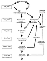
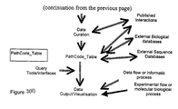
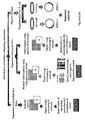
- Figure 2 displays a scheme and features for a set of data-tables suitable for managing such interaction data that could be implemented in either relational or object-orientated data-bases.
- the primary links between table-keys are indicated, as are the suggested fields or elements to be held within each table.
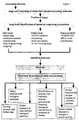
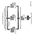
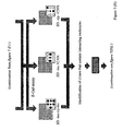
- An established computer data-base of protein interactions has many useful applications. For example, it may be used to predict the existence of new biological interactions or pathways, or to determine links between biological networks. Furthermore with this method, the function and localisation of previously unknown proteins can be predicted by determining their interaction partners. It also can be used to predict the response of a cell to changes in the expression of particular members of the networks without making a molecular, cellular or animal experiment. Finally, these data can be used to identify proteins or interactions between proteins within a medically relevant pathway, which are suitable for therapeutic intervention, diagnosis or the treatment of a disease ( Figure 1).
- a significant advantage of the method of invention over existing 2H systems is the ease of removal of false positives from sets of host cells designed for 2H screening experiments.
- a further advantage lies in the scale at which such identification of interactions and interaction members can be made. Due to the ease of automating the method of the invention at different stages, fast and reliable screening of large numbers of clones will be possible.
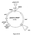
- the plasmid pBAD18- ⁇ RNAP expresses under the control of the arabiose promoter, fusion proteins between the ⁇ amino terminal domain (NTD) of the ⁇ -subunit of RNA polymerase and DNA fragments cloned into the multiple cloning site.
- NTD ⁇ amino terminal domain
- the presence of this plasmid in kanamycin-sensitive cells can be selected by plating on growth medium supplemented with kanamycin, or for its absence by the counterselective rpsL allele by plating on media supplemented with streptomycin (Murphy et al. 1995).
- the plasmid pBAD30-cI expresses under the control of the arabinose promoter, fusion proteins between the ⁇ cI protein and DNA fragments cloned into the multiple cloning site.
- the presence of this plasmid in amplicillin sensitive cells can be selected by plating on growth medium supplemented with amplicillin, or for its absence by the counterselective lac Y gene by plating on media supplemented with 2-nitrophenyl- ⁇ -D-thiogalactosidase (tONPG) (Murphy et al. 1995).
- the oriT sequence enables unidirectional genetic exchange of the pBAD30-cI plasmid and its derivatives from E.coli cells containing the F' fertility factor to F - strains lacking the fertility factor.
- Example 1 Construction of vectors yeast strains and read-out system for an improved yeast two-hybrid system
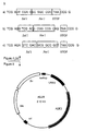
- the plasmids constructed for an improved yeast two-hybrid system pBTM118 a, b and c and pGAD428 a, b and c are shown in Figure 4. Both sets of vectors can be used for the construction of hybrid (fusion) proteins.
- the vectors contain the unique restriction sites Sal I and Not I located in the multiple cloning site (MCS) region at the 3'- end of the open reading frame for either the lex A coding sequence or the GAL4ad sequence Figure 4b).
- the two-hybrid plasmids shown in Figure 4a are shuttle vectors that replicate autonomously in both E. coli and S. cerevisiae .
- the three plasmids pBTM118 a, b and c are used to generate fusions of the LexA protein (amino acids 1-220) and a protein of interest cloned into the MCS in the correct orientation and reading frame.
- the plasmids pBTM118 a, b and c are derived from pBTM117c (Wanker et al., 1997) by insertion of the adapters shown in Table 1 into the restriction sites Sal I and Not I to generate the improved vectors with three different reading frames.
- the plasmids pBTM118 a, b and c carry the wild-type yeast CAN1 gene for counterselection, which confers sensitivity to canavanine in transformed yeast cells (Hoffmann, 1985).
- the plasmids also contain the selectable marker TRP1 , that allows yeast trp1-auxotrophs to grow on selective synthetic medium without tryptophan, and the selectable marker bla which confers ampicillin resistance in E . coli .
- the plasmids pGAD428 a, b and c are used to generate fusion proteins that contain the GAL4 activation domain (amino acids 768-881) operatively linked to a protein of interest.
- the plasmids pGAD428 a, b and c carry the wild type yeast CYH2 gene, which confers sensitivity to cycloheximide in transformed cells (Kaeufer et al., 1983), the selectable marker LEU2, that allows yeast leu2-auxotrophs to grow on selective synthetic medium without leucine, and the bacterial marker aphA (Pansegrau et al., 1987) which confers kanamycin resistance in E. coli .
- the plasmids pGAD428a, b and c were created from pGAD427 by ligation of the adapters shown in Table 1 into the MCS to construct the improved vectors with three different reading frames.
- pGAD427 For the construction of pGAD427, a 1.2 kb Dde I fragment containing the aphA gene was isolated from pFG101u (Pansegrau et al., 1987) and was subcloned into the Pvu I site of the pGAD426 using the oligonucleotide adapters 5'- GTCGCGATC-3' and 5'-TAAGATCGCGACAT-3'.
- the plasmid pGAD426 was generated by insertion of a 1.2 kb Eco RV CYH2 gene fragment, which was isolated from the pAS2-1 (Clonetech) into the Pvu II site of pGAD425 (Han and Collicelli, 1995).
- L40cc Saccharomyces cerevisiae strains L40cc, L40ccu and L40ccu ⁇ were created.
- the S. cerevisiae . L40cc was created by site specific knock-out of the CYH2 and CAN1 genes of L40 (Hollenberg et al., Mol. Cell. Biol. 15: 3813-3822), and L40ccu created by site specific knock-out of the URA3 gene of L40cc (Current Protocols in Molecular Biology, Eds. Ausubel et al.
- the strain L40ccu ⁇ was created by conducting a mating-type switch of the strain L40ccu by standard procedures (Ray BL, White CI, Haber JE (1991)).
- the genotype of the L40cc strain is: Mata his3 ⁇ 200 trp1-901 leu2-3,112 ade2 LYS2::(lexAop) 4 -HIS3 URA3::(lexAop) 8 -lacZ GAL4 can1 cyh2
- the genotype of the L40ccu strain is: Mata his3 ⁇ 200 trp1-901 leu2-3,112 ade2 LYS2::(lexAop) 4 -HIS3 ura3::(lexAop) 8 -lacZ GAL4 can1 cyh2
- that of L40ccu ⁇ is Mat ⁇ his3 ⁇ 200 trp1-901 leu2-3,112 ade2 LY
- FIG. 5 shows the URA3 read-out system carried by the plasmid pLUA.
- This URA3 read-out system under the control of a bacterial LexAop upstream activation sequence (UAS) can be used within the yeast 2-hybrid system both as a counter selective reporter gene and as a positive selection reporter gene to eliminate false positive clones.
- UAS bacterial LexAop upstream activation sequence
- the plasmid contains the features of the UAS lexAop -URA3 read-out system, the selectable marker ADE2 that allows yeast ade2-auxotrophs to grow on selective media without adenine and the bla gene which confers amplicillin resistance in E . coli .
- the plasmid pLUA is a shuttle vector that replicates autonomously in E . coli and yeast.
- pLUA For the construction of pLUA a 1.5 kb Sac I/ Cla I UAS lexAop -URA3 fragment was isolated from pBS-lexURA and ligated together with a 2.4 kb Sac I/ Cla I ADE2 fragment into Cla I digested pGAD425 ⁇ .
- pBS-lexURA was generated by ligating URA3 fragment together with a UAS lexAop fragment into pBluescript SK+.
- the URA3 and UAS lexAop fragments were obtained by PCR using genomic DNA from S .
- ADE2 gene was isolated by PCR using genomic DNA from SEY6210 ⁇ .
- pGAD425 ⁇ was generated by deleting of an 1.2 kb Sph I fragment from pGAD425 (Han and Colicelli, 1995) and religation of the vector.
- pBTM117c-HIP1 and pGAD427-HIP1 were obtained, by ligation of a 1.2 kb Sal I HIP1 fragment isolated from pGAD-HIP1 (Wanker et al., 1997). into pBTM117c and pGAD427, respectively.
- pBTM117c-MJD was created by inserting a 1.1 kb Sal I/ Not I MJD1 fragment (Kawagushi et al., 1994) into pBTM117c, and pGAD427-14-3-3 was generated by inserting a 1.0 kb EcoRI/NotI fragment of pGAD10-14-3-3 into pGAD427.
- pGAD427-HIPCT For the construction of pGAD427-HIPCT, a 0.5 kb Eco RI HIP1 fragment isolated from pGAD-HIPCT (Wanker et al., 1997) was ligated into pGAD427.
- pGAD427-lexA and pGAD427-ARNT were generated by insertion of a 1.2 kb Sal I/ Not digested lexA PCR fragment and a 1.4 kb Sal I/ Not I ARNT fragment into pGAD427, respectively.
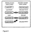
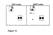
- fusion proteins LexA-SIM1 and GAL4ad-ARNT specifically interact with each other in the yeast two-hybrid system (Probst et al., 1997), because when both hybrids were coexpressed in Saccharomyces cerevisiae containing two integrated reporter constructs, the yeast HIS3 gene and the bacterial lacZ gene, which both contained binding sites for the LexA protein in the promoter region, the interaction between these two fusion proteins led to the transcription of the reporter genes.
- the fusion proteins by themselves were not able to activate transcription because GAL4ad-ARNT lacks a DNA binding domain and LexA-SIM1 an activation domain (Probst et al., 1997).
- the fusion proteins LexA-HIP1 and GAL4ad-LexA are capable of activating the HIS3 and lacZ reporter genes without interacting with a specific GAL4ad or LexA fusion protein, respectively.
- the yeast clones expressing the LexA-HIP1 protein have to be designated as false positives, because false positives are defined here as clones where a GAL4ad fusion protein or a LexA fusion protein alone without the respective partner protein activates the transcription of the reporter genes without the need for any interacting partner protein.
- the proteins LexA and GAL4ad and the fusion proteins LexA-MJD and GAL4ad-14-3-3 which are also present in the defined interaction library are unable to activate the reporter genes either alone or when present in the same cell with any other fusion proteins comprising the library.
- Example 2 Detection of clones expressing known interacting proteins from false positives using the improved two-hybrid system
- Pairs of the yeast two-hybrid plasmids pBTM117cSIM1 & pGAD427-ARNT, pBTM117c & pGAD427 and pBTM117c-HIP1 & pGAD427 were transformed into the yeast strain L40cc, and Trp+Leu+ transformants that contained at least one of each of the two plasmids were selected on SD-leu-trp plates.
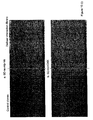
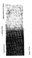
- Figure 9 shows that the yeast strains cells harboring both the plasmids pBTM117c-SIM1 & GAD427-ARNT or pBTM117c-HIP1 & pGAD427 grow on SD-leu-trp-his plates and turn blue after incubation in X-Gal solution, indicating that the HIS3 and lacZ reporter genes are activated in these strains.
- the yeast strain harboring both the negative control plasmids pBTM117c & pGAD427 was not able to grow on this medium and also showed no lacZ activity.
- Example 3 Generation of regular grid patterns of host cells expressing potentially interacting fusion proteins
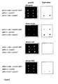
- the constructs for the expression of the fusion proteins shown in Figure 8 were pooled and 3 ⁇ g of the mixture was co-transformed into yeast strain L40cc by the method of Schiestel & Gietz (1989).
- the yeast cells co-transformed with the constructs described in Table 2 were plated onto large 24 x 24 cm agar trays (Genetix, UK) containing minimal medium lacking tryptophan leucine and histidine (SD-leu-trp-his).
- the agar trays were poured using an agar-autoclave and pump (Integra, Switzerland) to minimise tray-to-tray variation in agar colour and depth.
- the transformation mixture was plated such that between 200 and 2000 colonies per agar tray were obtained after incubation at 30°C for 4 to 7 days.
- Suitable changes to the hardware and software of a standard picking robot designed for the picking of E. coli cells as described by Lehrach et al. (1997) were made to account for the specific requirements of yeast cells.
- the illumination of. agar-trays containing plated colonies was changed from the dark-field sub-illumination to dark-field top-illumination to differentiate yeast colonies from the lawn of non-transformed cells.
- the existing vision guided motion system Karlnaswamy & Agapakis 1997) was modified to allow for a larger range of ⁇ blob' size when selecting yeast colonies to pick from the blob-feature-table returned by connectivity algorithms when applied to a digital image of the agar tray containing colonies.
- the clone inoculation routine was re-programmed to ensure that cell material which had dried on the picking pins during the picking routine was initially re-hydrated by 10 seconds of immersion in the wells of a microtiter plate before vigorous pin-motion within the well.
- This robotic procedure ensured that sufficient cell material was inoculated from each picking pin into an individual well of a microtiter plate.
- the picking pins were sterilised after inoculation to allow the picking cycle to be repeated by programming the robot to.brush the picking pins in a 0.3% (v/v) solution of hydrogen peroxide, followed by a 70% ethanol rinse from a second wash-bath and finally drying by use of a heat-gun to evaporate any remaining ethanol from the pins.
- an algorithm to automatically correct for height variation in the agar was incorporated by referencing the surface height of the agar in three corners and from these points automatically estimating the surface plane of the agar.
- the robot was further programmed to automatically adjust both the imaging and picking heights according to the agar surface height such that when a pin was extended into a colony, it removed cells only from the top surface of the colony and did not penetrate the whole colony into the growth medium.
- the connectivity algorithms would detect two "blobs" - one (the blue centre) lying directly on the other (the white surrounding) and although the former would be ignored since it was blue, the latter would be selected as its average grey-scale was greater than the discrimination value.
- Such cases were successfully selected against by ignoring any colonies that had 'holes' using a 'number of holes' function of the image analysis program, which flags those blobs which have a second blob within their boundary.
- yeast colonies were automatically picked from the agar-trays into individual wells of a sterile 384-well microtiter plate (Genetix, UK) containing sterile liquid minimal medium lacking leucine and tryptophan (SD-leu-trp) and containing 7% (v/v) glycerol.
- the resulting microtiter plates were incubated at 30°C for 36 hours, the settled colonies were dispersed by vigorous mixing using a 384-well plastic replicating tool (Genetix, UK) and then incubated for a further 2 to 4 days. A picking success of over 90% wells containing a growing yeast culture was achieved.
- each plate was labelled with a unique number and barcode.
- Each plate was also replicated to create two additional copies using a sterile 384-pin plastic replicator (Genetix, UK) to transfer a small amount of cell material from each well into pre-labelled 384-well microtiter plates and pre-filled with SD-leu-trp-his/7% glycerol liquid medium.
- the replicated plates were incubated at 30 °C for 3 days with a cell dispersal step after 36 hours, subsequently frozen and stored at -70°C together with the original-picked microtiter plates of the interaction library.
- a regular grid pattern of yeast cells expressing potentially interacting yeast clones was generated using a robotic and automated picking system.
- 384-well microtiter plates have a well every 4.5 mm in a 16 by 24 well arrangement. Therefore, for each 384-well microtiter plate a regular grid pattern at a density greater that 4 clones per square centimetre was automatically created.
- a computer-controlled 96-well pipetting system (Opal-Jena) with automatic plate-stacking, tip washing, liquid waste and accurate x-y positioning of the microtiter plate currently accessed by the tips was employed.
- the yeast two hybrid cells that had settled in the bottom of the wells of the arrayed interaction library as described above were re-suspended, and a stack of these 384-well plates were placed into the input stacker of the pipetting system.
- the system was programmed to take a single 384-well microtiter plate containing the arrayed yeast two-hybrid clones and parallel aspirate 10 ⁇ l of culture medium and cells into each of the 96 pipette tips from 96 wells of the 384-well plate.
- the inter-tip spacing of the 96 tips was 9mm and the wells of the 384-well microtiter plate were 4.5 mm so that cells were removed from only every other well along each dimension of the 384-well plate. 8 ⁇ l of the 96 aspirated samples contained in the tips were then pipetted in parallel into one set of wells of a sterile 1,536-well microtiter plate (Greiner, Germany).
- yeast cells were deposited into only 1 every 4 wells along each dimension of the 1,536-well plate. The remaining 2 ⁇ l of culture medium and cells was aspirated to waste before sterilising each 96 tips in parallel. Sterilisation was conducted by twice aspirating and washing to waste 50 ⁇ l of 0.3% (v/v) hydrogen peroxide stored in a first replenishable wash-bath on the system, and then aspirating and washing to waste 50 ⁇ l sterile distilled water stored in a second replenishable wash-bath.
- This plate-to-plate pipetting cycle was repeated 3 further times, each time aspirating a different set of 96-clones from the 384-well array of yeast 2-hybrid clones into a different set of 96-wells in the 1,536-well microtiter plate by moving the microtiter plates relative to the 96-tips using the accurate x-y positioning of the system.
- the first 384-well microtiter plate was automatically exchanged for the next 384-well microtiter plate, and the yeast 2-hybrid clones arrayed in this second 384-well plate were similarly arrayed into the 1,536-well plate.
- the yeast 2-hybrid clones contained within four 384-well microtiter plates had been automatically arrayed in the first 1,536-well plate, filling all wells, the 1,536-well plate was automatically exchanged for a second sterile 1,536-well plate stored in the second stacking unit of the pipetting system. The whole process was repeated until all yeast 2-hybrid clones of the interaction library had been automatically transferred form 384-well to 1,536-well microtiter plates.
- 1,536-well microtiter plates have a well every 2.25 mm in a 32 by 48 well arrangement. Therefore, for each 1,536-well microtiter plate we automatically created a regular grid pattern at a density greater than 19 clones per square centimetre.
- a high-throughput spotting robot such as that described by Lehrach et al . (1997) was used to construct porous planar carriers with a high-density regular grid-pattern of yeast clones from the defined interaction library contained within 384-well microtiter plates.
- the robot recorded the position of individual clones in the high-density grid-pattern by the use of a pre-defined duplicate spotting pattern and the barcode of the microtiter plate.
- Individually numbered membrane sheets sized 222 x 80 mm were pre-soaked in SD-leu-trp-his medium, carefully laid on a sheet of 3mm filter paper (Whatmann) pre-soaked in the same medium and placed in the bed of the robot.
- the interaction library was automatically arrayed as replica copies onto the membranes using a 384-pin spotting tool affixed to the robot.
- Five different microtiter plates from the first copy of the interaction library were replica spotted in a "3x3 duplicate" pattern around a central ink guide-spot onto 10 nylon membranes - corresponding to approximately 1,900 clones spotted at a density of approximately 40 spots per cm 2 .
- On each replica membrane three different control clones were spotted, each from a microtiter plate that contained the same control clone in every well.
- One control clone expressed the fusion proteins LexA-SIM1 & GAL4ad-ARNT, a second control clone the fusion protein LexA-HIP1, while a third expressed fusion protein GAL4ad-LexA, and all were spotted in order to test the selection, counterselection and the ⁇ -gal assay features of the method.
- the robot was programmed to spot onto each spot position 5 times from a slightly different position within the wells of the microtiter plates. The robot created a data-file in which the spotting pattern produced and the barcode that had been automatically read from each microtiter plate was recorded.
- Each membrane was carefully laid onto approximately 300 ml of solid agar media in 24 x 24 cm agar-trays. Six membranes were transferred to SD-leu-trp-his media and two each of the remaining membranes were transferred to either SD-trp+CHX or SD-leu+CAN media. The yeast colonies were allowed to grow on the surface of the membrane by incubation at 30 °C for 3 days.
- the plasmid pGNG1 (MoBiTec, Germany) carries a green fluorescent protein variant under the control of a LexA operator.
- This variant, GFPuv is up to 16 times brighter that the wild-type variant isolated from Aequora victoria (Ausubel et al., 1995; Short protocols in molecular biology, 3 rd ed. John Wiley & Sons, New York, NY.).
- the yeast 2um origin of replication and the auxotrophic marker URA3 maintains the plasmid in ura3 mutant yeast strains.
- This plasmid should act as a read-out system to detect single fusion proteins or interacting fusion proteins able to activate the read-out system in the method of invention described herein.
- green fluorescent proteins and its variants are considered suitable reporter genes in most host-cell types. Therefore, it would be possible for a person skilled in the art to incorporate this gene within other host-cell types and interaction systems as disclosed in this invention.
- the yeast strain L40ccu was transformed with the plasmid pGNG1 (MoBiTec, Germany) using the method of Schistel & Gietz (1989), and a resulting stable transformant clone cultured in minimal medium lacking uracil and subsequently used to generate two further yeast clones, each containing two genetic elements.
- the first strain, GNGp was generated by co-transformation of a mixture of the plasmids pBTM117c-SIM1 and pGAD427-ARNT co-transformed into L40ccu already carrying the reporter plasmid pGNG1.
- the second strain, GNGn was generated by co-transformation of a mixture of the plasmids pBTM117c-MJD and pGAD427-14-3-3 co-transformed into L40ccu already carrying the reporter plasmid pGNG1.
- the transformations were conducted using the method of Schistel & Gietz (1989), and transformants were selected by plating on minimal media lacking uracil, tryptophan and leucine.
- microtiter plates Individual colonies from the two transformations were picked into individual wells of 384-well microtiter plates as described in section 3.1 except that the microtiter plates contained liquid minimal medium lacking uracil, tryptophan and leucine.
- One microtiter plate was created that contained individual colonies of the GNGp yeast strain, and another carrying colonies of GNGn.
- a spotting robot (Lehrach et al., 1997) fitted with high precision spotting tool carrying 16 pins in a 4 x 4 pattern, the clones were arrayed onto poly-lysine coated glass-slide (Sigma, US).
- the clones were spotted at a spacing of 440 ⁇ m, with a spot diameter of approximately 300 ⁇ m generating a density of over 490 clones per square centimetre.
- the robot was programmed to spot onto each spot position 10 times from a slightly different position within the wells of the microtiter plates. It is well known in the art that piezo-ink-jet micropipetting systems (Kietzmann et al., 1997, Schober et al., 1993) can create regular grid pattern of clones at an even greater density. Indeed, grid densities of over 1600 spots per quare centimeter have been achieved with such systems.
- the fluorescent read-out system of cells in the regular grid pattern of cells was then visualised using a sensitive CCD camera (LAS1000, Fuji, Japan). Appropriate excitation light was provided and an emission filter appropriate for the emission spectrum of GFP uv was fitted to the lens.
- Other imaging systems could be utilised to investigate the regular grid pattern of clones. For example, laser-scanning systems including laser scanning confocal microscopes would be preferred when imaging very high density regular grid patterns, or for those formed from a small number of host cells deposited at each position.
- fusion proteins LexA-SIM1 and GAL4ad-ARNT can interact and activate a read-out system under control of the LexA operator. Since the GNG uv reporter gene is under the control of a LexA operator, a cell carrying the pGNG1 plasmid and expressing these fusion proteins should fluoresce under UV light. In contrast, the fusion proteins LexA-MJD and GAL4-14-3-3 were shown unable to activate the same read-out system. Image analysis of the digital image of the regular grid pattern of yeast cells, demonstrated that indeed, the GNGp yeast strain did fluoresce while the GNGn did not.
- pGNG1 As an alternative to pGNG1 a person skilled in the art could subclone an improved GFP mutant as described in Anderson et al. (1996). Replacement of the URA coding sequence in pLUA (section) with GFP is performed by using appropriate anchor primer to amplify the GFP mutant. Using the appropriate growth media the analysis can be performed as described above.
- Example 4 Detection of the read-out system in a regular grid pattern.
- Positive clones that expressed interacting fusion proteins were detected from false positive clones by considering the activity of ⁇ -galactosidase of clones grown on spotted membranes laid on the various selective media. Positive clones should activate the lacZ reporter gene on SD-leu-trp-his media and turn blue on incubation with X-Gal solution, but not on either of the two counterselective media. False positive clones should activate the reporter gene and turn blue on incubation with X-Gal solution on at least one counterselective media as well as on the SD-leu-trp-his medium.
- Figure 11 shows magnified images of a ⁇ -gal assay of clones grown on the membranes which had been placed on the three selective media.
- two clones were detected as positive clones that express interacting fusion proteins since they activated the lacZ reporter gene on SD-leu-trp-his media, but not on either of the two counterselective media, and whose spotted positions are circled.
- the two clones were identified by their microtiter plate address within the interaction library as 06L22 and 08N24 respectively. All other clones spotted within this region of the membrane were detected as false positive since they express ⁇ -galactosidase on SD-trp+CHX medium as well as on SD-leu-trp-his medium.
- LacZ reporter gene for the three control clones spotted onto the same membranes confirm these results.
- the positive control clone that expresses the interacting fusion proteins LexA-SIM1 & GAL4ad-ARNT should show a LacZ+ phenotype when grown on SD-leu-trp-his medium, but LacZ- when grown on either of the counterselective media.
- This control clone was spotted at position 03 in the region of the membranes shown in Figure 11b, of which one example is circled. The pattern of ⁇ -gal activity for this positive control clone on the three selective media is as predicted.
- the false positive control clone that expresses the fusion protein LexA-HIP1 and the false positive clone that expresses the fusion protein GAL4ad-LexA are spotted at positions 02 and 01, respectively. Both false positive control clones show a LacZ+ phenotype when grown on SD-leu-trp-his media, but are detected as false positive clones by the method of the invention since they also show a LacZ+ phenotype on SD-leu+CAN or SD-trp+CHX media, respectively.
- the clones spotted at position 04 are from the defined interaction library, and from their LacZ+ phenotype when grown on SD-leu+CAN media are predicted to be false positive clones.
- the image analysis system described above was used to automatically identify those individual clones on each high-density regular grid pattern that had activated the LacZ read-out system. This was conducted for each of the membranes grown on the three selective media, and the intensity of ⁇ -galactosidase activity for each clone grown on the three media was automatically recorded by the program using a scale from 0 to 3 (no activity, weak activity, medium activity, high activity). These data for all clones on a given membrane were saved in a computer file, and the ⁇ -galactosidase activity for a given clone was related to its activity when grown on the other two selective media using a computer program.
- the interaction library comprising the yeast cells as described in section 3.1 were screened in microtiter plate format to identify those cells that express interacting fusion proteins.
- microtiter plates containing the interaction library were removed from frozen storage and thawed to room temperature.
- each plate was replicated and labelled as described in section 3.1 to create additional copies for screening, each into 3 separate selective media.
- Cells were transferred into 384-well microtiter plates pre-filled with 40 ⁇ l of the liquid selective media SD-leu-trp, SD-leu+Can or SD-trp+CHX.
- the ⁇ -galactisidase substrate Galacton-Star in combination with Sapphire II (Tropix, US) generates detectable luminescent light on activation of the ⁇ -gal reporter gene in the yeast-cells, and an exposure time of 5 minutes was used to collect sufficient signal.
- the grey-scale digital images were captured, saved on computer and subsequently analysed using the image analysis system described in section 4.1. However, in this case, the position of each clone was far simpler to determine due to the lower density of the regular grid pattern of clones in the microtiter plate. Second, the size of the pixel mask used to measure the average pixel intensity was approximately that of the size of the microtiter plate well. Positive clones in the six microtiter plates were identified by image analysis of the digital images from clones grown in the three selective media, and these data processed by the computer program as described in section 4.1.
- the interaction library constructed for this example was composed of known fusion proteins with predicted interactions as shown in Figure 8.
- a real positive clone from this defined interaction library is therefore expected to express the interacting fusion protein-pairs LexA-SIM1 & GAL4ad-ARNT, LexA-HD1.6 & GAL4ad-HIP1 or LexA-HD3.6 & GAL4ad-HIP1 and hence contain the corresponding pairs of plasmid constructs pBTM117c-SIM1 & pGAD427-ARNT, pBTM117c-HD1.6 & pGAD427-HIP1 or pBTM117c-HD3.6 & pGAD427-HIP1, respectively.
- the four membranes which had been placed on the SD-leu-trp-his medium and had not been used to assay ⁇ -gal activity were processed according to the procedure described in Larin & Lehrach (1990) in order to affix the DNA contained within the clones of the interaction library onto the surface of the membrane.
- a 1.1 kb DNA fragment of SIM1 and a 1.3 kb DNA fragment of ARNT were radioactively labeled by standard random priming procedures for use as a hybridisation probe (Feinberg & Vogelstein, 1983).
- Each probe was heat denatured for 10 min at 95 °C and hybridised overnight at 65 °C in 15 ml of 5% SDS/0.5M sodium phosphate (pH 7.2)/1 mM EDTA with a high-density spotted membrane with DNA from the interaction library affixed to it as prepared above.
- the membranes were washed once in 40mM sodium phosphate/0.1%SDS for 20 min at room temperature and once for 20 min at 65 °C before wrapping each membrane in Saran wrap and exposing it overnight to a phosphor-storage screens (Molecular Dynamics, USA).
- a digital image of each hybridised membrane was obtained by scanning the phosphor-storage screen using a phosphor-imager (Molecular Dynamics, USA).
- the individual clone 06L22 was recovered from the frozen plates of the original interaction library and inoculated into SD-leu-trp-his liquid medium. This culture was allowed to grow for 3 days at 30 °C and the corresponding plasmids contained in the clone were isolated using a QiaPrep (Qiagen, Hilden) procedure. Duplex PCR was used to simultaneously amplify the inserts contained within the plasmid constructs using primer-pairs specific for either the pBTM117 or pGAD427 plasmids.
- PCR of the individual inserts from individual plasmids carried by clone 06L22 was conducted as above except by using only the respective primer pair for the required plasmid.
- the individual inserts were also amplified directly from the yeast culture using a Whole Cell Yeast PCR Kit (Bio 101, USA).
- the pairs of inserts isolated from clone 06L22 either by amplification from the extracted plasmid DNA or by direct PCR of the yeast clone were subjected to DNA sequencing by standard protocols.
- the 1.26Kb inserts amplified using the primers specific to plasmid pBTM117 were confirmed as the expected fragment of the SIM1 gene by comparison of the known sequence for this gene (Probst et al., 1997).
- the 1.37Kb inserts amplified using the primers specific to the pGAD427 plasmid were confirmed as the expected fragment of the ARNT gene.
- Example 6 Detection and identification of interacting proteins using a large-scale and automated application of the improved 2-hybrid system
- FIG. 6 A scheme utilizing the method of the invention within a large-scale and automated approach for the parallel detection of clones that express interacting fusion proteins and the identification of members comprising the interactions is shown in Figure 6.
- Yeast clones from an "interaction library" that express interacting proteins are identified on a large-scale by the use of visual inspection or digital image processing and analysis of high-density gridded membranes on which their ⁇ -galactosidase activity has been assayed after growth on various selective media. Automated methods as described in earlier examples are used to effect the production of the interaction library and high-density spotted membranes, and the analysis of digital images of the ⁇ -gal assay and hybridisation images.
- the resulting recombinant colonies were washed from the 5 trays using 50 ml of LB liquid media for each tray.
- the amplified cDNA library cloned into pSport was isolated from this wash mixture by a QiaPrep (Qiagen, Germany) plasmid extraction procedure. Approximately 1 ⁇ g of the library inserts were then isolated from the plasmid DNA by Not 1/ Sal 1 digestion and size selected (1-1.5Kb) by agarose gel purification using standard procedures.
- Two pools representing all three reading frames of the two vector series pGAD428 and pBTM118 were prepared by Not 1/ Sal 1 digestion and pooling of 1 ⁇ g each of vectors pGAD428 a, b & c and pBTM118 a, b & c, respectively.
- the insert mixture that was isolated as above was split into two equal fractions and 300 ng was ligated with 50 ng of each prepared vector-series pool. Following ligation, each reaction was then separately transformed into electro-competent E.coli cells, and recombinant clones for each library were selected on five 24 x 24 cm plates using kanamycin or ampicillin for the pGAD428 or pBTM118 libraries respectively.
- molar-equivalent amounts of the DNA binding and activation domain libraries were pooled, and 20 ⁇ g of this mixture was co-transformed into the yeast strain L40cc by the method of Gietz et al. (1992).
- the resulting transformation mix was plated on a single 24 x 24 cm agar tray.
- the agar-trays were prepared as described in section 1.3.1. A total of twenty transformations were prepared and plated onto separate agar trays yielding an average of 1500 yeast colonies per tray after 7 days of incubation at 30 °C.
- the agar trays containing yeast colonies were placed in the modified laboratory picking robot and individual clones were automatically picked as described in section 3.1.
- a total of 30 384-well microtiter plates were generated and represented an interaction library of greater than 10,000 clones for the study organism.
- the library was replicated to generate 3 further copies, labelled and all copies were stored at -70°C to provide for analysis at a later date as described in section 3.1.
- the clones comprising it were arrayed at high density on 222 x 222 mm porous membranes (Hybond N+, Amersham, UK) using the method described.in section 3.3.
- a total of twenty replica membranes, each arrayed in a " 3 x 3 duplicate" regular grid-pattern of clones using 23 384-well microtiter plates from a thawed copy of the stored interaction library were produced.
- one microtiter plate was aditionally arrayed in position 24 containing 8 different control clones representing known positive, negative and false positive clones.
- This pattern corresponded to over 9000 yeast two-hybrid clones spotted at a density of approximately 40 clones cm -2 .
- the robot was programmed to spot onto each spot position 5 times from a slightly different position within the wells of the microtiter plates. The robot created a data file in which the spotting pattern produced and the barcode that had been automatically read from each microtiter plate was recorded.
- Each membrane was carefully laid onto approximately 300 ml of solid agar media in 24 x 24 cm agar trays. Fourteen membranes were transferred to SD-leu-trp-his media and three each of the membranes which had been spotted five times were transferred to either SD-trp+CHX or SD-leu+CAN media. The yeast colonies were allowed to grow on the surface of the membrane by incubation at 30 °C for 3 days.
- This program was used to query these data and identify those clones that had activated the read-out system when grown on two out of three SD-leu-trp-his replica membranes, but not when grown on any of the two sets of three replica membranes placed on the two counterselective media SD-leu+CAN or SD-trp+CHX.
- the data-base correctly identified the eight different control clones each arrayed in 48 wells of the 24 th microtiter plate. A total of 7539 clones from the interaction library arrayed in 23 384-well microtiter plates were thus identified as positive clones - clones that only activated the read-out system when both plasmids (and hence fusion proteins) were expressed in the cell.
- 3983 clones were identified as false-positive clones as they also activated the read-out system when grown on SD-trp+CHX medium, the growth medium that eliminated the plasmid expressing the activation domain fusion protein.
- 113 clones were identified as false positive clones by activating the read-out system when grown on SD-leu+CAN medium, the growth medium that eliminated the plasmid expressing the DNA binding fusion protein.
- a total of 96 positive clones were randomly selected from the database and recovered from a frozen copy of the interaction library clones stored in 384-well microtiter plates.
- the DNA sequences cloned into the pGAD428 and pBTM118 vectors carried by each clone were directly amplified as described in section 5.2 except that the direct PCR reactions were conducted in 96-well microtiter plates using a high-thoughput water-bath thermocycling machine (Maier et al., 1994).
- Standard sequencing approaches were employed to characterise the nucleic acids encoding the DNA-binding domain fusion proteins of the positive clones following pBTM428- specific 96-well PCR as described above. Similarly, the sequence of the insert encoding for the activation-domain fusion protein following pGAD118-specific PCR was determined. Sequence comparison of these insets against published DNA sequences using standard sequence comparison tools (e.g. BAST), identified that one interaction involved two previously unidentified gene fragments that were expressed by the positive-clone located in plate 5, well K20. From the predicted protein sequence these two genes were designated Protein A and Protein B.
- standard sequence comparison tools e.g. BAST
- FIG. 18 shows a graphical representation of the hybridisation-positive and interaction-positive clones identified with probe B (open circles) and probe A (red circles). Two clones (5K20 and 3L11 marked by "A/B") gave a hybridisation signal with both probe A and Probe B, indicating that both these positive clones expressed the same interacting fusion proteins.
- the 3443 positive clones identified as described above were distributed across all 23 microtiter plates of the interaction library. To greatly facilitate further analysis of positive clones, it was advantageous to individually physically isolate clones and to generate a second, re-arrayed regular grid-pattern of positive clones, preferably within a further set of 384-well plates.
- the list of positive clones, together with their plate-well location was generated from the data-base described in Example 7 and automatically loaded as a computer file onto the re-arraying robot.
- the robot automatically took the mother plate containing the first positive yeast two-hybrid clone by reference to the data file and read and recorded the barcode of the plate.
- Individual and sequential pins of the 96-pin re-arraying head were positioned above and lowered into the required wells from this first plate, and the mother plate was automatically exchanged when all positive clones had been sampled.
- the head was automatically moved over to the first 384-well daughter plate containing SD-leu-trp/7%glycerol and inoculated all 96-pins in the first set of wells as described above.
- a data output file was then updated which related the new plate-well location of a given positive clone in the re-arrayed library to its old plate well location in the original interaction library. All pins were then sterilised as described, and the cycle completed until all positives clones had been transferred from the interaction library to a new plate well location comprising the re-arrayed library.
- the data output file was then transferred to the central computer database to append a table in the database described in Example 7 to record the correct location of a given positive clone in the re-arrayed interaction library.
- the resulting clones in the daughter plates were replicated into two further copies and stored at -70 °C as described in section 3.1.
- Example 7 Generation of a database of interactions.
- Figure 2 Central to the scheme ( Figure 2) is a data table holding relevant information on each member of an interaction - the cDNA-Table - where a separate record in the table represents each member of an interaction, and members are indicated to form interactions by sharing the same clone name. It is advantageous to structure the core data table in this way for several reasons. First, the same core table can be used to hold data on cDNAs from different kinds of genetic libraries (for example, standard cDNA or genomic libraries) which can be generated during a global analysis using various genomic techniques, not just interaction data. Secondly, each of the members of an interaction, or genetic fragments may be further characterised by a number of ways for different sets of data.
- genetic libraries for example, standard cDNA or genomic libraries
- the Gene_Table which provides a direct relationship to the fragment's DNA sequence, nucleotide homology match (for example through BLAST searching) and the corresponding gene name.
- the Domain_Table provides facility to directly access data of the fragment's in-frame translation, amino acid homology match (for example through BLASTN searching) and any 2 or 3-dimensional structural information which may be known or can be predicted.
- this database structure provides the facility to relate from the central cDNA_Table to any other table holding data describing said characterisation as may be appropriate.
- oligofingerpriting data can be used to identify each member of an interaction in a highly parallel manner and includes fields for data such as cluster number, confidence of cluster membership and predicted gene homology for that cluster (Maire et al., 1994).
- a database structure will more easily enable tertiary or higher order interactions to be incorporated within the same data table. This is in contrast to a structure in which interactions rather than members of an interaction were the basic object or record in a data table, and for each higher order interaction a new data-table would be needed or an existing data table modified.
- Y2H_Table In the case of a yeast two-hybrid interaction screen one related table would be the Y2H_Table. Said table may include information for a given clone pertaining to cloning and experimental details of its creation, the tissue and library from which it was derived, its physical location to enable easy access for further studies, whether it was derived from the mating of given Mat a and Mat ⁇ strains. Importantly, the Y2H_Table holds information pertaining to the interaction class of the clone, were said interaction class is defined as whether the clone was a positive clone, negative clone, or a false positive with respect to either the activation domain (AD) or biding domain (BD) fusion protein. The value for said interaction class is easily derived for a large number of clones by the method of invention described in earlier examples.
- AD activation domain
- BD biding domain
- the Hyb Table is provided. This table relates for a given clone, the hybridisation intensity obtained with a given probe in a hybridisation experiment using a given high-density array. Said high-density array to be related to tables holding data from the spotting robot such as the defined spotting pattern used, the method by which the array was produced and the identity of the library and clones arrayed on said array. The incorporation of these tables within a user interface will enable this embodiment of the method of invention to be easily conducted by displaying to the user the physical location of a given positive yeast two hybrid clone that hybridised to a given probe.
- Said two-hybrid clone can then be recovered, the members comprising the interaction isolated by PCR and sequenced.
- Said sequenced members of an interaction then provide data to be entered into the cDNA_Table and other related tables on further analysis.
- Said member to then be used as a second hybridisation probe onto an array to identify the next step in an interacting pathway by the same procedure.
- these data can be curated by manual and/or expert systems to update a definitive data table for example the PathCode_Table.
- Said definitive database to hold the highest quality information on interactions from the cDNA_Table, where said highest quality information on interactions to be those from the cDNA Table that pass a level of "certainty" as specified to the curator and/or expert system.
- all relevant data especially that of the translated frame of the cDNA and corresponding protein domain is related from other tables and presented in a usable form to the curator and/or expert system. This presentation allows for easy recognition and exclusion or correction of basic errors in the data such as poor quality sequencing, or incorrectly cloned cDNA fragments. These may include contaminating fragments which can be identified as originating from an organism which is different to that of the cDNA library.
- a given cDNA is entered into the PathCode_Table only once for each interaction in which it is found, together with a record for the corresponding interacting cDNA (or cDNAs for multimer complexes).
- a cDNA has different interactions, for example with different proteins or where different protein domains of the cDNA interacts with different proteins, then in each case a different record for the cDNA is created.
- These different records are linked by a common and unique "Interaction ID”.
- a given interaction is represented thus only once in the PathCode_Table, and is related to previous tables in the database by the host-cell clone that represents the interaction and the ID of each cDNA in the interaction. Said host cell that represents the interaction is selected by consideration and curation of all host-cells and the interacting fragments representing said interaction held in the cDNA_Table.
- a set of criteria can be implemented to assist in said curation and selection, and to derive a measure of confidence for the interaction.
- such criteria may have decreasing information value and include: First, if a given interaction is observed in both directions of the experiment ie proteinA-AD interacting with proteinB-BD, and proteinB-BD interacting with proteinA-DB. Second, if different examples of the same interaction are observed. Where different examples of the same interaction are defined as protein fragments of substantially different length and position (for example greater than 10% different) but from the same underlying protein domain and are also found to interact. Third, if the same examples of the same interaction are observed, for example by multiple cloning of the same fragments where the same fragments are of substantially the same length and position from the same underlying protein domain.
- the protein domains that interact may have biological relevance. That is, similar domains or genes are known to interact from public literature, or it is known that both genes are expressed or likely to be expressed in the same cellular location. This criterion can also be used as an internal quality control of the library cloning, interaction experiment and subsequent identification of interacting members since every interaction experiment should identify a certain set of published "house-keeping interactions", and the identification of such interactions can be used as quality measure for the overall interaction experiment.
- cDNA fragments representing the interacting proteins may be subcloned, and additional interaction experiments be conducted.
- Said additional interaction experiments may include testing each protein for interaction against a set of unrelated proteins to investigate the specificity of said interaction. Said testing may be conducted using the same interaction method that identified the interaction, for example the yeast two-hybrid, but preferable it is an independent method.
- Favoured is where a given interaction is biochemically validated using methods including tissue co-northern, cellular co-localisation or coprecipitation studies.
- the PathCode table has relational links to secondary or external databases holding data on nucleotide and protein sequences, and biochemical, structural, biological or Bibliographical information. These data, representing the complete relationships between all tables and databases can be queried by using simple user interfaces, designed for example using Java, or by more complicated commands such as those provided by SQL.
- Possible queries include those to locate from these data interactions, pathways or networks for a given nucleotide or amino acid sequence or motif, or for a given 3-dimensional structure or motif. Secondly, for highly established networks, these data may be queried to identify a given pathway between two given points. It may be that some queries are more efficiently conducted using a substantially different design of the PathCode_Table for example by representing a given interaction as the underlying record rather than a given member of an interaction. A person skilled in the art would be able to transfer data from one table design to another using standard data-parsing systems to enable said more efficient conduction of queries.
- the result of these queries is displayed using graphical methods to enable the investigator to interpret these data most efficiently.
- Said graphical methods to include elements activated by mouse clicks such as hotlinks to seamlessly link these data with other data sources, or to query and display further levels of interactions.
- Computer-based methods of generating visual representations of specific interactions, partial or complete protein-protein interaction networks can be employed to automatically calculate and display the required interactions most efficiently. Both finding the network paths and calculating the optimal display of the found paths can be based on algorithms well known in the art of mathematical graph theory. For example, algorithms simila to those which have been employed to display other biological relationships such as genetic pedigrees and phylogenetic relationships.
- An established computer database of protein interactions has many useful applications. For example, it may be used to predict the existence of new biological interactions or pathways, or to determine links between biological networks. Furthermore with this method, the function and localisation of previously unknown proteins can be predicted by determining their interaction partners. It also can be used to predict the response of a cell to changes in the expression of particular members of the networks without making a molecular, cellular or animal experiment. Finally, these data can be used to identify proteins or interactions between proteins within a medically relevant pathway, which are suitable for therapeutic intervention, diagnosis or the treatment of a disease.
- Example 8 Pre-selection against false positive clones and the automated creation of a regular grid-pattern of yeast cells expressing a fusion protein
- Three mating type-a yeast strains were constructed by co-transformation using the method of Schiestel & Gietz (1989). into L40ccu, of the plasmid pLUA containing the URA3 read-out system, and either the pBTM117c, pBTM117c-SIM1 or pBTM117c-HIP1 plasmids, respectively. Transformants that contained both the pLUA plasmid and one of the DNA binding domain plasmid were selected on SD-trp-ade medium.
- yeast strains were similarly constructed by cotransformation into L40ccu ⁇ of pLUA, and either the pGAD427, pGAD427-ARNT or pGAD427-LexA plasmids, respectively. Transformants that contained both the pLUA and one of the activation domain plasmids were selected on SD-leu-ade medium. The yeast strains thus obtained are listed in Table 3.
- yeast strains x1a, x2a and x3a were replica plated onto the selective media SD-trp-ade, SD-trp-ade containing 0.2% 5-FOA and SD-trp-ade-ura, while the yeast strains y1 ⁇ , y2 ⁇ and y3 ⁇ were replica plated onto the selective media SD-leu-ade, SD-leu-ade containing 0.2% 5-FOA and SD-leu-ade-ura.
- Table 4 shows that the two yeast strains x3a and y3 ⁇ which expressed the fusion proteins LexA-HIP1 and GAL4ad-LexA, respectively, were unable to grow on their respective media containing 5-FOA yet were able to grow on their respective media lacking uracil.
- all other yeast strains that contained plasmids that expressed fusion proteins that were alone unable to activate the read-out system could grow on their respective media containing 5-FOA, but could not grow on selective media lacking uracil.
- the URA3 read-out system successfully eliminated clones containing auto-activating fusion proteins prior to interaction mating.
- yeast strain L40ccu was transformed with the plasmid pLUA and a resulting stable transformant colony cultured in minimal medium lacking adenine. Cells from this culture were rendered competent and transformed with 3 ⁇ g pooled mixture of all six pBTM117c constructs shown in Table 2.
- yeast strain L40ccu ⁇ was transformed with the plasmid pLUA and a resulting stable transformant colony cultured in minimal medium lacking adenine. Cells from this culture were rendered competent and transformed with 3 ⁇ g pooled mixture of all six pGAD427 constructs shown in Table 2. In all cases, competent cells were prepared and transformations conducted using the method of Schiestel & Gietz (1989).
- the two transformation mixes were incubated at 30°C for 2 hours in 10 ml of YPD liquid medium before plating onto large 24 x 24 cm agar trays (Genetix, UK).
- the Mat a cells containing the pBTM117c fusion library were plated onto minimal medium lacking tryptophan and adenine but containing 0.2% 5-FOA (SD-trp-ade+FOA), while the Mat ⁇ cells containing the pGAD427 fusion library were plated onto minimal medium lacking leucine and adenine but containing 0.2% 5-FOA (SD-leu-ade+FOA).
- the agar trays were poured using an agar autoclave and pump (Integra, Switzerland) to minimise tray-to-tray variation in agar colour and depth. After plating, the colonies were grown by incubating the trays at 30°C for 4 to 7 days resulting in approximately 1500 colonies per tray.
- These fusion proteins were shown to activate the URA3 read-out system without any interacting fusion protein. Therefore, cells carrying these plasmids should be unable to grow on selective media containing 5-FOA. Hence, only those yeast clones expressing a single fusion protein unable to activate the URA3 reporter gene will form colonies on be picked by the modified robotic system.
- each plate was replicated to create two additional copies into labelled 384-well microtiter plates and pre-filled with the liquid growth medium containing 7% glycerol as was appropriate for the yeast strain.
- the replicated plates were incubated at 30 °C for 4 days with a cell dispersion step conducted after 36 hours as above, subsequently frozen and stored at -70 °C together with the original picked microtiter plates of the libraries of cells expressing fusion proteins.
- strains were plated on selective growth medium lacking leucine, buffered to pH 7.0 with potassium phosphate and containing 2 ug/ml of X-Gal (SD-leu/XGAL). After incubation at 30 °C for 7 days, the strains were inspected for growth and blue colour. Table 5 shows that although all yeast strains were able to grow on the selective media, only the L40ccu strain expressing the fusion protein LexA-HIP1 and the L40ccu ⁇ strain expressing the fusion protein GAL4ad-LexA turned blue. In contrast, all other yeast strains that contained plasmids that expressed fusion proteins unable to activate the readout system alone could grow on the selective media, but did not turn blue.
- the blue-colour generated by auto-activation of the ⁇ -galactosidase readout system developed faster than any pink-colour of other clones due to the ade2 mutation.
- the blue colour may develop slower than the pink colour for some fusion proteins that may affect the reliability of visual differentiation using automated systems with grey-scale vision systems. Therefore, a person skilled in the art will be able to incorporate colour recognition systems, colour filters or construct a yeast strain that does not develop the pink colour. For example, using a strain carrying the wild-type ADE2 gene, or the complementary mutation ade3 .
- Two defined fusion protein libraries were generated.
- Six pBTM117c constructs shown in Table 2 were pooled and 3 ⁇ g of the mixture was co-transformed into the yeast strain L40ccu.
- the resulting transformants were selected by plating the mixture onto five large 24 x 24 cm agar-tray (Genetix, UK) containing minimal medium lacking tryptophan, buffered to pH 7.0 with potassium phosphate and containing 2 ug/ml of X-Gal (SD-trp/XGAL).
- Second, the six pGAD427 constructs shown in Table 5 were pooled and 3 ⁇ g of the mixture was co-transformed into the yeast strain L40ccu ⁇ .
- the resulting transformants were selected by plating the mixture onto five large 24 x 24 cm agar-tray (Genetix, UK) containing minimal medium lacking leucine, buffered to pH 7.0 with potassium phosphate and containing 2 ug/ml of X-Gal (SD-leu/XGAL). These agar trays were poured using an agar autoclave and pump (Integra, Switzerland) to minimise tray-to-tray variation in agar colour and depth. The agar trays were incubated for 7 days to allow the yeast clones to grow and the blue colour of clones able to activate the ⁇ -galactosidase reporter gene to develop. In all cases, competent cells were prepared and transformations conducted using the method of Schiestel & Gietz (1989).
- the replicated plates were incubated at 30 °C for 4 days with a cell dispersion step conducted after 36 hours as above, subsequently frozen and stored at -70 °C together with the original picked microtiter plates of the libraries of cells expressing fusion proteins.
- the pBTM117c-HIP1 plasmid encoded for the LexA-HIP1 fusion protein, and the pGAD427-LexA encoded for the GAL4ad-lexA fusion protein were known to auto-activate the readout system without any partner protein. Hence, automatic visual differentiation has pre-selected against these false positive clones and automatically created a regular grid pattern of yeast clones expressing a single fusion protein unable to activate the read-out system.
- the yeast strains that did not express auto-activating fusion proteins in section 8.1 were mated using an automated approach.
- Each of the yeast strains x1a, x2a, y1 ⁇ and y2 ⁇ was grown in every well of one of four microtiter plates containing SD-trp-ade medium for the Mat a strains and SD-leu-ade medium for the Mat ⁇ strains.
- Each plate was labelled with a unique barcode and using a spotting robot such as described by Lehrach et al .
- yeast strains x1a and x2a were transferred in a defined 2 x 2 duplicate pattern with an inter-spot spacing of 2mm to Hybond-N+ membrane (Amersham) which had been pre-soaked with YPD medium.
- the spotting robot then automatically transferred the yeast strains y1 ⁇ and y2 ⁇ to the same respective spotting positions on each membrane as, and already containing the x1a and x2a clones.
- the robot automatically sterilised the spotting tool, changed the microtiter plate between each set of clones transferred and created a data file in which the spotting pattern produced and the barcode that had been automatically read from each microtiter plate was recorded.
- the spotted membranes were transferred to YPD plates and incubated for over night at 30°C to allow mating and growth to occur.
- Each membrane was assayed for ⁇ -Gal activity using the method of Breeden & Nasmyth (1985) and was subsequently air dried overnight.
- a digital image of each dried filter was captured using a standard A3 computer scanner and image processed as described in section 4.1.
- the processed image was stored on computer and the identity of clones that expressed ⁇ -Galactosidase was determined using the image analysis system described in section 4.1.
- Figure 21 shows the results of automated interaction mating between the strains x1a & y1 ⁇ and x2a & y2 ⁇ .
- yeast strain L40ccu was transformed with the plasmid pLUA and a resulting stable transformant colony cultured in minimal medium lacking adenine. Cells from this culture were rendered competent and transformed with 3 ⁇ g pooled mixture of all six pBTM117c constructs shown in Table 2.
- yeast strain L40ccu ⁇ was transformed with the plasmid pLUA and a resulting stable transformant colony cultured in minimal medium lacking adenine. Cells from this culture were rendered competent and transformed with 3 ⁇ g pooled mixture of all six pGAD427 constructs shown in Table 2. In all cases, competent cells were prepared and transformations conducted using the method of Schiestel & Gietz (1989).
- the cells in the two resulting transformation mixes were allowed to recover by incubation at 30°C in YPD liquid medium for 2 hours before plating onto large 24 x 24 cm agar trays (Genetix, UK).
- the Mata cells containing the pBTM117c fusion library were plated onto minimal medium lacking tryptophan and adenine but containing 0.2% 5-FOA (SD-trp-ade+FOA), while the Mat ⁇ cells containing the pGAD427 fusion library were plated onto minimal medium lacking leucine and adenine but containing 0.2% 5-FOA (SD-leu-ade+FOA).
- the colonies on the agar trays were grown by incubation at 30°C for 4 to 7 days.
- the colonies on the two agar trays were replica-plated onto new agar trays containing the same respective selective media as a given original tray using standard velvet replication. This replication procedure only transfered cells from the top of a growing colony and thus reduced the carry over of dormant cells and hence the number of false positive clones in the yeast two-hybrid system.
- These replica agar trays were incubated at 30°C for 4 to 7 days in order for the yeast cells to grow.
- Mat a and Mat ⁇ colonies were separately collected off both replica trays by washing with 20 ml of liquid minimal medium. These two mixtures of yeast clones were carefully resuspended, pelleted and washed with sterile distilled water before incubation in 100 ml of YPD in order to ensure that the cells in both mixtures were mating competent.
- the two populations of mating competent cells were combined in 500 ml of YPD liquid media contained within a 10 litre flat bottomed flask and incubated at 30°C with very gentle shaking ( ⁇ 60 rpm) overnight to allow interaction mating to proceed.
- the resulting mixture of diploid cells was pelleted by gentle centrifugation at 3000 rpm for 5 min, washed twice with 50 ml of sterile distilled water and finally, 10 ml of the resulting cell suspension was plated onto each of five 24 x 24 cm agar-trays containing 300 ml of minimal medium lacking leucine, tryptophan, adenine, histidine and uracil (SD-leu-trp-ade-his-ura).
- the agar trays were poured using an agar-autoclave and pump (Integra, Switzerland) to minimise tray-to-tray variation in agar colour and depth. After plating, the colonies were grown by incubating the trays at 30°C for 4 to 7 days.
- the resulting diploid yeast cells expressing interacting fusion proteins were automatically picked using our modified picking system as described in section 3.1 except that the picked clones were inoculated into microtiter plates containing the liquid selective medium SD-leu-trp-ade/7% glycerol.
- the interaction library comprising the diploid yeast cells contained in the microtiter plates were grown by incubation at 30°C as described in section 3.1. Two further copies of the interaction library were made into new microtiter plates containing SD-leu-trp-ade/7% glycerol growth medium, all plates were individually labelled with a unique barcode and stored at -70 °C until required for further analysis as described in section 3.1.
- Example 10 Application of the improved two-hybrid system to a prokaryotic two-hybrid system
- E . coli strains KS1-OR2HF + and KS1-OR2HF - were created that carry the sacB conterselective marker under the control of the plac O R 2-62 promoter, and also the tetracycline selective gene under the control of a second plac O R 2-62 promoter.
- Both strains have the sacB counterselective reporter gene stabley inserted within the E . coli chromosome by knock-out of the arabinose operon to enable arabinaose controlled inducible promoters to be utilised.
- the selective Tet. reporter gene is stabley inserted in within the chromosome by knock-out of the lactose operon which also enables a lacY counterselective marker to be utilised.
- Strain KS1-OR2HF + was created by transformation of the fertility conferring F' plasmid into KS1-OR2HF - .
- KS1-OR2HF - was created by site-specific knock-out and insertion of the sacB reporter gene construct into the arabinose operon of strain KS1-ORTet by transformation of the plasmid pKO3-araOrsacB and subsequent selection for stable insertions using the method of Link et al.
- pKO3-araOrsacB was prepared by blunt-ended ligation of a 1.4 Kb OrsacB fragment into Stu I digested pKO3-ARA to produce an insert of the OrsacB fragments flanked by 2.5 Kb bp and 1.0 Kb of the 3' and 5' ends of the E . coli arabinose operon respectively.
- pKO3-ARA carries the complete arabinose E . coli operon, which had been amplified by PCR from E.coli genomic DNA using tailed primers, digested with Sal I and cloned into the Sal I site of pKO3 by standard procedures.
- the OrsacB fragment was created by ligating together PCR fragments of the plac O R 2-62 promoter and the sacB gene.
- the plac O R 2-62 promoter and sacB PCR fragments were amplifed using standard procedures and anchor primers which gave rise to complementary overhangs between the two consecutive fragments which were subsequently annealed to generate the chimeric sequence (see, for example, Current Protocols in Molecular Biology, Eds. Ausubel et al. John Wiley & Sons: 1992) from the plasmids KJ306-31 and pKO3.
- the lac promotor derivative plac O R 2-62 carried by the plasmid KJ306-31 was prepared by cleaving the plasmid KJ306 with Hinc II and inserting a 31bp linker sequence (Dove et al. 1997).
- the strain KS1-ORTet was created by site-specific knock-out and insertion of a tetracycline reporter gene under the control of the plac O R 2-62 promoter into the lactose operon of strain KS1F - also by genomic knock-out utilising the pKO3 system.
- the tetracycline gene was obtained by PCR of the plasmid pACYC184.
- the E.coli strain KS1F - was constructed from KS1 (Dove et al.) by removal of the F' plasmid using standard plasmid curing procedures.
- Two vectors, pBAD18- ⁇ RNAP and pBAD30-cI were constructed to provide further genetic features to enable the method of invention ( Figure 22).
- the vectors are based on the pBAD series of vectors which provide tight inductive control expression of cloned genes using the promoter from the arabinose operon (Guzman et al., 1995 J. Bact. 177: 4141-4130, and can be maintained in the same E.coli cell by virtue of compatible origins of replication.
- the plasmid pBAD18- ⁇ RNAP expresses under the control of the arabiose promoter, fusion proteins between the ⁇ amino terminal domain (NTD) of the ⁇ -subunit of RNA polymerase and DNA fragments cloned into the multiple cloning site.
- NTD ⁇ amino terminal domain
- the presence of this plasmid in kanamycin sensitive cells can be selected by plating on growth medium supplemented with kanamycin, or for its absence by the counterselective rpsL allele by plating on media supplemented with streptomycin (Murphy et al. 1995).
- the plasmid pBAD30-cI expresses under the control of the arabinose promoter, fusion proteins between the ⁇ cI protein and DNA fragments cloned into the multiple cloning site.
- the presence of this plasmid in amplicillin sensitive cells can be selected by plating on growth medium supplemented with amplicillin, or for its absence by the counterselective lac Y gene by plating on media supplemented with 2-nitrophenyl- ⁇ -D-thiogalactosidase (tONPG) (Murphy et al. 1995).
- the 288 bp oriT sequence enables unidirectional genetic exchange of the pBAD30-cI plasmid and its derivatives from E.coli cells containing the F' fertility factor to F - strains lacking the fertility factor.
- the plasmid pBAD18- ⁇ RNAP was constructed by cloning a 0.7 Kb DNA fragment encoding the ⁇ amino terminal domain (NTD) (residues 1-248) of the ⁇ -subunit of RNA polymerase ( ⁇ -NTD) into Eco RI digested pBAD18-CS.
- NTD ⁇ amino terminal domain
- ⁇ -NTD ⁇ -subunit of RNA polymerase
- the plasmid pBAD18-CS was obtained by site-specific insertion assisted by PCR cloning of the 400 bp coding region and translational start site of the rpsL allele into pBAD18-Kan (Guzman et al 1995) before the transcriptional termination signal of the kanamycin gene to enable polycistronic transcription of the counterselective and selective markers.
- the rpsL allele was obtained by PCR amplification of the plasmid pNO1523 (Murphy et al. 1995).
- the plasmid pBAD30-cI was constructed by cloning a 730 bp DNA fragment encoding the ⁇ cI protein into Eco RI digested pBAD30-TCS.
- the 730 bp fragment encoding the ⁇ cI protein was isolated by PCR from the plasmid pAC ⁇ cI (Dove et al 1997).
- the plasmid pBAD30-TCS was obtained by site-specific insertion assisted by PCR cloning of the 1.3 Kb coding region and translational start site of the lac Y gene into pBAD30-T before the transcriptional termination signal of the ampicillin gene to enable polycistronic transcription of the counterselective and selective markers.
- the lac Y gene was obtained by PCR amplification of the plasmid pCM10 (Murphy et al. 1995).
- the plasmid pBAD30-T was obtained by site specific insertion of a 288 bp oriT sequence obtained by PCR from the F' plasmid between the M13 intergenic region and cat' locus of pBAD30 (Guzman et al 1995).
- the pSport1 plasmid extraction containing the amplified cDNA library of Strongylocentrotus purpuratus described in section 6.1 was used. Approximately 1 ⁇ g of the library inserts were then isolated from the plasmid DNA by Hind III/ Sal 1 digestion and size selective (1-1.5Kb) agarose gel purification using standard procedures.
- the two plasmids pBAD18- ⁇ RNAP and pBAD30-cI were prepared by digestion with Hind III/ Sal 1.
- the insert mixture that was isolated as above was split into two equal fractions and 300 ng was ligated with 50 ng of each of the two prepared plasmids.
- the pBAD18- ⁇ RNAP reaction was then transformed into competent KS1-OR2HF E . coli cells, and the pBAD30-cI was transformed into competent KS1-OR2HF + E . coli cells.
- the two transformation mixes were plated onto large 24 x 24 cm agar trays (Genetix, UK) containing selective media.
- the F - cells containing the pBAD18- ⁇ RNAP fusion library were plated onto LB selective medium supplemented with kanamycin (50 ug/ml), arabinose (0.2% w/v) and sucrose (5% w/v).
- the F + cells containing the pBAD30-cI fusion library were plated LB selective medium supplemented with amplicillin (100 ug/ml), arabinose (0.2%) and sucrose (5%).
- the agar trays were poured using an agar-autoclave and pump (Integra, Switzerland) to minimise tray-to-tray variation in agar colour and depth. After plating, the colonies were grown by incubating the trays at 37°C for 18 to 24 hours.
- the E . coli cells expressed fusion proteins under the control of the arabinose promoter, and those cells expressing single fusion proteins able to auto-activate the sacB reporter gene were unable to grow, since expression of the sacB gene confers sensitivity to sucrose supplemented in the growth media at high concentrations.
- the cells expressing the pBAD18- ⁇ RNAP fusion library were inoculated into liquid LB selective medium supplemented with kanamycin (50 ug/ml) and 10% (v/v) glycerol (LB+Kan/10%Gly), while the cells expressing the pBAD30-cI fusion library were inoculated into LB selective medium supplemented with amplicillin (100 ug/ml) and 10% (v/v) glycerol (LB+Amp/10%Gly).
- the resulting microtiter plates were incubated at 37°C for 18 to 24 hours, and after growth of E . coli strains within the microtiter plates, each plate was labelled with a unique number and barcode.
- the plates were also replicated to create two additional copies using a sterile 384-pin plastic replicator (Genetix, UK) to transfer a small amount of cell material from each well into pre-labelled 384-well microtiter plates and pre-filled with the liquid selective medium containing 10% glycerol as was appropriate for the E . coli strain.
- the replicated plates were incubated at 37 °C for 18 to 24 hours, subsequently labelled, frozen and stored at -70 °C together with the original picked microtiter plates of the libraries of E . coli cells expressing fusion proteins.
- the libraries of fusion proteins described in section 10.2.1 were utilised.
- the two transformation mixes were plated onto large 24 x 24 cm agar trays (Genetix, UK) containing selective media.
- the F - cells containing the pBAD18- ⁇ RNAP fusion library were plated onto LB selective medium supplemented with kanamycin (50 ug/ml), arabinose (0.2%) and X-Gal (2 ug/ml).
- the F + cells containing the pBAD30-cI fusion library were plated LB selective medium supplemented with amplicillin (100 ug/ml), arabinose (0.2%) and X-Gal (2 ug/ml).
- the agar trays were poured using an agar-autoclave and pump (Integra, Switzerland) to minimise tray-to-tray variation in agar colour and depth. After plating, the colonies were grown by incubating the trays at 37°C for 18 to 24 hours and to allow any blue colour of colonies to develop.
- the E . coli cells expressed fusion protein under the control of the arabinose promoter, and those cells expressing fusion proteins able to auto-activate the lacZ reporter gene turned blue by enzymatic reaction of the X-Gal substrate as is well known in the art.
- white E.coli cells expressing single fusion proteins unable to activate the read-out system were automatically visually differentiated from false positive E.coli cells that had turned blue and only white E.coli cells were arrayed in a regular grid pattern.
- a standard laboratory picking robot (Lehrach et al., 1997) was used except that the improvements relating to reliable sorting of white from blue yeast colonies as described in section 3.1 was also used to reliably discriminate between white and blue E.coli colonies.
- White E.coli colonies from the two sets of agar trays prepared above were automatically picked and inoculated into the appropriate selective media in 384-well microtiter plates as described in section 10.2. It will be recognised by a person skilled in the art that higher density regular grid patterns of these clones may easily be formed.
- the F - cells containing the pBAD18- ⁇ RNAP fusion library were plated onto LB selective medium supplemented with kanamycin (50 ug/ml), arabinose (0.2%) and sucrose (5%).
- the F + cells containing the pBAD30-cI fusion library were plated LB selective medium supplemented with ampicillin (100 ug/ml), arabinose (0.2%) and sucrose (5%).
- the resulting F - and F + colonies were separately collected off the agar trays by washing with 20 ml of liquid LB medium. These two mixtures of E . coli clones were carefully re-suspended, pelleted and washed with LB. The two populations of cells were combined in 500 ml of LB liquid media and incubated at 37°C with gentle shaking for 6 hours to allow interaction conjugation to proceed. The resulting mixture of E .
- coli cells was pelleted by gentle centrifugation at 3000 rpm for 5 min, washed twice with 50 ml of LB liquid media and finally, 10 ml of the resulting cell suspension was plated onto each of five 24 x 24 cm agar trays containing 300 ml of the solid LB selective medium supplemented with ampicillin (100 ug/ml), kanamycin (50 ug/ml), arabinose (0.2%) and tetracycline (35 ug/ml) (LA+Amp+Kan+Tet+ara).
- the agar trays were poured using an agar-autoclave and pump (Integra, Switzerland) to minimise tray-to-tray variation in agar colour and depth. After plating, the colonies were grown by incubating the trays at 37°C for 18 to 24 hours.
- E . coli cells that expressed interacting fusion proteins grew on the surface of the selective agar, and were automatically picked using a laboratory picking system as described in section 10.2 except that picked clones were inoculated into microtiter plates containing the liquid LB medium supplemented with ampicillin (100 ug/ml), kanamycin (50 ug/ml) and 10% (v/v) glycerol (LB+Amp+Kan/10%Gly).
- the interaction library comprising the E . coli cells contained in the microtiter plates were grown by incubation at 37°C for 18 to 24 hours.
- a high-throughput spotting robot such as that described by Lehrach et al. (1997) was used to construct porous planar carriers with a high-density regular grid pattern of E.coli clones from the defined interaction library contained within 384-well microtiter plates that is described above.
- the robot recorded the position of individual clones in the high-density grid pattern by the use of a pre-defined duplicate spotting pattern and the barcode of the microtiter plate.
- Individually numbered membrane sheets sized 222 x 222 mm were pre-soaked in LB medium, laid on a sheet of 3MM filter paper (Whatmann, UK) also pre-soaked in LB medium and placed in the bed of the robot.
- the interaction library was automatically arrayed as replica copies onto the membranes using a 384-pin spotting tool affixed to the robot.
- Microtiter plates from the first copy of the interaction library were replica spotted in a "5x5 duplicate" pattern around a central ink guide spot onto 10 nylon membranes - corresponding to positions for over 27,000clones spotted at a density of over 100 spots per cm2.
- the robot created a data file in which the spotting pattern produced and the barcode that had been automatically read from each microtiter plate was recorded.
- Each membrane was carefully laid onto approximately 300 ml of solid agar media in 24 x 24 cm agar trays.
- Six membranes were transferred to LB+Amp+Kan+Tet agar containing 0.2% arabinose and two each of the remaining membranes were transferred to either LB agar supplemented with kanamycin (50 ug/ml), arabinose (0.2%) and tONPG (1 mM) (LB+kan+ara+tONPG) or LB agar supplemented with amplicillin (100 ug/ml), arabinose (0.2%) and streptomycin (at an appropriate concentration for counterselction) (LB+Amp+ara+Sm).
- the E.coli colonies were allowed to grow on the surface of the membrane by incubation at 37 °C for 18 to 24 hours.
- a positive E.coli clone (identified as 15F09) that expressed interacting fusion proteins as determined by the computer systems as described above, was recovered from a stored frozen copy of the interaction library. Both members comprising the interaction were recovered by specific PCR amplification of the insets carried by the pBAD18- ⁇ RNAP and pBAD30-cI plasmids directly from the E.coli culture using plasmid-specific primers. Both members of the interaction were sequenced by standard procedures, and the information entered into a database as described in Example 7.
- high-density arrays of DNA representing interaction libraries or members comprising interaction libraries can be made by transfer to solid supports by a variety of means.
- DNA hybridisation to identify E . coli clones carrying plasmids that encode for interacting fusion proteins, one membrane that had been taken from the LB+Amp+Kan+Tet+ara growth medium was processed to affix the DNA carried by the E.coli cells comprising the interaction library according to the method of Hoheisel et al (1991).
- the insert carried by the pBAD30-cI plasmid of clone 15F09 was radioactively labelled by the method of Feinberg & Vogelstein, (1983) and used as a hybridisation probe to the DNA array, and positive signals identified as described in section 4.1.
- a clone (22C11) was identified as hybridising to the probe and was shown to be a positive clone by query of the data based described in section 4.1.
- further steps in a protein-protein interaction pathway can be identified by hybridisation, consideration of reporter gene activation of hybridisation-positive clones and recovery of plasmids encoding members comprising these interactions. Recovery of the plasmids allows further investigation such as DNA sequencing to identify the members or repeated hybridisation to identify further steps in the protein-protein interaction pathway and hence develop protein-protein interaction networks as described in section 6.6.
- Example 11 Application of the improved two-hybrid system to a mammalian two-hybrid system
- the human embryonic kidney fibroblast-derived cell line HEK 293 (or simply 293 cells) is especially suitable for mammalian 2H studies due to its high susceptibility for DNA during transfection (Graham; F.L. and Van der Eb, A.J. (1973), Virol. 54: 536-539; Graham, F.L., Smiley, J., Russel, W.C. and Nairn, R. (1977), J. Gen. Virol. 36: 59-72).
- the cell line is available from ATCC.
- Plasmids carrying the mammalian read-out systems named pG5E1bEGFPneo, pG5E1bEGFPhyg or pG5E1bEGFPpur are used. These plasmids contain the TATA element of the adenoviral E1b gene and five tandem copies of the GAL4 responsive element UAS G (5" CGGAGTACTGTCC TGCG 3') (Sadowski, I., Ma, J., T Schwarzenberg, S. and Ptashne, M. (1988), Nature 335: 559-560) positioned immediately upstream of the coding sequence for the enhanced green fluorescent protein (EGFP; Yang, T.T., Cheng, L. and Kain, S.R. (1996), Nucl.
- EGFP enhanced green fluorescent protein
- plasmids pMneo1, 2, 3 or pMhyg1,2,3, which are derived from pM1,2,3 (Sadowski, I., Bell, B., Broad, P. and Hollis, M. (1992), Gene 118: 137-141) by insertion of either neo r or hyg r marker gene using standard subcloning procedures, are series (1,2,3 correspond to three possible reading frames) of improved Gal4p-fusion vectors derived from the pSG424 plasmid, which was designed for mammalian expression of fusion proteins that contain the DNA-binding domain of the yeast Gal4 protein (Sadowski, I. and Ptashne, M. (1989), Nucl. Acids Res.
- This vector contains a polylinker preceded by coding sequences for Gal4p amino acids 1-147.
- a hybrid reading frame that encodes a Gal4p-fusion protein can be generated by inserting cDNA sequences into the polylinker region of pSG424/pM's. Transcripts of the hybrid reading frame are inititated from the SV40 early promoter and their processing is facilitated by the SV40 polyadenylation signal. Alternatively, the hybrid reading frames are subcloned into pLXSN or any other similar retroviral vector to allow packaging cell line-aided infection of target cells.
- the plasmids pVP-Nconeo and pVP-Ncohyg are derived from pVP-Nco vector (Tsan, J., Wang, Z., Jin, Y., Hwang, L., Bash, R.O., Baer, R.
- pVP-Nco in turn is an improved version of the pNLVP16 plasmid, which was constructed for the expression of herpes simplex virus protein VP16-fusion proteins in mammalian cells (Dang, C.V., Barrett, J., Villa-Garcia, M., Resar, L.M.S., Kato, G.J. and Fearon, E.R. (1991), Mol. Cell. Biol. 11: 954-962).
- a polylinker sequence is preceded by an artificial reading frame including the eleven amino-terminal residues of Gal4p (MKLLSSIEQAC), a nuclear localization signal from the SV40 large T antigen (PKKKRKVD) and the acidic transactivation domain (amino acids 411-456) of the VP16 protein.
- the hybrid reading frames comprising Gal4 (1-147) and individual sequences of a cDNA library are subcloned into pLXSN or any other similar retroviral vector to allow packaging cell line-aided infection of target cells.
- a number of monoclonal 293 cell lines stably containing the pG5E1bEGFPneo-, pG5E1bEGFPhyg or pG5E1bEGFPpur read-out system are generated by the method of calcium phosphate transfection (Chen, C. and Okayama, H. (1987), Mol. Cell. Biol. 7:2745-2752), lipofectamine transfection or any other common transfection method, followed by selection in G418, hygromycinB (HygB) or puromycin containing medium, respectively. It is tested subsequently which particular clone is most appropriate (number of readout system copies and site(s) of integration into the host chromosomes may influence expression levels and inducibility of the reporter gene) for the method of invention.
- the selected 293-G5E1bEGFPneo, 293-G5E1bEGFPhyg or 293-G5E1bEGFPpur reporter cell line is used as a "modified host cell strain" to perform the method of invention (detection and identification of interacting proteins).
- Two pools representing all three reading frames of the two vector series pMneo or Mhyg and pVP-Nconeo or pVP-Ncohyg were prepared by Not 1/ Sal 1 digestion and pooling of 1 ⁇ g each of vectors pMneo / pMhyg 1,2,3 and pVP-Nconeo / pVP-Ncohyg 1,2,3, respectively.
- 300 ng of a cDNA insert mixture that was isolated as described in section 6.1 was split into two equal fractions and was ligated with 50 ng of each prepared vector-series pool. Following ligation, each reaction was then separately transformed into electro-competent E .
- coli cells and recombinant clones for each library were selected on five 24 x 24 cm plates ampicillin.
- the pMneo1,2,3 or pMhyg1,2,3 plasmids containing the cDNA library fused to the Gal4-DNA binding domain were transfected into the selected 293 reporter cell line.
- designated packaging cell lines are transfected with the respective retroviral vectors and virus-containing supernatant from such cultures is then used to infect the reporter cell line (according to standard protocols; e.g. Redemann, N., Holzmann, v.Rüden, T., Wagner, E.F., Schlessinger, J. and Ullrich, A. (1992), Mol. Cell. Biol. 12: 491-498).
- Transfection and infection protocols can be optimized in a way to introduce on average only one plasmid per cell by adjusting the plasmid concentration for transfection or the virus titer during infection.
- Antibiotics G418 or HygB are employed to select for successfully transfected/infected reporter cells.
- the resultant polyclonal pool of stably transfected/infected reporter cells is then subjected to a preselection screen using the read-out system to visually differentiate cells that express auto-activating fusion proteins.
- cells expressing auto-activators can be identified by screening for expression of EGFP and consequently for the ability of the respective cells to emit a green fluorescent light (507 nm) upon stimulation with the appropriate excitatory wavelength (488 nm) (Yang, T.T., Cheng, L. and Kain, S.R. (1996), Nucl. Acids Res. 24 (22): 4592-4593).
- Monitoring read-out system activation is either done by eye using a fluorescence microscope or by an automated detection device.
- the cells that activated the GRP reporter system were visually differentiated and sorted from other cells not actiavting the reporter system using a flouorescent assisted cell sorting deivce (FACS).
- FACS flouorescent assisted cell sorting deivce
- elimination of false positive cells expressing auto-activators is either done manually or by removal/killing of cells by means of a suction pump or a micromanipulator or by a detector-linked automated system employing micromanipulators or a laser ablation device
- the remaining polyclonal pool of 293 reporter cells expressing DNA-binding fusion proteins are then subjected to a second transfection-/infection step as described above using pVP-Nconeo or pVP-Ncohyg plasmids or respective retroviral derivatives containing the cDNA library fused to the VP16 transactivator sequence.
- Selection for successfully transfected/infected cells employing antibiotics G418 or HygB is optional here. If selection is desired it has to be made sure that the resistance marker that forms part of the read-out system is different from the marker genes on previously transfected/infected vectors.
- Addition of the antibiotics selecting for the second transfection/infection-vector may be necessary, if the subsequent screening/final selection procedures take several days to complete, in order to prevent loss/diluting out of the plasmids in the absence of selective pressure.
- a complete selection also eliminates cells that have not been successfully co-transfected (i.e. have not received a pVP-Nco-plasmid), although such cells would not be a major problem (as long as transfection/infection efficiency is high) because they would not be identified by the interaction screening anyway. It is also noteworthy that the longer the cells are kept in culture until cell lysis (and molecular analyses of the interacting proteins and their corresponding cDNA sequences), the more likely it is to loose cDNAs that encode for more or less toxic fusion proteins.
- the resulting polyclonal pool of doubly transfected reporter cells is then subjected to visual screening for interacting proteins as described for the visual preselection.
- Green fluorescent ("positive") cells indicative of the expression of two interacting proteins were automatically sorted using a FACS system to arrange cells in a regualr grid patternin wells of a mirotitre plate.
- single cell PCR and DNA sequencing was conducted to identify members comprising the interactions.
- the positive cells can be seeded onto a culture dish in a regular array/grid pattern. Cells might also be placed one by one into small walls of a multiwell dish and provided with an appropriate growth factor-supplemented medium or conditioned medium to allow the cells to survive and grow in isolation from other cells.
- the co-transfection protocol described above only includes a single pre-selection (instead of a double pre-selection). It does not include the possibility of a pre-selection against false positive clones arising from pVP-Nco (transactivation domain-cDNA fusion library) plasmids. Although the number of false positives from pVP-Nco plasmids is usually much lower than from pM1, 2, 3 (DNA binding domain-cDNA fusion library) plasmids, it may under some circumstances be necessary to apply a double pre-selection strategy.
- a disadvantage of the above described double pre-selection method is that proteins with toxic or anti-proliferative effects and their corresponding cDNAs will be lost during the lengthy selection process necessary to establish polyclonal pools of stable cell lines for both cDNA-fusion library-sequences.
- the double pre-selection strategy In order to prevent elimination of cDNA sequences encoding for toxic/ anti-proliferative proteins one can combine the double pre-selection strategy with the following inducible system.
- the host cell strain is a 293 cell line which expresses a tetracycline (Tet)-controlled transactivator (tTA), which is a fusion of amino acids 1-207 of the tetracycline repressor (TetR) and the C-terminal activation domain (130 amino acids) of herpes simplex virus protein VP16.
- the cell line is called 293 Tet-Off as tTA is able to activate transcription from a Tet operator sequence (tetO)-controlled gene only in the absence of Tet.
- the reverse situation exists in the 293 Tet-On cell line, which stably expresses a reverse tTA ((r)tTA) that requires the presence of Tet to induce transcription from tetO-regulated genes.
- Both, 293 Tet-Off and 293 Tet-On cell lines are G418-resistant (neo r ). These cell lines are available through Clonetech Inc.. tTA plasmids used to generate 293 Tet-Off and 293 Tet-On cell lines are described in Gossen, M. and Bujard, H. (1992), Proc. Natl. Acad. Sci. USA 89: 5547-5551 and in Gossen M. , Freundlich, S., Bender, G., Müller, G., Hillen, W. and Bujard, H. (1995), Science 268: 1766-1769.
- Tet-On or -Off cell lines are then transfected with a read-out system (described in 11.1.) and the reporter cell lines 293 Tet-On- or -Off-pG5E1bEGFPhyg/pur are generated through selection in G418 or HygB.
- the sequences for the Gal4-DNA binding domain and for the SV40 nuclear localisation signal/VP16 transactivation domain are retrieved from pM and pVP-Nco plasmids and separately subcloned into the polylinker of pREV-TRE, a retroviral vector (Clonetech Inc.) to generate pREV-TRE-Gal4 and pREV-TRE-VP16.
- pREV-TRE contains the retroviral extended packaging signal, ⁇ +, which allows for production of infectious but replication-incompetent virus in conjunction with a packaging cell line such as PT67, followed by a hyg r gene (selectable marker) and seven copies of tetO fused to the cytomegalovirus (CMV) minimal promoter immediately 5' of the polylinker. ⁇ + and polylinker sequences are flanked by 5' and 3'LTRs, respectively.
- pREV-TRE is available from Clonetech Inc., cDNA libraries are subcloned into the polylinker of pREV-TRE.
- the above described reporter cell lines are separately infected with either pREV-TRE-Gal4- or pREV-TRE-VP16-derived retroviral particles.
- a polyclonal pool of new stable cell lines is selected in both cases using the resistance selection marker gene hyg r .
- Transient expression of fusion proteins from pREV-TRE plasmids has to be induced by withdrawal (Tet-Off) or addition (Tet-On) of Tet in order to allow for double preselection and elimination of false positives as described above.
- the remaining polyclonal pools of cell lines are then subjected to cell fusion as described above.
- the HygB concentration in the culture medium is increased to minimize a possible loss of either one component of the pairs of fusion protein cDNA sequences present in all fused cells.
- expression of fusion proteins has to be induced by addition or withdrawal of Tet.
Landscapes
- Health & Medical Sciences (AREA)
- Life Sciences & Earth Sciences (AREA)
- Engineering & Computer Science (AREA)
- Chemical & Material Sciences (AREA)
- Molecular Biology (AREA)
- Immunology (AREA)
- Biomedical Technology (AREA)
- Hematology (AREA)
- Urology & Nephrology (AREA)
- General Health & Medical Sciences (AREA)
- Biotechnology (AREA)
- Biochemistry (AREA)
- Physics & Mathematics (AREA)
- Organic Chemistry (AREA)
- Microbiology (AREA)
- Medicinal Chemistry (AREA)
- Bioinformatics & Cheminformatics (AREA)
- Analytical Chemistry (AREA)
- Pathology (AREA)
- Cell Biology (AREA)
- General Physics & Mathematics (AREA)
- Food Science & Technology (AREA)
- Genetics & Genomics (AREA)
- Wood Science & Technology (AREA)
- Proteomics, Peptides & Aminoacids (AREA)
- Zoology (AREA)
- Biophysics (AREA)
- General Engineering & Computer Science (AREA)
- Tropical Medicine & Parasitology (AREA)
- Bioinformatics & Computational Biology (AREA)
- Toxicology (AREA)
- Chemical Kinetics & Catalysis (AREA)
- General Chemical & Material Sciences (AREA)
- Crystallography & Structural Chemistry (AREA)
- Plant Pathology (AREA)
- Measuring Or Testing Involving Enzymes Or Micro-Organisms (AREA)
- Vending Machines For Individual Products (AREA)
- Micro-Organisms Or Cultivation Processes Thereof (AREA)
- Organic Low-Molecular-Weight Compounds And Preparation Thereof (AREA)
- Pharmaceuticals Containing Other Organic And Inorganic Compounds (AREA)
Abstract
Description
- A peptide or protein cloned into the bait hybrid might
itself have activating properties, activating transcription
of a reporter gene independent of an interaction with the
fish hybrid (herein: "
False Positives Class 1"). - A peptide or protein cloned into the fish hybrid might
itself constitute a DNA binding domain, binding to the DNA
binding site or to the basal portion of the promoter,
activating transcription of a reporter gene independent of an
interaction with the bait hybrid (herein: "
False Positives Class 2"). - A peptide or protein cloned into the fish hybrid might
specifically bind to the DNA binding domain of the bait
hybrid, or, vice versa, a peptide or protein cloned into the
bait hybrid might specifically bind to the activation domain
of the fish hybrid, reconstituting activation of the reporter
gene independent of an interaction between the bait and fish
proteins. This may include binding to epitope tags fused to
the DNA binding domain or activation domain (herein: "
False Positives Class 3"). - Certain peptides or proteins are able to bind non-specifically to many different other structures (commonly denoted: "Sticky Proteins"). These will result in a large number of positives with one common genetic element. A number of strategies have been previously described which remove some of the above classes of false positives (Allen et al., 1995; Bartel et al., 1993).
- The use of two reporter genes (Bartel et al., 1993): One
of these genes usually expresses a selectable marker (e.g.
HIS3) and the other reporter gene a measurable marker
activity (e.g. lacZ), and the reporter gene promoters usually
are different. By scoring positives according to activation
of both reporter genes, this allows removal of a certain part
of the
False Positives Class 2 since an interaction with both of the different promoters is less likely to occur. - The use of selectable markers and preselection (Bartel et
al., 1996): This method employs replica plating of yeast
clones that express one fusion protein from plates containing
selective medium corresponding to the selectable marker
introduced with the plasmid that encoded said one fusion
protein to plates containing selective medium corresponding
to a reporter gene product (e.g. LEU2 as selectable marker on
plasmid, HIS3 as reporter gene). Yeast clones that showed
growth on selective medium corresponding to the reporter gene
product where identified as
False Positives Class 1 orClass 2, respectively, and were subsequently not used for interaction mating. - The use of counterselectable genes and preselection (Vidal
et al., 1996a): Two populations of mating competent yeast
host cells of different mating type are provided that contain
(a) the bait-hybrid plasmid and one counterselectable
reporter gene in the population of cells of the first mating
type, and (b) the fish-hybrid plasmid and the same or another
counterselectable reporter gene in the population of cells of
the second mating type. When these first and second
populations are kept individually under conditions such that
expression of said counterselectable reporter gene inhibits
the growth of said host cells,
False Positives Class 1 andFalse Positives Class 2 are hypothetically removed. - The use of a second, different bait-hybrid protein:
Several approaches have been described, all of which are
performed on positive clones after scoring of positives:
(a) curing of the bait-hybrid plasmid, transfection with a
second bait-hybrid plasmid containing an unrelated bait
protein fused to the same DNA binding domain as in the
original bait-hybrid plasmid; expression of the reporter
gene(s) indicates
False Positives Class 2 as well as a Sticky Protein or FalsePositive Class 3 being fused to the activation domain (Harper et al., 1993); (b) curing of the bait-hybrid plasmid, transfection with a second bait-hybrid plasmid containing an unrelated bait-protein fused to a different DNA binding domain that binds to a second DNA binding site controlling a second site comprising the reporter gene; expression of the reporter gene indicates a Sticky Protein or certain types ofFalse Positives Class 3 being fused to the activation domain (Le Douarin et al., 1995); (c) transfection with a control hybrid plasmid encoding a fusion protein comprising the bait protein and a second DNA binding domain that binds to a second DNA binding site controlling a second reporter gene; lack of expression of the second reporter gene indicates a False Positive Class 1 (Hurd et al., 1997).
As referred to above, false positive clones have praven to dramatically reduce the overall usefulness of the 2H system. For example, by inclusion of a marker counterselecting for the absence of a genetic element that specifies one of a pair of the potentially interacting partners, clones that will grow and therefore only carry the second genetic element specifying the second partner can now be tested for the activation of the read-out system. If the clone containing only the fusion protein encoded by the second genetic element activates the read-out system in the absence of the other genetic element, then it will be classified as a false positive. Thus, only clones that activate the read-out system in the presence of both or all genetic elements, but do not activate the read-out system when one of the genetic elements is lost, are classified as positives. In order to save time and effort, preferably only the plasmid encoding the activation domain is removed, as the fusion protein comprising the DNA binding domain is more likely to have auto-activating properties.
In a further preferred embodiment, the present invention provides for a method further comprising:
Using the modified laboratory picking robot, individual yeast colonies were automatically picked from the agar-trays into individual wells of a sterile 384-well microtiter plates, as described in section 1.3.1 except that the Mata yeast strains were picked into microtiter plates containing the growth medium SD-trp-ade and 7% (v/v) glycerol, while the Matα yeast strains were picked into microtiter plates containing the growth medium SD-leu-ade and 7% (v/v) glycerol. The resulting microtiter plates were incubated at 30°C for 4 days with a cell-dispersal step after 36 hours section 3.1. After incubation, each plate was replicated to create two additional copies into labelled 384-well microtiter plates and pre-filled with the liquid growth medium containing 7% glycerol as was appropriate for the yeast strain. The replicated plates were incubated at 30 °C for 4 days with a cell dispersion step conducted after 36 hours as above, subsequently frozen and stored at -70 °C together with the original picked microtiter plates of the libraries of cells expressing fusion proteins.


| Two-hybrid vectors used for the expression of fusion proteins. | |||||
| Plasmid | Fusion-protein | Insert (kb) | Counter-selection | Selection in yeast | Fusion protein Reference |
| PBTM117c | LexA | - | CAN1 | TRP1 | N/A |
| pBTM117c-HD1.6 | LexA-HD1.6 | 1.6 | CAN1 | TRP1 | Wanker et al., 1997 |
| pBTM117c-HD3.6 | LexA-HD3.6 | 3.6 | CAN1 | TRP1 | Wanker et al., 1997 |
| pBTM117c-SIM1 | LexA-SIM1 | 1.1 | CAN1 | TRP1 | Probst et al., 1997 |
| pBTM117c-MJD | LexA-MJD | 1.1 | CAN1 | TRP1 | this work |
| pBTM117c-HIP1 | LexA-HIP1 | 1.2 | CAN1 | TRP1 | this work |
| PGAD427 | GAL4ad | - | CYH2 | LEU2 | N/A |
| pGAD427-ARNT | GAL4ad-ARNT | 1.4 | CYH2 | LEU2 | Probst et al., 1997 |
| pGAD427-HIP1 | GAL4ad-HIP1 | 1.2 | CYH2 | LEU2 | Wanker et al., 1997 |
| pGAD427-HIPCT | GAL4ad-HIPCT | 0.5 5 | CYH2 | LEU2 | Wanker et al., 1997 |
| pGAD427-14-3-3 | GAL4ad-14-3-3 | 1.0 | CYH2 | LEU2 | this work |
| pGAD427-LexA | Gal4ad-LexA | 1.2 | CYH2 | LEU2 | this work |
| Yeast strains used for the 5-FOA counterselection and the automated interaction mating | ||
| Strain | Plasmids | Selected on |
| x1a | pBTM117c / pLUA | SD-trp-ade |
| x2a | pBTM117c-SIM1 / pLUA | SD-trp-ade |
| x3a | pBTM117c-HIP1 / pLUA | SD-trp-ade |
| y1α | pGAD427 / pLUA | SD-leu-ade |
| y2α | pGAD427-ARNT / pLUA | SD-leu-ade |
| y3α | pGAD427-LexA / pLUA | SD-leu-ade |
| Identification of fusion proteins that activate the URA3 read-out system. | ||||
| a. | ||||
| Strain | Plasmids | SD-trp -ade | SD-trp-ade+5-FOA | SD-trp-ade-ura |
| x1a | pBTM117c / pLUA | + | + | - |
| x2a | pBTM117c-SIM1 / pLUA | + | + | - |
| x3a | pBTM117c-HIP1 / pLUA | + | - | + |
| SD-trp-ade: Selective medium lacking tryptophan and adenine. | ||||
| SD-trp-ade+5-FOA: Selective medium containing 0.2 % 5-FOA. | ||||
| SD-trp-ade-ura: Selective medium lacking tryptophan, adenine and uracil. | ||||
| b. | ||||
| Strain | Plasmids | SD-leu -ade | SD-leu-ade+5-FOA | SD-leu-ade-ura |
| y1α | pGAD427 / pLUA | + | + | - |
| y2α | pGAD427 - ARNT/pLUA | + | + | - |
| y3α | pGAD427 | + | - | + |
| -LexA/pLUA | ||||
| SD-leu-ade: Selective medium lacking leucin and adenine. | ||||
| SD-leu-ade+5-FOA: Selective medium containing 0.2 % 5-FOA. | ||||
| SD-leu-ade-ura: Selective medium lacking leucin, adenine and uracil. |
| Identification of fusion proteins that activate the LacZ read-out system. | |||
| A. L40ccu yeast cells transformed with pBTM117c plasmid constructs expressing a fusion protein comprising the LexA DNA binding domain are plated on minimal medium lacking trptophan, buffered to pH 7.0 with potassium phosphate and containing 2 ug/ml of X-Gal (SD-trp/XGAL): Results for the state of the read-out system for various auto-activating and non-auto-activating fusion proteins | |||
| Plasmid Construct | Fusion protein | Growth on SD-trp/XGAL | Blue colouration |
| pBTM117c | LexA | + | - |
| pBTM117c-HD1.6 | LexA-HD1.6 | + | - |
| pBTM117c-HD3.6 | LexA-HD3.6 | + | - |
| pBTM117c-SIM1 | LexA-SIM1 | + | - |
| pBTM117c-MJD | LexA-MJD | + | - |
| pBTM117c-HIP1 | LexA-HIP1 | + | + |
| B. L40ccuα yeast cells transformed with pGAD427 plasmid constructs expressing a fusion protein comprising the GAL4ad activation domain are plated on minimal medium lacking leucine, buffered to pH 7.0 with potassium phosphate and containing 2 ug/ml of X-Gal (SD-leu/XGAL): Results for the state of the read-out system for various auto-activating and non-auto-activating fusion proteins. | |||
| Plasmid Construct | Fusion protein | Growth on SD-leu/XGAL | Blue colouration |
| pGAD427 | GAL4ad | + | - |
| PGAD427-ARNT | GAL4ad-ARNT | + | - |
| PGAD427-HIP1 | GAL4ad-HIP1 | + | - |
| PGAD427-HIPCT | GAL4ad-HIPCT | + | - |
| PGAD427-14-3-3 | GAL4ad-14-3-3 | + | - |
| PGAD427-LexA | Gal4ad-LexA | + | + |
Claims (59)
- A method for the identification of at least one member of a pair or complex of interacting molecules from a pool of potentially interacting molecules, comprising:(A) providing at least one set of host cells, each set containing at least one genetic element comprising a selectable marker, said selectable marker being different between different sets of host cells, said genetic elements each comprising genetic information specifying one of said potentially interacting molecules, said host cells further carrying a read-out system that is activated upon the presence of auto-activating molecules;(B) selecting against host cells expressing a molecule able to auto-activate the read-out system by transferring at least one set of host cells or progeny of at least one set of host cells to at least one selective medium which allows growth of said host cells in the presence of said selectable marker different for each set of host cells and visual differentiation between those cells whose readout system has been activated from those host cells whose read-out system has not been activated;(C) combining in host cells at least two different genetic elements, wherein at least one set of host cells grows on said selective medium specified in (B);(D) allowing at least one interaction, if any, to occur;(E) selecting for said interaction by transferring said host cell or progeny of said host cells to a selective medium that allows identification of said host cells upon activation of the read-out system;(F) identifying host cells that contain interacting molecules that activate said read-out system on said selective medium;(G) identifying at least one member of said pair or complex of interacting molecules.
- The method of claim 1, wherein said pair or complex of interacting molecules is selected from the group consisting of RNA-RNA, RNA-DNA, RNA-protein, DNA-DNA, DNA-protein, protein-peptide, peptide-peptide or protein-protein interactions.
- The method of claim 1 or 2, wherein said genetic elements are plasmids, artificial chromosomes, viruses or other extrachromosomal elements.
- The method of any one of claims 1 to 3, wherein said interaction leads to the formation of a transcriptional activator comprising a DNA binding and a transactivating protein domain and which is capable of activating a response moiety driving the activation of said read-out system.
- The method of any one of claims 1 to 4, wherein said read-out system comprises at least one counterselectable gene.
- The method of claim 5, wherein said counterselectable gene is one of the genes URA3, LYS2, sacB, CAN1, CYH2, rpsL, or lacY.
- The method of claim 5 or 6, wherein the selective medium in step (B) comprises a counterselective compound.
- The method of claim 7, wherein said counterselective compound is 5-fluoro orotic acid, canavanine, cycloheximide, sucrose, tONPG or streptomycin.
- The method of any one of claims 1 and 5 to 8, wherein said read-out system comprises or further comprises at least one detectable protein.
- The method of claim 9, wherein said detectable protein is encoded by at least one of the genes lacZ, HIS3, URA3, LYS2, tetA, sacB, gfp, yfp, bfp, cat, luxAB, HPRT, bla, kan or a surface marker.
- The method of any one of claims 1 to 10, wherein said host cells are bacterial cells, mammalian cells, insect cells or plant cells.
- The method of any one of claims 1 to 11 further comprising transforming, infecting or transfecting at least one set of host cells of said sets of host cells with said genetic element or genetic elements prior to step (D).
- The method of any one of claims 1 to 11 further comprising transforming, infecting or transfecting each set of host cells of said sets of host cells with said genetic element prior to step (D).
- The method of any one of claims 1 to 11 further comprising transforming, infecting or transfecting one set of host cells of said sets of host cells with at least one genetic element prior to step (A), selecting against host cells in said one set of host cells expressing a molecule able to auto-activate said read-out system as specified in step (B), and transforming, infecting or transfecting said set of host cells with at least one further genetic element in step (D).
- The method of any one of claims 1 to 13, wherein cell fusion, conjugation or interaction mating is used for the generation of said host cells with said genetic elements prior to step (D), preferably in step (C).
- The method of claim 15, wherein said cell fusion, conjugation or interaction mating is effected or assisted by automation.
- The method of claim 16, wherein said automation is effected by an automated picking, spotting, re-arraying pipetting, micropipetting or cell sorting device.
- The method of claim 17, wherein said device is a picking robot, spotting robot, re-arraying robot, pipetting system, micropipetting system or fluorescent assisted cell sorting (FACS) system.
- The method of any one of claims 1 to 18, wherein said selectable marker is an auxotrophic or antibiotic marker.
- The method of claim 19, wherein said auxotrophic or antibiotic marker is LEU2, TRP1, URA3, ADE2, HIS3, LYS2, kan, bla, Zeocin, neomycin, hygromycin, pyromycin or G418.
- The method of any one of claims 1 to 20, wherein host cells or progeny of host cells of step (B) or (D) are transferred to a storage compartment.
- The method of claim 21, wherein the transfer to a storage compartment is effected or assisted by automation.
- The method of claim 21 or 22, wherein the transfer to a storage compartment is effected by an automated arraying, replicating, picking, spotting, pipetting or micropipetting or cell sorting device.
- The method of claim 23, wherein said device is a picking robot, spotting robot, pipetting system, micropipetting system or fluorescent assisted cell sorting (FACS) system.
- The method of any one of claims 21 to 24, wherein said storage compartment comprises an anti-freeze agent.
- The method of any one of claims 21 to 25, wherein said storage compartment is at least one microtiter plate.
- The method of claim 26, wherein said at least one microtiter plate comprises 96, 384, 846 or 1536 wells.
- The method of any one of claims 1 to 27, wherein the transfer of host cells or progeny of host cells in step (E) is effected or assisted by automation using a regular grid pattern.
- The method of claim 28, wherein the transfer of host cells or progeny of host cells in step (E) is effected by an automated replicating, picking, spotting, pipetting or micropipetting or cell sorting device.
- The method of claim 29, wherein said device is a replicating robot, picking robot, spotting robot, pipetting system, micropipetting system or fluorescent assisted cell sorting (FACS) system.
- The method of any one of claims 28 to 30, wherein the transfer of host cells or progeny of host cells in step (E) is made by multiple transfers carrying additional host cells to the same position in said regular grid pattern.
- The method of any one of claims 1 to 31, wherein the transfer of host cells or progeny of host cells in step (E) is made to at least one carrier.
- The method of claim 32, wherein said at least one carrier is a microtiter plate and the regular grid pattern is at densities greater than 1, preferably greater than 4, more preferably greater than 10, most preferably greater than 18 clones per centimeter square.
- The method of claim 32, wherein said at least one carrier is a porous support and the regular grid pattern is at densities in the range of 1 to 10, preferably 10 to 50, more preferably 50 to 100, most preferably greater than 100 clones per centimeter square.
- The method of claim 32, wherein said at least one carrier is a non-porous support and the regular grid pattern is at densities in the range of 1 to 100, preferably 100 to 500, more preferably 500 to 1000, most preferably greater than 1000 clones per centimeter square.
- The method of any one of claims 1 to 35, wherein the identification of host cells in step (F) from consideration of the activation state of said read-out system using visual means is effected or assisted by automation.
- The method of any one of claims 1 to 36, wherein the identification of host cells in step (F) from consideration of the activation state of said read-out system is effected or assisted by automated digital image capture, storage, processing, and/or analysis.
- The method of any one of claims 1 to 37, wherein the identification of said at least one member of said pair or complex of interacting molecules in step (G) is effected by nucleic acid hybridisation, oligonucleotide hybridisation, nucleic acid or protein sequencing, restriction digestion, spectrometry or antibody reactions.
- The method of any one of claims 1 to 38, wherein the identification of said at least one member of said pair or complex of interacting molecules in step (G) is effected using a regular grid pattern of said at least one member or of said genetic information encoding said at least one member.
- The method of claim 39, wherein construction of regular grid patterns in step (G) is effected or assisted by automation.
- The method of claim 40, wherein the automation is effected or assisted by an automated spotting, pipetting or micropipetting or cell sorting device.
- The method of claim 41, wherein automation in step (G) is implemented by employing a spotting robot, spotting tool, pipetting system or micropipetting system.
- The method of any one of claims 39 to 42, wherein said identification is effected by automated digital image capture, storage, processing and/or analysis.
- The method of any one of claims 1 to 43, wherein nucleic acid molecules, prior to said identification in step (G), are amplified by PCR or are amplified in a different host cell as a part of said genetic elements, preferably in bacteria and most preferably in E. coli.
- The method of any one of claims 1 to 44, further comprising:(H) providing at least one of said genetic elements in step (A), which additionally comprises or comprise a counterselectable marker, wherein said counterselectable markers are different for each type of genetic element;(I) selecting for interaction by transferring host cells or progeny of host cells in step (E) to(i) at least one selective medium that precludes growth of host cells in the presence of the counterselectable marker specified in (H) and allows growth in the presence of a selectable marker; and(ii) a further selective medium that allows identification of host cells upon activation of the read-out system;(J) identifying host cells in step (F) that contain interacting molecules that:(iii) do not activate said readout system on said at least one selective medium specified in (i); and(iv) activate said readout system on said selective medium specified in (ii).
- The method of claim 45, wherein the genetic element that additionally comprises a counterselectable marker further specifies an activation domain fusion protein.
- The method of any one of claims 1 to 44, further comprising:(K) providing at least two of said genetic elements in step (A), which additionally comprise different counterselectable markers;(L) selecting for interaction by transferring host cells or progeny of host cells in step (E) to(v) at least one selective medium, wherein said selective medium that precludes growth of host cells in the presence of the first counterselectable marker of the counterselectable markers specified in (K) and allows growth in the presence of a first selectable marker;(vi) at least one selective medium, wherein said selective medium precludes growth of host cells in the presence of the second counterselectable marker of the counterselectable markers specified in (K) and allows growth in the presence of a second selectable marker;(vii) a further selective medium that allows identification of said host cells upon activation of the read-out system; and(M) identifying host cells that contain molecules that:(viii) do not activate said read-out system on said at least one selective medium specified in (v);(ix) do not activate said read-out system on said at least one selective medium specified in (vi); and(x) activate said read-out system on said selective medium specified in (vii).
- The method of claim 47, wherein said at least two genetic elements that additionally comprise a counterselectable marker further specify a DNA binding domain fusion protein and an activation domain fusion protein, respectively.
- The method of any one of claims 45 to 48, wherein said counterselectable marker or counterselectable markers of step (H) or (K) are selected from the group consisting of URA3, LYS2, sacB, CAN1, CYH2, rpsL or lacY.
- The method of any one of claims 45 to 49, wherein the transfer of host cells or progeny of host cells in step (I) or (L) is effected or assisted by automation.
- The method of claim 50, wherein the said automation in step (I) or (L) is effected by an automated replicating, picking, spotting, pipetting or micropipetting or cell sorting device.
- The method of claim 51, wherein said automation in step (I) or (L) is implemented by employing a replicating robot, picking robot, spotting robot, spotting tool, automated pipetting or micropipetting system, or fluorescent assisted cell sorting (FACS) system.
- The method of claim 1, wherein said visual differentiation in step (B) is based on a difference between host cells in different activation states of the read-out system which can be detected by visual means.
- The method of claim 53, wherein said difference between host cells in different activation states that can be detected by visual means is brought about by activation of one of the genes lacZ, gfp, yfp, bfp, CAT, luxAB, or of a surface marker.
- The method of claim 53 or 54, wherein said visual means include digital image capture, storage, processing and/or analysis.
- The method of any one of claims 1 to 55, wherein said genetic information specifying one of said potentially interacting molecules is different for each host cell in a set of host cells or a majority of host cells in a set of host cells.
- The method of claim 56, wherein said genetic information specifying one of said potentially interacting molecules is identical in not more than 10%, preferably not more than 5%, more preferably not more than 2%, most preferably not more than 1 % of host cells in a set of host cells.
- Kit comprising:(P) Host cells comprising a read-out system which allows host cells to be selected against by visual differentiation upon activation of said read-out system; and(Q) at least one genetic element comprising a selectable marker and genetic information encoding an activation domain or a DNA binding domain, which activation domain and DNA binding domain are together able to activate said read-out system.
- Kit according to claim 58, wherein said host cells are bacterial cells or mammalian cells.
Priority Applications (1)
| Application Number | Priority Date | Filing Date | Title |
|---|---|---|---|
| EP98965193A EP1036324B1 (en) | 1997-11-27 | 1998-11-27 | Identification and characterization of interacting molecules by automated interaction mating |
Applications Claiming Priority (8)
| Application Number | Priority Date | Filing Date | Title |
|---|---|---|---|
| EP97120880 | 1997-11-27 | ||
| EP97120879 | 1997-11-27 | ||
| EP97120867 | 1997-11-27 | ||
| EP97120879 | 1997-11-27 | ||
| EP97120880 | 1997-11-27 | ||
| EP97120867 | 1997-11-27 | ||
| EP98965193A EP1036324B1 (en) | 1997-11-27 | 1998-11-27 | Identification and characterization of interacting molecules by automated interaction mating |
| PCT/EP1998/007657 WO1999028745A1 (en) | 1997-11-27 | 1998-11-27 | Identification and characterization of interacting molecules by automated interaction mating |
Publications (2)
| Publication Number | Publication Date |
|---|---|
| EP1036324A1 EP1036324A1 (en) | 2000-09-20 |
| EP1036324B1 true EP1036324B1 (en) | 2004-06-23 |
Family
ID=27238345
Family Applications (3)
| Application Number | Title | Priority Date | Filing Date |
|---|---|---|---|
| EP98962397A Withdrawn EP1036329A1 (en) | 1997-11-27 | 1998-11-27 | Identification and characterization of interacting molecules using automation |
| EP98965193A Expired - Lifetime EP1036324B1 (en) | 1997-11-27 | 1998-11-27 | Identification and characterization of interacting molecules by automated interaction mating |
| EP98965192A Expired - Lifetime EP1042670B1 (en) | 1997-11-27 | 1998-11-27 | Identification and characterization of interacting molecules |
Family Applications Before (1)
| Application Number | Title | Priority Date | Filing Date |
|---|---|---|---|
| EP98962397A Withdrawn EP1036329A1 (en) | 1997-11-27 | 1998-11-27 | Identification and characterization of interacting molecules using automation |
Family Applications After (1)
| Application Number | Title | Priority Date | Filing Date |
|---|---|---|---|
| EP98965192A Expired - Lifetime EP1042670B1 (en) | 1997-11-27 | 1998-11-27 | Identification and characterization of interacting molecules |
Country Status (10)
| Country | Link |
|---|---|
| US (1) | US6664048B1 (en) |
| EP (3) | EP1036329A1 (en) |
| JP (3) | JP2002508517A (en) |
| AT (2) | ATE269974T1 (en) |
| AU (3) | AU1757599A (en) |
| CA (3) | CA2311896A1 (en) |
| DE (2) | DE69822510T2 (en) |
| GB (1) | GB2338711B (en) |
| IL (3) | IL136394A0 (en) |
| WO (3) | WO1999028744A1 (en) |
Families Citing this family (30)
| Publication number | Priority date | Publication date | Assignee | Title |
|---|---|---|---|---|
| US7029847B2 (en) | 2000-05-16 | 2006-04-18 | Massachusetts Institute Of Technology | Methods and compositions for interaction trap assays |
| US20030044787A1 (en) | 2000-05-16 | 2003-03-06 | Joung J. Keith | Methods and compositions for interaction trap assays |
| DE60127867T2 (en) * | 2000-06-13 | 2008-01-17 | Glaxo Group Ltd., Greenford | HIGH-SPEED SCREENING OF COMPOUNDS FOR BIOLOGICAL EFFICACY |
| AU2001266172A1 (en) * | 2000-06-23 | 2002-01-02 | Domantis Limited | Matrix screening method |
| JP4024994B2 (en) | 2000-06-30 | 2007-12-19 | 株式会社東芝 | Semiconductor light emitting device |
| AU2001278872A1 (en) * | 2000-07-06 | 2002-01-21 | Genvec, Inc. | Method of identifying a binding partner of a gene product |
| EP1299950B1 (en) | 2000-07-13 | 2008-05-21 | CT-Concept Technologie AG | Method and device for a condition-dependent control of the transient behavior of power semiconductor switches |
| US7390458B2 (en) * | 2000-10-13 | 2008-06-24 | Irm Llc | High throughput processing system and method of using |
| US20030165873A1 (en) | 2001-03-02 | 2003-09-04 | Come Jon H. | Three hybrid assay system |
| EP1243327A1 (en) * | 2001-03-19 | 2002-09-25 | BioChip Technologies GmbH | Suface with a pattern of cells and method for its production |
| US7219016B2 (en) * | 2001-04-20 | 2007-05-15 | Yale University | Systems and methods for automated analysis of cells and tissues |
| WO2002103608A2 (en) * | 2001-06-14 | 2002-12-27 | Ramot University Authority For Applied Research & Industrial Development Ltd. | Method of expanding a biological network |
| DE10141522C1 (en) * | 2001-08-24 | 2003-03-06 | Siemens Ag | Process for producing a phosphor layer |
| US20020094519A1 (en) * | 2002-03-07 | 2002-07-18 | Agencourt Bioscience Corporation | Detection of protein interactions |
| US20040067532A1 (en) | 2002-08-12 | 2004-04-08 | Genetastix Corporation | High throughput generation and affinity maturation of humanized antibody |
| US20050074780A1 (en) * | 2003-10-01 | 2005-04-07 | Bryhan Marie D. | Device for analyzing slides |
| WO2006040154A2 (en) * | 2004-10-14 | 2006-04-20 | Dublin City University | Prokaryotic two hybrid system |
| US7988872B2 (en) | 2005-10-11 | 2011-08-02 | Applied Materials, Inc. | Method of operating a capacitively coupled plasma reactor with dual temperature control loops |
| US8034180B2 (en) | 2005-10-11 | 2011-10-11 | Applied Materials, Inc. | Method of cooling a wafer support at a uniform temperature in a capacitively coupled plasma reactor |
| US8157951B2 (en) | 2005-10-11 | 2012-04-17 | Applied Materials, Inc. | Capacitively coupled plasma reactor having very agile wafer temperature control |
| US8092638B2 (en) | 2005-10-11 | 2012-01-10 | Applied Materials Inc. | Capacitively coupled plasma reactor having a cooled/heated wafer support with uniform temperature distribution |
| US8021521B2 (en) | 2005-10-20 | 2011-09-20 | Applied Materials, Inc. | Method for agile workpiece temperature control in a plasma reactor using a thermal model |
| US7932030B2 (en) * | 2007-04-05 | 2011-04-26 | The Board Of Supervisors Of Louisiana State University And Agricultural And Mechanical College | System for pulling out regulatory elements using yeast |
| EP3124497B1 (en) | 2007-09-14 | 2020-04-15 | Adimab, LLC | Rationally designed, synthetic antibody libraries and uses therefor |
| US8877688B2 (en) | 2007-09-14 | 2014-11-04 | Adimab, Llc | Rationally designed, synthetic antibody libraries and uses therefor |
| US12529164B2 (en) | 2007-09-14 | 2026-01-20 | Adimab, Llc | Rationally designed, synthetic antibody libraries and uses therefor |
| KR102024922B1 (en) | 2010-07-16 | 2019-09-25 | 아디맵 엘엘씨 | Antibody libraries |
| WO2018011422A1 (en) * | 2016-07-15 | 2018-01-18 | Danmarks Tekniske Universitet | Plating cells and corresponding picking colonies |
| WO2018213343A1 (en) * | 2017-05-16 | 2018-11-22 | The Regents Of The University Of California | Fluorescence detection in yeast colonies |
| US20210147790A1 (en) * | 2019-11-06 | 2021-05-20 | General Automation Lab Technologies Inc. | Method for picking biological sample |
Family Cites Families (8)
| Publication number | Priority date | Publication date | Assignee | Title |
|---|---|---|---|---|
| US5283173A (en) * | 1990-01-24 | 1994-02-01 | The Research Foundation Of State University Of New York | System to detect protein-protein interactions |
| US5789184A (en) | 1993-03-31 | 1998-08-04 | Cadus Pharmaceutical Corporation | Yeast cells engineered to produce pheromone system protein surrogates, and uses therefor |
| US5503977A (en) | 1994-04-22 | 1996-04-02 | California Institute Of Technology | Split ubiquitin protein sensor |
| NZ502795A (en) * | 1995-04-11 | 2001-09-28 | Gen Hospital Corp | Method of identifying molecular interactions employing counterselection and at least two hybrid molecules or two hybrid systems |
| US5569588A (en) * | 1995-08-09 | 1996-10-29 | The Regents Of The University Of California | Methods for drug screening |
| EP0790304A1 (en) * | 1995-12-21 | 1997-08-20 | AMERSHAM INTERNATIONAL plc | Improvements relating to assay systems |
| US6083693A (en) * | 1996-06-14 | 2000-07-04 | Curagen Corporation | Identification and comparison of protein-protein interactions that occur in populations |
| AU4159697A (en) * | 1996-08-23 | 1998-03-06 | President And Fellows Of Harvard College | Interaction trap assay, reagents and uses thereof |
-
1998
- 1998-11-27 WO PCT/EP1998/007656 patent/WO1999028744A1/en not_active Ceased
- 1998-11-27 CA CA002311896A patent/CA2311896A1/en not_active Abandoned
- 1998-11-27 WO PCT/EP1998/007655 patent/WO1999031509A1/en not_active Ceased
- 1998-11-27 AT AT98965193T patent/ATE269974T1/en not_active IP Right Cessation
- 1998-11-27 JP JP2000539355A patent/JP2002508517A/en active Pending
- 1998-11-27 GB GB9923293A patent/GB2338711B/en not_active Expired - Fee Related
- 1998-11-27 AU AU17575/99A patent/AU1757599A/en not_active Abandoned
- 1998-11-27 CA CA002311705A patent/CA2311705A1/en not_active Abandoned
- 1998-11-27 EP EP98962397A patent/EP1036329A1/en not_active Withdrawn
- 1998-11-27 EP EP98965193A patent/EP1036324B1/en not_active Expired - Lifetime
- 1998-11-27 AU AU20504/99A patent/AU762954B2/en not_active Ceased
- 1998-11-27 EP EP98965192A patent/EP1042670B1/en not_active Expired - Lifetime
- 1998-11-27 AU AU20505/99A patent/AU760908B2/en not_active Ceased
- 1998-11-27 AT AT98965192T patent/ATE262178T1/en not_active IP Right Cessation
- 1998-11-27 DE DE69822510T patent/DE69822510T2/en not_active Expired - Fee Related
- 1998-11-27 IL IL13639498A patent/IL136394A0/en unknown
- 1998-11-27 IL IL13639398A patent/IL136393A0/en unknown
- 1998-11-27 WO PCT/EP1998/007657 patent/WO1999028745A1/en not_active Ceased
- 1998-11-27 CA CA002311927A patent/CA2311927A1/en not_active Abandoned
- 1998-11-27 JP JP2000523551A patent/JP2002507386A/en active Pending
- 1998-11-27 JP JP2000523550A patent/JP2001525186A/en active Pending
- 1998-11-27 DE DE69824757T patent/DE69824757T2/en not_active Expired - Fee Related
- 1998-11-27 IL IL13639298A patent/IL136392A0/en unknown
-
2000
- 2000-05-30 US US09/583,067 patent/US6664048B1/en not_active Expired - Fee Related
Also Published As
| Publication number | Publication date |
|---|---|
| DE69824757T2 (en) | 2005-10-20 |
| WO1999031509A1 (en) | 1999-06-24 |
| GB2338711A8 (en) | 2000-01-26 |
| JP2001525186A (en) | 2001-12-11 |
| ATE262178T1 (en) | 2004-04-15 |
| EP1036324A1 (en) | 2000-09-20 |
| GB2338711B (en) | 2000-11-15 |
| ATE269974T1 (en) | 2004-07-15 |
| CA2311896A1 (en) | 1999-06-24 |
| DE69822510T2 (en) | 2005-03-24 |
| JP2002507386A (en) | 2002-03-12 |
| GB9923293D0 (en) | 1999-12-08 |
| WO1999028745A1 (en) | 1999-06-10 |
| AU760908B2 (en) | 2003-05-22 |
| CA2311705A1 (en) | 1999-06-10 |
| IL136393A0 (en) | 2001-06-14 |
| EP1036329A1 (en) | 2000-09-20 |
| AU1757599A (en) | 1999-07-05 |
| IL136392A0 (en) | 2001-06-14 |
| WO1999028744A1 (en) | 1999-06-10 |
| AU762954B2 (en) | 2003-07-10 |
| DE69824757D1 (en) | 2004-07-29 |
| DE69822510D1 (en) | 2004-04-22 |
| EP1042670A1 (en) | 2000-10-11 |
| CA2311927A1 (en) | 1999-06-10 |
| JP2002508517A (en) | 2002-03-19 |
| IL136394A0 (en) | 2001-06-14 |
| US6664048B1 (en) | 2003-12-16 |
| EP1042670B1 (en) | 2004-03-17 |
| AU2050499A (en) | 1999-06-16 |
| GB2338711A (en) | 1999-12-29 |
| AU2050599A (en) | 1999-06-16 |
Similar Documents
| Publication | Publication Date | Title |
|---|---|---|
| AU760908B2 (en) | Identification and characterization of interacting molecules by automated interaction mating | |
| Causier et al. | Analysing protein-protein interactions with the yeast two-hybrid system | |
| Paiano et al. | Yeast two‐hybrid assay to identify interacting proteins | |
| US6132963A (en) | Interaction trap systems for analysis of protein networks | |
| Uetz | Two-hybrid arrays | |
| US7393318B2 (en) | Methods and compositions for interaction trap assays | |
| Lopez et al. | Mapping protein-protein interaction using high-throughput yeast 2-hybrid | |
| WO2006060646A2 (en) | Cell microarray for profiling of cellular phenotypes and gene function | |
| Serebriiskii et al. | Genomic analysis utilizing the yeast two-hybrid system | |
| Rajagopala et al. | Analysis of protein–protein interactions using high-throughput yeast two-hybrid screens | |
| Worseck et al. | A stringent yeast two-hybrid matrix screening approach for protein–protein interaction discovery | |
| US6905818B1 (en) | Method for the identification and characterization of interacting molecules by automated interaction mating | |
| Rajagopala et al. | Analysis of protein–protein interactions using array-based yeast two-hybrid screens | |
| AU752792B2 (en) | Methods and compositions for the determination of protein function and identification of modulators thereof | |
| Endoh et al. | [6] A green fluorescent protein-based reverse two-hybrid system: Application to the characterization of large numbers of potential protein-protein interactions | |
| US20020045188A1 (en) | Methods for validating polypeptide targets that correlate to cellular phenotypes | |
| Finley Jr et al. | Two-hybrid analysis of genetic regulatory networks | |
| Cagney et al. | High‐throughput screening for protein‐protein interactions using yeast two‐hybrid arrays | |
| AU2596800A (en) | Method for generating a pathway reporter system | |
| EP2080029B1 (en) | Means and methods for detecting protein-peptide interactions | |
| Kolanus | The two hybrid toolbox | |
| EP1895304A1 (en) | Methods for detecting protein-peptide interactions | |
| Uetz | Analysis of Protein–Protein Interactions Using High-Throughput Yeast Two-Hybrid Screens | |
| Armstrong et al. | Modular scale yeast two-hybrid screening | |
| Epinat | A Yeast-Based Recombination Assay for Homing Endonuclease Activity |
Legal Events
| Date | Code | Title | Description |
|---|---|---|---|
| PUAI | Public reference made under article 153(3) epc to a published international application that has entered the european phase |
Free format text: ORIGINAL CODE: 0009012 |
|
| 17P | Request for examination filed |
Effective date: 20000627 |
|
| AK | Designated contracting states |
Kind code of ref document: A1 Designated state(s): AT BE CH CY DE DK ES FI FR GB GR IE IT LI LU MC NL PT SE |
|
| AX | Request for extension of the european patent |
Free format text: AL PAYMENT 20000627;LT PAYMENT 20000627;LV PAYMENT 20000627;MK PAYMENT 20000627;RO PAYMENT 20000627;SI PAYMENT 20000627 |
|
| RIN1 | Information on inventor provided before grant (corrected) |
Inventor name: MEIER-EWERT, SEBASTIAN Inventor name: MAIER, ELMAR Inventor name: WEDEMEYER, NIELS Inventor name: LEHRACH, HANS Inventor name: BRANCROFT, DAVID Inventor name: WANKER, ERICH |
|
| 17Q | First examination report despatched |
Effective date: 20001129 |
|
| GRAP | Despatch of communication of intention to grant a patent |
Free format text: ORIGINAL CODE: EPIDOSNIGR1 |
|
| GRAS | Grant fee paid |
Free format text: ORIGINAL CODE: EPIDOSNIGR3 |
|
| GRAA | (expected) grant |
Free format text: ORIGINAL CODE: 0009210 |
|
| RIN1 | Information on inventor provided before grant (corrected) |
Inventor name: MEIER-EWERT, SEBASTIAN Inventor name: MAIER, ELMAR Inventor name: WEDEMEYER, NIELS Inventor name: LEHRACH, HANS Inventor name: BANCROFT, DAVID Inventor name: WANKER, ERICH |
|
| AK | Designated contracting states |
Kind code of ref document: B1 Designated state(s): AT BE CH CY DE DK ES FI FR GB GR IE IT LI LU MC NL PT SE |
|
| AX | Request for extension of the european patent |
Extension state: AL LT LV MK RO SI |
|
| PG25 | Lapsed in a contracting state [announced via postgrant information from national office to epo] |
Ref country code: NL Free format text: LAPSE BECAUSE OF FAILURE TO SUBMIT A TRANSLATION OF THE DESCRIPTION OR TO PAY THE FEE WITHIN THE PRESCRIBED TIME-LIMIT Effective date: 20040623 Ref country code: IT Free format text: LAPSE BECAUSE OF FAILURE TO SUBMIT A TRANSLATION OF THE DESCRIPTION OR TO PAY THE FEE WITHIN THE PRE;WARNING: LAPSES OF ITALIAN PATENTS WITH EFFECTIVE DATE BEFORE 2007 MAY HAVE OCCURRED AT ANY TIME BEFORE 2007. THE CORRECT EFFECTIVE DATE MAY BE DIFFERENT FROM THE ONE RECORDED.SCRIBED TIME-LIMIT Effective date: 20040623 Ref country code: FR Free format text: LAPSE BECAUSE OF FAILURE TO SUBMIT A TRANSLATION OF THE DESCRIPTION OR TO PAY THE FEE WITHIN THE PRESCRIBED TIME-LIMIT Effective date: 20040623 Ref country code: FI Free format text: LAPSE BECAUSE OF FAILURE TO SUBMIT A TRANSLATION OF THE DESCRIPTION OR TO PAY THE FEE WITHIN THE PRESCRIBED TIME-LIMIT Effective date: 20040623 Ref country code: CY Free format text: LAPSE BECAUSE OF FAILURE TO SUBMIT A TRANSLATION OF THE DESCRIPTION OR TO PAY THE FEE WITHIN THE PRESCRIBED TIME-LIMIT Effective date: 20040623 Ref country code: AT Free format text: LAPSE BECAUSE OF FAILURE TO SUBMIT A TRANSLATION OF THE DESCRIPTION OR TO PAY THE FEE WITHIN THE PRESCRIBED TIME-LIMIT Effective date: 20040623 |
|
| REG | Reference to a national code |
Ref country code: GB Ref legal event code: FG4D |
|
| REG | Reference to a national code |
Ref country code: CH Ref legal event code: EP |
|
| REG | Reference to a national code |
Ref country code: IE Ref legal event code: FG4D |
|
| REF | Corresponds to: |
Ref document number: 69824757 Country of ref document: DE Date of ref document: 20040729 Kind code of ref document: P |
|
| PG25 | Lapsed in a contracting state [announced via postgrant information from national office to epo] |
Ref country code: SE Free format text: LAPSE BECAUSE OF FAILURE TO SUBMIT A TRANSLATION OF THE DESCRIPTION OR TO PAY THE FEE WITHIN THE PRESCRIBED TIME-LIMIT Effective date: 20040923 Ref country code: GR Free format text: LAPSE BECAUSE OF FAILURE TO SUBMIT A TRANSLATION OF THE DESCRIPTION OR TO PAY THE FEE WITHIN THE PRESCRIBED TIME-LIMIT Effective date: 20040923 Ref country code: DK Free format text: LAPSE BECAUSE OF FAILURE TO SUBMIT A TRANSLATION OF THE DESCRIPTION OR TO PAY THE FEE WITHIN THE PRESCRIBED TIME-LIMIT Effective date: 20040923 |
|
| PG25 | Lapsed in a contracting state [announced via postgrant information from national office to epo] |
Ref country code: ES Free format text: LAPSE BECAUSE OF FAILURE TO SUBMIT A TRANSLATION OF THE DESCRIPTION OR TO PAY THE FEE WITHIN THE PRESCRIBED TIME-LIMIT Effective date: 20041004 |
|
| REG | Reference to a national code |
Ref country code: CH Ref legal event code: NV Representative=s name: PATENTANWAELTE SCHAAD, BALASS, MENZL & PARTNER AG |
|
| PGFP | Annual fee paid to national office [announced via postgrant information from national office to epo] |
Ref country code: LU Payment date: 20041123 Year of fee payment: 7 Ref country code: CH Payment date: 20041123 Year of fee payment: 7 |
|
| PGFP | Annual fee paid to national office [announced via postgrant information from national office to epo] |
Ref country code: IE Payment date: 20041124 Year of fee payment: 7 Ref country code: BE Payment date: 20041124 Year of fee payment: 7 |
|
| LTIE | Lt: invalidation of european patent or patent extension |
Effective date: 20040623 |
|
| PG25 | Lapsed in a contracting state [announced via postgrant information from national office to epo] |
Ref country code: MC Free format text: LAPSE BECAUSE OF NON-PAYMENT OF DUE FEES Effective date: 20041130 Ref country code: LU Free format text: LAPSE BECAUSE OF NON-PAYMENT OF DUE FEES Effective date: 20041130 |
|
| NLV1 | Nl: lapsed or annulled due to failure to fulfill the requirements of art. 29p and 29m of the patents act | ||
| PLBE | No opposition filed within time limit |
Free format text: ORIGINAL CODE: 0009261 |
|
| STAA | Information on the status of an ep patent application or granted ep patent |
Free format text: STATUS: NO OPPOSITION FILED WITHIN TIME LIMIT |
|
| 26N | No opposition filed |
Effective date: 20050324 |
|
| EN | Fr: translation not filed | ||
| PG25 | Lapsed in a contracting state [announced via postgrant information from national office to epo] |
Ref country code: IE Free format text: LAPSE BECAUSE OF NON-PAYMENT OF DUE FEES Effective date: 20051128 |
|
| PG25 | Lapsed in a contracting state [announced via postgrant information from national office to epo] |
Ref country code: LI Free format text: LAPSE BECAUSE OF NON-PAYMENT OF DUE FEES Effective date: 20051130 Ref country code: CH Free format text: LAPSE BECAUSE OF NON-PAYMENT OF DUE FEES Effective date: 20051130 Ref country code: BE Free format text: LAPSE BECAUSE OF NON-PAYMENT OF DUE FEES Effective date: 20051130 |
|
| REG | Reference to a national code |
Ref country code: CH Ref legal event code: PL |
|
| REG | Reference to a national code |
Ref country code: IE Ref legal event code: MM4A |
|
| PGFP | Annual fee paid to national office [announced via postgrant information from national office to epo] |
Ref country code: DE Payment date: 20061124 Year of fee payment: 9 |
|
| PGFP | Annual fee paid to national office [announced via postgrant information from national office to epo] |
Ref country code: GB Payment date: 20061127 Year of fee payment: 9 |
|
| BERE | Be: lapsed |
Owner name: *MAX-PLANCK-GESELLSCHAFT ZUR FORDERUNG DER WISSENS Effective date: 20051130 |
|
| PG25 | Lapsed in a contracting state [announced via postgrant information from national office to epo] |
Ref country code: PT Free format text: LAPSE BECAUSE OF NON-PAYMENT OF DUE FEES Effective date: 20041123 |
|
| GBPC | Gb: european patent ceased through non-payment of renewal fee |
Effective date: 20071127 |
|
| PG25 | Lapsed in a contracting state [announced via postgrant information from national office to epo] |
Ref country code: DE Free format text: LAPSE BECAUSE OF NON-PAYMENT OF DUE FEES Effective date: 20080603 |
|
| PG25 | Lapsed in a contracting state [announced via postgrant information from national office to epo] |
Ref country code: GB Free format text: LAPSE BECAUSE OF NON-PAYMENT OF DUE FEES Effective date: 20071127 |