EP1026241A1 - An improved method for preparing DNA from serum and plasma - Google Patents
An improved method for preparing DNA from serum and plasma Download PDFInfo
- Publication number
- EP1026241A1 EP1026241A1 EP00300762A EP00300762A EP1026241A1 EP 1026241 A1 EP1026241 A1 EP 1026241A1 EP 00300762 A EP00300762 A EP 00300762A EP 00300762 A EP00300762 A EP 00300762A EP 1026241 A1 EP1026241 A1 EP 1026241A1
- Authority
- EP
- European Patent Office
- Prior art keywords
- plasma
- serum
- dna
- alkalinized
- samples
- Prior art date
- Legal status (The legal status is an assumption and is not a legal conclusion. Google has not performed a legal analysis and makes no representation as to the accuracy of the status listed.)
- Granted
Links
- 238000000034 method Methods 0.000 title claims abstract description 85
- 210000002966 serum Anatomy 0.000 title claims abstract description 70
- 239000006228 supernatant Substances 0.000 claims abstract description 19
- 239000003513 alkali Substances 0.000 claims abstract description 17
- 244000005700 microbiome Species 0.000 claims abstract description 15
- 238000010438 heat treatment Methods 0.000 claims abstract description 9
- 230000003321 amplification Effects 0.000 claims description 24
- 238000003199 nucleic acid amplification method Methods 0.000 claims description 24
- HEMHJVSKTPXQMS-UHFFFAOYSA-M Sodium hydroxide Chemical compound [OH-].[Na+] HEMHJVSKTPXQMS-UHFFFAOYSA-M 0.000 claims description 18
- 238000001514 detection method Methods 0.000 claims description 18
- 239000000047 product Substances 0.000 claims description 14
- 241000701024 Human betaherpesvirus 5 Species 0.000 claims description 9
- 241000894006 Bacteria Species 0.000 claims description 3
- 241000700605 Viruses Species 0.000 claims description 3
- 239000003155 DNA primer Substances 0.000 claims description 2
- 108020004414 DNA Proteins 0.000 description 47
- 229920000642 polymer Polymers 0.000 description 33
- 239000000523 sample Substances 0.000 description 31
- 238000003752 polymerase chain reaction Methods 0.000 description 30
- 239000011324 bead Substances 0.000 description 23
- 238000006243 chemical reaction Methods 0.000 description 15
- 210000000265 leukocyte Anatomy 0.000 description 10
- 239000013612 plasmid Substances 0.000 description 10
- 238000012360 testing method Methods 0.000 description 10
- 238000003556 assay Methods 0.000 description 9
- 210000004369 blood Anatomy 0.000 description 9
- 239000008280 blood Substances 0.000 description 9
- 238000000605 extraction Methods 0.000 description 8
- 239000003112 inhibitor Substances 0.000 description 8
- 238000012408 PCR amplification Methods 0.000 description 7
- 238000002360 preparation method Methods 0.000 description 7
- 239000000203 mixture Substances 0.000 description 6
- 239000013615 primer Substances 0.000 description 6
- 239000000126 substance Substances 0.000 description 6
- 238000005119 centrifugation Methods 0.000 description 5
- 230000005764 inhibitory process Effects 0.000 description 5
- 108020004707 nucleic acids Proteins 0.000 description 5
- 102000039446 nucleic acids Human genes 0.000 description 5
- 150000007523 nucleic acids Chemical class 0.000 description 5
- 108090000623 proteins and genes Proteins 0.000 description 5
- 238000005464 sample preparation method Methods 0.000 description 5
- 239000000243 solution Substances 0.000 description 5
- 230000000007 visual effect Effects 0.000 description 5
- 238000007400 DNA extraction Methods 0.000 description 4
- TWRXJAOTZQYOKJ-UHFFFAOYSA-L Magnesium chloride Chemical compound [Mg+2].[Cl-].[Cl-] TWRXJAOTZQYOKJ-UHFFFAOYSA-L 0.000 description 4
- 238000011109 contamination Methods 0.000 description 4
- 238000002474 experimental method Methods 0.000 description 4
- 238000009396 hybridization Methods 0.000 description 4
- 239000013641 positive control Substances 0.000 description 4
- PEDCQBHIVMGVHV-UHFFFAOYSA-N Glycerine Chemical compound OCC(O)CO PEDCQBHIVMGVHV-UHFFFAOYSA-N 0.000 description 3
- 108060003393 Granulin Proteins 0.000 description 3
- 238000002944 PCR assay Methods 0.000 description 3
- KWYUFKZDYYNOTN-UHFFFAOYSA-M Potassium hydroxide Chemical compound [OH-].[K+] KWYUFKZDYYNOTN-UHFFFAOYSA-M 0.000 description 3
- 150000001875 compounds Chemical class 0.000 description 3
- 230000002452 interceptive effect Effects 0.000 description 3
- 239000013610 patient sample Substances 0.000 description 3
- 238000012545 processing Methods 0.000 description 3
- NKDFYOWSKOHCCO-YPVLXUMRSA-N 20-hydroxyecdysone Chemical compound C1[C@@H](O)[C@@H](O)C[C@]2(C)[C@@H](CC[C@@]3([C@@H]([C@@](C)(O)[C@H](O)CCC(C)(O)C)CC[C@]33O)C)C3=CC(=O)[C@@H]21 NKDFYOWSKOHCCO-YPVLXUMRSA-N 0.000 description 2
- CSCPPACGZOOCGX-UHFFFAOYSA-N Acetone Chemical compound CC(C)=O CSCPPACGZOOCGX-UHFFFAOYSA-N 0.000 description 2
- NLXLAEXVIDQMFP-UHFFFAOYSA-N Ammonia chloride Chemical compound [NH4+].[Cl-] NLXLAEXVIDQMFP-UHFFFAOYSA-N 0.000 description 2
- KCXVZYZYPLLWCC-UHFFFAOYSA-N EDTA Chemical compound OC(=O)CN(CC(O)=O)CCN(CC(O)=O)CC(O)=O KCXVZYZYPLLWCC-UHFFFAOYSA-N 0.000 description 2
- LFQSCWFLJHTTHZ-UHFFFAOYSA-N Ethanol Chemical compound CCO LFQSCWFLJHTTHZ-UHFFFAOYSA-N 0.000 description 2
- 102000035195 Peptidases Human genes 0.000 description 2
- 108091005804 Peptidases Proteins 0.000 description 2
- WCUXLLCKKVVCTQ-UHFFFAOYSA-M Potassium chloride Chemical compound [Cl-].[K+] WCUXLLCKKVVCTQ-UHFFFAOYSA-M 0.000 description 2
- 108010006785 Taq Polymerase Proteins 0.000 description 2
- 239000003146 anticoagulant agent Substances 0.000 description 2
- 229940127219 anticoagulant drug Drugs 0.000 description 2
- 230000009089 cytolysis Effects 0.000 description 2
- 229940079593 drug Drugs 0.000 description 2
- 239000003814 drug Substances 0.000 description 2
- 210000003743 erythrocyte Anatomy 0.000 description 2
- 238000002955 isolation Methods 0.000 description 2
- 239000007788 liquid Substances 0.000 description 2
- 229910001629 magnesium chloride Inorganic materials 0.000 description 2
- 102000004169 proteins and genes Human genes 0.000 description 2
- 238000000746 purification Methods 0.000 description 2
- 230000002441 reversible effect Effects 0.000 description 2
- 230000035945 sensitivity Effects 0.000 description 2
- 238000000926 separation method Methods 0.000 description 2
- 239000004094 surface-active agent Substances 0.000 description 2
- XLYOFNOQVPJJNP-UHFFFAOYSA-N water Substances O XLYOFNOQVPJJNP-UHFFFAOYSA-N 0.000 description 2
- 108091032973 (ribonucleotides)n+m Proteins 0.000 description 1
- VHUUQVKOLVNVRT-UHFFFAOYSA-N Ammonium hydroxide Chemical compound [NH4+].[OH-] VHUUQVKOLVNVRT-UHFFFAOYSA-N 0.000 description 1
- 108091008048 CMVpp65 Proteins 0.000 description 1
- 208000035473 Communicable disease Diseases 0.000 description 1
- 241000701022 Cytomegalovirus Species 0.000 description 1
- 102000053602 DNA Human genes 0.000 description 1
- 238000007399 DNA isolation Methods 0.000 description 1
- 241000450599 DNA viruses Species 0.000 description 1
- 229920002307 Dextran Polymers 0.000 description 1
- 101100175482 Glycine max CG-3 gene Proteins 0.000 description 1
- HTTJABKRGRZYRN-UHFFFAOYSA-N Heparin Chemical compound OC1C(NC(=O)C)C(O)OC(COS(O)(=O)=O)C1OC1C(OS(O)(=O)=O)C(O)C(OC2C(C(OS(O)(=O)=O)C(OC3C(C(O)C(O)C(O3)C(O)=O)OS(O)(=O)=O)C(CO)O2)NS(O)(=O)=O)C(C(O)=O)O1 HTTJABKRGRZYRN-UHFFFAOYSA-N 0.000 description 1
- 241000700721 Hepatitis B virus Species 0.000 description 1
- 241000701044 Human gammaherpesvirus 4 Species 0.000 description 1
- 208000026350 Inborn Genetic disease Diseases 0.000 description 1
- 206010028980 Neoplasm Diseases 0.000 description 1
- 101710163270 Nuclease Proteins 0.000 description 1
- MUBZPKHOEPUJKR-UHFFFAOYSA-N Oxalic acid Chemical compound OC(=O)C(O)=O MUBZPKHOEPUJKR-UHFFFAOYSA-N 0.000 description 1
- 239000004365 Protease Substances 0.000 description 1
- 241000700584 Simplexvirus Species 0.000 description 1
- 239000007983 Tris buffer Substances 0.000 description 1
- 206010058874 Viraemia Diseases 0.000 description 1
- 239000000654 additive Substances 0.000 description 1
- 235000019270 ammonium chloride Nutrition 0.000 description 1
- 239000000908 ammonium hydroxide Substances 0.000 description 1
- 238000004458 analytical method Methods 0.000 description 1
- 238000000137 annealing Methods 0.000 description 1
- 239000007864 aqueous solution Substances 0.000 description 1
- 238000009835 boiling Methods 0.000 description 1
- 244000309466 calf Species 0.000 description 1
- 201000011510 cancer Diseases 0.000 description 1
- 210000004027 cell Anatomy 0.000 description 1
- 239000003153 chemical reaction reagent Substances 0.000 description 1
- 230000000295 complement effect Effects 0.000 description 1
- 238000001816 cooling Methods 0.000 description 1
- 238000012864 cross contamination Methods 0.000 description 1
- 238000004925 denaturation Methods 0.000 description 1
- 230000036425 denaturation Effects 0.000 description 1
- 238000002405 diagnostic procedure Methods 0.000 description 1
- 238000011067 equilibration Methods 0.000 description 1
- 235000019441 ethanol Nutrition 0.000 description 1
- 238000011156 evaluation Methods 0.000 description 1
- 208000016361 genetic disease Diseases 0.000 description 1
- 239000011521 glass Substances 0.000 description 1
- 229960002897 heparin Drugs 0.000 description 1
- 229920000669 heparin Polymers 0.000 description 1
- 208000015181 infectious disease Diseases 0.000 description 1
- 230000002458 infectious effect Effects 0.000 description 1
- 230000002401 inhibitory effect Effects 0.000 description 1
- 238000013101 initial test Methods 0.000 description 1
- 238000007834 ligase chain reaction Methods 0.000 description 1
- 230000002934 lysing effect Effects 0.000 description 1
- 239000000463 material Substances 0.000 description 1
- 238000005259 measurement Methods 0.000 description 1
- 239000002207 metabolite Substances 0.000 description 1
- 238000002156 mixing Methods 0.000 description 1
- 239000013642 negative control Substances 0.000 description 1
- 238000006386 neutralization reaction Methods 0.000 description 1
- 239000008188 pellet Substances 0.000 description 1
- 210000005259 peripheral blood Anatomy 0.000 description 1
- 239000011886 peripheral blood Substances 0.000 description 1
- 238000002205 phenol-chloroform extraction Methods 0.000 description 1
- 239000001103 potassium chloride Substances 0.000 description 1
- 235000011164 potassium chloride Nutrition 0.000 description 1
- 239000003755 preservative agent Substances 0.000 description 1
- 230000002335 preservative effect Effects 0.000 description 1
- 229940024999 proteolytic enzymes for treatment of wounds and ulcers Drugs 0.000 description 1
- 230000002829 reductive effect Effects 0.000 description 1
- 238000005070 sampling Methods 0.000 description 1
- 238000004062 sedimentation Methods 0.000 description 1
- 239000002904 solvent Substances 0.000 description 1
- 238000000527 sonication Methods 0.000 description 1
- 238000001179 sorption measurement Methods 0.000 description 1
- 238000010998 test method Methods 0.000 description 1
- 238000010257 thawing Methods 0.000 description 1
- 210000001541 thymus gland Anatomy 0.000 description 1
- 238000002054 transplantation Methods 0.000 description 1
- 239000001226 triphosphate Substances 0.000 description 1
- 235000011178 triphosphate Nutrition 0.000 description 1
- 125000002264 triphosphate group Chemical class [H]OP(=O)(O[H])OP(=O)(O[H])OP(=O)(O[H])O* 0.000 description 1
- LENZDBCJOHFCAS-UHFFFAOYSA-N tris Chemical compound OCC(N)(CO)CO LENZDBCJOHFCAS-UHFFFAOYSA-N 0.000 description 1
- 238000005406 washing Methods 0.000 description 1
Classifications
-
- C—CHEMISTRY; METALLURGY
- C12—BIOCHEMISTRY; BEER; SPIRITS; WINE; VINEGAR; MICROBIOLOGY; ENZYMOLOGY; MUTATION OR GENETIC ENGINEERING
- C12Q—MEASURING OR TESTING PROCESSES INVOLVING ENZYMES, NUCLEIC ACIDS OR MICROORGANISMS; COMPOSITIONS OR TEST PAPERS THEREFOR; PROCESSES OF PREPARING SUCH COMPOSITIONS; CONDITION-RESPONSIVE CONTROL IN MICROBIOLOGICAL OR ENZYMOLOGICAL PROCESSES
- C12Q1/00—Measuring or testing processes involving enzymes, nucleic acids or microorganisms; Compositions therefor; Processes of preparing such compositions
- C12Q1/68—Measuring or testing processes involving enzymes, nucleic acids or microorganisms; Compositions therefor; Processes of preparing such compositions involving nucleic acids
- C12Q1/6806—Preparing nucleic acids for analysis, e.g. for polymerase chain reaction [PCR] assay
-
- C—CHEMISTRY; METALLURGY
- C12—BIOCHEMISTRY; BEER; SPIRITS; WINE; VINEGAR; MICROBIOLOGY; ENZYMOLOGY; MUTATION OR GENETIC ENGINEERING
- C12N—MICROORGANISMS OR ENZYMES; COMPOSITIONS THEREOF; PROPAGATING, PRESERVING, OR MAINTAINING MICROORGANISMS; MUTATION OR GENETIC ENGINEERING; CULTURE MEDIA
- C12N15/00—Mutation or genetic engineering; DNA or RNA concerning genetic engineering, vectors, e.g. plasmids, or their isolation, preparation or purification; Use of hosts therefor
- C12N15/09—Recombinant DNA-technology
- C12N15/10—Processes for the isolation, preparation or purification of DNA or RNA
- C12N15/1003—Extracting or separating nucleic acids from biological samples, e.g. pure separation or isolation methods; Conditions, buffers or apparatuses therefor
-
- C—CHEMISTRY; METALLURGY
- C12—BIOCHEMISTRY; BEER; SPIRITS; WINE; VINEGAR; MICROBIOLOGY; ENZYMOLOGY; MUTATION OR GENETIC ENGINEERING
- C12Q—MEASURING OR TESTING PROCESSES INVOLVING ENZYMES, NUCLEIC ACIDS OR MICROORGANISMS; COMPOSITIONS OR TEST PAPERS THEREFOR; PROCESSES OF PREPARING SUCH COMPOSITIONS; CONDITION-RESPONSIVE CONTROL IN MICROBIOLOGICAL OR ENZYMOLOGICAL PROCESSES
- C12Q1/00—Measuring or testing processes involving enzymes, nucleic acids or microorganisms; Compositions therefor; Processes of preparing such compositions
- C12Q1/68—Measuring or testing processes involving enzymes, nucleic acids or microorganisms; Compositions therefor; Processes of preparing such compositions involving nucleic acids
- C12Q1/6876—Nucleic acid products used in the analysis of nucleic acids, e.g. primers or probes
- C12Q1/6888—Nucleic acid products used in the analysis of nucleic acids, e.g. primers or probes for detection or identification of organisms
- C12Q1/689—Nucleic acid products used in the analysis of nucleic acids, e.g. primers or probes for detection or identification of organisms for bacteria
-
- C—CHEMISTRY; METALLURGY
- C12—BIOCHEMISTRY; BEER; SPIRITS; WINE; VINEGAR; MICROBIOLOGY; ENZYMOLOGY; MUTATION OR GENETIC ENGINEERING
- C12Q—MEASURING OR TESTING PROCESSES INVOLVING ENZYMES, NUCLEIC ACIDS OR MICROORGANISMS; COMPOSITIONS OR TEST PAPERS THEREFOR; PROCESSES OF PREPARING SUCH COMPOSITIONS; CONDITION-RESPONSIVE CONTROL IN MICROBIOLOGICAL OR ENZYMOLOGICAL PROCESSES
- C12Q1/00—Measuring or testing processes involving enzymes, nucleic acids or microorganisms; Compositions therefor; Processes of preparing such compositions
- C12Q1/70—Measuring or testing processes involving enzymes, nucleic acids or microorganisms; Compositions therefor; Processes of preparing such compositions involving virus or bacteriophage
- C12Q1/701—Specific hybridization probes
Definitions
- the present invention pertains to methods for preparing DNA from serum and plasma, particularly for use as a target in amplification reactions.
- WBCs peripheral blood leukocytes
- DNA extraction involves procedures such as, for example, boiling, sonication, or freeze-thawing of the WBCs, or the use of proteolytic enzymes and/or surfactants to lyse the cells and extract the DNA.
- DNA extraction may also involve an alkali lysis step (U.S. Pat. No. 5,639,599). Often, a more rigorous extraction/purification is performed to further ensure that purified DNA is obtained devoid of potential inhibitors, including, e.g., use of glass beads (Gene Clean II kits, Bio 101, Inc.), phenol-chloroform extraction procedures, polymer capture (U.S. Pat. No 5,582,988), spin-column adsorption (Qiagen QIAamp kits), and other commercially available DNA isolation kits (Puregene, Gentra Systems Inc.).
- Extracellular HCMV nucleic acid in infected individuals is present in plasma and serum. Serum and plasma are gaining acceptance as samples of choice for detecting HCMV nucleic acid using PCR.
- the target DNA does not exist as free DNA but rather as a complex association of DNA, RNA, and proteins.
- the DNA must be extracted from the complex and denatured in order to render it available for amplification. Serum and plasma samples are typically subjected to heat, surfactants, and treated with proteases. Often additional rigorous protocols are employed to extract the target DNA, such as those described above for WBCs (Spector et al., J. Clin. Microbiol. 30:2359-65, 1992; Nolte et al., J. Clin. Microbiol.
- Alkali treatment has also been used. However, this alkali treatment typically requires a high NaOH concentration followed by a neutralization step (Hansen et al., J. Infect. Dis. 170:1271-4, 1994).
- the present invention provides a method for extracting DNA from serum or plasma samples.
- the method comprises:
- the heated alkalinized serum or plasma produced by step (ii) is cooled or is allowed to cool to room temperature, i.e., about 25°C, prior to centrifugation in step (iii).
- the invention provides a method for detecting the suspected presence of a DNA-containing microorganism, for example human cytomegalovirus (HCMV), in serum or plasma.
- a DNA-containing microorganism for example human cytomegalovirus (HCMV)
- HCMV human cytomegalovirus
- the heated alkalinized serum or plasma is cooled or is allowed to cool to room temperature, i.e., about 25°C, prior to centrifugation in step (iii).
- the present invention provides a simple, rapid, and highly effective method for extracting DNA from serum and plasma samples which results in DNA preparations suitable for subsequent detection assays without further manipulation.
- the method comprises the steps of:
- the heated alkalinized serum or plasma is cooled or is allowed to cool to room temperature, i.e., about 25°C.
- the cooling can be passive, i.e., by simple equilibration with room air, or active, i.e., the heated alkalinized serum or plasma can be refrigerated, placed on ice, or put in a water bath, until the temperature of the heated alkalinized serum or plasma reaches room temperature.
- the suitable alkali includes, but is not limited to, sodium hydroxide, potassium hydroxide, ammonium hydroxide, or a mixture of any of the foregoing.
- sodium hydroxide is used.
- the final concentration of alkali in the mixture prepared in step (i) ranges from about 10 to about 90 mM, and most preferably from about 15 to about 50 mM. Special notice is made of an alkali concentration of about 20 mM.
- a preferred method involves forming a mixture of 1 part by volume of serum or plasma with about 4 parts by volume of an aqueous solution of about 25 mM alkali.
- the temperature to which the alkalinized serum or plasma is heated preferably is about 105°C and the time preferably is about 5 minutes.
- the centrifugation step is preferably carried out at ambient temperature for about 2 minutes at about 16,000 X g.
- the recovered supernatant from the centrifuged mixture can be added directly to a PCR amplification admixture.
- the supernatant can be modified by further treatment or addition of reagents as desired prior to addition to the PCR admixture.
- Treatment with alkali according to the invention serves to inactivate PCR inhibitors and denature proteins, particularly nucleases that can degrade the target DNA to a point where it cannot be amplified. It also serves to denature the DNA, making it more amenable to hybridization to amplification primers and thus to PCR amplification.
- the simplicity of the method greatly reduces the probability of sample-to-sample contamination and PCR product carryover. It also eliminates the need for costly and often unstable materials used routinely for nucleic acid isolation and purification.
- the present invention is useful in preparing DNA samples for diagnostic assays, particularly those that detect DNA viruses such as, e.g., cytomegalovirus, herpes simplex virus, Epstein-Barr virus, Hepatitis B virus and any blood-borne bacteria.
- Detection methods in which the DNA preparations can be used include, without limitation, any method involving hybridization, including, e.g., polymerase chain reaction, ligase chain reaction, and the like.
- the present invention encompasses methods for the detection of DNA-containing microorganisms which comprises alkalinizing serum or plasma from a subject, heating the alkalinized serum or plasma to between about 100 and about 110°C for a time period ranging from about 5 to about 20 minutes, centrifuging to obtain a DNA-containing supernatant, recovering the supernatant, amplifying the DNA in the supernatant using primers specific for the DNA of the DNA-containing organism, thereby forming amplification products specific to the microorganism, and then detecting the amplification products, where their presence indicates that the microorganism is present in the blood or serum of the subject.
- EDTA ethylenediamine tetraacetic acid
- other anti-coagulant such as heparin and oxalate
- the method of extraction of DNA using polymer capture was carried out as described in Example 3 of U.S. Patent. No 5,582,988, except that 20 ⁇ l of serum or plasma were used in the experiments described herein.
- the DNA-polymer complex was pelleted and the supernatant was discarded (which is expected to contain potential inhibitors of PCR).
- the DNA was then eluted from the pellet using 100 ⁇ l of a 20 mM NaOH solution and heated at 105°C for 5 minutes, followed by a 2 minute centrifugation step.
- the supernatant (25 ⁇ l) was added to 75 ⁇ l of PCR reaction admixture and amplified.
- the polymer capture procedure was used as a control for comparison with the simplified DNA extraction method of the present invention.
- the PCR reaction admixture at pH 8.0 contained 160 U/ml of recombinant Taq polymerase, 5.28 ⁇ g/ml of TP4-9.2 (a 5 fold excess) and 53.37 ⁇ g/ml of TP1-12.2 anti-Taq antibodies, 18 mM Tris buffer, 54 mM potassium chloride, 0.2 ⁇ M of each IPC primer (IPC-F1 (forward): Biotin- 5'-CGCCAGCGTGGACCATCAAGTAGTAA-3' ⁇ SEQ ID NO.: 1 ⁇ and IPC-R1 (reverse) 5'-CACGATCCTGGAGCAGACACTGAAGA-3' ⁇ SEQ ID NO.: 2 ⁇ ), 0.4 ⁇ M of each CMV Assay Specific Primer (LB42M (forward) with the sequence: Biotin-5'-TGCACTGCCAGGTGCTTCGGCTCAT-3' ⁇ SEQ ID NO.: 3 ⁇ and primer LB41 (reverse) 5'-CACCACGCAGCG
- the Taq polymerase and anti-Taq antibodies were allowed to pre-incubate at room temperature for 10 minutes, and the MgCl 2 was supplied as a separate solution to be added just prior to sample addition.
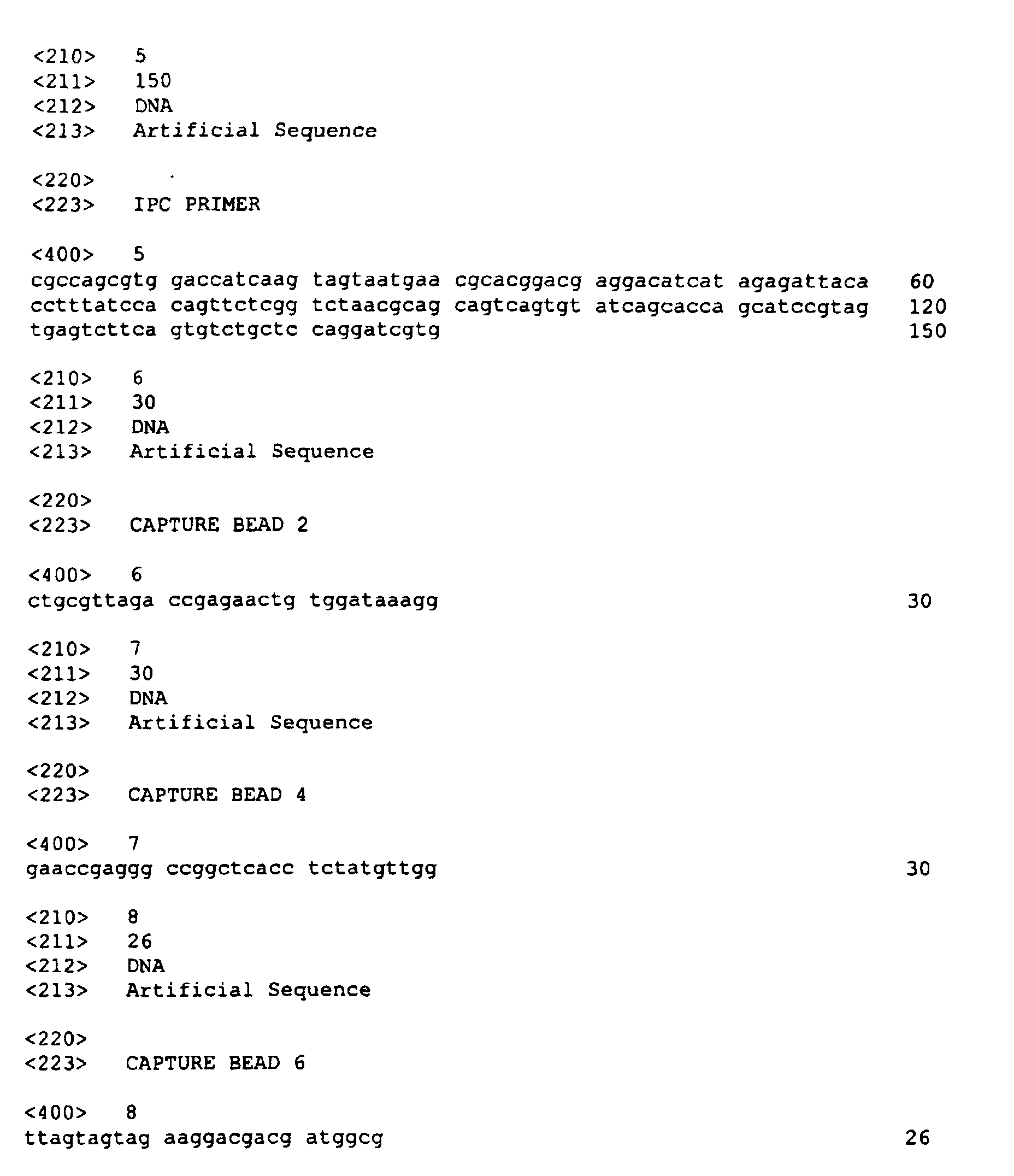
- the CMV Assay Specific Primers were designed to be complementary to the DNA encoding the pp65 Matrix Protein of the CMV Late gene in patient specimens. Both the IPC target and CMV pp65 Matrix protein target sequences were cloned into the Bluescript plasmid (Stratagene, La Jolla, CA) for use as assay controls. These were accurately quantitated by spectrophotometric analyses and fit to Poisson's distribution. The sequence of the internal positive control target DNA used was:
- PCR reaction admixture 25 ⁇ l of processed sample were added to 75 ⁇ l of the PCR reaction admixture. After addition and mixing, the reaction admixture was introduced into the amplification blister of a plastic PCR pouch containment system (Johnson & Johnson Clinical Diagnostics, Inc., US Pat. Nos. 5,089,233; 5,229,297; and 5,380,489). . Amplification and detection were carried out using a PCR amplification and detection processing instrument (Johnson & Johnson Clinical Diagnostics, Inc. US Pat. No. 5,567,617).
- reflectance density (D r ) measurements from the PCR processing instrument were recorded. D r readings above 0.13 indicate a positive result.
- Plasma samples from 65 patients were prepared according to the method of the present invention and the polymer capture procedure, as described above. Both 20 ⁇ l and 50 ⁇ l samples were used in the polymer capture method. These samples all had previously elicited a "No Test" result when evaluated using 50 ⁇ l sample volumes prepared by the polymer capture protocol in an initial evaluation. A "No Test" result occurs when the Internal Positive Control capture bead gives a false negative result.
- the IPC control plasmid that was introduced into the samples was present at 10 copies per final reaction admixture. All prepared samples were combined with the PCR reaction admixture and underwent amplification and detection, as described above.
- the 50 ⁇ l polymer capture protocol exhibited a 21% "No Test" frequency whereas the 20 ⁇ l polymer capture protocol and the 20 ⁇ l protocol of the instant invention had a 0% failure rate. With both polymer capture protocols, there was evidence of reduced signal on some samples and the Internal Positive Control bordered on negative.
- the method of the invention significantly outperformed the 50 ⁇ l polymer capture procedure and showed improvement over the 20 ⁇ l polymer capture procedure in terms of false negative results on the IPC capture bead.
- the samples prepared using the 50 ⁇ l polymer capture procedure evidently contained inhibitors of PCR or exhibited some other phenomenon associated with polymer capture.
- DNA was extracted from serum samples obtained from 100 CMV IgG-positive, IgM-negative OB/GYN patients provided by Texas Children's Hospital in Houston TX, using 20 ⁇ l of sample in the polymer capture procedure and the method of this invention (see Example 1 above).
- CMV control plasmid DNA was added to each sample to yield final single-stranded copy numbers of 20 CMV plasmid targets in the PCR reaction admixture.
- the IPC control plasmid also was introduced into the samples at a final level of 10 copies per PCR reaction admixture. All prepared samples were combined with the PCR reaction admixture, as described above. Amplification and detection, as before, were performed in duplicate in PCR pouches.
- Example 3 Extraction of DNA from Plasma from a CMV Patient Population Using the Method of the Present Invention: Comparison with GeneClean II Sample
- Plasma samples were either (i) extracted using the Gene Clean II method (Bio 101, Inc.) and characterized using both a very sensitive P 32 liquid hybridization CMV PCR assay and a direct gel CMV PCR assay or (ii) extracted using the method of the present invention and assayed as described in Example 1 above.
- the IPC control plasmid which was introduced into each sample, was present at 25 double strand copies per final reaction admixture. All samples were combined with the PCR reaction admixture followed by amplification and detection in duplicate in PCR pouches, as above.
- TCH plasma samples were considered to be very low positive samples, as all were positive only by the liquid hybridization assay and were negative on direct gel, which is indicative of a low copy level of CMV in the sample.
- Normal plasma sample pools were treated to vary the pH and add blood sample collection additives, and other substances known or suspected of being capable of interfering with sample preparation, amplification, or detection.
- the compounds were added to a sample at three times the estimated peak serum level (Table 5).
- DNA was extracted from 20 ⁇ l of each sample using the method of the present invention and the prior art polymer capture method, as described in Example 1 above.
- the IPC and CMV plasmids were present at 25 and 10 double strand copies, respectively, per final PCR reaction admixture.
- Half of the samples contained only the IPC plasmid to evaluate false positive results (color up on the CMV probe bead) that could arise due to sample cross-contamination or product carryover. All samples were combined with the PCR reaction admixture followed by amplification and detection in PCR pouches. For each compound, four pouches were run for both the 0 and the 10 CMV copy levels.
- the appropriate controls were run containing the various solvents (water, acetone, ethyl alcohol) used in preparing the compounds for inclusion in the assay.
- DNA extraction using the method of this invention does not result in inhibition of PCR amplification or detection of amplified products with any of the tested substances.
- This simple method represents a significant advance over more cumbersome, time consuming and laborious methods of the prior art.
Landscapes
- Chemical & Material Sciences (AREA)
- Life Sciences & Earth Sciences (AREA)
- Health & Medical Sciences (AREA)
- Engineering & Computer Science (AREA)
- Organic Chemistry (AREA)
- Wood Science & Technology (AREA)
- Zoology (AREA)
- Genetics & Genomics (AREA)
- Analytical Chemistry (AREA)
- Biotechnology (AREA)
- General Engineering & Computer Science (AREA)
- Bioinformatics & Cheminformatics (AREA)
- Proteomics, Peptides & Aminoacids (AREA)
- Biomedical Technology (AREA)
- Molecular Biology (AREA)
- Biophysics (AREA)
- Biochemistry (AREA)
- Microbiology (AREA)
- General Health & Medical Sciences (AREA)
- Physics & Mathematics (AREA)
- Immunology (AREA)
- Plant Pathology (AREA)
- Crystallography & Structural Chemistry (AREA)
- Chemical Kinetics & Catalysis (AREA)
- Virology (AREA)
- Measuring Or Testing Involving Enzymes Or Micro-Organisms (AREA)
- Medicines Containing Material From Animals Or Micro-Organisms (AREA)
- Investigating Or Analysing Biological Materials (AREA)
- Saccharide Compounds (AREA)
- Preparation Of Compounds By Using Micro-Organisms (AREA)
- Micro-Organisms Or Cultivation Processes Thereof (AREA)
Abstract
Description
- The present invention pertains to methods for preparing DNA from serum and plasma, particularly for use as a target in amplification reactions.
- Technology is rapidly advancing in the area of amplification and detection of nucleic acids, particularly as it relates to commercial diagnostic tests, which offer early detection of infectious diseases, cancer and genetic disorders. Highly sophisticated techniques for the amplification of minute quantities of nucleic acids, such as PCR (polymerase chain reaction), are now well known (see. U.S. Patents Nos. 4,683,195; 4,683,202; and 4,965,188). The inherent sensitivity of PCR, i.e., its ability to amplify very small concentrations of a target DNA, means that low level carryover of PCR products, and contamination between specimens, can yield false positive results. Carryover and sample contamination, among other things, are a function of the number of manipulations of the sample required during processing. Therefore, a simple procedure with a minimal number of steps that expose the sample to the environment is highly desirable.
- Traditionally, PCR identification of viremia due to infectious human cytomegalovirus (HCMV), and identification of other viruses and bacteria has been performed on peripheral blood leukocytes (WBCs) obtained from whole blood. Separation of WBCs from whole blood is usually required prior to extraction of the HCMV DNA from the WBCs. Procedures for this separation include erythrocyte sedimentation in a dextran solution (Rasmussen et al., J. Infect. Dis. 171:177-82, 1995), differential lysis using ammonium chloride solutions (U.S. Pat. No. 5,702,884 Ekeze et al), or the use of commercially available procedures (such as, e.g., CPT-Vacutainer™ tubes from Becton-Dickinson). Typically, these procedures include a wash step which results in the removal of potential inhibitors of amplification; alternatively, the isolation of WBCs serves to remove these inhibitors, which typically reside in high concentration in plasma or serum.
- Once the WBCs have been isolated, they are lysed and the DNA is extracted. This involves procedures such as, for example, boiling, sonication, or freeze-thawing of the WBCs, or the use of proteolytic enzymes and/or surfactants to lyse the cells and extract the DNA. DNA extraction may also involve an alkali lysis step (U.S. Pat. No. 5,639,599). Often, a more rigorous extraction/purification is performed to further ensure that purified DNA is obtained devoid of potential inhibitors, including, e.g., use of glass beads (Gene Clean II kits, Bio 101, Inc.), phenol-chloroform extraction procedures, polymer capture (U.S. Pat. No 5,582,988), spin-column adsorption (Qiagen QIAamp kits), and other commercially available DNA isolation kits (Puregene, Gentra Systems Inc.).
- Notably, procedures used for isolating and lysing WBCs, which serve to wash or remove potential inhibitors from the WBC preparation, cannot be used with plasma and serum. As the vast majority of endogenous biochemical substances and consumed drugs, metabolites of drugs, and the like, reside and are heavily concentrated in serum and plasma, there is a need in the art for a rapid and robust procedure that could be used with serum and plasma which would remove the potential inhibitors or render them ineffective.
- Extracellular HCMV nucleic acid in infected individuals is present in plasma and serum. Serum and plasma are gaining acceptance as samples of choice for detecting HCMV nucleic acid using PCR. Generally, the target DNA does not exist as free DNA but rather as a complex association of DNA, RNA, and proteins. The DNA must be extracted from the complex and denatured in order to render it available for amplification. Serum and plasma samples are typically subjected to heat, surfactants, and treated with proteases. Often additional rigorous protocols are employed to extract the target DNA, such as those described above for WBCs (Spector et al., J. Clin. Microbiol.30:2359-65, 1992; Nolte et al., J. Clin. Microbiol. 33:1263-66, 1995; Wolf et al., Transplantation 56:330-4, 1993; Patel et al., J. Clin. Microbiol. 32:1431-4, 1994). Alkali treatment has also been used. However, this alkali treatment typically requires a high NaOH concentration followed by a neutralization step (Hansen et al., J. Infect. Dis. 170:1271-4, 1994).
- Thus, there is a need in the art for a rapid and efficient procedure for extracting DNA from serum and plasma that is compatible with PCR amplification methods.
- The present invention provides a method for extracting DNA from serum or plasma samples. The method comprises:
- (i) contacting the serum or plasma with alkali to yield alkalinized serum or plasma;
- (ii) heating the alkalinized serum or plasma to a temperature between about 100 and about 110°C for a time between about 5 and about 20 minutes;
- (iii) centrifuging the heated alkalinized serum or plasma; and
- (iv) recovering the DNA-containing supernatant.
-
- In a preferred aspect, prior to centrifuging the heated alkalinized serum or plasma, the heated alkalinized serum or plasma produced by step (ii) is cooled or is allowed to cool to room temperature, i.e., about 25°C, prior to centrifugation in step (iii).
- In another aspect, the invention provides a method for detecting the suspected presence of a DNA-containing microorganism, for example human cytomegalovirus (HCMV), in serum or plasma. The method comprises:
- (i) contacting the serum or plasma with alkali to yield alkalinized serum or plasma;
- (ii) heating the alkalinized serum or plasma to a temperature between about 100 and about 110°C for a time between about 5 and about 20 minutes;
- (iii) centrifuging the heated alkcalinized serum or plasma;
- (iv) recovering the DNA-containing supernatant;
- (v) subjecting the DNA in the supernatant to amplification using oligonucleotide primers that recognize sequences within microorganism DNA, to form microorganism-specific amplification products; and
- (vi) detecting the amplification products, wherein detection of amplification products specific to the microorganism indicates the presence of the microorganism in the serum or plasma.
-
- In a preferred aspect, subsequent to step (ii), the heated alkalinized serum or plasma is cooled or is allowed to cool to room temperature, i.e., about 25°C, prior to centrifugation in step (iii).
- The present invention provides a simple, rapid, and highly effective method for extracting DNA from serum and plasma samples which results in DNA preparations suitable for subsequent detection assays without further manipulation.
- The method comprises the steps of:
- (i) contacting the serum or plasma with alkali to yield alkalinized serum or plasma;
- (ii) heating the alkalinized serum or plasma to a temperature between about 100 and about 110°C for a time between about 5 and about 20 minutes;
- (iii) centrifuging the heated alkalinized serum or plasma; and
- (iv) recovering the DNA-containing supernatant.
-
- It is preferred that subsequent to heating and prior to centrifugation, the heated alkalinized serum or plasma is cooled or is allowed to cool to room temperature, i.e., about 25°C. The cooling can be passive, i.e., by simple equilibration with room air, or active, i.e., the heated alkalinized serum or plasma can be refrigerated, placed on ice, or put in a water bath, until the temperature of the heated alkalinized serum or plasma reaches room temperature.
- In practicing the present invention, the suitable alkali includes, but is not limited to, sodium hydroxide, potassium hydroxide, ammonium hydroxide, or a mixture of any of the foregoing. Preferably, sodium hydroxide is used. The final concentration of alkali in the mixture prepared in step (i) ranges from about 10 to about 90 mM, and most preferably from about 15 to about 50 mM. Special notice is made of an alkali concentration of about 20 mM. A preferred method involves forming a mixture of 1 part by volume of serum or plasma with about 4 parts by volume of an aqueous solution of about 25 mM alkali.
- The temperature to which the alkalinized serum or plasma is heated preferably is about 105°C and the time preferably is about 5 minutes. The centrifugation step is preferably carried out at ambient temperature for about 2 minutes at about 16,000 X g.
- The recovered supernatant from the centrifuged mixture can be added directly to a PCR amplification admixture. However, the supernatant can be modified by further treatment or addition of reagents as desired prior to addition to the PCR admixture. Treatment with alkali according to the invention serves to inactivate PCR inhibitors and denature proteins, particularly nucleases that can degrade the target DNA to a point where it cannot be amplified. It also serves to denature the DNA, making it more amenable to hybridization to amplification primers and thus to PCR amplification. In addition, the simplicity of the method greatly reduces the probability of sample-to-sample contamination and PCR product carryover. It also eliminates the need for costly and often unstable materials used routinely for nucleic acid isolation and purification.
- The present invention is useful in preparing DNA samples for diagnostic assays, particularly those that detect DNA viruses such as, e.g., cytomegalovirus, herpes simplex virus, Epstein-Barr virus, Hepatitis B virus and any blood-borne bacteria. Detection methods in which the DNA preparations can be used include, without limitation, any method involving hybridization, including, e.g., polymerase chain reaction, ligase chain reaction, and the like. Thus, the present invention encompasses methods for the detection of DNA-containing microorganisms which comprises alkalinizing serum or plasma from a subject, heating the alkalinized serum or plasma to between about 100 and about 110°C for a time period ranging from about 5 to about 20 minutes, centrifuging to obtain a DNA-containing supernatant, recovering the supernatant, amplifying the DNA in the supernatant using primers specific for the DNA of the DNA-containing organism, thereby forming amplification products specific to the microorganism, and then detecting the amplification products, where their presence indicates that the microorganism is present in the blood or serum of the subject.
- The following examples illustrate the present invention without limitation.
- Whole blood samples were collected in Vacutainer™ tubes containing ethylenediamine tetraacetic acid (EDTA) or other anti-coagulant, such as heparin and oxalate, for obtaining plasma, or whole blood samples were collected without anticoagulant to obtain serum.
- 20 µl of serum or plasma were added to 80 µl of a 25 mM NaOH solution containing 0.334 µg/µl calf thymus DNA, and 1.67 double stranded copies per µl of the Internal Positive Control (IPC) plasmid target DNA in a screw-capped microcentrifuge tube. The mixture was vortexed, then heated at 105°C for five minutes. The samples were allowed to equilibrate to room temperature and centrifuged at 16,000 X g for two minutes at room temperature. 25 µl of the supernatant were added to 75 µl of the PCR reaction admixture (composition described below) and amplified.
- The method of extraction of DNA using polymer capture was carried out as described in Example 3 of U.S. Patent. No 5,582,988, except that 20 µl of serum or plasma were used in the experiments described herein. The DNA-polymer complex was pelleted and the supernatant was discarded (which is expected to contain potential inhibitors of PCR). The DNA was then eluted from the pellet using 100 µl of a 20 mM NaOH solution and heated at 105°C for 5 minutes, followed by a 2 minute centrifugation step. The supernatant (25 µl) was added to 75 µl of PCR reaction admixture and amplified. The polymer capture procedure was used as a control for comparison with the simplified DNA extraction method of the present invention.
- The PCR reaction admixture at pH 8.0 contained 160 U/ml of recombinant Taq polymerase, 5.28 µg/ml of TP4-9.2 (a 5 fold excess) and 53.37 µg/ml of TP1-12.2 anti-Taq antibodies, 18 mM Tris buffer, 54 mM potassium chloride, 0.2 µM of each IPC primer (IPC-F1 (forward): Biotin- 5'-CGCCAGCGTGGACCATCAAGTAGTAA-3' 〈SEQ ID NO.: 1〉 and IPC-R1 (reverse) 5'-CACGATCCTGGAGCAGACACTGAAGA-3' 〈SEQ ID NO.: 2〉), 0.4 µM of each CMV Assay Specific Primer (LB42M (forward) with the sequence: Biotin-5'-TGCACTGCCAGGTGCTTCGGCTCAT-3' 〈SEQ ID NO.: 3〉 and primer LB41 (reverse) 5'-CACCACGCAGCGGCCCTTGATGTTT-3' 〈SEQ ID NO.: 4〉), 1.2 mM total deoxynucleoside triphosphates (dNTPs), 4 mM magnesium chloride, 9.5% glycerol, and a preservative. Typically, the Taq polymerase and anti-Taq antibodies were allowed to pre-incubate at room temperature for 10 minutes, and the MgCl2 was supplied as a separate solution to be added just prior to sample addition. The CMV Assay Specific Primers were designed to be complementary to the DNA encoding the pp65 Matrix Protein of the CMV Late gene in patient specimens. Both the IPC target and CMV pp65 Matrix protein target sequences were cloned into the Bluescript plasmid (Stratagene, La Jolla, CA) for use as assay controls. These were accurately quantitated by spectrophotometric analyses and fit to Poisson's distribution. The sequence of the internal positive control target DNA used was:
- 5'-
CGCCAGCGTGGACCATCAAGTAGTAATGAACGCACGGACGAGGACATC
A
TAGAGATTACACCTTTATCCACAGTTCTCGGTCTAACGCAGCAGTCAGT
GTATCAGCACCAGCATCCGTAGTGAGTCTTCAGTGTCTGCTCCAGGATC
GTG-3' 〈SEQ ID NO.: 5〉. -
- 25 µl of processed sample were added to 75 µl of the PCR reaction admixture. After addition and mixing, the reaction admixture was introduced into the amplification blister of a plastic PCR pouch containment system (Johnson & Johnson Clinical Diagnostics, Inc., US Pat. Nos. 5,089,233; 5,229,297; and 5,380,489). . Amplification and detection were carried out using a PCR amplification and detection processing instrument (Johnson & Johnson Clinical Diagnostics, Inc. US Pat. No. 5,567,617). After an initial preheat for 3 minutes at 96°C, the sample was subjected to 40 cycles of amplification, alternating between 96°C (5 second) denaturations, and 70°C (40 second) annealing steps. After a 5 minute postheat at 103°C, the amplified products were introduced into the detection chamber of the pouch containing the control and assay specific probes linked to capture beads (Table 1).
Bead No. Designation Sequence SEQ ID NO. Bead 2 IPC-1P 5'-CTGCGTTAGACCGAGAACTGTGGAT AAAGG-3' 6 Bead 4 LBSA-11 (CMV) 5'-GAACCGAGGGCCGGCTCACCTCTAT GTTGG-3' (pp65 Matrix Protein) 7 Bead 6 INC-26.7a 5'-TTAGTAGTAGAAGGACGACGATGG CG-3' 8 INC-26.7a serves as an Internal Negative Control and is expected to give no signal in a properly functioning PCR pouch. - Color generation on the capture beads was then read visually and compared to a color score card having ten shades of increasing blue color and corresponding numbers ranging from 0 to 10 (0 = no signal, 10 = high positive signal). A visual score above a 2 indicates a positive test result on that capture bead. In addition, reflectance density (Dr) measurements from the PCR processing instrument were recorded. Dr readings above 0.13 indicate a positive result.
- The following experiment was performed to compare the extraction method of the present to the polymer capture method.
- Plasma samples from 65 patients were prepared according to the method of the present invention and the polymer capture procedure, as described above. Both 20 µl and 50 µl samples were used in the polymer capture method. These samples all had previously elicited a "No Test" result when evaluated using 50 µl sample volumes prepared by the polymer capture protocol in an initial evaluation. A "No Test" result occurs when the Internal Positive Control capture bead gives a false negative result. The IPC control plasmid that was introduced into the samples was present at 10 copies per final reaction admixture. All prepared samples were combined with the PCR reaction admixture and underwent amplification and detection, as described above.
- The "No Test" frequency in polymer capture vs. the method of the invention for plasma samples are summarized in Table 2 below.
Polymer Capture 50 ΦL sample Polymer Capture 20 ΦL sample Invention 20 ΦL sample IPC Capture Bead Results 14/65 0/65 0/65 "No Test" Frequency 21% 0% 0% Range (Visual Scores) 3-8 3-8 5-7.5 Number of samples <4 Visual Score 3/65 (4.6%) 1/65 (1.5%) 0/65 (0%) - The 50 µl polymer capture protocol exhibited a 21% "No Test" frequency whereas the 20 µl polymer capture protocol and the 20 µl protocol of the instant invention had a 0% failure rate. With both polymer capture protocols, there was evidence of reduced signal on some samples and the Internal Positive Control bordered on negative.
- Thus, the method of the invention significantly outperformed the 50 µl polymer capture procedure and showed improvement over the 20 µl polymer capture procedure in terms of false negative results on the IPC capture bead. The samples prepared using the 50 µl polymer capture procedure evidently contained inhibitors of PCR or exhibited some other phenomenon associated with polymer capture.
- In addition, there was one instance using the 20 µl polymer capture technique where a false positive result on the CMV (LBSA-11) capture bead was obtained from a CMV negative patient sample. As the corresponding 50 µl result was negative (and would have provided 2.5- fold more CMV target DNA from the patient sample if it were truly present), this result exemplifies the complications in a procedure with multiple manipulations, such as product carryover.
- DNA was extracted from serum samples obtained from 100 CMV IgG-positive, IgM-negative OB/GYN patients provided by Texas Children's Hospital in Houston TX, using 20 µl of sample in the polymer capture procedure and the method of this invention (see Example 1 above). Prior to sample preparation, CMV control plasmid DNA was added to each sample to yield final single-stranded copy numbers of 20 CMV plasmid targets in the PCR reaction admixture. The IPC control plasmid also was introduced into the samples at a final level of 10 copies per PCR reaction admixture. All prepared samples were combined with the PCR reaction admixture, as described above. Amplification and detection, as before, were performed in duplicate in PCR pouches.
- The frequency of false negative results in polymer capture versus the method of the invention using serum are summarized in Table 3 below.
Polymer Capture 20ΦL sample Method of the Present Invention 20 ΦL sample CMV Capture Bead Results (% Frequency) 5/100 (5%) 0/100 (0%) IPC Capture Bead Results (% Frequency) 0/100 (0%) 0/100 (0%) - Neither sample preparation procedure gave negative results on the IPC capture bead. However, addition of CMV plasmid target to the samples resulted in a 5% false negative frequency on the CMV bead when the polymer capture method was used, indicating potential inhibition of PCR. By contrast, there were no false negative CMV bead results using the method of the present invention.
- The following experiment was performed to test the efficacy of the method of the present invention in extracting DNA from patient samples.
- Plasma samples were either (i) extracted using the Gene Clean II method (Bio 101, Inc.) and characterized using both a very sensitive P32 liquid hybridization CMV PCR assay and a direct gel CMV PCR assay or (ii) extracted using the method of the present invention and assayed as described in Example 1 above. The IPC control plasmid, which was introduced into each sample, was present at 25 double strand copies per final reaction admixture. All samples were combined with the PCR reaction admixture followed by amplification and detection in duplicate in PCR pouches, as above.
- The results are on the CMV capture bead are shown in Table 4 below.

- All positive TCH plasma samples were considered to be very low positive samples, as all were positive only by the liquid hybridization assay and were negative on direct gel, which is indicative of a low copy level of CMV in the sample.
- There was an 80% concordance between the two sample preparation methods. Using the method of the present invention, 10 of the 12 samples gave positive results by the GeneClean method and two gave negative results. Upon retesting the sample preparations, of the original two false negative samples, one became positive. For the GeneClean negative plasma samples, a similar situation was encountered. With the 13 negative samples, both sample preparation methods gave concordant results for ten of the specimens (the same 10).
- In addition, there were three instances where the method of the present invention indicated a CMV positive sample. Upon retesting the sample preparations, two of these three samples again indicated a CMV positive result. Because of its inherent sensitivity, this is a common observation in PCR assays, where very low target samples give discrepant results. This is predicted based on an expected Poisson sampling distribution.
- The following experiment was performed to test the ability of the method of the invention to remove potentially interfering substances from plasma.
- Normal plasma sample pools were treated to vary the pH and add blood sample collection additives, and other substances known or suspected of being capable of interfering with sample preparation, amplification, or detection. The compounds were added to a sample at three times the estimated peak serum level (Table 5).
- DNA was extracted from 20 µl of each sample using the method of the present invention and the prior art polymer capture method, as described in Example 1 above. The IPC and CMV plasmids were present at 25 and 10 double strand copies, respectively, per final PCR reaction admixture. Half of the samples contained only the IPC plasmid to evaluate false positive results (color up on the CMV probe bead) that could arise due to sample cross-contamination or product carryover. All samples were combined with the PCR reaction admixture followed by amplification and detection in PCR pouches. For each compound, four pouches were run for both the 0 and the 10 CMV copy levels. In addition, the appropriate controls were run containing the various solvents (water, acetone, ethyl alcohol) used in preparing the compounds for inclusion in the assay.


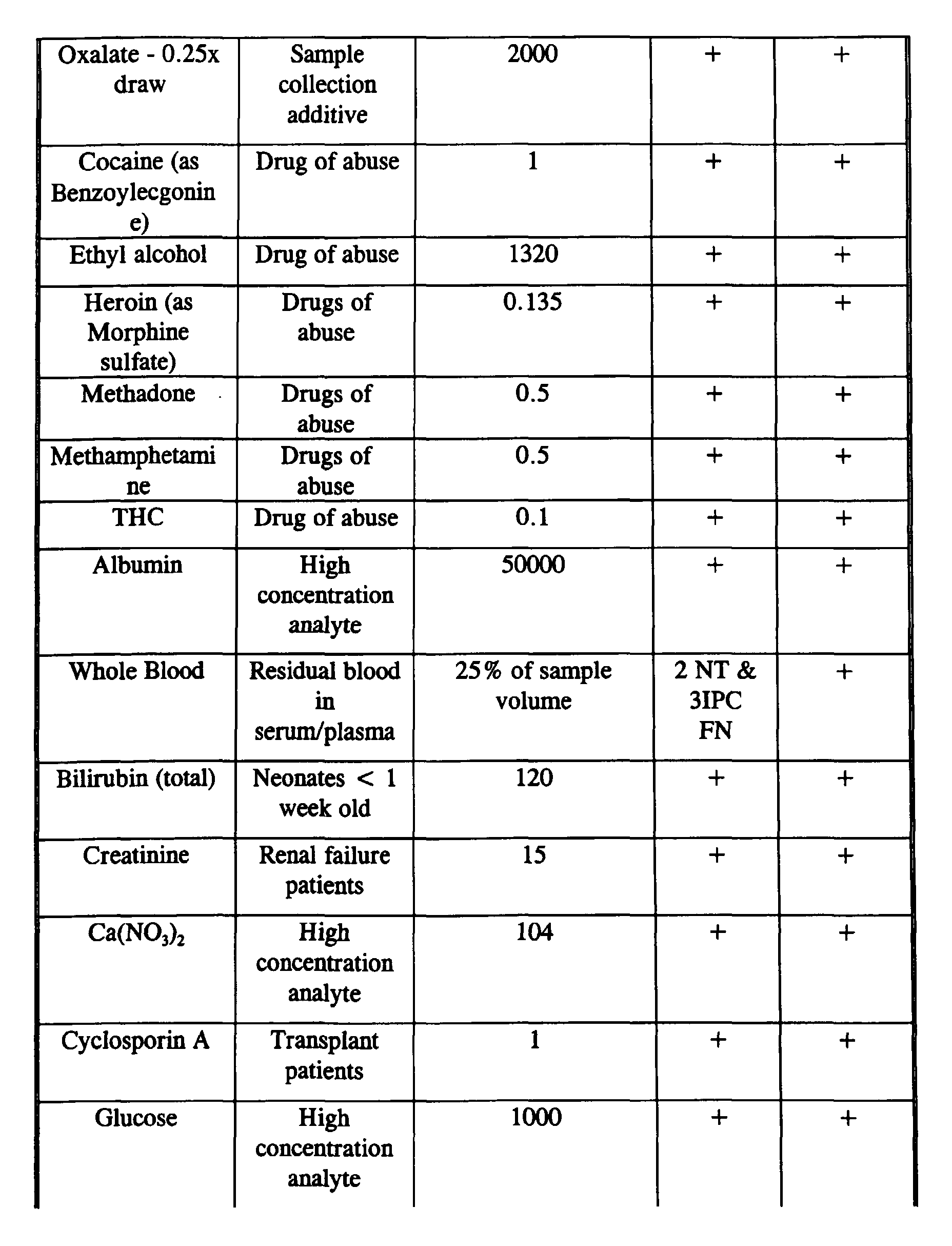
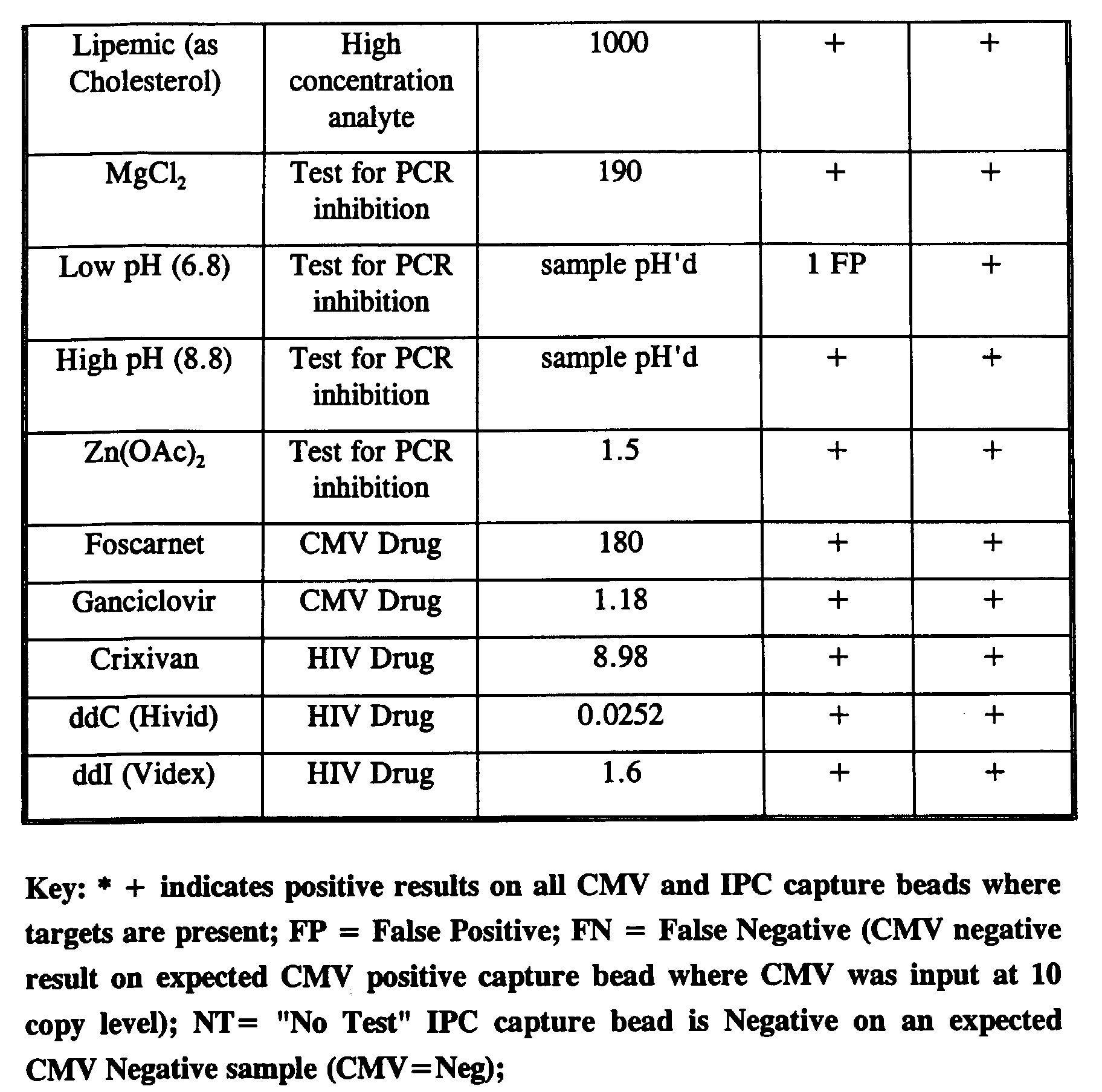
- During initial testing there were a few sporadic false negative results on both the CMV and IPC capture beads and several "No Test" results for each of the sample preparation methods, although they were predominantly seen on polymer capture samples. In addition, several of the controls gave unexpected negative results and there were indications of pouch failures contributing to these.
- Upon retesting the previously prepared samples, all results for all substances tested elicited positive results for both the polymer capture and the method of the present invention, with the exception of the 25% blood samples (25 parts by volume of whole blood added to 75 parts by volume of the plasma prior to DNA extraction) prepared using the polymer capture method. Two of the four samples containing only IPC target (no CMV target) again showed "No Test" results on the IPC capture bead. Also, three of the four samples containing both the IPC and CMV target DNA's showed inhibition on the IPC capture bead, with visual scores of only "1". For two of these, the CMV capture bead also indicated signs of inhibition (visual color scores of "3" and "4"). Also, there was again one instance of a false positive result using the polymer capture technique (at the low pH variation) again indicating the need for a simple procedure with a minimum number of steps to prevent carryover contamination.
- These results indicate that 25% whole blood is inhibitory in the HCMV assay when samples were prepared using the polymer capture technique. By contrast, extraction of DNA using the method of the present invention resulted in no inhibition of PCR amplification and detection. This is significant because serum and plasma samples in clinical laboratories are often contaminated with red blood cells. Furthermore, the polymer capture technique is more laborious and permits greater exposure of the sample to the environment compared with the method of the instant invention which compares favorably with polymer capture.
- DNA extraction using the method of this invention does not result in inhibition of PCR amplification or detection of amplified products with any of the tested substances. This simple method represents a significant advance over more cumbersome, time consuming and laborious methods of the prior art.
- All patents, applications, articles, publications, and test methods mentioned above are hereby incorporated by reference in their entirety.
- Many variations of the present invention will suggest themselves to those skilled in the art in light of the above detailed description. Such obvious variations are within the full intended scope of the appended claims.

Claims (14)
- A method for extracting DNA from serum or plasma, said method comprising:(i) contacting serum or plasma with alkali to yield alkalinized serum or plasma;(ii) heating said alkalinized serum or plasma to a temperature ranging from about 100 to about 110°C for a time ranging from about 5 to about 20 minutes to yield heated alkalinized serum or plasma;(iii) centrifuging said heated alkalinized serum or plasma to yield a DNA-containing supernatant; and(iv) recovering said DNA-containing supernatant.
- A method as defined in claim 1, wherein after said heating step (ii) and prior to said centrifuging step (iii), said heated alkalinized serum or plasma is cooled or allowed to cool to about 25°C.
- A method as defined in claim 2, wherein said alkali is sodium hydroxide.
- A method as defined in claim 2, wherein said temperature is about 105°C.
- A method as defined in claim 2, wherein said time is about 5 minutes.
- A method as defined in claim 2, wherein said centrifuging is at about 16,000 x g for about 2 minutes.
- A method as defined in claim 2, wherein said alkalinized serum or plasma comprises alkali at a concentration ranging from about 15 to about 50 mM
- A method for detecting a DNA-containing microorganism in serum or plasma, said method comprising:(i) contacting serum or plasma from a subject with alkali to yield alkalinized serum or plasma;(ii) heating said alkalinized serum or plasma to a temperature ranging between about 100 to about 110°C for a time ranging from about 5 to about 20 minutes to yield heated alkalinized serum or plasma;(iii) centrifuging said heated alkalinized serum or plasma to yield a DNA-containing supernatant;(iv) recovering said DNA-containing supernatant;(v) subjecting said DNA in said supernatant to amplification using oligonucleotide primers that recognize sequences within the DNA of said DNA-containing microorganism, to form amplification products specific to said DNA-containing microorganism; and(vi) detecting said amplification products,
wherein detection of said amplification products indicates the presence of said microorganism in said serum or plasma. - A method as defined in claim 8, wherein after said contacting step (ii) and prior to said centrifuging step (iii), said heated alkalinized serum or plasma is cooled or allowed to cool to about 25°C.
- A method as defined in claim 8, wherein said temperature is about 105°C.
- A method as defined in claim 8, wherein said microorganism is a bacteria.
- A method as defined in claim 8, wherein said microorganism is a virus.
- A method as defined in claim 12, wherein said virus is human cytomegalovirus.
- A method as defined in claim 8, wherein said alkalinized serum or plasma comprises alkali at a concentration of about 20 mM
Applications Claiming Priority (2)
| Application Number | Priority Date | Filing Date | Title |
|---|---|---|---|
| US11849699P | 1999-02-03 | 1999-02-03 | |
| US118496P | 1999-02-03 |
Publications (2)
| Publication Number | Publication Date |
|---|---|
| EP1026241A1 true EP1026241A1 (en) | 2000-08-09 |
| EP1026241B1 EP1026241B1 (en) | 2004-05-19 |
Family
ID=22378966
Family Applications (1)
| Application Number | Title | Priority Date | Filing Date |
|---|---|---|---|
| EP00300762A Expired - Lifetime EP1026241B1 (en) | 1999-02-03 | 2000-02-01 | An improved method for preparing DNA from serum and plasma |
Country Status (13)
| Country | Link |
|---|---|
| US (1) | US6268127B1 (en) |
| EP (1) | EP1026241B1 (en) |
| JP (1) | JP4468534B2 (en) |
| KR (1) | KR100845949B1 (en) |
| CN (1) | CN100334102C (en) |
| AT (1) | ATE267251T1 (en) |
| AU (1) | AU769709C (en) |
| CA (1) | CA2297547C (en) |
| DE (1) | DE60010776T2 (en) |
| DK (1) | DK1026241T3 (en) |
| ES (1) | ES2220340T3 (en) |
| NO (1) | NO324041B1 (en) |
| PT (1) | PT1026241E (en) |
Cited By (1)
| Publication number | Priority date | Publication date | Assignee | Title |
|---|---|---|---|---|
| KR20020028386A (en) * | 2000-10-09 | 2002-04-17 | 박제철 | PURITY SEPARATION METHOD OF mtDNA FROM ANIMALS OF A SMALL AMOUNT |
Families Citing this family (6)
| Publication number | Priority date | Publication date | Assignee | Title |
|---|---|---|---|---|
| AU2003287082A1 (en) * | 2002-10-11 | 2004-05-04 | Imutex Pharmaceuticals, Inc. | Heterologous plasma cocktail for hiv treatment |
| WO2016187780A1 (en) * | 2015-05-25 | 2016-12-01 | Coyote Bioscience Yixing Co., Ltd. | Device and method for sample collection |
| CN107683330A (en) * | 2015-05-25 | 2018-02-09 | 卡尤迪生物科技(北京)有限公司 | Devices and methods for sample collection |
| CN108795926A (en) * | 2018-06-27 | 2018-11-13 | 中国农业科学院麻类研究所 | A kind of DNA profiling fast preparation method |
| WO2020075696A1 (en) * | 2018-10-09 | 2020-04-16 | 公益財団法人筑波メディカルセンター | Sample pretreatment method |
| JP7329912B2 (en) * | 2018-10-09 | 2023-08-21 | 公益財団法人筑波メディカルセンター | Microorganism detection method |
Citations (1)
| Publication number | Priority date | Publication date | Assignee | Title |
|---|---|---|---|---|
| WO1997034015A1 (en) * | 1996-03-15 | 1997-09-18 | The Penn State Research Foundation | Detection of extracellular tumor-associated nucleic acid in blood plasma or serum using nucleic acid amplification assays |
Family Cites Families (5)
| Publication number | Priority date | Publication date | Assignee | Title |
|---|---|---|---|---|
| US5229297A (en) | 1989-02-03 | 1993-07-20 | Eastman Kodak Company | Containment cuvette for PCR and method of use |
| US5089233A (en) | 1989-06-12 | 1992-02-18 | Eastman Kodak Company | Processing apparatus for a chemical reaction pack |
| US5380489A (en) | 1992-02-18 | 1995-01-10 | Eastman Kodak Company | Element and method for nucleic acid amplification and detection using adhered probes |
| JP3494646B2 (en) | 1992-06-08 | 2004-02-09 | ジェン−プローブ・インコーポレイテッド | Preparation of nucleic acids from mononuclear cells |
| US5582988A (en) | 1994-09-15 | 1996-12-10 | Johnson & Johnson Clinical Diagnostics, Inc. | Methods for capture and selective release of nucleic acids using weakly basic polymer and amplification of same |
-
2000
- 2000-01-28 US US09/493,352 patent/US6268127B1/en not_active Expired - Lifetime
- 2000-02-01 EP EP00300762A patent/EP1026241B1/en not_active Expired - Lifetime
- 2000-02-01 CA CA002297547A patent/CA2297547C/en not_active Expired - Fee Related
- 2000-02-01 AT AT00300762T patent/ATE267251T1/en active
- 2000-02-01 DE DE60010776T patent/DE60010776T2/en not_active Expired - Lifetime
- 2000-02-01 ES ES00300762T patent/ES2220340T3/en not_active Expired - Lifetime
- 2000-02-01 PT PT00300762T patent/PT1026241E/en unknown
- 2000-02-01 DK DK00300762T patent/DK1026241T3/en active
- 2000-02-02 AU AU14880/00A patent/AU769709C/en not_active Ceased
- 2000-02-02 KR KR1020000005130A patent/KR100845949B1/en not_active Expired - Fee Related
- 2000-02-02 NO NO20000539A patent/NO324041B1/en not_active IP Right Cessation
- 2000-02-03 JP JP2000032644A patent/JP4468534B2/en not_active Expired - Fee Related
- 2000-02-03 CN CNB001046985A patent/CN100334102C/en not_active Expired - Fee Related
Patent Citations (1)
| Publication number | Priority date | Publication date | Assignee | Title |
|---|---|---|---|---|
| WO1997034015A1 (en) * | 1996-03-15 | 1997-09-18 | The Penn State Research Foundation | Detection of extracellular tumor-associated nucleic acid in blood plasma or serum using nucleic acid amplification assays |
Non-Patent Citations (1)
| Title |
|---|
| HANSEN, K. K. ET AL.: "Detection of Cytomegalovirus DNA in Serum Correlates with Clinical Cytomegalovirus Retinitis in AIDS.", J. INFECT. DIS., vol. 170, no. 6, November 1994 (1994-11-01), pages 1271 - 1274, XP000907454 * |
Cited By (1)
| Publication number | Priority date | Publication date | Assignee | Title |
|---|---|---|---|---|
| KR20020028386A (en) * | 2000-10-09 | 2002-04-17 | 박제철 | PURITY SEPARATION METHOD OF mtDNA FROM ANIMALS OF A SMALL AMOUNT |
Also Published As
| Publication number | Publication date |
|---|---|
| NO20000539D0 (en) | 2000-02-02 |
| US6268127B1 (en) | 2001-07-31 |
| JP2000279199A (en) | 2000-10-10 |
| DE60010776D1 (en) | 2004-06-24 |
| CN100334102C (en) | 2007-08-29 |
| CN1270962A (en) | 2000-10-25 |
| DE60010776T2 (en) | 2005-05-12 |
| EP1026241B1 (en) | 2004-05-19 |
| KR20000076596A (en) | 2000-12-26 |
| NO20000539L (en) | 2000-08-04 |
| ATE267251T1 (en) | 2004-06-15 |
| AU1488000A (en) | 2000-08-10 |
| AU769709C (en) | 2004-10-14 |
| PT1026241E (en) | 2004-09-30 |
| ES2220340T3 (en) | 2004-12-16 |
| NO324041B1 (en) | 2007-07-30 |
| CA2297547A1 (en) | 2000-08-03 |
| DK1026241T3 (en) | 2004-08-30 |
| CA2297547C (en) | 2006-12-19 |
| JP4468534B2 (en) | 2010-05-26 |
| AU769709B2 (en) | 2004-01-29 |
| KR100845949B1 (en) | 2008-07-11 |
Similar Documents
| Publication | Publication Date | Title |
|---|---|---|
| US5231015A (en) | Methods of extracting nucleic acids and pcr amplification without using a proteolytic enzyme | |
| EP0702090B1 (en) | Coamplification of target nucleic acids using volume exclusion agent, in reaction composition, test kit and test device useful therefor | |
| JP5399993B2 (en) | Improved method for extracting nucleic acids from tissue samples and paraffin-embedded tissues | |
| Andresen et al. | Diagnosis of visceral leishmaniasis by the polymerase chain reaction using blood, bone marrow and lymph node samples from patients from the Sudan | |
| US6210881B1 (en) | Method for reducing inhibitors of nucleic acid hybridization | |
| EP0511712B1 (en) | Nucleic acid amplification and detection methods using rapid polymerase chain reaction cycle | |
| WO2004013357A2 (en) | Nucleotide sequences specific to francisella tularensis and methods for the detection of francisella tularensis | |
| JP2575290B2 (en) | Mycobacterial sample processing method | |
| JP2012055311A (en) | Generic buffer for amplification | |
| JP2023126543A (en) | How to detect microorganisms | |
| EP1026241B1 (en) | An improved method for preparing DNA from serum and plasma | |
| JP7272202B2 (en) | Method and apparatus for testing target nucleic acid | |
| US5763185A (en) | Method for reducing inhibitors of nucleic acid hybridization | |
| CN115820819A (en) | One-step nucleic acid detection method free from PAM limitation and application thereof | |
| CN223535088U (en) | A component for a reaction tube body and a reaction tube | |
| CN223522537U (en) | Reaction tube for two-step method | |
| Wu et al. | Differential detection of cytomegalovirus immediate-early messenger RNA in clinical samples using ligation-dependent PCR | |
| EP4267756A1 (en) | Detecting a target nucleic acid in a biological sample | |
| JP2003174878A (en) | Nucleic acid synthesis method | |
| HK1004861B (en) | Nucleic acid amplification and detection methods using rapid polymerase chain reaction cycle | |
| AU9421198A (en) | Procedure for the detection of high virus concentrations in blood plasma and/or blood serum by means of the polymerase chain reaction |
Legal Events
| Date | Code | Title | Description |
|---|---|---|---|
| PUAI | Public reference made under article 153(3) epc to a published international application that has entered the european phase |
Free format text: ORIGINAL CODE: 0009012 |
|
| AK | Designated contracting states |
Kind code of ref document: A1 Designated state(s): AT BE CH CY DE DK ES FI FR GB GR IE IT LI LU MC NL PT SE |
|
| AX | Request for extension of the european patent |
Free format text: AL;LT;LV;MK;RO;SI |
|
| 17P | Request for examination filed |
Effective date: 20010112 |
|
| AKX | Designation fees paid |
Free format text: AT BE CH CY DE DK ES FI FR GB GR IE IT LI LU MC NL PT SE |
|
| 17Q | First examination report despatched |
Effective date: 20030430 |
|
| GRAP | Despatch of communication of intention to grant a patent |
Free format text: ORIGINAL CODE: EPIDOSNIGR1 |
|
| GRAS | Grant fee paid |
Free format text: ORIGINAL CODE: EPIDOSNIGR3 |
|
| GRAA | (expected) grant |
Free format text: ORIGINAL CODE: 0009210 |
|
| AK | Designated contracting states |
Kind code of ref document: B1 Designated state(s): AT BE CH CY DE DK ES FI FR GB GR IE IT LI LU MC NL PT SE |
|
| REG | Reference to a national code |
Ref country code: GB Ref legal event code: FG4D |
|
| REG | Reference to a national code |
Ref country code: CH Ref legal event code: EP |
|
| REG | Reference to a national code |
Ref country code: IE Ref legal event code: FG4D |
|
| REF | Corresponds to: |
Ref document number: 60010776 Country of ref document: DE Date of ref document: 20040624 Kind code of ref document: P |
|
| REG | Reference to a national code |
Ref country code: GR Ref legal event code: EP Ref document number: 20040402241 Country of ref document: GR |
|
| REG | Reference to a national code |
Ref country code: DK Ref legal event code: T3 |
|
| REG | Reference to a national code |
Ref country code: CH Ref legal event code: NV Representative=s name: E. BLUM & CO. PATENTANWAELTE |
|
| REG | Reference to a national code |
Ref country code: SE Ref legal event code: TRGR |
|
| REG | Reference to a national code |
Ref country code: PT Ref legal event code: SC4A Free format text: AVAILABILITY OF NATIONAL TRANSLATION Effective date: 20040729 |
|
| REG | Reference to a national code |
Ref country code: ES Ref legal event code: FG2A Ref document number: 2220340 Country of ref document: ES Kind code of ref document: T3 |
|
| ET | Fr: translation filed | ||
| PG25 | Lapsed in a contracting state [announced via postgrant information from national office to epo] |
Ref country code: CY Free format text: LAPSE BECAUSE OF FAILURE TO SUBMIT A TRANSLATION OF THE DESCRIPTION OR TO PAY THE FEE WITHIN THE PRESCRIBED TIME-LIMIT Effective date: 20050201 |
|
| PLBE | No opposition filed within time limit |
Free format text: ORIGINAL CODE: 0009261 |
|
| STAA | Information on the status of an ep patent application or granted ep patent |
Free format text: STATUS: NO OPPOSITION FILED WITHIN TIME LIMIT |
|
| 26N | No opposition filed |
Effective date: 20050222 |
|
| REG | Reference to a national code |
Ref country code: CH Ref legal event code: PFA Owner name: ORTHO-CLINICAL DIAGNOSTICS, INC. Free format text: ORTHO-CLINICAL DIAGNOSTICS, INC.#100 INDIGO CREEK DRIVE#ROCHESTER, NY 14626-5101 (US) -TRANSFER TO- ORTHO-CLINICAL DIAGNOSTICS, INC.#100 INDIGO CREEK DRIVE#ROCHESTER, NY 14626-5101 (US) |
|
| PGFP | Annual fee paid to national office [announced via postgrant information from national office to epo] |
Ref country code: MC Payment date: 20110126 Year of fee payment: 12 Ref country code: IE Payment date: 20110210 Year of fee payment: 12 Ref country code: DK Payment date: 20110210 Year of fee payment: 12 |
|
| PGFP | Annual fee paid to national office [announced via postgrant information from national office to epo] |
Ref country code: FI Payment date: 20110211 Year of fee payment: 12 Ref country code: CH Payment date: 20110214 Year of fee payment: 12 Ref country code: SE Payment date: 20110211 Year of fee payment: 12 Ref country code: PT Payment date: 20110110 Year of fee payment: 12 Ref country code: AT Payment date: 20110126 Year of fee payment: 12 Ref country code: FR Payment date: 20110218 Year of fee payment: 12 Ref country code: IT Payment date: 20110219 Year of fee payment: 12 Ref country code: DE Payment date: 20110126 Year of fee payment: 12 Ref country code: LU Payment date: 20110330 Year of fee payment: 12 Ref country code: NL Payment date: 20110216 Year of fee payment: 12 |
|
| PGFP | Annual fee paid to national office [announced via postgrant information from national office to epo] |
Ref country code: GR Payment date: 20110113 Year of fee payment: 12 |
|
| PGFP | Annual fee paid to national office [announced via postgrant information from national office to epo] |
Ref country code: GB Payment date: 20110126 Year of fee payment: 12 Ref country code: ES Payment date: 20110315 Year of fee payment: 12 |
|
| PGFP | Annual fee paid to national office [announced via postgrant information from national office to epo] |
Ref country code: BE Payment date: 20110722 Year of fee payment: 12 |
|
| REG | Reference to a national code |
Ref country code: PT Ref legal event code: MM4A Free format text: LAPSE DUE TO NON-PAYMENT OF FEES Effective date: 20120801 |
|
| BERE | Be: lapsed |
Owner name: *ORTHO-CLINICAL DIAGNOSTICS INC. Effective date: 20120228 |
|
| REG | Reference to a national code |
Ref country code: NL Ref legal event code: V1 Effective date: 20120901 |
|
| PG25 | Lapsed in a contracting state [announced via postgrant information from national office to epo] |
Ref country code: MC Free format text: LAPSE BECAUSE OF NON-PAYMENT OF DUE FEES Effective date: 20120229 |
|
| REG | Reference to a national code |
Ref country code: CH Ref legal event code: PL |
|
| GBPC | Gb: european patent ceased through non-payment of renewal fee |
Effective date: 20120201 |
|
| REG | Reference to a national code |
Ref country code: GR Ref legal event code: ML Ref document number: 20040402241 Country of ref document: GR Effective date: 20120905 |
|
| PG25 | Lapsed in a contracting state [announced via postgrant information from national office to epo] |
Ref country code: FI Free format text: LAPSE BECAUSE OF NON-PAYMENT OF DUE FEES Effective date: 20120201 Ref country code: CH Free format text: LAPSE BECAUSE OF NON-PAYMENT OF DUE FEES Effective date: 20120229 Ref country code: LI Free format text: LAPSE BECAUSE OF NON-PAYMENT OF DUE FEES Effective date: 20120229 Ref country code: SE Free format text: LAPSE BECAUSE OF NON-PAYMENT OF DUE FEES Effective date: 20120202 |
|
| REG | Reference to a national code |
Ref country code: DK Ref legal event code: EBP |
|
| REG | Reference to a national code |
Ref country code: IE Ref legal event code: MM4A |
|
| REG | Reference to a national code |
Ref country code: FR Ref legal event code: ST Effective date: 20121031 |
|
| PG25 | Lapsed in a contracting state [announced via postgrant information from national office to epo] |
Ref country code: IT Free format text: LAPSE BECAUSE OF NON-PAYMENT OF DUE FEES Effective date: 20120201 Ref country code: GR Free format text: LAPSE BECAUSE OF NON-PAYMENT OF DUE FEES Effective date: 20120905 Ref country code: PT Free format text: LAPSE BECAUSE OF NON-PAYMENT OF DUE FEES Effective date: 20120801 |
|
| REG | Reference to a national code |
Ref country code: AT Ref legal event code: MM01 Ref document number: 267251 Country of ref document: AT Kind code of ref document: T Effective date: 20120201 |
|
| REG | Reference to a national code |
Ref country code: DE Ref legal event code: R119 Ref document number: 60010776 Country of ref document: DE Effective date: 20120901 |
|
| PG25 | Lapsed in a contracting state [announced via postgrant information from national office to epo] |
Ref country code: BE Free format text: LAPSE BECAUSE OF NON-PAYMENT OF DUE FEES Effective date: 20120228 |
|
| PG25 | Lapsed in a contracting state [announced via postgrant information from national office to epo] |
Ref country code: FR Free format text: LAPSE BECAUSE OF NON-PAYMENT OF DUE FEES Effective date: 20120229 Ref country code: GB Free format text: LAPSE BECAUSE OF NON-PAYMENT OF DUE FEES Effective date: 20120201 Ref country code: AT Free format text: LAPSE BECAUSE OF NON-PAYMENT OF DUE FEES Effective date: 20120201 Ref country code: IE Free format text: LAPSE BECAUSE OF NON-PAYMENT OF DUE FEES Effective date: 20120201 Ref country code: NL Free format text: LAPSE BECAUSE OF NON-PAYMENT OF DUE FEES Effective date: 20120901 |
|
| PG25 | Lapsed in a contracting state [announced via postgrant information from national office to epo] |
Ref country code: DE Free format text: LAPSE BECAUSE OF NON-PAYMENT OF DUE FEES Effective date: 20120901 |
|
| REG | Reference to a national code |
Ref country code: ES Ref legal event code: FD2A Effective date: 20130708 |
|
| PG25 | Lapsed in a contracting state [announced via postgrant information from national office to epo] |
Ref country code: ES Free format text: LAPSE BECAUSE OF NON-PAYMENT OF DUE FEES Effective date: 20120202 |
|
| PG25 | Lapsed in a contracting state [announced via postgrant information from national office to epo] |
Ref country code: DK Free format text: LAPSE BECAUSE OF NON-PAYMENT OF DUE FEES Effective date: 20120229 |
|
| PG25 | Lapsed in a contracting state [announced via postgrant information from national office to epo] |
Ref country code: LU Free format text: LAPSE BECAUSE OF NON-PAYMENT OF DUE FEES Effective date: 20120201 |